Note : Les descriptions sont présentées dans la langue officielle dans laquelle elles ont été soumises.
1 2198243
WOUND DRAINAGE GOUIPMENT
The present invention relates to the healing of wounds and, more
particularly, but not by way of limitation, to an apparatus for closing wounds
that is compact, self-contained, and includes a disposable wound fluids
canister.
Wound closure involves epithelial and subcutaneous tissue adjacent the
wound migrating towards the centre of the wound until it closes.
Unfortunately,
closure is difficult with large wounds or wounds that have become infected. In
such wounds, a zone of stasis (i.e. an area in which localized swelling of
tissue
restricts the flow of blood to the tissues) forms near the surface of the
wound.
Without sufficient blood flow, the epithelial and subcutaneous tissues
surrounding the wound not only receive diminished oxygen and nutrients, but
are also less able to successfully fight bacterial infection and, thus are
less able
to close the wound naturally. Such wounds have presented difficulties to
medical personnel for many years. -
The most common technique for closing open wounds has been the use
of sutures or staples. Although such mechanical closure techniques are widely
practised and often effective, they suffer a major disadvantage by providing
tension on the skin tissue adjacent the wound. That is, the tensile force
required to achieve closure using sutures or staples causes very high
localized
stresses at the suture or staple insertion point. Such stresses commonly
result in
the rupture of the tissue at those points, which can eventually cause
dehiscence
in wounds, providing additional tissue loss.
Moreover, some wounds harden and inflame to such a degree due to
infection that closure by stapling or suturing is not feasible. Wounds not
reparable by suturing or stapling generally require prolonged hospitalisation,
2 2198243
vrith its attendant high cost, and major surgical procedures, such as grafts
of
surrounding tissues. Examples of wounds not readily treatable with staples or
suturing include large, deep, open wounds, decubitus ulcers, ulcers resulting
from chronic osteomyelitis, and partial thickness bums that subsequently
develop into full thickness bums.
The above problem is discussed in WO 93/09727 which proposes as a
solution a procedure for draining the wound by applying a continuous negative
pressure to the wound over an area sufficient to promote migration of
epithelial
and subcutaneous tissue toward the wound. Although WO 93/09727 deals in
some detail with the clinical considerations of this kind of treatment, the
apparatus described has certain practical shortcomings.
One problem with the apparatus described in the above prior document
is that no means are disclosed for avoiding spread of infection from one
patient
to another or re-infection of the patient being treated.
In accordance with the present invention, there is provided a therapeutic
apparatus for stimulating healing of wounds, said apparatus including a
housing
that contains a vacuum pump and a chamber for holding a disposable wound
drainage collection canister. The canister preferably resides within the
chamber and connects at an outlet with the vacuum pump and at an inlet with a
porous pad. The pad is placed over a wound and adhesively secured thereto to
create a sealed envirorunent at the wound. Thus, when the vacuum pump
activates, it evacuates air from the canister and thence the wound
environment,
resulting in the application of negative pressure to the wound, which in turn
tends to promote drainage of fluids flowing from the wound into the canister.
After the canister is filled, it is removed from the chamber, disposed of, and
replaced with another canister to continue therapy.
~ 2'98243
Although the vacuum pump is designed to be reusable because of its
more costly components, the apparatus utilizes a removable and disposable
canister adapted to prevent contamination of the vacuum pump or the remainder
of the apparatus. If the vacuum pump or other parts of the housing or the
tubing leading to the pump from the canister became contaminated, the wound
closure apparatus would have to be completely disassembled, thoroughly
cleaned and possibly discarded. Disassembly and cleaning of the wound
closure apparatus is extremely time and labour intensive, while disposal of
the
wound closure apparatus is expensive. Consequently, a removable and
disposable canister prevents either of the above undesirable circumstances
from
occurring.
It is, therefore, an object of the present invention to provide a wound
closure apparatus that closes wounds without stressing the surrounding skin.
It is another object of the present invention to render technology like that
disclosed in WO 93/09727 available in a convenient, compact and self-
contained, efficient and economically feasible system. It is also an object to
optimize the safety and effectiveness of such a device, particularly from an
infection control standpoint.
It is a further object of the present invention to provide a wound closure
apparatus that includes a removable and disposable wound fluids collection
canister to protect the wound closure apparatus from contamination.
Still other objects, features and advantages of the present invention will
become evident to those skilled in the art in light of the following.
Figure I is a perspective view depicting the vacuum pump unit of a
wound closure apparatus constructed according to the teachings of the present
invention.
4 2198243
Figure 2 is a right side plan view depicting the vacuum pump unit of
Figure 1.
Figure 2A is a detail view of the latch 26 portion of Figure 2, partially
cut-away to eliminate guide (or "key") 29 from the view and to show portions
of
latch 26 in sagital cross section.
Figure 3 is a perspective view depicting a wound drainage collection
canister for use in conjunction with the vacuum pump unit of Figure 1.
Figure 4 is a rear plan view depicting the wound drainage collection
canister of Figure 3.
Figure 5 is a perspective view depicting the connection of a wound
drainage collection canister of Figure 3 to a wound pad.
Figure 6 is a front plan view in partial cross section depicting the
connection of the wound drainage collection canister of Figure 3 within the
housing of the vacuum pump of Figure 1.
Figure 6A is a partial view of the apparatus shown in Figure 6 ekcept the
canister is removed.
Figure 7 is a perspective view depicting the filter carrier of the wound
drainage collection canister.
Ficure 8 is a top plan view depicting the filter cap of the wound drainage
collection canister. '
Figure 9 is a schematic view depicting the control system for a wound
closure apparatus constructed according to the teachings of the present
invention,and
Figure 10 is a section through a wound showing the wound pad in place.
As illustrated in Figures 1 and 2, front housing 11 and rear housing 12
connect together using any suitable means such as screws and fasteners to
2198243
provide wound closure vacuum pump 10 with a small, compact, and easily
portable carrying case. Consequently, front housing I1 and rear housing 12
connect together to form handle 13 that permits easy carrying of wound closure
apparatus 10. Except as maybe otherwise evident from this description, the
carrying case of vacuum pump 10 is substantially as described and shown in
V,IIPO Design No. DM/032185.
Front housing 11 includes power switch 15 that is movable between an
on and off position to pennit user control of the delivery of power to wound
closure apparatus 10. Keypad 16 and liquid crystal display (LCD) 17 mount to
front housing 11 to permit the programming of wound closure apparatus 10.
Chamber 18 is defined by integrally formed interior side walls 100 and 101,
top
wall 102, bottom wall 103 and rear wall 104. Side wall 100 is dependently
attached to the interior of front housing 11 by standard mounting hardware
(not
showm). The would fluids collection canister, illustrated in Figures 3-5, is
received within chamber 18. Side walls 100 and 101 each include a key 29 and
30, respectively, the aid in the alignment of wound fluids collection canister
19
within chamber 18. Furthermore, front housing I l includes latch 26 to secure
the wound fluids collection canister within chamber 18.
Rear housing 12 includes arm 14 pivotally mounted to it within recess
110. An identical arm pivotally mounts to the opposite side of rear housing 12
within an identical recess. Arm 14 and its corresponding arm mounted on the
opposite side of rear housing 12 pivot from within their recesses to a
position
where they support wound closure apparatus 10 at an angle. Arm 14 and its
corresponding arm angularly support wound closure apparatus 10 to pennit
easier user access to keypad 16. Arm 14 and its corresponding arm may also be
used to pennit hanging of apparatus 10 from a hospital bed foot board.
6 2198243
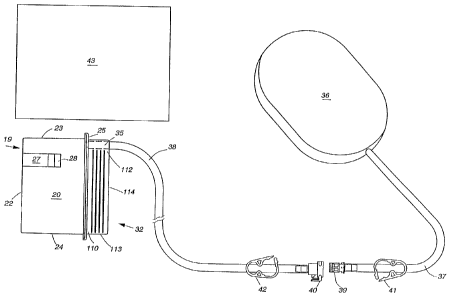
Canister 19 has a shape as shown in Figures 3 to 6_ As illustrated in
Figures 3 to 6, canister 19 includes sidewalls 20 and 21, top wall 23, bottom
wall 24, back wall 22 and front wall 25 that define the rectangular chamber
for
receiving blood, pus, and other fluids emitted from a wound. Sidewalls 20 and
21 include keyways 27 and 31 respectively, that receive a respective one of
keys
29 and 30 to provide easy alignment of canister 19 within chamber 18.
Furthermore, keyway 27 includes recess 28 that receives latch 26 to fasten
canister 19 within chamber 18.
Front wall 25 of canister 19 includes raised portion 32 extending
therefrom to fumish a window that permits a user to determine the level of
wound fluids within canister 19. Accordingly, raised portion 32 is transparent
so that the level of wound fluids within canister 19 may be visually
determined.
Raised portion 32 includes sidewalls I10 and I11, top wall 112, bottom wall
113, and front face 114 that define a chamber which opens into the chamber
defined by sidewalls 20 and 21, top wall 23, bottom wall 24, back wall 22 and
front wall 25 of canister 19. Front face 114 of raised portion 32 includes
graduations that demarcate the volume of wound fluid within canister 19.
Additionally, sidewalls 110 and 111 of raised portion 32 include ridges that
provide a gripping surface for the user during the insertion and removal of
canister 19 from chamber 18.
Although raised portion 32 is transparent to permit the determination of
the level of wound fluids within canister 19, sidewalls 20 and 21, back wall
22,
top wall 23, bottom wall 24, and front wall 25 are opaque or textured so that
they are only translucent. As an alternative, the portions of canister 19
surrounding filter 46 may also be transparent. This enables a user to visually
check for signs of contamination of filter 46. In this preferred embodiment,
- - - -- - -- -
7 2198243
sidewalls 20 and 21, back wall 22, top wall 23, bottom wall 24, front wall 25,
and raised portion 32 of canister 19 are fabricated from a plastics material.
Canister 19 includes inlet 35 that is formed integrally Nvith top wall 112
of raised portion 32. Inlet 35 is cylindrical in shape and communicates with
the
interior of canister 19 to permit the transfer of wound fluids into canister
19. In
thi,s preferred embodiment, inlet 35 is also fabricated from a plastics
material.
In order to prevent liquids sucked into the canister from splashing
directly onto cap 49, which masks the outlet 44, and to reduce foaming within
the canister, inlet 35 has a blind inner end. Inlet 35 has a slot 35A so that
drainage fluid is deflected downwardly into the raised handle portion 32 of
the
canister. Handle portion 32 may communicate with the main part of the
canister through one or more holes in wall 25. It is desirable to avoid
foaming
because this can give a false reading when a capacitance sensing device is
used
to sense when the canister is filled. An anti-foaming material, e.g. a
silicone
may be added to the canister, e.g. by coating the interior walls. It may also
be
advantageous to include a gel-forming substance, e.g. a polyacrylamide or
modified starch in order to immobilise the drainage fluid. This is
particularly
useful if the apparatus is likely to be tilted.
Wound fluids (i.e. drainage) are communicated through inlet 35 into
canister 19 via pad 36 and hoses 37 and 38. In this preferred embodiment, pad
36 is fabricated from an open cell polyurethane or polyether foam. Hose 37 is
inserted within pad 36 by making an incision in pad 36 and inserting the end
of
hose 37. Hose 37 can then be secured within pad 36 using any suitable means
such as an adhesive or a flange. Preferably, the foam pad is moulded or formed
with an elongated hole for the drainage tube which is an interference fit with
the
tube. The hoses are preferably made from medical grade PVC tube. Hose 38
8 219820
mounts within inlet 35 using any suitable means such as an adhesive or
welding.
Hoses 37 and 38 include luer lock connectors 39 and 40, respectively, (or the
equivalent such as any known quick disconnect type coupling) that attach
together to permit communication between hoses 37 and 38. Furthermore,
hoses 37 and 38 include pinch clamps 41 and 42, respectively, that are capable
of sealing their respective hose 37 or 38 to prevent the flow of wound fluids.
The foam pad is preferably packaged in a sterile container together with its
connector and clamp. When packaged, the clamps will be in their open
condition.
The communication of wound fluids into canister 19 requires the
securing of pad 36 over a wound. Pad 36 is secured over a wound using cover
43 which is fabricated from a plastics material and includes an adhesive on
one
side that sticks to human skin. Wound cover 43 is conveniently a surgical
drape material comprising a sheet of elastomeric material coated peripherally
or
overall with a pressure-sensitive adhesive, such as an acrylic adhesive. The
elastomeric or rubbery nature of the wound cover is important because it
accommodates changes in pressure in the wound area during intermittent
operation of the vacuum pump. The wound cover is preferably a polyurethane ,
film with a removable backing sheet, i.e. of polythene to protect the adhesive
surface.
A high degree of reticulation in the polymer foam is desirable to achieve
good permeability when the foam is under suction. Foams having at least 90%
and especially at least 95% of interconnecting cells are preferred.
In use, the foam pad is cut to a size which corresponds closely to the
edge of the wound with the objective of packing the foam into the wound cavity
210 so that it contacts the surface of the cavity, rather than bridging the
cavity.
~ 2198243
9
As depicted in Figure 10, the cavity may be extensive and there may be little
or
no tissue coverage to the bone 212. This is illustrated diagrammatically in
Figure 10. Figure 10 is a cross-section through a wound showing the foam pad
36 packed into the wound cavity 210. It is important that the foam should be
firmly packed into the recesses of the wound cavity. Drainage tube 37
terminates within the centre of the foam pad 36. Surgical drape 43 extends
over the foam pad and is adhered to intact skin 211 around the periphery of
the
wound. Drape 43 is also firmly adhered around the tube 37 to prevent leakage,
of air. A wound cover is then adhered to the surrounding skin and around the
drainage tube to provide an air-tight sea] around the wound.
As illustrated in Figures 2, 4 and 6, canister 19 includes outlet 44 that
mounts over port 45 to permit wound closure apparatus 10 to draw wound fluids
into canister 19. Outlet 44 is cylindrically shaped and formed as an integral
part of back wall 22 by outer wall 33 and inner wall 50 which are
interconnected by end wall 34. Passageway 52, defined in part by interior wall
50 and in part by filter cap 49, provides the actual conduit for outlet 44
between
the interior and exterior of canister 19. The placement of canister 19 within
recess 18 such that outlet 44 resides over port 45 couples canister 19 to a
vacuum pump. The vacuum pump removes air from canister 19 to create
vacuum pressure within canister 19. That vacuum pressure is then transmitted
to a wound site through hoses 37 and 38, thereby not only enabling therapeutic
use of system 10, but also tending to promote wound drainage. Any wound
drainage fluid is then drawn through pad 36 and hoses 37 and 38 into canister
19.
Outlet 44 resides near top wall 23 of canister 19 to ensure efficient
operation of the vacuum pump. That is, the vacuum pump removes the most
- - -
r 2)98243
air from canister 19 when the air does not have to first bubble through wound
fluids contained in canister 19. Consequently, with outlet 44 positioned near
the top of canister 19, the vacuum pump removes air directly from canister 19,
and it is only during the final filling of canister 19 that air must bubble
through
wound fluids. Preferably, as described below, the apparatus includes detecting
and warning means which operates before the level of the drainage fluid
reaches
either the inlet or outlet tube so that a fresh canister can be installed.
In removing fluids from a wound utilizing wound closure apparatus 10, a
major safety concem is preventing wound fluids from contaminating the
vacuum pump. Accordingly, filter 46 mounts over outlet 44 utilizing filter
carrier 48 and filter cap 49 to block the flow of wound fluids to outlet 44 so
that
wound fluids remain within canister 19 and do not flow into the vacuum pump.
In this preferred embodiment, filter 46 is a 0.2 micron hydrophobic membrane
filter providing a bacterial barrier, although other filters may be
substituted as
appropriate.
As illustrated in Figure 7, filter carrier 48 includes face 53 formed
integrally with lip 54. Face 53 includes groove 56 formed therein, while lip
54
supports brace 55 in its interior. Filter 46 fits within groove 56 of face 54
and
is supported within filter carrier 48 by brace 55 of lip 54. An '0' ring 53A
is
fitted in peripheral recess of filter carrier 48 to accommodate manufacturing
tolerances and ensure a fluid tight seal in filter cap 49.
As illustrated in Figures 6 and 8, filter cap 49 includes cylindrical
portions 57 and 58 which are formed integrally (with annulus 57' spanning
therebetween), to hold filter carrier 48 within passageway 52 of outlet 44. To
mount filter 46 over passageway 52, filter 46 is first placed within filter
carrier
48 as described above. Filter carrier 48 is then positioned within filter cap
49
~ 11 2198243
such that face 53 abuts annulus 57' of filter cap 49 and lip 54 of filter
carrier 48
resides within annular lip 50' of outlet 44. Accordingly, when cylindrical
portion 57 of filter cap 49 mounts over outlet 44, the front face 53 of filter
carrier 48 and the outer edges of filter 46 abut annulus 57' to secure filter
46
within passageway 52. Filter cap 49 attaches to outlet 44 using any suitable
means such as an adhesive or welding. Filter cap 49 is completely sealed
except for aperture 51 positioned on top of filter cap 49. Aperture 51
communicates with port 45 via passageway 52 of outlet 44 to permit the vacuum
pump to draw air from the interior of canister 19.
As illustrated in Figures 2 and 6, port 45 includes 0-ring 59 mounted
thereabout to provide a fluid tight sea] between port 45 and inner wall 50 of
outlet 44. Port 45 mounts through rear wall 104 of chamber 18 using any
suitable means such as nuts 60 and 61. Furthermore, hose 62 attaches to the
rear of port 45 using any suitable means such as a clamp to couple port 45 to
the
vacuum pump.
Switch 63 protrudes through rear wall 104 of chamber 18 to produce a
signal indicating when canister 19 properly and securely resides within
chamber
18. In this preferred embodiment, switch 63 is a normally open push button
switch that mounts on rear wall 104 of chamber 18 using any suitable means
such as a bracket. When canister 19 is properly positioned within chamber 18,
its rear wall 22 presses the head of switch 63, closing switch 63 so that it
provides a signal indicating that canister 19 properly resides within chamber
18.
Fill sensor 64 resides adjacent side wall 101, exterior to chamber 18. Fill
sensor 64 provides a signal that indicates when canister 19 is filled with
wound
debris. In this preferred embodiment, fill sensor 64 is a capacitive sensor
that
mounts on side wall 101 of chamber 18 using any suitable means such as a
2198243
12
bracket or appropriate adhesive material. Fill sensor 64 has a sensing profile
64A which determines the point at which the capacitance measurement is made.
When wound fluids have reached the level within canister 19 which corresponds
to the location of the sensing profile 64A, the capacitance within canister 19
as
'seen' by fill sensor 64 changes, resulting in fill sensor 64 outputting a
signal
indicating that canister 19 is filled with wound fluids to the level at which
the
sensing profile is located. The position of this sensing profile behind wall
101
can be changed (see Figure 6A) to provide an optimum balance of space and
volume utility.
As illustrated in Figure 2A, latch 26 generally comprises latch pin 65,
handle 66 latch guide sleeve 68A and spring 67. Latch pin 65 comprises a
proximal end 65A and distal end 65B. Latch guide sleeve 68A abuts the inner
surface of front housing 11 and is held securely in place from the outer side
of
front housing 11 by nut 68B. Handle 66 screws onto the proximal end 65A of
latch pin 65 and is locked in position by nut 69A. In the preferred
embodiment,
cover 68 over nuts 69A and 68B provides a surface against which handle 66
abuts, thus preventing end 65B from excessively entering chamber 18 as will be
uriderstood further herein. Cover 68 also provides aesthetic enclosure of nuts
69A and 68B. Dependent attachment of side wall 100 (chamber 18), as
described hereinabove, is such that side wall 100 abuts latch guide sleeve 68A
on the side distal front housing 11. Further, this arrangement causes distal
end
65B of latch pin 65 to project into chamber 18 under the force of spring 67
(shown partially cut away). Spring 67 resides circumferential ly about latch
pin
65 within an axial bore of latch pin guide 68A. Spring 67 exerts force between
distal end 65B of latch pin 65 and an annulus within the axial bore of latch
pin
guide 68A. A transverse slot in the distal end of latch pin guide 68A receives
~ 2198243
13
end 65B of latch pin 65, providing rotational alignment of end 65B and further
recess for end 65B when a user "pulls" handle 66 in an axial direction.
Latch 26 operates to ensure canister 19 remains secured within chamber
18. End 65B of latch 26 terminates in a point that protrudes through key 29
into
chamber 18. During the placing of canister 19 within chamber 18, key way 27
of canister 19 forces the point 65B of the latch pin within key 29. However,
once canister 19 has been properly positioned within chamber 18, recess 28
resides below latch pin end 65B so that spring 67 biases the point 65B of
latch
pin 65 into recess 28 to prevent the removal of canister 19 from chamber 18.
TYie removal of canister 19 from chamber 18 is accomplished by grasping
handle 66 and pulling the point 65B of latch pin 65 from recess 28. With the
point of latch pin 65 no longer within recess 28, canister 19 may be pulled
from
chamber 18 using its raised portion 32.
As illustrated in Figure 9, wound closure apparatus 10 preferably plugs
into a standard 115/120 VAC power source (e.g. an outlet) to supply power to
control system 70. Alternative embodiments (not shown, although similar) are
readily adapted for 220 VAC power by changing the power cord and
appropriatelv re-wiring the tops of the transformer within the DC power supply
71 as is readily know in the art. The application of power to control system
70
is regulated by power switch 15 which is a standard push button on/off switch.
With power switch 15 depressed, DC power supply 71 receives the 115/120
VAC signal and converts it into a 12 VDC signal for use by fan 74 and motor
83. A conventional voltage regulator 96 steps down the voltage to +5V or 12V
for use by each of the other DC components 63, 16, 17, 82, 72 and 75.
Voltage regulator 96 connects to keypad 16, LCD 17, switch 63, microcontroller
72, transducer 75, and tilt sensor 82 to supply each of them with the +5V DC
14 2198243
signal. Microcontroller 72 links to solid state relays (MOSFETs) 97 and 98 for
controlling the provision of the 12 VDC power supply to fan 74, pump motor 83
atid fill sensor 64, respectively.
As illustrated in Figure 1, once power switch 15 is depressed, a user
employs keypad 16 and LCD 17 to select the operating parameters foi wound
closure apparatus 10. Wound closure apparatus 10 stores the previously
selected operating parameters so that upon power initialization, LCD 17
displays the phrase "NEW PATIENT" with the word "NO" over arrow button
76, and the word "YES" over arrow button 77. If the user presses arrow button
76 to answer no, wound closure apparatus 10 will operate at the previously
selected parameters. After answering no, the user presses on/off button 78 to
begin operation of wound closure apparatus 10.
Conversely, if the user presses arrow button 77 to indicate a new patient,
wound closure apparatus 10 will operate either under default values or allow
the
user to select the operating parameters. To operate under default parameters,
the user presses on/off button 78 after pressing arrow button 77. However, to
select his or her own values, the user presses option button 79 after pressing
arrow button 77.
Upon the pressing of option buttons 79, LCD 17 displays a bar graph
representing the spectrum of available vacuum pump pressures and a numerical
representation of the vacuum pump pressure presently displayed by the bar
graph. The user changes vacuum pump pressure using arrow buttons 76 and
77. The pressing of arrow button 76 reduces vacuum pump pressure, while the
pressing of arrow button 77 increases vacuum pump pressure. After selecting
the desired vacuum pump pressure, the user presses option button 79 to save
the
selected vacuum pump pressure.
15 ~198243
Once the selected vacuum pump pressure has been saved, LCD 17
displays the pump operation times available to the user. The user may program
wound closure apparatus 10 to pump either continuously or intermittently.
Thus, LCD 17 displays the word "CONTINUOUS" over arrow button 76 and
"IlVTERMTfTENT" over arrow button 77. The user selects continuous
operation by pressing arrow button 76 followed by on/off button 78 to activate
the vacuum pump. In its continuous mode, wound closure apparatus 10 runs its
vacuum pump continuously until on/off button 78 is pressed again.
If the user presses arrow button 77 to select intermittent operation, LCD
17 displays a bar graph or figures representing the minimum and maximum on
titnes for the vacuum pump. LCD 17 also displays the phrase "ON TIME" and
the numerical value presently displayed. A user decreases the on time of the
vacuum pump by pressing arrow button 76 and increases the on time of the
vacuum pump by pressing arrow button 77. After selecting the desired on time,
the user presses options button 79 to save the selected on time value.
LCD 17 then displays a second bar graph or figures representing the off
tiine for the vacuum pump with the phrase "OFF TIME" and the numerical value
presently depicted by the bar graph. Again, arrow buttons 76 and 77 are
pressed to increase or decrease, respectively, the off time for the vacuum
pump.
After selecting the off time, the user presses options button 79 followed by
on/off button 78 to operate wound closure apparatus 10 using the selected
parameters.
Keypad 16 includes setting button 80 to permit the user to sequentially
display the currently selected operating parameters of wound closure apparatus
10. Keypad 16 further includes delay button 81 to permit the user to
deactivate
an alarm sounded in response to an improper operating condition of wound
= 2198243
16
closure apparatus 10. Delay button 81 provides the user with the ability to
silence alarms so that the alarm will not have to be listened to during the
correction of the problem.
Any new alarm conditions occurring within the fifteen minute period
("delay period") after the pressing of delay button 81 will not be indicated
by an
audible alarm. However, the pump will still be deactivated when appropriate,
even during the delay period.
Again referring to Figure 9, microcontroller 72 is a multi-port
microprocessor with a eight-bit analog to digital (A/D) converter having
associated memory that stores the program directing microcontroller 72 during
its control of wound closure apparatus 10. After receiving and storing the
user
selected operational parameters and receiving an on signal due to the pressing
of
on/off button 78, microcontroller 72 activates pump motor 83 which, in turn,
drives vacuum pump 84 to begin the removal of air from canister 19.
As vacuum pump 84 operates, it draws air from within canister 19, into
hose 62 via outlet 44 of canister 19 and port 45. Hose 62 connects to filter
85
and transducer 75 via Tjunction 91. Filter 85 is similar to filter 46 and thus
ensures no wound fluids contaminate vacuum pump 84. Filter 85
communicates with pump 84 via T-junction 88 and one arm of the latter is
connected to bleed valve 86. Bleed valve 86 communicates with the
atmosphere to release pressure developed within line 62 by vacuum pump 84
after microcontroller 72 deactivates vacuum pump 84. Bleed valve 86 is
sufficiently small to ensure that it generally does not affect the vacuum
pressure
levels achieved by vacuum pump 84 as it evacuates air from canister 19, except
to prevent over pressurisation beyond 250 mnnHg and to prevent erratic
operation of the vacuum pump at very low pressure settings..
17 2198243
In the preferred embodiment, an orifice of 0.5 mm diameter is especially
preferred for bleed valve 86. Valve 86 or the equivalent is particularlv
important for enabling intermittent application of negative pressure, as the
orifice allows for gradual release of the negative pressure (over a period of
about fifteen seconds) when the pump motor 83 is de-actuated. Bleed valve 86
is positioned outside housing 11 to facilitate un-clogging of aperture 86 in
the
event of a blockage. An aperture is provided in bleed valve 86, which is
machined from stainless steel. Flow control orifices would be alternatives.
Line 62 also includes T-connector 91 to connect it with line 92. Line
92 is connected to tank 94 which acts as a damper to pressure changes in line
62. This dampening effect, facilitated by restrictor 89 in line 93 between
transducer 75 and T junction 91, causes the pressure measured by transducer 75
to be an accurate indication of actual wound site pressure. Transducer 75
communicates with line 62 via line 93 to measure tank 94 pressure and produce
an electrical signal representative of that pressure. Transducer 75 outputs
its
pressure signal to microcontroller 72.
Microcontroller 72 utilizes the pressure signal to control the speed of
pump motor 83. As previously described, the user selects either a default
vacuum pump pressure or a desired vacuum pump pressure for the operation of
wound closure apparatus 10. After receiving the wound pressure signal from
transducer 75, microcontroller 72 compares the wound pressure with the user
selected pressure. If the wound pressure is higher than the user selected
vacuum pump pressure, microcontroller 72 reduces pump motor speed to
decrease vacuum pump pressure and thus the pressure at the wound.
Conversely, if the wound pressure is less than the user selected vacuum pump
18 2198243
pressure, microcontroller 72 increases the speed of pump motor 83 resulting in
an increase in the vacuum pressure applied at the wound.
Microcontroller 72 controls pump motor 83 by varying the amount of
voltage received by pump motor 83. That is, microcontroller 72 receives the
12V DC signal from DC power supply 71 and outputs a voltage between 0 and
12V DC to pump motor 83 to control its speed in accordance with the user
selected vacuum pump pressure value. Accordingly, microcontroller 72
employs feedback to ensure that the wound experiences the user selected
vacuum pump pressure. If the target pressure is not reached after a period of
five minutes, microcontroller 72 deactivates motor 83 and sounds the audible
alarm. Additionally, the feedback signal prevents maximum vacuum pump
pressure from being exceeded. If the wound pressure measured by transducer
75 exceeds a maximum safe vacuum pump pressure, microcontroller 72
deaptivates pump motor 83.
Wound closure apparatus 10 includes fan 74 to cool pump motor 83 and
printed circuit board or chassis 200 during the operation of the wound closure
apparatus 10. In the preferred embodiment, microcontroller 72 controls fan 74,
to always operate while power is being supplied In alternative embodiments,
however, microcontroller 72 controls fan 74 to operate only in relation to
motor
83, because it is only necessary for fan 74 to operate if motor 83 is also
operating. In such alternative, as long as pump motor 83 operates,
microcontroller 72 runs fan 74. However, when microcontroller 72 deactivates
pump motor 83 it also deactivates fan 74.
Control system 70 includes fill sensor 64 to provide a signal to
microcontroller 72 that indicates when canister 19 is completely filled with
wound fluids. After receiving a signal from fill sensor 64, microcontroller 72
-- - - -
19 2198243
deactivates pump motor 83 and fan 74 and activates alarm 95 to signal the user
that canister 19 must be replaced.
Control system 70 includes switch 63 to prevent users from operating
wound closure apparatus 10 without a canister properly installed. If a
canister
is not properly installed, switch 63 remains open and therefore outputs no
signal
to microcontroller 72. If microcontroller 72 receives no signal from switch
63,
indicating no canister within chamber 18, it will not supply power to pump
motor 83 even after a user has pressed on/off button 78. Furthermore,
microcontroller 72 activates alarm 95 to signal the user that either a
canister is
not properly installed or is improperly installed within chamber 18 Nvhen
therapy
is activated. Microcontroller 72 operates pump motor 83 only if switch 63 is
depressed to provide a signal indicating the proper placement of a canister
within chamber 18.
Control system 70 includes tilt sensor 82 to prevent operation of wound
closure apparatus 10 if it is tilted excessively. Excessive tilting of wound
closure apparatus 10 during operation diminishes the efficiency of removal of
wound fluids and, more importantly, might result in either the contamination
of
vacuum pump 84 or the spilling of wound fluids. Thus, if wound closure
apparatus 10 tilts along any of its axes beyond a predetermined angle
(approximately 450 in this preferred embodiment), tilt sensor 82 outputs a
signal
to microcontroller 72. In response, microcontroller 72 deactivates pump motor
83 and activates alarm 95 to signal the user of the excessive tilt situation.
In
this preferred embodiment, tilt sensor 82 may be implemented with any standard
mercury switch. The tilt circuiting and alarm operates as follows. If therapy
is
in progress and the pump unit is tilted, the alarm will sound and the liquid
crystal display 17 will state 'unit tilted'. Therapy is automatically stopped.
')0
When the unit is returned to the vertical, therapy will be automatically
reinstated
after a time delay (e.g. about 30 seconds) has elapsed..