Note : Les descriptions sont présentées dans la langue officielle dans laquelle elles ont été soumises.
CA 02242360 1998-07-06
WO 97/24981 PCT/IL97/00010
CARDIAC EL.ECTRO-ME(`HANICS
FIELD OF THE INVENTION
The present invention relates to the field of cardiac medicine and more
particularly to
diagnosing and treating diseased hearts based on the interaction between
cardiac electro-
physiological and cardiac bio-mechanical activity.
BACKGROUND OF THE INVENTION
Cardiovascular diseases accounted for approximately 43 percent of the
mortality in the
United States of America in 1991 (923,000 persons). However, many of these
deaths are not
directly caused by an acute myocardial infraction (AMI). Rather, many patients
suffer a
general decline in their cardiac output known as heart failure. Once the overt
signs of heart
failure appear, half the patients die within five years. It is estimated that
between two and three
million Americans suffer from heart failure and an estimated 200,000 new cases
appear every
year. In many cases heart failure is caused by damage accumulated in the
patient's heart, such
as damage caused by disease, chronic and acute ischemia and especially (-75%)
as a result of
hypertension.
A short discussion of the operation of a healthy heart is useful in order to
appreciate the
complexity of the functioning of the heart and the multitude of pathologies
which can cause
heart failure. Fig. 1A is a schematic drawing of a cross-section of a healthy
heart 20. In general
heart 20 comprises two independent pumps. One pump comprises a right atrium 22
and a right
ventricle 24 which pump venous blood from an inferior and a superior vena cava
to a pair of
lungs (not shown) to be oxygenated. Another pump comprises a left atrium 26
and a left
ventricle 28, which pump blood from pulmonary veins (not shown) to a plurality
of body
systems, including heart 20 itself. The two ventricles are separated by a
ventricular septum 30
and the two atria are separated by an atrial septum 32.
Heart 20 has a four phase operational cycle in which the two pumps are
activated
synchronously. Fig. 1B shows a first phase, called systole. During this phase,
right ventricle 24
contracts and ejects blood through a pulmonic valve 34 to the lungs. At the
same time, left
ventricle 28 contracts and ejects blood through an aortic valve 36 and into an
aorta 38. Right
atrium 22 and left atrium 26 are relaxed at this point and they begin filling
with blood,
however, this preliminary filling is limited by distortion of the atria which
is caused by the
contraction of the ventricles.
Fig. I C shows a second phase, called rapid filling phase and indicates the
start of a
1
CA 02242360 1998-07-06
WO 97/24981 PCT/IL97/00010
diastole. During this phase, right ventricle 24 relaxes and fills with blood
flowing from right
atrium 22 through a tricuspid valve 40, which is open during this phase.
Pulmonic valve 34 is
closed, so that no blood leaves right ventricle 24 during this phase. Left
ventricle 28 also
relaxes and is filled with blood flowing from left atrium 26 through a mitral
valve 42, which is
open. Aortic valve 36 is also closed to prevent blood from leaving left
ventricle 26 during this
phase. The filling of the two ventricles during this phase is affected by an
existing venous
pressure. Right atrium 22 and left atrium 26 also begin filling during this
phase. However, due
to relaxation of the ventricles, their pressure is lower than the pressure in
the atria, so tricuspid
valve 40 and mitral valve 42 stay open and blood flows from the atria into the
ventricles.
Fig. 1D shows a third phase called diastatis, which indicates the middle of
the diastole.
During this phase, the ventricles fill very slowly. The slowdown in filling
rate is due to the
equalization of pressure between the venous pressure and the intra-cardiac
pressure. In
addition, the pressure gradient between the atria and the ventricles is also
reduced.
Fig. 1 E shows a fourth phase called atrial systole which indicates the end of
the
diastole and the start of the systole of the atria. During this phase, the
atria contract and inject
blood into the ventricles. Although there are no valves guarding the veins
entering the atria,
there are some mechanisms to prevent backflow during atrial systole. In left
atrium 26, sleeves
of atrial muscle extend for one or two centimeters along the pulmonary veins
and tend to exert
a sphincter-like effect on the veins. In right atrium 22, a crescentic valve
forms a rudimentary
valve called the eustachian valve which covers the inferior vena cava. In
addition, there may
be muscular bands which surround the vena cava veins at their entrance to
right atria 22.
Fig. 1F is a graph showing the volume of left ventricle 24 as a function of
the cardiac
cycle. Fig. IF clearly shows the additional volume of blood injected into the
ventricles by the
atria during atrial systole as well as the variance of the heart volume during
a normal cardiac
cycle. Fig. 1 G is a graph which shows the time derivative of Fig. i F, i.e.,
the left ventricle fill
rate as a function of cardiac cycle. In Fig. 1G two peak fill rates are shown,
one in the
beginning of diastole and the other during atrial systole.
An important timing consideration in the cardiac cycle is that the atrial
systole must
complete before the ventricular systole begins. If there is an overlap between
the atrial and
ventricular systoles, the atria will have to force blood into the ventricle
against a raising
pressure, which reduces the volume of injected blood. In some pathological and
induced cases,
described below, the atrial systole is not synchronized to the ventricular
systole, with the effect
2
CA 02242360 1998-07-06
WO 97/24981 PCT/IL97/00010
of a lower than optimal cardiac output.
It should be noted that even though the left and the right sides of heart 20
operate in
synchronization with each other, their phases do not exactly overlap. In
general, right atrial
systole starts slightly before left atrial systole and left ventricular
systole starts slightly before
right ventricular systole. Moreover, the injection of blood from left
ventricle 26 into aorta 38
usually begins slightly after the start of injection of blood from right
ventricle 24 towards the
lungs and ends slightly before end of injection of blood from right ventricle
24. This is caused
by pressures differences between the pulmonary and body circulatory systems.
When heart 20 contracts (during systole), the ventricle does not contract in a
linear
fashion, such as shortening of one dimension or in a radial fashion. Rather,
the change in the
shape of the ventricle is progressive along its length and involves a twisting
effect which tends
to squeeze out more blood. Fig. 2 shows an arrangement of a plurality of
muscle fibers 44
around left ventricle 28 which enables this type of contraction. When muscle
fibers 44 are
arranged in a spiral manner as shown in Fig. 2 and the activation of muscle
fibers 44 is started
from an apex 46 of left ventricle 28, left ventricle 28 is progressively
reduced in volume from
the bottom up. The spiral arrangement of muscle fibers 44 is important because
muscle fibers
typically contract no more than 50% in length. A spiral arrangement results in
a greater change
of left ventricular volume than is possible with, for example, a flat
arrangement in which the
fibers are arranged in bands around the heart. An additional benefit of the
spiral arrangement is
a leverage effect. In a flat arrangement, a contraction of 10% of a muscle
fiber translates into a
reduction of 10% of the ventricular radius. In a spiral arrangement with, for
example, a spiral
angle 48 of 45 , a 10% contraction translates into a 7.07% contraction in
ventricular radius and
a 7.07% reduction in ventricular length. Since the ventricular radius is
typically smaller than
the ventricular length, the net result is that, depending on spiral angle 48,
a tradeoff is effected
between a given amount of contraction and the amount of force exerted by that
contraction.
Spiral angle 48 is not constant, rather, spiral angle 48 changes with the
distance of a
muscle fiber from the outer wall of the ventricle. The amount of force
produced by a muscle
fiber is a function of its contraction, thus, each layer is optimized to
produce an optimal
amount of force. Since the contraction of each muscle fiber is synchronous
with the increase in
the ventricular pressure (caused by the muscle contraction), it might be
expected that the
muscle fibers produce a maximum force at maximum contraction. However,
physiological
constraints on muscle fibers denote that maximal force is generated before
maximal
3
CA 02242360 1998-07-06
WO 97/24981 PCT/IL97/00010
contraction. In addition, the force exerted by a muscle fiber begins to fall
soon after maximum
force is exerted. The varying spiral angle is a mechanism which makes it
possible to increase
the contractile force on the ventricle after maximum force is reached by a
particular muscle
fiber.
As described above, activation of the heart muscle is from the apex up. Thus,
the
muscle on the top of the ventricle could theoretically exert more force than
the muscle at apex
46, which would cause a distention at apex 46. The varying spiral angle is one
mechanism to
avoid distention. Another mechanism is that the muscle near apex 46, which is
activated first,
is slightly more developed than the muscle at the top of the ventricle, which
is activated last.
As a result of the above described mechanisms, the force exerted by the
ventricular wall is
more evenly distributed over time and space. It should be appreciated that
blood which
remains in one place without moving, even in the heart, can clot, so it is
very important to eject
as much blood as possible out of the heart.
As can be appreciated, a complicated mechanism is required to synchronize the
activation of muscle fibers 44 so that an efficient four phase cycle is
achieved. This
synchronization mechanism is provided by an electrical conduction system
within the heart
which conducts an electrical activation signal from a (natural) cardiac
pacemaker to muscle
fibers 44.
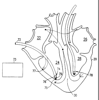
Fig. 3 shows the main conduction pathways in heart 20. An SA node 50, located
in
right atrium 22, generates an activation signal for initiating contraction of
muscle fibers 44.
The activation signal is transmitted along a conduction pathway 54 to left
atria 26 where the
activation signal is locally disseminated via Bachman bundles and Crista
terminals. The
activation signal for contracting the left and right ventricles is conducted
from SA node 50 to
an AV node 52, where the activation signal is delayed. The ventricles are
normally eiectrically
insulated from the atria by non-conducting fibrous tissue, so the activation
signal must travel
through special conduction pathways. A left ventricle activation signal
travels along a left
pathway 58 to activate left ventricle 28 and a right ventricle activation
signal travels along a
right pathway 56 to activate right ventricle 24. Generally, the conduction
pathways convey the
activation signal to apex 46 where they are locally disseminated via Purkinje
fibers 60 and
propagation over the rest of the heart is achieved by conduction in muscle
fibers 44. In general,
the activation of the heart is from the inner surface towards the outer
surface. It should be
noted that electrical conduction in muscle fibers 44 is generally faster along
the direction of
4
CA 02242360 1998-07-06
WO 97/24981 PCT/1L97/00010
the muscle fibers. Thus, the conduction velocity of the activation signals in
heart 20 is
generally anisotropic.
As can be appreciated, the delay in AV node 52 results, in a healthy heart, in
proper
ventricular systolic sequencing. The temporal distribution of the activation
signal in the
ventricular muscle results in the activation of the ventricles from the apex
up. In a healthy
heart the activation signal propagates across left ventricle 28 in
approximately 60 milliseconds.
In an externally paced heart, where the activation signal is not conducted
through Purkinje
fibers 60 or in a diseased heart, the propagation time is typically longer,
such as 150
milliseconds. Thus, disease and extemal pacing affect the activation profile
of the heart.
Cardiac muscle cells usually exhibit a binary reaction to an activation
signal; either the
cell responds normally to the activation signal or it does not respond at all.
Fig. 4 is a graph
showing changes in the voltage of a single cardiac muscle cell in reaction to
the activation
signal. The reaction is generally divided into five stages. A rapid
depolarization stage 62
occurs when the muscle cell receives an activation signal. During this stage,
which lasts a few
milliseconds, the potential of the cell becomes rapidly positive. After
depolarization, the
muscle fiber rapidly repolarizes during a rapid repolarization stage 64 until
the cell voltage is
approximately zero. During a slow repolarization stage 66, also known as the
plateau, the
muscle cell contracts. The duration of stage 66, the plateau duration, is
directly related to the
amount of work performed by the muscle cell. A relatively fast repolarization
stage 68 follows,
where the muscle cell repolarizes to its original potential. Stage 66 is also
known as the
refractory period, during which the cell cannot be activated by another
activation signal.
During stage 68, the cell is in a relative refractory period, during which the
cell can be
activated by an exceptionally strong activation signal. A steady state 70
follows in which the
muscle cell is ready for another activation.
It should be appreciated that the contraction of cardiac muscle cells is
delayed in time
from their activation. In addition the duration of the contraction is
generally equal to the
duration of the plateau.
An important factor which may affect the length of the plateau is the
existence of an
ionic current resulting from the voltage potentials generated by the local
depolarizations. The
ionic current starts at the last activated portion of the heart and progresses
back along the path
of the activation. Thus, it is the later activated portions of the heart which
are first affected by
the ionic current. As a result, the repolarization of these cells is
relatively faster than the
5
CA 02242360 1998-07-06
WO 97/24981 PCT/IL97/00010
repolarization of the first activated muscle fibers, and their contraction
time is relatively
shorter. As can be appreciated, in a healthy heart, where the propagation time
of the activation
signal is relatively short, the ionic currents are significantly smaller than
in a diseased or
externally paced heart.
One of the main results of the contraction of the ventricles is increased
intra-ventricular
pressure. In general, when the intra-cardiac pressure is higher, the outflow
from the heart into
the circulatory system is stronger and the efficiency of the heart is higher.
A mathematical
relationship termed Laplace's law can be used to model the relationship
between the pressure
in the ventricle and the tension in the wall of the ventricle. Laplace's law
was formulated for
generally spherical or cylindrical chambers with a distentible wall, however,
the law can be
applied to the ventricles since they are generally elongated spherical in
shape. Figs. 5A-C
show three formulations for determining the tension in a portion of the
ventricle wall, all of
which are based of the law of Laplace. In Fig. 5A, the tension across a cross-
section of the
wall is shown wherein T, the tension in the wall, is equal to the product of
P, the transmural
pressure across the wall, r (squared), the radius of the ventricle, and zt.
Figs 5B and C show
formulas for calculating the tension per unit in portions of the ventricular
wall, for example in
Fig. 5C, for a unit cross-sectional area of muscle in a wall of thickness S.
As can be appreciated, if r, the radius of the ventricle, is large, a higher
tension is
needed to produce the same pressure change as in a ventricle with a smaller
radius. This is one
of the reasons that ventricular dilation usually leads to heart failure. The
heart muscle is
required to produce a higher tension is order to achieve the same pressure
gradient. However,
the heart is not capable of producing the required tension, so, the pressure
gradient, and thus
the cardiac efficiency, are reduced.
Unfortunately, not all people have healthy hearts and vascular systems. Some
types of
heart problems are caused by disease. HCM (hypertrophic cardiomyopathy or
HOCM) is a
disease in which the left ventricle and, in particular, the ventricular
septum, hypertrophy,
sometimes to an extent which blocks the aortic exit from the left ventricle.
Other diseases,
such as atrophy causing diseases, reduce the amount of muscle fibers in
portions of the heart.
A very common cause of damage to the heart is ischemia of the heart muscle.
This
condition, especially when manifesting itself as an acute myocardial
infraction (heart attack),
can create dead zones in the heart which do not contain active muscle. An
additional, and
possibly more important effect, is the non-conducting nature of these dead
zones which may
6
CA 02242360 1998-07-06
WO 97/24981 PCT/1L97/00010
upset the natural activation sequence of the heart. In some cases, damaged
heart tissue
continues to conduct the activation signal, albeit at a variable or lower
velocity, which may
cause arrhythmias.
A chronic ischemic condition is usually caused by blockage of the coronary
arteries,
usually by arteriosclerosis, which limits the amount of oxygen which can reach
portions of the
heart muscle. When more work (i.e., more tension) is required of the heart
muscle and an
increase in oxygen supply is not available, the result is acute pain, and if
the supply is cut off
for an extended period, death of the starved muscle will follow.
When the output of the heart is insufficient, a common result is hypertrophy
of the
heart, usually of the left ventricle. Hypertrophy is a compensatory mechanism
of the heart for
increasing the output volume. However, in a chronic condition, hypertrophy has
generally
negative effects. For example, arrhythmias, congestive heart failure (CHF) and
permanent
changes in the morphology of the heart muscle (ventricular modeling) may
result from
hypertrophy.
One of the most common cardio-vascular diseases is hypertension. A main effect
of
hypertension is increased cardiac output demand, which causes hypertrophy
since the blood
must be pumped against a higher pressure. Furthermore, hypertension usually
aggravates other
existing cardiac problems.
The human heart has many compensatory and adaptive mechanisms, termed cardiac
reserve, so that not all cardiac pathologies manifest as heart disease. Once
the cardiac reserve
is used up, the heart cannot keep up with the demand and heart failure may
result. One
measure of heart function and efficiency is the left ventricle ejection
factor, which is the ratio
between the amount of blood in the left ventricle during diastole and the
amount of blood
exiting during systole. It should be noted that a significant portion of the
change in ventricular
volume between systole and diastole is due to the thickening of activated
muscle fibers.
Another measure of heart function is the left ventricle stroke volume, which
is the amount of
blood which is ejected from the left ventricle each heart beat. It should be
noted that once the
cardiac reserve is used up it is difficult, if not impossible, for the heart
to increase its output
when needed, such as during exercise.
There are many ways in which non-optimal timing of the activation of the heart
can
result in lower cardiac output. In AF (atrial fibrillation) one or both atria
does not contract in
correct sequence with its associated ventricle. As a first result, the atria
does not inject blood
7
CA 02242360 1998-07-06
WO 97/24981 PCT/IL97/00010
into its associated ventricle during atrial systole, so the ventricle volume
is not maximized
before ventricular systole, and stroke volume is slightly reduced. If the
right atria is
fibrillating, sequencing of the AV node is non-regular, which results in the
ventricles
contracting at an irregular rate, and the heart output is further reduced.
In some cases of a conduction block between the SA node and the ventricles,
such as
caused by a damaged AV node, the contraction of the atria is not synchronized
to the
contraction of the ventricles, which also results in a lower heart output.
Another type of timing deficiency results when there are large dead areas in
the heart
muscle which do not conduct electrical signals. The activation signal must
circumvent the
dead areas, which results in a longer pathway (and longer delay time) for the
activation signal
reaching some portions of the heart. In some cases, these portions of the
heart are activated
long after the rest of the heart has already contracted, which results in a
reduced contribution
of these portions to the total cardiac output.
Heart muscle which is stressed before it is activated, heart muscle which is
weakened
(such as by ischemia) and portions of the heart which have turned into scar
tissue, may form
aneurysms. As can be appreciated from Laplace's law, portions of the ventricle
wall which do
not generate enough tension to offset the tension induced by the intra-cardiac
pressure must
increase their local radius in response to the pressure overload. The
stretched wall portion thins
out and may burst, resulting in the death of the patient. The apex of the left
ventricle is
especially susceptible to aneurysms since it may be very thin. In addition,
the totai pressure in
the ventricle and the flow from the ventricle are reduced as the aneurysm
grows, so the heart
output is also reduced. Although weak muscle should be expected to hypertrophy
in response
to the greater need, in some cases, such as after an AMI, hypertrophy may not
occur before
irreversible tissue changes are caused by the stretching.
Perfusion of the heart muscle usually occurs during diastole. However, if the
diastole is
very long, such as when the activation signal is propagated slowly, some
portions of the heart
may not be oxygenated properly, resulting in functional ischemia.
As mentioned above, one of the adaptation mechanisms of the heart is
hypertrophy, in
which the size of the heart increases to answer increased demand. However,
hypertrophy
increases the danger of arrhythmias, which in some cases reduce heart output
and in others,
such as VF (ventricular fibrillation) are life threatening. Arrhythmias are
also caused by
damaged heart tissues which generate erroneous activation signals and by
blocks in the
8
CA 02242360 2005-04-28
conduction system of the heart.
In some cases arrhythmias of the heart are treated using medicines, in others,
by
implanting a pacemaker or a defibrillator. A common pacemaker implanting
procedure,
for example for treating the effects of AF, includes:
(a) ablating or removing the AV node; and
(b) implanting a pacing electrode in the apex of the heart. The location of
the
pacing electrode may be changed (during the procedure) if the heart does not
beat at a
desired sequence for a given output of the pacemaker.
It is also known to pace using multiple electrodes, where the activation
signal is
initiated from a selected one or more of the electrodes, depending on sensed
electrical
values, such as sequence, activation time and depolarization state. Typically,
the pacing
regime is adapted to a specific arrhythmia. Sometimes, logic is included in
the pacemaker
which enables it to identify and respond to several types of arrhythmia.
U.S. Patent 5,403,356 to Hill et al. describes a method of preventing atrial
arrhythmias by adapting the pacing in the right atrium in response to a sensed
atrial
depolarization, which may indicate an arrhythmia.
Sometimes the pacing is performed for more than one chamber. For example, in
dual chamber pacing, both left and right ventricles are separately paced.
There have been
attempts to use dual chamber pacing to relive aortic obstruction caused by
HCM. The
aortic exit from the left ventricle is located between the left and right
ventricle, so that
when both ventricles contract simultaneously, the aorta is squeezed from all
sides. In a
healthy heart, the ventricular septum does not obstruct the aorta, however, in
an HCM-
diseased heart, the enlarged septum obstructs the aortic exit from the left
ventricle. When
pacing to reduce aortic obstruction, the contractions of the left and right
ventricles are
stepped, so that when the left ventricle contracts, the right ventricle
dilates and the aorta
is less compressed.
Lameh Fananapazir, Neal D. Epstein, Rodolfo V. Curiel, Julio A. Panza, Dorothy
Tripodi and Dorothea McAreavey, in "Long-Term Results Of Dual-Chamber (DDD)
Pacing In Obstructive Hypertrophic Cardiomyopathy", Circulation, Vol. 90, No.
60, pp.
2731-2742, December 1994, describes the effects of pacing a HCM-diseased heart
using
DDD pacing at the apex of the right ventricle. One effect is that the muscle
mass near the
pacing location is reduced, i.e., the ventricular septum is atrophied. The
atrophy is
9
CA 02242360 2005-04-28
hypothesized to be caused by the changes in workload at the paced location
which are due
to the late activation time of ventricular segments far from the pacing
location.
Margarete Hochleitner, Helmut Hortnagl, Heide Hortnagl, Leo Fridrich and Franz
Gschnitzer, in "Long-Term Efficiency Of Physiologic Dual-Chamber Pacing In The
Treatment Of End-Stage Idiopathic Dilated Cardiomyopathy", American Journal of
Cardiology, volume 70, pp. 1320-1325, 1992, describes the effect of DDD pacing
on hearts
which are dilated as a result of idiopathic dilated cardiomyopathy. DDD pacing
resulted in an
improvement of cardiac function and in a reduction in hypertrophy in several
patients. In
addition, it is suggested that positioning the ventricular electrode of the
DDD pacemaker in
near the apex of the right ventricle reduced the stress at the apex of the
left ventricle, by its
early activation. No method is suggested for choosing the implantation
location of the
electrodes.
Xavier Jeanrenaud, Jean-Jacques Goy and Lukas Kappenberger, in "Effects Of
Dual
Chamber Pacing In Hypertrophic Obstructive Cardiomyopathy", The Lancet, Vol.
339, pp.
131$-1322, May 30, 1992, teaches that to ensure success of DDD pacing in HCM
diseased
hearts, an optimum AV interval (between atrial activation and ventricular
activation) is
required. In addition, it is suggested that this optimal AV interval is
modified by performing
exercise.
Several methods may be used to treat heart failure. One method is to connect
assist
pumps to the patient's circulatory system, which assist the heart by
circulating the blood. To
date, no satisfactory long-term assist pump has been developed. In some cases,
a diseased
heart is removed and replaced by another human heart. However, this is an
expensive,
complicated and dangerous operation and too few donor hearts are available.
Artificial hearts
suffer from the same limitations as assist pumps and, like them, are not yet
practical.
Certain types of heart failure, such as those caused by conduction blocks in
the AV
node or by AF can be helped by the implantation of a pacemaker, as described
above.
Some cases of heart failure can be helped by medicines which either strengthen
the
heart, correct arrhythmias or reduce the total volume of blood in the body
(which reduces
blood pressure): However, many cases of heart failure can only be treated by
reducing the
activity of the patient. Ultimately, once the cardiac reserve is used up, most
cases of heart
failure cannot be treated and result in death.
U.S. patent 5,391,199 discloses apparatus and method for mapping the
electrical
CA 02242360 2005-04-28
activity of the heart.
"Biomedical Engineering Handbook", ed. Joseph D. Bronzino, chapter 156.3, pp.
2371-2373, IEEE press/ CRC press, 1995, describes modeling strategies in
cardiac
physiology. On page 2373 a model is described, including experimental support,
according to which model the shape of a ventricle is determined by the (local)
amount of
oxygen consumption. In addition, this model differentiates between pressure
overload on
the heart, which causes thickening of muscle fibers, denoted concentric
hypertrophy, and
volume overload which causes an increase in the ventricular volume (by
stretching),
denoted eccentric hypertrophy. Eccentric hypertrophy may also be caused by
reducing the
amount of oxygen available to the cardiac muscle.
R. S. Reneman, F. W. Prinzen, E. C. Cheriex, T. Arts and T. Delhass, in
"Asymmetrical Changes in Left Ventricular Diastolic Wall Thickness Induced by
Chronic
Asynchronous Electrical Activation in Man and Dogs", FASEB J., 1993;7;A752
(abstract), abstract number 4341, describe results of studies in paced hearts
and which
show that earlier activated ventricular wall portions were thinner than later
activated wall
portions, showing an asymmetrical hypertrophy as a result of the pacing.
C. Daubert, PH. Mabo, Veronique Berder, D. Gras and C. LeClercq, in "Atrial
Tachyarrhythmias Associated with High Degree Interatrial Conduction Block:
Prevention by
Permanent Atrial Resynchronisation", European Journal of C.PE., Vol. 4, No. 1,
pp. 35-
44, 1994, describes a method of treating atrial fibrillation by implanting
pacemaker
electrodes in various locations in the heart, including two electrodes in the
right atrium.
Frits W. Prinzen, Cornelis H. Augustijn, Theo Arts, Maurits A. Allessie and
Robert Reneman, in "Redistribution of Myocardial Fiber Strain and Blood Flow
by
Asynchronous Activation", American Journal of Ph,ysiology No. 259 (Heart
Circulation
Physiology No. 28), H300-H308, 1990, describes studies which show that the
location of
pacing electrodes in a paced heart significantly affect the distribution of
strain, and
perfusion (blood flow) in the heart.
SUMMARY OF THE INVENTION
It is an object of some aspects of the present invention to provide methods of
augmenting the compensatory mechanisms of the heart.
11
CA 02242360 1998-07-06
WO 97/24981 PCT/IL97/00010
Another object of some aspects of the present invention is to provide methods
of
mapping the local physiological values and/or the shape of the heart to
determine the
activation profile of the heart and, preferably, to analyze the resulting maps
to determine
possible optimizations in the activation profile.
Yet another object of some aspects of the present invention is to control the
adaptation
mechanisms in the heart so that the heart output or some other parameter of
the heart is
optimized. Alternatively or additionally, the adaptation mechanisms of the
heart are utilized to
effect change in the morphology of the heart, such as by redistributing muscle
mass.
Still another object of some aspects of the present invention is to control
the activation
sequence of the heart so that the heart output or some other physiological
variable of the heart
is optimized, preferably, in real-time.
When used herein, the terms "physiological variable" and "cardiac parameter"
do not
include electrical activity, rate, arrhythmia or sequencing of the heart. The
tenn "local
physiological value" does not include electrical activity, per se, rather it
refers to a local
physiological state, such as contraction of local heart muscle, perfusion or
thickness. The term
"location" refers to a location on or in an object, such as the heart muscle.
For example, a
valve or an apex of the heart. "Position" refers to a position in space,
usually relative to a
known portion of the heart, for example, 1.5 inches perpendicular from the
apex of the heart.
The term "local information" includes any information associated with the
location on the
heart wall, including position and electrical activity.
An object of some aspects of the present invention is related to pacemakers
which are
adapted to control the adaptation mechanisms of the heart and/or to optimize
heart parameters.
In a preferred embodiment of the invention, the mechanical motion of the heart
muscle
is mapped using a catheter having a position sensor near its distal end. The
mapping includes:
(a) placing the catheter into contact with the heart wall;
(b) determining the position of the distal end of the catheter; and
(c) repeating step (b) for additional locations in the heart.
Preferably, the catheter is in contact with the heart wall through the entire
cardiac
cycle. It should be appreciated that contact with the heart wall can be
achieved either from the
inside or from the outside of the heart, such as outside contact being
achieved by inserting the
catheter into the coronary arteries and/or veins. Alternatively, the catheter
is directly inserted
into the body (not through the vascular system), such as through a
throactoscope or during
12
CA 02242360 1998-07-06
WO 97/24981 PCT/IL97/00010
surgery.
Preferably, (b) includes determining the position of the catheter at at least
two instants
of an entire heart cycle. More preferably, it includes determining the
position with time over
the cycle. Alternatively or additionally, the catheter has a plurality of
distal ends, each with a
position sensor and (b) includes determining the position of each one of the
ends.
Preferably, the catheter does not move between sequential diastoles. This can
be
asserted, for example, by using an impedance sensor, by determining changes in
a locally
sensed electrogram, by detennining that the position sensor repeats its
trajectory during heart
cycles or by determining that the catheter returns to the same location each
diastole or other
recognizable portion of the cardiac cycle.
Preferably, the mapping further includes determining the geometry and/or
changes in
the geometry of at least a portion of the heart as a function of time and/or
phase of the cardiac
cycle. For example, the existence of an aneurysm can be determined from a
characteristic
bulge of the aneurysm during systole. Likewise, a dilated ventricle can be
determined from the
determined volume. Additionally or alternatively, the mapping includes
determining the local
radius of a portion of the heart wall.
Preferably the catheter comprises a pressure sensor which measures the intra-
cardiac
pressure. Further preferably, the forces on the heart wall are calculated
using the local radius
and/or the determined pressure, preferably using Laplace's law.
Preferably, the catheter includes at least one electrode for determining the
local
electrical activity of the heart. Preferably, the local activation time and/or
the activation signal
is measured and incorporated in a map of the heart. Additionally or
alternatively, local
electrical conductivity is measured, since fibrous scar tissue does not
conduct as well as viable
muscle tissue.
A preferred embodiment of the invention provides a map which compares the
local
activation time to the movement of a segment of local heart wall. Preferably,
the map
compares activation time of the segment to movement of the segment relative to
the movement
of surrounding segments. Thus, the reaction of a muscle segment to the
activation signal can
be determined from the local geometrical changes.
In a preferred embodiment of the invention, the instantaneous thickness of the
heart
wall at the point of contact is also determined. Preferably, the thickness is
measured using an
ultrasonic transducer, preferably mounted on the distal portion of the
catheter. Preferably,
13
CA 02242360 1998-07-06
WO 97/24981 PCT/IL97/00010
changes in the thickness of the cardiac wall are used to determine the
reaction of the heart
muscle to the activation signal. Typically, when the muscle contracts, the
wall thickens, while
if the muscle does not react and the intra-cardiac pressure rises, the wall
thins.
In a preferred embodiment of the invention provides a map of the local energy
expenditure of the heart. Preferably, the local energy expenditure is
determined using
Laplace's law, local changes in thickness and a pressure sensor, mounted on
the catheter,
which determines the intra-cardiac pressure.
In preferred embodiments of the invention, additional or alternative sensors
are
mounted on the distal end of the catheter and are used in constructing cardiac
maps. For
example, a Doppler ultrasonic sensor which measures perfusion may be used to
determine the
local perfusion as a function of time and workload. Additionally or
alternatively, an ionic
sensor is used to sense changes in ion concentrations.
Although the above maps are described as being time based or cardiac-phase
based, in
a preferred embodiment of the invention, measurements are binned based on
geometrical
characteristics of the heart or on ECG or electrogram characteristics.
Preferably, the ECG
characteristics comprise pulse rate and/or ECG morphology. Maps associated
with different
bins can be compared to determine pathologies and under utilization of the
heart, for example,
an abnormal activation profile due to a conduction abnormality, such as a
block, for assessing
the effects of tachycardia or for assessing changes in the activation profile
as a function of
heart rate.
Preferably, maps constructed before a cardiac procedure are compared to maps
constructed after a procedure to determine the effect of the procedure. In
some instances, maps
of the heart are constructed while the heart is artificially paced.
A preferred embodiment of the invention provides for changing the distribution
of
muscle-mass in the heart from an existing muscle-mass distribution to a
desired muscle-mass
distribution. This is achieved by adjusting the pacing of the heart to achieve
an activation
profile which affects such change. Preferably, portions of the heart which are
relatively
atrophied are activated so that relatively more effort is required of them
than previously.
Alternatively or additionally, portions of the heart which are hypertrophied
are activated so
that less effort is required of them than previously. Preferably, the decision
how to change the
activation profile of the heart is based on a map of the heart, further
preferably, using a map
which shows the local energy expenditure and/or the local work performed by
each portion of
14
CA 02242360 1998-07-06
WO 97/24981 PCT/IL97/00010
the heart. Alternatively or additionally, a map which shows the ratio between
local perfusion
and local energy expenditure is used. Preferably, the activation profile of
the heart is changed
when the heart approaches the desired muscle mass distribution. Typically, the
heart is paced
using an implanted pacemaker. Preferably, a map is used to determine the
optimal location for
the pacing electrode(s). Additionally or alternatively, a treatment course of
pharmaceuticals for
affecting the activation of the heart, may be designed using such a map and a
model of the
reaction of the heart to the pharmaceuticals.
Other cardiac treatment options may also be planned and/or decided between
using
such maps. For example, bypass surgery is only an option if the unperfused
tissue (whose
ischemia will be relived by the surgery), is viable and its activity (and
contribution to the
heart) will be improved by the surgery. Thus, before deciding between bypass
surgery, PCTA
and other reperfusion treatments, it is possible to acquire and analyze a map
to help with the
decision. In one example, tissue which induces arrhythmia due to ischemia can
be detected
using a map of the types described herein and a decision to reperfuse made. In
another
example, performing bypass surgery to increase perfusion to scar tissue, is
traumatic to the
patient and may actually reduce the perfusion of other parts of the heart. If,
before the surgery,
a map is consulted, unnecessary surgery may be averted or at least reduced in
complexity
(double instead of triple bypass)
One aspect of the invention relates to the optimal placement of pacemaker
electrodes.
A preferred method of determining electrode placement includes:
(a) pacing a heart from a first location;
(b) determining a value of a physiological variable while pacing at the first
location;
(c) repeating (a) and (b) at least at a second location; and
(d) implanting the pacing electrode at a location of the first and second
locations which
yields an optimal value for the physiological variable or at a location with a
response known to
yield an optimal value in the future.
One preferred physiological variable is the stroke volume. Preferably, the
physiological
variable is measured using a catheter.
Yet another aspect of the invention relates to pacing a heart to reduce
stress. A
preferred method of pacing the heart includes:
(a) measuring a local physiological value at a plurality of locations in the
heart;
(b) determining a pacing regime which will change the distribution of the
value at the
CA 02242360 1998-07-06
WO 97/24981 PCT/IL97/00010
plurality of locations; and
(c) pacing the heart using the new pacing regime.
Preferably, the new pacing regime is determined such that the stress on
certain portions
of the heart will be reduced, preferably, by keeping the local physiological
value within a
range. Further preferably, the range is locally determined based on local
conditions in the
heart. One preferred local physiological value is blood perfusion. Preferably,
(a)-(c) are
performed substantially in real time. Further preferably, measuring the
physiological value is
performed substantially simultaneously at the plurality of locations.
Still another aspect of the invention relates to increasing the efficiency of
a heart using
adaptive pacing. A preferred method of adaptive pacing includes:
(a) determining a preferred pacing regime for a heart which is optimal with
respect to a
physiological variable; and
(b) pacing the heart using the preferred pacing regime.
Preferably, the preferred pacing regime is determined using a map of the
heart. The
map is preferably analyzed to determine which portions of the heart are under-
utilized due to
their activation time. The preferred pacing is preferably initiated by
implanting a pacer,
preferably, with a plurality of electrodes. Alternatively or additionally, the
preferred pacing is
initiated by changing the electrification of a plurality of previously
implanted pacemaker
electrodes.
In a preferred embodiment of the invention, the pacing regime is regularly
changed so
that each pacing regime optimizes the utilization of different portions of the
heart.
Additionally or alternatively, the pacing regime is regularly changed to
temporally distribute
workload between different portions of the heart.
Another aspect of the invention relates to pacemakers having adaptive pacing
regimes.
A preferred pacemaker includes:
a plurality of electrodes;
a source of electricity for electrifying the electrodes; and
a controller which changes the electrification of the electrodes in response
to a plurality
of measured local physiological values of a heart to achieve an optimization
of a physiological
variable of the heart.
The measured physiological values preferably include plateau length and/or
activation
time. Preferably, the measurement is performed using the pacemaker electrodes.
Alternatively
16
CA 02242360 1998-07-06
WO 97/24981 PCT/IL97/00010
or additionally, measurement is performed using at least one additional
sensor. One preferred
physiological variable is stroke volume. Further preferably, the physiological
variable is
measured by the pacemaker, such as measuring intra-cardiac pressure using a
solid-state
pressure sensor.
There is therefore provided in accordance with a preferred embodiment of the
invention, a method of constructing a cardiac map of a heart having a heart
cycle including:
(a) bringing an invasive probe into contact with a location on a wall of the
heart;
(b) determining, at at least two different phases of the heart cycle, a
position of the
invasive probe;
(c) determining a local non-electrical physiological value at the location;
(d) repeating (a)-(c) for a plurality of locations of the heart; and
(e) combining the positions to form a time-dependent map of at least a portion
of the
heart. Preferably, the method includes:
(f) determining at least one local relationship between changes in positions
of the
invasive probe and a determined local non-electrical physiological value.
There is provided in accordance with another preferred embodiment of the
invention, a
method of constructing a cardiac map of a heart having a heart cycle
including:
(a) bringing an invasive probe into contact with a location on a wall of the
heart;
(b) determining a position of the invasive probe;
(c) determining a local non-electrical physiological value at the location at
a plurality
of different phases of the heart cycle;
(d) repeating (a)-(c) for a plurality of locations of the heart; and
(e) combining the positions to form a map of at least a portion of the heart.
Preferably,
the method includes determining at least a second position of the invasive
probe at a phase at
which the local non-electrical value is found, which position is different
from the position
determined in (b). Preferably, the method includes determining at least one
local relationship
between changes in positions of the invasive probe and determined local non-
electrical
physiological values.
Preferably, the method includes determining a trajectory of the probe as a
function of
the cardiac cycle. Preferably, the method includes analyzing the trajectory.
Additionally or alternatively, the local physiological value is determined
using a sensor
external to the probe. Preferably, the sensor is external to a body which
includes the heart,
17
CA 02242360 1998-07-06
WO 97/24981 PCT/IL97/00010
Alternatively, the local physiological value is determined using a sensor in
the invasive probe.
Alternatively or additionally, the local physiological value is detertnined at
substantially the
same time as the position of the invasive probe. Alternatively or
additionally, the map includes
a plurality of maps, each of which corresponds to a different phase of the
cycle of the heart.
Alternatively or additionally, the map includes a difference map between two
maps, each of
which corresponds to a different phase of the cycle of the heart.
Alternatively or additionally,
the local physiological value includes a chemical concentration.
Altematively or additionally, the local physiological value includes a
thickness of the
heart at the location. Preferably, the thickness of the heart is determined
using an ultrasonic
transducer mounted on the invasive probe. Preferably, the method includes
determining a
reaction of the heart to an activation signal by analyzing changes in the
thickness of the heart.
Alternatively or additionally, the local physiological value includes a
measure of a
perfusion at the location. Altematively or additionally, the local
physiological value includes a
measure of work performed at the location. Altematively or additionally, the
method includes
determining a local electrical activity at each of the plurality of locations
of the heart.
Preferably, the electrical activity includes a local electrogram.
Alternatively or additionally,
the electrical activity includes a local activation time. Alternatively or
additionally, the
electrical activity includes a local plateau duration of heart tissue at the
location. Aiternatively
or additionally, the electrical activity includes a peak-to-peak value of a
local electrogram.
Alternatively or additionally, the method includes determining a local change
in the
geometry of the heart. Preferably, the local change includes a change in a
size of an area
surrounding the location. Alternatively or additionally, the local change
includes a warp of an
area surrounding the location. Alternatively or additionally, the local change
includes a change
in a local radius of the heart at the location. Preferably, the method
includes determining an
intra-cardiac pressure of the heart. Preferably, the method includes
determining a relative
tension at the location. Preferably, the relative tension is determined using
Laplace's law.
In a preferred embodiment of the invention, the method includes determining an
absolute tension at the location.
In a preferred embodiment of the invention, the method includes determining a
movement of the location on the heart wall relative to the movement of
neighboring locations.
Alternatively or additionally, the method includes determining the activity of
the heart at the
location. Preferably, determining the activity includes determining a relative
motion profile of
18
CA 02242360 1998-07-06
WO 97/24981 PCT/IL97/00010
the location on the heart wall relative to neighboring locations.
Alternatively, the activity
includes determining a motion profile of the heart at the location.
In a preferred embodiment of the invention, the method includes monitoring
stability
of the contact between the invasive probe and the heart. Preferably,
monitoring includes
monitoring the stability of the contact between the probe and the heart based
on the motion
profile. Alternatively or additionally, monitoring includes detecting changes
in the motion
profile for different heart cycles. Alternatively or additionally, monitoring
includes detecting
differences in positions of the probe at the same phase for different heart
cycles. Alternatively
or additionally, monitoring includes detecting changes in a locally measured
impedance of the
invasive probe to a ground. Altennatively or additionally, monitoring includes
detecting
artifacts in a locally determined electrogram.
In a preferred embodiment of the invention, the method includes reconstructing
a
surface of a portion of the heart. Alternatively or additionally, the method
includes binning
local information according to characteristics of the cycle of the heart.
Preferably, the
characteristics include a heart rate. Alternatively or additionally, the
characteristics include a
morphology of an ECG of the heart. Preferably, the ECG is a local electrogram.
Alternative}y
or additionally, the method includes separately combining the information in
each bin into a
map. Preferably, the method includes determining differences between the maps.
In a preferred embodiment of the invention, the positions of the invasive
probe are
positions relative to a reference location. Preferably, the reference location
is a predetermined
portion of the heart. Alternatively or additionally, a position of the
reference is determined
using a position sensor. Alternatively or additionally, the method includes
periodically
determining a position of the reference location. Preferably, the position of
the reference
location is acquired at the same phase in different cardiac cycles.
In a preferred embodiment of the invention, the invasive probe is located in a
coronary
vein or artery. Alternatively, the invasive probe is located outside a blood
vessel.
In a preferred embodiment of the invention, local information is averaged over
a
plurality of cycles.
There is also provided in accordance with a preferred embodiment of the
invention, a
method of determining the effect of a treatment including constructing a first
map of a heart,
prior to the treatment; constructing a second map of the heart, after the
treatment; and
comparing the first and second maps to diagnose the effect of the treatment.
19
CA 02242360 1998-07-06
WO 97/24981 PCT/IL97/00010
There is also provided in accordance with a preferred embodiment of the
invention, a
method including constructing a map of a heart; and analyzing the map to
determine
underutilized portions of the heart.
There is also provided in accordance with a preferred embodiment of the
invention, a
method including constructing a map of a heart; and analyzing the map to
select a procedure
for treating the heart.
There is also provided in accordance with a preferred embodiment of the
invention, a
method including constructing a map of a heart; and analyzing the map to
determine
optimization possibilities in the heart.
There is also provided in accordance with a preferred embodiment of the
invention, a
method including constructing a map of a heart; and analyzing the map to
determine
underperfused portions of the heart.
There is also provided in accordance with a preferred embodiment of the
invention, a
method including constructing a map of a heart; and analyzing the map to
determine over-
stressed portions of the heart.
There is also provided in accordance with a preferred embodiment of the
invention, a
method including constructing a map of a heart; and analyzing the map to
determine local
pathologies in the heart.
There is also provided in accordance with a preferred embodiment of the
invention, a
method including constructing a map of a heart; and analyzing the map to
assess the viability
of portions of the heart.
There is also provided in accordance with a preferred embodiment of the
invention, a
method of determining the effect of a change in activation of a heart,
including constructing a
first map of a heart, prior to the change; constructing a second map of the
heart, after the
change; and comparing the first and second maps to diagnose the effect of the
change in
activation.
There is also provided in accordance with a preferred embodiment of the
invention, a
method of determining the effect of a change in activation of a heart,
including constructing a
first map of a heart, prior to the change; constructing a second map of the
heart, after the
change; constructing a second map of the heart; and comparing the first and
second maps,
wherein the two maps are acquired in parallel by acquiring local inforrnation
at a location over
several cardiac cycles, wherein the activation changes during the several
cardiac cycles.
CA 02242360 1998-07-06
WO 97/24981 PCT/IL97/00010
There is also provided in accordance with a preferred embodiment of the
invention, a
method of assessing viability including constructing a first map of a heart,
prior to a change in
activation of the heart; constructing a second map of the heart, after the
change; and comparing
the first and second maps to assess the viability of portions of the heart.
Preferably, changing
the activation includes changing a pacing of the heart. Alternatively or
additionally, changing
the activation includes subjecting the heart to chemical stress. Alternatively
or additionally,
changing the activation includes subjecting the heart to physiological stress.
In a preferred embodiment of the invention, the heart is artificially paced.
There is also provided in accordance with a preferred embodiment of the
invention, a
method of cardiac shaping including generating a map of a heart; choosing a
portion of the
heart having a certain amount of muscle tissue thereat; and determining a
pacing regime for
changing the workload of the portion. Preferably, the method includes pacing
the heart using
the determined pacing regime. Preferably, the method includes waiting a period
of time; then
determining the effect of the pacing regime; and repeating choosing,
determining and pacing if
a desired effect is not reached. Preferably, the workload of the portion is
increased in order to
increase the amount of muscle tissue therein. Alternatively, the workload of
the portion is
decreased in order to decrease the amount of muscle tissue thereat. In a
preferred embodiment
of the invention, the workload is changed by changing an activation time of
the portion.
Preferably, the map includes electrical activation information. Alternatively
or additionally,
the map includes mechanical activation information.
There is also provided in accordance with a preferred embodiment of the
invention, a
method of determining an optimal location for implanting a pacemaker electrode
including:
(a) pacing a heart from a first location;
(b) determining a cardiac parameter associated with pacing at the location;
and
(c) repeating (a) and (b) for a second location; and
(d) selecting an optimal location based on the determined values for the
cardiac
parameters. Preferably, the method includes:
(e) implanting the electrode at the location for which the cardiac parameter
is optimal.
Preferably, pacing a heart includes bringing an invasive probe having an
electrode to a
first location and electrifying the electrode with a pacing cuirent.
Preferably, the cardiac parameter includes stroke volume. Alternatively or
additionally,
the cardiac parameter includes intra-cardiac pressure. Alternatively or
additionally,
21
CA 02242360 1998-07-06
WO 97/24981 PCT/IL97/00010
determining the cardiac parameter includes measuring the cardiac parameter
using an invasive
probe.
There is also provided in accordance with a preferred embodiment of the
invention, a
method of determining a regime for pacing a heart, including:
(a) determining a local physiological value at a plurality of locations in the
heart; and
(b) determining a pacing regime which changes a distribution of the
physiological
value in a desired manner. Preferably, the distribution includes a temporal
distribution.
Alternatively or additionally, the distribution includes a spatial
distribution. Preferably, the
method includes pacing the heart using the determined pacing regime.
Alternatively or
additionally, changing the distribution includes maintaining physiological
values within a
given range. Preferably, the range includes a locally determined range.
Alternatively or
additionally, the range includes a phase dependent range, whereby a different
range is
preferred for each phase of a cardiac cycle. Alternatively or additionally,
the range includes an
activation dependent range, whereby a different range is preferred for each
activation profile of
the heart. Preferably, different heart rates have different ranges.
Alteinatively or additionally,
different arrhythmia states have different ranges.
In a preferred embodiment of the invention, the physiological values are
determined
substantially simultaneously. Preferably, the physiological value includes
perfusion.
Alternatively or additionally, the physiological value includes stress.
Altematively or
additionally, the physiological value includes plateau duration.
There is also provided in accordance with a preferred embodiment of the
invention, a
method of determining a preferred pacing regime, including generating a map of
the heart; and
determining, using the map, a preferred pacing regime for a heart which is
optimal with
respect to a physiological variable. Preferably, the method includes pacing
the heart using the
preferred pacing regime. Alternatively or additionally, the map includes an
electrical map.
Preferably, determining a preferred pacing regime includes generating a map of
the activation
profile of the heart. Alternatively or additionally, the map includes a
mechanical map.
Preferably, determining a preferred pacing regime includes generating a map of
the reaction
profile of the heart. Alternatively or additionally, the method includes
analyzing an activation
map or a reaction map of the heart to determine portions of the heart which
are under-utilized
due to an existing activation profile of the heart. Alternatively or
additionally, pacing is
initiated by implanting at least one pacemaker electrode in the heart.
Preferably, the at least
22
CA 02242360 1998-07-06
WO 97/24981 PCT/IL97/00010
one pacemaker electrode includes a plurality of individual eiectrodes, each
attached to a
different portion of the heart.
In a preferred embodiment of the invention, pacing is initiated by changing
the
electrification of a plurality of previously implanted pacemaker electrodes.
Alternatively or
additionally, the physiological variable includes a stroke volume.
Alternatively or additionally,
the physiological variable includes a ventricular pressure profile.
There is also provided in accordance with a preferred embodiment of the
invention, a
method of pacing including:
(a) pacing a heart using a first pacing scheme; and
(b) changing the pacing scheme to a second pacing scheme, wherein the change
in
pacing is not directly related to a sensed or predicted arrhythmia,
fibrillation or cardiac output
demand in the heart. Preferably, each of the pacing regimes optimizes the
utilization of
different portions of the heart. Alternatively or additionally, the changing
of the pacing
regimes temporally distributes workload between different portions of the
heart.
There is also provided in accordance with a preferred embodiment of the
invention, a
pacemaker which performs any of the above described pacing based methods.
There is also provided in accordance with a preferred embodiment of the
invention, a
pacemaker including: a plurality of electrodes; a source of electricity for
electrifying the
electrodes; and a controller which changes the electrification of the
electrodes in response to a
plurality of values of local information of a heart, measured at different
locations, to achieve
an optimization of a cardiac parameter of the heart. Preferably, the local
information is
measured using the electrodes. Alternatively or additionally, the local
information is measured
using a sensor.
There is also provided in accordance with a preferred embodiment of the
invention, a
2 5 pacemaker including a plurality of electrodes; a source of electricity for
electrifying the
electrodes; and a controller which changes the electrification of the
electrodes in response to a
stored map of values of local information of a heart at different locations,
to achieve an
optimization of a cardiac parameter of the heart.
Preferably, the local information includes a local activation time.
Altematively or
additionally, the local information includes a local plateau duration.
Alternatively or
additionally, the local information includes local physiological values.
Alternatively or
additionally, the local information includes phase dependent local positions.
Alternatively or
23
CA 02242360 1998-07-06
WO 97/24981 PCT/IL97/00010
additionally, the cardiac parameter includes a stroke volume. Alternatively or
additionally, the
cardiac parameter is measured by the pacemaker. Alteinatively or additionally,
the cardiac
parameter includes an intra-cardiac pressure.
There is also provided in accordance with a preferred embodiment of the
invention, a
method of detecting structural anomalies in a heart, including:
(a) bringing an invasive probe into contact with a location on a wall of the
heart;
(b) determining a position of the invasive probe;
(c) repeating (a)-(b) for a plurality of locations on the wall;
(d) combining the positions to form a time-dependent map of at least a portion
of the
heart; and
(e) analyzing the map to determine structural anomalies in the heart.
Preferably, the
structural anomaly is an insipid aneurysm.
Preferably, the method includes repeating (b) at least a second time, at the
same
location and at a different phase of the cardiac cycle than (b).
There is also provided in accordance with a preferred embodiment of the
invention, a
method of adding a conductive pathway in a heart between a first segment of
the heart and a
second segment of the heart, including: generating a mechanical map of the
heart; providing an
activation conduction device having a distal end and a proximal end;
electrically connecting
the distal end of the device to the first segment; and electrically connecting
the proximal end
of the device to the second segment.
There is also provided in accordance with a preferred embodiment of the
invention, a
conductive device for creating conductive pathways in the heart, including: a
first lead adapted
for electrical connection to a first portion of the heart; a second lead
adapted for electrical
connection to a second portion of the heart; a capacitor for storing
electrical charge generated
at the first portion of the heart and for discharging the electrical charge at
the second portion of
the heart.
There is also provided in accordance with a preferred embodiment of the
invention, a
method of viewing a map, including: providing a map of local information of a
heart; and
overlaying a medical image on the map. Preferably, the medical image is an
angiogram.
Alternatively or additionally, the medical image is a three-dimensional image.
Alternatively or
additionally, the map contains both spatial and temporal information.
There is also provided in accordance with a preferred embodiment of the
invention, a
24
CA 02242360 2008-09-23
method of diagnosis including: generating a map of a heart; and correlating
the map with
a library of maps. Preferably, the method includes diagnosing the condition of
the heart
based on the correlation.
There is also provided in accordance with a preferred embodiment of the
invention, apparatus including: a memory having a plurality of maps stored
therein; and
a correlator which correlates an input map with the plurality of maps.
There is also provided in accordance with a preferred embodiment of the
invention, a method of analysis, including generating a map of electrical
activation of a
heart; generating a map of mechanical activation of the heart; and determining
local
relationships between the local electrical activation and mechanical
activation.
Preferably, the mechanical activation includes a profile of movement.
Preferably, the
electrical activation includes an activation time.
There is also provided in accordance with a preferred embodiment of the
invention, apparatus adapted to generate a map in accordance with any of the
mapping
methods described herein. Preferably, the apparatus includes a display adapted
to display
the map.
There is also provided, in accordance with a preferred embodiment of the
invention,
an apparatus for constructing a map of a heart having a heart cycle,
comprising:
a probe, having a distal end which contacts a location on the heart wall;
a sensor for determining a non-electrical physiological value at the location;
means
for determining a position in space of the distal end of the probe, at the
location; and
means for generating a map of the heart, which combines the position and
physiological value respectively determined at each of a multiplicity of
different locations on
the heart, including information received at a plurality of different phases
of the heart cycle,
to generate a time-dependent map.
There is also provided, in accordance with a preferred embodiment of the
invention,
an apparatus for detecting structural anomalies in a heart, comprising:
a probe having a distal end which contacts a location on a wall of the heart;
means for determining the position in space of the distal end o the probe, at
the
location;
a computer which combines a multiplicity of positions of the distal end of the
probe
determined at different locations on the heart to form a time-dependent map of
at least a
portion of the heart, and analyzes the map to determine a structural anomaly
in the heart.
25a
CA 02242360 2008-09-23
There is also provided, in accordance with a preferred embodiment of the
invention,
an apparatus for constructing a viability map of a heart having a heart cycle,
comprising:
a probe, having a distal end which contacts a location on the heart wall;
a sensor for determining a physiological value indicative of viability at the
location;
means for determining a position in space of the distal end of the probe, at
the
location;
means for generating a map of the heart, which combines the position and
physiological value respectively determined at each of a multiplicity of
different locations
on the heart, including information received at a plurality of different
phases of the heart
cycle, to generate a viability map.
There is also provided, in accordance with a preferred embodiment of the
invention,
an apparatus for determining a preferred pacing regime of a heart, comprising:
a sensor for generating a map of the heart and determining a physiological
value at a
plurality of locations from said map of the heart; and
a processor which receives the physiological values from the sensor and
determines a pacing
regime which changes a distribution of the physiological value in a desired
manner.
There is also provided, in accordance with a preferred embodiment of the
invention,
an apparatus for generating a map of a heart, comprising:
a probe which contacts a location on the heart and acquires local information
at the
location;
a computer which combines local information from a plurality of locations to
form a
map and which receives a medical image of the heart; and
a display on which the map and image are displayed such that the image is
overlaid
on the map.
There is also provided, in accordance with a preferred embodiment of the
invention,
an use of an invasive probe, operable to determine a local non-electrical
physiological value
at a plurality of locations on the wall of a heart, at least two different
phases of the heart
cycle, for constructing a time-dependent cardiac map of at least a portion of
the heart.
There is also provided, in accordance with a preferred embodiment of the
invention,
the use of an apparatus described herein for constructing a cardiac map of at
least a portion
of the heart.
There is also provided, in accordance with a preferred embodiment of the
invention,
an apparatus for mapping local geometric changes of heart, the apparatus
comprising:
a catheter characterised by
25b
CA 02242360 2008-09-23
a multi-head at a distal end of the catheter, each head having a position
sensor for
mapping local geometric changes of the heart.
Although the description of the present invention focuses on the heart,
apparatus
and methods described herein are also useful for mapping and affecting other
organs,
such as the stomach and other muscles. For example, in treating atrophied
muscles using
stimulation, an electro-mechanical map of the muscle is preferably acquired
during a test
stimulation to help in determining and optimal stimulation regime.
BRIEF DESCRIPTION OF THE DRAWINGS
Fig. 1 A is a schematic cross-section diagram of a heart;
Fig.1B - 1E are schematic cross-section diagrams showing the heart in each of
four phases of a cardiac cycle;
Fig. 1F is a graph showing the blood volume in a left ventricle of the heart
during
a cardiac cycle;
Fig. 1 G is a graph showing the filling rate of the left ventricle during a
cardiac
cycle;
Fig. 2 is a partial schematic view of a heart showing the arrangement of
cardiac
muscle fibers around a left ventricle;
Fig. 3 is a schematic cross-section diagram of a heart showing the electrical
conduction system of the heart;
Fig. 4 is a graph showing changes in the voltage potential of a single cardiac
muscle
25c
CA 02242360 2005-04-28
cell in reaction to an activation signal;
Figs. 5A-C are partial schematic cross-sectional perspective views of a heart
showing
application of Laplace's law to the determination of tension in the heart
muscle;
Fig. 6 is a schematic cross-sectional side view of a heart showing a preferred
apparatus for
generating a map of the heart;
Fig. 7 is a flowchart of a preferred method of constructing the map utilizing
the apparatus of
Fig. 6;
Fig. 8 is a generalized graph showing the dependence of a resistance on the
distance of the
catheter from heart muscle tissue;
Figs. 9A-D show various local changes in the geometry of the heart;
Fig. 10 shows a multi-headed catheter for sensing local geometric changes
according to a
preferred embodiment of the invention;
Fig. 11 is a flowchart showing a preferred binning method;
Fig. 12A-D show pathological cases where a change in pacing of a heart is
desirable; and
Fig. 13 is a schematic side view of an implanted pacemaker according to a
preferred
embodiment of the invention.
DETAILED DESCRIPTION OF THE PREFERRED EMBODIMENTS
A first preferred embodiment of the invention relates to mapping the geometry
of the heart and
time related changes in the geometry of the heart. Fig. 6 is a schematic side
view of a preferred
apparatus for performing the mapping. Fig. 7 is a flowchart showing a
preferred method for
performing a mapping.
Referring to Fig. 6, a distal tip 74 of a mapping catheter 72 is inserted into
heart 20 and
brought into contact with heart 20 at a location 75. Preferably, the position
of tip 74 is determined
using a position sensor 76. Sensor 76 is preferably a position sensor as
described in International
Publication No. WO 96/05768, "Medical diagnosis, treatment and imaging
systems", filed January 24,
1995, in U.S. Patent 5,391,199 or in U.S. Patent 5,443,489, all assigned to
the same assignee as the
instant application, and which typically require an external magnetic field
generator 73. Alternatively,
other position sensors as known in the art are used, for example, ultrasonic,
RF and rotating magnetic
field sensors. Alterna.tively or additionally, tip 74 is marked with a marker
whose position can be
determined from outside of heart 20, for example, a radio-opaque marker for
use with a
fluoroscope. Preferably, at least one reference catheter 78 is inserted into
heart 20 and
placed in a fixed position relative to heart 20. By comparing the positions of
catheter 72 and
26
CA 02242360 2005-04-28
catheter 78, the position of tip 74 relative to the heart can be accurately
determined even if
heart 20 exhibits overall motion within the chest. Preferably the positions
are compared at
least once every cardiac cycle, more preferably, during diastole.
Alternatively, position
sensor 76 determines the position of tip 74 relative to catheter 78, for
example, using
ultrasound, so no external sensor or generator 73 is required. Alternatively,
catheter 78 is
outside the heart, such as outside the body or in the esophagus.
It should be appreciated that a geometric map can be constructed even if
position
sensor 76 only determines position and not orientation. However, since sensor
76 is
typically located at a small distance from tip 74, at least two orientation
angles are desirable
to increase the accuracy of the position determination of tip 74.
Referring to Fig. 7, a typical mapping process includes:
(a) bringing catheter tip 74 into contact with the wall of heart 20, at
location 75;
(b) determining at least one position of tip 74;
(c) adding the position value to the map;
(d) moving catheter 72 to a second location, such as a location 77;
(e) repeating steps (b)-(d); and
(f) (optionally) reconstructing the surface of heart 20 from the determined
positions.
Reconstructing the surface of heart 20 may comprise reconstructing inner or
outer
surfaces of heart 20, depending on the location of catheter tip 74. Methods of
reconstructing
a surface from a plurality of data points are well known in the art.
Preferably, catheter 72 is a steerable tip catheter, so that repositioning of
tip 74 is
facilitated. Steerable catheters are further described in International
Publication No. WO
96/05768 and in U.S. Patents 5,404,297, 5,368,592, 5,431,168, 5,383,923,
5,368,564,
4,921,482, 5,195,968.
In a preferred embodiment of the invention, each position value has an
associated time
value, preferably relative to a predetermined point in the cardiac cycle.
Preferably, multiple
position determinations are performed, at different points in the cardiac
cycle, for each
placement of tip 74. Thus, a geometric map comprises a plurality of geometric
snapshots of
heart 20, each snapshot associated with a different instant of the cardiac
cycle. The cardiac
cycle is preferably determined using a standard ECG device. Alternatively or
additionally, a
27
CA 02242360 1998-07-06
WO 97/24981 PCT/IL97/00010
local reference activation time is determined using an electrode on catheter
72. Heart 20 may
be paced in a known manner, such as by catheter 78 or may be naturally paced.
In an alternative preferred embodiment of the invention, position values are
acquired
also while tip 74 is not in contact with heart 20. These position values can
be used to help
generation of an image of the inner surface of heart 20 by a process of
elimination, since any
point inside the heart, not in contact with the surface, is not on its inner
surface.
As can be appreciated, contact between tip 74 and heart 20 must be assured. In
particular, it is important to know when tip 74 comes into contact with heart
20 after
repositioning of tip 74 and the stability of tip 74 at a location, such as
whether tip 74 moves
from location 75 without operator intervention as a result of motion of heart
20 must be
known. One method of monitoring the contact between tip 74 and location 75 is
through
analysis of the trajectory of tip 74. The inner wall of heart 20 has many
crevices and tip 74
typically lodges in one of these crevices, such that tip 74 moving together
with location 75. It
can be expected that tip 74 will return to the same spatial position each
cardiac cycle. Thus, if
tip 74 does not return to the same position each diastole, contact between tip
74 and location
75 is not stable. Further, some types of slippage can be detected by
determining whether the
entire trajectory of tip 74 substantially repeats itself. Furthermore, some
types of slippage add
artifacts to the trajectory which can be detected by comparing the trajectory
against trajectories
of nearby segments of the heart or against a model of the motion of the heart.
It is also known that initiation of contact between tip 74 and heart 20 causes
artifacts in
a locally measured electrogram. Thus, in a preferred embodiment of the
invention, tip 74
includes an electrode 79 which measures the local electrical activity.
Artifacts in the measured
activity indicate that tip 74 is not in stable contact with location 75.
Preferably, the local
electrical activity and in particular, the local activation time and local
plateau length, are stored
in association with each location in heart 20.
In an additional embodiment of the invention, the contact pressure between tip
74 and
location 75 is measured, using a pressure sensor, to determine the existence
and stability of
contact therebetween.
In a preferred embodiment of the invention electrode 79 is used to measure the
impedance between tip 74 and a ground outside the patient. The impedance
between tip 74 and
the ground is affected by the distance of tip 74 from the wall of heart and by
the quality of
contact therebetween. The effect can be explained in the following manner.
Long cells such as
28
CA 02242360 1998-07-06
WO 97/24981 PCT/IL97/00010
muscle cells and nerves exhibit electrical conductivities which are non-
isotropic and frequency
dependent. Blood, which fills heart 20, exhibits conduction which is
relatively frequency
independent and isotropic, and its resistance is approximately half the
average resistance of
muscle tissue. The greatest amount of frequency dependence of body structures
is found
between 30 and 200 Hz. However, frequencies in the range 30 Hz-10 MHz are
useful. For
example, at 50 KHz, contact can be most easily determined from changes in the
impedance
and at .5 MHz, accumulation of residue on the catheter from charring of heart
muscle during
ablation can be determined from changes in the impedance.
Fig. 8 is a generalized graph showing the dependence of a resistance, between
tip 74
and an external lead attached to the patient, on the distance of tip 74 from
location 75, at 50
KHz.
Local geometric changes in the heart are also clinically interesting. Fig. 9A
shows a
segment 90 of heart 20 and Figs. 9B-9D show various aspects of local movement
of segment
90. The timing of movement of segment 90 relative to the cardiac cycle and/or
relative to the
movement of other segments of heart 20 indicates forces acting at segment 90.
These forces
may be as a result of local contraction at segment 90 or as a result of
contraction of other
portions of heart 20. Movement of segment 90 before an activation signal
reaches segment 90
may indicate that segment 90 is not activated at an optimal time and, thus,
that it does not
contribute a maximum amount to the output of heart 20. Movement without an
activation
signal usually indicates non-muscular tissue, such as scar tissue. The
activation time is
preferably measured using electrode 79 (Fig. 6).
Fig. 9B shows another way of determining the reaction of muscle tissue to an
activation signal. A first location 92 is located a distance D 1 from a second
location 94 and a
distance D2 from a third location 96. In a normal heart D1 and D2 can be
expected to contract
at substantially the same time by a substantially equal amount. However, if
the tissue between
location 92 and location 94 is non-reactive, D1 might even grow when D2
contracts (Laplace's
law). In addition a time lag between the contraction of D1 and of D2 is
probably due to blocks
in the conduction of the activation signal. A map of the reaction of the heart
to an activation
signal may be as important as an activation map, since it is the reaction
which directly affects
the cardiac output, not the activation.
Figs. 9C and 9D show the determination of local changes in the radius of heart
20,
which can be together with the pressure to determine the local tension using
Laplace's law. In
29
CA 02242360 2005-04-28
Fig. 9C a plurality of locations 98, 100 and 102 exhibit a local radius Rl and
in Fig. 9D, the
local radius decreases to R2, which indicates that the muscle fiber at
locations 98, 100 and
102 is viable. It should be noted, that since the pressure in. heart 20 is
spatially equalized, a
ratio between the tension at different parts of heart 20 can be determined
even if an absolute
value cannot be determined.
In a preferred embodiment of the invention, a plurality of catheters are
placed at
locations 98, 100 and 102, so that changes in the local geometry can be
determined in a
single cardiac cycle. Alternatively or additionally, a multi-head catheter,
each head having a
position sensor, is used to map local geometrical changes. Fig. 10 shows a
multi-head
catheter 104 having a plurality of position sensors 106 for mapping local
geometric changes.
Another clinically important local change is a change in the thickness of a
wall
segment of heart 20. Muscle fibers thicken when they contract, so an increase
in the
thiclrness of the wall segment indicates that muscle fibers in the wall
segment are
contracting. Thinning of the wall segment indicates that the wall segment is
stretching.
Either there are not enough muscle fibers in the wall segment to overcome the
tension on the
wall segment or the muscle fibers in the wall segment are not activated in
synchrony with the
rest of heart 20, resulting in pressure increases which are not counteracted
by local tension
increases. Late increases in the thickness of the wall segment usually
indicate that the
activation signal was delayed at the segment. Local changes in thickness can
also be
compared to a locally determined activation time, to determine a local
reaction time. In
addition, comparison of differences in thickening between several adjacent
wall segments is
indicative of the activation time, much like changes in local geometry.
The local thickness of the wall segment is preferably determined using an
ultrasonic
sensor mounted on catheter 72 or catheter 78. Forward looking ultrasonic
sensors (FLUS),
suitable for mounting on catheter 72 for determining the local thickness of
the wall segment
are described in International Publication No. WO 96/05768 and in U.S. Patent
5,373,849. A
side looking ultrasonic sensor (SLUS), suitable for mounting on catheter 78 is
described in
PCT publication WO 95/07657. Alternatively or additionally, an external
sensor, such as an
echocardiograph determines the thickness of the wall segment adjacent tip 94.
In a preferred embodiment of the invention sensors, additional to position
sensor 76,
are mounted at tip 74. As already described, at least one electrode 79 is
preferably mounted
at tip 74 to map the local electrical activity which can be integrated with
the geometric
CA 02242360 2005-04-28
map to form an electro-mechanical map. For example, contraction duration can
be
compared to local electrical plateau length or local activation time can be
compared to
local reaction time using an electro-mechanical map.
Additionally or alternatively, a chemical sensor is mounted at tip 74 to
determine
changes in the local ionic concentrations or local chemical concentrations.
Typically, such a
chemical sensor is mounted on a needle which is inserted into the myocardium.
Alternatively or additionally, a perfusion meter is mounted on tip 74 to
determine
the amount of perfusion. Examples of perfusion meters include: a Doppler
ultrasound
perfusion meter or a Doppler laser perfusion meter, such as disclosed in
"Design for an
ultrasound-based instrument for measurement of tissue blood flow", by Burns,
S.M. and
Reid, M.H., in Biomaterials, Artificial Cells and Artificial Organs, Volume
17, Issue 1
page 61-68, 1989. Such a perfusion meter preferably indicates the flow volume
and/or
the flow velocity.
Alternatively or additionally, a scintillation detector is mounted on tip 74
to
detect radiation emitted by radio-pharrnaceuticals injected into or ingested
by the patient. If
a suitable low energy radio-pharmaceutical is used, the scintillation detector
will be
sensitive to radiation from portions of heart 20 substantially in contact with
tip 74. For
example, local perfusion can be determined.
In another preferred embodiment of the invention, an optical sensor is mounted
on tip
74. As is known in the art, oxygenated blood reflects a spectrum which is
different from the
spectrum reflected by non-oxygenated blood. By determining the reflectance of
portions of
heart 20, the perfusion thereof can be determined. Additionally or
alternatively, optical
reflectivity patterns or texture is used to differentiate between different
tissue types, for
example, fibrous, viable muscle and damaged muscle. Preferably, the optical
sensor is a
camera or a fiber-optic image guide. Further preferably, an IR (infra-red)
sensitive sensor
is used. Typically, illumination at tip 74 is provided by a light source
mounted on tip 74
or by light transmitted through a fiber-optic light-guide.
Alternatively or additionally, a cold-tip catheter is used to map the effect
of ablating
a portion of the heart. It is-known in the art that hypothermic cardiac muscle
does not initiate
or react to electrical signals. Cold-tip catheters, such as disclosed in PCT
publication WO
95/19738 of 27 July 1995, can be
31
CA 02242360 1998-07-06
WO 97/24981 PCT/IL97/00010
used to inhibit the electrical activity of a local wall segment while
simultaneously mapping the
local geometrical effects of the inhibition.
Other locally sensed variables include, temperature, which may indicate
perfusion or
activation, osmolarity, conduction velocity, repolarization time,
repolarization duration, and
impedance, which may indicate tissue type and viability.
Mapping is typically performed when heart 20 is externally paced, such as
using
another catheter, either to set a constant heart rate or to generate certain
arrhythmias. Electrode
79 is useful in identifying and analyzing arrhythmias. In addition electrode
79 can be used as a
pacemaker to determine the effect of pacing from a certain location, such as
initiating VT. The
location of the catheter may be displayed as a relatively fixed location, such
as end diastolic
position. Alternatively, the movement of the catheter with the cardiac cycle
is shown (with or
without a changing map of the heart) as a navigational aid.
Several types of maps are generally acquired. One type maps local
physiological values
as a function of location on the heart, for example conductance. In this type
of map, the
position of tip 74 is typically determined at the same phase of the cardiac
cycle for each new
location and is unrelated to the acquisition of the local value. The local
value may be time
dependent. For example, a map of the instant local thickness of the heart wall
as a function of
the phase of the cardiac cycle. Another example is a local electrogram as a
function of time.
The value may be continuously acquired over the entire cardiac cycle, only
over a portion
thereof or at a single instant synchronized to the position determination
and/or the cardiac
cycle. A geometric map includes information about the geometry of the heart,
for example
shape and volume, and/or changes in the geometry of the heart as a function of
time, for
example, thickness, local curvature and shape. An electro-mechanical map
includes
information about the coupling between electrical signals and mechanical
changes in the heart,
for example, thickening as a function of activation time. Other types of maps
include
chemical-mechanical maps, which correlate mechanical and chemical action of
the heart,
energy expenditure maps which show local expenditures of energy, perfusion
maps which
show local perfusion of cardiac muscle and a map of the ratio between energy
expenditure and
local perfusion. One important type of map displays the delay between
electrical activation
time and various parameters of mechanical reaction. The mechanical reaction
displayed may
be a start of contraction, a maximum contraction or an end of contraction.
Further, such a map
may show the relative delays between any portion of the local electrical
activity and the
32
CA 02242360 1998-07-06
WO 97/24981 PCT/IL97/0001 0
mechanical activity, for example, the electrical activity may be the end of
the plateau or the
beginning of the rapid depolarization. This information is useful in
differentiated between
healthy and diseased tissue, as the lag between electrical and mechanical
activity tends to be
more pronounced in diseased tissue.
Several different types of analysis are useful in preferred embodiments of the
invention. In one, basic type of analysis, acquisition of local information is
repeated at the
same point over a number of cycles for binning. Preferably, the pacing of the
heart is changed
between the acquisitions and each measured value is associated with a
particular pacing
regime. Alternatively, this type of analysis may be practiced while performing
ablations in the
heart or otherwise changing the activation profile of the heart.
Alternatively, the acquired
values are averaged over several cardiac cycles to reduce noise.
In accordance with another preferred embodiment of the invention, the
trajectory of the
catheter is analyzed over a period of several cardiac cycles. This analysis is
useful to determine
changes in the activation profile of the heart over time or as a function of
respiration and body
position.
In a preferred embodiment of the invention, one or more different types of
local
analysis may be performed to assess cardiac function, locally and as a whole.
One type of local
analysis determines the location, velocity and or acceleration of the probe as
a function of the
cardiac cycle. Also, local voltage or any other type of local information may
be used instead of
position information. Such local information is expected to form a loop of
values, where the
values rises and/or lowers as a function of the cardiac cycle and retums to
substantially the
same value at the same phase of every cycle. In some diseased tissue, the loop
may close (i.e.,
return to the same value at the same phase) only after several cycles. The
stability of these
loops is another indicator of cardiac health. The form of the loop may be
compared between
different locations to assess the relationship between the local information
values and electrical
activation time, mechanical activation and other indicators of the action
potential, including
the plateau start and end.
In accordance with a preferred embodiment of the invention, local mechanical
activation and/or other local mechanical activity, such as the end of the
contraction, may be
determined based on a change in the velocity direction or in the acceleration
direction at a
location. It should be appreciated that the velocity and acceleration may be
referred to as three
dimensional vectors in space or as simple one dimensional vectors. Thus, one
map in
33
CA 02242360 1998-07-06
WO 97/24981 PCT/IL97/00010
accordance with a preferred embodiment of the invention, graphs changes in the
velocity and
acceleration profile as a function of the movement of the catheter.
Another type of map in accordance with a preferred embodiment of the
invention,
shows the absolute peak-to-peak voltage at each location. In healthy tissue
the value of this
voltage may be one or more orders of magnitude higher than in scar tissue,
with diseased
tissue having intermediate values. Thus, different types of cardiac tissues
may be identified
based on the measured peak-to-peak voltage.
Another type of analysis relates to changes in area at. a location. In a
preferred
embodiment of the invention, the surface of the heart is reconstructed using a
star based
algorithm, as polygons, preferably triangles, with each point being a
location. The area
surrounding a location is defined as the area in the polygons which include
the location. One
type of map in accordance with a preferred embodiment of the present invention
shows the
changes in the area surrounding the location as a function of time. The area
generally indicates
local contractile performance. Another type of analysis is determining warping
of the polygons
as a function of the cardiac cycle. This analysis can be used to calculate
stress and/or strain at
the location.
In a preferred embodiment of the invention, maps are compared before and after
a
medical procedure to assess its success. In addition, it may be desirable to
compare maps taken
at different times and at different levels of cardiac activity and demand, for
example, before,
during and after exercise. In some patients it may not be practical to perform
exercise, so a
chemical , test, such as using Dobutamine, may be applied instead of a
physical stress test.
As explained above, maps can be used to determine clinical information about
the
heart. Preferably, maps are constructed and analyzed in preparation for a
therapeutic procedure
or in assessing the success of a therapeutic procedure. For example, scar
tissue neither reacts to
nor conducts an electrical signal, while hibetnating muscle tissue conducts
the activation
signal but does not react to it. A map, as described above, can be used to
differentiate between
these and other types of tissue.
Aneurysms are readily detectable on a geometric map, as bulges during systole.
Furthermore, potential aneurysms can be detected soon after an AMI (acute
myocardial
infraction) from local reactions to an activation signal and local reactions
to changes in intra-
cardiac pressure, even if they are not visible to the naked eye. Automatic
detection may be
based on paradoxical movement, in which an over-stressed portion of the heart
expands (and
34
CA 02242360 1998-07-06
WO 97/24981 PCT/IL97/00010
bulges out) when the heart contracts and contracts when the heart expands.
The maps can be used to improve pumping efficiency of the heart. In an
efficiently
operating heart, each heart segment has an optimal relation between its
activation time and the
cardiac cycle. Using one of the above described maps, the relationship between
the local
activation time and the cardiac cycle can be determined. Using a finite-
element model of the
heart as a pump, underutilized segments of the heart can be determined. The
potential for
improvement in the heart output can be determined from the model and different
methods of
improving heart function, such as described below, can be tested.
A preferred embodiment of the invention provides a solution to mapping when
heart 20
has a non-constant rate. In one case, the heart rate varies, however, it is
not arrhythmic. In this
case, each heart beat may be treated as one time unit, with an appropriate
scaling. Where heart
beat is arrhythmic, either naturally, or by choice (manual pacing), position
and other sensed
values are binned according to ECG or electrogram morphology, beat length,
activation
location, relative activation time or other determined cardiac parameters.
Thus, a plurality of
maps may be constructed, each of which corresponds to one bin. Fig. 11 is a
flowchart of a
preferred binning method. Local information is acquired simultaneously with an
associated 12
lead body surface ECG. The morphology of the acquired ECG is correlated with a
plurality of
stored ECG traces. The local information is stored in a bin which has the
highest correlation.
Preferably, if the correlation is below a predetermined limit, a new bin is
created having the
acquired ECG as its associated ECG.
It should be appreciated that locally determined characteristics, such as
local
electrogram, are associated with a particular segment of heart 20, so that
local twisting,
moving and contractions can be determined. In many prior art systems, a map of
the electrical
activity of heart 20 is not associated with specific segments of heart 20 but
with general
features.
A preferred embodiment of the invention utilizes adaptive mechanisms of the
human
heart to change the heart, in particular the distribution of muscle mass in
the heart.
A general property of muscle tissue, including cardiac muscle, is that muscle
tissue
hypertrophies in reaction to increased stress and atrophies in reaction to
reduced stress.
3 0 According to a preferred embodiment of the invention, the stress and/or
workload in the heart
are redistributed to affect the distribution of cardiac muscle mass.
Preferably, redistribution of
stress and/or workload is achieved by changing the location of pacing in the
heart. Muscle
CA 02242360 1998-07-06
WO 97/24981 PCT/IL97/00010
tissue that is activated sooner has a longer plateau, and as a result has a
longer working time.
Muscle which is activated later has a greater initial contractile force (due
to its longer initial
length caused by the raise in intra-cardiac pressure), but has a shorter
plateau and a shorter
working time, which mean lower workload. Thus, workload can be redistributed
by changing
the pacing location.
It should be noted that increasing the plateau duration of a muscle segment
can cause
both atrophy and hypertrophy of the muscle segment. In general, increasing the
plateau
duration increases the both the amount of work performed by the muscle segment
and the
force that the muscle exerts. As a result, the muscle segment may atrophy.
However, if the
muscle is diseased, the exerted force may not be increased. Further, changing
the activation
time may reduce the effectiveness of the muscle, so that it hypertrophies,
even if the plateau
duration was increased. Further, it may be desirable to activate a muscle
portion early and/or to
extend its activation duration so that better perfused muscle will take over
the work of less
perfused muscle. Thus, even if the contractile force exerted by the muscle is
increased by the
increase in plateau duration, this increase is not sufficient to compensate
for the increase in
workload requirement, with the result that the muscle hypertrophies. Also,
since the extent of
ionic currents is usually different in healthy and diseased hearts, the effect
of changing the
plateau duration may be different.
Local uncompensated stress is caused by an increase in intra-cardiac pressure
before
the muscle is activated (to compensate). In healthy tissue, this stress
results in a small amount
of stretching, however, in weakened tissue, the stretching may be considerable
and cause
damage to the muscle. Since changing the pacing affects the amount of local
stress which is
not compensated for by muscle contraction, stress can also be redistributed by
changing the
pacing.
Fig. 12A shows a heart 20' having a hypertrophied ventricular septum 109. The
activation of the left ventricle of heart 20' typically starts from a location
108 at the apex of
heart 20', with the result that the activation times of a location 110 in an
external wall 111 is
substantially the same as the activation time of a location 112 in septum 109.
If the initial
activation location is moved from location 108 to location 112, e.g. by
external pacing, septum
109 will be more efficiently utilized, while wall I I I will be activated
later in the systole,
resulting in a shorter plateau duration of wall 111. As a result, wall 111
will hypertrophy and
septum 109 will atrophy, which is a desired result. It should be appreciated,
that not all
36
CA 02242360 1998-07-06
WO 97/24981 PCT/1L97/00010
pathological changes in muscle-mass distribution are reversible, especially if
slippage of
muscle fibers and/or formation of scar tissue are involved.
Another preferred embodiment of the invention relates to changing the
activation
profile of the heart in order to reduce the stress on certain portions of the
heart. Fig. 12B shows
a heart 20" having a partially infarcted portion 114. Portion 114 has less
muscle mass than
other parts of wall 111 and, in addition, may be activated later in the
cardiac cycle than
optimal. As a result, an aneurysm can be expected to form at portion 114.
Pacing at location
116, with or without pacing at location 108, both stimulates the existing
muscle tissue at
portion 114 and, since portion 114 is always contracted when other portions of
the left
ventricle are contracting, reduces the chances of stretching.
Instead of redistributing stress, other local physiological values can
redistributed, for
example, a local oxygen requirement. As is well known, the local oxygen
requirement is
directly related to the local workload. In some diseased hearts, the coronary
arteries perfusing
a first portion of the heart are more limited in their oxygenation capability
than the coronary
arteries perfusing a second portion of the heart. In a patient suffering from
chronic ischemia in
the first portion of the heart, it may be advantageous to redistribute the
workload so that the
first portion has less workload and the second portion has more workload. Fig.
12C shows
heart 20" having a first portion 120 that suffers from chronic ischemia and a
second portion
122 that is well oxygenated. If the pacing of the left ventricle of heart 20"
is moved from its
normal location 108 to a location 124, portion 122 takes over part of the
workload of portion
120.
Another type of redistribution relating to perfusion utilizes the fact that
the coronary
muscle perfuses best during diastole. In a heart having long conduction
pathways, some
portions may have a very late systole and, as a result, be poorly perfused. In
a preferred
embodiment of the invention, late activated portions of the heart are paced so
that they are
activated earlier and, as a result, are better perfused.
As can be appreciated, many physiological values can be redistributed in a
more
optimal manner by correctly pacing the heart. In particular, local
physiological values can be
kept within a preferred range by temporal or spatial redistribution. For
example, by pacing
once from a first location and once from a second location, the average stress
at the first
location can be equalized to the average stress at the second location.
Another aspect of the present invention relates to optimizing a global
parameter of
37
CA 02242360 1998-07-06
WO 97/24981 PCT/IL97/00010
cardiac operation (physiological variable), for example, increasing the
cardiac efficiency
which ultimately increases the cardiac output and may reduce hypertrophy. The
amount of
work actually performed by a cardiac muscle segment is dependent on its
plateau length
(which is dependent on its activation time) and on the correct sequencing of
activation of
different muscle segments. In an extreme case, a healthy portion of the heart
is not activated at
all during the cardiac cycle due to a conduction block. In a preferred
embodiment of the
invention, the output of the heart is increased by changing the activation
profile of the heart to
better utilize the existing muscle tissue.
Fig. 12D shows heart 20" having a substantially inactive muscle segment 126
which is
closer to natural pacing location 108 of the left ventricle and a healthy
muscle segment 130
which is further away from pacing location 108. Muscle segment 130 is not
called upon to
perfonn as much work as it can because of its late activation time, on the
other hand, segment
126 cannot perform as much work as it should since it is infarcted. Pacing the
left ventricle
from location 128 transfers the demand from segment 126 to segment 130, which
is able to
answer the demand. As a result, the output and efficiency of heart 20"
increase. If heart 20"
hypertrophied to compensate for its reduced output, the hypertrophy may be
reversed. Other
compensatory mechanisms, such as increased heart rate may also be reversed,
resulting in less
stress on heart 20".
It should be appreciated that changing the pacing location also affects the
utilization of
ventricular septum 30. Using a multi-location pacing scheme it is possible to
pace at location
128 and simultaneously pace ventricular septum 30, so that it is properly
utilized.
Other cardiac physiological variables can also be optimized using the methods
of the
present invention. For example, by changing the activation profile of the
heart, the pressure
gradient of the heart can be matched to the impedance of the circulatory
system. For example,
hypertrophy is an adaptive mechanism for hardening arteries. The increase in
size of the left
ventricle results in a less pulsile flow which more readily enters the
hardened arteries. By
changing the activation profile of the heart, the pulse can be made less
pulsile without
hypertrophy. Other variables which may be optimized include, but are not
limited to, heart
rate, diastolic interval, long axis and/or short axis shortening, ejection
fraction, valvular cross-
sectional area, and parameters of the vascular system, such as blood volume
and velocity,
blood-vessel cross-sectional area and blood pressure. It should be appreciated
that such a
variable may have a single value or a have a continually changing value whose
profile is to be
38
CA 02242360 1998-07-06
WO 97/24981 PCT/IL97/00010
optimized.
In an additional embodiment of the invention, the activation profile of the
heart is
changed to reduce the maximum intra-cardiac pressure. Although such a
reduction typically
reduces the heart output, it may be lifesaving in case of an aortic or cardiac
aneurysm.
Pacing the heart in the above described embodiments of the invention can be
performed in many ways. One pacing method does not require implanting a
cardiac
pacemaker. Rather, the conduction pathways in the heart are mapped and several
of the
pathways are disconnected to permanently change the activation profile of the
heart.
Disconnecting the pathways can be achieved by surgically removing portions of
pathways or
by ablating those portions, using methods known in the art. Alternatively, new
conduction
pathways can be formed in the heart, by surgically connecting pathways, by
implanting
conductive tissues or by implanting electrical conductors. For example, an
electrical lead
having a distal end and a proximal end, which are both highly conductive, and
which can act
as a conduction pathway. Optionally, the lead includes a miniature circuitry
which charges a
capacitor with the plateau voltage from the proximal end and discharges the
voltage as an
activation signal at the distal end.
Alternatively, a pacemaker can be implanted. Typically, the AV node is ablated
and the
ventricle is paced as described hereinabove. Alternatively, the AV node is not
ablated, the SA
node activation signal is sensed and the ventricles are activated artificially
before the signal
from the AV node arrives at the ventricles. In some embodiments of the
invention, such as
those explained with reference to Fig. 12B, pacing can proceed in parallel
both through the
natural pathways and through the artificial ones, with similar beneficial
results.
It should be appreciated that the use of multi-electrode pacemakers widens the
variety
of possible activation profiles and enables a better optimization. In
particular, activation times
can be more precisely controlled using a multi-electrode pacemaker. Also, the
local plateau
length can be better controlled when using multi-location pacing.
Another preferred embodiment of the invention provides a pacemaker utilizing
one of
the above described pacing methods. In such an embodiment, the pacemaker
includes sensors
for determining the state of global or local cardiac parameters. For example,
the intra-cardiac
pressure can be monitored, and if it exceeds a certain amount, the pacing
regime is changed to
effect a change in the activation profile, which in tum affects the intra-
cardiac pressure. In
another example, the pacemaker measures the stress in certain segments of the
heart, and if the
39
CA 02242360 1998-07-06
WO 97/24981 PCT/1L97/00010
stress in one of the segments exceeds a certain limit, the pacing regime is
changed so that the
stress in the segment is reduced.
In a preferred embodiment of the invention, the pacemaker determines local
ischemic
conditions, by measuring an injury current. As is known in the art, when the
activity of a
segment of muscle tissue is impaired, such as by oxygen starvation, the local
voltage at rest is
higher than in normal muscle. This change in voltage can be directly measured
using local
sensors. Alternatively, isotonic currents caused by the voltage difference can
be measured.
Further alternatively, the effect of the voltage changes on an ECG, which are
well known in
the art, can be utilized to diagnose an ischemic condition.
In an additional embodiment of the invention, the pacing regime is changed so
that the
stress is temporally redistributed between different segments of the heart.
This type of
distribution may be required if a high cardiac output is required and most of
the heart is
chronically ischemic. By cycling the workload, each portion of the heart gets
a recuperation
period. A temporal redistribution may also be required if it is not possible
to efficiently
activate two portions of the heart simultaneously, but activation of both is
desired so that
neither one atrophies as result of non-use.
In a preferred embodiment of the invention, portions of heart 20 are exercised
by
changing the pacing temporarily to increase the workload, stress or other
local values. After a
short time, the pacing is returned to a previous regime, which demands less of
the exercised
portions of heart 20.
There are several ways in which an optimal activation profile and its optimal
pacing
regime can be determined. In one preferred embodiment of the invention, a map
of the heart is
constructed and analyzed to determine an optimal activation profile. Such
determination is
usually performed using a model of the heart, such as a finite-element model.
It should be
appreciated that a relatively simple map is sufficient in many cases. For
example, an
activation-time map is sufficient for detenmining some portions of the heart
which are
activated too late in the cardiac cycle and are, thus, under utilized. In
another example, A map
of thickness changes is sufficient to determine portions of the heart which
are inactive and/or
to detect aneurysms.
Additionally or alternatively, an iterative method is used. A first pacing
regime may be
determined by analyzing a map or by heuristic methods. After application of
the pacing
regime, an optimization variable or a distribution of a local variable are
measured and the
CA 02242360 1998-07-06
WO 97/24981 PCT/IL97/00010
pacing regime changed appropriately. The cycle length of an iteration may be
very short, such
as for an optimizing pacemaker. In muscle mass redistribution, for example,
the determination
of the final pacing regime may take longer. First an initial pacing regime is
determined for a
heart diseased with HCM, after two or three weeks the heart is imaged and the
improvement in
the condition is determined. Based on the morphological changes in the heart a
new pacing
regime may be determined. This may be changed a number of times.
A preferred embodiment of the invention relates to optimal placement of
pacemaker
electrodes. In the past, when a pacemaker is implanted in a heart, the
location of the electrodes
is determined based on one of the following factors:
(a) the quality and stability of the electrical contact between the electrodes
and the
heart;
(b) the existence of artifacts in the electrogram; and
(c) the effect of the electrode placement and activation timing (for multi-
electrode
pacemakers) on the heart rhythm.
It should be noted, that since pacemaker electrodes are typically implanted
using a
fluoroscope, the precision of their placement is low. In a preferred
embodiment of the
invention, pacemaker electrode placement and/or the pacing regime of the
pacemaker are
determined such that at least one cardiac parameter or the distribution of
local physiological
values is optimized, as described above.
In a further preferred embodiment of the invention, an electrode is test-
implanted, or
simulated by pacing from a catheter, in each of a plurality of electrode
locations and the heart
output associated with each pacing location is measured. After determining the
pacing location
which yields the highest cardiac output, the electrode is implanted in that
location. Preferably,
the electrode is mounted on a position sensing catheter to aid in
repositioning of the electrode.
Preferably, the catheter comprises a peelable sheath enclosing the electrodes,
where the sheath
contains at least one position sensor. Further preferably, a steerable
catheter is used.
Preferably, the operation of the heart is re-evaluated after one or two weeks
to determine the
effect of the cardiac-adaptation mechanisms on the position of the optimal
pacing position. If
necessary, one or more electrodes are moved. Altematively or additionally,
when a multi-
electrode pacemaker is used, the pacing location can be changed by activating
alternative
electrodes.
Fig. 13 shows an implanted pacemaker according to a preferred embodiment of
the
41
CA 02242360 2005-04-28
invention. A control unit 140 electrifies a plurality of electrodes 142
implanted in various
locations in heart 20", in accordance with at least one of the pacing regimes
described above.
Various local physiological values of the heart can be determined using
electrodes 142, for
example, local activation time and plateau length. Alternatively or
additionally, at least one
implanted sensor 146 is used to determine local physiological values, such as
perfusion and
thickness. Alternatively or additionally, a cardiac physiological variable is
measured using a
sensor 144. Examples of physiological variables include, the intra-cardiac
pressure which
may be measured using a solid state pressure transducer and the stroke volume,
which may
be measured using a flow velocity sensor in the aorta. Other variables
include: heart rate,
diastolic interval, long and short axis shortening, ejection fraction and
valvular cross-section.
In addition, vascular variables may be measured in any particular vessel, for
example, blood-
vessel cross-section, vascular flow velocity, vascular flow volume and blood
pressure. Any
one of these variables can be used to asses the functionality of the heart
under a new pacing
regime.
It should be appreciated that cardiac mapping can be performed both from the
inside
of the heart by inserting a catheter into the heart and from the outside of
the heart by inserting
the catheter into the coronary veins and arteries. Further, mapping,
especially electrical
mapping, can be performed inside the heart muscle, such as by inserting an
electrode carrying
needle into the muscle.
Cardiac mapping in accordance with preferred enibodiments of the invention, is
preferably performed using the Carto system (for electrical mapping) and the
Noga system (for
electromechanical mapping), both available form Biosense (Israel) Ltd., Tirat
HaCarmel,
Israel. Some preferred types of mapping catheters are described in
International Publication
No. WO 97/24981 filed in Israel on January 8, 1997, by applicant "Biosense"
and titled
"Cardiac Electro-Mechanics".
It should also be appreciated that once the position of the catheter is known,
external
sensors can be used to provide local physiological values of heart tissue
adjacent to the tip of
the sensor. For example, if the tip of the catheter caries an ultra-sound
marker, an ultrasound
image including the marker can be used to determine the local wall thickness.
Another
example is a combination with SPECT (single photon emission tomography). If
the catheter
incorporates a radioactive marker suitable for SPECT, local functional
information can be
gleaned from a SPECT image. Yet another example is determining local perfusion
from
42
CA 02242360 1998-07-06
WO 97/24981 PCT/IL97/00010
Doppler-ultrasound images of the coronaries, from nuclear medicine images or
from X-ray or
CT angiography and overlaying the perfusion map on the geometrical map. In
general, a map
in accordance with the present invention may be overlaid on or combined with
many types of
medical data, for example three-dimensional CT data and the like.
One method of aligning an angiogram or a perfusion map with a catheter-
acquired map
is to acquire both maps substantially simultaneously. The image of the
catheter in the
perfusion map can then be used to determine if the catheter is near a perfused
tissue or non-
perfused tissue. Altematively or additionally, a plurality of reference
locations are identified in
both the catheter-based map and the perfusion map, so that the two maps can be
aligned. The
reference locations can be locations either inside or outside the body and
they may be
identified by placing a position-sensing sensor at the location during the
catheter-based
mapping. Preferably, the reference locations are also identified during the
perfusion mapping
by using a position-sensitive sensor, so that the frames of reference for the
two maps can be
automatically aligned, for example, using the reference catheter as described
above.
Alternatively or additionally, an appropriate type of radio-opaque or
radiative marker is placed
on the body so that it is visible during the perfusion mapping. Alternatively,
the reference
locations are identified from anatomical or functional details in the two
maps.
It should be appreciated that a two dimensional angiogram can be aligned, in a
clinically useful manner, with a two-dimensional projection of a map of the
heart. The
appropriate projection direction can be determined from the relative positions
of the patient
and the angiographic system during the angiography. Preferably, a bi-plane
angiogram is
aligned with two two-dimensional projections of a map of the heart,
alternatively, other types
of angiograms or perfusion maps are used. Alignment may be automatic, using
fiduciary
marks or reference locations as described above. Alternatively, manual
alignment or analysis is
performed.
It should be appreciated that a catheter can be positioned in almost any part
of the body
via the vascular system and via body orifices. In addition, a positioning
sensing catheter can be
surgically inserted in any portion of the body, for example, inserting the
catheter into the
abdomen or into the thigh. Thus, the above described mapping and pacing
(stimulating)
methods and apparatus can also be applied to mapping and stimulating atrophied
and injured
muscles, mapping the bowels and mapping the electrical and chemical activity
of the brain.
The present invention has been described in a plurality of preferred
embodiments, each
43
CA 02242360 1998-07-06
WO 97/24981 PCT/IL97/00010
of which has been separately described. It should be appreciated that the
present invention
contemplates combining various aspects of different embodiments, for example,
various types
of mappings and various types of pacing may be combined in accordance with
preferred
embodiments of the invention. Further, many different types of mapable local
physiological
variables have been described. In various preferred embodiments of the
invention, any number
of these variables may be mapped and their coupling analyzed to yield
information about the
activity of a heart. The scope of the invention also includes a pacemaker
designed to or
programmed to perform any of the above described pacing regimes. Further, the
scope of the
present invention also encompasses the act of programming a pacemaker to
perform any of the
above described pacing regimes and the act of modifying pulse parameters in
accordance with
any embodiment of the present invention. Also, the scope of the invention
should be construed
to include analyzing such maps, as described herein and apparatus, such as a
computer
workstation with software, for performing such analyses. In addition the scope
of the invention
should be construed to include apparatus for acquiring maps as described
herein, and in
particular software suitable for converting individual local positions, sensed
physiological
values and electrical activity into such maps. Also such apparatus preferably
displays such
maps to an operator, either as a snap shot or as a dynamic map.
Another aspect of the present invention relates to computer aided diagnosis. A
library
of maps representing different types of pathologies, from many patients may be
stored on a
computer. Since the maps are typically acquired using a computerized system,
inputting the
maps is easy. When a patient is diagnosed, the diagnosis is stored along with
the map, as well
as any additional information, such as history, development of the disease,
effects of various
drugs (with maps to show these effects), effect of new pacing regimes and the
like. When a
new map is made, this map may be correlated with the maps in the library to
more easily
diagnose the patient. Maps may be correlated using anatomical landmarks,
fiduciary marks
inputted by the user, or geometrical alignment. In addition a map may be
correlated with a
previous map of the same patient to asses the success of a treatment. In a
preferred
embodiment of the invention, the computer system include an expert system
which helps with
the diagnosis and/or suggests an appropriate treatment. It should be
appreciated, that even
though each person may have a different anatomy and different cardiac
disorders, there will be
many similarities between maps of different people having similar disorders,
such as ischemia
due to the blockage of a particular coronary artery.
44
CA 02242360 1998-07-06
WO 97/24981 PCT/IL97/00010
It will be appreciated by persons skilled in the art that the present
invention is not
limited to what has thus far been described. Rather the scope of the present
invention is limited
only by the claims which follow.