Note : Les descriptions sont présentées dans la langue officielle dans laquelle elles ont été soumises.
CA 02268073 1999-04-O1
WO 98/25516 PCT/US97/18503
NON-INVASIVE CUFFLESS DETERMINATION OF BLOOD PRESSURE
Background of the Invention
Several distinct arterial blood-pressure parameters yield medically useful
information, among them pressure at systole, pressure at diastole, mean
arterial
pressure, pulse pressure, and continuous arterial pressure. The traditional
ways of
measuring these may be categorized as follows: sphygmomanometry (cuff
measurement), automated sphygmomanometry, and indwelling arterial-line
transduction (A-line.
The importance of continuous arterial blood pressure as a medical indicator
has spurred the development of new methods of measuring it. These include
external pressure transduction, photoplethysmography, and pulse-wave transit
timing. To date these latter methods have been used mainly experimentally.
Sphygmomanometry, the most widely used traditional method, gives pressure
at systole and pressure at diastole. The automated cuff uses a machine-
actuated
pump for cuff inflation, and algorithms and sensors to listen for initial and
unrestricted arterial flow. However the cuff methods restrict blood flow
during
each measurement so they are unsuited to continuous use, and the
determinations
of blood pressure made by many automatic cuff systems fail to meet accuracy
standards. The cuff also produces discomfort to the patient, which can
influence
blood pressure readings.
A-lines, which are used when continuous measurement is necessary, are
reasonably accurate during periods free from signal artifact, from sources
such as
?5 line-crimping, blood-clotting, and contact between the indwelling
transducer and the
arterial wall. However the transducer needs to be inserted surgically, and can
cause
thrombosis and infection. Because the method necessitates a surgical
procedure, it
is used sparingly, and frequently not recommended for use even when continuous
pressure measurement would otherwise be desirable.
The experimental methods noted all attempt to circumvent the drawbacks
of the A-line by measuring continuous blood pressure externally. Both direct
external pressure sensing and indirect calculation methods have been devised.
CA 02268073 1999-04-O1
WO 98/25516 PCT/US97/18503
The direct non-invasive methods use external pressure transduction. A
pressure transducer is placed against an artery that lies just beneath the
skin, such
as the radial artery, and by pushing against the arterial wall senses pressure
mechanically. However, because the transducer is sensing force, it is
extremely
subject to mechanical noise and motion artifact. Continuous measurement is
problematical in that the transducer impedes blood flow. Difficulty also
arises in
keeping the transducer positioned properly over the artery. Thus, indirect-
measurement methods have been considered.
Pulse-wave transit-time measurement is an indirect way of inferring.arterial
blood pressure from the velocity of the pulse wave produced at each heart
cycle.
however, though the velocity is related to blood pressure, the methods devised
to
date assume that the relationship is linear, and even if that were the case,
it is
probable that transit time by itself provides too little information about the
pulse
wave to permit the determination of blood pressure accurately. Another
shortcoming of the method is that it is incapable of giving pressures at both
systole
and diastole, which many medical practitioners find useful.
Photoplethysmography, a technique of tracking arterial blood-volume and
blood oxygen content, gives rise to the other indirect way of inferring blood
pressure continuously. However, the methods based on it derive information
from
the volumetric data as though it were the same as blood pressure; that is,
they
assume that blood-pressure and blood-volume curves are similar -- which is
true
sometimes but not in general. Furthermore, photoplethysmographic measurements
are made at bodily extremities such as the earlobe or finger, and blood
pressure
observed at the body's periphery is not generally the same as from more
central
measurements.
Because the insertion of an A-line is frequently judged to be too invasive a
procedure to undertake in order to determine blood pressure, and no practical
non-
surgical method of continuous measurement has yet supplanted it, the need for
one
remains.
2
CA 02268073 2004-07-19
73766-83
Summarv of the Invention
In one aspect, the method according to the
invention for determining arterial blood pressure in a
subject includes detecting an EKG signal for the subject for
a series of pulses in a time window; selecting a fiducial
point for each pulse on the EKG signal; monitoring blood
volume versus time waveshape at a selected location on the
subject's body for the series of pulses; determining
instantaneous heart rate for each pulse from the EKG signal;
calculating arterial pressure for the instantaneous heart
rate and the blood volume versus time waveshape for each
pulse; determining distribution of a function of at least
one of the EKG signal, the blood volume versus time
waveshape, and the arterial pressures for the series of
pulses; and detecting artifacts from the distribution. In
one embodiment, the fiducial point is the R-wave and
arterial pressure is calculated utilizing a selected change
in blood volume from the blood volume versus time wave
shape. It is preferred that the selected change in blood
volume be in the range of 20o to 800 on the upslope on the
wave shape. It is more preferred that the selected change
in blood volume is in the range of 40o to 600. The most
preferred selected change in blood volume is approximately
500 on the upslope of the volume waveform. It is preferred
that the selected body portion is a distal location such as
a fingertip.
In another aspect, the method according to the
invention for determining arterial blood pressure in a
subject includes detecting an EKG for the subject for a
series of pulses in a time window; selecting a fiducial
point for each pulse on the EKG signal; monitoring blood
volume versus time at a selected location on the subject's
3
CA 02268073 2004-07-19
73766-83
body for the series of pulses; determining time difference
between occurrence of the selected fiducial point and
occurrence of a selected change in blood volume at the
selected body location for each pulse; determining heart
rate for each pulse from the EKG; computing arterial
pressure based on the time difference and heart rate for
each pulse; determining distribution of the time difference,
heart rate, and arterial pressure for the series of pulses;
and detecting artifacts from the distribution. In a
preferred embodiment, the fiducial point is the R-wave and
the body portion is a distal location such as a fingertip.
A preferred method for monitoring blood volume utilizes
photoplethysmography. The computed arterial pressure may be
diastolic pressure, systolic pressure, or mean arterial
pressure.
According to another aspect of the invention,
there is provided a method for determining arterial blood
pressure in a subject comprising: detecting an EKG for the
subject; selecting a fiducial point on the EKG during a
pulse; monitoring blood volume versus time at a selected
location on the subject's body; determining a time
difference between occurrence of the selected fiducial point
and the sum of time to beginning of a volume change and the
time to a selected change in blood volume which is a
function of blood volume waveshape; calculating arterial
blood pressure; and detecting artifacts by determining
whether any of the calculated arterial blood pressure and
the determined time difference lie outside of a selected
range.
There is also provided method for determining mean
arterial blood pressure in a subject, comprising: detecting
an EKG for the subject; selecting a fiducial point on the
3a
CA 02268073 2004-07-19
73766-83
EKG during a pulse; monitoring blood volume versus time at a
selected location on the subject's body; determining time
difference between occurrence of the selected fiducial point
and occurrence of a selected change in blood volume at the
selected body location; determining heart rate from the EKG;
computing mean arterial pressure based on the time
difference and heart rate, wherein mean arterial pressure,
PMci), is determined by computing PM~i) - ( (Kgcal * Vp(i)2) +
Ksconst - (KDv * Vp(i)2) + (Kpihr * IHR~i) ) + KDcal) * 1~3 + (KDv
vp~i)2) + (KDinr * IHR~i)) + KD~al wherein TR-oci) is the time
difference between occurrence of the selected fiducial point
and a change in blood volume at the selected body location;
KD~. KDihr and Ks~onst are constants and Kp~ai and Ks~ai are
calibration constants; IHR~i) is the instantaneous heart
rate; vp~i) is the pulse velocity; and i refers to the itr.
pulse.
In another aspect, apparatus according to the
invention for determining arterial blood pressure includes
EKG apparatus for detecting electrical activity of the
heart. Apparatus responsive to change in blood volume may
include photoplethysmography apparatus. Outputs from the
EKG apparatus and the blood
3b
CA 02268073 1999-04-O1
WO 98/25516 PCT/US97/18503
volume monitoring apparatus are introduced into a signal processor or computer
which computes arterial blood pressure.
In yet another aspect, the signal processing and computing apparatus is
adapted to detect artifacts in the blood pressure measurement and to reject
such
artifacts. These techniques allow for the reliable assessment of the
confidence of the
blood pressure calculation for each pulse. The techniques disclosed herein
provide
a much more reliable measure of blood pressure during times of good input
signal
and informs the user that there are no available measures during times of poor
input
signal quality.
The present invention provides an improved method and apparatus for
measuring arterial blood pressure continuously, non-invasively, and without
the use
of a blood pressure cuff. Because of the automated artifact detection and
rejection,
a reliable assessment of the confidence of the blood pressure computation for
each
pulse can be made.
Brief Description of the Drawing
Fig. 1 is a schematic illustration of the apparatus of the present invention.
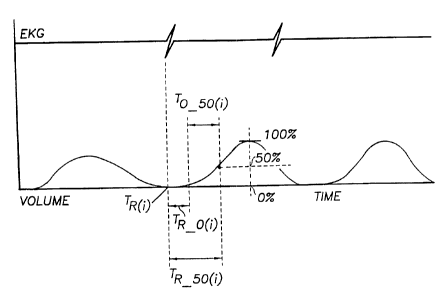
Fig. 2 is a graph of EKG and blood volume versus time.
Description of the Preferred Embodiment
The physics of wave propagation in elastic tubes is an important factor to
understand the underlying concept of the present invention. The simplest
equation
for the velocity of propagation of a pressure pulse in an elastic tube was
first
described by Moens-Kortweg who from experimental evidence and theoretical
grounds established the formula
Eh
2R8
where c is the wave velocity, E and h are Young's modulus and thickness of the
arterial wall, b the density of the fluid and R the mean radius of the tube.
To eliminate the experimental difficulties of measuring the wall thickness and
Young's modulus the Moens-Kortweg equation was modified by Bramwell and Hill
4
CA 02268073 2004-07-19
73766-83
(1922) so that the elastic behavior of the tube was expressed in terms of its
pressure-
volume distensibility. The formula can then be reduced to
Y YdP
c = -
8(aYIc7P) 8c7Y
or
~.POC C2(~Y)
Y
where V is the initial volume of the artery, a,V is the change in volume
resulting
in the pressure pulse DP and c is the pulse wave velocity.
The problem then involves determining a non-invasive way of measuring
both the pulse wave velocity, and percent change in arterial volume. In order
to
accomplish this, we have chosen to use the standard EKG signal and any stable
1~ measure of blood volume versus time (such as photoplethysmography~ in the
preferred embodiment).
The method of utilizing the EKG signal and blood volume versus time
signals include first measuring the TR ~~ (duration of R-wave on EIiG to
50°/o point
on volume versus time up-slope) for the i'th pulse. This duration is the sum
of the
time between the.R-wave and the arrival of the pulse 0% point (TR-~) added to
the
duration of the pulse 0% point to the 50°/o point on the up-slope (To
~~;~). The
inverse of TR o~;~ is proportional to the pulse velocity as defined above (or
coc 1/TR o~;>) and
1"°-~,; is more related to ~V and V. Therefore the measure TR_~;y is a
measure that
is related to c, ~'' and V.
2~ Then, the combined pulse velocity measure for the i'th pulse (v~;~) is
therefore defined as the inverse of TR so~;> and the combined pulse velocity
squared
(vp~~~ is obtained by simply squaring vP~,,. Also the instantaneous R-R
interval and
thereby instantaneous heart rate for the i'th pressure pulse (RR; and IHR~;~
respectively) are determined and used in the calculation of diastolic,
systolic and
mean pressures for the i'th pulse (Pp~~, P~S~;~ and PM~;~ respectively). The
theoretical
basis for the importance of the R-R interval or IHR in the calculation of
diastolic
pressure can be summarized as follows. The diastolic pressure is defined as
that
5
CA 02268073 2004-07-19
73766-83
arterial pressure that exists at the end of the .diastolic pressure decay.
This
exponential diastolic pressure decay starts at the closure of the aortic
valve, and ends
at the opening of the aortic valve. The pressure decay rate depends on a
variety of
factors, including the aortic pressure built up during systole, and the
systenuc
azvterial impedance (related to the stiffness of the walls of the arterial
system,
especially the arteriole). For a given individual, the pressure to which this
decay falls for
any given heart beat (or diastolic pressure) is therefore related to the
duration this
decay is allowed to continue. This duration of decay for any given pulse is
directly
proportional to the instantaneous R-R interval or inversely proportional to
the IHR
of that pulse. Therefore, the shorter the decay duration (higher IHR), the
higher
the diastolic pressure is expected to be, and the longer the decay duration
(lower
IHR), the lower the diastolic pressure is expected to be._ In summary the
equations
for the calculation of pressures for the i'th pressure pulse are as follows:
IHR~ = 17RR~~
. vP~;~z = ( 1/TR ~~;~ ) '~ ( 1/TR sx) )
1'n(7 ° ( Kn, '~ vn~~~z ) + ( Kn~h~ ''' IHR~;~ ) + K~,t
Pscp ~ ( KS<n '~ VPcpZ ) + Ks~~e
PMca ~ C Psn - PDc~, ) ~' 1/3 + PDT;,
In these equations, Kn", KD;," and K~",S, are constants that in the preferred
embodiment are equal to 2.5, 0.5 and 35 respectively, and where K~ and Kx~ are
calibration constants. Pp~;~, P s~;a and P M~~are diastolic, systolic and mean
arterial
pressure respectively.
?he practice of the present invention will be described in conjunction with
the figures. In Fig. 1 a human subject 10 is monitored by EKG leads
represented
generally at 12. Those skilled in the art recognize that multiple leads are
typically
utilized $or measuring the EKG. 1'hotoplethysmography apparatus 14 monitors
blood volume at a fingertip 16 of the subject 10. The outputs from the EKG
apparatus 12 and photoplethysmography apparatus 14 are processed in a computer
or signal processor 18 and produces as an output blood pressure which, as
discussed
above, may be diastolic pressure, systolic pressure or mean arterial pressure
for each
pulse. With reference to Fig. 2, the processor 18 detects the R-wave arrival.
6
CA 02268073 1999-04-O1
WO 98/25516 PCT/ITS97/18503
Thereafter, the blood volume measuring apparatus 14 detects the onset of a
change
in volume at time TR o~,~ and determines the time when volume has reached the
50%
(To ~~apoint on the volume versus time upslope. As can be seen in Fig. 2, the
time
from the arrival of the pulse zero percent point (TR ot;)) to the 50% point on
the
upslope (To ~~;)) depends on the shape of the volume versus time curve.
Because the
present invention utilizes both the time from R-wave arrival to the zero
percent
volume change point, and from the zero percent volume change point to the 50%
point, pressure determinations are more accurate than in the prior art in
which
either pulse arrival time or wave shape was utilized but not both in
combination as
in the present invention.
Another aspect of the invention includes methods for automated artifact
detection and rejection thereby providing a reliable assessment of the
confidence of
each blood pressure calculation for each pulse. These artifact rejection
methods
include the calculation of two additional variables for each pulse. For the
i'th pulse
thev are as follows:
qVP~~)z ( ~VPI3)Z 'VP~~)z ) / ~VP~z)z
where 'vP~,)'- is obtained by sorting five consecutive vPZ terms { vP~;_z)z,
vPt;_,)z, vP~;)z,
vPc+,)z, vP~;+z)z } and is the second lowest value, 'vP~z)z is the median of
the values, and
'vP~~)z is the second highest of the values.
And
z z z
diffvP~;) = vP~;) - v P~~.,)
The algorithm for the detection of whether the i'th pulse is artifact involves
testing if these variables are above predetermined thresholds. In this
preferred
embodiment, the test includes whether either
qvP~;)z > THRESH_qv
diffvP~;)z > THRESH_diffv
where the preferred values of THRESH-qv = 0.8 and THRESH diffv = 8.0 More
specifically, these variables are used in addition to the following others to
determine
the Pp{;) calculation artifact. The algorithm includes whether:
qvP~;)z > THRESH'qv
or
PDT;) < PD TOOLOW
7
CA 02268073 1999-04-O1
WO 98!25516 PCTIIJS97/18503
or
PD~;~ > PD TOOHIGH
or
PDc) > Psc~)
where in the preferred embodiment, PD TOOLOW = 30 and PD TOOHIGH =
150. If any of the above are true then it is deemed that the diastolic
pressure for
the i'th pulse (PDT;)) is not evaluable.
Specifically, and in like manner, the artifact determination for Ps~;~
calculation
includes whether:
qvP!>>Z > THRESH-qv
or
diffvP~;~z > THRESH diffv
or
Ps~;~ < PS TOOLOW
or
Ps~;~ > PS TOOHIGH
or
PD(7 > Pso
where in the preferred embodiment, PS TOOLOW = 50 and PS TOOHIGH =
200. If any of the above are true then it is deemed that the systolic pressure
for the
i'th pulse (Ps~;~) is not evaluable.
Finally and specifically, the determination if the PM~~ calculation would
result
in artifact for the i'th pulse if:
Pp~;~ is not evaluable
or
Ps~;~ is not evaluable
and if either is true, then the mean pressure for the i'th pulse is deemed to
be not
evaluable.
What is claimed is:
8