Note : Les descriptions sont présentées dans la langue officielle dans laquelle elles ont été soumises.
CA 02283214 2006-08-03
PROTECTIVE SHEATH FOR
TRANSVAGINAL ANCHOR IMPLANTATION DEVICE
Technical Field
This invention relates to a protective sheath or cap for a bone anchor
implantation device. The bone anchor implantation device is used in
maintaining or
improving urinary continence.
Background Information
Urinary incontinence, the inability to control urination from the bladder, is
a
widespread problem in the United States and throughout the world. Urinary
incontinence affects people of all ages and can severely impact a patient both
physiologically and psychologically.
In approximately 30% of the women suffering from urinary incontinence,
incontinence is caused by intrinsic sphincter deficiency (ISD), a condition in
which the
valves of the urethral sphincter do not properly coapt. In approximately
another 30% of
incontinent women, incontinence is caused by hypermobility, a condition in
which the
muscles around the bladder relax, causing the bladder neck and proximal
urethra to
rotate and descend in response to increases in intraabdominal pressure.
Hypermobility
may be the result of child delivery or other conditions which weaken, stretch
or tear the
muscles. In an additional group of women with urinary incontinence, the
condition is
caused by a combination of ISD and hypermobility.
In males, urinary incontinence may be the consequence of post radical
prostatectomy, which can destroy the valves of the urethral sphincter.
In addition to the conditions described above, urinary incontinence has a
number of other causes, including birth defects, disease, injury, aging, and
urinary tract
infection.
CA 02283214 1999-09-08
WO 99/37216 PCT/US99/01805
-2-
Numerous approaches for treating urinary incontinence are available. In one
procedure,
referred to as bladder neck stabilization (BNS), sutures are placed around the
muscles on either
side of the urethra and affixed to the rectus fascia or pubic bone and
tensioned to treat
hypermobility. Other procedures which treat both hypermobility and intrinsic
sphincter deficiency
(ISD) involve the placement of a sling under the urethra/bladder which
compresses the sphincter
while simultaneously acting as a stabilizer of the bladdemeck (preventing
excessive downward
mobility). The bone anchors which support the sling sutures may be inserted
into rectus fascia or
various locations on the pubis bone to provide a non-moveable anchoring
method.
Summary of the Invention
The present invention generally relates to devices and methods for inserting
anchors, such
as bone anchors, into a bone or tissue and more particularly to a protective
sheath or cap for
isolating the bone anchor to prevent both accidents with the sharp bone anchor
before it is
inserted into a target site and contamination of the target site by insertion
of the bone anchor
therethrough.
Bone anchors are often attached into bones in order to provide support for a
"sling" useful
in improving or maintaining a patient's urinary incontinence. In one
procedure, a suture-carrying
anchor is driven through the vaginal wall and into the posterior portion of
the pubic bone or
symphysis pubic, and the suture(s) attached to the bone anchor(s) extend
through the vaginal wall
and may be attached to the endopelvic fascia, the vaginal wall, a sling, or
other material to
stabilize and/or slightly compress the urethra thereby improving the patient's
urinary incontinence.
The present invention effectively addresses concerns in affixing an anchor to
bone or tissue.
The present invention is directed to a protective sheath for the bone anchor.
The
protective sheath prevents accidents with the sharp bone anchor tip before
insertion into the target
site, and it prevents infection of the pubic bone. The protective sheath
prevents exposure and
accidental puncture of the surgeon's gloves as well as premature insertion
into unintended tissue
in the patient. It also provides a sterile barrier around the bone anchor. The
protective sheath
shields the bone anchor from contacting microorganisms in the vagina and area
surrounding the
implantation site during insertion. The protective sheath of the present
invention ensures that the
bone anchor implants into the bone implantation site free from contaniination
and thus prevents
the occurrence of biological complications.
CA 02283214 1999-09-08
WO 99/37216 PCTIUS99/01805
-3-
One aspect of the present invention is a bone anchor implantation device
comprising an
elongated member having a first end and a second end, and a related method. A
bone anchor is
releasably engaged to the elongated member in the vicinity of the first end. A
protective sheath is
mounted over the bone anchor. The protective sheath can be axially movable
relative to the bone
anchor such that the bone anchor is exposed from the sheath as the bone anchor
is pressed into a
bone by the elongated member. Alternatively, the protective sheath can be a
balloon, gelatin
structure, or other covering that encapsulates or covers the bone anchor prior
to implantation.
The balloon or thin film can be hermetically sealed around the bone anchor,
but in any case the
balloon isolates the bone anchor from contact with tissue and prevents
contamination prior to
implantation of the bone anchor. The balloon is perforated by the bone anchor
as the bone anchor
is pressed into the bone by the elongated member or shaft. The balloon may be
made of a variety
of materials such as plastic, thermoplastic, elastromers, PET, PETG, rubber,
vinyl, latex, or
silicone. In one preferred embodiment, the balloon is made of latex. The
balloon can also be
made of a biodegradable material. In another preferred embodiment, the balloon
comprises a
polymer such as a synthetic polymer. Nonlimiting examples of useful polymers
include the
following: polyglycolic acid (PGA), polyactic acid(PLA), poly (dioxanone)
(PDO), poly (1-
lactide) (LPLA), poly (dl-lactide) (DLPLA), poly (glycolide-co-trimethylene
carbonate) (PGA-
TMC), poly (1-lactide-co-glycolide) (PGA-LPLA), poly (di-lactide-co-glycolide)
(PGA-DLPLA),
poly (1-lactide-co-dl-lactide) (LPLA-DLPLA), poly(glycolide-co-trimethylene
carbonate-co-
dioxanone) (PDO-PGA-TMC), poly(s-caprolactone), poly(dioxanone)(a polyether-
ester), poly
(lactide-co-glycotide), poly(SA-HDA anhydride), poly(orthoester), and
polyglyconate. The
protective sheath can also take the form of a gelatin structure (similar to a
pill capsule).
In some embodiments, the protective sheath (e.g. balloon or gelatin structure)
can contain
an antibiotic which is released when the sheath is perforated by the bone
anchor. The antibiotic
prevents infection at the site where the bone anchor is pressed into the bone.
Nonlimiting
examples of antibiotics which can be used include the following: nafcillin,
aminogylcoside,
ciprofloxin, clindamcin, piperacillin/tazobactum, ampicillin/sulbactum,
aminoglcoside,
vancomycin, cephalosporin, TMP/SMX, ampicillin, gentaminicin, tobramycin, and
ciprofloxacin.
Those skilled in the art will appreciate that there are numerous ways to
insert the antibiotic into
the balloon or the gelatin structure. In one embodiment, the bone anchor
implantation device has
CA 02283214 2006-08-03
-4-
a port which extends from the first end to the second end of the shaft into
the balloon or
gelatin structure. Antibiotics can be inserted into the protective sheath
through a port.
In general, in another aspect, the invention features a bone anchor
implantation
device which has a spring attached to the sheath within the balloon which
retracts when
the sheath is pressed against the bone by the shaft, thereby causing the bone
anchor to
perforate the balloon and implant into the bone. The spring element may be an
open-
coiled helical spring which surrounds the bone anchor. The spring element
retracts
when pressure is applied to the sheath causing the bone anchor to puncture the
balloon.
Thus, in one aspect, there is provided a bone anchor implantation device,
comprising: a hook-shaped shaft having a first end and a second end; a bone
anchor
releasably engaged to one end of the shaft; a protective sheath for
encapsulating the
bone anchor prior to implantation, wherein the protective sheath comprises a
balloon.
In another aspect, there is provided use of a bone anchor implantation device
for
inserting a bone anchor into a bone, the bone anchor implantation device
comprising: a
hook-shaped shaft having a first end and a second end; a bone anchor
releasably
engaged to one end of the shaft; a protective sheath for encapsulating the
bone anchor
prior to implantation, wherein the protective sheath comprises a balloon.
In some embodiments, the shaft of the bone anchor implantation device can
have a hollow section which accommodates one or more sutures coupled to the
releasably engaged bone anchor. Also, the shaft preferably is hook-shaped.
A method for inserting such a bone anchor that is releasably engaged to such a
bone anchor implantation device can include the steps of locating a bone
anchor
implantation site on the bone and applying a retrograde force to the bone
anchor to
implant the bone anchor into the bone or to retract the spring to cause the
bone anchor
to perforate the sheath and implant into the bone.
CA 02283214 2006-08-03
- 4a-
The foregoing and other objects, aspects, features, and advantages of the
invention will become more apparent from the following description and from
the
claims.
Brief Description of the Drawings
In the drawings, like reference characters generally refer to the same parts
throughout the different views. Also, the drawings are not necessarily to
scale,
emphasis instead generally being placed upon illustrating the principles of
the
invention.
Figure 1 is a plan view of the bone anchor implantation device.
Figure 2 is an exploded view of the anchor implantation device.
Figure 3 is a cross-sectional view of the distal end of the cannula showing
the
protective cap therein taken along line 3-3 of Figure 2.
CA 02283214 1999-09-08
WO 99/37216 PCT/US99/01805
-5-
Figure 4 is a cross-sectional view of the bone anchor implantation device of
Figure 1 taken
along line 4-4 of Figure 1.
Figure 5 is a cross-sectional view of the bone anchor implantation in its
locked
configuration.
Figure 6 is a plan view of an alternate embodiment of the bone anchor
implantation device
having a keyhole-shaped bore in the second handle.
Figure 7 is a cross-sectional view taken along line 7-7 of the alternate
embodiment shown
in Figure 6 locked in the position in which the inserter shaft is fully
extended from the cannula.
Figure 8 is a cross-sectional view of the alternate embodiment of Figure 6
locked in the
position in which the inserter shaft is fully retracted within the cannula.
Figure 9 is a cross-sectional view of the distal end of the inserter shaft in
the cannula
showing location of the bone anchor implantation site by sliding the cannula
along the endopelvic
fascia.
Figure 10 is a cross-sectional view of the distal end of the inserter shaft
and the cannula
showing the inserter shaft penetrating the protective cap near the distal end
of the cannula.
Figure 11 is a cross-sectional view of the distal ends of the inserter shaft
in the cannula
showing the bone anchor being drive into the bone.
Figure 12 shows the bone anchor with sutures extending therefrom after
implantation into
the bone.
Figure 13 is a side view of a bone anchor implantation device having a hooked
shaft.
Figure 14 is an enlarged side view of a distal portion of the bone anchor
implantation
device taken along line 14-14 of Figure 13 showing the internal structure of
the bone anchor
mount.
Figure 15 is a perspective view of the bone anchor mount.
Figure 16 is a cross sectional view of the bone anchor mount of Figure 15
taken along
line 16-16.
CA 02283214 1999-09-08
WO 99/37216 PCT/US99/01805
-6-
Figure 17 is a schematic view showing the interior structure of the handle an
alternate
embodiment of the bone anchor implantation device inserted into the vagina
with the proximal end
of the second telescoping cylinder contacting the pubic bone.
Figure 18 is an enlarged view of the shaft of the alternate embodiment of the
bone anchor
implantation device illustrated in Figure 17.
Figure 19 is a cross sectional view of the shaft of the bone anchor
implantation device
shown in Figure 18 taken along line 19-19 of Figure 18.
Figure 20 is a schematic view showing the interior structure of the handle of
an alternate
embodiment of the bone anchor implantation device illustrated in Figure 17
inserted into the
vagina showing the implantation of a bone anchor into the pubic bone and the
compression of the
spring.
Figure 21 is a side view of the bone anchor implantation device of Figure 13
showing a
protective sheath contacting the pubic bone.
Figure 22 is a side view of the bone anchor implantation device of Figure 13
showing the
bone anchor implanted into the pubic bone.
Figure 23 is a cross sectional view of the bone anchor mount and protective
sheath when
the protective sheath is containing the pubic bone.
Figure 24 is a cross sectional view of the bone anchor mount and the
protective sheath
when the bone anchor is being implanted into the pubic bone.
Figure 25 is a side view of a bone anchor implantation device having a hooked-
shaped
shaft and a balloon.
Figure 26a is a perspective view of the bone anchor and a balloon.
Figure 26b is a perspective view of a bone anchor and a gelatin structure.
Figure 27 is a schematic view showing the interior structure of the handle of
an alternate
embodiment of the bone anchor implantation device with balloon inserted into
the vagina with the
proximal end of the second telescoping cylinder contacting the pubic bone.
Figure 28 is a side view of the bone anchor implantation device of Figure 25
showing a
balloon contacting the pubic bone.
*rB
CA 02283214 1999-09-08
WO 99/37216 PCTIUS99/01805
-7-
Figure 29 is a side view of the bone anchor implantation device of Figure 25
showing the
bone anchor implanted into the pubic bone.
Figure 30 is a side elevational view of a bone anchor.
Figure 31 is a side elevational view of the spear member of bone anchor shown
in Figure
30.
Figure 32 is a rear elevational view of the spear member of the bone anchor
shown in
Figure 30.
Figure 33 is an enlarged sectional view of a shaft of the spear member of the
bone anchor
taken along the lines 4--4 of Figure 31.
Figure 34 is a side elevational view of a collar member of the bone anchor
shown in Figure
30.
Figure 35 is a rear elevational view of the collar member of the bone anchor
shown in
Figure 30.
Figure 36 is a side elevational view of the collar member of the bone anchor
taken along
the direction of the lines 7--7 of Figure 35.
Figure 37 is a sectional view of the collar member of the bone anchor taken
along the lines
8--8 of Figure 35.
Figure 38 is a perspective view of the bone anchor tip on a bone anchor
implantation
device having a balloon with a spring element.
Figure 39 is a side view of a bone anchor implantation device with a
telescoping sheath.
Figure 40a is an enlarged view of the telescoping sheath of Figure 39 with the
telescoping
sheath in an expanded position.
Figure 40b is an enlarged view of the telescoping sheath of Figure 39 with the
telescoping
sheath in a retracted position.
Figure 41 a is a view of a bone anchor implantation device with a balloon
sheath.
Figure 41b is an enlarged view of the balloon sheath on bone anchor
implantation device
of Figure 4 1 a.
CA 02283214 1999-09-08
WO 99/37216 PCT/US99/01805
-8-
Figure 42a is a view of a bone anchor implantation device with a latex sheath
and a spring
element.
Figure 42b is another view of the bone anchor implantation with a latex sheath
and a
spring element.
Figure 43 is a view of a bone anchor implantation device with a caplet sheath.
Figure 44 is a view of a bone anchor implantation device that has a lumen that
can hold
one or more sutures.
Figure 45 is a view of a bone anchor implantation device of Figure 39 inserted
into the
vagina showing the implantation of a bone anchor into the pubic bone.
Description
The present invention relates to a device for affixing a bone anchor to a
bone. More
particularly, the invention relates to a protective sheath for protecting the
bone anchor from
contacting tissue during implantation and thereby preventing contamination of
the bone anchor. It
also relates to methods for improving or maintaining a patient's urinary
continence in which bone
anchors are inserted transvaginally into the posterior portion of the pubic
bone or symphysis pubis
and devices for use in such methods. As used herein, the terms
"transvaginally" or "transvaginal
access" refer to access through the vaginal introitus or from within the
vagina, as opposed to
access from the patient's abdominal side.
As will be described in more detail below, the methods and devices of the
present
invention drive a bone anchor through the vaginal wall and into the posterior
portion of the pubic
bone or symphysis pubis. The public bone may also be accessed through a
suprapublic bone
incision. Preferably, at least one bone anchor is driven into the pubic bone
on either side of the
urethra. However, one of skill in the art will appreciate that a single bone
anchor may also be
used. The sutures attached to the bone anchors extend through the vaginal wall
and may then be
attached to the endopelvic fascia, the vaginal wall, a sling, or other
material to stabilize and/or
slightly compress the urethra, thereby improving or maintaining the patient's
urinary continence.
CA 02283214 1999-09-08
WO 99/37216 PCT/US99/01805
-9-
Two Handle Bone Anchor Imnlantation Device
In one embodiment, the anchor implantation device has a first handle having an
inserter
shaft attached thereto. The inserter shaft is adapted to releasably engage or
attach to a bone
anchor. A second handle is hingedly attached to the first handle and has a
cannula attached
thereto. The cannula has a central bore extending therethrough. The cannula is
aligned with the
inserter shaft such that the inserter shaft is inside the central bore of the
cannula and is extendible
from and retractable in the cannula. Preferably, a biasing member is disposed
between the first
handle and the second and biases the first handle and the second handle apart.
Figures 1 and 2 provide a plan view and an exploded view of an anchor
implantation
device 10 for introducing a bone anchor 22 transvaginally and driving it into
the pubic bone or
symphysis pubis. The device comprises a first handle 12 having a proximal end
14, a central
region 16, and a distal end 18. The first handle 12 may be made of any
relatively firm material,
including plastic or metal. Preferably, the first handle 12 is made of
plastic, aluminum, stainless
steel, or titanium. However, those skilled in the art will appreciate that a
wide range of other
material may also be employed.
The first handle 12 may be configured in any of a variety of shapes compatible
with
vaginal insertion. Preferably, the first handle 12 is rectangular. However,
those skilled in the art
will appreciate that a variety of configurations may be employed, such as a
handle which tapers
towards the distal end, and the present invention contemplates the use of any
handle configuration
compatible with vaginal insertion.
The dimensions of the first handle 12 are also compatible with vaginal
insertion. The first
handle 12 may be from about 4 inches to about 8 inches in length, about 0.25
inches to about 1.25
inches in width, and about 0.05 inches to about 0.5 inches in height.
Preferably, the first handle
12 is about 5 inches to about 7 inches in length, about 0.5 inches to about 1
inch in width, and
about 0.1 inches to about 0.3 inches in height. More preferably, the first
handle 12 is has a length
of 6 inches, a width of 0.75 inches and a height of 0.2 inches.
An inserter shaft 20 adapted for releasably engaging a bone anchor 22 is
located near the
distal end 18 of the first handle 12. A variety of bone anchors 22 can be
used. In a preferred
embodiment, illustrated in Figure 30 the bone anchor comprises a spear member
112 which is
able to pierce and securely engage the bone. The spear member 112 has a
generally cone shaped
*rB
CA 02283214 1999-09-08
WO 99/37216 PCT/[JS99/01805
-10-
head portion 114 which is used to pierce the bone to which the and a shaft
portion 116 with an
oval eyelet 118 therethrough for receiving and holding a suture strand(s). To
provide means for
retaining the spear member 112 within the bone, the bone anchor 122 further
comprises a collar
member 120. The collar member is used for retaining the bone anchor 122 in
place, once it has
been driven into the bone, by lodging within the bone in a manner to resist
removal of the bone
anchor 122. The bone anchor 122 and its component parts are more fully
described below.
The spear member 112 of the bone anchor 122 will now be described with
additional
reference to Figures 31-33. The shaft portion 116 of the spear member 112 is
generally
cylindrical in shape and has the eyelet 118, or bore, formed radially
therethrough proximate one of
its ends. The eyelet 118 may be oval, round or other suitable shapes and is of
a sufficient size to
permit suture strand or strands to pass therethrough. The circumference of
each outer end of the
eyelet 118 is chamfered or grounded to provide a bevel portion 122. It should
be appreciated that
the bevel portion 122 provides a generally smooth surface for contacting
suture strand which has
been passed through the eyelet 118. The eyelet 118 is located on the shaft
portion 116 of the
spear member 112 such that the transverse axis of the eyelet 118 intersect the
longitudinal axis of
the spear member 112.
The generally cone-shaped head portion 114 of the spear member 112 is located
at an end
of the shaft portion 116 opposite the end having the eyelet 118. As best shown
in Figures 30 and
31, the apex of the cone-shaped head portion is a point 124 which is suitable
for piercing and
being driven into bone. The diameter of the cone-shaped head portion 114
increases, when
viewed along a longitudinal direction rearwardly from the point 124 towards
the shaft portion
116. The cone angle along this region is preferably about 30 degrees. The
diameter of the cone-
shaped head portion 114 increases at a greater rate along approximately the
rearward half thereof,
when viewed along the same longitudinal direction. Thus, the rearward half of
the cone-shaped
head portion 114 arcs outwardly from the central longitudinal axis of the
spear member 112. As
show in Figures 31 and 32, the base 126 of the cone-shaped head portion 114 is
a ring-shaped
planar surface which is oriented substantially perpendicular to the
longitudinal axis of the shaft
portion 116.
Preferably, the cone-shaped head portion 114 is formed integrally with the
shaft portion
116 of the spear member 112. Alternatively, the cone-shaped head portion 114
and the shaft
CA 02283214 1999-09-08
WO 99/37216 PCT/US99/01805
-11-
portion 116 may initially be formed separately and then subsequently attached
to one another by
any suitable means.
The collar member 120 of the bone anchor 122 will now be described with
particular
reference to Figure 30 and Figures 34-37. The collar member 120 is provided
with a ring-shaped
generally planar forward surface 130 which is adapted to bear against, and
mate with the base 126
of the cone-shaped head portion 114 of the spear member 112. A circular bore
132 is located
centrally through the planar forward surface 130 and is adapted to receive the
shaft portion 116 of
the spear member 112 therethrough. The circumference of the planar forward
surface 130 may
be, but is not necessarily, chamfered to formed a beveled outer rim portion
133.
Four separate flanges 134 extend rearwardly from the planar forward surface
130 of the
collar member 120 as shown in Figures 34-39. The flanges 134 are separated
from one another
by longitudinally extending slots 136. The portions of the flanges 134 which
are proximate to the
planar forward surface 130 run generally parallel to the central longitudinal
axis of the collar
member 120. Each of the flanges 134 arcs generally outward from the central
longitudinal axis as
the flange 134 extends in a direction away from the planar forward surface
130. The lateral width
of each of the flanges 134 increases as the flange 134 extends in a direction
away from the planar
forward surface 130. The extreme rearward end of each of the flanges 134
curves away from the
planar forward surface 130 in the form a shallow C-shape, thereby providing
two trailing tips 140
for each flange 134.
As set forth above, the collar member 120 is rotatably fitted over the shaft
portion 116 of
the spear member 112 to form the assembled bone anchor 122 as shown in Figure
1. While there
is no need to permanently secure the collar member 120 to the spear member
112, the planar
forward surface 130 may nevertheless be securely attached to the base 126 of
the cone-shaped
head member 114 of the spear member 112 by any suitable means. It will be
appreciated,
however, that by permitting the spear member 112 to freely rotate with respect
to collar member
120, the suture strand 150 can be rotated by the surgeon after implantation to
a position where
the forces acting on the suture strand 150 by the bone anchor 122 are more
evenly distributed
around the region of the shaft portion 116 adjacent to the eyelet 118. Such a
position of the
suture strand 150 is shown in Figure 33.
CA 02283214 1999-09-08
WO 99/37216 PCT/US99/01805
-12-
In addition, it should also be appreciated that the two-piece construction of
the bone
anchor 122, affords machining advantages over a single-piece bone anchor. That
is, it is easier to
machine each of these components separately and to subsequently assemble them
together, as
opposed to machining the same basic structural features from a single piece of
material. Any
known materials suitable for orthopedic anchor devices may be employed to
construct the bone
anchor 122 of the present invention. Preferably, the bone anchor 122 is formed
from a metallic
material possessing sufficient strength to penetrate the bone. Such materials
include titanium 316
LVM stainless steel, CoCrMo alloy, Nitinol alloy, or other suitable materials.
Preferably, the
bone anchor is made of titanium.
Referring now back to Figures 1 and 2, the inserter shaft 20 has a distal end
24, a central
region 26, and a proximal end 28. Preferably, the inserter shaft extends at an
angle of about 90
from the first handle 12.
The inserter shaft 20 may be made of any of a variety of materials, including
steel,
stainless steel, aluminum, titanium, and plastic, but is preferably made of
stainless steel.
Additionally, the inserter shaft 20 may have a variety of cross sectional
shapes including
rectangular, hexagonal, or triangular but preferably the inserter shaft 20 has
a circular cross
section.
The inserter shaft 20 maybe located from about 0.05 inches to about 0.5 inches
from the
distal end 18 of the first handle 12. preferably, the inserter shaft 20 is
located from about 0.1
inches to about 0.3 inches from the distal end 18. More preferably, the
inserter shaft 20 is located
0.2 inches from the distal end 18 of the handle.
The length of the inserter shaft 20 is consistent with transvaginal delivery
of the releasable
bone anchor 22. Thus, the inserter shaft 20 maybe from about 0.5 inches to
about 1.5 inches
long. Preferably, the inserter shaft 20 is from about 0.75 inches to about
1.25 inches in length.
More preferably, the inserter shaft 20 is 1 inch in length.
Preferably, the proximal end 28 and the central region 26 of the inserter
shaft 20 have an
equal cross sectional area, which is larger than the cross sectional area of
the bone anchor 22 and
distal end 24. Thus, a shoulder 17 is formed at the junction between the
central region 25 of the
inserter shaft and the distal region of the inserter shaft. The shoulder 17
acts as a stop which will
not penetrate the cortical shell of the bone.
CA 02283214 1999-09-08
WO 99/37216 PCT/US99/01805
- 13 -
The diameter of the inserter shaft is dependent upon the size of the bone
anchor. In
embodiments in which the inserter shaft 20 is cylindrical, the diameter of the
proximal end 28 and
central region 26 of the inserter shaft is from about 0.1 inches to about 0.3
inches, and that of the
distal end 24 is from about 0.04 inches to about 0.2 inches. Preferably, the
diameter of the
proximal end 28 and central region 26 of the inserter shaft is from about 0.15
inches to about 0.25
inches, and that of the distal end 24 is from about 0.07 inches to about 0.11
inches. More
preferably, the diameter of the proximal end 28 and central region 26 of the
inserter shaft is 0.2
inches and that of the distal end 24 is 0.09 inches.
Preferably, the inserter shaft 20 is curved as shown in Figures 1 and 2. As
will be
appreciated by those of skill in the art, the inserter shaft 20 may also be
straight. In those
embodiments in which the inserter shaft 20 is curved, the radius of curvature
of the inserter shaft
is the distance between the pivot point of the hinge and the center of the
inserter shaft. The
radius of curvature of the inserter shaft 20 may be from about 3.5 inches to
about 7.9 inches.
Preferably, the radius of curvature of the inserter shaft 20 is from about 4.7
inches to about 6.9
15 inches. More preferably, the radius of curvature of the inserter shaft 20
is 5. 8 inches.
The distal end 24 of the inserter shaft 20 is adapted to releasably engage a
bone anchor 22.
In one embodiment, the bone anchor 22 is housed within a notch 30 at the
distal end 24 of the
inserter shaft, and frictionally engages the inner wall of the distal end 24
of the inserter shaft.
However, it will be appreciated by those of skill in the art that the inserter
shaft 20 may releasably
20 engage the bone anchor 22 through a variety of means other than that
described above, and such
means are specifically contemplated by the present invention.
The distal end 24 of the inserter shaft maybe hollow or solid and has a
complementary
shape to the proximal end of the bone anchor 22 to permit the bone anchor 22
to frictionally
engage the distal end 24 of the inserter shaft. For example, the distal end 24
of the inserter shaft
and the proximal end of the bone anchor may be square, rectangular,
pentagonal, triangular or
hexagonal in cross section. Preferably, the distal end 24 of the inserter
shaft and the proximal end
of the bone anchor are cylindrical. However, those skilled in the art will
appreciate that numerous '
shapes may be employed, and the present invention specifically contemplates
any such shape.
The central region 26 of the inserter shaft has a pair of grooves 32 therein
for receiving a
suture 54 attached to the bone anchor as illustrated in Figures 1 and 2.
Preferably, the grooves 32
CA 02283214 1999-09-08
WO 99/37216 PCT/US99/01805
-14-
in the inserter shaft are coextensive with slots 34 in the outer cannula and
are aligned with the
slots 34.
The device also comprises a second handle 36 hingedly connected to the first
handle 12
and having a proximal end 38, a central region 40, and a distal end 42. The
second handle 36 may
be fabricated from any of the materials discussed above with regard to the
first handle 12.
Additionally, the second handle 36 may have any of the dimensions and shapes
discussed above
with regard to the first handle 12. The preferred materials, dimensions, and
shapes for the second
handle 36 are the same as those discussed above with regard to the first
handle 12.
The second handle 36 has a cannula 44 positioned near its distal end 42 and
fixed within a
bore 11 in the second handle by screws 13. The cannula 44 has a proximal end
46, a central
region 48, and a distal end 50, with a central bore 52 running through its
entire length.
Preferably, the cannula 44 extends at an angle of about 90 from the second
handle 36.
The cannula 44 may be fabricated from any of the materials described above
with regard
to the inserter shaft 20. Preferably, the cannula 44 is made of stainless
steel.
The cannula 44 may have any of the shapes discussed above with regard to the
inserter
shaft 20. Preferably, the shape of the cannula 44 is the same as that of the
inserter shaft 20.
The cannula 44 is located approximately the same distance from the distal end
42 of the
second handle as the inserter shaft 20 is from the distal end 18 of the first
handle and the central
bore 52 of the cannula has an inner diameter larger than the outer diameter of
the inserter shaft
20. In this way, the inserter shaft 20 extends into the central bore in the
cannula as depicted in
Figure 1. The inserter shaft 20 is extendible and retractable relative to the
cannula 44.
Preferably, the cannula 44 has two oppositely disposed slots 34 therein
through which the
suture 54 attached to the bone anchor passes. These slots reduce the
possibility of the suture 54
becoming tangled. Preferably, the slots 34 in the cannula are aligned with and
coextensive with
the grooves 32 in the inserter shaft.
Alternatively, the sutures can be contained within the cannula and extend out
another
portion of the device such as the first handle 12.
Preferably, the distal end 50 of the cannula has a sharp tip 56 to facilitate
its use in
piercing tissue.
CA 02283214 1999-09-08
WO 99/37216 PCT/US99/01805
-15-
As illustrated in Figures 1 and 2, in the embodiments in which the inserter
shaft 20 is
curved, the cannula 44 is preferably also curved in the same arc as the
inserter shaft 20. By
curving the cannula 44, the diameter of the cannula 44 can be reduced in
comparison with
embodiments in which the inserter shaft and the cannula are not curved. Thus,
in the
embodiments in which the inserter shaft 20 and cannula 44 are curved, the
inner diameter of the
cannula 44 is from about 0.1 inches to about 0.25 inches and the outer
diameter of the cannula 44
is from about 0.14 inches to about 0.31 inches. Preferably the inner diameter
of the cannula 44 is
from about 0.15 inches to about 0.2 inches. In a highly preferred embodiment,
the inner diameter
of the cannula is 0.170 inches, the wall is about 0.02 inches, and the outer
diameter is about 0.210
inches. These dimensions also apply to the devices in Figures 5 and 6 in which
the inserter shaft
and the cannula are also curved.
In a preferred embodiment, the cannula 44 has a protective cap 58 inside the
central bore
52 and located at the distal end 50 of the cannula, as shown in Figures 2, 3
and 5. The protective
cap 58 may be made of a variety of materials, such as plastic, thermoplastic
elastomers, PET,
PETG, rubber material, vinyl, gelatin, latex, thermoset rubbers and silicone.
Preferably, the
protective cap 58 is made of silicone or plastic.
The internal protective cap 58 acts to shield the bone anchor 22 from
contamination, e.g.,
from contact with microorganisms in the vagina which could cause infection if
introduced into the
pubic bone during implantation of the bone anchor. In one embodiment, the
protective cap 58 has
a vertical slit 60 and a horizontal slit 62 therein which intersect to form a
cross. Alternatively, the
protective cap 58 has slits three slits which intersect to form a Y.
In one embodiment, the slits 60 and 62 penetrate entirely through the material
of the
protective cap 58, thereby dividing the protective cap into discrete segments.
Preferably, the slits
60 and 62 are scored in the material of the protective cap 58 but do not
extend entirely
therethrough.
The slits 60 and 62 permit the bone anchor 22 to move through the protective
cap 58
during implantation. In embodiments in which the slits 60 and 62 penetrate
entirely through the
material of the protective cap 58, the bone anchor 22 forces the segments of
the protective cap 58
to separate as the bone anchor 22 is extended through the protective cap 58.
The protective cap
58 remains in contact with the external surface of the bone anchor 22 as it is
inserted into the
CA 02283214 1999-09-08
WO 99/37216 PCT/US99/01805
-16-
bone, thereby shielding the bone anchor from contact with potentially
infectious microorganisms
in the vaginal wall.
The operation of the protective cap 58 in embodiments in which the slits 60
and 62 are
scored in the protective cap is identical to that described above. However, in
such embodiments,
the tip of the bone anchor 22 pierces the material of the protective cap 58 as
the bone anchor 22
is extended, thereby causing the protective cap 58 to separate into segments
along the scores.
The proximal ends of the first and second handles, 14 and 38 respectively, are
hingedly
connected to permit them to move towards and away from one another. Any
suitable type of
hinge can be used, for example, this can be accomplished using a hinge 64
similar to that
commonly found on doors as shown in Figures 1 and 2. Alternatively, a piece of
rubber may be
interposed between the first and second handles at their proximal ends 14 and
38 and secured
thereto by bolts extending into holes in each of the handles. Those skilled in
the art will
appreciate that other means of hingedly connecting the first and second
handles may be employed,
and the present invention specifically contemplates embodiments in which such
other hinging
mechanisms are employed.
The first handle 12 and the second handle 36 are biased apart. In one
embodiment, the
biasing force is provided by a spring 66, as discussed below. The spring 66
can be metal, resilient
polymer, pneumatically driven, or of any other suitable design. However, those
skilled in the art
will appreciate that a number of other structures can be employed to achieve
the same biasing
effect. The present invention specifically contemplates such other means of
biasing the handles
apart.
When sufficient force is applied to the distal ends of the first and second
handles (18 and
42) to overcome the resistance of the spring, the distal ends (18 and 42) of
the first and second
handles move closer together. In the position in which the distal ends of the
first and second
handles are maximally separated, the inserter shaft 20 and bone anchor 22
thereon are fully
retracted inside the cannula 44. As increasing force is applied to the handles
and the distal ends
approach one another, the inserter shaft 20 and bone anchor 22 thereon emerge
from the distal
end 50 of the cannula. At the point where the distal ends (18 and 42) of the
first and second
handles are touching, the inserter shaft 20 and bone anchor 22 thereon are
maximally extended
from the distal end 50 of the cannula.
CA 02283214 1999-09-08
WO 99/37216 PCTIUS99/01805
-17-
At the point of maximum extension, the length of the inserter shaft 20
extending from the
cannula 44 is from about 0.05 inches to about 0.8 inches. Preferably, at the
position of maximum
extension, the length of the inserter shaft 20 extending from the cannula 44
is about 0.1 inches to
about 0.5 inches. More preferably, the length of the inserter shaft 20
extending from the cannula
44 at the position of maximum extension is 0.2 inches.
In the above embodiment, the bone anchor 22 is inserted in the bone by
manually moving
the inserter shaft 20 axially through the bore 52 in the cannula until the
inserter shaft and bone
anchor 22 thereon extend from the cannula 44. However, the those skilled in
the art will
appreciate that approaches other than manually moving the inserter shaft may
also be used to
implant the bone anchor into the bone. For example, the bone anchor 22 can be
forced into the
bone by applying sufficient pneumatic pressure through the inserter shaft to
eject the bone anchor
from the inserter shaft with sufficient force to implant the bone anchor in
the bone. Alternatively,
the bone anchor may be driven into the bone by a spring mechanism.
Preferably, the device further comprises a locking mechanism for locking the
device in the
position in which the inserter shaft is fully retracted within the cannula in
order to avoid accidental
insertion of the bone anchor into tissue. As those skilled in the art will
appreciate, a variety of
locking structures may be used to achieve such locking.
One exemplary locking mechanism is shown in Figure 2. The locking mechanism
comprises a locking plate 15 slidably mounted over the first handle 12 and
having a bore 68
therein. The locking plate 15 is separated from the first handle 12 by a
spacer 70 having an
internally threaded bore 72 therein which is aligned with the bore 68 in the
locking plate. The first
handle 12 has an elongate hole 74 herein having a proximal end 75 and a distal
end 77. A locking
screw 76 extends through the elongate hole 74 in the first handle and the
bores 72.68 in the
spacer 70 and locking plate 15. The locking screw 76 is secured to the locking
plate 15 by a nut
78.
The second handle 36 has a bore 80 therethrough having a diameter larger than
that of the
head of the locking screw 76. In the unlocked position, the bore 80 can be
aligned with the
locking screw 76 as shown in Figure 4 thereby permitting the first handle 12
and the second
handle 36 to be squeezed together such that the inserter shaft 20 extends from
the cannula 44.
CA 02283214 1999-09-08
WO 99/37216 PCTIUS99/01805
-18-
As illustrated in Figure 5, in the locked position, the locking plate 15 is
positioned at the
proximal end 75 of the elongate hold 74 in the first handle. In this position,
the locking screw 76
is disposed between the first and second handles such that the head of the
screw abuts the inner
side of the second handle 36, thereby preventing the first handle 12 and the
second handle 36
form being squeezed together.
In an alternate embodiment, the bone anchor implantation device 110 may have a
dual
position lock permitting the device to be locked in a position in which the
inserter shaft 120 is
fully retracted within the cannula 144 or in a position in which the inserter
shaft 120 is fully
extended from the cannula 144. In this embodiment, illustrated in Figure 6 the
bore 180 of the
second handle 136 is keyhole shaped. The narrower part 121 of the keyhole
shaped bore is
sufficiently narrow to prevent the head of the locking screw 176 from passing
therethrough. As
illustrated in Figure 7, when the locking plate 115 is positioned at the
proximal end 175 of the
elongate bore in the first handle, the head of the locking screw 176 is over
the narrow part 121 of
the keyhole shaped aperture 180. As shown in Figure 7, in this position the
inner side of the head
of locking screw 176 contacts the outer side of the second handle. The device
is locked in the
position in which the inserter shaft and bone anchor thereon are fully
extended.
When the locking plate 115 is positioned at the distal end 177 of the elongate
bore in the
first handle, the head of locking screw 176 is aligned with the wide portion
123 of the keyhole
shaped bore. The locking plate 115 can then be returned to the proximal end
175 of the elongate
2o hole, such that the head of the locking screw 176 is disposed between the
first handle 112 and the
second handle 136 and the head of the locking screw 176 abuts the inner side
of the second
handle 136, as shown in Figure 8. In this position the inserter shaft 120 is
fully retracted and the
first handle 112 and the second handle 136 cannot be squeezed together.
The above locking mechanisms may be used in the embodiments where the inserter
shaft
and cannula are straight. As those skilled in the art will appreciate, a
variety of other locking
structures may be used to achieve such dual position locking. Such other
locking mechanisms are
specifically contemplated by the present invention.
In the embodiments described above, the force biasing the two handles apart is
preferably
provided by a spring 66 disposed between two depressions 82 and 84 in the
first handle 12 and
the second handle 36. In the embodiments described above, the spring 66 is
located in the central
CA 02283214 1999-09-08
WO 99/37216 PCT/US99/01805
-19-
regions 16 and 40 of the first and second handles. However, those skilled in
the art will
appreciate that the location of the spring is not critical to the operation of
the present invention.
Additionally, it will be appreciated that biasing members other than a spring
may be employed to
bias the handles apart.
Using the present bone anchor implantation device, the bone anchor is
transvaginally
introduced into the pubic bone as follows.
After making an incision in the anterior vaginal wall, the endopelvic fascia
is accessed
using techniques well known to those of skill in the art, such as with a
conventional retractor. A
Foley catheter may be introduced to assist in locating the bladder neck. The
bone anchor
implantation device is inserted into the vaginal introitus and the first
desired site for bone anchor
implantation is located by digital palpation of the urethra, pubic symphysis
or other anatomical
landmark or other techniques known to those of ordinary skill in the art. The
device is locked in
the position in which the inserter shaft is fully retracted during this
procedure.
Once the desired site for bone anchor implantation is located, the sharp tip
56 on the distal
end of the cannula is driven through the endopelvic fascia 17. The pointed end
of the cannula can
also be employed to locate the desired implantation site by inserting the
device into the vaginal
introitus and through the incision, piercing the endopelvic fascia, and moving
the cannula along
the pubic bone 19 to the desired implantation site, as shown in Figure 9.
The device is then unlocked from the position in which the inserter shaft 20
is fully
retracted. In the embodiment having a single position lock, the first and
second handles (12 and
36) are pressed together with enough pressure to extend the inserter shaft 20
out of the cannula
44 and drive the bone anchor 22 into the posterior portion of the bone 19.
Alternatively, in the
embodiment having a dual position lock, the device is locked in the position
in which the inserter
shaft 120 is fully extended from the cannula 144 and the manual pressure is
applied to drive the
bone anchor 122 into the posterior portion of the pubic bone 19.
As shown in Figure 10, when the first and second handles are squeezed towards
one
another, the inserter shaft moves towards the bone 19. The bone anchor 22
pierces the protective
cap 58 which separates as the bone anchor 22 passes therethrough. The
protective cap 58 shields
the bone anchor 22 from contact with the vaginal tissue.
CA 02283214 1999-09-08
WO 99/37216 PCTIUS99/01805
-20-
As shown in Figure 11, when the inserter shaift 20 is extended beyond the
distal tip of the
cannula 56, the bone anchor contacts the bone 19 and is driven therein.
The inserter shaft 20 is then retracted into the cannula 44, leaving the bone
anchor 22
implanted in the bone 19 with the attached suture 54 extending through the
wound in the vaginal
wall and the endopelvic fascia as shown in Figure 12.
The above site location and bone anchor implantation procedure is repeated to
implant a
second bone anchor on the opposite side of the urethra from the first bone
anchor.
In one embodiment, the sutures are attached to a needle, looped back through
the vaginal
wall, and attached to tissue such as the endopelvic fascia or the vaginal wall
so as to bias the
tissue surrounding the urethra towards the urethra. The biasing force
compresses or stabilizes the
bladder neck thereby maintaining or improving urinary continence.
Alternatively, the sutures attached to the bone anchors can be attached to a
sling which
compresses or stabilizes the bladder neck. In such procedures, an incision is
made midline to the
urethra. An opening or pocket for receiving the sling is created in the tissue
between the urethra
and the upper vaginal wall. The bone anchor implantation device is inserted
through the incision,
into the pocket, and through the endopelvic fascia to contact the pubic bone.
At least one bone
anchor is inserted into the pubic bone on each side of the urethra. The sling
is introduced into the
opening or pocket and attached to the sutures. The tension on the sling
provided by the sutures is
adjusted to provide the appropriate biasing force to the urethra.
Example 1 describes one method of using the present bone anchor implantation
device to
compress or stabilize the bladder neck with sutures. it will be appreciated
that the bone anchor
implantation device can be used with other methods in which sutures compress
or stabilize the
bladder neck.
EXAMPLE 1
Compression or Stabilization of the Bladder Neck with Sutures
The bone anchor implantation device can be used in incontinence treatments in
which the
bladder neck is compressed or stabilized with sutures. A Foley catheter is
inserted into the
urethra to indicate its location. An incision is then made through the
anterior vaginal wall,
preferably approximately 1 cm lateral to midline and adjacent to the bladder
neck. The vaginal
CA 02283214 2006-08-03
-21-
wall is retracted to allow access to the endopelvic fascia. The bone anchor
implantation device,
having a bone anchor with sutures attached thereto releasably engaged with the
inserter shaft, is
introduced through the opening in the vaginal wall with the device locked in
the position in which
the inserter shaft is fully retracted within the cannula, and the sharp point
is pressed through the
fascia to contact the posterior pubic bone. Preferably, the anchor
implantation site is located
lateral to the symphysis pubis and cephalad to the inferior edge of the pubic
bone. The anchor
implantation site is located by palpating the inferior rim of the pubic bone
and the symphysis
pubis, moving laterally until the lower border of the obturator foramen is
located. Preferably, the
anchor is located from about 0.5 to 4 cm lateral to the symphysis pubis and
from about 0.5 to 3
cm cephalad to the inferior edge. More preferably, the anchor implantation
site is located
approximately I cm lateral to the symphysis pubis and 1 cm cephalad to the
inferior edge of the
pubic bone. In addition, the anchor implantation site can be located on the
pubic ramus.
The locking mechanism of the bone anchor implantation device is then placed in
the
unlocked position, and the two handles are squeezed together such that the
inserter shaft is in the
extended position. Alternatively, for devices having a dual position locking
mechanism, the bone
anchor may be exposed by locking the device in the position in which the
inserter shaft is fully
extended from the cannula. In either case, the anchor is driven into the pubic
bone using manual
pressure and opposing thumb pressure on the external pubic section if
necessary.
The bone anchor implantation device is withdrawn, leaving the two free ends of
the
2( anchored suture exiting the endopelvic f scia 17. A device such as a Mayo
needle is then
attached to one free end of the anchored suture and a "bite of fascia" is
taken adjacent to the
bladder neck. Preferably, the entry and exit points of the suture are adjacent
to the bladder neck
approximately 0.5 cm lateral to the urethra. This step is then repeated with
the other free end of
the suture, and the two ends are tied together. The vaginal wall incision is
then closed.
Alternatively, the entry and exit points of the suture can be made as
illustrated in Figure
13a of U.S. Patent No. 5,611,515 to Benderev.
The above procedure is then repeated on the opposite side of the urethra to
complete the
bladder neck suspension. The sutures are then appropriately tensioned.
Appropriate tension is
confirmed using well known means such as cystoscopy or a standard Q tip test.
CA 02283214 1999-09-08
WO 99/37216 PCT/US99/01805
-22-
EXAMPLE 2
Example 2 describes use of the bone anchor implantation device in a procedure
in which
the bladder neck is compressed or stabilized with a sling. However, it will be
appreciated that the
bone anchor implantation device can be used with other methods in which the
bladder neck is
compressed or stabilized with a sling.
Double Anchor Placement: For Sling Or Bolster Procedure
The bone anchor implantation device can also be used in incontinence
treatments in which
the bladder neck is compressed or stabilized using a sling. Preferably, in
such procedures two
bone anchors are placed on either side of the urethra. However, one of
ordinary skill in the art
will appreciate that one or more than two bone anchors per side can be used.
The procedure is
performed as follows.
A Foley catheter is inserted into the urethra to indicate its location.
Starting adjacent to
the bladder neck on either side of the urethra, a 1 cm incision is made
through the anterior vaginal
wall approximately 1 cm lateral to and parallel to the midline of the urethra.
The vaginal wall is
retracted to allow access to the endopelvic fascia 17. Blunt dissection is
used to tunnel under the
urethra and form a pocket for the sling.
The bone anchor implantation device is introduced through the opening in the
vaginal wall
with the device locked in position in which the inserter shaft is fully
retracted within the cannula,
and the sharp point of the cannula is pressed through the fascia 17 near the
distal end of the
vaginal wall incision closer to the bladder neck, to contact the posterior
aspect of the pubic bone.
Preferably, the first anchor implant site is located lateral to the symphysis
pubis and cephalad to
the inferior edge of the pubic bone. More preferably, the first anchor implant
site is located
approximately 1 cm lateral to the symphysis pubis and 1 cm cephalad to the
inferior edge of the
pubic bone.
The locking mechanism of the bone anchor implantation device is then placed in
the
unlocked position, and the two handles are squeezed together to expose the
anchor.
Alternatively, for devices having a dual position locking mechanism, the bone
anchor may be
exposed by locking the device in the position in which the inserter shaft is
fully extended from the
CA 02283214 1999-09-08
WO 99/37216 PCT/US99/01805
-23-
cannula. The anchor is driven into the pubic bone using manual pressure and
opposing thumb
pressure on the extemal pubic region if necessary.
The bone anchor implantation device is withdrawn leaving the two free ends of
suture
exiting the endopelvic fascia.
The above bone anchor implantation procedure is repeated to introduce a second
anchor
on the same side of the urethra as the first anchor. The second anchor implant
site is located by
palpating the obturator foramen in the pelvis just cephalad to the ramus. For
implantation of the
second anchor, the fascial tissue near the proximal end of the vaginal wall
incision farther from the
bladder neck is pierced. The second anchor is implanted on the superior
(cephalad) aspect of the
ramus.
The bone anchor implantation device is removed as before trailing the two free
ends of
each suture from the vaginal wall incision.
The above procedures for implantation of the first and second anchors are
repeated on the
opposite side of the urethra.
The sling is then positioned in the pocket under the urethra. The free ends of
suture from
the two anchors on each side of the urethra are then tied to the corresponding
corners of the sling.
The sutures are then tied off with the appropriate amount of tension to
suspend or stabilize the
bladder neck. The vaginal wall incisions are then closed on each side.
Alternatively, the above procedure can also be utilized in techniques in which
only a single
bone anchor is inserted on either side of the urethra. Preferably, in such
procedures the anchor
implantation site is located approximately 1 cm lateral to the symphysis pubis
and 1 cm cephalad
to the inferior edge of the pubic bone. More preferably, the anchor
implantation site is located
approximately 1 cm lateral to the symphysis pubis and 1 cm cephalad to the
inferior edge of the
pubic bone.
Bone Anchor Implantation Device With Hooked Shaft
In another embodiment, the anchor implantation device of the present invention
has a
hooked shaft with a bone anchor mount for releasably engaging a bone anchor on
the distal end of
the shaft. This embodiment reduces the amount of force required to drive the
bone anchor into
the bone by utilizing the patient's body weight to provide an opposing force.
CA 02283214 1999-09-08
WO 99/37216 PCT/US99/01805
-24-
In this embodiment, the anchor implantation device comprises a handle, a
hooked shaft
secured to the handle and a bone anchor mount adapted to releasably engage a
bone anchor
attached to the distal end of the shaft. The bone anchor mount generally
points toward the
handle, such that the user can drive the bone anchor into the bone by simply
pulling back on the
handle and using the patient's body weight to provide an opposing force.
Preferably, the
longitudinal axis of the bone anchor mount is aligned with the longitudinal
axis of the handle.
Preferably, a protective sheath is attached to the bone anchor mount such that
the bone anchor
releasably engaged to the bone anchor mount is enclosed within the protective
sheath and isolated
from tissue contact during placement of the device. More preferably, the
protective sheath is a
telescoping sheath or a balloon.
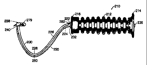
A representative anchor implantation device having a hooked shaft is shown in
Figure 13.
As illustrated in Figure 13, the anchor implantation device 210 has a handle
212 having a proximal
end 214 and a distal end 216. The handle 212 may be made of a variety of
materials, such as
plastic or metal.
The shaft 220 may be made of a variety of materials such as stainless steel
engineering
plastics, fiber-bearing components, or other materials. Preferably, the shaft
is made of stainless
steel.
In the embodiment of the bone anchor implantation device shown in Figure 13,
shaft 220
comprises a straight proximal section 222, a first generally curved section
224 distal to the
straight proximal section, a second generally curved section 226 distal to the
first curved section,
a third generally curved section 228 distal to the second curved section, and
a fourth generally
curved section 230 distal to the third curved section. However, one skill in
the art would
appreciate that the shaft could also comprise a series of straight segments
angled relative to one
another to form a hook.
The straight proximal section 222 of the shaft 220 has an annular shoulder 232
which
abuts the distal end 216 of the handle. The straight proximal section 222
passes through a lumen
(not shown) extending through the handle. The proximal end of the straight
proximal section 222
has a threaded bore which is adapted to receive a screw 236 which secures the
shaft 220 to the
handle. If desired, a washer (not shown) niay be placed between the proximal
end 214 of the
handle and the screw 236.
CA 02283214 1999-09-08
WO 99/37216 PCT/US99/01805
- 25 -
While one means of securing the shaft 220 to the handle 212 was described
above, those
skilled in the art will appreciate that a variety of other means may be
employed. For example, a
plastic handle may be formed over the shaft such that the shaft is integral
with the handle.
The straight proximal section 222 of the shaft 220 may be from about 3 inches
to about 6
inches in length. Preferably, the straight proximal section 222 is from about
4 inches to about 5
inches in length. More preferably, the straight proximal section 222 is about
4.5 inches in length.
The handle 212 defines an axis at the proximal end of the anchor implantation
device 210,
and ten moving distally from the handle 212 the shaft 220 first curves away
from the axis of the
handle and then back toward the axis of the handle 212. The distal end of the
shaft 220 preferably
is located in the vicinity of the axis of the handle 212. In some preferred
embodiments, the shaft
220 at the distal end can be generally perpendicular to the axis of the handle
or can actually be
curving back toward the handle 212. Preferably the distance from the distal
end of the handle 212
to the tip of the tapered bone anchor receptacle 246 measured along the
longitudinal axis of the
handle 212 is about 33/8 inches. Preferably, the distance from the distal end
of the handle 212 to
the distal end of the bone anchor mount 238 is about 4 inches. Preferably, the
distance of a line
perpendicular to the longitudinal axis of the handle 212 extending from the
bottom of the third
curved section 228 is about 2 inches.
Referring to Figures 13-16, a bone anchor mount 238 is attached to the distal
end 240 of
the fourth curved section 230 of the shaft 220. The bone anchor mount 238 may
be oriented at an
angle from about 60 to about 120 relative to the distal end 240 of the
fourth curved section.
Preferably, the bone anchor mount 238 is oriented at an angle from about 80
to about 100
relative to the distal end 240 of the fourth curved section. More preferably,
the bone anchor
mount 238 is oriented at an angle of approximately 90 relative to the distal
end 240 of the fourth
curved section, as illustrated in Figure 13.
The bone anchor mount comprises an outer cylinder 242, an inner cylinder 244,
and a
tapered bone anchor receptacle 246 for releasably engaging a bone anchor 248.
As was the case
with the two handle bone anchor implantation device discussed above, a variety
of bone anchors
can also be used with the bone anchor implantation device having a hooked
shaft. Preferably, the
bone anchor used with the hooked shaft device is the bone anchor discussed
above with respect to
the two handle bone anchor implantation device.
CA 02283214 1999-09-08
WO 99/37216 PCT/US99/01805
-26-
In any event, it is preferred that the bone anchor mount 238 and the bone
anchor
receptacle 246 are oriented so that the bone anchor 248 is pointed in the
general direction of the
handle 212. In one preferred embodiment, the axis of the bone anchor 248 is
generally aligned
with the axis of the handle 212, with the bone anchor pointed toward the
handle 212.
The bone anchor mount 238 may be fabricated from the same materials as the
shaft 220
and may be attached to the shaft 220 by a variety of methods known to those
skilled in the art,
such as brazing. As best shown in Figure 15 the distal end 250 of the outer
cylinder 242 has a
pair of holes 252 therein sized to accommodate a suture 254.
The outer cylinder 242 may have a diameter from about 0.18 inches to about 0.6
inches.
Preferably, the outer cylinder 242 has a diameter from about 0.25 inches to
about 0.5 inches.
More preferably, the outer cylinder 242 has a diameter of about 0.375 inches.
As best shown in the cross section of Figure 16, the outer cylinder 242 has a
cavity 258
formed therein, creating a cup in the proximal region of the outer cylinder
242. The proximal end
260 of the outer cylinder 242 has an annular shoulder 262 thereon.
The inner cylinder 244 is connected to the outer cylinder 242 and extends into
the cavity
258 as best shown in Figure 16. The inner cylinder 244 may be connected to the
outer cylinder
242 in a variety of ways known to those skilled in the art. For example, the
inner cylinder 244
may be fused to the outer cylinder 242. As best shown in Figure 15, the inner
cylinder 244 has
grooves 264 therein adapted to accommodate the suture 254.
A tapered bone anchor receptacle 246 extends from the proximal end 266 of the
inner
cylinder 244. The tapered bone anchor receptacle 246 has grooves 268 therein
adapted to
accommodate the suture 254.
The tapered bone anchor receptacle 246 may extend from the proximal end 266 of
the
inner cylinder 244 by a distance of from about 0.3 inches to about 0.7 inches.
Preferably, the
tapered bone anchor receptacle 246 extends from the proximal end 266 of the
inner cylinder 244
by a distance of from about 0.4 inches to about 0.6 inches. More preferably,
the tapered bone
anchor receptacle 246 extends from the proximal end 266 of the inner cylinder
244 by a distance
of about 0.5 inches.
CA 02283214 1999-09-08
WO 99/37216 PCT/US99/01805
-27-
The distal end 270 of the tapered bone anchor receptacle 246 preferably has a
width
smaller than that of the proximal end 266 of the inner cylinder 244. This
configuration produces a
shoulder 272 which may serve as a depth stop to ensure that the bone anchor
248 is driven into
the bone to the desired depth.
The distal end 270 of the tapered bone anchor receptacle 246 may be from about
0.08
inches to about 0.12 inches in width. Preferably, the distal end 270 of the
tapered bone anchor
receptacle 246 is from about 0.09 inches to about 0.110 inches in width. More
preferably, the
distal end of the tapered bone anchor receptacle 246 is 0.1 inches in width.
The proximal end 274 of the tapered bone anchor receptacle 246 may be from
about 0.110
inches to about 0.15 inches in width. Preferably, the proximal end 274 of the
tapered bone anchor
receptacle 246 is from about 0.12 inches to about 0.14 inches in width. More
preferably, the
proximal end 274 of the tapered bone anchor receptacle 246 is 0.13 inches in
width.
The proximal end 274 of the tapered bone anchor receptacle 246 may have a
variety of
cross sectional shapes adapted to releasably engage the bone anchor 248. For
example, the
proximal end 274 of the tapered bone anchor receptacle 246 may be square,
rectangular,
pentagonal, triangular or hexagonal in cross section.
As depicted in Figures 14-16, the tapered bone anchor receptacle 246 may have
a notch
276 therein in which the bone anchor 248 is releasably seated.
Alternatively, the outer cylinder, inner cylinder, and tapered bone anchor
receptacle may
be a single integral component.
Preferably, the bone anchor implantation device has a protective sheath
connected to the
bone anchor mount which protects the point of the bone anchor from tissue
contact during
placement of the device and also protects the bone anchor from contacting
potentially infectious
microorganisms.
One embodiment of the protective sheath 278 is shown in Figures 13-16. In this
embodiment, the protective sheath 278 comprises a first telescoping cylinder
280 and a second
telescoping cylinder 282. A spring 284 biases the first telescoping cylinder
280 and the second
telescoping cylinder 282 to a position in which they extend from the outer
cylinder 242 and cover
the bone anchor 248.
CA 02283214 1999-09-08
WO 99/37216 PCT/US99/01805
-28-
The first and second telescoping cylinders 280, 282 may be made of a variety
of materials
such as stainless steel or plastic. Preferably, the first and second
telescoping cylinders 280, 282
are made of stainless steel.
The first telescoping cylinder 280 has a lumen 286 extending therethrough. The
first
telescoping 280 cylinder has a first shoulder 288 which engages shoulder 262
on the outer
cylinder 242 and a second shoulder 290 which engages a first shoulder 292 on
the second
telescoping cylinder 282.
The second telescoping cylinder 282 has a first shoulder 292 which engages the
second
shoulder 290 on the first telescoping cylinder 280 as described above. A
second shoulder 294 is
located at the proximal end of the second telescoping cylinder 282 and engages
the spring 284.
The second telescoping cylinder 282 also has a lumen 296 extending
therethrough which is in
fluid communication with the lumen 286 of the first telescoping cylinder 280
and the cavity 258 in
the outer cylinder 242.
The inner diameter of the first telescoping cylinder 280 is slightly larger
than the outer
diameter of the second telescoping cylinder 282 such that the second
telescoping cylinder 282 can
retract inside the first telescoping cylinder 280. The first telescoping
cylinder 280 and the second
telescoping 282 can retract inside the cavity 258 of the outer cylinder 242.
The first telescoping cylinder 280 may be from about 0.2 inches to about 0.3
inches in
length, with an inner diameter of from about 0.27 inches to about 0.33 inches
and an outer
diameter of about 0.3 inches to about 0.36 inches. Preferably, the first
telescoping cylinder 280 is
from about 0.23 inches to about 0.27 inches in length, with an inner diameter
of from about 0.29
inches to about 0.31 inches and an outer diameter of about 0.32 inches to
about 0.34 inches.
More preferably, the first telescoping cylinder 280 is about 0.25 inches in
length, with an inner
diameter of about 0.3 inches and an outer diameter of about 0.33 inches.
The second telescoping cylinder 282 may be from about 0.2 inches to about 0.3
inches in
length, with an inner diameter of from about 0.22 inches to about 0.31 inches
and an outer
diameter of about 0.25 inches to about 0.35 inches. Preferably, the second
telescoping cylinder
282 is from about 0.23 inches to about 0.27 inches in length, with an inner
diameter of from about
0.24 inches to about 0.29 inches and an outer diameter of about 0.27 inches to
about 0.33 inches.
CA 02283214 1999-09-08
WO 99/37216 PCT/US99/01805
-29-
More preferably, the second telescoping cylinder 282 is about 0.25 inches in
length, with an inner
diameter of about 0.27 inches and an outer diameter of about 0.3 inches.
As illustrated in Figures 13-16, a spring 284 biases the first and second
telescoping
cylinders 280 and 282 towards a position in which the first telescoping
cylinder 280 and the
second telescoping cylinder 282 are extended from the outer cylinder 242.
Another embodiment of the protective sheath is depicted in Figures 25 and 26a.
The bone
anchor implantation device as described above with respect to Figures 14-17
has a balloon which
encapsulates the bone anchor. The bone anchor implantation device 210 has a
balloon 279 which
is coupled to the bone anchor mount 238 and which covers the bone anchor 279.
The balloon
279 protects the bone anchor from contacting potentially infections
microorganisms prior to
implantation.
The balloon 279 may be made of any material which exhibits a strength that
allows it to be
punctured by the bone anchor. Examples of suitable materials include plastic,
thermoplastic,
elastromers, PET, PETG, rubber, vinyl, latex, gelatin or silicone. In a
preferred embodiment, the
balloon is made of latex.
Alternatively, the balloon 279 can be made of a biodegradable material. A
suitable
biodegradable material dissolves within the patient after a predetermined
period of time.
Following implantation of the bone anchor the punctured sheath remains are
simply metabolized
by natural biological processes.
In a preferred embodiment, the balloon comprises a biodegradable polymer. The
polymer
may be either natural or synthetic. Synthetic polymers offer greater
advantages that natural
materials in that they can be tailored to give a wider range of properties and
are more uniform
than materials from natural sources. Synthetic polymers also offer a more
reliable source of raw
materials which reduce the risks of invoking an immunogenic response.
Biodegradable polymers are synthesized from chemical functional groups such
as, for
example, esters, anhydrides, orthoesters and amides, which have hydrolytically
unstable linkages
in the backbone. Preferably, the balloon is made of a.biodegradable materials
such as polyglycotic
acid (PGA), polylactic acid (PLA), poly (dioxanone) (PDO), poly (1-lactide)
(LPLA), poly (dl-
lactide) (DLPLA), poly (glycolide-co-trimethylene carbonate) (PGA-TMC), poly
(1-lactide-co-
glycolide) (PGA-LPLA), poly (dl-lactide-co-glycolide) (PGA-DLPLA), poly (1-
lactide-co-dl-
*rB
CA 02283214 1999-09-08
WO 99/37216 PCT/US99/01805
-30-
lactide) (LPLA-DLPLA), poly(glycolide-co-trimethylene carbonate-co-dioxanone)
(PDO-PGA-
TMC), poly(e-caprolactone), poly(dioxanone)(a polyether-ester), poly (lactide-
co-glycotide),
poly(SA-HDA anhydride), poly(orthoester), polyglyconate.
The balloon or structure can be filled with air, water, antibiotic or any
substance which
inflates the balloon so as to prevent the bone anchor from puncturing the
balloon prior to
implantation. Alternatively, as illustrated in Figure 38 and discussed below,
the device may
further include a spring which is attached to the shaft within the balloon to
maintains the shape of
the balloon and prevent the bone anchor from puncturing the balloon
prematurely. In a preferred
embodiment, the balloon 278 contains an antibiotic that is released when the
sheath is perforated
by the bone anchor during implantation. The balloon may contain several
antibiotics that have
complementary activity. The antibiotic prevents infection at the site where
the bone anchor is
pressed into the bone. Non-limiting examples of suitable antibiotics for use
in the invention
include nafcillin, aminogylcoside, ciprofloxin, clindamcin,
piperacillin/tazobactum,
ampicillin/sulbactum, aminoglcoside, vancomycin, cephalosporin, TMP/SMX,
ampicillin,
gentaminicin, tobramycin and ciprofloxacin.
There are numerous ways to insert an antibiotic or other desired substance
into the
balloon. In one embodiment, the bone anchor implantation device further
comprises an port 280
which allows antibiotics to be inserted into the balloon. As shown in Figure
25, the port 280
extends from the balloon 279 into a lumen which extends from one end of the
shaft to the other.
The port 280 has an opening 282 at the distal end of the shaft. Antibiotics
inserted into the
opening 282 travel through the port 280 into the balloon 279. In an alternate
embodiment (not
shown), the port extends from the balloon into a lumen which extends from one
end of the shaft
to the other end and through the handle to an opening. Those skilled in the
art will appreciate
other ways of inserting the antibiotic into the balloon.
In general, in another aspect illustrated in Figure 38, the invention features
a bone anchor
implantation 210 device that has a spring element attached to the shaft within
the balloon which
retracts when the balloon contacts the bone anchor implantation site causing
the bone anchor to
perforate the balloon and implant in the bone. The spring element reduces the
amount of force
required to perforate the sheath and implant the bone anchor in the bone. The
spring element 285
can be any type of spring which is elastically or plastically deformable. The
spring element 285
has a first end 286 which is attached or grounded to the bone anchor mount 238
and a second
CA 02283214 1999-09-08
WO 99/37216 PCT/US99/01805
-31-
floating end 287 which contacts the balloon. Preferably, the spring is a
compression spring which
is normally in an open position. A compression spring retracts when pressure
is applied causing
the point to puncture the balloon. As illustrated in Figure 38, the spring
element 285 can be an
open-coiled helical spring which surrounds the bone anchor 248. The spring
mechanism 285 must
have some minimum deflection strength so as to prevent the bone anchor from
puncturing the
balloon prematurely during insertion but must have enough deflection strength
to puncture the
balloon when force is applied to the bone anchor implantation device at the
desired location. The
balloon 279 which covers the spring and the bone anchor has the same
attributes as the balloon
discussed above with respect to Figures 25-26a.
Another embodiment of the protective sheath is illustrated in Figure 26b. The
bone
anchor implantation device 210 as described above with respect to Figures 14-
17 has a gelatin
structure 290 which covers the bone anchor. The bone anchor implantation
device 210 has a
gelatin structure or caplet which is coupled to the bone anchor mount 238 and
which encapsulates
the bone anchor 248. The gelatin structure 290 protects the bone anchor 248
from
contamination. As discussed above with reference to Figures 25-26a, the
gelatin structure may be
filled with an antibiotic. The device may also include a port for inserting
antibiotic into the gelatin
structure.
An alternative embodiment of the bone anchor implantation device 310 is shown
in
Figures 17-20 and Figure 27.
As illustrated in Figures 17 and 18, the shaft 320 has a generally straight
proximal section
399, a first generally bent section 397, a generally straight median section
395, a second bent
section 393, a generally curved section 391, and a distal generally straight
section 389.
The straight proximal section 399 may be from about 3.0 inches to about 6.0
inches in
length. Preferably, the straight proximal section 399 is from about 4.0 inches
to about 5.0 inches
in length. More preferably, the straight proximal section 399 is about 4.5
inches in length.
The first bent section 397 may be from about 1.0 inches to about 3.0 inches in
length.
Preferably, the first bent section 397 is from about 1.5 inches to about 2.5
inches in length. More
preferably, the first bent section 397 is about 2 inches in length.
The first bent section 397 may bend at an angle of from about 35 to about 55
relative to
the straight proximal section 399. Preferably, the first bent section 397
bends at an angle of from
CA 02283214 1999-09-08
WO 99/37216 PCT/US99/01805
-32-
about 40 to about 50 relative to the straight proximal section 399. More
preferably, the first
bent section 397 bends at an angle of about 45 relative to the straight
proximal section 399.
The straight median section 395 may be from about 2 inches to about 4 inches
in length.
Preferably, the straight median section 395 is from about 2.5 inches to about
3.5 inches in length.
More preferably, the straight median section 395 is about 3 inches in length.
The second bent section 393 may be from about 0.5 inches to about 2.5 inches
in length.
Preferably, the second bent section 393 is from about 1.0 inches to about 2.0
inches in length.
More preferably, the second bent section 393 is about 1.5 inches in length.
The second bent section 393 may bend at an angle of from about 125 to about
145
relative to the straight median section 395. Preferably, the second bent
section 393 bends at an
angle of from about 130 to about 140 relative to the straight median section
395. More
preferably, the second bent section 393 bends at an angle of about 135
relative to the straight
median section 395.
The curved section 391 may curve through an arc of from about 70 to about 110
with a
radius from about 0.2 inches to about 0.6 inches. Preferably, the curved
section curves 391
through an arc of from about 80 to about 100 with a radius from about 0.3
inches to about 0.5
inches. More preferably, the curved section 391 curves through an arc of about
90 with a radius
of 0.4 inches.
The distal straight section 389 may be from about 0.5 inches to about 0.9
inches in length.
Preferably, the distal straight section 389 is from about 0.6 inches to about
0.8 inches in length.
More preferably, the distal straight section 389 is about 0.7 inches in
length.
The shaft 320 has a lumen extending therethrough. The lumen may have a
diameter from
about 0.03 inches to about 0.07 inches and the shaft 320 may have an outer
diameter from about
0.2 inches to about 0.3 inches. Preferably, the lumen has a diameter from
about 0.04 inches to
about 0.06 inches and the shaft 320 has an outer diameter from about 0.24
inches to about 0.26
inches. More preferably, the lumen has a diameter of about 0.05 inches and the
shaft 320 has an
outer diameter of about 0.250 inches.
CA 02283214 1999-09-08
WO 99/37216 PCT/US99/01805
-33-
Preferably, the shaft 320 has an insert 387 therein with a lumen 385 extending
therethrough as best illustrated in the cross section of Figure 19. The insert
387 may be made of
a variety of materials such as stainless steel or plastic.
The insert 387 has an outer diameter approximately that of the diameter of the
lumen in
the shaft such that the insert 387 fits snugly within the lumen of the shaft.
The insert 387 may
have an outer diameter from about 0.2 inches to about 0.3 inches. Preferably,
the insert 387 has
an outer diameter from about 0.21 inches to about 0.27 inches. More
preferably, the insert 387
has an outer diameter of about 0.23 inches.
The insert 387 has a lumen 385 extending therethrough having a diameter large
enough to
accommodate a suture 354. The diameter of the lumen 385 may be from about 0.02
inches to
about 0.100 inches. Preferably, the diameter of the lumen 385 is from about
0.04 inches to about
0.08 inches. More preferably, the diameter of the lumen 385 is about 0.06
inches.
As illustrated in Figures 17, 18 and 20, the shaft 320 has a bore therein
which is large
enough to permit the suture 354 to exit from the shaft 320. In the embodiment
shown in Figures
17, 18 and 20, the bore is located in the straight median section 395 at a
position in which it is
located outside of the patient's body when the bone anchor 348 has been
inserted into the
patient's bone. However, those skilled in the art will appreciate that the
bore may be located in
other locations such as the first bent section 397.
As illustrated in Figures 17 and 20, the shaft 320 extends through a lumen 383
in the
handle 312. The lumen 383 has a narrow distal section 381 having a diameter
slightly larger than
the outer diameter of the shaft 320 and a wide proximal section 373 adapted to
receive a spring
371.
The shaft 320 passes through the interior of the spring 371 as depicted in
Figures 17 and
20. The distal end of the spring 371 contacts the distal end of the wider
proximal section 373 of
the lumen. The proximal end of the spring contacts a plug 369. The plug 369
has a lumen
through which the shaft 320 passes and a bore adapted to receive a screw 367.
The screw 367
passes through the bore in the plug 369 and a bore in the shaft 320 which is
aligned with the bore
in the plug, thereby securing the shaft 320 to the plug 369.
The resistance of the spring 371 is selected to be equal to the force with
which the bone
anchor 348 is to be driven into the bone. For example, where the bone anchor
348 is to be driven
CA 02283214 1999-09-08
WO 99/37216 PCT/US99/01805
-34-
into the bone by applying 20 pounds of force, the spring 371 is a 20 pound
spring. The spring
indicates when the desired amount of force has been applied because the user
can sense when the
spring has been completely compressed.
The spring 371 may have a resistance of from about 5 to about 35 pounds.
Preferably, the
spring 371 has a resistance from about 15 to 25 pounds. More preferably, the
spring 371 has a
resistance of 20 pounds.
Those skilled in the art will appreciate that the anchor implantation device
shown in
Figures 13-16 may also be adapted to include a force indicating spring in the
handle.
As illustrated in Figures 17, 18 and 20, a bone anchor mount 338 and a
protective sheath
378 as described above with respect to the embodiment of Figures 13-16 are
attached to the end
of the distal straight section 389.
In an alternate embodiment, illustrated in Figure 27, the protective sheath on
the bone
anchor implantation device 310 is a balloon 279 as described above with
respect to the
embodiments of Figures 25 and 26a. The balloon is attached to the distal
straight section 3 89 of
the bone anchor implantation device 310 as described above with respect to the
embodiment of
Figures 24 and 25. The device may further comprise a spring element as
discussed above with
respect to Figure 38.
The hooked bone anchor implantation devices 210, 310 are used as follows. An
incision
in the anterior vaginal wall is made as described above. The site for bone
anchor implantation is
located by palpation as described above.
The hooked bone anchor implantation device 210, 310 is inserted into the
vagina as shown
in Figures 17 and 21 with the patient in the lithotomy position and the
surgeon located between
the patient's legs. The shaft 220, 320 is inserted through the incision and
the protective sheath
278, 378 is positioned such that the proximal end of the second telescoping
cylinder 282, 382
contacts the pubic bone 219, 319 as shown in Figures 17, 21 and 23. At this
time, the first and
second telescoping cylinders 280, 380, 282, 382 are biased to a position in
which they extend
from the outer cylinder 242, 342 to cover the bone anchor. The bone anchor is
inserted into the
bone by applying a retrograde force to the bone anchor. The retrograde force
can be applied in a
number of ways as will be apparent to one of skill in the art. Preferably, the
bone anchor is
implanted by pulling the handle. For example, the handle may be pulled in a
retrograde direction
CA 02283214 1999-09-08
WO 99/37216 PCT/US99/01805
-35-
(toward the user) to implant the anchor as shown in Figures 20 and 22. As the
device is pulled in
a retrograde motion, the first and second telescoping cylinders 280, 282, 380,
382 retract inside
the cavity 258, 358 of the outer cylinder as shown in Figures 20, 22 and 24
and the bone anchor
248, 348 is driven into the pubic bone 219, 319. Because the patient's body
weight provides an
opposing force, the user need only apply a small amount of force, such as 10-
20 pounds, in order
to drive the bone anchor 248, 348 into the bone 219, 319. The device 210, 310
is then pushed
away from the implanted anchor to disengage the device from the anchor. The
device is then
removed from the vagina, leaving the bone anchor 248, 319 in the bone 219, 319
with the suture
extending therefrom. The bladder neck is then compressed, suspended or
stabilized using the
suture(s) extending from the bone anchor(s) as described above.
As shown in Figure 20, in the device 310 having a spring 371 inside the handle
312, the
spring 371 is compressed when the handle is pulled in a retrograde direction
to drive the bone
anchor into the bone. In this embodiment, the user can detect when the spring
371 has been
completely compressed, or compressed by a predetermined amount, indicating
that the desired
amount of force for driving the bone anchor into the bone has been applied.
The hooked bone anchor implantation device with balloon illustrated in Figures
25-27 is
used as follows. An incision in the anterior vaginal wall is made as described
above. The site for
bone anchor implantation is located by palpation of the urethra, pubic
symphsis or other
anatomical landmark or other techniques known by those skilled in the art.
The hooked bone anchor implantation device with a balloon or gelatin structure
210, 310
is inserted into the vagina as shown in Figures 27 and 28 with the patient in
the lithotomy position
and the surgeon located between the patient's legs. The shaft 220, 320 is
inserted through the
incision and the balloon or gelatin structure 279, 379 is positioned such that
the balloon or gelatin
structure 279 contacts the pubic bone 219, 319 as shown in Figures 27 and 28.
The bone anchor
is inserted into the bone by applying a retrograde force to the bone anchor
238,348. The force is
transmitted through the bone anchor implantation device 210, 310 to the bone
anchor 279, 379
and causes the balloon or gelatin structure 279, 379 to press against the
pubic bone 279, 379.
The application of additional force causes the bone anchor point to puncture
the balloon or gelatin
structure 279, 379 and drives the bone anchor 248, 348 into the pubic bone
219, 319. Because
the patient's body weight provides an opposing force, the user need only apply
a small amount of
force, such as 10-20 pounds, in order to drive the bone anchor 248, 348 into
the bone 219, 319.
CA 02283214 1999-09-08
WO 99/37216 PCT/US99/01805
-36-
In one embodiment (not shown), the retrograde force retracts a spring element
attached to the
shaft within the balloon which causes the bone anchor to perforate the sheath
and implant into the
bone. In a preferred embodiment, an antibiotic is inserted into the balloon or
gelatin structure and
is released when the sheath is punctured. The antibiotic may be inserted into
the balloon or
gelatin structure via a port which extends from one end of the shaft to the
other end of the shaft
into the balloon. The device 210, 310 is then pushed away from the implanted
anchor to
disengage the device from the anchor. The device is then removed from the
vagina, leaving the
bone anchor 248, 319 in the bone 219, 319 with the suture extending therefrom.
The bladder
neck is then compressed, suspended or stabilized using the suture(s) extending
from the bone
anchor(s) as described above.
Other embodiments of the invention, particularly various types of protective
sheaths which
prevent premature insertion, are illustrated in Figures 39-45.
Figures 39-40b illustrate a bone anchor implantation device with a telescoping
sheath
which retracts upon insertion. As discussed above with reference to Figures 13-
16, the bone
anchor implantation device with telescoping sheath comprises a handle 512, a
hooked shaped
shaft 520 secured to the handle 212, a bone anchor mount 538 adapted to
releasably engage a
bone anchor 548 and attached at the distal end of the shaft 520, and a
telescoping sheath 550
which attaches to the bone anchor mount 538 and covers the bone anchor 548.
Figure 40a
depicts the telescoping sheath 550 in an extended or open position covering
the bone anchor 548.
Figure 40B illustrates the telescoping sheath 550 in a retracted position.
Details regarding the
telescoping sheath are discussed above with reference to Figures 13-16. A
spring biases the
telescoping sheath 550 between extended and retracted positions.
As illustrated in Figure 45 the bone anchor implantation device with
telescoping sheath
shown in Figure 39 is inserted into the vagina 908 and positioned so that the
telescoping sheath
950 contacts the pubic bone 918. The bone anchor 948 is then inserted by
applying a retrograde
force to the bone anchor 948 by pulling the handle 912 of the device in a
retrograde direction.
There is an annular shoulder 910 on the bone anchor 948 which acts as a depth
stop to ensure
adequate penetration. Further details regarding the method of inserting the
bone anchor are
described above with reference to Figures 17 and 21.
CA 02283214 1999-09-08
WO 99/37216 PCT/US99/01805
-37-
Figures 41 a and 41 b illustrate a hooked-shaped bone anchor implantation
device which a
balloon 650 covering the bone anchor 648. The device comprises a handle 612, a
hooked-shaped
shaft secured to the handle 612, a bone anchor mount 638 attached to the
distal end of the shaft
and adapted to releasably engage a bone anchor 648, and a balloon 650 which
covers the bone
anchor 648. Features of the balloon sheath are discussed above with reference
to Figures 25 and
26a. The balloon 650 may be filled with one or more antibiotics to combat
infection at he
implantation site. The device may also comprise a port 680 for inserting
antibiotics into the
balloon. The balloon is perfed away upon implantation.
Figures 42a and 42b illustrate a bone anchor implantation device having a
latex sheath 750
with a spring element 779 which covers the bone anchor 748. As described above
with reference
to Figure 38, the spring element can be any type of spring which is
elastically or plastically
deformable. Preferably the spring is a compression spring such as a coiled
helical spring which
surrounds the bone anchor and which retracts when pressure is applied. The
latex sheath can be
hermetically sealed to the bone anchor implantation device. The sheath may
also be filled with
one or more antibiotics to prevent infection at the implantation site. The
latex sheath perfs away
upon implantation.
Figure 43 illustrates a bone anchor implantation device with gelatin structure
or a capsule
890 that covers the bone anchor 888. The capsule 890 can be coupled to the
bone anchor mount
838. The capsule 890 can be made of biodegradable materials such as those
described above with
reference to the balloon sheath illustrated in Figures 25 and 26a. Preferably
the caplet is made of
gelatin. The bone anchor 888 perfs the capsule 890 and any remaining capsule
particles are
absorbed or metabolized.
Figure 44 illustrates a bone anchor implantation device that has a sheath 802
with a lumen
805 that a diameter large enough to accommodate one or more sutures 808 which
are preattached
to the bone anchor 848. The sheath 802 protect the sutures 808 from
contamination. Further
details regarding a shaft with a lumen that accommodates one or more sutures
are discussed
above with respect to Figures 17-20. The bone anchor 848 with attached sutures
808 has a screw
or locking device 804 which secures the bone anchor 848 to the bone anchor
implantation device.
The locking mechanism 804 prevents accidental insertion of the bone anchor 848
into tissue.
CA 02283214 1999-09-08
WO 99/37216 PCT/US99/01805
-38-
Although this invention has been described in terms of certain preferred
embodiments,
other embodiments which will be apparent to those of ordinary skills in the
art in view of the
disclosure herein are also within the scope of this invention. Accordingly,
the scope of the
invention is intended to be defined only by reference to the appended claims.
What is claimed is: