Note : Les descriptions sont présentées dans la langue officielle dans laquelle elles ont été soumises.
CA 02318367 2008-02-20
28472-122
1
Antibiotic Sensitivity Testing
The present invention relates to a method for
testing the growth characteristics of bacteria, in
particular to testing for sensitivity to particular
antibiotics or biostatic agents, as well as to kits for use
in the method.
Bacteria with antibiotic resistance are becoming
an increasingly serious problem. The current method for
determining the antibiotic resistances of a strain of
bacteria is very time consuming. It requires first the
isolation of the organism in pure culture. A "lawn" of the
bacteria is then prepared and allowed to grow in the
presence of a set of antibiotics. Zones of inhibition of
growth around a particular antibiotic show that the bacteria
are susceptible (with the size of the zone indicating the
degree of susceptibility). Uninhibited growth in the
presence of an antibiotic indicates resistance. The process
takes at least two days to complete which is far from ideal,
particularly in a clinical situation, where the optimum
treatment regime of an infected individual may be determined
as a result of these tests.
There is a need for a test which allows relatively
rapid assessment of antibiotic resistance or susceptibility,
for example within a few hours.
JP 04 370 100 discloses a method for examining the
drug sensitivity of a micro-organism which is based on the
determination of the amount of adenosine triphosphate (ATP)
in a culture containing the drug. US 3 933 592 discloses a
method for selection of an antibiotic for treatment of
bacterial infection which is based on a comparison of the
results of an ATP assay of cultures containing an antibiotic
at various time intervals.
CA 02318367 2008-02-20
28472'-122
2
Assays for the detection of micro-organisms by
measurement of adenylate kinase are known for example from
International Patent Application Nos. PCT/GB94/00118 (WO
94/17202) and PCT/GB94/01513 (WO 96/02665).
In particular, WO 94/17202, which has equivalent
US 5 648 232, discloses methods and test kits for
determining the presence and amount of micro-organisms using
an adenylate kinase assay based on the construction of
calibration curves.
Adenylate kinase is an essential enzyme in all
living cells which, in the presence of ADP, catalyses the
ATP producing reaction shown below.
2ADP ATP + AMP
In this assay, ADP is added as a reagent to the
sample under test, preferably in the presence of magnesium
ions. ATP produced as a result of the above-mentioned
adenylate kinase reaction can be detected for example using
firefly bioluminescence. For this, reagents such as the
combination of luciferin/luciferase are added to the
mixture, generally after a short incubation period, for
example of about 5 minutes, and the luminescent signal
produced is monitored.
The sensitivity of this assay is limited only by
the background level of adenylate kinase and the purity of
reagents used. For example, using E. coif as a model
system, the adenylate kinase activity from fewer than 100
cells was measured in a 5 minute incubation assay as
illustrated in Figure 1 hereinafter. In the tests used to
generate this Figure, the sample volume was 200 pl.
CA 02318367 2008-12-09
28472-122
3
The applicants have found that adenylate kinase
can be used as a sensitive marker of biomass and that the
above-mentioned assay technique can be utilised in studies
which give much more detailed information regarding growth
characteristics of bacterial cells.
Thus the invention provides the use of an assay
for adenylate kinase in an in vitro test for the effect of
external conditions on the growth characteristics of
bacterial cells, which test is to assess the growth stage of
the bacteria by comparison of the extracellular adenylate
kinase content of a cell culture thereof with the total
intracellular and extracellular content.
The adenylate kinase assay provides a rapid and
sensitive means of investigating many aspects of bacterial
growth and inhibition. The sort of external conditions
which may be investigated using the invention are various.
For example, the adenylate kinase assay may be used in
methods to determine the sensitivity of a particular
bacterial strain or mixed culture to particular antibiotic
or biostatic reagents, or the methods may be adapted for use
in the screening of reagents for antibiotic or biostatic
properties. It has also been found that a comparison of the
extracellular adenylate kinase content of a cell culture
with the total intracellular content is indicative of the
growth status and health of the cell culture and thus the
adenylate kinase assay may be used to assess these features.
The configuration of the test will take into
account the nature of the investigations being undertaken,
the type of bacterial samples available, the nature of the
samples and reagents if any, which are to be tested and in
particular whether they have lytic or non-lytic effects on
CA 02318367 2010-02-18
28472-122
3a
the cells. Various forms of these tests will be described
in more detail hereinafter.
In particular, however, the invention provides a
method for determining susceptibility of a bacteria to a
reagent selected from an antibiotic, a biostatic agent or a
compound suspected of having antibiotic or biostatic
properties by comparing levels of extracellular adenylate
kinase content in a cell culture of the bacteria containing
the reagent, a cell culture of the bacteria prior to
addition of the reagent, and/or a cell culture of the
bacteria containing a lytic agent.
In another aspect, the invention provides a method
of determining the growth phase of a bacterial culture which
method comprises (a) subjecting a first sample of the
bacterial culture to a lytic reagent so as to lyse bacterial
cells therein; (b) assaying for extracellular adenylate
kinase in said first sample; (c) assaying for extracellular
adenylate kinase in a second sample of the culture which has
not been exposed to the lytic reagent; and (d) comparing the
results obtained from said first and second cultures and
assessing the growth stage of the culture, wherein results
showing that adenylate kinase levels in the second sample
which are of the order of 1% of the levels found in the
first sample is indicative of a healthy, log phase culture
and levels in excess of 1% are indicative of a progression
into stationary phase.
In yet another aspect, the invention provides a
test kit for use in the method as described herein, which
kit comprises ADP, a source of magnesium ions, luciferin and
luciferase and one or more of a lytic and/or a non-lytic
antibiotic or biostatic agent.
CA 02318367 2010-02-18
28472-122
3b
In a further aspect, the invention provides use of
an assay to determine the effect of an external condition on
the growth characteristics of a bacteria, wherein the assay
comprises comparing the extracellular adenylate kinase
content of a first sample of the bacteria to that of a
second sample of the bacteria, which has been exposed to the
external condition.
The reagents tested may be known antibiotics or
biostatic agents or they may be novel compounds or reagents not
CA 02318367 2000-07-20
WO 99/37799 PCT/GB99/00089
4
previously known as antibiotics so that the test forms part oZ
a screening program.
Some reagents, e.g. antibiotics such as (3-lactam antibiotics
such as penicillins like ampicillin and amoxycillin, will
cause lysis of bacteria in the culture. Where this does not
occur however, it may be necessary to lyse the bacteria prior
to effecting the assay. This may be done by various
techniques as understood in the art, including treatment with
lytic agents as well as physical methods such as subjecting
the bacteria to magnetic or electical fields, or sonication.
Agents producing lysis of bacteria include detergents and
enzymes such as bacteriolysin. These are non-specific
however and will liberate AK from all living material present
in the sample. This may be suitable where the sample
comprises a pure culture. However, where the bacteria under
investigation is a component of a mixed culture, other
strategies may be adopted. Specific measurements from target
cells in a mixed sample may be achieved for example by: 1)
specific capture of the cells of interest to separate them
from contaminating organisms followed by non-specific
adenylate kinase measurements; 2) use of a method which only
lyses the target cells so only the adenylate kinase from these
is measured; or 3) a combination thereof.
Adenylate kinase from the target bacterial cells only (2
above) may be liberated by using a lytic agent which is
specific for the particular bacteria under investigation, for
example a bacteriophage which is specific for the target
bacteria and which brings about lysis of that bacteria. These
bacteriophages are viruses which infect bacteria, causing
lysis of the cells and release of intracellular components,
CA 02318367 2000-07-20
WO 99/37799 PCT/GB99/00089
including adenylate kinase, into the external medium. This
release generally occurs about 30 to 60 minutes after
infection. It has been found that fewer than 500 cells are
detectable using this method in an assay taking 40 minutes.
5
Phages can infect target cells equally well in pure or mixed
culture. By comparing the amount of adenylate kinase which can
be chemically extracted from a sample with the amount released
after a set time with phage infection, the presence or absence
of target cells can be determined and the effects of the test
material on their growth measured.
In order for bacteriophage to reproduce and therefore bring
about lysis, the host cell must be in the log phase of growth.
If growth is inhibited for example, as a result of the
presence of a bacteriostatic agent or antibiotic, the
bacteriophage will not be able to grow and lyse the cells.
This can be used as a basis for a further embodiment of the
invention as illustrated below.
Alternatively or additionally, a mixed culture may be
subjected to a pretreatment step wherein the target bacterial
cells are either enriched in the culture and/or separated from
it. Such steps are well known in the art. For example,
separation may be effected using immunocapture techniques
where antibodies or binding fragments thereof which are
specific for particular bacteria are used to immobilise those
cells on a solid surface, such as a beads, microtitre plates,
filter membranes or columns. Magnetic beads may provide a
particularly preferred solid surface. Separation of the
beads, where appropriate using magnetic separation techniques
leads to substantial isolation of the target bacteria as
CA 02318367 2000-07-20
WO 99/37799 PCT/GB99/00089
6
illustrated hereinafter. It has been found that typically,
using magnetic beads as the solid support, detection of fewer
than 1000 cells can be achieved with a total assay time of
about 30 min. Other materials may also be used as a solid
support.
Further specificity may be gained by the use of selective
growth media. This can be used in the enrichment step to
establish a healthy growing culture, either prior to the
immunocapture assay, or infection by a bacteriophage.
Additionally, selective media can be used throughout the
course of the bacteriophage infection.
Such media will minimise overgrowth by non-target organisms,
which may be present, sometimes vastly in excess of the target
bacteria.
As mentioned above, the invention may be adapted for use in
the testing of bacteria for susceptibility.to particular
antibiotics or bacteriostatic agents.
Antibiotics such as P-lactams like penicillins work by
disrupting cell wall synthesis thereby causing the cells to
lose integrity and lyre during replication. This occurs
relatively rapidly, about 10-15 minutes after exposure to the
antibiotic, provided that the bacteria are actively growing.
Other antibiotics, such as chloramphenicol, do not cause cell
lysis but inhibit cell growth in other ways, as do biostatic
agents. The mode of action of any particular agent in use is
generally understood. The invention may be adapted for use in
testing the sensitivity to bacteria to any of these agents.
CA 02318367 2008-02-20
28472122
7
Specific embodiments of the invention include a
method for determining the sensitivity of a bacteria to a
lytic antibiotic, said method comprising the steps of (i)
separating said bacteria from other microbial species, for
example using an immunocapture step; (ii) determining the
extracellular adenylate kinase content of a culture of said
bacteria; (iii) adding the lytic antibiotic to the culture
and incubating it for a period sufficient to allow the
antibiotic to exert its lytic effect; and (iv) determining
the extracellular adenylate kinase content of the culture to
assess whether lysis has taken place.
In this test, sensitive bacteria would be lysed by
the antibiotic soon after addition thereof, generally within
about 15 minutes. Hence the free adenylate kinase content
of the culture would increase significantly following
addition of the antibiotic as the bacterial cells break open
liberating intracellular adenylate kinase. Optimally
measurement of the adenylate kinase levels would be taken
shortly before antibiotic addition, and then again at least
15 minutes after antibiotic addition. The free adenylate
kinase content of cultures of resistant bacteria would
remain largely constant. Only the normally present, low
level extracellular adenylate kinase content would therefore
be measured and, as explained above, this remains
approximately steady for healthy growing cells. Using this
method, an assessment of antibiotic sensitivity may be
achieved in a period of approximately 40 minutes.
The culture of bacteria used in this method may
comprise a selective growth medium which favours said
bacteria as discussed above as this will minimise any false
positive results as a result of contamination.
CA 02318367 2008-02-20
28472122
8
Other embodiments of the invention avoid the need
for using isolated or pure cultures of bacteria. In
particular the invention further provides a method for
determining the sensitivity of a bacteria to an antibiotic
or biostatic agent, said method comprising
CA 02318367 2000-07-20
WO 99/37799 PCT/GB99/00089
9
(a) incubating a first sample of a culture of said
bacteria, a second sample in the presence of said antibiotic,
a third sample in the presence of a bacteriophage which will
specifically lyse said target bacteria and a fourth sample in
the presence of both said bacteriophage and said antibiotic;
(b) determining the adenylate kinase content of each of the
first to fourth samples after culture, and
(c) determining the sensitivity or resistivity of the
bacteria on the basis of the adenylate kinase assay results
and on the mode of action of the antibiotic or biostatic
agent.
Since bacteria must be actively growing in order to be
suceptible to antibiotic effect,-selective media could be used
for an initial enrichment step. This may comprise an
incubation of about an hour. After this time the target cells
may, if desired, be concentrated by immunocapture into fresh
selective medium. The effect of adding antibiotics to the
target cells could be determined using adenylate kinase
measurements in combination with bacteriophage mediated lysis.
In order for a bacteripphage to reproduce, the host cell must
be in the log phase of growth. If growth is inhibited, e.g.
due to the presence of an antibiotic, then the phage will not
be able to replicate and will, therefore, not be able to cause
the host cells to be lysed. Using the adenylate kinase assay
in conjunction with bacteriophage, the antibiotic
sensitivities of bacteria can be determined within 3 hours.
The additional time is needed to establish .healthy, growing
cells prior to exposure to the antibiotic or infection by the
phage. Two types of test outcome are possible depending upon
the mode of action of the antibiotic concerned.
CA 02318367 2000-07-20
WO 99/37799 PCT/GB99/00089
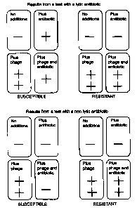
The results obtained are summarised in Figure 4 where "-"
indicates a result which is consistent with the detection of
extracellular adenylate kinase only, "+" indicates a
5 moderately positive result consistent with the detection of
intra and extracellular adenylate kinase of the existing
sample with no growth, and "++" indicates the detection of
elevated levels of adenylate kinase consistent with lysis of
the growing culture.
As is clear from Figure 4, the pattern of the results obtained
using this series of tests can allow ready distinction between
susceptible and resistant bacteria, provided the mode of
action (lytic or non-lytic) of the agent is understood. The
different effects are created as a result of the interaction
of the various reagents with the bacteria as will be explained
in more detail in the Examples given below.
It.has been found that a comparison of the extracellular
adenylate kinase content of a cell culture with the total
intracellular and extracellular content is indicative of the
growth status and health of the cell culture.
Therefore, yet a further embodiment of the invention provides
a method of determining the growth phase of a bacterial
culture which method comprises
(a) subjecting a first sample of said bacterial culture to a
l~tic reagent so as to lyse bacterial cells therein,
(b) assaying for adenylate kinase in said first sample and
also in a second sample of said culture which has not been
exposed to the lytic agent; and
CA 02318367 2008-02-20
28472122
11
(c) comparing the results obtained from said first
and second cultures and assessing the growth stage of the
culture.
Healthy, log phase cultures have relatively low
extracellular adenylate kinase levels (about 1 % of the
total adenylate kinase) and the proportion of extracellular
adenylate kinase stays relatively constant throughout the
log phase, and increases as the culture approaches
stationary phase. Stationary phase cultures may have as
much as a third of the total adenylate kinase in the culture
medium. Therefore, using adenylate kinase, the health of
cells, as well as their number can be rapidly determined.
This method can be used to, for example to confirm
that a particular cell culture is growing well, for example
where optimum growth is required, for example in
fermentation or other processes where bacterial products are
required. Alternatively, it may be necessary to confirm
cells are growing strongly when screening for antibiotic or
bacteriostatic compounds so that false positive results are
avoided because of weak or stationary phase cultures being
used in the test. Furthermore, it may be used to determine
what effect environmental factors, such as temperature or
culture media, have on the growth of any particular culture.
In each case, the adenylate kinase content may be
assessed by removing samples of the culture and carrying out
an adenylate kinase assay for example as described in
International Patent Application Nos. PCT/GB94/00118 (WO
94/17202) and PCT/GB94/01513 (WO 96/02665).
The invention also provides test kits for
effecting the methods of the invention. The test kit will
contain suitable components which would allow the particular
assay to be
CA 02318367 2000-07-20
WO 99/37799 PCT/GB99/00089
12
carried out. For example, for antibiotic sensitivity testing
kits may comprise a range of antibiotics, for example in
freeze-dried or other preserved states. It may also comprise
reagents for extracting adenylate kinase from a sample such as
detergents or other chemical lytic agents as well as reagents
necessary for assaying for adenylate kinase, such as
luciferin/luciferase etc.. In addition, for use in
situations where mixed bacterial cultures are to be tested,
the kits may contain suitable bacteriophages, also in
preserved states such as freeze-dried bacteriophages.
Additionally, the kits may comprises suitable selective growth
media. The reagents may be supplied in a suitable reaction
container such. as a multi-well plate.
The invention will now be particularly described by way of
example with reference to the accompanying diagrammatic
drawings in which:
Figure 1 is a graph showing the results of experiments to
measure the adenylate kinase activity from E. coli cells;
Figure 2: shows the results of magnetic bead immunocapture
assays for Salmonella typhimurium in a pure culture and in the
presence of 3.5x10⁵ Bacillus subtilis var niger vegetative
cells; where ^ shows adenylate kinase activity from cells
captured by beads; and 4 shows adenylate kinase activity from
residual cells in sample (i.e. in the left hand graph from
uncaptured Salmonella cells and in the right hand graph from
these plus non-specific cells;.
Figure 3 is a graph showing results from an experiment to
investigate the time course of phage mediated release of
adenylate kinase from a culture of Escherichia coli cells;
CA 02318367 2008-02-20
28472.-122
13
Figure 4 is a summary of antibiotic test results
obtainable using an embodiment of the invention;
Figure 5 is a diagram showing an assay plate for
testing bacterial samples for antibiotic resistance;
Figure 6 shows the effect of Phage 10359 and 50
mg/litre ampicillin with ampicillin-sensitive and resistant
cultures of E. coli 10243, in which 0 represents the results
with sensitive E. colt 10243 with ampicillin and phage
10359, 8 represents the results with no lytic agents and ^
represents the results with resistant E. coli 10243 with
ampicillin and phage; and
Figure 7 shows the effect of chloramphenicol (34
mg/litre) and Phage 10359 on cultures of E. coli 10243, in
which 0 represents phage only, t represents phage 10359 and
chloramphenicol, ^ represents chloramphenicol alone and 0
represents no phage or chloramphenicol.
Example 1
Comparison of extracellular and total adenylate kinase
contents of a culture
For example, in one embodiment, a sample of
bacterial cells is divided into first and second samples.
The first sample of bacterial cells is mixed with a solution
containing ADP and a detergent in the presence of magnesium
ions. This extracts the adenylate kinase from all the cells
present in the sample, thus allowing the ATP generating
reaction to occur. The reaction is allowed to proceed for
the required time e.g. 5 minutes, after which
bioluminescence reagent is added and the
CA 02318367 2000-07-20
WO 99/37799 PCT/GB99/00089
14
resulting light measured in a luminometer. An assay performed
in this way determines the total amount of adenylate kinase in
a sample, be it extracellular or intracellular.
The second sample is subjected to a similar assay but in the
absence of detergent so that only extracellular adenylate
kinase is measured.
Example 2
Use of Immunocapture to separate target cells from a mixed
s snen~ lion.
An immunocapture assay for Salmonella typhimurium from a pure
sample and from a sample also containing 3.5x10⁵ Bacillus
subtilis var niger (BG) vegetative cells was carried out.
Immunocapture assay carried out in a total volume of 300 l.
The immunocapture step, onto magnetic microbeads coated with
S. typhimurium specific antibody, took 10 minutes. The
immobilised beads were washed to remove unbound particles, and
a non-specific lysis step was carried out'to release adenylate
kinase from bound material. This was detected using a 5 minute
adenylate kinase assay.
The results are shown in Figure 2. The graphs show that about
70% of the target cells can be selectively removed from
suspension, and that this is largely unaffected by the
presence of contaminating material (the BG cells).
Example 3
Use of Phage Mediated s y ; a
The time course of adenylate kinase release from a culture of
Escherichia coli cells, some of which were infected with E.
coli specific bacteriophage was studied. 100 l samples (each
CA 02318367 2000-07-20
WO 99/37799 PCT/GB99/00089
containing just 350 cells) were removed at timed intervals
from a culture that had been infected with an E.coli specific
bacteriophage and then assayed for extracellular adenylate
kinase activity after 40 minutes. The results are shown in
5 Figure 3 where 0 = infected culture, and = non-infected
control.
It is clear from these results that fewer than 500 cells are
detectable using this method.
Example 4
Anti b; oti c sensitivity assays : -lyti c antibiotic
Sample cells (which may be mixed or pure cultures) are split
into 2 fractions and one infected with bacteriophage. Each of
these fractions is then further split into 2 fractions with
one being exposed to antibiotic and the other left untreated.
The relative levels of extracellular adenylate kinase produced
in a set time shows the effects of both the antibiotic and
bacteriophage on the target cells. Test results achieved-in
practise are illustrated in Table 1.
The results show both the antibiotic resistance state of the
cells and controls to ensure that the test has functioned
correctly.
CA 02318367 2000-07-20
WO 99/37799 PCT/GB99/00089
16
Table 1
Susceptible Bacteria
(1) No antibiotic, no phage (2) Antibiotic. no age
Low extracellular adenylate adenylate kinase released
kinase levels only, (no through lysis due to antibiotic.
lysis).
(3) No antibiotic. plus (4) Antibiotic plus ,phase
phage adenylate kinase released though
adenylate kinase released lysis due to antibiotic and
through lysis caused by phage. Levels lower than (3)
phage. because of reduced cell growth
and inhibition of phage
replication.
Resistant Bacteria
(1) No antibiotic. no phage (2) Antibiotic, nophaae
Low extracellular adenylate Low extracellular adenylate kinase
kinase levels only, (no levels only, (no lysis). Same as (1).
lysis).
(3) No antibiotic, plus (4) Antibiotic plus phage
phage adenylate kinase released through
adenylate kinase released lysis due to phage. Same as (3).
through lysis caused by
phage.
Example 5
Antibiotic sensitivity assays,-non-lyric antibiotic or
hiostat3oag=
The susceptibility of bacteria to antibiotics which do not
cause cell lysis, but inhibit cell growth in other ways, (and
other chemicals which inhibit bacterial cell growth, such as
CA 02318367 2000-07-20
WO 99/37799 PCT/GB99/00089
17
biostatic agents) can also be rapidly determined using a
similar method.
Sensitivity testing of bacteria in mixed culture to non-lytic
antibiotics would be carried out in the same way as testing
for susceptibility to antibiotics causing lysis. However,
since the bacteria must be actively growing to permit
bacteriophage infection, susceptibility to antibiotic would be
indicated by a lack of phage mediated lysis in the treated
sample.
The results which would be observed are'summarised in Table 2.
Table- 2
Susceptible Bacteria
(1) No antibiotic. no phagee (2) Antibiotic, nom a e
Low extracellular adenylate Low extracellular adenylate
kinase levels only, (no kinase levels only, (no lysis).
lysis). May be even lower than (1)
through inhibition of growth.
(3) No antibiotic, Plug (4) Antibiotic plus phage
phage Low extracellular adenylate
adenylate kinase released kinase levels only, (no lysis).
through lysis caused by May be even lower than (1)
phage. through inhibition of growth.
CA 02318367 2008-02-20
28472.-122
18
Table 2 contd
Resistant Bacteria
(1) No antibiotic, no phage (2) Antibiotic, no phage
Low extracellular adenylate Low extracellular adenylate
kinase levels only, (no lysis) kinase levels only, (no
lysis). Same as (1).
(3) No antibiotic, plus phage (4) Antibiotic plus phage
adenylate kinase released Adenylate kinase released
through lysis caused by phage. through lysis due to phage.
Same as (3).
Example 6
Test Kit for testing for antibiotic resistance
A suitable test kit comprises a sample container
which typically might be a plastic plate in which are formed a
number of wells as illustrated in Figure 5. The total volume
of liquid that could be held in each well may be approximately
0.5 ml. The plates are suitably pre-prepared so that
particular wells would contain appropriate freeze-dried (or
otherwise preserved) preparations of antibiotics and/or
bacteriophages, and optionally also selective growth media.
In a plate such as shown in Figure 5, each row of
4 wells is designed to be used to test for resistance to a
particular antibiotic, so using this plate, 3 antibiotics
could be tested simultaneously.
In use, about 0.2 ml volume of the samples for test,
which may be pure cultures, preparations enriched by immunocapture,
CA 02318367 2000-07-20
WO 99/37799 PCT/GB99/00089
19
samples from selective media or neat samples as appropriate,
are added to each well. After incubation at, for example 37 C
for 1 hour, reagents to measure the adenylate kinase activity,
may be added. These may be reagents which produce a
colorimetric signal in the presence of adenylate kinase, or
more preferably, reagents which generate a bioluminescence
signal.
In particular ADP and a source of magnesium ions, followed
after 5 minutes by luciferin and luciferase, would be added.
The light emission would either be determined 1 well at a time
by transfer to tubes and measurement in-a tube luminometer,
or, preferably, the plate would be assayed automatically in a
plate luminometer or be imaged as a whole using a CCD camera
system.
Example 7
Comparison of the effect of lytic and non-),ytic antibiotics on_
culture growth
The effect of lytic (ampicillin) and non-lytic
(chloramphenicol) antibiotics on E. coli culture growth were
examined.
A plasmid encoding ampicillin resistance (pUC18) was
introduced into a pure culture of E. coli 10243 in order to
induce resistance without altering phage host specificity. The
resistant strain was also tested to ensure that carrying the
plasmid did not alter its growth rate or infection by phage
10359, and was seen to be the same as the sensitive strain
regarding growth and infection.
CA 02318367 2008-02-20
284721-122
The adenylate kinase released from sensitive
(untransformed) bacteria and resistant bacteria in the
presence of E. coli bacteriophage 10359 and either a lytic
(ampicillin) or non-lytic (chloramphenicol) antibiotic were
5 compared.
Log phase cultures of both resistant and sensitive
strains of E. coli 10243 were infected with 105 phage 10359
at To and 50 gg/ml ampicillin at T5. Cell lysis due to
ampicillin was evident at T10 and significant at T20 in the
10 sensitive strain, masking any lytic effects due to
bacteriophage. The resistant strain was unaffected by the
antibiotic but showed lysis due to phage infection after 20
minutes, although this was not significant until T40. The
results are shown in Figure 6. This shows that after only
15 40 min incubation in the presence of ampicillin and phage,
the susceptibility of a culture of E. coif 10243 towards
ampicillin can be determined.
Log phase cultures of E. coli were incubated in
the presence of phage 10359 and/or chloramphenicol (34
20 gg/ml) over a period of 80 min and assayed for adenylate
kinase as before. The results are shown in Figure 7.
Greater cell lysis was demonstrated where cultures
were incubated with both bacteriophage and chloramphenicol
compared with chloramphenicol alone. Although
chloramphenicol is not a lytic antibiotic, cell lysis was
exhibited over the course of the incubation, probably due to
cell death. The degree of phage mediated lysis was
considerably less in cultures containing the antibiotic,
because the bacteria were not growing well and thus
prevented the phage from completing their replication cycle.
CA 02318367 2000-07-20
WO 99/37799 PCT/GB99/00089
21
Following the results obtained with the ampicillin resistant
mutant, it would be expected that a chloramphenicol resistant
mutant would behave the same in both the presence and absence
of the'antibiotic. Therefore, after a 60 min incubation, the
degree of increase in background luminescence would indicate
whether or not a culture was susceptible to chloramphenicol.