Note : Les descriptions sont présentées dans la langue officielle dans laquelle elles ont été soumises.
CA 02363257 2001-08-24
WO 00/53082 PCT/US00/05892
METHOD AND CIRCUIT FOR STORING AND PROVIDING
HISTORICAL PHYSIOLOGICAL DATA
BACKGROUND OF THE INVENTION
The present invention relates to physiological test instruments and, in
particular, sensors that include a mechanism for storing and providing to a
monitor
historical physiological data such as blood oxygen saturation data.
Pulse oximetry is typically used to measure various blood flow
characteristics including, but not limited to, the blood oxygen saturation of
hemoglobin in
arterial blood, the volume of individual blood pulsation supplying a tissue,
and the rate of
blood pulsation corresponding to each heartbeat of a patient. Measurement of
these
characteristics has been accomplished by the use of a non-invasive sensor that
passes
light through a portion of a patient's blood perfused tissue and photo-
electrically senses
the absorption and scattering of light in such tissue. The amount of light
absorbed is then
used to estimate the amount of blood constituent in the tissue. The "pulse" in
pulse
oximetry comes from the time varying amount of arterial blood in the tissue
during the
cardiac cycle. The signal processed from the sensed optical signal is the
familiar
plethysmographic waveform due to cycling light attenuation.
To estimate blood oxygen saturation of a patient, conventional two-
wavelength pulse oximeters emit light from two light emitting diodes (LEDs)
into a
pulsatile tissue bed and collect the transmitted light with a photodiode (or
photo-detector)
positioned on an opposite surface (i.e., for transmission pulse oximetry) or
an adjacent
surface (i.e., for reflectance pulse oximetry). One of the two LEDs' primary
wavelength
is selected at a point in the electromagnetic spectrum where the absorption of
oxyhemoglobin (HbO~) differs from the absorption of reduced hemoglobin (Hb).
The
second of the two LEDs' wavelength is selected at a different point in the
spectrum where
the absorption of Hb and Hb O~ also differs from each other, and further
differs from
those at the first wavelength. Commercial pulse oximeters typically utilize
one
wavelength in the near red part of the visible spectrum near 660 nanometers
(nm) and one
in the near infrared (IR) part of the spectrum in the range of 880-940 nm.
CA 02363257 2001-08-24
WO 00/53082 PCT/US00/05892
2
Oxygen saturation can be estimated using various techniques. In one
common technique, the photo-current generated by the photo-detector is
conditioned and
processed to determine the modulation ratio of the red to infrared signals.
This
modulation ratio has been observed to correlate well to arterial oxygen
saturation. The
pulse oximeters and sensors are empirically calibrated by measuring the
modulation ratio
over a range of in vivo measured arterial oxygen saturations (Sa02) on a set
of patients,
healthy volunteers, or animals. The observed correlation is used in an inverse
manner to
estimate blood oxygen saturation (SpOz) based on the measured value of
modulation
ratios of a patient. The estimation of oxygen saturation using modulation
ratio is
described in U.S. Patent No. 5,853,364, entitled "METHOD AND APPARATUS FOR
ESTIMATING PHYSIOLOGICAL PARAMETERS USING MODEL-BASED
ADAPTIVE FILTERING", issued December 29, 1998, and U.S. Patent No. 4,911,167,
entitled "METHOD AND APPARATUS FOR DETECTING OPTICAL PULSES",
issued March 27, 1990. The relationship between oxygen saturation and
modulation ratio
is further described in U.S. Patent No. 5,645,059, entitled "MEDICAL SENSOR
WITH
MODULATED ENCODING SCHEME," issued July 8, 1997. All three patents are
assigned to the assignee of the present invention and incorporated herein by
reference.
The LEDs and photo-detector are typically housed in a reusable or
disposable oximeter sensor that couples to the pulse oximeter electronics and
the display
unit (hereinafter referred to as the monitor). The sensors are often connected
to patients
for long periods of time. Conventionally, historical physiological data for
the patient is
collected, if at all, by the monitor coupled to the sensor. The historical
data can be
valuable to a clinician or medical personnel for diagnostic and monitoring
purposes.
Patients are often moved to various locations during treatment. For
example, a patient may be picked up in an ambulance, delivered to an emergency
room,
moved to an operating room, transferred to a surgical recovery room,
transferred to an
intensive care unit, and then moved to a nursing floor or other locations.
Thus, the patient
may be moved between various locations within the same hospital, or between
different
hospitals. In many instances, the sensor employed to monitor the conditions of
the patient
is adhesive in their attachment and therefore remains with the patient. The
monitors,
however, are typically local to particular locations within the facility. The
sensor is
normally disconnected from the monitor at the departure site and reconnected
to another
monitor at the destination site. Consequently, any historical physiological
data collected
CA 02363257 2001-08-24
WO 00/53082 PCT/US00/05892
by the monitor at the departure site is normally unavailable to the clinician
attending the
patient at the destination site.
In the medical art, a combination of a catheter sensor and a memory unit is
disclosed in U.S. Patent No. 4,858,615, entitled "CATHETER SENSOR AND
MEMORY UNIT," and issued August 22, 1989. In this patent, the sensor assembly
(34)
is located at a distal end of the catheter (32) and the memory unit (38) is
connected by a
mufti-conductor lead (40) to the sensor (see Fig. 5). The catheter is an
invasive
instrument typically used at a particular location and removed during
transport. Neither
the catheter nor the memory unit would travel with the patient as he or she is
moved to
different locations. Thus, any data captured and stored in the memory unit
(38) is also not
available when the catheter is removed from the patient.
Accordingly, it is highly desirable to provide mechanisms for storing and
providing historical physiological data that travels with a patient.
SUMMARY OF THE INVENTION
The invention provides a mechanism for storing and providing historical
physiological data, such as blood oxygen saturation data, for a patient. In
particular, the
historical physiological data is stored in a storage medium that "travels"
with the patient
and is accessible wherever the patient is moved. This is achieved by storing
the
physiological data within the sensor assembly. At the destination site, a
monitor or a
device capable of interfacing with the sensor electronics can retrieve and
display the data.
The historical physiological data allows a clinician or medical personnel at
the destination
site to assess the condition of the patient for the entire time that the
patient has been
monitored. The invention can be used to store and provide various types of
physiological
data including, but not limited to, blood oxygen saturation, heart rate, and
temperature
data.
A specific embodiment of the invention provides a physiological sensor
that includes a number of light sources, at least one photo-detector, and a
memory circuit.
The light sources are selected to operate at different wavelengths. The photo-
detector
receives light emitted by the plurality of light sources. And the memory
circuit stores
physiological data and provides the data when requested. The physiological
data is
indicative of a physiological condition of a patient being monitored by the
sensor.
CA 02363257 2001-08-24
WO 00/53082 PCT/US00/05892
4
Another specific embodiment of the invention provides a physiological
test instrument that includes a monitor and a sensor. The monitor includes
conditioning
circuitry and processing circuitry. The conditioning circuitry receives an
electrical signal
and processes the electrical signal to provide sampled data. The processing
circuitry
processes the sampled data to provide physiological data, wherein the
physiological data
is indicative of a physiological condition of a patient. The sensor couples to
the monitor
and includes a number of light sources, at least one photo-detector, and a
memory circuit.
The light sources are selected to operate at different wavelengths. The photo-
detector
receives light emitted by the light sources. The memory circuit stores the
physiological
data and provides the data when requested. An encoder can optionally be
coupled to the
processing circuitry to code and compress the physiological data before
storage to the
memory circuit. The test instrument can be an oximeter system for storing and
providing
historical saturation data of a patient.
Yet another specific embodiment of the invention provides a method for
storing physiological data. The method detects, via a sensor, at least one
signal indicative
of a physiological condition and conditions the detected signal to generate
data samples.
The data samples are processed to generate the physiological data, wherein the
physiological data describes the physiological condition. The physiological
data is stored
within a memory located within the sensor. The physiological data can be coded
and
compressed before storage to the memory.
The foregoing, together with other aspects of this invention, will become
more apparent when refernng to the following specification, claims, and
accompanying
drawings.
BRIEF DESCRIPTION OF THE DRAWINGS
Fig. 1 shows a simplified block diagram of an embodiment of a
physiological measurement system;
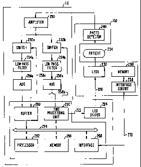
Fig. 2 shows a block diagram of an embodiment of a monitor and a sensor;
and
Fig. 3 shows a block diagram of one compression scheme for oxygen
saturation data.
CA 02363257 2001-08-24
WO 00/53082 PCT/US00/05892
DESCRIPTION OF THE SPECIFIC EMBODIMENTS
Fig. 1 shows a simplified block diagram of an embodiment of a
physiological measurement system 100. System 100 includes a monitor 110 that
couples
to a display unit 120 via an electrical cable 122. Monitor 110 further couples
via a second
5 electrical cable 128 to a sensor 130 that is applied to a patient 132.
Sensor 130 includes
light sources (e.g., LEDs) and a photo-detector along with suitable components
to couple
the electro-optical components to electrical cable 128. Sensor 130 is shown in
Fig. 1 as a
clip-on sensor. However, the invention can be applied to many sensor
implementations,
including those attached to a patient by adhesive and other attachment means.
In a
specific embodiment, monitor 110 is a pulse oximeter.
For estimating blood oxygen saturation, light from light sources at two or
more wavelengths (e.g., red and infrared) is transmitted through a patient's
blood
perfused tissues (e.g., in a finger) and detected by a photo-detector. The
selection of the
wavelengths is based on a number of factors. Such factors include the
absorption
characteristics of the patient and transmission medium. The light sources and
photo-
detector are typically housed within a sensor that couples to the monitor
(e.g., the pulse
oximeter). The detected optical signal is then provided to the monitor for
processing.
Fig. 2 shows a block diagram of an embodiment of monitor 110 and sensor
130. Within monitor 110, a time processing unit (TPU) 220 provides control
signals 222
to an LED driver 224 that, via data lines) 226, alternately activates LEDs 230
within
sensor 130. Depending on the particular implementation, LEDs 230 include two
or more
LEDs and LED driver 224 provides the necessary drive signals for the LEDs.
When
activated, the light from LEDs 230 passes through a medium (e.g., air or a
fiber optic
cable, depending on the implementation) into a patient's tissues 234. After
being
transmitted through or reflected from the tissues, the light is received by a
photo-detector
240 via another medium (e.g., air or another fiber optic cable). Photo-
detector 240
converts the received light into a photo-current, which is then provided to an
amplifier
250 that amplifies the photo-current.
~As shown in Fig. 2, the amplified signal from amplifier 250 is provided to
circuitry for two different channels, one channel for each of the red and
infrared
wavelengths. For a tlu-ee-wavelength implementation, circuitry is provided for
three
channels. Each channel circuitry includes an analog switch 252 coupled in
series with a
low pass filter 254 that is further coupled in series with an analog-to-
digital converter
CA 02363257 2001-08-24
WO 00/53082 PCT/US00/05892
6
(ADC) 256. Control lines 258 from time processing unit 220 select the sampled
data
from the channel corresponding to the LED being activated. Specifically, the
sampled
data from ADC 256a is selected when the red LED is activated and the sampled
data from
ADC 256b is selected when the infrared LED is activated. The sampled data from
ADCs
256 is provided to a buffer 260 that stores the data for further processing.
In an
implementation, as buffer 260 periodically fills up, a processor 262 coupled
to a bus 264
directs the transfer of the data from buffer 260 into a memory 266. The
monitor
implementation shown in Fig. 2 is one of many implementations. Another pulse
oximeter
implementation is disclosed in the aforementioned U.S. Patent No. 5,853,364.
The
present invention can be adapted for application in various monitor
implementations.
The sensor of the invention further includes circuitry that stores historical
physiological data and provides the data when requested. As shown in Fig. 2,
sensor 130
includes a memory 236 coupled to an interface circuit 238. Interface circuit
238 provides
signal conditioning, and can also provide other functions such as address
decoding, and
so on. Interface circuit 238 couples via a bus 270 to a data interface circuit
268 within
monitor 110. Through interface circuits 238 and 268, physiological data is
transferred
between monitor 110 and sensor 130.
In an embodiment, to enhance compatibility of the sensor of the invention
with conventional sensors and conventional monitors, bus 270 is implemented
using new
signal lines (i.e., not using or sharing the existing signal lines of
conventional sensors).
Bus 270 can be implemented as a serial bus, a parallel bus, or other bus
architectures.
With this implementation, when sensor 130 of the invention is plugged into a
monitor not
capable of supporting the features of the invention, the signals on interface
circuit 238 are
simply ignored by the monitor, or alternatively not requested by the monitor.
In another embodiment, interface circuits 238 and 268 interact via signal
lines) or wires) existing in conventional sensors and monitors. For example,
interface
circuits 238 and 268 can couple via data lines) 226 and time multiplex with
the LED
drive signals from LED driver 224.
Time processing unit 220, buffer 260, processor 262, memory 266, and
data interface circuit 268 can be implemented in various manners. For example,
these
elements can be implemented within a single integrated circuit, such as a
DMC68HC16
micro-controller from Motorola. These elements can also be implemented within
an
CA 02363257 2001-08-24
WO 00/53082 PCT/US00/05892
7
application specific integrated circuit (ASIC), a digital signal processor, a
micro-
controller, or other circuits.
Memory 236 can be implemented as a random access memory (RAM), a
FLASH memory, a programmable read only memory (PROM), an erasable PROM
(EPROM), an electrically erasable PROM (EEPROM), a similar programmable and/or
erasable memory, any kind of erasable memory, a write once memory, or other
memory
technologies capable of write operations. Memory 236 and interface circuit 238
can be
integrated within one integrated circuit for reduced size and cost.
In a specific embodiment, to preserve the historical data and prevent
accidental erasure, the sensor memory can be written once. This memory
characteristic
also prevents erasure of the data during sensor processing. A specific example
of a
memory device that can be written once is a 2-wire EPROM device available from
Dallas
Semiconductor Corp.
In another embodiment, the memory can be erased and overwritten
multiple times. This memory characteristic may be advantageous, for example,
for non-
disposable sensors that may include a large amount of memory. Such "specialty"
sensors
may be better suited for applications where there is a higher propensity to
use reusable
sensors, such as inside an operating room or an intensive care unit or during
an
ambulance transport. Specific examples of memory devices that can be erased
and
overwritten are Flash, EEPOM, battery backed RAM, and other technologies.
The invention is applicable for various oximeter system implementations.
For example, in an embodiment, an adapter module and a fiber optic cable can
be
interposed between cable 128 and sensor 130 (see Fig. 1). The adapter module
can
include the light sources, the detector, and suitable optics to couple the
electro-optical
components to the fiber optic cable that guides light to and receives light
from the patient.
The fiber optic cable can also be partitioned into a long extension cable and
a relatively
short "sensor" cable. The fiber optic cables can be either glass or plastic
fiber. This
embodiment allows the electro-optical components to be reused, and only the
short sensor
cable is replaced from patient to patient.
Fig. 2 shows an oximeter implementation using light at two wavelengths.
However, light from more than two LEDs can be used (i.e., for improved
accuracy).
Light from a single light source can also be used, typically along with
appropriate optical
filter. Moreover, light sources other than LEDs can be used. For example,
lasers or white
CA 02363257 2001-08-24
WO 00/53082 PCT/US00/05892
light sources can be used with appropriate filters at either the transmitting
or receiving
end.
The sensor can include different numbers of elements, depending on the
implementation of the sensor or the application for which the sensor is used.
In one
implementation, the sensor includes the LEDs and the photo-detector. This
implementation reduces the transmission loss by placing the light source and
the detector
near the patient. In another implementation, the sensor includes only the
transmission
medium (e.g., a short fiber optic cable), but no LEDs or photo-detector. This
implementation reduces cost, since the LEDs and photo-detector are included
within an
adapter module and are reusable. In yet another implementation, the 'sensor
can include
either the LEDs or the photo-detector, as a compromise to reduce cost and
transmission
loss. For these various variations, the sensor includes the memory for storing
historical
physiological data.
During normal operation, when the sensor is plugged into the monitor, the
monitor receives the signal from the photo-detector within the sensor and
processes this
signal to obtain the desired physiological data. In some conventional
monitors, the
physiological data is stored in a memory within the monitor and retrieved at a
later time
when requested. However, when a patient is moved to new locations and
different
monitors are used, the data stored in the monitor at the previous site is
typically not
available at the current site.
In accordance with the invention, the physiological data is processed,
displayed, and stored in the monitor in the nominal manner. In addition, the
data is
compressed and provided to the sensor for storage in a memory 236 located
within the
sensor. When the sensor is plugged into another monitor, the new monitor can
retrieve
the data stored in the sensor memory, decompress the retrieved data, and
display the
decompressed data. In an embodiment, when the sensor is first plugged into a
new
monitor, the monitor retrieves and displays the historical physiological data
for the most
recent predetermined period (i.e., the last 20 or 30 minutes). This
predetermined period
can be programmed by the clinician or can be preprogrammed into the sensor
memory.
Alternatively, the monitor can be configured to retrieve and display the
historical physiological data at any time upon request by a health care giver
(or a
clinician), by the health care giver simply activating a control knob on the
monitor. The
control knob optionally can be preset so as to automatically retrieve the data
upon
CA 02363257 2001-08-24
WO 00/53082 PCT/US00/05892
occurrence of a predetermined event, such as a sensor being plugged into the
monitor, or
can by preconfigured so that the data is only retrieved upon explicit command
by a health
care giver.
As noted above, the invention can be used to store and provide various
S physiological data including, but not limited to, blood oxygen saturation
and heart rate
data. For clarity, the invention is described in the context of the storage
and retrieval of
blood oxygen saturation (Sp02) data. Based on the received signals
representative of the
intensity of the light detected by photo-detector 240, processor 262 estimates
oxygen
saturation using algorithms that are known in the art. These algoritluns
utilize calibration
coefficients that may be empirically determined and correspond to, fbr
example, the
wavelengths of the lights used.
The saturation data for a particular patient is processed by the monitor
attached to the sensor, and the processed data is provided to the sensor for
storage in the
sensor memory. The selection of the sensor memory is dependent on numerous
factors
1 S including cost, the amount of data that needs to be stored for a
particular application, the
amount of achievable data compression, the physical dimensions, and so on. For
oxygen
saturation, storage of approximately seven days of historical data is adequate
for many
applications.
In an embodiment, to reduce the amount of data to be stored in the sensor
memory, the physiological data is compressed before storage. In an embodiment,
the
compression is performed by facilities located within the monitor.
Alternatively, the
encoding circuit can be on the sensor itself. The monitor further includes
facilities to
decompress the data retrieved from the sensor memory. Compression allows for
the use
of a smaller-sized memory in the sensor. This is particularly advantageous
since the
sensor is typically disposed after use on a patient. Compression also allows
more data to
be stored into a memory of a given size. The ability to store large amount of
data is
important for many diagnostic applications that require data collected over
hours or days.
The compression scheme can be designed to take advantage of known
characteristics of the physiological data being stored. For example, it is
known that
oxygen saturation generally do not change rapidly. This characteristic can be
exploited to
achieve significant compression, as described below.
Fig. 3 shows a block diagram of one compression scheme for oxygen
saturation data. The saturation data is provided to a filter 312 that filters
the data. The
CA 02363257 2001-08-24
WO 00/53082 PCT/US00/05892
filtered data is provided to a differential pulse code encoder (DPCM) 314 that
determines
difference values between successive filtered data samples. The difference
data is
provided to a quantizer 316 that "re-quantizes" the difference data. The
quantized data is
provided to a run-length coder (RLC) 318 that codes the quantized data using
an efficient
5 set of codes. Each of these elements is further described below.
In an embodiment, since it is known that oxygen saturation does not
change rapidly, the saturation data is averaged over a predetermined time
period (herein
referred to as an epoch) and one averaged saturation sample is provided as
representative
of the saturation during that epoch. In a specific embodiment, an epoch is a
time period
10 having a duration of one to five minutes, although any different duration
can be used.
The epoch can also be set based on the characteristics of the physiological
data being
stored (i.e., a longer epoch for slow changing physiological data and a
shorter epoch for
fast changing data).
Filter 312 filters the saturation data. Filter 312 can be a digital filter
designed in a manner known in the art. In an embodiment, filter 312 is a
lowpass filter
having a bandwidth related to the epoch (i.e., BW ~ a/ tEPOCH~ where BW is the
filter
bandwidth, oc is a proportionality constant, and tEPOCH is the period of an
epoch. The
characteristics of filter 312 can also be equalized (i.e., spectrally shaped)
to match the
characteristics of the data being filtered.
To further smooth the data and increase the amount of compression, the
saturation data can be filtered over a period of several epochs. However,
averaging the
saturation data over a longer time interval masks rapid changes in saturation,
which are
smoothed out and lost in the averaging process. To capture rapid change
events, a
moving average filter can be used.
In an embodiment, the moving average filter includes a filter that filters
the saturation data over an epoch (i.e., a single-epoch filter) and another
filter that filters
the data over multiple epochs (i.e., a multiple-epoch filter). The moving
average filter
monitors the averaged saturation data from the single-epoch filter and detects
averaged
saturation samples that fall outside a predetermined window. The predetermined
window
can be set at plus and minus several saturation points around the current
averaged
saturation value. For example, if the current averaged saturation sample has a
value of 90
saturation points, the predetermined window can be set at ~2 saturation points
centered
CA 02363257 2001-08-24
WO 00/53082 PCT/US00/05892
11
around 90 (e.g., 88 to 92). The moving average filter then activates a flag if
the next
averaged saturation sample has a value below 88 or greater than 92. If the
averaged
saturation sample from the single-epoch filter is within the window, the
averaged sample
from the multiple-epoch filter is used. Otherwise, an averaged saturation
sample from the
S single-epoch filter falling outside the window indicates a rapid change in
saturation. This
detected sample is used to restart the moving average and cause a change of
the averaged
saturation sample to the new value from the single-epoch filter. The moving
average
filter allows for the detection of rapid changes and the capture of their
magnitudes while
maintaining a filtered data stream that enhances compression of nominal data.
The slow varying nature of oxygen saturation suggests the use of
differential coding since less bits would be required to represent the
differences between
samples than the actual sample values. With differential coding, the first
saturation
sample is stored using the actual sample value. A subsequent saturation sample
is
represented as a delta value from a preceding saturation sample. Periodically,
the actual
sample value is stored to prevent an accumulation of error in the differential
coding and
to limit the propagation of error. DPCM 314 determines the difference values
between
successive saturation samples. The difference value is calculated by
subtracting the
current saturation sample from a preceding saturation sample.
For many applications, it is not necessary to store saturation data with a
great deal of precision. For example, for some applications, it is sufficient
and acceptable
to indicate a change of ~ one saturation point as no change in saturation.
Thus, the
difference values from DPCM 314 can be re-quantized by quantizer 316.
In an embodiment, quantizer 316 is a window comparator having a
quantization window of, for example, ~ one saturation point. If the difference
value falls
within the quantization window, quantizer 316 indicates a "no change" in
saturation and
outputs a zero. If the difference value falls outside the quantization window,
quantizer
316 passes this value without additional processing. Quantizer 316 can also be
implemented in other mannersz for example, as a quantizer having a step size
twice that of
the saturation sample.
Requantization by quantizer 316 introduces quantization error in the
reconstnicted data. This error can accumulate over successive samples and
exceed an
acceptable threshold. To avoid this phenomenon, an error accumulator 320
coupled to
CA 02363257 2001-08-24
WO 00/53082 PCT/US00/05892
12
quantizer 316 accumulates the error introduced by quantizer 316 and provides
the
accumulated error to DPCM 314. DPCM 314 takes the accumulated error into
account
when calculating the difference values.
Because of the slow varying nature of oxygen saturation and the use of
differential coding and requantization, many of the data values from quantizer
316 are
zero. In an embodiment, run length coder (RLC) 318 receives the quantized data
from
quantizer 316,. transmits the non-zero values, and sends a code representative
of the
number of zero values between the non-zero values. For example, for a sequence
of (3, 0,
0, 0, 0, 0, 0, 4, ...), RLC 318 transmits the first "3", then a code
indicating six consecutive
zeros, then "4". In an embodiment, the code representative of the number of
consecutive
zeros are generated such that the most likely sequences of consecutive zeros
are assigned
codes having shorter code widths, and the more unlikely sequences are assigned
codes
having longer code widths. This code characteristic is similar to that of a
Huffman code
that is known in the art.
The elements shown in Fig. 3 can be implemented in various manners.
For example, these elements can be implemented within a processor (i.e.,
processor 262
in Fig. 2), a digital signal processor, an ASIC, or other circuits. The
functions of the
elements in Fig. 3 can also be provided by a program code executed on
processor 262
with the supported of memory 266.
Fig. 3 shows one compression embodiment. In another compression
embodiment, the non-zero difference values are transmitted along with their
epoch
numbers. For the sequence shown above, the transmitted values may be (3, 1 ),
(4, 8), and
so on. The first number in the pair is the difference value and the second
number is the
epoch number. For some applications, this embodiment may provide additional
compression over the embodiment shown in Fig. 3.
In yet another compression embodiment, the saturation value and the
number of epochs over which the value is within a predetermined quantization
window
are recorded. In this embodiment, it is not necessary to compute the
difference values.
Again, this embodiment may significantly reduce the data storage requirement
for some
type of physiological data.
Several compression embodiments have been described for oxygen
saturation data. Although the invention can be practiced without the use of
compression,
additional capabilities are provided by the judicious use of compression. As
used herein,
CA 02363257 2001-08-24
WO 00/53082 PCT/US00/05892
13
compression includes any processing that alters, however slightly, the
original form of the
physiological data as they are generated (in the nominal manner) by the
monitor. Other
compression schemes can also be used and are within the scope of the
invention. Of
course, no compression could optionally be used.
Additional data besides oxygen saturation data can be stored in the sensor
memory (i.e., to assist in diagnostics or monitoring of patients). For
example, a time
stamp of the data can be stored. In this case, the first data sample includes
the specific
time (e.g., date and time) when the data is recorded. Subsequent data samples
can be
indicated by the number of epochs away from the first (or a previous) data
sample. The
sensor memory can also store an indication of a disconnection of the sensor
from the
monitor. This data allows the clinician or medical personnel to delineate the
events
retrieved from the sensor memory.
The sensor memory can also include a field that indicates when the sensor
memory is full. The information in this field can be provided to the monitor
to direct the
monitor to cease sending data to the sensor memory. The information in this
field can be
prominently displayed by the monitor to notify the clinician or medical
personnel. Also,
in response, the monitor can generate an alarm (i.e., blinking light or an
audio alarm, or
both) to draw the attention of the clinician to the operating state of the
sensor.
In a specific embodiment, the saturation data is stored in a data format that
includes an N-bit data field and a field containing the number of epochs over
which the
data value is maintained. However, many other data formats can be used and are
within
the scope of the invention.
As noted above, in a specific embodiment, the sensor memory is
implemented as a write-once memory device. A field in the sensor memory can be
set
when the sensor is reprocessed so that the monitor can determine that it is
coupled to a
reprocessed sensor. The monitor can use the information in this field to
disable the
display of the historical data (for example, if the memory is write once and
relatively
full). Alternatively, if the memory is erasable, a field for storing
historical physiological
data could be erased during sensor reprocessing.
Disabling the data display may be preferable in some applications to
ensure the integrity of the collected data. For a memory device that can be
written once
and has a fixed memory size, it may not be possible to determine where the
"old" data
came from or how much memory may still be available on a reprocessed sensor.
CA 02363257 2001-08-24
WO 00/53082 PCT/US00/05892
14
Moreover, it is highly desirable to avoid having data from an old patient
being displayed
and potentially mistaken as valid data for the patient to which the sensor
couples. Since it
is not easy to control or determine the amount of available unwritten memory
after a use,
which can vary from zero to the full amount, inconsistency and potential
customer
dissatisfaction may result from using a sensor having widely varying amounts
of available
memory. By not displaying data from reprocessed sensors, these potential
problems are
avoided.
The invention has been described for the storage of blood oxygen
saturation data. However, the sensor memory can also store data for other
physiological
characteristics such as, for example, heart beat, temperature, and so on. For
example,
anything, the sensor memory can be used to store hIIBP, IBP, and ECG
waveforms.
Moreover, as memory costs continue to fall and larger memories become
available, more
complex physiological parameters can be measured and stored.
Additionally, information about the monitor can be stored or embedded
1 S along with the physiological data. This additional information may
include, for example,
the serial number of the monitor to which the sensor couples, the sensor
connect/disconnect times, monitor diagnostics, and others. This information
would allow
the clinician access to historical information on the instrument as well as
the
physiological data, which might be useful, for example, in litigation or in
troubleshooting
and instrument.
The invention provides advantages not available in conventional monitors
and sensors. For example, the invention allows for monitoring of a patient in
transit who
may be connected to two or more monitors over a period of time. One such
situation is a
patient who is transported in an ambulance to an emergency room and later
transferred to
an intensive care unit. The invention is especially beneficial in this
application since this
particular patient is more likely to be in need of close monitoring.
The invention can also be used to document physiological characteristics.
For example, for a patient in home care who requires oxygen, documentation of
oxygen
saturation is typically needed. In this case, the sensor of the invention can
be used to
store saturation data for the patient over a predetermined time period (i.e.,
one week). At
the end of this period, the caregiver can simply remove the sensor and sends
it away as
documentation of the patient's saturation. The invention can also be used to
collect data
for other applications such as, for example, sleep diagnostics, de-saturation,
and so on.
CA 02363257 2001-08-24
WO 00/53082 PCT/US00/05892
The sensor of the invention has been described for use in combination with
a monitor that performs the signal processing of the detected signal and
compression of
the processed data. In another embodiment, the sensor of the invention
includes the
facility to process (and compress, if necessary or desirable) the detected
signal. This
5 embodiment advantageously allows for independent operation of the sensor
without
support from a monitor. The data stored within the sensor can be provided to a
monitor
for display. The amount of signal processing and compression that can be
achieved by
circuitry within the sensor is only limited by the available technology, which
inevitably
improves over time. In the near term, physiological data that does not require
extensive
10 signal processing and compression (e.g., temperature, peak amplitude in a
waveform,
heart rate, and so on) can be collected and stored by the sensor.
For further understanding of the invention in its use for the storage of
blood oxygen saturation data, a description of the derivation of oxygen
saturation from
photo-detected signals is included in the aforementioned U.S. Patent Nos.
4,911,167,
15 5,645,059, and 5,853,364.
The data stored can correspond to a value of an actual physiological
condition (i.e., oxygen saturation) or can be indicative of the condition
value with the
condition value being determinable by the monitor upon reference to a look-up
table or by
the monitor calculating the condition value from the stored data using a
predetermined
algorithm.
The previous description of the preferred embodiments is provided to
enable any person skilled in the art to make or use the present invention. The
various
modifications to these embodiments will be readily apparent to those skilled
in the art,
and the generic principles defined herein may be applied to other embodiments
without
the use of the inventive faculty. For example, the invention can be applied to
the storage
of other physiological data, such as data for a patient's heartbeat,
temperature, volume of
individual blood pulsation supplying the tissue or the rate of blood
pulsation, and so on.
Thus, the present invention is not intended to be limited to the embodiments
shown herein
but is to be accorded the widest scope consistent with the principles and
novel features
disclosed herein.