Note : Les descriptions sont présentées dans la langue officielle dans laquelle elles ont été soumises.
CA 02417609 2003-O1-28
WO 02/10748 PCT/USO1/21657
APPARATUS AND METHOD FOR REPRODUCIBLY
MODIFYING LOCALIZED ABSORPTION AND SCATTERING
COEFFICIENTS AT A TISSUE MEASUREMENT SITE DURING
s OPTICAL SAMPLING
BACKGROUND OF THE INVENTION
FIELD OF THE INVENTION
The invention relates to minimally invasive and non-invasive clinical testing.
More particularly, the invention relates to an apparatus and method for
modifying localized absorption and scattering coefficients at a tissue
measurement site during optical sampling.
DESCRIPTION OF THE RELATED ART
Conventional methods of clinical testing have required the use of invasive
procedures, such as biopsy and phlebotomy, to sample blood and tissue.
Subsequently, the samples were transported to a central location, such as a
laboratory, for examination and analysis. There is an increasing trend,
however, toward point of care testing and even in-home testing. One of the
benefits of this trend is to minimize the turnaround time from when a
sample is taken to being able to take action based on the test results. At the
same time, sampling procedures are becoming less and less invasive.
Since they minimize or eliminate the need to handle blood and tissue
specimens, minimally invasive and noninvasive procedures drastically
reduce biohazard risk, both to the subject and the practitioner. Additionally,
the decreased use of expendable reagents minimizes cost of testing and
the environmental and health risks posed by the use of chemical
substances.
Analyzers are being developed for point of care and in home use that either
sample in a minimally invasive fashion or are completely noninvasive, often
by sampling tissue optically. During use, it is necessary for many of these
-1-
CA 02417609 2003-O1-28
WO 02/10748 PCT/USO1/21657
analyzers to contact the surface of a tissue measurement site directly, in
order to control test conditions such as:
~ stability of the analyzer during measurement;
~ minimization of spectral reflectance;
~ avoidance of stray light; and
~ reproducibly hitting the targeted sampling area.
Pressure on the sampled tissue (skin) site induced by contact with the
analyzer can result in localized sampling variations. For example, pressure
applied to the tissue measurement site forces water from the vicinity of the
site, decreasing the water concentration. As water concentration changes,
there is a corresponding change in the local absorption coefFicient. In
addition, decreasing water concentration increases the density of the
scattering centers present in the sampled tissue volume, thereby altering
the reduced scattering coefficient. It would be desirable to modify local
absorption and reduced scattering coefficients in a controlled, reproducible
manner, allowing differential measurements to optimize the signal-to-noise
ratio of one or more target analytes.
It would also be advantageous to provide sampling devices that either
maintain a constant pressure or displacement between the analyzer and the
subject s skin or that reproducibly control changes in pressure or
displacement over time.
SUMMARY OF THE INVENTION
The invention provides a subject interface module for modifying localized
absorption and scattering coefficients by controlling the pressure applied to
a tissue measurement site by an analyzer during optical sampling; the
applied pressure may be maintained at a constant level, or it may be
applied in a controlled, reproducible manner as a function of time, so that
absorption and reduced scattering coefficients may be varied in a controlled,
reproducible manner. The invention is also embodied as a method of
-2-
CA 02417609 2003-O1-28
WO 02/10748 PCT/USO1/21657
modifying localized absorption and scattering coefficients in a controlled
and reproducible manner by varying pressure or displacement during
optical sampling.
The preferred embodiment of the invention includes a placement device for
receiving a body part such as an arm, so that the body part is held in a fixed
position and at a fixed elevation. The invention further includes an applied
force mechanism for advancing the fiber optic probe of an analyzer until it
makes contact with the body part, and maintaining the contact at a constant
pressure. The applied force is supplied by a counterweight on a single arm
balance. The invention further provides a temperature control, for
equilibrating the temperature of the fiber optic probe with the surface
temperature in the immediate vicinity of the tissue measurement site.
Alternate embodiments of the invention provide a means for bringing the
fiber optic probe into contact with the surface of the tissue measurement
site, and then displacing it by a known distance. In one embodiment, an
LED and a detector define a starting location prior to displacement and the
fiber optic probe is displaced a given distance after the LED is detected. In
another embodiment, the displacement of the probe is dictated by the
elimination of spectral reflectance. In a further embodiment, the probe is
displaced into the tissue until analysis of the spectral information indicates
that the preferred depths of the sample are being probed.
BRIEF DESCRIPTION OF THE DRAWINGS
Figure 1 provides a three-dimensional view of an arm support guide,
according to the invention;
-3-
CA 02417609 2003-O1-28
WO 02/10748 PCT/USO1/21657
Figure 2 provides a three-dimensional view of the arm support guide of
Figure 1 with a wrist guide and hand guide removed, according to the
invention;
Figure 3 shows a schematic view of an applied force mechanism for
advancing a fiber optic probe, according to the invention;
Figure 4 provides a three-dimensional view of a constant displacement
subject interface module, according to the invention; and
Figure 5 provides two noninvasive diffuse reflectance spectra of a tissue
measurement site on a human forearm, according to the invention.
DETAILED DESCRIPTION
The application of pressure to a sampling area in a noninvasive
measurement may affect the measurement site in a number of ways,
including:
~ localized changes in analyte concentration;
~ localized changes in physical parameters, such as temperature; and
~ changes in absorption and scattering coefficients.
For example, as pressure is applied to a region of the body, the localized
water concentration changes due to the applied pressure forcing water out
of the area. Subsequently, internal blood pressure is increased to maintain
blood flow to the area. Both affects alter the localized water concentration
with different time constants. As the water concentration changes, multiple
additional localized parameters change. In the near-IR spectral region, the
absorption coefficient, ,~.~, decreases as water concentration decreases.
With less water, the density of the scattering centers increases, with a
resulting increase in the reduced scattering coefficient, ,u'S. Naturally, the
,~a~,~'S ratio also changes, since both coefficients have changed. In
addition,
-4-
CA 02417609 2003-O1-28
WO 02/10748 PCT/USO1/21657
the concentrations of all analytes carried in the blood or interstitial fluid
change over a localized volume as they are expelled from the area along
with the water. As a result of water movement, non-aqueous analytes will
also experience localized concentration changes. For example, as water
departs a given volume of tissue, the relative concentration of the remaining
non-aqueous analytes increases.
During a non-invasive measurement, the penetration of photons into the
tissue layers is dependent upon the pressure applied to the tissue. As
previously indicated, pressure applied to a localized area changes the water
concentration, resulting in a localized change in the scattering and
absorption coefficients. As the scattering properties of the tissue change,
indicated by changes in the scattering coefficient, the depth of penetration
of
photons changes. As a result, the sampled volume of the tissue changes.
Since the tissue measurement site is not of a homogeneous nature, but i s
rather composed of layers, alterations in sampled volume can have a
pronounced affect on the measurement. To a first approximation, the skin
comprises a series of layers, starting with the stratum corneum at the
surface, followed in turn by the epidermis, the dermis, and a subcutaneous
fat layer, with internal structures, such as organs and bone, finally found
far
beneath the skin. Each layer has a different mean concentration of each
analyte and interferent. Accordingly, as the mean depth of penetration of the
probing photons changes, so does the mean concentration of analytes and
interferents. Thus, for a given sample, application of differing pressures
results in spectra that sample different tissue volumes, each containing
different concentrations of target analyte and interferents. Pressure on the
measurement site must either be kept constant or varied in a controlled,
reproducible manner, so that the impact of variation of pressure on the
sampling site may be well characterized, allowing appropriate development
of algorithms that compensate for or take advantage of the different
sampled volumes.
-5-
CA 02417609 2003-O1-28
WO 02/10748 PCT/USO1/21657
In noninvasive analysis, pressure effects are most evident in the near-IR
and mid-IR regions, which sample the surface layers. Applied pressure
changes localized concentrations over a limited radial distance from the
point of contact and to limited depths. Thus, photons that predominantly
sample the affected area are most affected by pressure. The depth of
penetration of near-IR and mid-IR photons is limited by the strong
absorbance of water. Scattering centers in the tissue also limit the depth of
penetration of light, from the ultraviolet through the visible and into the
near-
IR range. Since these spectral regions sample depths in tissue where
pressure has the most effect, they will be the most sensitive to pressure. It
should be noted that the affects will be observed the most in diffuse
reflection based analyzers but will also affect transflectance based
measurements and will have some affect on transmission based
measurements.
Advantageously, the foregoing effects on localized absorption and scattering
coefficients are applied in a method that utilizes differential spectral
°
measurements during which the applied force is varied by a known amount
to modify localized absorbance and scattering coefficients in a controlled
manner. The resulting values for the ,ualu'S ratio are then utilized in a
differential measurement to enhance the signal-to-noise ratio of a target
analyte signal. For example, the observed absorbances of particular
components such as water, protein, fat or urea reach a known level or a
given ratio versus another component. These ratios may be calibrated at
known pressures or displacement levels for individuals or groups of
subjects using any of a large number of combined wavelengths with known
chemometric techniques.
In summary, the invented method includes the steps of:
~ providing a tissue measurement site;
~ providing a spectroscopic analyzer having a subject interface adapted to
make direct contact with the tissue measurement site during
measurement;
-6-
CA 02417609 2003-O1-28
WO 02/10748 PCT/USO1/21657
~ making an initial spectral measurement, in which the applied pressure
or displacement by the analyzer is known and maintained during the
measurement;
~ calculating the absorbance and scattering coefficients;
~ making subsequent measurements in which the applied pressure or
displacement is varied by a known amount, and calculating absorbance
and scattering coefficients for each measurement; and
~ determining an optimal sampling depth for detecting a target analyte
based on the ratio of the measured absorption coefficients and
scattering coefficients.
The invention is further embodied as an apparatus for modifying localized
absorption and scattering coefficients by varying pressure or displacement
on a tissue measurement site in a controlled and reproducible manner.
According to a preferred embodiment, the invention provides a subject
interface module for adjustably maintaining pressure applied to a tissue
measurement site from a fiber optic probe at a constant level during optical
sampling. While the preferred embodiment of the invention utilizes a
bifurcated fiber optic bundle that couples light from the light source of an
analyzer to the tissue measurement site and from the tissue measurement
site to the detector element of the analyzer, other means of coupling light
from a light source to a target site would be suitable in the invention as
well.
The constant force subject interface module consists of two major
elements: a placement guide for securing the subject s body part upon
which the tissue measurement site is located, and an adjustable applied
force mechanism.
While the invention has been described herein with reference to human
subjects, this description is exemplary only and not intended to limit the
scope of the invention. Additionally, the placement guide has been
described with respect to the human arm. The principles of the invention
will suggest other guides to those skilled in the art that are applicable to
_7_
CA 02417609 2003-O1-28
WO 02/10748 PCT/USO1/21657
other limbs and body parts, both human and non-human, that are
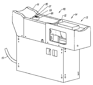
consistent with the spirit and scope of the invention. Referring now to Figure
1, shown is an arm placement guide 10. The arm placement guide is
equipped with an elbow guide 11 and a wrist guide 12. While the invented
guide also aids in supporting and immobilizing the arm, its primary function
is to enable reproducible placement of the tissue measurement site on the
analyzer, critical in producing accurate, consistent noninvasive
measurements. During use, a subject in a sitting position places the arm to
be sampled in the arm placement guide, so that the elbow is received by the
elbow guide 11 and the wrist and hand are positioned on the ergonomically
shaped wrist guide 12 and hand guide 13. In the resulting position, the
sample arm is at the subject s side with the elbow flexed to 90°. In
the
current embodiment, the arm placement device exhibits handedness; that
is, arm placement devices are separately adapted to receive right or left
arms, respectively.
It is preferred that the subject be in a sitting position during actual
sampling,
to minimize the effects of size difference between subjects. During tests of
the invented device, sampling with the subject in a sitting position resulted
in only a 2 difference in the height of the arm between an adult male and 10
year old boy, allowing the current embodiment of the invention to be built
with a relatively small range of travel being required by the movable fiber
optic probe. The wrist / hand guide unit 15 is detachably mounted on a
mechanical slide 20 (Figure 2) allowing the wrist support to be positioned
directly under the subject s wrist regardless of arm length. For optimal
reproducibility in placement of the arm on the analyzer, a custom elbow
guide 11 and wrist/hand guide 14 are constructed by creating custom molds
of a subject s elbow, wrist and hand. In the preferred embodiment, the
molds are formed from a substance such as the 5-minute RTV (room
temperature vulcanization) silicone putty supplied by Micro-Mark of Berkeley
Heights NJ, which is FDA approved for skin contact. However, other
products used for mold making having the appropriate toxicity profile would
be equally suitable.
_g_
CA 02417609 2003-O1-28
WO 02/10748 PCT/USO1/21657
As previously indicated, a fiber optic probe employs a bifurcated fiber optic
cable 15 to deliver light energy to the tissue measurement sight from an
energy source (not shown). The same probe collects light energy reflected
or transmitted from the tissue measurement site and delivers it to detectors
(not shown). A subject interface includes a cylindrical housing 16 with the
fiber optic probe tip 17 protruding from a terminal surface of the cylindrical
housing. An aperture 18 in the arm placement guide provides the subject
interface access to the tissue measurement site.
The subject s arm is positioned in the arm guide 10 such that the lowest
point of the suspended forearm is suspended directly over the tip of the fiber
optic probe 17. While the arm is being positioned, the fiber probe tip 17 is
locked into a down position using the beam movement brake 34 (Figure 3)
described in greater detail below.
0
Once the arm is positioned, an applied force mechanism 30 incorporating a
conventional single arm balance is employed to move the fiber optic probe
tip 17 upward until it contacts the arm with a constant upward force 31,
shown in Figure 3. In order to apply a very small, known amount of force to
the arm with the fiber optic probe, the point of contact between the forearm
and the probe should be limited to the tip of the probe. It is preferable that
the fiber optic probe be rectangular, with the long side of the rectangle
oriented lengthwise on the arm, so that the entire probe may contact the arm
with a minimal application of pressure. Additionally, the head of the fiber
optic probe needs to be as small as possible; again, in order to minimize
the amount of pressure required for complete contact between the probe
and the tissue measurement site. In the current embodiment, the applied
force is provided by a counterweight 33 on a single arm balance. The
balance comprises a hinged beam 32, mounted on an upright mount 37,
that rotates about a point of rotation defined by the point of attachment to
the
upright mount. A bearing 38 allows free movement of the beam about the
point of rotation. As the adjustable weight 33 is moved along the axis of the
_g_
CA 02417609 2003-O1-28
WO 02/10748 PCT/USO1/21657
beam, the force 31 applied to the tissue measurement site by the fiber optic
probe is changed. An alternative arrangement (not shown) for the
adjustable weight incorporates a weight that slides along the arm of the
balance, which is provided with gradations for different pressure levels. A
screw with a small circular weight mounted on it may be used for fine
adjustments to the applied force. In the present embodiment of the
invention, the total applied pressure may be varied in a continuous fashion
from 0 to 2 kg/in2. Additional weights may be added to vary the applied force
as required. Once the fiber optic probe is positioned, the probe may be
locked into position with the beam movement brake/lock mechanism 34.
The beam movement brake functions by means of a friction plate, which is
compressed into the upright mount 37 to lock the beam at a desired
position. In addition, the subject interface floats on a gimbal mount 35 to
insure that the optical axis of the probe is normal to the subject s arm at
the
point of contact. The gimbal mount includes a gimbal locking mechanism
36 that locks the gimbal by means of a compression or pinch element. The
fiber optic probe tip may be locked into position with the gimbal locking
mechanism 36 to maintain the stability of the probe against the arm. In
order to further assure the reproducibility of arm placement, it is necessary
to protect the invented apparatus from structural deformation due to
excessive pressure applied by the subject in the event that they lean on the
analyzer. The entire structure of the current embodiment is designed,
therefore, to withstand a force of 200 pounds exerted upon the arm support
structure, without deforming.
In addition to pressure control, the apparatus is capable of controlling the
temperature of the fiber optic probe so that it may equilibrate to the
localized
temperature in the vicinity of the tissue measurement site. In the current
embodiment, the housing 16 is cylindrical and completely surrounds the
fiber optic probe, with the probe tip 17 protruding from a terminal surface of
the cylindrical housing 16. Within the housing is a metallic core that is
maintained at a given temperature by means of a low voltage temperature
device (not shown). In the current embodiment, the core is fabricated from
-10-
CA 02417609 2003-O1-28
WO 02/10748 PCT/USO1/21657
aluminum, although other metals that are lightweight and conduct heat
readily would also be suitable. The temperature device is equipped with a
feedback control, allowing it to maintain a constant temperature. It should be
noted that the temperature of the sampled area may be predicted from the
near-IR spectra by using the shifts of the water bands, which absorb at
1450, 1950 and 2600 nm. As the temperature of the water increases, these
bands shift to higher energy.
The localized temperature of the forearm may also be measured directly. A
thermistor 19 encapsulated in a housing protrudes frorri the housing 16 into
the forearm slightly at a distance of approximately 7mm from the edge of the
fiber optic probe tip. In combination with temperature readings inside the
housing, the localized forearm temperature at the tissue measurement site
may be calculated.
One skilled in the art will recognize that the pressure may be applied by a
variety of other means, including but not limited to: a lever arm, spring
force,
air pressure or counter weights. While the above system is calibrated with
counter weights, one skilled in the art will recognize that the applied
pressure may be measured by a variety of means, including but not limited
to: balances, air pressure gauges, or by calculation.
An alternate version of the arm placement guide is reproducibly attached to
the arm and has guide rods that couple to the spectrometer to aid in
reproducibly coupling the sample to the analyzer.
While the preferred embodiment described above utilizes an applied force
to generate an applied pressure between the analyzer and the tissue
measurement site, in an alternate embodiment, the analyzer is brought into
contact with the tissue measurement site and subsequently displaced a
known distance against the skin at the tissue measurement site. In the
current, alternate embodiment of the invention, the fiber optic probe i s
maintained in a fixed vertical position, as it protrudes from a platform upon
-11-
CA 02417609 2003-O1-28
WO 02/10748 PCT/USO1/21657
which the subject s limb rests. The platform is raised and lowered, allowing
the tip of the fiber optic probe to compress the skin at the tissue
measurement site by varying amounts. DifFerent versions of the current
embodiment, each employing a different method for determining degree of
displacement, are provided. First, an LED and detector define a starting
location prior to displacement and the subject interface module may be
displaced a given distance after the LED is detected. Second, the analyzer
may be moved until spectral reflectance is removed, or, optionally, moved a
fixed distance after elimination of spectrally reflected light. In the near-IR
this would be when the light intensity at 1950 nm, where water has a strong
absorbance, approaches zero. Third, the analyzer may be displaced into the
tissue until analysis of the spectral information indicates that the preferred
depths of the sample are being probed, indicated by the detection of
chemical bands that serve as markers for an individual subject or class of
subjects; described in detail in the commonly assigned U.S Patent
Application Ser. No. 09/359,191, An Intelligent System For Noninvasive
Blood Analyte Prediction, S. Malin, T. Ruchti (July 22, 1999). Each of these
versions is described in greater detail below.
Referring now to Figure 4, the current embodiment of the invention provides
ergonomically designed elbow 11, wrist 12 and hand 13 guides mounted
on an arm support platform 40. Protruding through the arm support platform
40 is the fiber optic probe 17. The arm support platform 40 is moved
vertically up and down by a linear actuator mechanism composed of an
actuator arm 41 and vertical guides 42. The linear actuator mechanism is
driven by a conventional electric motor 45, which is, in turn, controlled by a
digital processor (not shown). An LED 43 situated at one side of the
subject s arm aims directly above the fiber bundle 17 and is detected by a
detector 44 situated at the opposite side of the subject s arm. During use,
the subject rests their arm on the provided elbow 11, wrist 12, and hand
guides 13. The linear actuator lowers the platform bearing the subject s arm
toward the fiber optic probe by lowering the arm support platform 40. As the
arm breaks the plane defined by the LED 43 and the corresponding detector
-12-
CA 02417609 2003-O1-28
WO 02/10748 PCT/USO1/21657
44, the LED signal is lost, and the system recognizes that the tissue
measurement site is a known distance from the tip of the fiber optic probe
17, the zero position. The arm support plane may be further lowered in a
controlled manner allowing known displacements of the fiber optic probe
into the subject s forearm. Naturally, the elasticity of living tissue allows
varying pressures to be applied to the surface of the tissue measurement
site without actual penetration of the fiber bundle into the skin of the arm.
A second version of the constant displacement subject interface module
defines the zero position of the translating arm support plane by detecting
spectrally reflected light collected by the fiber optic probe. The zero
position
constitutes the point at which no spectrally reflected light is detected. When
the tissue measurement site is not in contact with the surface of the fiber
optic probe, spectrally reflected light may be collected in the probe and
IS detected. This spectrally reflected light is an interferent that hinders
analysis. When the tissue measurement site first makes complete contact
with the tip of the fiber optic probe, the spectrally reflected light
approaches
zero intensity. In a diffuse reflectance based measurement of the skin in the
near-IR region, water has several strong absorbance bands located at
1450, 1950 and 2600 nm. Two noninvasive diffuse reflectance spectra of a
tissue measurement site on a human forearm are shown in Figure 5. The
top curve 50 shows that light is being detected at 1950 and 2500nm, in a
region where water has sufficiently high absorbance levels that a zero
signal should be observed. The detection of light indicates that spectrally
reflected light is being collected and that the fiber optic probe and the
tissue
measurement site are not in contact. The lower curve 51 shows zero
intensity (noise limited intensity) at 1950 and 2500nm, indicating that the
fiber optic probe tip and the tissue measurement site are in direct contact.
The zero point is defined as the point when intensity at 1950nm first reaches
zero. Known displacements beyond this point are determined using the
distance of travel of the computerized arm support platform.
-13-
CA 02417609 2003-O1-28
WO 02/10748 PCT/USO1/21657
A third version of the constant displacement subject interface module
establishes the displacement of the fiber optic probe into the forearm using
spectral information. As previously discussed, the scattering and
absorption coefficients of the sample change with different degrees of
applied pressure. Therefore, the sampled volume and resulting spectra are
a function of the displacement of the fiber versus the zero position. Thus,
the
spectra may be used to create a feedback to the linear drive system as to
the desired displacement of the subject interface module.
Other systems for raising and lowering the arm support platform are
possible, including: a hand crank, a lever arm, a scissors jack and drive, a
hinge point in conjunction with a linear drive and a worm drive. Other
systems consistent with the spirit and scope of the invention will be
apparent to those skilled in the art.
There are many situations in which it is beneficial to control the amount of
pressure exerted by an analyzer on the sample being analyzed. In the
biomedical field, analyzers are under development for a variety of important
analytes; for example, glucose, for monitoring diabetics, urea, for use with
dialysis patients, and oxygen. As previously mentioned, point of care testing
using minimally invasive and non-invasive methods is rapidly supplanting
more conventional methods of sampling and laboratory analysis in the field
of clinical testing. The invention finds application in any minimally invasive
and non-invasive measurements of this type, in which an analyzer must
make contact with a tissue measurement site.
While the foregoing description has presented the invention in the context of
medical applications with human subjects, the invention finds broad
application in a number of technical fields where solid samples are
analyzed that are not homogeneous at or near the surface and are elastic,
or where spectral reflectance must be eliminated by directly contacting a
sample with an analyzer. For example, the invention may be readily adapted
-14-
CA 02417609 2003-O1-28
WO 02/10748 PCT/USO1/21657
for veterinary or research use with non-human subjects. Additionally, optical
sampling of agricultural products is exceedingly common. For example,
analyses of fruits, vegetable and grains are affected by the degree of
pressure applied to the sample by the analyzer. The invention also provides
an apparatus for the removal of spectrally reflected light off of a sample in
diffuse reflectance mode, which is critical to quantitative analysis of small
analyte signals. Within the pharmaceutical and chemical arts, intimate
contact of the analyzer with tablets, capsules, pellets, chips and other such
items is beneficial in diffuse reflectance based measurements.
Although the invention has been described herein with reference to certain
preferred embodiments, one skilled in the art will readily appreciate that
other applications may be substituted for those set forth herein without
departing from the spirit and scope of the present invention. Accordingly, the
invention should only be limited by the Claims included below.
-15-