Note : Les descriptions sont présentées dans la langue officielle dans laquelle elles ont été soumises.
CA 02502366 2005-04-14
WO 2004/037123 PCT/US2003/031034
PROSTHETIC MESH ANCHOR DEVICE
BACKGROUND OF THE INVENTION
FIELD OF THE INVENTION
The invention relates to the laparoscopic repair of groin hernias.
DESCRIPTION OF RELATED ART
Laparoscopic repair of groin hernias, including inguinal ayzd femoral
hernias, using prosthetic mesh is gaining increasing popularity among
surgeons.
It offers comparable results to conventional open repair, with significantly
less
pain and disability. Despite its solid anatomical and physiological
principles, as
well as excellent patient acceptance, it still offers significant technical
challenges
to the average surgeon. Specifically, proper anatomical placement of the mesh,
sufficient coverage of all anatomically weak areas, and the avoida~.zce of
wrinkles or folds, constitute the most crucial determinants for a successful
procedure, leading to decreased post-operative pain and reduced recurrence
rate. These important features are among the objects of this invention.
To the best of our knowledge, there is currently no system devised to
secure mesh placement for laparoscopic groin hernia repair using a
percutaneous anchoring device. The current standard technique uses a
2 5 rectangular mesh usually rolled into a cylinder, introduced through a
trocar and
dropped into the pre-peritoneal space.
The mesh is then unfolded by manipulation with two laparoscopic
forceps and placed against the anterior abdominal wall, where it is stapled
into
3 0 place. During this process, in most instances, the mesh is entirely loose
inside
the pre-peritoneal space and its proper placement depends wholly on the
surgeon's ability, and quick reflexes. In order to facilitate this difficult
pr ocess of
free manipulation, several mesh designs are available, addressing the issues
of
CA 02502366 2005-04-14
WO 2004/037123 PCT/US2003/031034
2
conformability, porosity and elasticity. All available methods to facilitate
proper
placement of the mesh only address the issues of mesh design and different
means to deploy and secure the mesh by instrumental manipulation within the
pre-peritoneal space.
BRIEF SUMMARY OF THE INVENTION
The invention hereof addresses this singular problem by providing a
simple and quick percutaneous anchoring system for the mesh, which allows its
proper anatomical placement against the abdominal wall, thereby avoiding the
difficult process of free manipulation and greatly facilitating the technical
procedure while reducing operative time and cost.
The prosthetic mesh anchor device of the invention includes a tubular,
cylindrical sleeve having a closed end and an open end, the sleeve encasing a
spirally-wound mesh roll and an anchor which has a string or tether secured
thereto and extending outwardly through provided orifices in the mesh and the
sleeve.
2 0 The current invention consists of a unitary basic unit which includes an
anchoring system for the mesh, enabling prompt and accurate placement of the
mesh in a single step. An anchor firmly secures the mesh in a stationary
position in the exact desired anatomical location, allowing the surgeon to
staple
the mesh against the abdominal wall, while it remains secured in the correct
2 5 position. The device eliminates the difficL~lt manipulation process that
is
characteristic of the currently available tecluziques and mesh designs. Once
correct position and vertical orientation of the medial portion of the mesh is
secured, the surgeon assumes total control of the mesh and the remaining steps
of the procedure become considerably easier and faster.
The procedure utilizing the prosthetic mesh anchor device hereof consists
of the following steps:
CA 02502366 2005-04-14
WO 2004/037123 PCT/US2003/031034
3
Step 1. Creation of the Pre-peritoneal Space
A small incision is made below the umbilicus, slightly toward the side of
the hernia. The fascia is opened and a peritoneal distention balloon trocar is
.
introduced into the pre-peritoneal space and tunneled toward the pubis. A
laparoscope is introduced and the balloon is then gradually inflated. This
process is monitored both visually and manually by the surgeon to a volume
that provides satisfactory exposure of the anatomical structures. Once the
space
is created, the balloon trocar is removed and replaced with a blunt sealing
infra-umbillcal trocar and the laparoscope reinserted.
The pre-peritoneal space is re-insufflated to a pressure of ~ to l2mm of
Hg. Two additional tocars are then inserted along the midline, under direct
vision, namely, a 5mm supra-pubic tocar which is inserted just above the
pubis,
and another 5mm or l0mm middle tocar which is inserted halfway between the
pubis and the umbilicus.
Step 2. Dissection of the Hernia Sac and Spermatic Cord
Once the pre-peritoneal space is Lender direct vision, a broader dissection
of the pre-peritoneal space is undertaken with two blunt forceps, with a two
handed technique, in order to identify the anatomical landmarks and to create
sufficient space for placement of the mesh. Cooper's ligament, the inferior
epigastric vessels, the pubis and the femoral vessels are identified. In the
case of
a direct hernia, the sac and pre-peritoneal contents are dissected and
separated
from the inguinal floor.
In the case of an indirect inguinal hernia, the internal ring is identified
with the cord elements. The sac is then reduced and separated from the cord
3 0 structures.
CA 02502366 2005-04-14
WO 2004/037123 PCT/US2003/031034
4
The cord structures are freed from their attachments and suspended
from the posterior abdominal wall. In the case of femoral hernia repair, a
similar dissection is performed.
Step 3. Placement of the Mesh
A blunt forceps is introduced through the supra-pubic trocar and
adva~.iced toward the infra-umbilical trocar. Under direct laparoscopic
vision,
the forceps is then inserted into the infra-umbilical trocar in a retrograde
fashion
and advanced until it exits the proximal end of the trocar, as the
laparoscopic is
gradually pulled out.
The mesh-anchor device of the invention and its string or tether are
brought to the field and the string is grasped. The forceps is then pulled
back
into the preperitoneal space and out of the supra-pubic trocar bringing the
string out with it.
The surgeon will then hold and pull the string out gradually as the mesh
anchor device is introduced through the infra-umbilical trocar and advanced
towards the pubis.
As the mesh anchor device reaches the lowest level of penetration,
additional tension is applied to the string, bringing the mesh anchor device
tight
against the abdominal wall. Tile surgeon will manipulate the sleeve in such a
way that the mesh orifice will be placed just beneath the trocar site, in a
two-handed coordinated maneuver. At the completion of this maneuver, the
string will assume a perpendicular position in relation to the anchor.
When this position is secured, the cylindrical sleeve is pulled out of the
pre-peritoneal space by simple traction, leaving behind the anchor and furled
mesh held tightly against the abdominal wall by tension exerted on the string
by the surgeon.
CA 02502366 2005-04-14
WO 2004/037123 PCT/US2003/031034
A laparoscope is reintroduced through the infra-umbilical trocar and the
anatomical position and orientation of the mesh are adjusted if necessary,
The medial edge of the mesh assumes a vertical orientation and, once the
5 mesh is stabilized by traction obtained by continued tension exerted on the
string, the mesh is partially unrolled laterally by the forceps inserted
through
the middle trocar. The medial portion of the mesh is then stapled alongside
the
anchor, providing the desired stability that allows completion of the
procedure.
Step 4. Removal of the Anchor
Once the mesh is placed and stapled along its medial, vertical edge, the
anchor is removed. A forceps is introduced through the middle trocar, under
direct vision, and the superior edge of the anchor is grasped and pulled out
of
the pre-peritoneal space, bringing along the string. Alternatively, the string
can
also be cut with laparoscopic scissors and pulled out through the supra pubic
trocar.
Once the anchor has been removed, the surgeon can resume unrolling
and placing the mesh with bi-manual control whereupon the remaining edges
of the mesh may be stapled to the abdominal wall. Two laparoscopic forceps
are re-introduced through the middle and supra-pubie trocars. T'11e eircular
orifice of the mesh will allow insertion of the supra-pubic trocar into the
pre-peritoneal space.
It should be noted that the mesh orifice is much larger than the 5 mm
trocar orifice. This will provide two advantages: Just before placing the
first
staples on the medial vertical side of the mesh, the surgeon has some "wiggle
roorri', loosening the anchor and making some final adjustment to the mesh,
3 0 before stapling, in addition, the larger size mesh orifice facilitates re-
insertion of
the suprapubic trocar after the initial stapling of the mesh. The mesh orifice
can
be actually fairly large, without compromising the effectiveness of the
system.
CA 02502366 2005-04-14
WO 2004/037123 PCT/US2003/031034
6
All trocars are removed from the abdomen and the pre-peritoneal space
is deflated.
The wourids are closed.
The open or distal end of the tubular sleeve of the mesh anchor device
hereof has a tapered, curvilinear configuration. This is an anatomical design
which allows the surgeon to reach down behind the pubic bone, with the end of
the sleeve conforming to the curvilinear shape of the bone. When the surgeon
inserts the sleeve/mesh assembly, he does it blindly. The curvilinear tip of
the
sleeve will help him sense the deepest portion of the cavity, as the sleeve
falls
into the anatomical slot.
With the mesh anchor device hereof, the anchor is disposed immediately
below the outer convolution of the mesh roll, which in turn is encased by the
sleeve, with the string being fixed at one end to the anchor and having a free
end which extends outwardly through aligned orifices in the mesh and the
sleeve where it may be grasped by the surgeon using forceps or the like.
In the preferred embodiment of the anchor, a centrally-located tab is
provided to which one end of the string is attached.
In a first modified form of anchor, a pair of vertically-spaced,
centrally-located orifices are provided, with one end of the string being
threaded through one orifice and brought back on itself through the other
orifice and knotted.
In a second modified form of anchor, a pair of vertically-spaced, centrally-
located diagonal slots are provided, with one end of the string being threaded
through one slot and brought back on itself through the other slot and
knotted.
CA 02502366 2005-04-14
WO 2004/037123 PCT/US2003/031034
7
In the modified forms of anchor, the centrally-located orifices and/or
slots will also be positioned at the center of the mesh orifice as the sleeve
is
placed against the abdominal wall while being pulled by the string.
The trocar perforation through the abdominal wall will coincide with the
mesh orifice and the central orifices and slots of the modified forms of
anchor.
Once the anchor is withdrawn, the mesh orifice will be co-axial with the
trocar
opening and the laparoscopic trocar can be re-inserted through the abdominal
wall and through the mesh orifice. '
In all anchor embodiments, the mesh extends beyond both the upper and
lower ends of the anchor. The vertical edges of the mesh also extend beyond
the longitudinal edges of the anchor. This configuration provides sufficient
mesh exposure in a safe anatomical area for placement of staples through the
mesh against the abdominal wall.
The anchors have a semicircumferential cross-section with a smaller
diameter than a 5mm trocar. This is a distinct advantage, since the anchor
must
be removed through such a laparoscopic trocar.
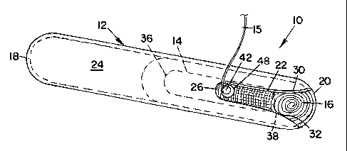
BRIEF DESCRIPTION OF THE SEVERAL VIEWS OF THE DRAWINGS
Fig. 1 is a perspective view of a prosthetic mesh anchor device
embodying a preferred form of the invention;
Fig. 2 is a perspective view of the sleeve of the mesh anchor device of Fig.
1;
Fig. 2A is an end elevational view of the sleeve of Fig. 2 as seen from the
3 o left;
Fig. 3 is a top plan view of the anchor of the mesh anchor device of Fig.1;
CA 02502366 2005-04-14
WO 2004/037123 PCT/US2003/031034
8
Fig. 3A is an end elevational view of the anchor of Fig. 3;
Fig. 4 is a cross-sectional view taken on line 4-4 of Fig. 3;
Fig, 5 is a top plan view of a first modified form of anchor,
Fig. 5A is a top plan view of a second modified form of anchor;
Fig. 6 is a top perspective view of the surgical mesh roll of the mesh
anchor device of Fig. 1;
Fig. 7 is a cross-sectional view showing a preperitoneal space following
insufflation and the placement of an upper or infra-umbilical trocar, a middle
trocar and a lower or supra-pubic trocar preparatory to use of the mesh anchor
device of Fig. 1 for laparoscopic groin hernia repair;
Fig. 8 is a cross-sectional view similar to Fig. 6 showing the mesh anchor
device of the invention following its advancement through the infra-umbilical
trocar into the preperitoneal space, where it is grasped by a forceps or the
like
2 0 which have been inserted through the supra-pubic trocar;
Fig. 9 is an enlarged, fragmentary, cross-sectional view of the mesh
anchor device of the invention following its advancement through the
preperitoneal space to a position wherein the innermost end of its sleeve is
disposed behind the pubic bone and its string or tether has been placed under
tension so as to be disposed perpendicular to the sleeve, with its outer free
end
disposed outside the abdomen, the middle trocar having been omitted for
clarity of illustration;
Fig. 10 is an enlarged, fragmentary, cross-sectional view of the mesh
anchor device of the invention following partial removal of its sleeve from
the
preperitoneal space through the infra-umbilical trocar, with its anchor and
furled mesh roll being held firmly against the iruzer wall of the abdomen by
CA 02502366 2005-04-14
WO 2004/037123 PCT/US2003/031034
9
tension placed on its string, the middle trocar having been omitted for
clarity of
illustration;
Fig. 11 is a fragmentary front elevational view of the mesh roll and
anchor of the mesh anchor device of the invention shown in solid lines before
the mesh is fully unrolled and shown in dash lines showing the final
positioning
of the mesh after it is fully unrolled to cover inguinal structures;
Fig.12 is a cross-sectional view of the mesh anchor device of the
invention following stapling of the mesh, with tension on the string having
been
released so that the anchor can be grasped by forceps and removed from the
preperitoneal space through the middle trocar, bringing the string with it;
Fig. 13 is a transverse cross-sectional view of the mesh anchor device of
the invention taken at the level of the supra-pubic trocar site, with the
string of
the device anchored at its lower end to the anchor of Fig. 3 and having its
upper
free end disposed outwardly of the abdomen; and
Fig.14 is a transverse cross-sectional view similar to Fig.12 taken at the
2 0 level of the supra-pubic trocar site following removal of the sleeve, with
the
anterior portion of the mesh disposed between the anchor and abdominal wall,
the string passing through a mesh orifice and the supra-pubic trocar orifice,
and
a loop of the string encircling the inferior portion of the tab of the anchor.
2 5 DETAILED DESCRIPTION OF THE INVENTION
Referring to Figs. 1-4, a prosthetic mesh anchor device embodying a
preferred form of the invention is generally indicated by 10 and includes a
hollow, tubular, cylindrical sleeve 12, which encases an anchor 14 having one
3 0 end of a string or tether 15 attached thereto, and a spirally-wound,
cylindrical
roll of prosthetic mesh 16.
CA 02502366 2005-04-14
WO 2004/037123 PCT/US2003/031034
Sleeve 12 and anchor 14 are preferably fabricated from a thermoplastic
material such as polycarbonate or the like.
Sleeve 12 is hollow throughout its length and has a rounded, semi-
5 circular, closed end 18 and an open end or tip 20, with end 18 being closed
to
avoid gas leakage during use.
A somewhat rectangular slot 22 is provided in an upper wall 24 of sleeve
12 and extends longitudinally inwardly from open end 20 along the central axis
10 of the sleeve for approximately one-quarter the length of the sleeve.
Slot 22 has a closed inner end 26 and an opposite open outer end 28.
Slot inner end 26 is of semi-circular configuration, while slot outer end 28
is of tapered, curvilinear configuration wherein the outer ends of the slot
curve
outwardly as at 30 and 32 from upper wall 24 and merge at their lower ends
with a lower wall 34 of sleeve 12, thereby imparting a curvilinear
configuration .
to the open end or tip 20 of the sleeve, all for purposes to appear.
2 0 Anchor 14, best seen in Fig.1, 3, 3A and 4, is relatively thin in
thickness
and narrow in width, is of substantially rectangular configuration in plan,
has a
semi-circumferential transverse cross-section and has curved opposite ends 36
and 38.
2 5 A crescent shaped opening 40 cut centrally through anchor 14, provides
an integral tab 42 which is joined to the body of the anchor by a neck 44 of
reduced width, whereby an end of string 15, not shown, may be looped
there-around and knotted so as to be readily secured to the anchor.
30 A first modified form of anchor 114, as shown in Fig. 5, is identical to
anchor 14 except that tab 42 of anchor 14 has been replaced by a pair of
vertically spaced openings 142 and 144 located on the central vertical axis of
the
anchor.
CA 02502366 2005-04-14
WO 2004/037123 PCT/US2003/031034
11
Anchor 114 is preferably fabricated from a thermoplastic material such as
polycarbonate or the like.
In the case of anchor 114, one end of string 15, not show~.l, may be
threaded through one of the openings 142 or 144 and brought back on itself
through the other opening and knotted to the string, whereby the string is
secured to the anchor.
A second modified form of anchor 214, as shown in Fig. 5A, is identical to
anchor 14 except that tab 42 of anchor 14 has been replaced by a pair of
oppositely inclined, diagonally directed slots 242 and 244 located on the
central
vertical axis of the anchor, with the slots extending inwardly from opposite
side
edges of anchor 214, and the direction of inclination of the slots being
toward a
forward edge 238 of anchor 214.
A~.zchor 214 is preferably fabricated from a thermoplastic material such as
polycarbonate or the like.
2 0 In the case of anchor 214, one end of string 15, not shown, may be
threaded through one of the slots 242 or 244 and brought back on itself
through
the other slot and knotted to the string, whereby the string is secured to the
anchor.
2 5 Spirally-wound prosthetic mesh roll 16 is approximately one-third the
length of sleeve 12 and is approximately one inch greater in length than
anchors
14,114 or 214.
Anchors 14,114 or 214, axe disposed centrally of the length of mesh roll
30 16 along the longitudinal central axis of the mesh roll immediately below
an
outer convolution 46 of the mesh roll.
CA 02502366 2005-04-14
WO 2004/037123 PCT/US2003/031034
12
Outer convolution 46 of mesh roll 16 is provided with a central circular
orifice 48 so positioned relative to tab 42 of anchor 14, or relative to
openings
142 and 144 of anchor 114, or relative to slots 242 and 244 of anchor 214, as
to
permit a free end of string 15 to pass there through.
The transverse widths of each of anchors 14, or 114, or 214 correspond to
approximately one-half the diameter of mesh roll 16, for purposes to appear.
As best seen in Fig.11, mesh roll 16 is provided with a horizontally
inwardly extending lateral slit 50 to accommodate a trio of cord structures 74
of
the patient adjacent the hernia site, as will appear.
Referring to Figs. 7-14, the manner of use of mesh anchor device 10 will
be explained.
In Fig. 7, a 5mm infra-umbilical trocar 52, a 5mm or l0mm middle trocar
54 and a 5mm supra-pubic trocar 56 have been inserted along the midline of the
patient's abdomen 58 following insufflation of a preperitoneal space 60 above
the peritoneum 62 and abdominal cavity 64 and adjacent pubis 66 in preparation
for laparoscopic hernia repair, with infra-umbilical trocar 52 positioned
below
the umbilicus and slightly toward the side of the hernia, middle trocar 54
positioned half-way between the pubis and umbilicus, and supra-pubic trocar 56
positioned immediately above the pubis.
Following dissection of the hernia sac and spermatic cord, the surgeon
introduces mesh anchor device 10 into preperitoneal space 60, as seen in Fig.
8,
with a forceps F advanced through supra-pubic trocar 56 grasping string 15 and
drawing mesh anchor device 10 through infra-umbilical trocar 52 with the open
end 20 of sleeve 12 of the mesh anchor device facing the supra-pubic trocar.
As shown in Fig. 9, the surgeon continues to pull string 15 until open end
20 of sleeve 12 is brought into contact with pubis 66.
CA 02502366 2005-04-14
WO 2004/037123 PCT/US2003/031034
13
As mesh anchor device 10 reaches the lowest level of penetration,
additional tension is applied to string 15, bringing the mesh anchor device
tight
against the abdominal wall as shown in Fig.10. The surgeon will manipulate
sleevel2 in such a way that orifice 48 of mesh roll 16 is placed just beneath
the
site of supra-pubic trocar 56 in a two handed maneuver. At this time, string
15
will have assumed a perpendicular position in relation to sleeve 12.
When this position is secured, cylindrical sleeve 12 is pulled out of
pre-peritoneal space 60 through infra-umbilical trocar 52 by simple traction,
leaving behind anchor 14, or 114, or 214, with furled mesh 16 held tightly
against the abdominal wall 58 by tension exerted by the surgeon on string 15.
At this time, a laparoscope is reintroduced through infra-umbilical trocar
52 and the anatomical position and orientation of mesh roll 16 are adjusted,
if
necessary.
As seen in Fig. 11, a medial edge portion 68 of mesh roll 16 assumes a
vertical orientation and, once the mesh is stabilized by traction exerted
through
string 15, it is partially unrolled laterally by forceps, not shown, inserted
through middle trocar 54. Medial edge portion 68 of the mesh roll is then
stapled adjacent anchor 14 to the posterior abdominal wall as by staples 70,
providing desired stability to allow completion of the procedure.
It should be noted that the upper and lower edges of mesh roll 16 extend
beyond both the upper and lower ends 36 and 38 respectively of a~.vchor 14.
The
vertical edges of the mesh also extend beyond the longitudinal edges of the
anchor. This configuration provides sufficient mesh exposure in a safe
anatomical area for placement of staples through the mesh against the
abdominal wall.
As shown in dash lines in Fig.11, a superior portion 76 of mesh roll 16 is
passed over a pair of inferior epigastric vessels 73, while an inferior
portion 72
of mesh roll 16 is passed under cord structures 74, a femoral artery 75 and a
CA 02502366 2005-04-14
WO 2004/037123 PCT/US2003/031034
14
femoral vein 77, with the mesh being positioned according to the surgeon's
preference.
As aforesaid, horizontal lateral slit 50 in mesh 16 accommodates the cord
structures 74.
The upper and lower horizontal edges of the mesh are secured against
the posterior abdominal wall by staples 80 as the placement proceeds according
to the surgeon's preference.
Once the mesh has been placed and stapled along its medial vertical edge,
as shown in Fig. 11, anchor 14, or 114, or 214 is removed, as seen in Fig.12.
Forceps, not shown, are introduced through middle\trocar 54, under direct
vision, and the curved edge 36 of the anchor is grasped and pulled out of
pre-peritoneal space 60 through middle trocar 54 bringing along string 15.
Alternatively, string 15 can also be cut with laparoscopic scissors S and
pulled
out through supra-pubic trocar 56.
Once the anchor has been removed, the surgeon can resume unrolling
2 0 and placing the mesh with bi-manual control. Two laparoscopic forceps, not
shown, are reintroduced through middle trocar 54 and supra-pubic trocar 56.
Circular orifice 48 of mesh 16 will allow insertion of the supra-pubic trocar
into
the pre-peritoneal space through the mesh.
It should be noted that mesh orifice 48 is much larger than the 5mm
trocar orifice. This will provide two advantages:
1. Just before placing the first staples on the medial vertical side of the
mesh,
the surgeon has some "wiggle room', loosening the anchor and making
some final adjustment to the mesh before stapling; and
CA 02502366 2005-04-14
WO 2004/037123 PCT/US2003/031034
2. The larger size mesh orifice facilitates re-insertion of the supra-pubic
trocar after the initial stapling of the mesh. The mesh orifice can be large,
without compromising the effectiveness of the system.
5 The surgeon now removes trocars 52, 54 and 56 from the abdomen, pre-
peritoneal space 60 is deflated and the wounds are closed.
Fig. 13 is a transverse cross-sectional showing of mesh anchor device 10
taken at the level of the supra-pubic trocar site, with the lower end of
string 15
10 looped around neck 44 of tab 42 of anchor 14 and with the upper free end of
the
string passing through a supra-pubic trocar site orifice 82 and being disposed
outwardly of abdomen 58.
Fig. 14 is a transverse cross-sectional view similar to Fig. 12 taken at the
15 level of the supra-pubic trocar site following removal of sleeve 12, with
outer
convolution 46 of mesh roll 16 disposed between anchor 14 and abdominal wall
58, with string 15 passing through mesh orifice 48 and supra-pubic trocar
orifice
82 and the loop of string 15 encircling neck 44 of tab 42 of anchor 14.
It will be understood that, while reference has been made herein to a
"string" or "tether", I do not wish to be limited thereto, since any suitable
means,
such as a cord, or line, or the like may be employed.