Note : Les descriptions sont présentées dans la langue officielle dans laquelle elles ont été soumises.
CA 02591716 2007-06-14
WO 2006/065940 PCT/US2005/045322
PROGNOSTIC AND PREDICTIVE MARKERS IN
CANCER TREATMENT
By
Soonmyung Paik, MD and Chungyeul Kim, MD
REFERENCE TO RELATED APPLICATION
This application claims priority to U.S. Provisional application number
60/636,169,
filed December 15, 2004, application number 60/698,112 filed July 11, 2005,
and 60/717,485
filed September 14, 2005, all of which are incorporated by reference herein.
BACKGROUND OF THE INVENTION
Breast cancer is a heterogeneous disease with respect to clinical behavior and
response
to therapy. This variability is a result of the differing molecular make up of
cancer cells within
each subtype of breast cancer. However, only two molecular characteristics are
currently being
exploited as therapeutic targets. These are estrogen receptor and HER2, which
are targets of
antiestrogens and Herceptin respectively. Efforts to target these two
molecules have been
proven to be extremely productive. Nevertheless, those tumors that do not have
these two
targets are often treated with chemotherapy which generally targets
proliferating cells. Since
some important normal cells are also proliferating, they are damaged by
chemotherapy at the
same time. Therefore, chemotherapy is associated with severe toxicity.
Identification of
molecular targets in tuinors in addition to ER or HER2 is critical in the
development of new
anticancer therapy.
Recent studies using combination of cDNA array based expression profiling and
comparative genomic hybridization ("CGH") have elucidated the role of gene
amplification in
the transcriptional program of breast cancer.
In a study by Pollack et al, copy number alteration and expression levels
across 6691
mapped human genes were examined in 44 locally advanced breast cancer and 10
breast cancer
CA 02591716 2007-06-14
WO 2006/065940 PCT/US2005/045322
cel~"'"~~ne~ Hi~e IÃR; T; 'erou CM et al., Proc Natl Acad Sci U S A 2002;
99(20):12963-12968). The data from this study suggests that at least 12% of
all the variation in
gene expression among the breast cancer is directly attributable to underlying
variation in gene
copy numbers. The total number of genomic alterations (gains and losses)
correlated
significantly with high grade (p=0.008), negative ER (P=0.04), and p53
mutation (p=0.0006).
Of 117 high level amplifications (representing 91 different genes) 62%
(representing 54 genes)
were found to be associated with at least moderately elevated mRNA levels, 42%
(representing
36 different genes) with highly elevated mRNA levels. In a similar effort,
Hyman et al have
exainined correlation between copy number changes and expression levels in 14
breast cancer
cell lines using cDNA microarray of 13,824 genes. Hyman E, Kauraniemi P,
Hautaniemi S et
al., Cancer Res 2002; 62(21):6240-6245. They found 44% of highly amplified
genes resulted in
overexpression with 10.5% of overexpressed genes being amplified.
Together these results indicate a profound role of gene amplification in
transcriptional
control of gene expression in breast cancer and provide rationale for pursuing
amplified genes
as a preferred target for developing therapeutics and diagnostics.
Unfortunately, no study has correlated clinical outcome with a comprehensive
list of
amplified genes in breast cancer although amplification of a handful of genes
has been
identified by array CGH and have been examined by fluorescence in-situ
hybridization
("FISH") and found to be prognostic. The biggest barrier for the screening of
amplification
pattern is the cost and need for high quality DNA for array CGH assays.
On the other hand, FISH is a stable method that works with formalin fixed
paraffin
embedded sections in a routing clinical setting. FISH probes for HER2 have
been FDA
approved as a predictive test for Herceptin response. Due to the stability of
DNA in the paraffin
embedded sections, it is more reliable than RNA based or immunohistochemistry
based clinical
assays. However, FISH probes for potentially important amplified genes have
not been
comprehensively developed. In fact, there is only one vendor (Vysis, Downers
Grove, IL) that
2
CA 02591716 2007-06-14
WO 2006/065940 PCT/US2005/045322
sO~'Y~~s ~na~~~ of these probes have not been clinically validated at this
point as prognostic factors. These probes are also very expensive (cost about
$300 per case)
and of limited variety, barely scratching the repertoire of potentially
important amplicons in
solid tumors such as breast and colon cancer.
In a recent survey of five Vysis supplied commercial FISH probes (HER2, MDM2,
MYC, CCND 1, EGFR) for potentially presumed important amplicons in breast
cancer in 1100
cases, Al-Kuraya et al found some but not all the five gene amplifications
correlate with
survival outcome in a poorly defined clinical cohort with no treatment
information. Al-Kuraya
K, Schraml P, Torhorst J et al., Cancer Res 2004; 64(23):8534-8540.
Nevertheless, they did
find that 60% of the cases did not have any amplification of the five genes
examined. In
addition, a gene amplification dosage effect was found in which survival rate
was in the
following order; no amplification> 1 amplified> 2 amplified> 3 amplified. This
data supports
the so called "amplificatory" phenotype with an increased level of genomic
instability and high
likelihood for amplification development and therefore supports the need for a
comprehensive
clinical correlation of ainplicons in breast cancer.
Despite recent advances in molecular taxonomy of breast cancer, only two
molecular
characteristics are currently being exploited as therapeutic targets. These
are estrogen receptor
and HER2, which are targets of antiestrogens (tamoxifen and aromatase
inhibotors) and
Herceptin respectively. Efforts to target these two molecules have been proven
to be extremely
productive. Nevertheless, those tumors that do not have these two targets are
often treated with
chemotherapy which generally targets proliferating cells. Since some important
normal cells
are also proliferating, they are damaged by chemotherapy at the same time.
Therefore,
chemotherapy is associated with severe toxicity. Identification of molecular
targets in tumors
in addition to ER or HER2 is critical in the development of new anticancer
therapy.
Approximately 15 to 20% of all breast cancer has overexpression of HER2
protein on its
cell surface. Paik S, Hazan R, Fisher ER et al., J Clin Oncol 1990; 8(1):103-
112. Such tumors
3
CA 02591716 2007-06-14
WO 2006/065940 PCT/US2005/045322
Itõt~ ..,j .:. ,,. tk tf~:"= Et..,I II; i ..W ij'... ..'.;e.., fF :,R.tt
aref kno~nt~ ha~ ~a wors~' pfo~~ than those without HER2 protein
overexpression Paik S.
Hazan R, Fisher ER et al., J Clin Oncol 1990; 8(1):103-112. Overexpression of
HER2 protein is
almost invariably due to amplification or increased copy number of gene
encoding HER2.
Multiple drugs have been developed to target HER2 signaling as means to stop
growth
of cancer cells that have overexpression of HER2 protein on its surface. One
of these drugs is
Trastuzumab (Herceptin), developed by Genentech. Herceptin has recently been
shown to be
effective in prolonging survival in patients diagnosed with advanced breast
cancer with HER2
overexpression. Slamon DJ, Leyland-Jones B, Shak S et al., N Engl J Med 2001;
344(11):783-
792. Recently it has also been shown to reduce recurrences and death in
patients with early
stage breast cancer which have HER2 protein overexpression or HER2 gene
amplification
Romond EH, Pesez EA, Bryant J et. al, N Eng J Med 2005; 353(16); 1673-1684.
The overall
reduction in recurrence rate is about 50% with Herceptin when compared to
chemotherapy
alone in adjuvant setting. Romond EH, Pesez EA, Bryant J et. al, N Eng J Med
2005; 353(16);
1673-1684. Not all patients seem to gain benefit from this expensive
treatment, which also has
potential serious cardio toxicity. A method to identify those patients who
will benefit most
from Herceptin or other HER2 targeting drugs is required. Slamon DJ, Leyland-
Jones B, Shak
S et al., N Engl J Med 2001; 344(11):783-792; Goldman B., J Natl Cancer Inst
2003;
95(23):1744-1746. Many laboratories have been pursuing abnormalities in the
components of
HER2 signaling pathway, such as PTEN, as predictors of response to Herceptin,
with the
hypothesis that such abnormalities will render tumor cells resistant to
Herceptin even in the
presence of HER2 protein overexpression. Crowder RJ, Lombardi DP, and Ellis
MJ., Cancer
Cell 2004; 6(2):103-104; Nagata Y, Lan KH, Zhou X et al., Cancer Cell 2004;
6(2):117-127.
Such studies have concentrated only on molecules that may have direct role in
HER2 signaling
pathway, however, none have been substantiated in clinical studies and there
is no marker used
for the prediction of response to Herceptin in the clinical practice.
4
CA 02591716 2007-06-14
WO 2006/065940 PCT/US2005/045322
Tt~ere re' any''"gfte. -"t~a~ "i'are amplified in breast cancer as
demonstrated by CGH
studies. As stated previously, about 10% of genes overexpressed in breast
cancer are due to
gene amplification. Pollack JR, Sorlie T, Perou CM et al., Proc Natl Acad Sci
U S A 2002;
99(20):12963-12968. One of the frequently amplified gene in human cancers is
cMYC located
on chromosome 8. In normal cells cMYC is expressed in highly regulated manner
driving cells
from G 1 to S phase. Perhaps due to its important role in normal cell
proliferation, efforts to
block cMYC has not been a major focus of pharmaceutical industry. Only one
company
currently has a drug that is going through clinical testing (Cylene
Pharmaceuticals). Studies
have suggested that cMYC has an important role as a molecular switch that
determines the
cell's fate to go through programmed cell death or cell proliferation
Pelengaris S, Khan M,
Evan G., Nat Rev Cancer 2002; 2(10):764-776; Pelengaris S, Khan M, Evan GI.,
Cell 2002;
109(3):321-334. When cMYC is overexpressed, cells go into uncontrolled cell
proliferation and
become susceptible to programmed cell death in the absence of a survival
signal (see Figure
la). cMYC induces apoptosis by regulating many components of the programmed
cell death
pathway, but the main effector seems to be Bax. Pelengaris S, Khan M, Evan G.,
Nat Rev
Cancer 2002; 2(10):764-776.
Eventually cells with cMYC overexpression will go through mass suicide due to
the
exhaustion of locally available survival factors. At the same time, cMYC
overexpression has
been shown to cause genomic instability. This could cause amplification of
other oncogenes
such as HER2. Fest T, Mougey V, Dalstein V et al., Oncogene 2002; 21(19):2981-
2990.
Amplification of other genes could generate anti-apoptotic signals and
therefore the inhibition
of the apoptotic pathway. For example, in the case of HER2 amplification,
studies have
demonstrated that HER2 induces Bcl-2, an anti-apoptotic protein that inhibits
Bax. Milella M,
Trisciuoglio D, Bruno T et al., Clin Cancer Res 2004; 10(22):7747-7756.
5
CA 02591716 2007-06-14
WO 2006/065940 PCT/US2005/045322
n6' =~~~Iiighs to identify markers/genes that provide prognostic
indicators of therapy efficacy. The references cited above and in the Appendix
hereto are
hereby incorporated by reference as if fully set forth herein.
SUMMARY OF THE INVENTION
The present disclosure describes a new prognostic and therapeutic target,
HTPAP gene,
which when amplified confers poor prognosis in breast cancer patients even
after treatment with
standard chemotherapy containing doxorubicin, cyclophosphamide, and
paclitaxel. HTPAP
amplification is an independent prognosticator of tumor size, treatment,
number of positive
axillary lymph node, age and hormone receptor status, HER2 amplification, and
cMYC
amplification. Furthermore, cMYC, is a predictor of response to Herceptin, in
such a way that
for patients with cMYC amplification together with HER2
amplification/overexpression, there
is a 75% reduction in cancer recurrence rate when Herceptin is added to
chemotherapy,
compared to only 45% reduction in recurrence rate for those patients without
cMYC
amplification. cMYC is amplified in approximately 30% of the breast cancer
patients with
HER2 amplification or overexpression. Inhibition of HER2 signaling by
Trastuzumab
apparently changes the cMYC role from proliferation switch to pro-apoptotic
switch. The
invention has the following clinical applications: optimization of methods for
patient selection
and determining treatments using Trastuzumab and other drugs that target a
HER2 signaling
pathway: optimization of methods for patient selection for future clinical
studies that test the
addition of other drugs or targeted therapies, such as Bevacizumab (Avastin)
that targets
angiogenesis, by allowing identification of patients who are at high risk of
relapse even after
Trastuzumab or HER2 targeted therapy: PCR-based assay that will detect the
gene
amplification status of both HER2 and cMYC in a single tube assay for
prognostication and
prediction of response in breast cancer patients: and rational development of
cMYC targeted
therapy through indirect modulation of its pro-apoptotic activity by
inhibiting anti-apoptotic
signal from other activated oncogenes.
6
CA 02591716 2007-06-14
WO 2006/065940 PCT/US2005/045322
kbTION OF THE FIGURES
Figure 1 a shows a schematic of cMYC as a pro-apoptotic switch.
Figure lb shows a schematic of cMYC as a proliferation switch.
Figure 1 c shows a schematic of an anti-apoptotic signal from HER2.
Figure 2 shows a flow chart describing a method of identifying therapeutic
targets
Figure 3 shows the results of a clustering study.
Figure 4 shows a chart of recurrence by amplification.
Figure 5 shows a Kaplan Meier plot for APPBP2.
Figure 6 shows a Kaplan Meier plot for BMP7.
Figure 7 shows a Kaplan Meier plot for bm 009.
Figure 8 shows a Kaplan Meier plot for CACNB 1.
Figure 9 shows a Kaplan Meier plot for chk.
Figure 10 shows a Kaplan Meier plot for c_myc.
Figure 11 shows a Kaplan Meier plot for cyclindl.
Figure 12 shows a Kaplan Meier plot for decrl.
Figure 13 shows a Kaplan Meier plot for FLJ 10783.
Figure 14 shows a Kaplan Meier plot for GRO1.
Figure 15 shows a Kaplan Meier plot for GRB2.
Figure 16 shows a Kaplan Meier plot for HBS 1 L.
Figure 17 shows a Kaplan Meier plot for HER2.
Figure 18 shows a Kaplan Meier plot for MAL2.
Figure 19 shows a Kaplan Meier plot for HTPAP.
Figure 20 shows a Kaplan Meier plot for MLN64.
Figure 21 shows a Kaplan Meier plot for MRPS7.
Figure 22 shows a Kaplan Meier plot for PPM1D.
Figure 23 shows a Kaplan Meier plot for NC043.
7
CA 02591716 2007-06-14
WO 2006/065940 PCT/US2005/045322
Figur~ 2~ s~io QlaritM Qe?-'P7bFforRPS6KB1.
Figure 25 shows a Kaplan Meier plot for SEB4D.
Figure 26 shows a Kaplan Meier plot for stk6.
Figure 27 shows a Kaplan Meier plot for SIP2 28.
Figure 28 shows a Kaplan Meier plot for TPD52
Figure 29 shows a Kaplan Meier plot for TRAF4.
Figure 30 shows a Kaplan Meier plot for ZNF217.
Figure 31 shows a Kaplan Meier plot for ZHX1.
Figure 32 shows a Kaplan Meier plot for any amplicon.
Figure 33 shows a diagram of the HTPAP gene.
Figure 34 shows a recurrence free survival.
8
CA 02591716 2007-06-14
WO 2006/065940 PCT/US2005/045322
24 ll(Ã !';~;iJ ,r Et=J)tk," W~ION OF THE INVENTION
One reason for the high cost of commercially available FISH probes is the cost
and
difficulty of directly fluorescence labeling bacterial artificial clones (BAC)
representing the
probes. This disclosure provides a method for fluorescently labeling BAC
clones representing
known amplicons efficiently by combining a series of whole genome
amplification methods
and an efficient FISH method for paraffin embedded tissue which has been
archived more than
years (see overview in Figure 2). This labeling and FISH method is a log order
less
expensive as compared to commercially available probes. Using paraffin block
tissue samples
for over 30,000 breast and colon cancer cases that are all annotated with
clinical follow up
10 information and treatment received provided a unique source for clinical
correlative science
studies. Combining the FISH method with tissue micro array (TMA) allows
screening of more
than 100 cases using a single microscopic section making screening of multiple
amplicons in
thousands of cases a reality. One of ordinary skill in the art will readily
recognize that any
number of methods well known in the art can be used to label probes for FISH
applications.
Furthermore, because FISH is used to determine amplification, numerous other
quantitative or
semi-quantitative methods may be used, including, but not limited to, antibody
based assay
(such as ELISA (enzyme-linked immunosorbent assay)) and qt PCR.
Pilot Proiect=
In a pilot demonstration project, more than 987 cases from National Surgical
Adjuvant
Breast and Bowel Project ("NSABP") trial B-28 were screened comparing 4 cycles
of
ariamycin cyclophosphamide versus same drugs followed by four cycles of
paclitaxel. In this
study, tissue microarrays were constructed and FISH assays performed for 10
different in-
housed developed probes based on array CGH data (two sets are very close to
each other, i.e.
HER2 and MLN64, APPBP2 and PPM1D). The amplicons and their chromosomal
locations are
shown as follows:
SEB4D 20q13.32
9
CA 02591716 2007-06-14
WO 2006/065940 PCT/US2005/045322
Z~!F~h
APPBP2 17q23.2
TPD52 8q21
MLN64 17q11-q12
PPM1 D 17q23.2
HER2 17q21.1
CYCLIND1 11q13
MAL2 8q23
C-MYC 8q24.12-q24.13
After hybridization of individual probes, cases were scored as either
amplified (if signal
more than 3 copies per nuclei) or not-amplified (2 copies or less). In order
to find the natural
class of amplification patterns of these 10 amplicons, non-supervised
Hierarchical clustering
was performed. The results of the pilot study are shown in Figure 3. What is
notable in the
result is close correlation of amplification status of PPM1D and APPBP2, and
HER2 and
MLN6 as expected based on their very close proximity in their chrmosomal
location. This data
proves that our method for BAC labeling as claimed results in highly
reproducible results.
In addition, there are cases with no amplification of any of the 10 amplicons.
While the
proportion of such cases will decrease as more amplicons are screened, it is
likely that such
subgroups do exist that are relatively resistant to amplification.
The prognostic value of non-amplification versus any amplification in B-28
according to
treatment was examined. Recurrence free survival of those patients with no
amplification of any
of the 10 amplicons were significantly better than those with amplification of
any of the genes
(Figure 4), while as expected from the nature of the genes in the 10 selected
amplicons in this
pilot, there was no interaction with the benefit from adding taxol to AC based
on amplification
phenotype in general in this protocol.
As a result of systematic screening of 27 candidate amplicons that are
associated with
overexpression (as shown in Table 1), three amplicons (HER2: cMYC, and HTPAP
which is
also knows as PPAPDCIB) were identified that are independently prognostic in
node positive
breast cancer treated with standard chemotherapy when they are tested in
multivariate analysis
including other prognostic variables. These three amplicons were identified
using the following
BACs: HER2-PathVysion HER2 Assay from Vysis; cMYC-LSI C-MYC from Vysis; HTPAP-
CA 02591716 2007-06-14
WO 2006/065940 PCT/US2005/045322
RP'2 ! 15 1~D5~ ~~I~e~~'rthel~5''s~ bd~IroF"ordinary skill in the art would
readily recognize multiple
other probe sources for the same genes can be utilized with this invention.
One of ordinary skill
in the art would readily recognize multiple other method of labeling any probe
sources for the
same genes can be utilized with this invention. These could include both
fluorogenic and
chrmogenic probe labeling methods.
These 27 amplicons were screened by FISH on TMA constructed from a NSABP trial
B-28, in which auxiliary node positive breast cancer patients were randomly
assigned to receive
4 cycles of arimycin (doxorubicin) plus cyclophosphamide (AC) or same regimen
followed by
taxol (N=1901). This means that approximately 51,327 FISH assays were
performed
(27x 1901). Selection of the 27 amplicons was based on the following criteria:
1) selected
amplicons had been all shown to be associated with moderate to high level of
gene expression
of the coded genes when amplified in breast cancer tumors or cell lines in
both studies
conducted by Pollack et al and Hyman et al (Pollack JR, Sorlie T, Perou CM et
al., Proc Natl
Acad Sci U S A 2002; 99(20):12963-12968; Hyman E, Kauraniemi P, Hautaniemi S
et al.,
Cancer Res 2002; 62(21):6240-6245); 2) the public genome sequence map was
examined and
FISH validated BAC clones were selected that corresponded best with the
selected amplicons;
and 3) some amplicons, such as MLN64, which were located very close to HER2
were included
as an internal control for reproducibility and validity of the assay (that is
HER2 and MLN64
amplification were expected to correlate extremely tightly due to their close
proximity in
chromosome location).
Amplification status was categorized as either amplified or non-amplified,
with gene
amplification defined as having more than 4 signals (4 dots per single tumor
cell nucleus) from
in situ hybridization. Correlation with clinical outcome using univariate Cox
proportional
hazard model showed that HER2, MLN64 (which is very close to HER2 and highly
correlated),
cMYC, HTPAP, TPD52, MAL2, and ZNF217 are significantly correlated with
clinical outcome
of patients entered into the B-28 trial (Table 1). In addition, the presence
of any amplification
11
CA 02591716 2007-06-14
WO 2006/065940 PCT/US2005/045322
and ''nGm1er 'arfr~r~lffrcadal:,A'owed significant correlation with outcome.
Kaplan Meier
plots for each of the 27 amplicons screened are shown in the Figures 5 to 31.
A Kaplan Meier
plot comparing cases with no amplification versus any amplification is shown
in Figure 32.
Multivariate analysis including conventional prognostic markers (tumor size,
number of
positive nodes, hormone receptor status, and age) was performed. Three
amplicons remained
significant: HER2; cMYC; and HTPAP (as shown in Table 2).
HTPAP:
Both HER2 and cMYC have previously been shown to be prognostic in breast
cancer.
HER2 is the therapeutic target for Herceptin. However their prognostic role in
chemotherapy
treated patients has not been clearly demonstrated. On the other hand, HTPAP
is a novel gene
which translates into a protein with a phosphatidic acid phosphatase homology
domain and a 5'
transmembrane domains as well as signal peptide that indicates that the
protein product is
secreted (Figure 33). The Bacterial Artificial Chromosome clone used for
generation of FISH
probe for HTPAP (clone RP11-513D5) has only three genes in it: HTPAP; WHSCILI;
and
DDHD2. Of these, other studies correlating gene amplification with expression
in breast cancer
cell lines have shown that HTPAP is the one that is overexpressed when this
region is
amplified. Pollack JR, Sorlie T, Perou CM et al., Proc Natl Acad Sci U S A
2002;
99(20):12963-12968; Hyman E, Kauraniemi P, Hautaniemi S et al., Cancer Res
2002;
62(21):6240-6245; Ray ME, Yang ZQ, Albertson D et al., Cancer Res 2004;
64(1):40-47. In a
review of data from microarray analysis of gene expression in breast cancer,
Jenssen et al
reported that HTPAP overexpression is associated with poor prognosis of
patients with breast
cancer together with 94 other genes. Jenssen TK, Kuo WP, Stokke T, Hovig E.
Associations
between gene expressions in breast cancer and patient survival. Hum Genet
2002; 111(4-
5):411-420. These results demonstrate that amplification of the HTPAP gene is
an independent
prognosticator for breast cancer even after treatment with standard
chemotherapy.
12
CA 02591716 2007-06-14
WO 2006/065940 PCT/US2005/045322
'c~~~~iV6npreviously been shown to be prognostic in breast cancer.
HER2 is the therapeutic target for Herceptin. On the other hand, HTPAP is a
novel gene which
translates into a protein with a phosphatidic acid phosphatase homology domain
and a 5'
transmembrane domains as well as signal peptide that indicates that the
protein product is
secreted (Figure 33).
While amplification and overexpression of HTPAP in a limited number of breast
cancers with 8pll-12 amplification has been described before by other
investigators, these
studies have not pinpointed HTPAP as the main driver gene in those
amplifications since there
are other genes that are overexpressed from the region of amplification. By
taking advantage of
the use of relatively small FISH probes containing only three genes in which
HTPAP is the only
overexpressed gene, and screening of large number of cases with defined
treatment from a
single prospective clinical trial, this disclosure is the first to demonstrate
its role as a prognostic
factor independent of other prognosticators in breast cancer. Since it is
amplified and correlated
with poor prognosis even after standard chemotherapy, HTPAP is also an
important therapeutic
target for breast cancer.
The following characteristics of HTPAP make it an ideal therapeutic and
diagnostic
target in breast cancer: 1) HTPAP is amplified and stable clinical diagnostic
assay using FISH
or PCR can be used to detect the amplification status; 2) it is an independent
prognostic factor
in heavily treated patients; 3) it is transmembrane protein with enzyme
activity; and 4) it is also
secreted.
The amplification of this gene being highly correlated with poor prognosis
indicates that
the blocking of these activities will have beneficial therapeutic effects (as
exemplified by the
HER2 gene which has a similar characteristic of being amplified, prognostic
factor, and a cell
surface receptor).
Certain embodiments of the present invention include monoclonal antibodies or
series of
monoclonal antibodies with specificity for the extracellular domain of the
HTPAP protein.
13
CA 02591716 2007-06-14
WO 2006/065940 PCT/US2005/045322
Thgie'Ã::antiliob6 ~'-Ab-'c~' tEalone or in combination with chemotherapeutic
drugs or
antibodies to other targets. The generation of such antibodies can be
performed via any number
of methods for monoclonal production which are well known in the art.
In certain embodiments of the present invention, these anti-HTPAP antibodies
used to
detect HTPAP protein secreted in the serum or plasma or body fluid (such as
nipple aspirate
from the patients) and compared to normal levels in the diagnosis or
monitoring of disease
during therapy. Detection may be accomplished by any number of methods well
known in the
art, including but not limited to radioimmunoassay, flow cytometery, ELISA, or
other
colormetric assays.
Phosphatidic acid phosphatase domain typically acts as an important signaling
molecule
in the cancer cells. Certain embodiments of the present invention include the
use of these
domains of the HTPAP gene in targeting the development of small molecules that
interfere or
modulate such activity. Furthermore, the use of anti-bodies to HTPAP can be
used to identify
down stream signaling molecules to HTPAP and subsequently targeted by small
molecule
therapeutics.
Certain other embodiments include the blocking of HTPAP gene activity using
siRNA,
antisense oligonucleotide, or Ribozyme approaches that are well known in the
art.
Other genes found to be of marginal prognostic power in this study cohort of
AC or
ACT Treated node positive breast cancer may have significant prognostic power
in untreated or
node negative patients - these include TPD52, MAL2, ZNF217, NCOA3, ZHXl, B1V1
009,
BMP7, and STK6 and they also may provide attractive target for therapeutic
development. In
certain embodiments of the present invention, three prognostic amplified genes
HER2, cMYC,
and HTPAP can be utilized to create a prognostic index to guide treatment
decision making for
breast cancer patients. Certain other embodiments include same three genes
together with
clinical variables to generate a prognostic index to guide treatment decision
making.
14
CA 02591716 2007-06-14
WO 2006/065940 PCT/US2005/045322
Y~ i: , f=., i='':nf .m 5
c1VI1
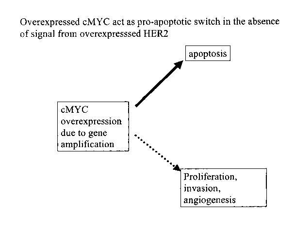
Cells primed for malignant transformation by cMYC amplification seem to be
able to
escape the fate of apoptosis with the help of HER2 amplification, however, it
is believed that
this also makes them dependent on HER2 signaling to survive (Figure 2b).
Therefore inhibition
of the HER2 signal by Trastuzumab could trigger pro-apoptotic function of cMYC
in such
cancer cells (Figure 2c). This was verified in retrospective analysis of tumor
specimens
collected as part of NSABP trial B-3 1, in which patients diagnosed with HER2
overexpressing
tumors were randomized to receive chemotherapy or chemotherapy plus Herceptin.
The results
of this analysis clearly deinonstrated that tumors with co-amplification of
both HER2 and
cMYC gene are sensitive to Trastuzumab.
In an effort to identify clinically important gene amplifications in breast
cancer, 27
different commonly amplified genes in breast cancer were screened using FISH.
As previously
stated, in a unpublished study correlating clinical outcomes of 1900 patients
with the status of
gene amplification of 27 different genes/loci, HER2, cMYC, and HTPAP were
identified as
three independent amplified genes that confer a worse prognosis even after
standard
combination taxane-containing adjuvant chemotherapy. Furthermore, cases that
had co-
amplification of HER2 and cMYC had much worse prognosis than cases with
amplification of
either one of the genes.
The status of cMYC in 1344 patients enrolled in the NSABP B-31 trial were
examined
to test the potential benefits of addition of Trastuzumab to chemotherapy in
the treatment of
patients diagnosed with early stage breast cancer with HER2 gene
amplification/overexpression. FISH was used to enumerate the cMYC gene copy
number using
a commercially available DNA probe (Vysis). Any tumor with more than 10% of
cells showing
more than 4 copies of cMYC gene was classified as cMYC gene amplified in this
analysis. 399
cases out of 1344 total cases studied were classified as cMYC amplified.
Tumors with cMYC
amplification were believed to be sensitive to inhibition of HER2 signaling
due to its activation
CA 02591716 2007-06-14
WO 2006/065940 PCT/US2005/045322
of ~.' p~-~~bpijtie''~~~ 'gri~l' wei t~~ ?R2 signal is inhibited by
Trastuzumab and that this would
translate into much more significant reduction in recurrence rate in cMYC
amplified cohort in
comparison to patients with no amplification of cMYC.
Recurrence free survival of B-31 patients according to cMYC amplification
status is
shown in Figure 34. In patients with no amplification of cMYC gene (N=945),
there was a 34%
reduction in recurrence rate when Trastuzumab was added to chemotherapy
(p=0.02). On the
other hand, in patients with cMYC amplification (N=399), there was a 74%
reduction in
recurrence rate when Trastuzumab was added to chemotherapy (p<0.0001). The P-
value for the
interaction test was 0.014 to determine if this difference between the two
cohort is statistically
meaningful, thus verifying the cMYC by Trastuzumab interaction. In spite of
starting with a
very poor prognosis, patients with tumors that have co-amplification of HER2
and cMYC end
up enjoying near cure of their disease with Trastuzumab plus chemotherapy.
Although Trastuzuinab does not cure all HER2 overexpressing tumors, strategies
to add
other targeted therapies such as inhibitor of angiogenesis may be useful.
However, such an
approach is highly toxic and very expensive. cMYC amplified cases should not
need additional
therapy (other than Trastuzumab) due to their sensitivity to Trastuzumab.
Therefore, one
invention of the present disclosure is the screening of patients for
approaches that add other
targeted therapies to Trastuzumab. Furthermore, the present disclosure
includes a method of
detennining a cancer patient's amplification of cMYC and HER2 status. The
present disclosure
is also applicable to other HER2-targeted therapies since the effect is an
indirect one through
activation of pro-apoptotic role of cMYC. In other words, the invention
disclosed herein
includes methods of determining treatments and treating patients with
Trastuzumab and other
materials based on a patient's cMYC and HER2 status.
In other embodiments, the present invention can be applied in exploiting pro-
apoptotic
function of cMYC in cMYC amplified tumors without HER2 amplification. Instead
of directly
16
CA 02591716 2007-06-14
WO 2006/065940 PCT/US2005/045322
f
inhlbiilin~ ~c~ti~ity,~'~i~~ffe~t=~afproaches inhibiting survival signals will
likely make such
tumors go through programmed cell death by activation of cMYC's pro-apoptotic
function.
The test for cMYC in the present disclosure can be either in the format of
FISH,
quantitative polymerase chain reaction, immunohistochemistry or other
immunological
detection method in homogenized tumor tissue, including a single tube, "real-
time" quantitative
polymerase chain reaction (at PCR) assay that includes HER2, cMYC, HTPAP, and
a reference
gene to simultaneously detect the presence of amplification of these three
genes and provide
both prognostic information as well as prediction of response to Trastuzumab
or other HER2
targeted therapies, as well as the assay and methods of treating a patient
based on the results of
such an assay. In other embodiments, the present invention can be applied in
exploiting pro-
apoptotic function of cMYC in cMYC ainplified tumors without HER2
amplification. Instead
of directly inhibiting cMYC activity, indirect approaches inhibiting survival
signals will likely
make such tumors go through programmed cell death by activation of cMYC's pro-
apoptotic
function.
17