Note : Les descriptions sont présentées dans la langue officielle dans laquelle elles ont été soumises.
CA 02601524 2007-09-19
WO 2006/104820
PCT/US2006/010540
TREATMENT OF BIOPROSTHETIC TISSUES TO MITIGATE POST
IMPLANTATION CALCIFICATION
FIELD OF THE INVENTION
[0001] This invention pertains generally to medical methods/devices
and more particularly to a method for fixing (e.g., tanning or crosslinking)
and
sterilizing biological tissue to decrease the fixed tissue's propensity for
post-
implantation calcification and decrease the thrombogenicity of the fixed
tissue.
BACKGROUND OF THE INVENTION
[0002] Implantable biological tissues can be formed of human tissues
preserved by freezing (i.e., cryopreserving) the so called homograft tissues,
or
of animal tissues preserved by chemically fixing (i.e., tanning) the so called
bioprosthesis (Carpentier, Biological Tissues in Heart Valve Replacement,
Butterworth (1972), Ionescu, Ed.). The type of biological tissues used as
bioprostheses include cardiac valves, blood vessels, skin, dura mater,
pericardium, small intestinal submucosa ("SIS tissue"), ligaments and tendons.
These biological tissues typically contain connective tissue proteins (i.e.,
collagen and elastin) that act as the supportive framework of the tissue. The
pliability or rigidity of each biological tissue is largely determined by the
relative amounts of collagen and elastin present within the tissue and/or by
the
physical structure and configuration of its connective tissue framework.
Collagen is the most abundant connective tissue protein present in most
tissues.
Each collagen molecule is made up of three (3) polypeptide chains intertwined
in a coiled helical configuration.
[0003] The techniques used for chemical fixation of biological
tissues
typically involve the exposure of the biological tissue to one or more
chemical
fixatives (i.e., tanning agents) that form cross-linkages between the
polypeptide
chains within a given collagen molecule (i.e., intramolecular crosslinkages),
or
between adjacent collagen molecules (i.e., intermolecular crosslinkages).
CA 02601524 2007-09-19
WO 2006/104820
PCT/US2006/010540
2
[0004] Examples
of chemical fixative agents that have been utilized to
cross-link collagenous biological tissues include: formaldehyde,
glutaraldehyde,
dialdehyde starch, hexamethylene diisocyanate and certain polyepoxy
compounds. Of the various chemical fixatives available, glutaraldehyde has
been the most widely used since the discovery of its antiimmunological and
antidegenerative effects by Dr. Carpentier in 1968. See Carpentier, A., J.
Thorac. Cardiovascular Surgery, 58: 467-69 (1969). In
addition,
glutaraldehyde is one of the most efficient sterilization agents.
Glutaraldehyde
is used as the fixative and the sterilant for many commercially available
bioprosthetic products, such as porcine bioprosthetic heart valves (e.g., the
Carpentier-EdwardsTM stented porcine Bioprosthesis), bovine pericardial heart
valves (e.g., Carpentier-EdwardsTM Pericardial Bioprosthesis) and stentless
porcine aortic valves (e.g., Edwards PRIMA P1usTM Stentless Aortic
Bioprosthesis), all manufactured and sold by Edwards Lifesciences LLC, Irvine,
CA.
[0005] Fixation
provides mechanical stabilization, for example, by
preventing enzymatic degradation of the tissue. Glutaraldehyde has been
extensively employed as a cross-linking agent to react with amino acid
residues
of collagen, such as the 8-amino groups of lysine and hydroxylysine or the
carboxyl groups of aspartic acid and glutamic acid. The chemical nature of the
glutaraldehyde-amine reaction is complex due to the reactivity of the
glutaraldehyde molecule as well as the self-polymerization of dialdehydes. The
most important component of the reaction products of an aldehyde and a
primary amine involves the formation of a Schiff base wherein the nitrogen
forms a double bond with the aldehyde carbon, replacing the double bond
between the carbonyl carbon and the oxygen.
[0006] One
problem associated with the implantation of many
bioprosthetic materials is that the connective tissue proteins (i.e., collagen
and
elastin) within these materials can become calcified following implantation
within the body. Such calcification can result in undesirable stiffening or
CA 02601524 2007-09-19
WO 2006/104820
PCT/US2006/010540
3
degradation of the bioprosthesis. Two types of calcification--intrinsic and
extrinsic--are known to occur in fixed collagenous bioprostheses. Intrinsic
calcification follows the adsorption by the tissue of lipoproteins and calcium
binding proteins. Extrinsic calcification follows the adhesion of cells (e.g.,
platelets) to the bioprosthesis and leads to the development of calcium
phosphate-containing surface plaques on the bioprosthesis.
[0007] The factors that affect the rate at which fixed tissue
bioprostheses
undergo calcification have not been fully elucidated. However, factors thought
to influence the rate of calcification include the patient's age, the
existence of
metabolic disorders (i.e., hypercalcemia, diabetes, etc.), dietary factors,
the
presence of infection, parenteral calcium administration, dehydration, in situ
distortion of the bioprosthesis (e.g., mechanical stress), inadequate
anticoagulation therapy during the initial period following surgical
implantation
and immunologic host-tissue responses.
[0008] In addition, glutaraldehyde fixation may have effect on tissue
calcification. Further, in many cases, the fixed tissues are stored in media
containing glutaraldehyde to maintain sterility. Unreacted glutaraldehyde or
glutaraldehyde adsorbed during storage can leach out into the body post-
implantation and cause side effects, as glutaraldehyde is suspected to be
cytotoxic. In addition, unreacted aldehyde groups are typically present on the
fixed tissue, which can become oxidized to carboxylic moieties. These moieties
can attract calcium ions in vivo and contribute toward initiating
calcification.
[0009] Efforts at retarding the calcification of bioprosthetic tissue
have
been numerous in recent years. The techniques resulting from these efforts may
be broadly divided into two categories; those involving the pre- or post-
treatment of glutaraldehyde-fixed tissue with one or more compounds that
inhibit calcification (or modify the fixed tissue to be less prone to
calcification)
and those involving the fixation of the tissue with compounds other than
glutaraldehyde, thereby reducing calcification.
CA 02601524 2013-04-10
4
[0010] The former category of techniques includes, but is not limited to,
treatment with such
compounds as: a) detergent or surfactant, after glutaraldehyde fixation; b)
diphosphonates,
covalently bound to the glutaraldehyde-fixed tissue or administered via
injection to the
recipient of the bioprosthesis or site-specifically delivered via an osmotic
pump or controlled-
release matrix; c) amino-substituted aliphatic functional acid, covalently
bound after
glutaraldehyde-fixation; d) sulfated polysaccharides, especially chondroitin
sulfate, after
glutaraldehyde fixation and preferably followed by treatment with other matrix-
stabilizing
materials; e) chitosan/heparin coupling after fixation; f) ferric or stannic
salts, either before or
after glutaraldehyde fixation; g) polymers, especially elastomeric polymers,
incorporated into
the glutaraldehyde-fixed tissue; or h) water-soluble solutions of a phosphate
ester or a
quaternary ammonium salt or a sulfated higher aliphatic alcohol, after
glutaraldehyde-fixation.
[0011] The latter category of techniques for reducing the calcification of
bioprosthetic tissue, i.e.,
techniques involving the fixation of the tissue with compounds other than
glutaraldehyde,
includes but is not limited to, the following: a) treatment by soaking the
bioprosthetic tissue in
an aqueous solution of high osmolality containing a photo-oxidative catalyst
and then exposing
said tissue to light thereby fixing the tissue via-photo-oxidization; and b)
fixation via treatment
with a polyepoxy compound, such as polyglycidyl ether (polyepoxy) compound.
[0012] Recently a new technique of calcium mitigation was described in U.S.
Patent Publication
No. 2003/0125813 Al. This method involves contacting fixed, unfixed or
partially fixed tissue
with a glutaraldehyde solution that has previously been heat-treated or pH
adjusted prior to its
contact with the tissue. Lee, et al. (J. Biomed. Mater. Res., 58(1);27-35
(2001)) have disclosed a
method of mitigating unreacted glutaraldehyde residues by blocking with amino
compounds,
e.g., NH2-PEO- SO3 or heparin containing amino groups.
#10904527 v1
CA 02601524 2007-09-19
WO 2006/104820
PCT/US2006/010540
[0013] Although
some of these techniques have proven to be efficient in
reducing calcification, there remains a need in the art for further
improvements
of the existing techniques or for the development of new calcification-
mitigating techniques to lessen the propensity for post-implantation
calcification
5 of fixed bioprosthetic tissues.
SUMMARY OF THE INVENTION
[0014] The
present invention provides methods for treating biological
tissue to inhibit post- implant calcification of the tissue. According to one
method of this invention, a tissue is immersed in or otherwise contacted with
a
pretreated glutaraldehyde solution. In a preferred embodiment of the present
invention, the glutaraldehyde solution is pretreated by adjusting its pH to a
pH
within the range of about 5.0 to 7.0, and preferably to about 6Ø This
pretreated
glutaraldehyde solution is then used to treat the tissue, preferably at a
temperature in the range of about 30 to 70 C, more preferably at a temperature
between about 40 to 60 C, and most preferably, at a temperature of about 45 to
55 C. In a preferred embodiment, the tissue is treated for a period of time
between about one hour to six months, and more preferably for about one day to
two months. The tissue is at least partially fixed prior to, after, or
concurrently
with the step of contacting the tissue with the pretreated gluteraldehyde,
wherein the tissue is fixed by, immersing the tissue in a solution containing
gluteraldehyde as a crosslinking agent. Contact
with the pretreated
gluteraldehyde produces free amine groups on the tissue, which are
subsequently blocked by contacting the crosslinked tissue with a blocking
agent.
[0015] In yet another embodiment of a method of the present invention,
a tissue is contacted with either a glutaraldehyde solution or a pH-adjusted
glutaraldehyde solution for a period of time sufficient to crosslink the
tissue.
The crosslinked tissue is first heated and then treated with a reducing agent
that
reduces aldehyde and carboxylic acid groups on the fixed tissue.
CA 02601524 2013-04-10
,
6
[0016] In each method of this invention, the pretreated glutaraldehyde
solution may also be
used as a terminal sterilization solution subsequent to the blocking step. In
addition, the
glutaraldehyde solution, whether pretreated or not, may also contain other
chemicals to
enhance its efficacy, such as surfactants (e.g., TweenTm 80), alcohol (e.g.,
ethanol) and/or
aldehydes (e.g., formaldehyde).
[0017] Further in accordance with the invention, there are provided
bioprosthetic devices or
articles that are formed, wholly or partially, of tissue that has been treated
in accordance with
the various embodiments of the methods of the present invention. Examples of
biological
tissues of human or animal origin which may be used in bioprosthetic devices
or articles of the
present invention include, but are not limited to, heart valves, venous
valves, blood vessels,
ureter, tendon, dura mater, skin, pericardium, cartilage (e.g., meniscus),
ligament, bone,
intestine (e.g., intestinal wall), small intestinal submucosa ("SIS tissue"),
and periostium.
[0018] Further in accordance with the present invention, there are provided
methods for
treating diseases and disorders of mammalian patients, by implanting
bioprosthetic materials
that have undergone the calcification mitigating treatment of the various
embodiments of the
method of the present invention. Such treatment methods include, but are not
limited to, a) the
surgical replacement of diseased heart valves with bioprosthetic heart valves
that have been
treated with glutaraldehyde in accordance with the present invention, b) the
repair or
bypassing of blood vessels by implanting biological vascular grafts that have
been treated with
glutaraldehyde in accordance with the present invention, c) the surgical
replacement or repair
of torn or deficient ligaments by implanting bioprosthetic ligaments that have
been prepared in
accordance with the present invention and, d) the repair, reconstruction,
reformation,
enhancement, bulking, ingrowth, reconstruction or regeneration of native
tissues by implanting
one or more biopolymeric or bioprosthetic tissue
#10904527 v1
CA 02601524 2007-09-19
WO 2006/104820
PCT/US2006/010540
7
scaffolds that have been prepared in accordance with the present invention
(e.g.,
tissue engineering with a natural tissue or biopolymeric scaffold).
[0019] The various embodiments of the method of mitigating post-
implantation calcification of bioprosthetic tissues offer significant
advantages
over previous practices, as the desirable features of blocking the free amine
groups or reducing aldehyde and acid functional groups lessens the potential
for
untoward or undesirable reactions between the fixed tissue and glutaraldehyde
that is present in storage and/or sterilization solutions.
[0020] Also contemplated is the use of pH buffer formulations that
contain antioxidants (e.g., ascorbic acid) that promote long-term, low acid
stability for storage.
[0021] Additional advantages and novel features of this invention
shall
be set forth in part in the description that follows, and in part will become
apparent to those skilled in the art upon examination of the following
specification or may be learned by the practice of the invention. The
advantages of the invention may be realized and attained by means of the
instrumentalities, combinations, compositions, and methods particularly
pointed
out in the appended claims.
BRIEF DESCRIPTION OF THE FIGURES
[0022] The accompanying drawings, which are incorporated herein and
form a part of the specification, illustrate preferred embodiments of the
present
invention and, together with the description, serve to explain the principles
of
the invention.
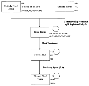
[0023] Figure 1 is a flow diagram for mitigating calcification of a
bioprosthetic tissue by blocking free amine groups on the tissue.
[0024] Figure 2 is a flow diagram for mitigating calcification of a
bioprosthetic tissue by reducing aldehyde and acid groups coupled to the
tissue.
DETAILED DESCRIPTION OF THE INVENTION
[0025] It has previously been reported that cross-linked
bioprosthetic
tissue post-treated in 0.625% glutaraldehyde phosphate solution for 2 months
at
CA 02601524 2013-04-10
8
50 C, with fluid movement (e.g., shaking), exhibited less calcification in
the rat subcutaneous
and rabbit intramuscular implant models than control cross-linked
bioprosthetic tissue fixed in
0.625% glutaraldehyde phosphate solution under typical conditions (i.e., room
temperature for
1-14 days). See U.S. Patent No. 5,931,969.
[0026] It has also been previously reported that it is advantageous to conduct
the heating step
on the glutaraldehyde solution prior to its contact with the tissue. See U.S.
Publication Serial
No. 2003/0125813. The heat-treated glutaraldehyde may then be cooled to a
lower temperature
and the tissue may then be added to the cooled glutaraldehyde solution under
conditions of
reduced severity, greater convenience, or both (e.g., shorter time, lower
temperature, or both).
By heat- treating the glutaraldehyde solution in the absence of the tissue,
higher temperatures,
concentrations or both can be used during the heat-treating process without
risking or causing
any adverse effect on the tissue.
[0027] It was also reported in U.S. Publication Serial No. 2003/0125813 that
alternatively the
glutaraldehyde solution can be buffered, rather than heat- treated, by
adjusting the pH of the
solution to within a range of about 5.0 to 7.0, preferably about 6Ø The
buffered glutaraldehyde
solution has a similar, although slightly less, advantageous effect as the
heat-treated
glutaraldehyde solution.
[0028] Since the above disclosures, Applicants have discovered that it is
advantageous to
remove carboxylic acid residues and aldehyde residues, which have the
potential to be oxidized
to carboxylic acids, from the fixed tissue, since it has been proposed that
the carboxylic moiety
attracts calcium ions and contributes towards initiating calcification of
bioprosthetic tissue.
Accordingly, in one method of this invention a tissue is immersed in or
otherwise contacted
with a glutaraldehyde solution (less than about 5% by weight). The tissue is
at least partially
fixed prior to, after, or concurrently with the step of contacting the tissue
with the
gluteraldehyde, wherein the tissue is fixed by immersing the
#10904527 v1
CA 02601524 2007-09-19
WO 2006/104820
PCT/US2006/010540
9
tissue in a solution containing gluteraldehyde as a crosslinking agent.
Contact
with the solution in this manner hydrolyzes labile Schiff base bonds located
at
or near the surface of the collagen superhelix of the tissue, thus removing
aldehyde and acid groups coupled to the tissue via the Schiff base bonds and
producing free amine groups on the tissue. The Schiff base bonds deeper within
the superhelix are sterically protected and therefore are not hydrolyzed. The
free
amine groups are then blocked in a subsequent step by contacting the
crosslinked tissue with a blocking agent.
[0029] In an alternative embodiment of the present invention, the
tissue
is treated with a pretreated glutaraldehyde that is prepared by adjusting the
pH
of the glutaraldehyde solution to a pH within the range of about 5.0 to 7.0,
and
preferably to about 6Ø The tissue is at least partially fixed prior to,
after, or
concurrently with the step of contacting the tissue with the pH adjusted
gluteraldehyde, wherein the tissue is fixed by immersing the tissue in a
solution
containing gluteraldehyde as a crosslinking agent. The pH-adjusted
glutaraldehyde solution is then used to treat the tissue, preferably at a
temperature in the range of about 30 to 70 C, more preferably at a
temperature
between about 40 to 60 C, and most preferably, at a temperature of about 45 to
55 C. In a preferred embodiment, the tissue is treated for a period of time
between about one hour to six months, and more preferably for about one day to
two months. Contact with the pH adjusted glutaraldehyde in this manner
hydrolyzes labile Schiff base bonds located at or near the surface of the
collagen
superhelix, thus removing aldehyde and acid groups coupled to the tissue via
the Schiff base bonds and producing free amine groups on the tissue. The free
amine groups are then blocked in a subsequent step by contacting the
crosslinked tissue with a blocking agent.
[0030] In yet another embodiment of a method of the present
invention,
the tissue is contacted with an untreated or pH adjusted glutaraldehyde
solution
without heating for a period of time sufficient to promote crosslinking. The
CA 02601524 2007-09-19
WO 2006/104820
PCT/US2006/010540
crosslinked tissue is then treated with a reducing agent that reduces aldehyde
and carboxylic acid groups coupled to the fixed tissue.
[0031] A. Method
for Mitigating Calcification of Bioprosthetic
Material using Pretreated Glutaraldehyde
5 [0032]
Figure 1 is a flow diagram that generally illustrates one
embodiment of the method of the present invention. As shown in Figure 1, the
first step of the process is to prepare a pretreated glutaraldehyde solution,
e.g., a
heat-treated or pH adjusted glutaraldehyde, in the absence of tissue.
[0033] 1.
Preparation of heat-treated Glutaraldehyde in Absence of
10 Tissue
[0034] Briefly,
a heat-treated glutaraldehyde solution is prepared in the
absence of the tissue by heating the solution to a first temperature for a
first
period of time. The temperature of the glutaraldehyde solution is then
adjusted
to a second temperature (preferably lower than the first temperature) before
contacting the tissue. However, this step may also be carried out with an
aqueous solution or a solution of unheated glutaraldehyde.
[0035] It will
be appreciated that the concentration of glutaraldehyde in
the starting solution may be varied. Thereafter, the solution concentration
may
be adjusted, if desired, prior to addition of the tissue. It is believed that
glutaraldehyde concentrations of as little as 0.1% and as much as 25% or more
may be used during the heat-treating step. Reduced
glutaraldehyde
concentrations of 0.6% to 2.5% have, to date, been successfully obtained and
used by Applicant, and those skilled in the art will recognize that higher or
lower concentrations of glutaraldehyde may indeed prove to be advantageous
during the heat-treating step of the process. The preferred concentration for
use
during the heat-treating step (Figure 1) is 1.0-2.0%. This heat-treatment of
the
glutaraldehyde may be accomplished by heating of the solution until the free
aldehyde content of the solution has fallen about 25% or more and remains
stable at that level (e.g., a solution of 1.8% falls to about 0.6% or less).
Initially, the solution containing glutaraldehyde may be buffered to a pH of
7.4
CA 02601524 2007-09-19
WO 2006/104820
PCT/US2006/010540
11
with a phosphate buffer, a non-phosphate buffer such as a HEPES buffer, or
other suitable buffered solutions, and, in such cases, heating of the solution
to
cause the free aldehyde content to fall will also cause the pH of the solution
to
fall.
[0036] In one embodiment, an aqueous solution of 1.8% by weight
glutaraldehyde is prepared in a clean, inert vessel (e.g., a vessel made of
stainless steel, plastic or borosilicate glass) and such solution is then
buffered to
the pH of approximately 7.4 by adding phosphate buffered saline solution.
[0037] The first temperature to which the glutaraldehyde is heated is
20 procedure.
[0038] The heat-treatment of the glutaraldehyde may be accomplished
by any suitable means. For example, the glutaraldehyde can be pre-heated to
and maintained at a temperature between about 20 to 90 C, preferably between
about 60 to 80 C, and most preferably 65 to 75 C for a period of time
sufficient
CA 02601524 2007-09-19
WO 2006/104820
PCT/US2006/010540
12
the step of heat treating the glutaraldehyde may take anywhere from one hour
to
six months or more depending on the temperature used, and typically between
1-14 days. The preferred method is to heat the glutaraldehyde solution to
approximately 65 to 75 C, for approximately 1 day to 2 months or until the
desired fall of at least 25% or more in free aldehyde concentration and a pH
of
approximately 6.0, are observed. Higher temperatures ranging up to
approximately 90 C may be used, and the use of such higher temperatures will
typically speed the desired fall in free aldehyde concentration and
accompanying change in pH (e.g., a solution having a starting pH adjusted to
7.4 will fall to a pH of about 6.0 after approximately 1-3 days at 90 C).
Lower
temperatures, ranging downward to approximately 20 C, may also be used, and
the use of such lower temperatures will typically cause the desired free
aldehyde
content and pH changes to take longer.
[0039] After the heat-treatment of the glutaraldehyde has been
completed the solution is filtered and cooled to a second temperature that
does
not cause damage to the tissue (e.g., about 30 to 70 C, preferably about 40 to
60 C, or most preferably at about 50 C).
[0040] Optionally, after the glutaraldehyde has been heat-treated,
the
solution is allowed to cool to about 50 C and its pH is adjusted to
approximately 7.4 by adding phosphate buffered saline or some other suitable
buffer.
[0041] 2. Preparation of pH adjusted Glutaraldehyde
[0042] In another embodiment of this invention, the glutaraldehyde
solution is not pre-heated, but rather the pH of the glutaraldehyde solution
is
adjusted to a pH within the range of about 5.0 to 7.0, and preferably to about
6Ø
[0043] 3. Harvesting and Preparation of Tissue
[0044] The desired biological tissue is harvested from a human
cadaver
or animal donor, and prepared for subsequent fixation and treatment. The
tissue
is typically harvested by surgical cutting or removal from its host animal.
CA 02601524 2013-04-10
13
Thereafter, it is typically trimmed or cut to size and washed with sterile
water, basic salt
solution, saline or other suitable washing solution.
[0045] In one embodiment, the tissue may be heat treated in a surfactant
solution (e.g., Tween 80
with or without ethanol and/or formaldehyde) or in a physiologic solution
(e.g. saline or a
balanced salt solution) prior to fixation at a temperature between about 37 C
and 60 C,
preferably about 45 C, for about one hour to six months, preferably about one
to 15 days, and
then heat treated in a heat treated glutaraldehyde solution as described
above.
[0046] In one embodiment, the tissue is treated with a surfactant prior to
fixation to remove
lipids, fatty acids, cholesterol, etc. to ensure that the tissue will be fixed
throughout rather than
merely on the surface. However, care must be taken not to overdo the cleaning
action and
thereby damage the base tissue by using too strong a solution. Thus, it is
preferred to use the
surfactant in the form of an aqueous solution containing 0.5 to 6% by weight
of surfactant. A
suitable treatment time is from two to six hours, preferably about three hours
(See U.S. Patent
No. 4,553,974). The surfactant may be an anionic surfactant, a non-ionic
surfactant, an
amphoteric surfactant or a mixture thereof. Examples of suitable anionic
surfactants are sodium
dodecyl sulfate, sodium dodecyl sulfoacetate and sodium salt of alkaryl
polyether sulfonate.
Examples of suitable non-ionic surfactants are octylphenoxy polyethoxy ethanol
(Triton X-
100Tm), polyoxyethylene (20) sorbitan monooleate (Tween 80), polyoxyethylene
(20) sorbitan
monostearate (Tween 60). Examples of suitable amphoteric surfactants are
sulfobetaines
commonly known as ZwittergentsTM.
[0047] Alternatively, lipids are removed by immersing the tissue in a high
osmolality aqueous
solution, such as a solution of a salt and a sugar, wherein the salt is
capable of penetrating the
sample and the sugar functions to maintain the high osmolality of the solution
as described in
U.S. Patent No. 6,350,732. Examples of suitable salts include, but are not
limited to, sodium
chloride and potassium
#10904527 v1
CA 02601524 2007-09-19
WO 2006/104820
PCT/US2006/010540
14
chloride, and examples of suitable sugars include, but are not limited to,
sucrose
and fructose.
[0048] 4. Fixation of Biological Tissue
[0049] The biological tissue may be fixed prior to, during, or after
its
treatment with the pretreated glutaraldehyde. In the example illustrated in
Figure 1, the tissue is fixed prior to undergoing the treatment with
pretreated
glutaraldehyde. In this example the fixation is carried out by immersing the
tissue in a solution of 0.625% by weight glutaraldehyde buffered to a pH of
approximately 7.4 by a suitable buffer such as a phosphate buffer, for 1-14
days
at ambient temperature.
[0050] Preferably, tissue fixation is carried out by immersing the
tissue
in a solution comprising a glutaraldehyde solution that has a low acid-forming
potential, such as high purity glutaraldehyde monomer (molecular weight (MW)
100), high purity glutaraldehyde dimer (MW 182), a mixture of the two, low
acid dialyzed or commercial gluteraldehyde.
[0051] In order to enhance fixation or sterilization, other chemical
compounds such as surfactants (e.g. Tween 80) and/or ethanol and/or
formaldehyde can be added to the glutaraldehyde.
[0052] After the tissue is removed from the fixative solution, it is
thoroughly rinsed with saline solution, basic salt solution, free
glutaraldehyde
solution, or some other suitable washing solution.
[0053] 5. Heat treatment of unfixed, partially-fixed, or fixed tissue
[0054] The unfixed, partially fixed, or fixed tissue is then
contacted with
a pretreated glutaraldehyde solution (either heat-treated or pH adjusted)
prepared as described above. Tissue that has been "fully fixed" in this regard
means that the tissue has been fixed to an extent suitable for use as an
implant,
while "partially fixed" means that the tissue has been fixed to some extent
short
of being fully fixed.
[0055] The tissue treatment step according to the example in Figure 1
is
preferably accomplished by immersing fixed, partially fixed or unfixed tissue
in
CA 02601524 2007-09-19
WO 2006/104820
PCT/US2006/010540
the pretreated glutaraldehyde solution while maintaining the solution at about
30 to 70 C, preferably about 40 to 60 C, or most preferably at about 50 C,
with
or without fluid movement. It is preferable that the pH of the solution be
left at
about 6.0 prior to placement of the tissue within the solution. Thereafter,
the
5 temperature of the solution is maintained at approximately 50 C with the
tissue
immersed in the solution to allow the pretreated glutaraldehyde solution to
interact with or modify the tissue. The tissue's susceptibility to post-
implant
calcification will be significantly reduced after immersion in the pretreated
glutaraldehyde for as little as one hour to as much as six months or more
10 (depending primarily on the temperature used), but typically occurs
within 1 to
15 days at 50 C. Thereafter, the tissue is removed from the solution. The
tissue
is typically brown in color at this time. After it has been removed from the
pretreated glutaraldehyde solution, the tissue is thoroughly rinsed with
saline
solution, basic salt solution, or some other suitable washing solution.
15 [0056] 6. Blocking free amine groups
One end result of treating the tissue with glutaraldehyde is the
hydrolysis of the carbon-nitrogen double bonds of the less stable Schiff base
bonds on and/or near the surface of the tissue, thereby simultaneously
removing
aldehyde and acid groups that were coupled to the tissue via the Schiff base
bonds. This is desirable, since the unreacted aldehyde groups can become
oxidized to carboxylic moieties, which then attract calcium ions in vivo and
contribute toward initiating calcification. However, the treatment with
glutaraldehyde results in a cross-linked tissue with free amine residues at
and/or
near the surface of the tissue.
Because since hydrolysis of the Schiff base bonds also results in the
presence of primary amine residues on the tissue that could react with
glutaraldehyde present in the post-sterilization and storage solutions (Figure
1),
the present inventors discovered that it is advantageous to contact the
primary
amines with a solution comprising a blocking reagent that will react with and
block the primary amine, thus avoiding reactions between free amines and
CA 02601524 2013-04-10
16
glutaraldehyde that is present in solutions used in subsequent steps according
to this invention.
This in turn reduces the amount of free aldehyde groups coupled to the tissue
that could
potentially get oxidized to acids. Therefore, after treatment with the
pretreated glutaraldehyde,
the tissue is rinsed and then contacted with a blocking agent (Figure 1). As
used herein, a
"blocking agent" is any compound having a functional group or chemical moiety
that is
sufficiently reactive with an amine group. Blocking agents reactive with an
amine group and
suitable for use in this invention include, but are not limited to,
monoaldehydes (i.e., a molecule
containing a single aldehyde functionality, such as formaldehyde), sugars,
water-soluble
polyepoxys such as ethylene glycol diglycidyl ether (also know as DenacolTm),
collagen, and any
other agents known in the art that contain amine reactive functionalities,
provided the product
of the reaction between the amine group and the blocking agent does not
contain a free
aldehyde or carboxylic acid group.
[0057] For example formaldehyde, which has a single aldehyde functional group,
can react with
a primary amine on the tissue to form a Schiff base bond wherein the nitrogen
of the primary
amine forms a double bond with the formaldehyde carbonyl carbon. In contrast
to the reaction
between a primary amine and glutaraldehyde, a Schiff base bond formed from a
reaction
between a primary amine and formaldehyde does not have a free aldehyde moiety
that can
become oxidized to a carboxylic acid, and therefore blocking the amine with
formaldehyde will
not increase the propensity of the tissue towards calcification post-
implantation. A further
advantage of utilizing formaldehyde as the blocking reagent is that the Schiff
base bond will
slowly hydrolyze post-implantation, thereby releasing formaldehyde into the
region
surrounding the implanted tissue. The slow-released formaldehyde will depress
hyperplasia on
the tissue implant (i.e., an abnormal increase in the number of tissue cells)
and therefore will
reduce or prevent an overgrowth of tissue on the implanted bioprosthetic
tissue.
#10904527 v1
CA 02601524 2007-09-19
WO 2006/104820
PCT/US2006/010540
17
[0058] Another example of a suitable blocking agent is a polyglycidyl
ether, which readily reacts with amines. Examples of polyglycidyl ether
blocking agents include, but are not limited to, any of the various Denacols
and
their individual reactive species, including mono, di, tri, and multi-
functional
epoxides.
[0059] Sugars also react with amines and therefore are also suitable
as
blocking agents according to this invention. Suitable sugars include reducing
sugars, which can form Schiff base bonds with the free amine groups on the
tissue. Examples of reducing sugars include, but are not limited to,
glycerose,
threose, erythrose, lyxose, xylose, arabinose, ribose, allose, altrose,
glucose,
mannose, gulose, idose, galactose, talose, or any other diose, triose,
tetrose,
pentose, hexose, septose, octose, nanose or decose.
[0060] B. Method for Mitigating Calcification of Bioprosthetic
Material using Untreated or pH-adjusted Glutaraldehyde and
Subsequent Reduction
[0061] An alternative method of the present invention is illustrated
in
Figure 2.
[0062] A biological tissue is harvested from a human cadaver or
animal
donor, and prepared for subsequent fixation and treatment as described herein.
The tissue is optionally treated with a surfactant or a high osmolality
aqueous
solution prior to fixation to remove lipids, fatty acids, cholesterol, etc. to
ensure
that the tissue will be fixed throughout rather than merely on the surface as
described herein.
[0063] The tissue is then contacted with either a non-pretreated
glutaraldehyde or a pH-adjusted glutaraldehyde solution wherein the pH is
within the range of about 5.0 to 7.0, and preferably to about 6.0 for a period
of
time sufficient to crosslink the tissue.
The crosslinked tissue is then treated with a reducing agent that reduces
aldehyde and carboxylic acid groups coupled to the fixed tissue. In this the
crosslinked tissue is treated with a reducing agent that will reduce
carboxylic
CA 02601524 2007-09-19
WO 2006/104820
PCT/US2006/010540
18
acid or potential acid-forming functional groups such as aldehydes. Removing
all or substantially all of the carboxylic acid and/or potential acid forming
functional groups on the crosslinked tissue thus removes potential nucleation
sites for calcification to occur.
[0064] Although in theory any reducing agent that will effectively
reduce carboxylic acid and aldehyde functional groups may be used for this
step, for example, hydrides, thiols, formic acid, etc., preferably the
reducing
agent is a borohydride, and more preferably sodium borohydride. Other
reducing agents include hydrogen, i.e., as used in standard reduction methods
that utilize hydrogen, typically under pressure and 1-ethy1-3-(3-
dimethylaminopropyl)carbodiimide (EDAC). It is known to those skilled in the
art to use EDAC with N-Hydroxysuccinimide (or
alternatively, N-
Hydroxysulfosuccinimide) (NHSS) to improve yields. EDAC will "reduce"
(couple) any free carbonyl group by coupling to any available amine and,
therefore, it can also be used as a blocking agent for amines.
[0065] C. Additional Calcification Mitigation Procedures
[0066] It is
known that the presence of ions such as phosphate ions tends
to increase the occurrence of calcification. Therefore, the buffers or
solutions
used in any or all of the process steps of a method of this invention
preferably
include a buffer or antimineralization solution having a level of phosphates,
sulfates, carbonates, calcium, and/or magnesium decreased to an amount
effective in reducing calcification of the tissue after implantation. In one
embodiment, the buffer is a phosphate-deficient solution. The phosphate-
deficient solution has a level of phosphate decreased to an amount effective
in
reducing calcification of said tissue after implantation, said solution
further
being non-destructive or non-destabilizing to the tissue.
Substantially
phosphate-free solutions are those containing only trace amounts of
phosphates,
as in contaminating amounts found in most chemicals used in the preparation of
conventional tissue-treating solutions. Examples
of phosphate-deficient
solutions include, but are not limited to, borate, bicarbonate, cacodylate,
CA 02601524 2013-04-10
19
HEPES, MPRS, and PIPES. Other examples of antimineralization solutions or
buffers include,
but are not limited to, sodium chloride, ascorbic acid, and glutaric acid
solutions.
[0067] In another embodiment, the buffer solutions utilized in any or all of
the process steps of
the methods of this invention include a non-isotonic buffer, that is, either
hypertonic or
hypotonic buffer, wherein the osmolality of the buffer has been adjusted to
induce the desired
tissue properties (e.g., density, modulus, tensile strength, elongation,
etc.). For example, a
hypertonic buffer (i.e., a buffer with a higher salt concentration than in
normal cells) will pull
water out of the tissue. As a result, the tissue components are pulled closer
together, which
allows them to cross-link easier and thus increases the density.
Alternatively, a hypotonic buffer
(i.e., a buffer with a lower salt concentration than in normal cells) will
swell the tissue and allow
deeper penetration of the cross-linking agent.
[0068] In another embodiment, one or more of the steps of the methods of this
invention is
performed under non-oxidizing conditions, including, but not limited to,
performing the steps
under a nitrogen blanket, low actinic safety lights, and/or mechanical covers.
[0069] D. Post-sterilization, Assembly/ Fabrication and Storage of
Bioprosthesis
[0070] 1. First Bioburden Reduction (BREP I)
[0071] After the tissue has been fixed, treated to mitigate post-implant
calcification according to
a method of this invention, and rinsed, it is subjected to a first bioburden
reduction treatment.
For Example, the tissue is immersed in or otherwise contacted with a mixture
containing i) a
crosslinking agent, ii) a denaturing agent and iii) a surfactant (i.e., a CDS
solution). One
preferred CDS solution (described in U.S. Patent No. 4,885,005 and U.S. Patent
No. 4,648,881,) is
a mixture of i) formaldehyde,
ethanol and iii) surfactant (e.g., Tween 8OTM surfactant,
available from ICI Americas, Brantford, Ontario). Such preferred CDS solution
#109045271
CA 02601524 2013-04-10
may also be referred to by the acronym "FETS" and has a preferred formulation
as follows:
Formaldehyde (4.0 0.4% by weight), Ethanol (22.0 2.2% by weight) and Tween
(80 1.2 0.2%
by weight). The tissue is preferably immersed in the CDS solution for 2 hours
to 7 days and
typically about 2 hours. During this immersion period, the CDS solution is
maintained at a
temperature of 4-50 C, and preferably at about 20-37 C.
[0072] Those skilled in the art will appreciate that various alternative
chemical compounds or
solutions may be substituted for each component of the CDS solution, as
described below.
[0073] Potential alternative denaturing agents include, but are not limited
to: alcohols/solvents:
(e.g., ethanol, or isopropyl alcohol); acidified ethers (e.g., sulfuric acid/
ether mixture, acetone,
ethers of small alkyl size such as methyl, ethyl, etc.); ketones (e.g., methyl
ethyl ketone):
commercial solvent systems (e.g., GenesolveTM (Allied Signal, Inc.,
Morristown, N.J.)); glycols
(e.g., glycerol ethylene glycol, polyethylene glycol, low molecular weight
carbowax; and high
concentration salt solutions (e.g., magnesium chloride, and sodium chloride).
[0074] Potential alternative surfactants include, but are not limited to:
[0075] a) anionic surfactants: e.g., esters of lauric acid, including but not
limited to sodium
laurel sulfate (also called sodium dodecyl sulfate); and alkyl sulfonic acid
salts (e.g., 1-
decanesulfonic acid sodium salt).
[0076] b) non-ionic compounds: e.g., compounds based on the polyoxyethylene
ether structures,
including Triton X-100, 114, 405, N-101 (available commercially from Sigma
Chemical, St. Louis,
MO) and related structures; Pluronic and Tetronic surfactants (available
commercially from
BASF Chemicals, Mount Olive, N.J.).
[0077] c) alkylated phenoxypolyethoxy alcohols: e.g., NP4OTM, NonidetTM P40,
IgepalTM,
CA63OTM, hydrolyzed/ functionalized animal and plant compounds including Tween
80, Tween
20, octyl-derivatives, octyl (3-glucoside, octyl b- thioglucopyranoside,
deoxycholate and
derivatives thereof, zwitterionic
#10904527 v1
CA 02601524 2013-04-10
21
compounds, 3 -( [cholamidopropyl] -dimethyl amino)-1-propanesulfonate (CHAPS),
3-
([cholamidopropyll-dimethyl amino)-2-hydroxy-l- propanesulfonate (CHAPSO)
(available from
Pierce Biotec Company, Rockford, IL).
[0078] The above surfactant compounds can be used individually or in mixtures
such as
deoxycholate/ Triton or commercially available mixtures such as Micro-80/
90TM.
[0079] 2. Fabrication/ Assembly
[0080] After the first bioburden reduction has been completed, the tissue may
again be rinsed
with a suitable rinsing solution such as isotonic saline or 0.625%
glutaraldehyde and
transported into a clean room or aseptic environment. Thereafter, the tissue
may be further
trimmed or shaped (if necessary) and attached to or assembled with any non-
biological
components (e.g., stents, frames, suture rings, conduits, segments of
polyester mesh to prevent
suture tear-through, etc.) to form the desired bioprosthetic device. Examples
of bioprosthetic
devices that are assembled of both biological tissue and non-biological
components include
stented porcine bioprosthetic heart valves (e.g., the Carpentier-EdwardsTM
Bioprosthesis), and
bovine pericardial heart valves (e.g., Carpentier-EdwardsTM Pericardial
Bioprosthesis), stentless
porcine aortic valves that incorporate fabric reinforcements (e.g., Edwards
PRIMA PlusTM
Stentless Aortic Bioprosthesis), and conduit valves for bio- mechanical
ventricular assist devices
(e.g., the Novacor N-100PC model), all available from Edwards Lifesciences
LLC, Irvine, CA.
[0081] 3. Second Bioburden Reduction (BREP II)
[0082] After the bioprosthesis has been fabricated and assembled it is
subjected to a second
bioburden reduction that is essentially a repeat of the first bioburden
reduction described
above, however, in this second bioburden reduction step, the solution is
preferably maintained
at about 37 C for approximately 2 hours to 10 days, preferably about 9 hours.
[0083] 4. Terminal Sterilization and Storage
#10904527 v1
CA 02601524 2013-04-10
22
[0084] After completion of the second bioburden reduction, the tissue (or
bioprosthesis) is
rinsed with a suitable rinsing solution (such as isotonic saline or 0.625%
glutaraldehyde
solution) and then placed in a terminal solution for storage and
sterilization. A preferred
terminal sterilization solution is a glutaraldehyde solution having a
concentration of about 0.2
to 1.0% by weight glutaraldehyde, and most preferably about 0.625% by weight
glutaraldehyde.
This solution has a strong sterilizing effect that can be enhanced by a
terminal heating of the
solution. Another preferred terminal sterilization solution comprises an
osmotically balanced
salt solution in combination with at least one chemical sterilant.
[0085] In one embodiment of the terminal sterilization step, the tissue (or
bioprosthesis) is
immersed in or contacted with the terminal sterilization solution and heated
for a period of time
sufficient to ensure sterility of the bioprosthesis until the time of
implantation. The period of
heating varies depending upon the temperature utilized, i.e., the lower the
temperature the
longer the period of time. For example, from 1 or 2 hours to 1 month for
temperatures between
about 50 C and 20 C, respectively. Preferably, the period of time is 1 to 6
days at 37 C or 6
hours to 2 days at 50 C, however one of skill in the art will recognize that
these temperature or
time values can be modified within the scope of the invention.
[0086] In order to avoid additional transfer and manipulation, the terminal
sterilization is
preferably carried out in the sealed storage container or package in which the
bioprosthesis will
be shipped and stored until the time of implantation. The tissue (or
bioprosthesis) is aseptically
deposited in the storage container that has been pre-filled with the 0.625%
glutaraldehyde
aqueous solution buffered to a pH of 7.4 with sodium hydroxide, such that the
tissue (or
bioprosthesis) is fully immersed in the buffered glutaraldehyde solution.
Thereafter, the
container is sealed and placed at room temperature for at least 7 days, or in
an oven at 37 C for
24 hours, or at 50 C for 6 hours to enhance the sterilization power of
glutaraldehyde.
Thereafter, the container is cooled to room temperature and shipped to the
hospital or other
location(s) where it is stored until the time of use of the bioprosthesis.
#10904527 v1
CA 02601524 2007-09-19
WO 2006/104820
PCT/US2006/010540
23
[0087] In another embodiment, the tissue is sterilized by an in-
container
terminal sterilization process comprising the steps of: providing a container
which contains a quantity of a terminal sterilant solution comprising 0.2-1.0%
by weight glutaraldehyde buffered to a pH of approximately 7.4; immersing the
tissue in the terminal sterilant solution within said container; sealing the
container; heating the container, the terminal sterilant solution and
bioprosthesis
contained therein to a temperature of about 37-50 C for a period of about six
hours to six days; cooling the container, the terminal sterilant solution and
the
bioprosthesis contained therein to room temperature; and allowing the
container
to remain sealed until it is desired to implant the bioprosthesis in a
mammalian
patient.
[0088] In another embodiment, the terminal sterilization is carried
out
before placing the tissue or bioprosthesis in the storage container.
[0089] In some cases, glutaraldehyde that has been heat-treated in
accordance with this invention may be used as the terminal solution and, in
such
cases, it may be possible to shorten or completely eliminate the previous step
of
immersing the tissue in previously heat-treated glutaraldehyde, opting instead
to
accomplish some or all of the treatment of the tissue according to the methods
of this invention until the last step of storage, i.e., concurrently with the
terminal
sterilization step.
[0090] In a preferred embodiment, the tissue with which the present
method is practiced includes substantially any mammalian tissue that is useful
in preparing a prosthetic device having a biological component thereto. For
example, in one embodiment, the tissue is derived from an organ. In another
embodiment, the tissue is selected from nerve tissue, glandular tissue (e.g.,
lymphatic tissue), respiratory tissue, digestive tissue, urinary tract tissue,
sensory tissue (e.g., cornea, lens, etc.), and reproductive tissue. In a
related
embodiment where the biological material is a biological fluid, however,
addition of liquid is not likely to be necessary, unless to dilute the ionic
strength
of the biological fluid to permit miscibility of the extraction solvent.
CA 02601524 2007-09-19
WO 2006/104820
PCT/US2006/010540
24
[0091] In presently a preferred embodiment, the tissue is selected
from
muscle tissue, adipose tissue, epithelial tissue and endothelial tissue. In
particularly preferred embodiments, the tissue is selected from myocardial
tissue and vascular tissue. In a related embodiment, the tissue is selected
from
the group including, without limitation, heart valve, venous valve, blood
vessel,
ureter, tendon, dura mater, skin, pericardium, intestine (e.g., intestinal
wall), or
periostium. In a particularly preferred embodiment, the tissue is derived from
bone, cartilage (e.g. meniscus), tendon, ligament, or any other connective
tissue.
[0092] As the source of the material used for this purpose may vary
with
regard to both tissue type, the source may also vary with regard to species
type
(autologous, homologous or heterologous tissue). The artisan will appreciate
that the methods of the present invention may be used with bioprosthetic
devices that include one or more types of tissues or materials.
[0093] In a preferred embodiment where the biological material is a
solid tissue or product, it may first be suspended in an aqueous solution so
that
it will be suitable for the extraction process. For example, brain tissue may
be
suspended in sucrose solution (e.g., 0.32 M sucrose) at 10% weight to volume.
Other hypotonic or isotonic solutions include 5% dextrose, phosphate buffered
saline, tri-buffered saline, HEPES-buffered saline, or any of the foregoing
buffers. The biological material in the aqueous solution can also be
homogenized, ground, or otherwise disrupted to maximize contact between the
treatment agents and the biological material.
[0094] In a particularly preferred embodiment, the biological
material
will form part or all of a bioprosthetic tissue that is designed and intended
for
implantation into a graft recipient.
[0095] In yet another preferred embodiment, the structural integrity
of
the tissue is maintained. Structural integrity can be defined as the ability
of
tissue to perform it's necessary biological function. The artisan will
appreciate
that the degree of structural integrity required for the tissue to perform
it's
necessary function may vary among different types of tissues. Further,
CA 02601524 2013-04-10
particular applications for which the tissue is used may require different
levels of structural
integrity.
[0096] The foregoing description is provided for the purpose of describing and
illustrating a few
exemplary embodiments of the invention only. One skilled in the art will
recognize that other
embodiments of the invention are possible, but are not described in detail
here. Thus, these
examples are not intended to limit the scope of the invention in any way.
Unless defined
otherwise, all technical and scientific terms used herein have the same
meaning as commonly
understood by one of ordinary skill in the art to which this invention
belongs. Although the
preferred methods and materials are now described any methods and materials
similar or
equivalent to those described herein can be used in the practice or testing of
the present
invention. The words "comprise," "comprising," "include," "including," and
"includes" when
used in this specification and in the following claims are intended to specify
the presence of
stated features, integers, components, or steps, but they do not preclude the
presence or
addition of one or more other features, integers, components, steps, or groups
thereof.
[0097]
[00981 While the foregoing is a complete description of the preferred
embodiments of the
invention, various alternatives, modifications, and equivalents may be used.
Moreover, it will
be obvious that certain other modifications may be practiced within the scope
of the appended
claims.
#10904527 v1