Note : Les descriptions sont présentées dans la langue officielle dans laquelle elles ont été soumises.
CA 02640794 2008-07-30
WO 2007/093431 PCT/EP2007/001360
1
A METHOD FOR OSTEOGENIC DIFFERENTIATION OF BONE MARROW STEM CELLS (BMSC) AND
USES THEREOF
Field of the Invention
In an aspect, the invention relates to in vitro or ex vivo methods for
osteogenic, and preferably
at least in part also endothelial, differentiation of bone marrow stem cells
(BMSC), and to
applications of so differentiated cells. In a further aspect, the invention
relates to particular
types and populations of cells displaying characteristics akin to but new over
previously
disclosed osteoprogenitors and osteoblasts. In related aspects, the invention
provides uses,
in particular in the field of therapy, preferably bone therapy, of the above
methods, cells and
cell populations obtainable using the methods, and of the cell types and cell
populations
specifically described herein.
Background to the Invention
The feasibility of allogeneic bone marrow transplantation was demonstrated in
children with
severe osteogenesis imperfecta (Horwitz et al. 1999. Nat Med 5(3): 309-13). In
that study,
functional marrow-derived mesenchymal cells engrafted and contributed to the
formation of
new dense bone, indicating that the transplanted cells differentiated to bone-
producing
osteoblasts. Autologous bone marrow transplantation was also reported in a
patient suffering
from osteonecrosis of the humeral head (Hernigou et al. 1997. J Bone Joint
Surg Am
79:1726-1730). Therefore, transplantation of stem cells capable of undergoing
osteogenic
differentiation or of cells that are committed towards osteogenic
differentiation may be a
promising avenue for the treatment of bone-related diseases, in particular
when the treatment
requires production of new bone.
Hence, there exists a great need for efficient techniques which provide
sufficient quantities of
cells, in particular autologous cells, suitable for transplantation as a
remedy for bone-related
disorders.
While undifferentiated bone marrow stem cells may be transplanted, these cells
are not yet
committed to an osteogenic lineage and therefore a considerable proportion of
the
transplanted stem cells may not eventually contribute to formation of bone
tissue. In addition,
it has been demonstrated (Banfi et al. 2000. Exp Hematol 28: 707-15) that in
vitro culturing of
bone marrow stem cells decreases their proliferation potential as well as
their capability to
CONFIRMATION COPY
CA 02640794 2008-07-30
WO 2007/093431 PCT/EP2007/001360
2
undergo differentiation when treated with growth factors like FGF-2. For
example, in that
study, bone forming efficiency of in vitro cultured bone marrow stem cells was
decreased 36
times already at first passage, compared to freshly isolated bone marrow.
Hence, in vitro
expansion of bone marrow stem cells may decrease their efficiency as a source
of bone-
forming osteoblasts upon transplantation.
Martin et al. (Endocrinology 138: 4456-4462, 1997) demonstrates that in vitro
culturing of
bone marrow stem cells in the presence of fibroblast growth factor type 2 (FGF-
2), in
combination with foetal calf serum (FCS) components, keeps the cells in an
immature state
(less alkaline phosphatase and fibroblast-like morphology), albeit the cells
are competent to
undergo osteogenic differentiation in vitro under specific osteogenic culture
conditions.
However, differentiation of such immature cells into bone-producing
osteoblasts in vivo still
depends on the provision of the appropriate signals upon transplantation.
Hence, although
such cells might be capable of osteogenic differentiation in vitro, a
considerable proportion
thereof may still not become osteoblasts in vivo. Also, Chaudhary et al. (Bone
34: 402-11,
2004) shows that human bone marrow stem cells treated with FGF-2 do not
demonstrate any
osteogenic phenotype (no alkaline phosphatase expression) and in fact have
dystrophic
morphology. Similarly, Kalajzic et al. (J Cell Biochem 88: 1168-76, 2003)
demonstrates that
FGF-2 inhibits osteogenic differentiation.
In addition, preparation of materials for use in human therapy should avoid
the use of non-
human animal components, such as serum components (e.g., FCS) in the culture
media.
However, as shown by Kuznetsov et al. (Transplantation 70: 1780-1787, 2000),
the use of
homologous or autologous human sera greatly diminishes the ability of human
bone marrow
stem cells to form colonies and expand in vitro, and to form bone in vivo.
Hence, Kuznetsov
et al. suggest that the use of FCS is prerequisite for efficient expansion of
bone marrow stem
cells and for their capacity to form bone.
Takagi et al. 2003 (Cytotechnology 43: 89-96) incubated human bone marrow
aspirates in
donor serum supplemented with FGF-2, under specific conditions. The cell
population so-
obtained by Takagi et al. 2003 only showed chondrogenic differentiation
potential and was
thus contemplated by the authors for use in the regeneration of cartilage.
Kobayashi et al. 2005 (J Bone Joint Surg Br 87: 1426-3) describes particular
conditions for
isolation and maintenance of human BMSC in autologous donor serum, concluding
that these
CA 02640794 2008-07-30
WO 2007/093431 PCT/EP2007/001360
3
conditions may provide for sufficient ex vivo expansion of human BMSC, while
preserving
their multi-differentiation potential. FGF-2 is employed by Kobayashi et al.
2005 in secondary
culture to promote further BMSC expansion without differentiation. These
authors do not
disclose conditions which would cause their cells to progress towards
osteoprogenitors or
osteoblast phenotype cells.
Lin et al. 2005 (Transplant Proc 37: 4504-5) reported prolonged expansion of
multi-potential
human BMSC in autologous donor serum. Addition of FGF-2 and EGF to the cells
under
certain conditions did not influence cell proliferation and did not cause
progression of the cells
towards osteogenic fate.
In order to provide for maximum bone formation, it would be desired to
transplant cells which
already show an osteoblastic phenotype, since such cells are essentially the
only ones with a
demonstrated bone-forming activity. However, in vitro differentiation of bone
marrow stem
cells into osteoblasts involves culturing in osteogenic medium (Jaiswal et al.
1997. J Cell
Biochem 64: 295-312) and may lead to decreased proliferation of such cells in
vitro.
Moreover, the use of osteogenic medium involves addition of further components
to the cells,
which may increase the risk of contamination of the cell culture.
Hence, there exists a need in the art for a simple and reliable method to
produce
osteoprogenitors, osteoblasts or osteoblastic phenotype cells from human adult
stem cells, in
particular human bone marrow stem cells, in vitro while maintaining high
expansion capacity
of the cells, ensuring autologous conditions and minimising the number of
components
involved in culturing of the cells.
There also exists need in the art for osteoprogenitor or osteoblastic cells
having specific
useful characteristics, e.g., in the context of bone therapy, and for cell
populations comprising
such cells.
Summary of the Invention
The present invention addresses the above and other problems of the prior art.
In particular, the inventors realised that adult stem cells, in particular
bone marrow stem cells
(BMSC), advantageously of human origin, can be readily expanded ex vivo and
directed
towards useful osteoprogenitor or osteoblast phenotypes, and useful cell
populations
comprising such and other phenotypes, using herein disclosed culture
conditions.
CA 02640794 2013-12-19
3a
Various embodiments of this invention relate to an isolated cell population
comprising: (a)
human osteoblasts or osteoblast phenotype cells that co-express (1) at least
one osteoblast
marker selected from alkaline phosphatase (ALP) of the bone-liver-kidney type,
procollagen
type 1 amino-terminal propeptide (P1NP) and bone sialoprotein (BSP) with (2)
at least one
stem cell / immature osteoprogenitor marker selected from CD63 and CD166; and
(b)
endothelial cells or progenitors thereof which are CD34 positive and further
express one or
more of: von Willebrand factor (vWF), VEGF and CD133, said isolated cell
population
comprising at least 80% of human osteoblasts or osteoblast phenotype cells as
defined under
(a), and wherein the endothelial cells or progenitors thereof as defined under
(b) constitute
less than 20% of all cells defined under (a) and (b).
Various embodiments of this invention relate to an isolated cell population
comprising: (a)
human osteoblasts or osteoblast phenotype cells that co-express (1) at least
one osteoblast
marker selected from alkaline phosphatase (ALP) of the bone-liver-kidney type,
procollagen
type 1 amino-terminal propeptide (P1NP) and bone sialoprotein (BSP) with (2)
the
hematopoietic / endothelial progenitor marker CD34; and (b) endothelial cells
or progenitors
thereof which are CD34 positive and further express at least one of von
Willebrand factor
(vWF), VEGF and CD133; said isolated cell population comprising at least 50%
of human
osteoblasts or osteoblast phenotype cells as defined under (a), and wherein
the endothelial
cells or progenitors thereof as defined under (b) constitute less than 20% of
all cells defined
under (a) and (b).
CA 02640794 2008-07-30
WO 2007/093431 PCT/EP2007/001360
4
Accordingly, in an aspect, the invention relates to a method for obtaining
osteoprogenitors,
osteoblasts or osteoblast phenotype cells from human bone marrow stem cells in
vitro or ex
vivo, comprising contacting the bone marrow stem cells with human plasma or
serum and a
growth factor or a biologically active variant or derivative thereof.
The inventors observed that methods of the invention can differentiate a
substantial fraction,
e.g., a majority, of exposed stem cells toward the osteoprogenitor or
osteoblast phenotypes.
Consequently, in an aspect the methods of the invention can be employed for
obtaining
osteoprogenitors, osteoblasts or osteoblast phenotype cells per se.
Nevertheless, it can be appreciated that the methods of the invention
generally produce cell
populations comprising osteoprogenitors, osteoblasts or osteoblast phenotype
cells, usually
populations comprising a substantial portion, e.g., a majority, of such cells.
The inventors also
realised that cell populations resulting from the method may comprise further
cell types, at
least some of which can augment the useful characteristics of the
osteoprogenitor or
osteoblast phenotype cells present in such populations, in particular in the
context of bone
therapy. For example, in an embodiment a cell population resulting from the
methods of the
invention may also comprise endothelial cells or endothelial progenitors.
Accordingly, in an aspect, the invention relates to a method for obtaining a
cell population
comprising osteoprogenitors, osteoblasts or osteoblast phenotype cells from
human bone
marrow stem cells in vitro or ex vivo, comprising contacting the bone marrow
stem cells with
human plasma or serum and a growth factor or a biologically active variant or
derivative
thereof.
As shown by experimental evidence, the method of the invention may provide for
expansion
of the bone marrow stem cells between 40,000 to 710,000 times over three
weeks, more
particularly twenty one days. Such high degree of expansion is surprising in
view of prior art
teaching that the use of human serum markedly diminishes expansion of human
bone
marrow stem cells (Kuznetsov et al. 2000). Hence, the present invention allows
for generation
of a high number of cells for the purposes of transplantation. This
advantageously decreases
the size of the bone marrow sample which needs to be drawn from a subject in
order to
provide for the stem cells. In addition, the invention allows for shortening
the time when the
differentiated cells can be transplanted into a patient, thus resulting in
faster therapy.
CA 02640794 2008-07-30
WO 2007/093431 PCT/EP2007/001360
In a preferred embodiment, the method uses fibroblast growth factor and, in
particular, FGF-
b, i.e., FGF-2. It is surprising, in view of the prior art teaching that FGF-2
causes a more
immature phenotype of bone marrow stem cells (Martin et al. 1997, Kalajzic et
al. 2003), that
the use of FGF-2 in combination with human plasma or serum components
stimulates
5 differentiation of bone marrow stem cells to attain the phenotypic
characteristics of
osteoprogenitors, osteoblasts or osteoblast phenotype cells.
Hence, the present method provides for unexpected advantageous effect - high
expansion
and osteoblast phenotype - by combining elements which have been known in
prior art to
provide for opposite effects when used separately. Even more strikingly, the
prior art taught
that in vitro differentiation into osteoblasts requires osteogenic medium,
containing
components such as dexamethasone, ascorbic acid phosphate and beta-
glycerolphosphate.
The present invention surprisingly shows that such components are not needed
for obtaining
osteoprogenitors, osteoblasts or osteoblast phenotype cells. Hence, the number
of
components in a medium may be advantageously decreased, resulting in less
chances of
error or contamination, or carryover of such components upon transplantation.
In further preferred embodiments, the method uses human plasma or serum which
is
autologous to the bone marrow stem cells and/or does not include any non-human
animal
material (such as serum components) in the culture of bone marrow stem cells.
This makes
the method particularly advantageous for use in human therapy, e.g., by
decreasing the risk
of rejection of the obtained cells and/or by decreasing the risk of
contamination with
pathogens.
Additional preferred embodiments of the method define other features, e.g.,
without limitation,
incubation times, passages, component quantities, etc., which alone or in
combination further
delimit the method from prior art and underlie the provision of cells and cell
populations of
advantageous characteristics, e.g., of superiority in bone transplantation.
The inventors realised that osteoprogenitors, osteoblasts or osteoblast
phenotype cells, as
well as cell populations, obtainable using the methods of the invention show
exemplary
advantages over the prior art. Firstly, at least during the ex vivo culturing,
the said cells show
a fast proliferation rate, with an estimated doubling time of approximately 2
days. Hence,
sufficient numbers of the cells can be generated within comparably short time,
which
advantageously limits the patient treatment periods. Secondly, the cells show
a relatively fast
CA 02640794 2008-07-30
WO 2007/093431 PCT/EP2007/001360
6
rate of substrate mineralization, which allows for enhanced bone-formation
upon
transplantation of the cells into patients. Third, the cells display little or
substantially no
propensity for differentiation towards other mesenchymal phenotypes, in
particular towards
adipocytes or chondrocytes. This can advantageously limit the formation of
tissue other than
bone when the cells are transplanted.
Accordingly, in other aspects, the invention provides for osteoprogenitors,
osteoblasts or
osteoblast phenotype cells, as well as for cell populations and cultures
comprising such,
which are obtainable or directly obtained using the methods of the invention,
and also for
therapeutic uses thereof in bone-related disorders and corresponding
pharmaceutical
formulations comprising such.
In a further development of the invention, the inventors analysed in detail
the cells and cell
populations obtained carrying out the methods of the invention, in order to
define new
osteogenic cell types and new cell populations comprising such, that may offer
particular
superiority in therapy, especially in bone transplantation therapy.
Consequently, the invention
also contemplates such new cell types, populations comprising such, as well as
uses thereof,
especially in bone therapy.
Accordingly, in an aspect, the invention provides osteoprogenitor, osteoblast
or osteoblast
phenotype cells (herein, "OOP-1 cells") characterised in that they co-express
(1) at least one
osteoblast marker chosen from alkaline phosphatase (ALP), more specifically
ALP of the
bone-liver-kidney type, procollagen type 1 amino-terminal propeptide (P1NP)
and bone
sialoprotein (BSP) with (2) at least one stem cell / immature osteoprogenitor
marker chosen
from CD63 (by means of example, as recognised by antibody HOP-26; see
Zannettino et al.
2003. J Cell Biochem. 89: 56-66) and CD166. Under (1): in a preferred
embodiment, the said
00P-1 cells may express at least ALP; in further preferred embodiments, the
said 00P-1
cells may express at least two markers chosen from ALP, P1NP and BSP, e.g., at
least ALP
and P1NP, at least ALP and BSP or at least P1NP and BSP; in a yet further
preferred
embodiment, the said 00P-1 cells may express at least all three of ALP, BSP
and P1NP.
Under (2): in a preferred embodiment, the said 00P-1 cells may express at
least CD63; in
another preferred embodiment, the said 00P-1 cells may express at least CD166;
in a further
preferred embodiment, the said 00P-1 cells may express at least CD63 and
CD166.
CA 02640794 2008-07-30
WO 2007/093431 PCT/EP2007/001360
7
To the inventors' best knowledge the prior art only observed CD63 and/or CD166
in stem
cells / immature osteoprogenitors when ALP, P1NP and BSP were negative. Such
CD63
and/or CD 166 positive cells of prior art showed multi-potency and could
differentiate to
chondrocytes, adipocytes as well as osteoblasts. Hence, the concomitant
presence of at least
one of ALP, P1NP or BSP with CD63 and/or CD166 marks the osteoprogenitor or
osteoblast
phenotype cells of the invention (00P-1 cells) as a new, previously
undisclosed cell type.
Moreover, this new cell type displays at least some of advantageous properties
such as high
proliferation rate, high mineralization rate and substantially absent
propensity towards
chondrocytic and adipocytic differentiation, which are particularly useful,
e.g., in bone
therapeutic context.
In a preferred embodiment, the said osteoprogenitor or osteoblast phenotype
cells (00P-1
cells) are negative for osteocalcin (OCN). It has been known in the art that
OCN becomes
expressed preferentially in mature osteoblasts. Hence, the absence of OCN
expression
signifies the less mature character of these cells.
In a further aspect, the invention provides osteoprogenitor, osteoblast or
osteoblast
phenotype cells (herein, "OOP-2 cells") characterised in that they co-express,
i.e., are
positive for, (1) at least one osteoblast marker chosen from alkaline
phosphatase (ALP), more
specifically ALP of the bone-liver-kidney type, procollagen type 1 amino-
terminal propeptide
(P1NP) and bone sialoprotein (BSP) with (2) the hematopoietic / endothelial
progenitor
marker CD34. Under (1): in a preferred embodiment, the said 00P-2 cells may
express at
least ALP; in further preferred embodiments, the said 00P-2 cells may express
at least two
markers chosen from ALP, P1NP and BSP, e.g., at least ALP and P1NP, at least
ALP and
BSP or at least P1NP and BSP; in a yet further preferred embodiment, the said
00P-2 cells
may express at least all three of ALP, BSP and P1NP.
To the inventors' knowledge, osteoprogenitor or osteoblast phenotype cells
expressing CD34
have never before been described, and the absence of CD34 is typically
considered one of
the features of bone marrow mesenchymal stem cells. Accordingly, the
concomitant presence
of at least one of ALP, P1NP or BSP with CD34 marks the osteoprogenitor or
osteoblast
phenotype cells of the invention (00P-2 cells) as a new, previously
undisclosed cell type.
Moreover, this new cell type displays at least some of advantageous properties
such as high
proliferation rate, high mineralization rate and substantially absent
propensity towards
CA 02640794 2008-07-30
WO 2007/093431 PCT/EP2007/001360
8
chondrocytic and adipocytic differentiation, which are particularly useful,
e.g., in bone
therapeutic context.
In a preferred embodiment, the said osteoprogenitor or osteoblast phenotype
cells (00P-2
cells) are negative for osteocalcin (OCN). It has been known in the art that
OCN becomes
expressed preferentially in mature osteoblasts. Hence, the absence of OCN
expression
signifies the less mature character of these cells.
The inventors also contemplate overlaps between the above described
osteoprogenitor or
osteoblast cell types of the invention. For example, in some embodiments, the
00P-1 cells
may further co-express CD34. In other embodiments, the 00P-2 cells may further
express at
least one, e.g., one or both, of CD63 and CD166.
As mentioned, the invention also encompasses cell populations comprising the
above
osteoprogenitor or osteoblast phenotype cells of the invention, e.g.,
comprising the 00P-1
and/or 00P-2 cell types discussed above. An exemplary cell population may
comprise at
least 10%, preferably at least 30%, more preferably at least 50%, e.g., at
least 60%, yet more
preferably at least 70%, e.g., at least 80%, and even more preferably at least
90%, e.g., at
least 95% of the 00P-1 and/or 00P-2 cell types. In preferred embodiments, the
cell
population may comprise less than 50%, preferably less than 40%, even more
preferably less
than 30%, yet more preferably less than 20% and still more preferably less
than 10%, e.g.,
less than 7%, less than 5% or less than 2% of cell types other than the above
00P-1 and/or
00P-2 cell types.
In a preferred embodiment, the said cell population may also comprise
endothelial cells or
progenitors thereof. Preferably, such endothelial cells or progenitors may
express at least
one, e.g., at least two, or at least all three, of von Willebrand factor (vWF)
VEGF and CD133.
In a further embodiment, the said endothelial cells can also co-express CD34.
Advantageously, the inventors have realised that the presence of such
endothelial cells or
progenitors thereof in a cell population alongside osteoprogenitors or
osteoblast phenotype
cells of the invention may improve the engraftment of the said osteogenic
lineage cells in
patients, presumably, but without limitation, by instigating the formation of
vessels supporting
and oxygenating the implanted cells and tissues and/or by releasing growth
factors, such as,
e.g., VEGF.
CA 02640794 2013-12-19
9
In related aspects, the invention provides pharmaceutical formulations
comprising the above
defined cells and cell populations, and therapeutic uses thereof.
These and other features of the invention are further explained here below and
in the
appended claims, as well as illustrated by non-limiting examples.
Brief Description of the Figures
Figure 1 shows results of injection of a cell population prepared according to
the present
invention in a patient with osteonecrosis of the femoral head (solid line with
diamonds). A: VAS
score; B: WOMAC score. "B"-baseline, "3m"-3 months, "6m"-6 months, dashed line-
historical
controls (control biopsy).
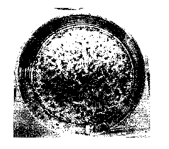
Figure 2 shows mineralization by the cells / populations of the invention.
Detailed Description of the Invention
As used herein, the singular forms "a", "an", and "the" include both singular
and plural
referents unless the context clearly dictates otherwise. By way of example, "a
cell" refers to
one or more than one cell.
The terms "comprising", "comprises" and "comprised of as used herein are
synonymous with
"including", "includes" or "containing", "contains", and are inclusive or open-
ended and do not
exclude additional, non-recited members, elements or method steps.
The recitation of numerical ranges by endpoints includes all numbers and
fractions subsumed
within that range, as well as the recited endpoints.
The term "about" as used herein when referring to a measurable value such as a
parameter,
an amount, a temporal duration, and the like, is meant to encompass variations
of +/-20% or
less, preferably +/-10% or less, more preferably +/-5% or less, even more
preferably +/-1% or
less, and still more preferably +/-0.1% or less from the specified value,
insofar such variations
are appropriate to perform in the disclosed invention.
CA 02640794 2008-07-30
WO 2007/093431 PCT/EP2007/001360
Unless otherwise defined, all terms used in disclosing the invention,
including technical and
scientific terms, have the meaning as commonly understood by one of ordinary
skill in the art
to which this invention belongs.
1. Methods of invention
5 As detailed in the Summary section, in an aspect, the invention relates
to a method for
obtaining osteoprogenitors, osteoblasts or osteoblast phenotype cells, as well
as for obtaining
cell populations comprising osteoprogenitors, osteoblasts or osteoblast
phenotype cells, from
human bone marrow stem cells in vitro or ex vivo, comprising contacting the
bone marrow
stem cells with human plasma or serum and a growth factor or a biologically
active variant or
10 derivative thereof.
Bone marrow is the soft tissue occupying medullar cavities of long bones, some
haversian
canals, and spaces between trabeculae of cancellous or spongy bone. Two types
of bone
marrow are conventionally distinguished: red, which is found in all bones in
early life and in
restricted locations in adulthood (e.g., in the spongy bone) and is primarily
concerned with the
production of blood cells (haematopoiesis); and yellow, which comprises
primarily fat cells
and connective tissue.
As a whole, bone marrow is a complex tissue comprised of hematopoietic stem
cells, red and
white blood cells and their precursors, mesenchymal stem cells (MSC), stromal
cells and their
precursors, and a group of cells including fibroblasts, reticulocytes,
adipocytes, and cells
which form a connective tissue network called "stroma".
Bone marrow cells contribute to many diverse tissues after systemic
transplantation in both
mice and humans. This capacity may reflect the activities of multiple stem
cells present in
bone marrow, such as, e.g., haematopoietic stem cells, mesenchymal stem cells
and/or
marrow multipotent stem cell. For example, Krause et al. (Cell 105: 369-377,
2001) showed
that a single bone marrow derived stem cell can generate cells of both the
haematopoietic
and non-haematopoietic lineages. This is confirmed by Dominici et al. (PNAS
101(32): 11761-
6, 2004) who showed that hematopoietic cells and osteoblasts can be derived
from a
common marrow progenitor after bone marrow transplantation. In another
example, US Pat.
5,486,359 discloses the isolation from bone marrow of mesenchymal stem cells,
capable of
generating cells of mesenchymal lineages, e.g., of bone, cartilage, muscle,
tendon,
connective tissue, fat or marrow stroma. Further, Horwitz et al. (Nat Med
5(3): 309-13, 1999)
CA 02640794 2008-07-30
WO 2007/093431 PCT/EP2007/001360
11
showed that allogeneic bone marrow transplantation is effective in children
with severe
osteogenesis imperfecta. In that study, functional marrow-derived mesenchymal
cells
engrafted and contributed to the formation of new dense bone. As shown by
Horwitz et al.
(PNAS 99(13): 8932-7, 2002), the percentage of grafted osteoblasts was not
significantly
improved after the transplantation of only mesenchymal stem cells (plastic-
adherent bone
marrow cells), leading to the conclusion that bone marrow cells other than
those in the
adherent population, where mesenchymal stem cells are thought to reside, can
be potent
transplantable progenitors of osteoblasts. In view of the above, bone marrow
may contain
several types of stem cells with the potential to generate cells of the
osteocytic (bone)
lineage.
The term "bone marrow stem cell" or "BMSC" as used herein thus refers to any
adult stem
cell present in bone marrow, and particularly present in or (partly) isolated
from a sample of
bone marrow. A sample of bone marrow (BMSC) may be obtained, e.g., from iliac
crest,
femora, tibiae, spine, rib or other medullar spaces of a subject. The term
BMSC also
encompasses the progeny of BMSC, e.g., progeny obtained by in vitro or ex vivo
propagation
of BMSC obtained from a sample of a subject.
The term "stem cell" as used herein denotes any cell that, if exposed to
appropriate
conditions, is capable of giving rise to at least one, and preferably two or
more different cell
types. Such a stem cell may be capable of extensive, or perhaps indefinite,
proliferation in
vivo and, under specific conditions, also in vitro, wherein the progeny of
such a stem cell may
retain the phenotypic features and the proliferative capacity of the mother
cell, or else may, if
exposed to appropriate conditions, give rise to more specialized, i.e. more
differentiated,
cell(s). A stem cell is said to "give rise" to another, more differentiated,
cell when, for example,
the stem cell differentiates to become the other cell without previously
undergoing cell
division, or if the other cell is produced after one or more rounds of cell
division and/or
differentiation of the stem cell.
The term "adult stem cell" as used herein refers to a stem cell present in or
obtained from an
organism at the foetal stage or after birth.
Preferable bone marrow stem cells according to the invention have the
potential of generating
cells of at least the osteogenic (bone) lineage, such as, e.g., osteogenic
cells and/or
osteoprogenitors and/or pre-osteoblasts and/or osteoblasts and/or osteocytes,
etc.
CA 02640794 2008-07-30
WO 2007/093431 PCT/EP2007/001360
12
Preferably, at least some bone marrow stem cells according to the invention
may also have
the potential to generate further cells comprised in the cell populations
resulting from the
methods of the invention, such as, e.g., cells of endothelial lineage, for
example endothelial
progenitor cells and/or endothelial cells.
An exemplary, but non-limiting, type of BMSC having the potential of
generating cells of at
least the osteogenic lineage are mesenchymal stem cells. The term "mesenchymal
stem cell"
or "MSC" (also known as "marrow stromal cells") as used herein refers to an
adult,
mesoderm-derived stem cell that is capable of generating cells of mesenchymal
lineages,
typically of two or more mesenchymal lineages, e.g., osteocytic (bone),
chondrocytic
(cartilage), myocytic (muscle), tendonocytic (tendon), fibroblastic
(connective tissue),
adipocytic (fat) and stromogenic (marrow stroma) lineage. MSC may be isolated
from, e.g.,
bone marrow, blood, umbilical cord, placenta, foetal yolk sac, skin (dermis),
specifically foetal
and adolescent skin, periosteum and adipose tissue. Human MSC, their
isolation, in vitro
expansion, and differentiation, have been described in, e.g., US Pat. No.
5,486,359; US Pat.
No. 5,811,094; US Pat. No. 5,736,396; US Pat. No. 5,837,539; or US Pat. No.
5,827,740. Any
MSC described in the art and isolated by any method described in the art may
be suitable in
the present invention, provided such MSC are capable of generating cells of at
least the
osteocytic (bone) lineage, such as, e.g., osteogenic cells and/or
osteoprogenitors and/or pre-
osteoblasts and/or osteoblasts and/or osteocytes, etc.
Potentially, but without limitation, at least some MSC might also be able to
generate further
cells comprised in the cell populations resulting from the methods of the
invention, such as,
e.g., cells of endothelial lineage, for example endothelial progenitor cells
and/or endothelial
cells.
The term MSC also encompasses the progeny of MSC, e.g., progeny obtained by in
vitro or
ex vivo propagation of MSC obtained from a biological sample of an animal or
human subject.
As shown in the examples, the present method of entails selecting those BMSC
cells which,
upon contacting with human plasma or serum and a growth factor or a
biologically active
variant or derivative thereof, adhere to a substrate surface, e.g., the
surface of the culture
vessel. It is known in the art that MSC can be isolated from bone marrow (or
other sources)
by selecting those (mononuclear) cells which can adhere to a substrate
surface, e.g., plastic
surface (indeed, MSC are sometimes referred to as plastic-adherent cells or
colony forming
CA 02640794 2013-12-19
13
unit fibroblasts). Therefore, without being limited to any hypothesis, the
present inventors
speculate that in the present method, MSC may at least partly contribute to
obtaining of
osteoblasts or osteoblast phenotype cells from BMSC.
Therefore, in an aspect, the present invention also contemplates a method for
obtaining
osteoblasts or osteoblast phenotype cells from human mesenchymal stem cells in
vitro or ex
vivo, comprising contacting the MSC with human plasma or serum and a growth
factor.
MSC may be comprised in a biological sample, e.g., in a sample comprising
BMSC, or may be
at least partly isolated therefrom as known in the art. Moreover, MSC may be
at least partly
isolated from bone marrow or from sources comprising MSC other than bone
marrow, e.g.,
blood, umbilical cord, placenta, foetal yolk sac, skin (dermis), specifically
foetal and adolescent
skin, periosteum and adipose tissue.
In a preferred embodiment, BMSC or MSC present in or at least partly isolated
from the
biological sample may be contacted with human plasma or serum and a growth
factor or a
biologically active variant or derivative thereof, without prior propagation
in conditions which
allow for cell growth and doubling of BMSC or MSC without differentiation.
It is further known that preparations of MSC from bone marrow comprise a
subpopulation of
cells which are small, proliferate rapidly, undergo cyclical renewal when re-
plated at low
density and are precursors of more mature MSC in the same culture. This
subpopulation of
cells is termed "rapidly self-renewing cells" and may have at least two
components identified
as RS-1 and RS-2 (Colter et al. PNAS 97(7): 3213-8, 2000). Therefore, without
being limited to
any hypothesis, the present inventors speculate that in the present method, RS
cells as
described by Colter et al. 2000 may at least in part contribute to obtaining
of osteoprogenitors,
osteoblasts or osteoblast phenotype cells from BMSC, possibly leading through
an
intermediate of more mature MSC. Potentially, but without limitation, the
inventors speculate
that RS cells might also be able to generate further cells comprised in the
cell populations
resulting from the methods of the invention, such as, e.g., cells of
endothelial lineage, for
example endothelial progenitor cells and/or endothelial cells.
Accordingly, in an embodiment, the present invention also contemplates a
method for
obtaining osteoprogenitors, osteoblasts or osteoblast phenotype cells, or for
obtaining cell
CA 02640794 2013-12-19
14
populations comprising such, from human rapidly self-renewing cells (RS) in
vitro or ex vivo,
comprising contacting the RS with human plasma or serum and a growth factor.
It is further known that bone marrow contains a precursor cell population
termed "side
population" (SP). These cells are identified as CD3ewineg hematopoietic
precursors, but have
remarkable plasticity in terms of regenerating hematopoietic as well as non-
hematopoietic
tissue (Goodell et al. 1997. Nat Med 3(12):1337-45). Therefore, without being
limited to any
hypothesis, the present inventors speculate that in the present method, SP
cells as described
by Goodell et al. 1997 may at least in part contribute to obtaining of
osteoprogenitors,
osteoblasts or osteoblast phenotype cells from BMSC, possibly leading through
an
intermediate of more mature MSC. Potentially, but without limitation, the
inventors speculate
that SP cells might also be able to generate further cells comprised in the
cell populations
resulting from the methods of the invention, such as, e.g., cells of
endothelial lineage, for
example endothelial progenitor cells and/or endothelial cells.
Accordingly, in an embodiment, the present invention also contemplates a
method for
obtaining osteoprogenitors, osteoblasts or osteoblast phenotype cells, or for
obtaining cell
populations comprising such, from human side population cells (SP) in vitro or
ex vivo,
comprising contacting the SP with human plasma or serum and a growth factor.
It is further known that bone marrow comprises a population of osteogenic
precursor cells
which are initially identified by their low density (e.g., upon density
gradient centrifugation),
non-adherent nature and low-level of expression of osteogenic markers (Long et
al. 1995. J
Clin Invest. 1995 Feb;95(2):881-7; US 5,972,703). However, as such cells are
induced to
differentiate towards osteoblasts, they also become adherent to substrate
surface. Therefore,
without being limited to any hypothesis, the present inventors speculate that
in the present
method, osteogenic precursors as described by Long et al. 1995 may at least in
part contribute
to obtaining of osteoprogenitors, osteoblasts or osteoblast phenotype cells
from BMSC.
Accordingly, in an embodiment, the present invention also contemplates a
method for
obtaining osteoprogenitors, osteoblasts or osteoblast phenotype cells, or for
obtaining a cell
population comprising such, from human osteogenic precursors (OP) in vitro or
ex vivo,
comprising contacting the OP with human plasma or serum and a growth factor.
CA 02640794 2008-07-30
WO 2007/093431 PCT/EP2007/001360
It is further known that bone marrow comprises a population of primitive
precursor cells which
can generate cells of both the haematopoietic and non-haematopoietic lineages
(Krause et al.
2001. Cell 105:369-377; Dominici et al. 2004. PNAS 101(32): 11761-6).
Therefore, without
being limited to any hypothesis, the present inventors speculate that in the
present method,
5 such primitive precursors may at least in part contribute to obtaining of
osteoprogenitors,
osteoblasts or osteoblast phenotype cells from BMSC, possibly leading through
an
intermediate of more mature MSC. Potentially, but without limitation, the
inventors speculate
that such primitive precursors might also be able to generate further cells
comprised in the
cell populations resulting from the methods of the invention, such as, e.g.,
cells of endothelial
10 lineage, for example endothelial progenitor cells and/or endothelial
cells.
It is to be understood that, given the complexity of bone marrow stem cell
populations, the
present invention should not be seen as limited to one or more particular BMSC
types.
Rather, in the present method, one or more BMSC cell types, e.g., as described
above, may
contribute, perhaps to different extent, to obtaining osteoprogenitors,
osteoblasts or
15 osteoblast phenotype cells, or to obtaining cell populations comprising
such. On the other
hand, it is to be understood that the present method may also employ a
particular BMSC
population, e.g., MSC, at least partly isolated from other BMSC populations.
According to the present aspect, the obtaining of osteoprogenitors,
osteoblasts or osteoblast
phenotype cells from human bone marrow stem cells is in vitro or ex vivo. The
term "in vitro"
as used herein is to denote outside, or external to, animal or human body. The
term "in vitro"
as used herein should be understood to include "ex vivo". The term "ex vivo"
typically refers
to tissues or cells removed from an animal or human body and maintained or
propagated
outside the body, e.g., in a culture vessel.
In an embodiment, BMSC are obtained from a biological sample of a human
subject.
The term "biological sample" or "sample" as used herein refers to a sample
obtained from a
biological source, e.g., from an organism, such as an animal or human subject,
cell culture,
tissue sample, etc. A biological sample of an animal or human subject refers
to a sample
removed from an animal or human subject and comprising cells thereof. The
biological
sample of an animal or human subject may comprise one or more tissue types and
may
comprise cells of one or more tissue types. Methods of obtaining biological
samples of an
animal or human subject are well known in the art, e.g., tissue biopsy or
drawing blood.
CA 02640794 2008-07-30
WO 2007/093431 PCT/EP2007/001360
16
A useful biological sample of a human subject comprises bone marrow stem cells
thereof.
Such sample may be typically obtained from bone marrow, e.g., from iliac
crest, femora,
tibiae, spine, rib or other medullar spaces of a subject. Another useful
biological sample
comprises mesenchymal stem cells, and may be derived, e.g., from, blood,
umbilical cord,
placenta, foetal yolk sac, skin (dermis), specifically foetal and adolescent
skin, periosteum, or
adipose tissue of a subject.
The term "subject" as used herein refers to a eukaryotic organism, in
particular an animal or
human organism. Animal subjects include prenatal forms of animals, such as,
e.g., foetuses.
Human subjects may include foetuses, and not embryos.
In another embodiment, BMSC are obtained from a human subject who is at risk
for or has a
bone-related disorder. The present inventors have realised that administering
osteoblasts or
osteoblast phenotype cells obtained from the BMSC of such subject according to
the methods
of the invention can be useful for treating the bone-related disorder in the
said subject, e.g.,
by de novo bone formation or increasing bone density.
Accordingly, the term "bone-related disorder" as used herein refers to any
type of bone
disease, the treatment of which may benefit from the administration of
osteogenic lineage
cells, e.g., osteoprogenitors, osteoblasts or osteoblast phenotype cells to a
subject having the
disorder. In particular, such disorders may be characterised, e.g., by
decreased bone
formation or excessive bone resorption, by decreased number, viability or
function of
osteoblasts or osteocytes present in the bone, decreased bone mass in a
subject, thinning of
bone, compromised bone strength or elasticity, etc.
By way of example, but not limitation, bone-related disorders which can
benefit from
administration of osteoblasts or osteoblast phenotype cells of the present
invention may
include local or systemic disorders, such as, any type of osteoporosis or
osteopenia, e.g.,
primary, postmenopausal, senile, corticoid-induced, any secondary, mono- or
multisite
osteonecrosis, any type of fracture, e.g., non-union, mal-union, delayed union
fractures or
compression, conditions requiring bone fusion (e.g., spinal fusions and
rebuilding), maxillo-
facial fractures, bone reconstruction, e.g., after traumatic injury or cancer
surgery, cranio-
facial bone reconstruction, osteogenesis imperfecta, osteolytic bone cancer,
Paget's Disease,
endocrinological disorders, hypophsophatemia, hypocalcemia, renal
osteodystrophy,
osteomalacia, adynamic bone disease, rheumatoid arthritis,
hyperparathyroidism, primary
CA 02640794 2008-07-30
WO 2007/093431 PCT/EP2007/001360
17
hyperparathyroidism, secondary hyperparathyroidism, periodontal disease,
Gorham-Stout
disease and McCune-Albright syndrome.
As recited above, the method of the present invention is for obtaining
osteoprogenitors,
osteoblasts or osteoblast phenotype cells, or cell populations comprising
such, from BMSC.
As used herein in the context of the methods of the invention, the recitation
osteoprogenitors,
osteoblasts or osteoblast phenotype cells generally encompass cells which can
contribute to,
or are capable of developing to cells which can contribute to, the formation
of bone material
or bone matrix. It is to be understood that this aspect of the invention
provides methods
resulting in cells and cell populations which, as experimentally substantiated
by the inventors,
are useful for restoring bone formation in therapeutic settings. Consequently,
the recitation
osteoprogenitors, osteoblasts or osteoblast phenotype cells should be
construed as wishing
to encompass any such useful cells of the osteogenic lineage resulting from
the methods of
the invention.
The present disclosure provides sufficient guidance to perform the method of
the invention,
such as to arrive at cells and cell populations contemplated herein and
generally referred to
by the above recitation. By means of verification whether desired cells or
cell populations
have been obtained, a skilled person can apply, e.g., the phenotypical
assessment methods
disclosed in the examples or further tests in the art. Nevertheless, by
further guidance and not
limitation, an osteoprogenitor, osteoblast or osteoblast phenotype cell may
encompass any
cell of the osteogenic cell lineage which will have at least one
characteristic, and may display
at least two, at least three, at least four or at least five characteristics,
from the following list:
(a) positive for CD90, CD73 and CD105 (b) positive for alkaline phosphatase
(ALP) (more
specifically, ALP of the bone-liver-kidney type); (c) positive for osteocalcin
(specific for mature
osteoblasts); (d) density between 1.050 and 1.090 g/cm3; (e) positive for
osteonectin (positive
in osteoblasts and precursors); (f) a cell diameter between 6 to 70pm and
substantially
cuboidal shape; (g) positive for type I collagen (procollagen) and/or for
vimentin and/or bone
sialoprotein; (h) positive for other osteoblast-specific markers, such as BMP
receptors, PTH
receptors; (i) evidence of ability to mineralize the external surroundings, or
synthesize
calcium-containing extracellular matrix, when exposed to osteogenic medium
(Jaiswal et al.
1997. J Cell Biochem 64: 295-312).
CA 02640794 2008-07-30
WO 2007/093431 PCT/EP2007/001360
18
The expression of the above cell-specific markers can be detected using any
suitable
immunological technique known in the art, such as immuno-cytochemistry or
affinity
adsorption, Western blot analysis, FACS, ELISA, etc., or by any suitable
biochemical assay of
enzyme activity (e.g., for ALP), or by any suitable technique of measuring the
quantity of the
marker mRNA, e.g., Northern blot, semi-quantitative or quantitative RT-PCR,
etc. Sequence
data for markers listed in this disclosure are known and can be obtained from
public
databases such as GenBank. Calcium accumulation inside cells and deposition
into matrix
proteins can be measured by culturing in 45Ca2+, washing and re-culturing, and
then
determining any radioactivity present inside the cell or deposited into the
extracellular matrix
(U.S. Pat. No. 5,972,703), or by assaying culture substrate for mineralization
using a Ca2+
assay kit (Sigma Kit #587), or as described in the examples.
Wherein a cell is said to be positive for a particular marker, this means that
a skilled person
will conclude the presence of a distinct signal for that marker when carrying
out the
appropriate measurement. Where the method allows for quantitative assessment
of the
marker, positive cells may generate a signal that is at least 2-fold higher
than such signal
generated by control cells (e.g., by BMSC cells before applying the method of
the present
invention, or by any other non-osteogenic cells), e.g., at least 4-fold, at
least 10-fold, at least
20-fold, at least 30-fold, at least 40-fold or at least 50-fold higher.
As explained, BMSC present in or at least partly isolated from the biological
sample are
contacted with human plasma or serum and a growth factor or a biologically
active variant or
derivative thereof, in order to obtain osteoprogenitors, osteoblasts or
osteoblast phenotype
cells.
A skilled person appreciates that human plasma and serum are complex
biological
compositions, which may comprise one or more growth factors, cytokines or
hormones.
Therefore, the term "contacted with human plasma or serum and a growth factor
or a
biologically active variant or derivative thereof" denotes that the said
growth factor or a
biologically active variant or derivative thereof is provided in addition to,
i.e., exogenously to
or in supplement to, the plasma or serum. Hence, BMSC are contacted, besides
the growth
factors that may be comprised in the plasma or serum, with a growth factor or
a biologically
active variant or derivative thereof provided in addition to, i.e.,
exogenously to or in
supplement to, the plasma or serum.
CA 02640794 2008-07-30
WO 2007/093431 PCT/EP2007/001360
19
The said growth factor or a biologically active variant or derivative thereof
may be one that is
not present in the plasma or serum. In such case, BMSC are contacted with a
(i.e., the said)
growth factor or a biologically active variant or derivative thereof with
which they would not be
contacted if they were contacted with the plasma or serum alone. The said
growth factor or a
biologically active variant or derivative thereof may also be one that is
present in the plasma
or serum. In such case, BMSC are contacted with a greater amount or
concentration of a (i.e.,
the said) growth factor or a biologically active variant or derivative thereof
than if they were
contacted with the plasma or serum alone.
The term "growth factor" as used herein refers to a biologically active
substance which
influences proliferation, growth, differentiation, survival and/or migration
of various cell types,
and may effect developmental, morphological and functional changes in an
organism, either
alone or when modulated by other substances. A growth factor may typically act
by binding,
as a ligand, to a receptor (e.g., surface or intracellular receptor) present
in cells responsive to
the growth factor. A growth factor herein may be particularly a proteinaceous
entity
comprising one or more polypeptide chains.
By means of example and not limitation, the term "growth factor" encompasses
the members
of the fibroblast growth factor (FGF) family, bone morphogenic protein (BMP)
family, platelet
derived growth factor (PDGF) family, transforming growth factor beta (TGFbeta)
family, nerve
growth factor (NGF) family, the epidermal growth factor (EGF) family, the
insulin related
growth factor (IGF) family, the hepatocyte growth factor (HGF) family,
hematopoietic growth
factors (HeGFs), the platelet-derived endothelial cell growth factor (PD-
ECGF), angiopoietin,
vascular endothelial growth factor (VEGF) family, glucocorticoids, and the
like.
In a preferred embodiment, the growth factor is a member of the fibroblast
growth factor
(FGF) family. In a further embodiment, the said member of the FGF family is
chosen from the
group consisting of acidic and basic FGF, FGF-1 and FGF-2, respectively, int2
(FGF-3), hst
(FGF-4), FGF-5, hst2 (FGF-6), keratinocyte growth factor (FGF-7), androgen-
induced growth
factor (FGF-8); glia-activating factor (FGF-9) and any of FGF-10 to 23.
In a preferred embodiment, the fibroblast growth factor is basic FGF, also
denoted FGF-b,
FGF-2, BFGF, HBGH-2, prostatropin, or heparin-binding growth factor 2
precursor (HBGF-2).
The inventors have realised that FGF-b is particularly effective in the method
of the present
invention.
CA 02640794 2008-07-30
WO 2007/093431 PCT/EP2007/001360
In an embodiment, FGF-2 or a biologically active variant or derivative thereof
is the only
growth factor, provided exogenously to those potentially present in human
serum or plasma,
with which BMSC are contacted in the method of the invention.
In a further embodiment, BMSC are contacted with FGF-2 or a biologically
active variant or
5 derivative thereof and one or more additional growth factors other than
FGF-2. Preferably, the
said one or more additional growth factors do not include EGF.
In another embodiment, the growth factor is a member of the bone morphogenic
protein
(BMP) family. In a further embodiment, the said member of the BMP family is
any chosen
from the group consisting of BMP-2, BMP-3, BMP-4, BMP-5, BMP-3b/GDF-10, BMP-6,
BMP-
10 7, BMP-8 and BMP-15.
In an embodiment, the growth factor is a member of the platelet derived growth
factor (PDGF)
family. In a further embodiment, the said member of the PDGF family is any
chosen from the
group consisting of neuropilin-2, PDGF, PDGF-A, PDGF-B, PDGF-C, PDGF-D, PDGF-
AB
and PIGF.
15 In an embodiment, the growth factor is a member of the transforming
growth factor beta
(TGFbeta) family. In a further embodiment, the said member of the TGFbeta
family is any
chosen from the group consisting of TGF-beta-1, TGF-beta-2, TGF-beta-3, TGF-
beta-4,
GDF1 (Growth/differentiation factor 1), GDF-2, GDF-3, GDF-5, GDF-6, GDF-7, GDF-
8, GDF-
9, GDF-11, GDF-15, INHA (inhibin alpha chain), INHBA (inhibin beta A chain),
INHBB (inhibin
20 beta B chain), INHBC (inhibin beta C chain), INHBE (inhibin beta E
chain), MIS (Muellerian-
inhibiting factor), and further of members of GDNF subfamily, including GDNF
(glial cell line-
derived neurotrophic factor), NRTN (neurturin), PSPN (persephin).
In an embodiment, the growth factor is a member of the nerve growth factor
(NGF) family. In
a further embodiment, the said member of the NGF family is any chosen from the
group
consisting of BDNF (brain-derived neurotrophic factor), NGF (beta-nerve growth
factor), NT3
(neurotrophin-3) and NT5.
In an embodiment, the growth factor is a member of the epidermal growth factor
(EGF) family.
In a further embodiment, the said member of the EGF family is any chosen from
the group
consisting of amphiregulin, betacellulin, EGF, epiregulin, HB-EGF (heparin-
binding EGF-like
growth factor), NRG1 (neuregulin-1) isoform GGF2, NRG1 isoform SMDF, NRG1-
alpha,
NRG1-beta, TGFalpha, Tomoregulin-1 and TMEFF2.
CA 02640794 2008-07-30
WO 2007/093431 PCT/EP2007/001360
21
In an embodiment, the growth factor is a member of the insulin related growth
factor (IGF)
family. In a further embodiment, the said member of the IGF family is any
chosen from the
group consisting of insulin, IGF1A (insulin-like growth factor 1A), IGF1B,
IGF2, INSL3 (insulin-
like 3), INSL5, INSL6 and relaxin.
In an embodiment, the growth factor is a member of the vascular endothelial
growth factor
(VEGF) family. In a further embodiment, the said member of the VEGF family is
any chosen
from the group consisting of VEGF, VEGF-B, VEGF-C and VEGF-D.
In an embodiment, the growth factor is a glucocorticoid. In a further
embodiment, the said
glucocorticoid is any chosen from the group consisting of dexamethasone,
hydrocortisone,
prednisolone, methylprednisolone, prednisone, triamcinolone, corticosterone,
fluocinolone,
cortisone, betamethasone.
In a preferred embodiment, the growth factor used in the present method is a
human growth
factor. As used herein, the term "human growth factor" refers to a growth
factor substantially
the same as a naturally occurring human growth factor. For example, where the
growth factor
is a proteinaceous entity, the constituent peptide(s) or polypeptide(s)
thereof may have
primary amino acid sequence identical to a naturally occurring human growth
factor. The use
of human growth factors in the present method is preferred, as such growth
factors are
expected to elicit a desirable effect on cellular function.
The term "naturally occurring" is used to describe an object or entity that
can be found in
nature as distinct from being artificially produced by man. For example, a
polypeptide
sequence present in an organism, which can be isolated from a source in nature
and which
has not been intentionally modified by man in the laboratory, is naturally
occurring. When
referring to a particular entity, e.g., to a polypeptide or protein, the term
encompasses all
forms and variants thereof which occur in nature, e.g., due to a normal
variation between
individuals. For example, when referring to a proteinaceous growth factor, the
term "naturally
occurring" encompasses growth factors having differences in the primary
sequence of their
constituent peptide(s) or polypeptide(s) due to normal allelic variation
between individuals.
The present method may employ a biologically active variant or derivative of a
growth factor.
In the method of the invention, "biologically active" variants or derivatives
of a growth factor
achieve at least about the same degree of obtaining osteoblasts or osteoblast
phenotype
CA 02640794 2008-07-30
WO 2007/093431 PCT/EP2007/001360
22
cells from BMSC as the respective growth factor, when other conditions are
substantially the
same.
Where a growth factor exerts its effects by binding to its cognate receptor,
biologically active
variants or derivatives of the said growth factor may display affinity and/or
specificity for
binding to that cognate receptor, which is at least about as high as the
affinity and/or
specificity of the growth factor for binding thereto. For example, the said
biologically active
variants or derivatives may have affinity and/or specificity for binding to
the cognate receptor
which is at least 80%, e.g., at least 85%, preferably at least 90%, e.g., at
least 90%, or even
100% or more of the affinity and/or specificity of the respective growth
factor for binding to
that receptor. The above parameters of the binding may be readily determined
by a skilled
person using in vitro or cellular assays which are known per se.
Where the activity of a given growth factor can be readily measured in an
established assay,
e.g., an in vitro or cellular assay (such as, for example, measurement of
mitogenic activity in
cell culture), biologically active variants or derivatives of the said growth
factor may display
activity in such assays, which is at least about as high as the activity of
the growth factor. For
example, the said biologically active variants or derivatives may show
activity which is at least
80%, e.g., at least 85%, preferably at least 90%, e.g., at least 90%, or even
100% or more of
the activity of the respective growth factor.
A "variant" of a polypeptide has an amino acid sequence which is substantially
identical (i.e.,
largely but not wholly identical) to the amino acid sequence of the
polypeptide. Herein,
"substantially identical" refers to at least 85% identical, e.g., at least 90%
identical, preferably
at least 95% identical, e.g., least 99% identical. Sequence differences may
result from
insertion (addition), deletion and/or substitution of one of more amino acids.
Sequence identity between two polypeptides can be determined by aligning the
amino acid
sequences of the polypeptides and scoring, on one hand, the number of
positions in the
alignment at which the polypeptides contain the same amino acid residue and,
on the other
hand, the number of positions in the alignment at which the two polypeptides
differ in their
sequence. The two polypeptides differ in their sequence at a given position in
the alignment
when the polypeptides contain different amino acid residues at that position
(amino acid
substitution), or when one of the polypeptides contains an amino acid residue
at that position
while the other one does not or vice versa (amino acid insertion/addition or
deletion).
CA 02640794 2008-07-30
WO 2007/093431 PCT/EP2007/001360
23
Sequence identity is calculated as the proportion (percentage) of positions in
the alignment at
which the polypeptides contain the same amino acid residue versus the total
number of
positions in the alignment.
At least some of the differences between the amino acid sequences of a variant
and of the
respective polypeptide with which the variant is substantially identical, can
involve amino acid
substitutions. Preferably, at least 85%, e.g., at least 90%, more preferably
at least 95%, e.g.,
100% of the said differences can be amino acid substitutions. Preferably, the
said amino acid
substitutions may be conservative. The term "conservative substitution" as
used herein
denotes that one amino acid residue has been replaced by another, biologically
similar amino
acid residue. Non-limiting examples of conservative substitutions include the
substitution of
one hydrophobic amino acid residue, such as isoleucine, valine, leucine or
methionine for
another, or the substitution of one polar residue for another, such as between
arginine and
lysine, between glutamic and aspartic acids or between glutamine and
asparagine, and the
like.
A variant growth factor may be comprised of one or more peptide(s) or
polypeptide(s), at least
one of which is a variant as defined above of the respective constituent
peptide or polypeptide
of the growth factor.
A "derivative" of a polypeptide may be derivatised by chemical alteration of
one or mode
amino acid residues and/or addition of one or more moieties at one or more
amino acid
residues, e.g., by glycosylation, phosphorylation, acylation, acetylation,
sulphation, lipidation,
alkylation, etc. Typically, less than 50%, e.g., less than 40%, preferably
less than 30%, e.g.,
less than 20%, more preferably less than 15%, e.g., less than 10% or less than
5%, e.g., less
than 4%, 3%, 2% or 1% of amino acids in a derivative polypeptide may be so
derivatised. A
derivative proteinaceous growth factor may be comprised of one or more
peptide(s) or
polypeptide(s), at least one of which may be derivatised on at least one amino
acid residue.
In another embodiment, the growth factor used in the present method may be a
non-human
animal growth factor, and particularly a non-human mammal growth factor, or a
biologically
active variant or derivative thereof. As used herein, the terms "non-human
animal growth
factor" and "non-human mammal growth factor" refer to a growth factor
substantially the same
as, respectively, a naturally occurring non-human animal or non-human mammal
growth
factor. For example, where the growth factor is a proteinaceous entity, the
constituent
CA 02640794 2008-07-30
WO 2007/093431 PCT/EP2007/001360
24
peptide(s) or polypeptide(s) thereof may have primary amino acid sequence
identical to a
naturally occurring non-human animal or non-human mammal growth factor. A
skilled person
will understand that non-human animal or non-human mammal growth factors may
be
applicable in the present method, albeit to a lesser extent than human animal
growth factors,
since the latter are of the same origin as the BMSC cells. In particular, non-
human animal or
non-human mammal growth factors may be used if they elicit the desired effect,
e.g., an
effect similar to an (analogous) human growth factor.
In a preferred embodiment, the growth factor or a biologically active variant
or derivative
thereof is recombinant, i.e., produced by a host organism through the
expression of a
recombinant nucleic acid molecule, which has been introduced into the host
organism or an
ancestor thereof, and which comprises a sequence encoding the said
polypeptide. The term
"recombinant nucleic acid molecule" as used herein refers to a nucleic acid
molecule (e.g., a
DNA or cDNA molecule) which is comprised of segments joined together using
recombinant
DNA technology.
The use of recombinantly expressed growth factors or biologically active
variants or
derivatives thereof may be particularly advantageous. For example, if the
growth factor is a
human growth factor, it may be more readily prepared from a recombinant source
than by
isolation from human biological material. Moreover, isolation of growth
factors form human or
animal material may entail the risk of transmission of pathogenic agents. Such
risk can be
more effectively controlled or eliminated during recombinant expression,
particularly if this
employs cell expression systems that can be routinely inspected for the
presence of
pathogenic agents. Advantageously, such risk can be further diminished if the
cell expression
systems are distant from humans, e.g., bacteria cells, yeast cells, plant
cells or insect cells,
since pathogenic agents possibly present in such cultures would be less likely
to harm human
cells or humans.
Suitable expression systems, e.g., expression vectors, such as plasmid and
viral vectors;
host organisms, such as bacteria (e.g., E. coli, S. tymphimurium, Serratia
marcescens,
Bacillus subtilis), yeast (e.g., S. cerevisiae and Pichia pastoris), cultured
plant cells (e.g., from
Arabidopsis thaliana and Nicotiana tobaccum) and animal cells (e.g., mammalian
cells and
insect cells), and multi-cellular organisms, such as plants or animals; and
procedures for
isolation of the expressed recombinantly produced proteins, such as growth
factors or
biologically active variants or derivatives thereof, are known in the art.
Reference is made to
CA 02640794 2013-12-19
well-known textbooks, including, e.g., "Molecular Cloning: A Laboratory
Manual, 2nd Ed."
(Sambrook et al., 1989), Animal Cell Culture (R. I. Freshney, ed., 1987), the
series Methods in
Enzymology (Academic Press), Gene Transfer Vectors for Mammalian Cells (J. M.
Miller & M. P.
Cabs, eds., 1987); "Current Protocols in Molecular Biology and Short Protocols
in Molecular
5 Biology, 3rd Ed." (F. M. Ausubel et al., eds., 1987 & 1995); Recombinant
DNA Methodology II (R.
Wu ed., Academic Press 1995). Recombinant growth factors are also commonly
commercially
available (e.g., from Sigma, Biological Industries, R&D Systems, Peprotech,
etc.).
The term "plasma" is as conventionally defined. Plasma is usually obtained
from a sample of whole
blood, which is provided or contacted with an anticoagulant, such as heparin,
citrate (e.g., sodium
10 citrate or acid citrate dextrose), oxalate or EDTA, upon or shortly
after drawing the blood sample, to
prevent clotting. Subsequently, cellular components of the blood sample are
separated from the
liquid component (plasma) by an appropriate technique, typically by
centrifugation. The term
"plasma" therefore refers to a composition which does not form part of a human
or animal body.
The term "serum" is as conventionally defined. Serum can be usually obtained
from a sample of
15 whole blood by first allowing clotting to take place in the sample and
subsequently separating the
so formed clot and cellular components of the blood sample from the liquid
component (serum) by
an appropriate technique, typically by centrifugation. Clotting can be
facilitated by an inert catalyst,
e.g., glass beads or powder. Advantageously, serum can be prepared using serum-
separating
tubes (SST) known in the art, which contain the inert catalyst to facilitate
clotting and further
20 include a gel with density designed to become positioned between the
liquid component and the
clot and cellular components after centrifugation, thus simplifying
separation. Alternatively, serum
can be obtained from plasma by removing the anticoagulant and fibrin. The term
"serum" hence
refers to a composition which does not form part of a human or animal body.
The isolated plasma or serum can be used directly in the method of the present
invention. They
25 can also be appropriately stored for a later use in the method of the
present invention. Typically,
plasma or serum can be stored for shorter time periods, e.g., up to about 1-2
weeks, at a
temperature above the respective freezing points of plasma or serum, but below
ambient
temperature. Usually, this temperature will be about 15 C or less, preferably
about 10 C or less,
more preferably about 5 C or less, e.g., about 5 C, 4 C, 3 C, 2 C or about 1
C,
CA 02640794 2008-07-30
WO 2007/093431 PCT/EP2007/001360
26
most preferably about 5 C or about 4 C. Alternatively, plasma or serum can be
stored at
below their respective freezing points, i.e., by freeze storage. As usual in
the art,
advantageous temperatures for freeze storage of plasma or serum can be about -
70 C or
less, e.g., about -75 C less or about -80 C or less. Such temperatures may
advantageously
prevent any thawing of the stored plasma or serum, thereby preserving the
quality thereof.
Freeze storage can be used irrespective of the time period for which the
plasma or serum
need to be stored, but may be particularly suitable if longer storage is
required, e.g., for
longer than a few days or for longer than 1-2 weeks.
Prior to storage or use, the isolated plasma or serum can be heat inactivated.
Heat
inactivation is used in the art mainly to remove the complement. Where the
present method
employs plasma or serum autologous to the cells cultured in the presence
thereof, it may be
unnecessary to heat inactivate the plasma or serum. Where the plasma or serum
is at least
partly allogeneic to the cultured cells, it may be advantageous to heat
inactivate the plasma or
serum. Heat inactivation typically involves incubating the plasma or serum at
56 C for 30 to
60min, e.g., 30min, with steady mixing, after which the plasma or serum is
allowed to
gradually cool to ambient temperature. A skilled person will be aware of any
common
modifications and requirements of the above procedure.
Optionally, the plasma or serum may also be sterilised prior to storage or
use. Usual means
of sterilisation may involve, e.g., filtration through one or more filters
with pore size smaller
than 1pm, preferably smaller than 0.5pm, e.g., smaller than 0.45pm, 0.40pm,
0.35pm,
0.30pm or 0.25pm, more preferably 0.2pm or smaller, e.g., 0.15pm or smaller,
0.10pm or
smaller.
In an embodiment, the present method employs human plasma or serum which is
autologous
to human BMSC contacted therewith. The term "autologous" with reference to
plasma or
serum denotes that the plasma or serum is obtained from the same subject as
are BMSC to
be contacted with the said plasma or serum. The present inventors have
realised that the use
of autologous plasma or serum provides advantageous conditions for obtaining
osteoblasts
and osteoblast phenotype cells from BMSC. In addition, when the obtained
osteoblasts or
osteoblast phenotype cells are to be administered to the same human subject
from which the
BMSC were obtained, the use of autologous plasma or serum may ensure optimal
acceptance of the cells by the subject and/or avoid accidental transmission of
infectious
agents from, e.g., other sera.
CA 02640794 2008-07-30
WO 2007/093431 PCT/EP2007/001360
27
In another embodiment, the method may employ human plasma or serum which is
"homologous" to human BMSC contacted therewith, i.e., obtained from one or
more (pooled)
human subjects other than the subject from which the BMSC are obtained.
In a further embodiment, the method may employ a mixture of autologous and
homologous
plasma or sera as defined above.
The term "contacting" as used herein means bringing together, either directly
or indirectly,
one or more molecules, components or materials with another, thereby
facilitating interactions
there between. Typically, a growth factor or a biologically active variant or
derivative thereof,
and human plasma or serum, may be contacted with BMSC by means of their
inclusion in the
media, in which the BMSC are cultured.
In embodiments, human plasma or serum may be included in the media at a
proportion
(volume of serum / volume of medium) between 0.5% and 30%, preferably between
1% and
20%, more preferably between 2% and 10%, such as between 5% and 10%, e.g., at
about
5%, 6%, 7%, 8%, 9% or 10%. The inventors have surprisingly realised that human
plasma or
serum in a relatively low amount, e.g., at about 5 volume A3 or below, e.g.
between 1% and
5%, between 2% and 5%, between 3% and 5% or between 4% and 5% may be
sufficient for
obtaining osteoprogenitors, osteoblasts or osteoblast phenotype cells from
BMSC. This
allows to advantageously decrease the volume of plasma or serum that needs to
be obtained
from a donor (e.g., from a patient in case of autologous plasma or serum) in
order to culture
the BMSC.
A growth factor or a biologically active variant or derivative thereof may be
included in the
media at a concentration sufficient, in combination with human plasma or serum
included in
the same media at one of the above indicated proportions, to induce
differentiation of BMSC
into osteoblasts or osteoblast phenotype cells, thereby obtaining the latter.
Typically, the
growth factor, e.g., FGF-2, or a biologically active variant or derivative
thereof can be included
in the media at a concentration of between 0.01 and 10Ong/ml, preferably
between 0.1 and
5Ong/ml, e.g., between 0.5 and 3Ong/ml, more preferably between 1 and 2Ong/ml,
such as
between 1 and 1Ong/ml, e.g., preferably less than 5ng/ml, e.g., 1, 2, 3, 4, or
5ng/ml. Where a
growth factor would be glucocorticoid, e.g., dexamethasone, such concentration
may
preferably be between 10-9 to 10-5 mMol. It will be understood that the above
concentrations
refer to that growth factor or a biologically active variant or derivative
thereof, which is
CA 02640794 2008-07-30
WO 2007/093431 PCT/EP2007/001360
28
provided in addition to, i.e., exogenously to or in supplement to, the plasma
or serum (see
elsewhere in the specification).
In a preferred embodiment, FGF-2 or a biologically active variant or
derivative thereof is
included at a concentration lower than 2Ong/ml, preferably lower than 1Ong/ml,
yet more
preferably lower than 5ng/ml, e.g., at 1, 2, 3, 4 or 5ng/ml. The inventors
hypothesise that such
lower FGF-2 concentration may be particularly preferred for achieving
differentiation.
In an embodiment, the above concentrations may refer to the total
concentration of the said
growth factor or a biologically active variant or derivative thereof in the
medium, i.e., to the
sum concentration of the growth factor or a biologically active variant or
derivative thereof as
contributed by the plasma or serum and as provided in addition thereto.
In another embodiment, the above concentrations may refer to the concentration
of the said
growth factor or a biologically active variant or derivative thereof as
provided in addition to
that already contributed by the plasma or serum. Understandably, if the growth
factor to-be-
added is normally not present (not detectable) in the plasma or serum, the
total and added
concentration of the growth factor will be (substantially) the same.
In an embodiment, BMSC may be continuously contacted with human plasma or
serum and a
growth factor or a biologically active variant or derivative thereof, for a
time period sufficient to
induce differentiation of BMSC into osteoprogenitors, osteoblasts or
osteoblast phenotype
cells, thereby obtaining the latter. The term "continuously contacted" may
mean that the said
components are included in all media, in which the BMSC, the progeny thereof
and/or or cells
derived therefrom, are cultured during the said time period. Typically, human
plasma or
serum and the growth factor or a biologically active variant or derivative
thereof may be
supplied at substantially identical respective concentrations in all (fresh)
media used for
culturing the BMSC during the said time period.
Hence, typically, t=0 days would correspond to the time point when isolated
BMSC are first
plated in the presence of a growth hormone and human serum or plasma (primary
culture).
In embodiments, the above exposure time period may be at least 5 days,
preferably at least
10 days, more preferably at least 15 days and even more preferably at least 18
days, e.g., at
least 20 days. For example, the time period may be between 5 and 30 days,
preferably
between 10 and 30 days, more preferably between 15 and 25 days, and even more
preferably about 20 days, e.g., 20, 21, 22 or so days. For example, in a
preferred
CA 02640794 2008-07-30
WO 2007/093431 PCT/EP2007/001360
29
embodiment, the time period may be between 12 and 16 days, e.g., 12, 13, 14,
15 or 16
days, particularly preferably about 14 days. In another preferred embodiment,
the time period
may be between 18 and 24 days, e.g., 18, 19, 20, 21, 22, 23 or 24 days,
particularly
preferably about 21 days.
In further embodiments, the above time period may involve one or more passages
of the
cells, such as, 1, 2, 3, 4 or more passages, and preferably it would involve
1, 2 or 3
passages, even more preferably 1 or 2 passages, e.g., 1 passage. The passage
number
refers to the number of times that a cell population has been removed from a
culture vessel
and undergone a subculture, i.e., a passage. As will be understood by a
skilled person, cells
grown in culture are typically passaged when they have reached a given degree
of
confluence. For example, if cells are present as monolayers, they may be
passaged when
their confluence is 60% or more, e.g., 70% or more, 80% or more, or 90% or
more, or even
100%. The cells may be typically passaged at a ratio between of 1/8 to 2/3,
such as 1/4 to
1/2. This ratio expresses the proportion of cells which are introduced into
the same volume of
medium after passaging. If cells are present in colonies, they may be
passaged, e.g., after a
given number of days in culture, e.g., at between 4 and 20 days, e.g., between
8 and 15
days, more preferably between 10 to 15 days, e.g., at 10, 11, 12, 13, or 14
days. For
example, they may be passaged once the average number of cells per colony is
20 or more,
or 50 or more, or 100 or more, 500 or more.
In a particular preferred embodiment, where the total time of contacting the
cells with a
growth factor, especially FGF-2, and human serum or plasma is, preferably,
between 12 and
16 days, the primary plated BMSC need not be passaged, but may be directly
collected. In
another preferred embodiment, where the total time of contacting the BMSC with
a growth
factor, especially FGF-2, and human serum or plasma is, preferably, between
about 18 and
about 24 days, one passage of the primary culture may be performed preferably
between 8
and 17 days, yet more preferably between 12 and 16 days, e.g., most preferably
at day 14.
The inventors observed that contacting BMSC with a growth factor, esp. with
FGF-2, and
human serum or plasma for about 12-16 days may be preferable, as at the end of
this period
the cells show phenotype closest to osteoblasts. Thereafter, the cells appear
to undergo at
least partial dedifferentiation, as evidenced by changing morphology, lower
ALP and higher
levels of differentiation factors. Nevertheless, the above period may be
advantageously
extended to about 18 to 24 days in order to obtain more resulting osteogenic
lineage cells,
CA 02640794 2008-07-30
WO 2007/093431 PCT/EP2007/001360
without a yet substantial dedifferentiation of the cells. Although further
prolongation of these
periods may be possible, potentially involving further passages, the inventors
hypothesise
that such prolonged presence of a growth factor, esp. FGF-2, might cause
further, unwanted
dedifferentiation of the cells. Therefore, the above cited shorter time
periods are preferred.
5 In an embodiment, any medium capable of supporting the growth of
fibroblasts in cell culture
may be used in the present method. Media formulations that will support the
growth of
fibroblasts include, but are not limited to, Minimum Essential Medium (MEM),
Dulbecco's
Modified Eagle's Medium (DMEM), alpha modified Minimum Essential Medium (alpha-
MEM),
Basal Medium Essential (BME), BGJb, F-12 Nutrient Mixture (Ham), and the like,
which are
10 commercially available (e.g., lnvitrogen, Carlsbad, California). A
particularly suitable medium
for use in the present method may be alpha-MEM, IMDM, X-Vivo-10, X-Vivo 20
serum free
medium (clinical grade), available from Invitrogen or Cambrex (New Jersey).
Such liquid
culture Media contain ingredients necessary for mammal cell development, which
are known
per se. For example, these ingredients include inorganic salts (in particular
Na, K, Mg, Ca,
15 and possibly Cu, Fe, and Zn), amino acids, vitamins, and sources of
carbon (e.g. glucose),
etc. Typically, 5-20% of a serum component, e.g., Fetal Calf Serum (FCS), may
need to be
added to the above media in order to support the growth of fibroblasts.
However, a defined
serum-free medium could be used if the factors in FCS necessary for fibroblast
growth were
identified and provided in the growth medium. Advantageously, in the present
method, this
20 serum component may by represented by the human plasma or serum with
which BMSC are
contacted, such that no further serum component is added to the medium. The
media may
further contain one or more compounds of interest, including, but not limited
to, sodium
bicarbonate, antibiotic and/or antimycotic components, such as, penicillin,
streptomycin
and/or amphotericin, etc.
25 In a preferred embodiment, BMSC are not contacted with any component
obtained from a
non-human animal, in particular non-human mammal. If osteoprogenitors,
osteoblasts or
osteoblast phenotype cells obtained from BMSC are to be administered to a
human subject,
the absence of contact between the BMSC and components obtained from non-human
animals ensures optimal acceptance of the cells by the subject and avoids
accidental
30 transmission of infectious agents thereto. The latter concern becomes
increasingly important
due to the appearance of prion diseases, e.g., BSE, which can be transmitted
from animals to
humans.
CA 02640794 2008-07-30
WO 2007/093431 PCT/EP2007/001360
31
In a particular embodiment, BMSC are not contacted with any serum component
derived from
a non-human animal. For example, media used to culture and differentiate BMSC
in the
present method may not contain any serum component from a non-human animal. As
noted
above, addition of, e.g., FCS to cell culture media is commonplace in the art
to sustain cell
culture growth. Hence, media used to culture and differentiate BMSC in the
present method
may not include any FCS or other non-human animal serum components. The
inventors
realised that when BMSC are not contacted with any serum component from a non-
human
animal, this generates advantageous conditions for obtaining osteoblasts and
osteoblast
phenotype cells from the BMSC.
In another embodiment, the medium in which BMSC are cultured, does not contain
any
antibiotic or antimycotic components. Absence of these components allows to
more readily
discern possible contamination of the culture. If the osteoblasts or
osteoblast phenotype cells
obtained from the BMSC are to be administered to a human subject, this avoids
introducing
pathogenic microorganisms to the subject.
In an embodiment, the medium does not contain components which are commonly
used in
the art to induce osteogenic differentiation, such as, a glucocorticoid (e.g.,
dexamethasone),
ascorbic acid-2-phosphate and/or beta-glycerolphosphate.
Hence, taking into account the above preferred features, in exemplary
embodiments, the
method for obtaining osteoprogenitors, osteoblasts or osteoblast phenotype
cells, or a cell
population comprising such (and optionally further comprising other cell
types, e.g.,
endothelial cells or progenitors) in vitro or ex vivo comprises the steps:
(a) recovering cells from a biological sample of a human subject comprising
BMSC,
preferably a sample of bone marrow;
(b) optionally, isolating mono-nucleated cells from the cells recovered in
(a), e.g., using
suitable density gradient centrifugation or other methods;
(c) adding cells of (a) or, preferably, (b) to a medium comprising human
plasma or serum and
a growth factor or a biologically active variant or derivative thereof, and
culturing the cell-
medium mixture, such as to allow for adherence of cells to a substrate
surface, e.g., glass or
plastic surface, e.g., of a culture vessel;
(d) removing non-adherent matter and further culturing adherent cells in the
medium as
defined in (c), such as to allow for obtaining osteoprogenitors, osteoblasts
or osteoblast-like
cells, or a cell population comprising such.
CA 02640794 2008-07-30
WO 2007/093431 PCT/EP2007/001360
32
In a preferred embodiment, the method may further comprise collecting the
cells or cell
population obtained in (d), preferably at between 12 and 16 days, e.g., at day
14.
In another preferred embodiment, the method may comprise passaging the cells
or cell
population of (d) between about days 12 and 16 and collecting the so-cultured
cells or cell
population between about days 18 and 24.
In a preferred embodiment, the culture vessel may provide for a plastic
surface to enable cell
adherence. In another embodiment, the surface may be a glass surface. In yet
another
embodiment, the surface may be coated with an appropriate material conducive
to growth of
the cells, e.g., Matrigel(R), laminin or collagen.
The term "isolating" with reference to a particular component denotes
separating that
component from at least one other component of a composition from which the
former
component is being isolated. Hence, isolating BMSC involves separating BMSC
from at least
one other component of a composition comprising BMSC. Isolating BMSC also
involves
increasing the proportion of BMSC in a composition relative to other
components, in particular
to other cellular components, compared to a composition from which the BMSC
are being
isolated. For example, isolating BMSC from a biological sample denotes
separating BMSC
from other components, in particular cellular components, of the sample.
The term "isolated" as used herein in relation to any cell population also
implies that such cell
population does not form part of an animal or human body.
In an embodiment, the cells of (a) or (b) may be plated for culturing at
between 1 and 1x10⁶
cells/mm2, e.g., between 1 and 5x10⁵ cells/mm2, between 1 and 1.5x10⁵
cells/mm2, e.g.,
between 1x10³ and 5x10⁵ cells/mm2, preferably between 1x10⁴ and 5x10⁵
cells/mm2, e.g.,
between 1x10⁴ and 1x10⁵ cells/mm2, or between 5x10⁴ and 5x10⁵ cells/mm2, or
between
5x10⁴ and 1x10⁵ cells/mm2, e.g., about 1x10⁴, 2x10⁴, 3x10⁴, 4x10⁴, 5x10⁴,
6x10⁴, 7x10⁴,
8X10⁴, 9x10⁴, or 1x10⁵ cells/mm2.
In preferred embodiments, the said removing non-adherent matter in (d) is
carried out after
between 1 to 8 days, e.g., 2 to 6 days, preferably between 1 and 4 days, more
preferably at
about 4 days, e.g., at 4 days, and even more preferably at day 1, 2 or 3, yet
more preferably
at day 1 or 2.
CA 02640794 2008-07-30
WO 2007/093431 PCT/EP2007/001360
33
Further, in preferred embodiments, the said further culturing of adherent
cells in (d) may be
performed for between 5 and 30 days, e.g., for about 10 to 25 days, more
preferably about 18
to 22 days, yet more preferably between 18 and 24 days, e.g., 18, 19, 20, 21,
22, 23 or 24
days.
Further in preferred embodiments, the said further culturing of adherent cells
in (d) may
involve one or more than one, e.g., 2 or 3 passages of the cells, preferably 1
or 2, more
preferably 1 passage. Preferably, it may involve a passage at about 10 - 18
days, e.g., at
about 12 to 16 days, e.g., at 14 days, following step (c).
In a preferred embodiment, such passage may be at a constant time in order to
standardise
the culturing procedure, e.g., at about 14 days, e.g., at 14 days of culture.
Preferably, the cells may be, after passaging, re-plated for further culturing
in (d) at between 1
and 1x10⁶ cells/mm2, e.g., at between 1x10² and 1x10⁵ cells/mm2, at, between
1x10³ and
1x10⁵ cells/mm2, preferably between 5x10³ and 5x10⁴ cells/mm2, e.g. about
5x10³, 6x10³,
7x10³, 8x10³, 9x10³, 1x10⁴, 2x10⁴, 3x10⁴, 4x10⁴, or 5x10⁴ cells/mm2, more
preferably between
8x10³ and 2x10⁴ cells/mm2, e.g., about 1x10⁴ cells/mm2.
In an embodiment, the cells may be re-plated at a confluence of at least 5%,
or at least 10%,
e.g., at least 30% and no more than 90% or no more than 80% or no more than
50%, and
preferably between 30 to 80%.
In an embodiment, the step of passaging includes treatment of the cells with a
bivalent ion
chelator (e.g., EDTA or EGTA) and/or treatment with trypsin. In a preferred
embodiment, the
step of passaging includes treatment of the cells with a bivalent ion chelator
(e.g., EDTA or
EGTA) and not with trypsin. This is advantageous, since trypsin may be derived
from animal
sources and may therefore carry the risk of introducing pathogenic agents.
Further preferred embodiments of the process steps (a) to (d) are as described
above.
As explained above, the methods of the invention, and preferred embodiments
thereof, yield
osteoprogenitors, osteoblasts or osteoblast phenotype cells, as well as cell
populations
comprising such, which show superior characteristics, such as, e.g., fast
proliferation, rapid
mineralization, and substantially absent potential to differentiate towards
adipocytes or
chondrocytes. The inventors have also realised that these cells and cell
populations perform
superiorly when implanted in patient bone tissue. Given such surprising
characteristics of
CA 02640794 2008-07-30
WO 2007/093431 PCT/EP2007/001360
34
cells and cell populations obtained by the methods of the invention, these
cells and
populations are in itself a valuable contribution to the art. Moreover, as
further explained in
the following section and corroborated by the experimental data, the methods
of the invention
provide new types of osteogenic cells, as evidenced by new and unexpected
combinations of
markers on these cells, as well as new cell populations particularly suited
for bone therapy.
Hence, it will be appreciated by a skilled person that when BMSC are treated
according to the
invention to obtain osteoprogenitors, osteoblasts or osteoblast phenotype
cells, a cell
population is obtained comprised mainly of osteoblasts or osteoblast phenotype
cells.
However, due to, e.g., fluctuations in cellular responses, the cell population
may comprise a
minor proportion of cells which are not osteoprogenitors, osteoblasts or
osteoblast phenotype
cells.
Accordingly, in a related aspect, the invention provides human
osteoprogenitors, osteoblasts
or osteoblast phenotype cells obtainable or directly obtained using the
methods of the
invention as described above.
In an embodiment, the invention provides an isolated cell population
comprising
osteoprogenitors, osteoblasts or osteoblast phenotype cells, said population
obtainable or
directly obtained using the methods of the invention as described above. For
example, such
cell population may comprise at least 60%, e.g., at least 65%, preferably at
least 70%, e.g., at
least 75%, more preferably at least 80%, e.g., 85%, even more preferably at
least 90%, e.g.,
95% or even at least 96%, at least 97%, at least 98% or at least 99% of
osteoprogenitors,
osteoblasts or osteoblast phenotype cells.
In a preferred embodiment, the cell population may comprise one or more cell
types other
than the said osteoprogenitors, osteoblasts or osteoblast phenotype cells. For
example, the
population may comprise less than 50%, e.g., less than 40%, preferably less
than 30%, e.g.,
less than 20%, more preferably less than 15% or less than 10%, e.g., less than
7%, less than
5% or less than 2% of cell types other than the said osteoprogenitors,
osteoblasts or
osteoblast phenotype cells.
The actual phenotype of the remaining cells in the cell mixture may be of
importance, since
rapid proliferation and differentiation of osteoblasts might be at least in
part dependent upon
endogenous production of substances effecting osteoblasts, e.g., growth
factors and/or
differentiating factors, by the remaining cells in the mixture. In this
respect, it may be
CA 02640794 2008-07-30
WO 2007/093431 PCT/EP2007/001360
interesting that bone development and remodelling is known to be at least in
part dependent
upon complex interactions between bone-forming osteoblasts and other cells
present within
the bone microenvironment, particularly endothelial cells, which may be
important members
of a complex interactive communication network in bone. Cell cooperation
between human
5 osteoprogenitor cells and endothelial cells has been previously
demonstrated (Guillotin et al.
2004. Cell Physiol Biochem 14(4-6): 325-32). In addition, the presence of
endothelial cells
may give rise to in situ formations of vessels or capillaries which will
irrigate the newly formed
bone tissue. The present inventors have found that such remaining cells in the
present cell
mixture may be endothelial-like and may, in terms of specific markers, be
positive for the
10 marker CD133 and/or CD34, and potentially negative for the marker CD45;
and in particular
and preferably, positive for at least any one, two or all of vWF, VEGF and
CD133; and
optionally also positive for CD34.
Accordingly, in a further preferred embodiment, the cell population may
comprise endothelial
cells or progenitors. In a further embodiment, the cell population may
comprise
15 osteoprogenitors, osteoblasts or osteoblast phenotype cells and
endothelial cells or
progenitors.
In another aspect, the invention relates to human osteoprogenitors,
osteoblasts or osteoblast
phenotype cells obtainable or directly obtained using the methods of the
invention as
described above, for use in therapy and/or for the manufacture of a medicament
for the
20 treatment of bone-related disorders.
In an embodiment, the invention relates to an isolated cell population
comprising
osteoprogenitors, osteoblasts or osteoblast phenotype cells, said population
obtainable or
directly obtained using the methods of the invention as described above, for
use in therapy
and/or for the manufacture of a medicament for the treatment of bone-related
disorders.
25 In an aspect, osteoprogenitors, osteoblasts or osteoblast phenotype
cells obtainable by or
directly obtained by methods of the present invention, or an isolated cell
population
comprising osteoprogenitors, osteoblast or osteoblast phenotype cells, said
population
obtainable or directly obtained using the methods of the invention as
described above, may
be administered at a site of bone lesion, e.g., surgery or fracture.
30 In another aspect, the invention provides a method for preventing and/or
treating bone
disease, comprising administration of osteoprogenitors, osteoblasts or
osteoblast phenotype
CA 02640794 2008-07-30
WO 2007/093431 PCT/EP2007/001360
36
cells obtainable by or directly obtained by methods of the present invention,
or of an isolated
cell population comprising osteoprogenitors, osteoblast or osteoblast
phenotype cells, said
population obtainable or directly obtained using the methods of the invention
as described
above, to a subject in need of such treatment.
In an aspect, the invention relates to a method for preventing and/or treating
bone disease,
comprising:
(a) obtaining a biological sample comprising BMSC from a subject in need of
such treatment;
(b) obtaining osteoprogenitors, osteoblasts or osteoblast phenotype cells, or
obtaining an
isolated cell population comprising osteoblast or osteoblast phenotype cells,
from the BMSC
in vitro or ex vivo according to methods of the invention; and
(c) administering the so-obtained osteoprogenitors, osteoblasts or osteoblast
phenotype cells
or the said cell population comprising such to the subject.
In a preferred embodiment, the step (b) may involve methods of the present
invention using
autologous human plasma or serum, and more preferably devoid of non-human
animal
components, e.g., serum components. Such condition may be referred herein as
"pure
autologous" conditions of obtaining the osteoblasts or osteoblast phenotype
cells of the
invention.
In a further aspect, the invention relates to a pharmaceutical composition
comprising
osteoprogenitors, osteoblasts or osteoblast phenotype cells obtainable or
directly obtained by
methods of the present invention, or comprising an isolated cell population
comprising
osteoprogenitors, osteoblast or osteoblast phenotype cells, said population
obtainable or
directly obtained using the methods of the invention as described above, and
suitable for
administration at a site of bone lesion.
2. Cells and populations of the invention
As explained in the Summary section, further study of the cells and cell
populations resulting
from the methods of the invention allowed the inventors to define new
osteoprogenitor,
osteoblast or osteoblast phenotype cell types, as well as specific cell
populations comprising
such, which underlie the advantageous properties observed upon use in bone
therapy.
In particular, in an aspect the invention provides osteoprogenitors,
osteoblasts or osteoblast
phenotype cells (herein, "OOP-1 cells"), preferably of human origin,
characterised in that they
CA 02640794 2008-07-30
WO 2007/093431 PCT/EP2007/001360
37
co-express (1) at least one osteoblast marker chosen from alkaline phosphatase
(ALP), more
specifically ALP of the bone-liver-kidney type, procollagen type 1 amino-
terminal propeptide
(P1NP) and bone sialoprotein (BSP) with (2) at least one stem cell / immature
osteoprogenitor marker chosen from CD63 and CD166.
Hence, in exemplary embodiments (a) to (u), the osteoprogenitor, osteoblast or
osteoblast
phenotype (00P-1) cells co-express: (a) at least ALP and CD63, (b) at least
P1NP and
0063, (c) at least BSP and CD63, (d) at least ALP, P1NP and CD63, (e) at least
ALP, BSP
and CD63, (f) at least P1NP, BSP and CD63, (g) at least ALP, P1NP, BSP and
CD63, (h) at
least ALP and CD166, (i) at least P1NP and CD166, (j) at least BSP and CD166,
(k) at least
ALP: P1NP and CD166, (I) at least ALP, BSP and CD166, (m) at least P1NP, BSP
and
CD166, (n) at least ALP, P1NP, BSP and CD166, (o) at least ALP, CD63 and
CD166, (p) at
least P1NP, CD63 and CD166, (q) at least BSP, CD63 and CD166, (r) at least
ALP, P1NP,
CD63 and CD166, (s) at least ALP, BSP, CD63 and CD166, (t) at least P1NP, BSP,
CD63
and CD166, or (u) at least ALP, P1NP, BSP, CD63 and 00166.
In a preferred embodiment (v), the osteoprogenitor, osteoblast or osteoblast
phenotype
(00P-1) cells, in particular as defined in any of the above embodiments (a) to
(u), are
negative for osteocalcin (OCN).
In a further preferred embodiment (w), the osteoprogenitor, osteoblast or
osteoblast
phenotype (00P-1) cells, in particular as defined in any of the above
embodiments (a) to (v),
are positive for CD34.
In a further preferred embodiment (x), the osteoprogenitor, osteoblast or
osteoblast
phenotype (00P-1) cells, in particular as defined in any of the above
embodiments (a) to (v),
are negative for CD34.
In a further preferred embodiment (y), the osteoprogenitor, osteoblast or
osteoblast
phenotype (00P-1) cells, in particular as defined in any of the above
embodiments (a) to (x),
are also positive for any one, two or all three of CD90, CD73 and CD105.
In a further preferred embodiment (z), the osteoprogenitor, osteoblast or
osteoblast
phenotype (00P-1) cells, in particular as defined in any of the above
embodiments (a) to (y),
are negative for any one, two or all three of CD45, CD19 and CD14.
CA 02640794 2008-07-30
WO 2007/093431 PCT/EP2007/001360
38
In a further preferred embodiment (ab), the osteoprogenitor, osteoblast or
osteoblast
phenotype (00P-1) cells, in particular as defined in any of the above
embodiments (a) to (z),
are negative for CD133.
In a further preferred embodiment (ac), the osteoprogenitor, osteoblast or
osteoblast
phenotype (00P-1) cells, in particular as defined in any of the above
embodiments (a) to
(ab), show evidence of ability to mineralize the external surroundings, or
synthesize calcium-
containing extracellular matrix, when exposed to osteogenic medium (Jaiswal et
al. 1997. J
Cell Biochem 64: 295-312). The amount of mineralization after 1 week (measured
by the total
proportion of Red Alizarin stained surface in mineralization medium, as known
in the art) may
be at least 40%, preferably at least 45%, more preferably at least 50%, even
more preferably
at least 55%, and most preferably at least 60%, such as, at least 65%, at
least 70%, at least
80%, at least 85%, at least 90% or at least 95% or even 100%. Advantageously,
mineralization by these cells is much faster than by classical BMSC, where the
above levels
can be achieved only after 3 to 4 weeks in osteogenic medium.
Interestingly, the doubling time of the above osteoprogenitors, osteoblasts or
osteoblast
phenotype cells of the invention in the mineralization medium may be between 1
and 3 days,
e.g., about 2 days. This is considerably faster than the doubling time of
classical osteoblasts
in these conditions, which is about 6-7 days.
In a still further preferred embodiment (ad), the osteoprogenitor, osteoblast
or osteoblast
phenotype (00P-1) cells, in particular as defined in any of the above
embodiments (a) to
(ac), substantially do not differentiate towards any one of, and preferably
towards neither of
cells of adipocytic lineage (e.g., adipocytes) or chondrocytic lineage (e.g.,
chondrocytes). The
absence of differentiation towards these cell lineages may be tested using
standard
differentiation inducing conditions established in the art (e.g., see
Pittenger et al. 1999.
Science 284: 143-7), and assaying methods (e.g., when induced, adipocytes
typically stain
with oil red 0 showing lipid accumulation; chondrocytes typically stain with
alcian blue or
safranin 0).
Substantially lacking propensity towards adipogenic or chondrogenic
differentiation may
typically mean that less than 50% of the tested cells, e.g., tested 00P-1
cells, preferably less
than 40%, e.g., less than 30%, more preferably less than 20%, even more
preferably less
than 10%, and still more preferably less than 5%, e.g., less than 4%, less
than 3%, less than
CA 02640794 2008-07-30
WO 2007/093431 PCT/EP2007/001360
39
2%, or less than 1% or even less than 0.1%, would show signs of adipogenic or
chondrogenic
differentiation when applied to the respective test.
In a further aspect the invention provides osteoprogenitor, osteoblast or
osteoblast phenotype
cells (herein, "OOP-2 cells"), preferably of human origin, characterised in
that they co-express
(1) at least one osteoblast marker chosen from alkaline phosphatase (ALP),
more specifically
ALP of the bone-liver-kidney type, procollagen type 1 amino-terminal
propeptide (P1NP) and
bone sialoprotein (BSP) with (2) the hematopoietic / endothelial progenitor
marker CD34.
Hence, in exemplary embodiments (a') to (g'), the osteoprogenitor, osteoblast
or osteoblast
phenotype (00P-2) cells co-express: (a') at least ALP and CD34, (b') at least
P1NP and
CD34, (c') at least BSP and CD34, (d') at least ALP, P1NP and CD34, (e') at
least ALP, BSP
and CD34, (f) at least P1NP, BSP and CD34, or (g') at least ALP, P1NP, BSP and
CD34.
In a preferred embodiment (h'), the osteoprogenitor, osteoblast or osteoblast
phenotype
(00P-2) cells, in particular as defined in any of the above embodiments (a')
to (g'), are
negative for osteocalcin (OCN).
In a further preferred embodiment (i'), the osteoprogenitor, osteoblast or
osteoblast
phenotype (00P-2) cells, in particular as defined in any of the above
embodiments (a') to (g'),
are positive for CD63.
In a further preferred embodiment (j'), the osteoprogenitor, osteoblast or
osteoblast
phenotype (00P-2) cells, in particular as defined in any of the above
embodiments (a') to (i'),
are positive for CD166.
In a further preferred embodiment (k'), the osteoprogenitor, osteoblast or
osteoblast
phenotype (00P-2) cells, in particular as defined in any of the above
embodiments (a') to (j'),
are also positive for any one, two or all three of CD90, CD73 and CD105.
In a further preferred embodiment (I'), the osteoprogenitor, osteoblast or
osteoblast
phenotype (00P-2) cells, in particular as defined in any of the above
embodiments (a') to (k'),
are negative for any one, two or all three of CD45, CD19 and CD14.
In a further preferred embodiment (m'), the osteoprogenitor, osteoblast or
osteoblast
phenotype (00P-2) cells, in particular as defined in any of the above
embodiments (a') to (I'),
are negative for CD133.
CA 02640794 2008-07-30
WO 2007/093431 PCT/EP2007/001360
In a further preferred embodiment (n'), the osteoprogenitor, osteoblast or
osteoblast
phenotype (00P-2) cells, in particular as defined in any of the above
embodiments (a') to
(m'), show evidence of ability to mineralize the external surroundings, or
synthesize calcium-
containing extracellular matrix, when exposed to osteogenic medium (Jaiswal et
al. 1997).
5 The amount of mineralization after 1 week (measured by the total
proportion of Red Alizarin
stained surface in mineralization medium, as known in the art) may be at least
40%,
preferably at least 45%, more preferably at least 50%, even more preferably at
least 55%,
and most preferably at least 60%, such as, at least 65%, at least 70%, at
least 80%, at least
85%, at least 90% or at least 95% or even 100%. Advantageously, mineralization
by these
10 cells is much faster than by classical BMSC, where the above levels can
be achieved only
after 3 to 4 weeks in osteogenic medium.
Interestingly, the doubling time of the above osteoprogenitors, osteoblasts or
osteoblast
phenotype cells of the invention in the mineralization medium may be between 1
and 3 days,
e.g., about 2 days. This is considerably faster than the doubling time of
classical osteoblasts
15 in these conditions, which is about 6-7 days.
In a still further preferred embodiment (o'), the osteoprogenitor, osteoblast
or osteoblast
phenotype (00P-2) cells, in particular as defined in any of the above
embodiments (a') to (n'),
substantially do not differentiate towards any one of, and preferably towards
neither of cells of
adipocytic lineage (e.g., adipocytes) or chondrocytic lineage (e.g.,
chondrocytes).
20 Wherein a cell is said to be positive for a particular marker, this
means that a skilled person
will conclude the presence or evidence of a distinct signal, e.g., antibody-
detectable or
detection possible by reverse transcription polymerase chain reaction, for
that marker when
carrying out the appropriate measurement, compared to suitable controls. Where
the method
allows for quantitative assessment of the marker, positive cells may on
average generate a
25 signal that is significantly different from the control, e.g., but
without limitation, at least 1.5-fold
higher than such signal generated by control cells, e.g., at least 2-fold, at
least 4-fold, at least
10-fold, at least 20-fold, at least 30-fold, at least 40-fold, at least 50-
fold higher or even
higher.
The expression of cell-specific markers can be detected using any suitable
immunological
30 technique known in the art, such as flow cytometry, immuno-cytochemistry
or affinity
adsorption, Western blot analysis, ELISA, etc., or by any suitable technique
of measuring the
CA 02640794 2008-07-30
WO 2007/093431 PCT/EP2007/001360
41
quantity of the marker mRNA, e.g., Northern blot, semi-quantitative or
quantitative RT-PCR,
etc.
In embodiments, the above osteoprogenitors, osteoblasts or osteoblast
phenotype cells,
when said to be ALP positive, will be ALP positive as determined by FACS. In
embodiments,
the ALP positive cells will contain at least 300mU ALP per 1mg total cellular
protein,
preferably at least 400mU ALP per lmg total cellular protein, more preferably
at least 450mU
ALP per 1mg total cellular protein, e.g., at least 500mU, at least 600mU, at
least 700mU, at
least 800mU, at least 900mU or at least 1U of ALP per 1mg total cellular
protein. For
example, the ALP activity may be between 400 and 1500mU per 1mg total cellular
protein,
e.g., between 450 and 1500mU, between 500 and 1500mU, between 550 and 1500mU
or
between 600 and 1500mU per 1mg total cellular protein, and may be typically
between 400
and 800mU per 1mg total cellular protein, e.g., about 500mU, about 550mU,
about 600mU,
about 650mU, about 700mU, about 750mU or about 800mU per 1mg of cellular
protein. The
above ALP activity may be present in combination with one or more other
characteristics (a)
to (h) as defined above.
In embodiments, osteoprogenitors, osteoblasts or osteoblast phenotype cells of
the invention,
when said to be P1NP positive, will produce the procollagen type 1 amino-
terminal propeptide
(P1NP) in the culture media in the following amounts of (P1NP is expressed in
ng per ml of
medium per 106 cells): at least 0,4ng, preferably at least 0,5, more
preferably at least 1,0,
even more preferably at least 1,2 and most preferably at least 1,5, e.g., at
least 1,6, at least
1,7, at least 1,8 or at least 2. For example, P1NP may be between 0,4 and 3,5,
e.g. between
0,5 and 3,5, between 0,8 and 3,5, between 1,0 and 3,5, between 1,2 and 3,5,
between 1,5
and 3,5, between 1,8 and 3,5, between 2,0 and 3,5, between 2,2 and 3,5,
between 2,5 and
3,5, between 2,8 and 3,5 or between 3,0 and 3,5; e.g., about 1,5, about 1,6,
about 1,7, about
1,8, about 1,9 or about 2,0. The above P1NP production may be present in
combination with
one or more other characteristics (a) to (h) as defined above.
In other embodiments, osteoprogenitors, osteoblasts or osteoblast phenotype
cells of the
invention, when said to be BSP positive, may contain a moderate to high
quantity of bone
sialoprotein. When measured by quantitative methods (e.g., quantitative RT-
PCR), the signal
generated in osteoprogenitors, osteoblasts or osteoblast phenotype cells of
the invention may
be at least 2-fold higher than that generated by control cells (e.g., any
other non-osteogenic
cells), and may be, preferably, at least 4-fold, at least 10-fold, and more
preferably at least
CA 02640794 2008-07-30
WO 2007/093431 PCT/EP2007/001360
42
20-fold, at least 30-fold, at least 40-fold or at least 50-fold higher. The
above bone
sialoprotein production may be present in combination with one or more other
characteristics
(a) to (h) as defined above.
Generally, the above recited CD and other markers are known in the art and
ways of and
reagents for their detection in cells are available to a skilled one.
In a further aspect, the invention encompasses cell populations comprising the
above
osteoprogenitor, osteoblast or osteoblast phenotype cells, e.g., comprising
the 00P-1 and/or
00P-2 cell types as above. Preferably, such cell population may comprise at
least 10%,
preferably at least 30%, more preferably at least 50%, e.g., at least 60%, yet
more preferably
at least 70%, e.g., at least 80%, and even more preferably at least 90%, e.g.,
at least 95%, or
even at least 96%, at least 97%, at least 98% or at least 99% of the 00P-1
and/or 00P-2 cell
types.
For example, of the total fraction of osteoprogenitor, osteoblast or
osteoblast phenotype cells,
00P-1 cells may constitute at least 5%, or at least 10%, or at least 20%, or
at least 30%, or
at least 40%, or at least 50%, or at least 60%, or at least 70%, or at least
80% or at least 90%
or at least 95% or even 100%, while 00P-2 may substantially constitute
remaining cells of
the said fraction.
In preferred embodiments, the cell population may comprise less than 50%,
preferably less
than 40%, even more preferably less than 30%, yet more preferably less than
20% and still
more preferably less than 10%, e.g., less than 7%, less than 5% or less than
2% of cell types
other than the above osteoprogenitors, osteoblasts or osteoblast phenotype
cells, esp. OOP-
1 and/or 00P-2 cell types.
In a preferred embodiment, the said cell population may comprise endothelial
cells or
progenitors thereof.
Preferably, such endothelial cells or progenitors may express at least one,
e.g., at least two,
or at least all three, of von Willebrand factor (vWF), VEGF and CD133.
Accordingly, in embodiments (a") to (g"), the said endothelial cells express:
(a") at least vWF,
(b") at least VEGF, (c") at least CD133, (d") at least vWF and VEGF, (e") at
least vWF and
CD133, (f") at least VEGF and CD133, or (g") at least vWF, VEGF and CD133.
CA 02640794 2008-07-30
WO 2007/093431 PCT/EP2007/001360
43
In a further embodiment (h"), the said endothelial cells, esp. of embodiments
(a") to (g") as
above, further express CD34.
Accordingly, in an embodiment, the cell population comprises (A)
osteoprogenitors,
osteoblasts or osteoblast phenotype cells, especially the 00P-1 and/or 00P-2
cells as
defined above, and further comprises (B) endothelial cells or progenitors as
defined above.
In an embodiment, the osteogenic cells under (A) and the endothelial cells
under (B) together
constitute at least 50%, e.g., at least 60%, preferably at least 70%, e.g., at
least 80%, more
preferably at least 90%, even more preferably at least 95%, and most
preferably at least 96%,
e.g., at least 97%, at least 98%, or at least 99%, or even 100% of cells
forming the said cell
population.
Of this (A+B) fraction, the osteogenic cells under (A) preferably constitute
at least 50%, e.g.,
at least 60%, more preferably at least 70%, e.g., at least 80%, and even more
preferably at
least 90%, e.g., at least 95%, at least 96%, at least 97%, at least 98%, or
even at least 99%,
in preferred examples, between 90% and 99%, between 90% and 95%, or between
95% and
99%.
Consequently, of this (A+B) fraction, the endothelial cells under (B)
preferably constitute less
than 50%, e.g., less than 40%, more preferably less than 30%, e.g., less than
20%, and even
more preferably less than 10%, e.g., less than 5%, less than 4%, less than 3%,
less than 2%,
or even less than 1%, in preferred examples, between 1% and 10%, between 5%
and 10%,
or between 1% and 5%.
It shall be appreciated that the herein disclosed cell types and cell
populations can be
advantageously arrived at using the differentiation methods of the invention.
Such methods
may be optionally supplemented by further separation or isolation of
particular cell types (e.g.,
using FACS based on the marker profile), and optionally combination of such
cell types to
form desired populations.
Nevertheless, it is to be understood that the invention defines the cell types
and populations
by their structural and functional characteristics, i.e., per se, and is not
limited to any way of
preparation thereof. By means of example and not limitation, osteoprogenitors,
osteoblasts or
osteoblast phenotype cells as defined above, might be obtained using other
conditions of
differentiating cells into osteogenic lineage, and selecting, e.g., by FACS,
cells having the
particular marker profiles as defined herein. Similarly, the endothelial cells
might also be
CA 02640794 2008-07-30
WO 2007/093431 PCT/EP2007/001360
44
generated by applying, e.g., angiogenic factors, or from hematopoietic cells,
followed by
selection of cells with the desired marker profile.
In further aspects, the invention also relates to particular populations
having particular marker
profiles.
For example, the invention relates to a cell population ("POP 1") wherein
between 70% and
100%, preferably between 80% and 100%, more preferably between 90% and 100%,
even
more preferably between, 95% and 100%, e.g., up to between 90% and 100% or
between
90% and 98%, or between 95% and 98%, are CD105 positive and, preferably, also
CD45
negative, CD19 negative, CD14 negative, CD90 positive and CD73 positive.
The invention further relates to population 2 ("POP 2") having the features as
POP 1, and
further wherein between 70% and 100%, preferably between 80% and 100%, more
preferably
between 90% and 100%, even more preferably between, 95% and 100% of CD105
positive
cells are also ALP positive and/or P1NP positive and/or BSP positive.
The invention further relates to population 3 ("POP 3") having the features as
POP 1, and
further wherein between 50% and 100%, e.g., between 60% and 100%, preferably
between
70% and 100%, e.g., between 80% and 100%, more preferably between 90% and
100%,
even more preferably between, 95% and 100% of CD105 positive cells are also
CD63
positive and/or CD166 positive.
The invention further relates to population 4 ("POP 4") having the features as
POP 2, and
further wherein between 50% and 100%, e.g., between 60% and 100%, preferably
between
70% and 100%, e.g., between 80% and 100%, more preferably between 90% and
100%,
even more preferably between, 95% and 100% of ALP positive and/or P1NP
positive and/or
BSP positive cells are also CD63 positive and/or CD166 positive.
The invention further relates to population 5 ("POP 5") having the features as
any of POP 1 or
POP 3, and wherein between 1% and 20%, preferably between 1% and 10%, more
preferably between 1% and 5% of cells are vWF positive and/or VEGF positive
and/or CD133
positive.
The invention also relates to a population 6 ("POP 6"): wherein between about
50% and
about 98%, preferably between about 70% and about 98% and even more preferably
between about 80% and about 98%, e.g., between about 90% and about 98% of
cells are
CA 02640794 2008-07-30
WO 2007/093431 PCT/EP2007/001360
ALP positive; wherein between about 30% and about 98%, preferably between
about 40%
and about 98% and even more preferably between about 50% and about 98%, e.g.,
between
about 60% and about 98%, between about 70% and 98%, between about 80% and 98%,
or
even between about 90% and 98% of cells are CD166 positive; wherein between
about 30%
5 and about 98%, preferably between about 40% and about 98% and even more
preferably
between about 50% and about 98%, e.g., between about 60% and about 98%,
between
about 70% and 98%, between about 80% and 98%, or even between about 90% and
98% of
cells are CD63 positive; wherein between about 0.5% and about 10%, preferably
between
about 1% and 10%, even more preferably between about 1% and 4% of cells are
CD133
10 positive; wherein between about 0.5% and about 10%, preferably between
about 1% and
10%, even more preferably between about 1% and 4% of cells are VEGF positive;
and
wherein between about 0.5% and about 10%, preferably between about 2% and 10%,
even
more preferably between about 5% and 10%, e.g., between about 8% and 10% of
cells are
vWF positive.
15 In related aspects, the invention relates to the above defined cells or
cell populations for use
in therapy and/or for the manufacture of a medicament for the treatment of
bone-related
disorders.
In an aspect, the above defined cells or cell populations may be administered
at a site of
bone lesion, e.g., surgery or fracture.
20 In another aspect, the invention provides a method for preventing and/or
treating bone
disease, comprising administration of the above defined cells or cell
populations to a subject
in need of such treatment.
In an aspect, the invention relates to a method for preventing and/or treating
bone disease,
comprising:
25 (a) obtaining the above defined cells or cell populations, and
(b) administering the so-obtained cells or cell populations to the subject.
In a preferred embodiment, the step (a) may involve methods of the present
invention using
autologous human plasma or serum, and more preferably devoid of non-human
animal
components, e.g., serum components. Such condition may be referred herein as
"pure
30 autologous" conditions of obtaining the osteoblasts or osteoblast
phenotype cells of the
invention.
CA 02640794 2008-07-30
WO 2007/093431 PCT/EP2007/001360
46
In a further aspect, the invention relates to a pharmaceutical composition
comprising cells
and cell populations as defined above, and suitable for administration at a
site of bone lesion.
3. Further aspects relating to cells and populations of the invention
The present aspects relate to Osteoprogenitors, osteoblasts or osteoblast
phenotype cells and
populations comprising such, as obtained or obtainable by methods described in
section 1
above; as well as to osteoprogenitors, osteoblasts or osteoblast phenotype
cells, and cell
populations comprising such, per se, as described in section 2 above.
In a further aspect, the invention relates to an arrangement comprising a
surgical instrument
for administration of a composition at a site of bone lesion and further
comprising the
pharmaceutical composition comprising the cells or cell populations of the
invention as
defined above, wherein the arrangement is adapted for administration of the
pharmaceutical
composition at the site of bone lesion. For example, a suitable surgical
instrument may be
capable of injecting a liquid composition comprising cells of the present
invention at the site of
bone lesion.
According to the above aspects, the cells or cell populations of the invention
may be
introduced into the bone of a human subject at the site of surgery or
fracture. Introduction of
osteoblasts to bone is useful in the treatment of bone fractures and bone-
related disorders.
As noted, preferably, the osteoblasts are obtained from BMSC of the subject
into which the
differentiated osteoblasts may be introduced. However, BMSC may also be
isolated from an
organism of the same or different species as the subject. The subject may be
any organism
having bone tissue. Preferably the subject is mammalian, most preferably the
subject is
human.
The BMSC cells or the cells or cell populations of the invention may be stably
or transiently
transformed with a nucleic acid of interest prior to introduction into the
bone lesion, e.g., a
surgery or fracture site, of the subject. Nucleic acid sequences of interest
include, but are not
limited to those encoding gene products that enhance the growth,
differentiation and/or
mineralization of osteoblasts. For example, an expression system for BMP-4,
can be
introduced into the BMSC in a stable or transient fashion for the purpose of
treating non-
healing fractures or osteoporosis. Methods of transformation of BMSC and
osteoblasts are
CA 02640794 2008-07-30
WO 2007/093431 PCT/EP2007/001360
47
known to those skilled in the art, as are methods for introducing osteoblasts
into a bone at the
site of bone lesion, e.g., surgery or fracture.
The cells or cell populations of the invention may be introduced alone or in
admixture with
further components useful in the repair of bone wounds and defects. Such
compositions
include, but are not limited to bone morphogenetic proteins,
hydroxyapatite/tricalcium
phosphate particles (HA/TCP), gelatin, poly-lactic acid, poly-lactic glycolic
acid, hyaluronic
acid, chitosan, poly-L-lysine, and collagen. For example, osteoblasts
differentiated from
adipose stromal cells may be combined with Demineralized Bone Matrix (DBM) or
other
matrices to make the composite osteogenic (bone forming in it own right) as
well as osteo-
inductive. Similar methods using autologous bone marrow cells with allogeneic
DBM have
yielded good results (Connolly et al. 1995. Clin Orthop 313: 8-18).
When cells or cell populations of the invention are introduced alone or in
admixture with
further components, the composition (e.g., pharmaceutical composition) may
contain further
components ensuring the viability of such cells, e.g., osteoprogenitors,
osteoblasts or
osteoblast phenotype cells therein. In particular, the cells or cell
populations can be supplied
in the form of a pharmaceutical composition, comprising an isotonic excipient
prepared under
sufficiently sterile conditions for human administration. For general
principles in medicinal
formulation, the reader is referred to Cell Therapy: Stem Cell
Transplantation, Gene Therapy,
and Cellular Immunotherapy, by G. Morstyn & W. Sheridan eds, Cambridge
University Press,
1996; and Hematopoietic Stem Cell Therapy, E. D. Ball, J. Lister & P. Law,
Churchill
Livingstone, 2000. Choice of the cellular excipient and any accompanying
elements of the
composition will be adapted in accordance with the device used for
administration. For
example, the composition may comprise a suitable buffer system to suitable pH,
e.g., near
neutral pH (e.g., phosphate or carbonate buffer system), and may comprise
sufficient salt to
ensure iso-osmotic conditions for the cells or cell populations, i.e.,
preventing osmotic stress.
For example, suitable solution for these purposes may be phosphate-buffered
saline (PBS)
as known in the art. Further, the composition may comprise a carrier protein,
e.g., albumin,
which may increase the viability of the cells. Preferably, to ensure exclusion
of non-human
animal material, the albumin may be of human origin (e.g., isolated from human
material or
produced recombinantly). Suitable concentrations of albumin are generally
known.
The cells or cell populations can be administered in a manner that permits
them to graft or
migrate to the intended tissue site and reconstitute or regenerate the
functionally deficient
CA 02640794 2008-07-30
WO 2007/093431 PCT/EP2007/001360
48
area. Administration of the composition will depend on the musculoskeletal
site being
repaired. For example, osteogenesis can be facilitated in concordance with a
surgical
procedure remodel tissue or insert a split, or a prosthetic device such as a
hip replacement. In
other circumstances, invasive surgery will not be required, and the
composition can be
administered by injection or (for repair of the vertebral column) using a
guidable endoscope.
If desired, the cell preparation can further include or be co-administered
with a
complementary bioactive factor such as a bone morphogenic protein, such as BMP-
2 or
BMP-4, or any other growth factor. Other potential accompanying components
include
inorganic sources of calcium or phosphate suitable for assisting bone
regeneration (WO
00/07639). If desired, cell preparation can be administered on a carrier
matrix or material to
provide improved tissue regeneration. For example, the material can be a
granular ceramic,
or a biopolymer such as gelatin, collagen, osteonectin, fibrinogen, or
osteocalcin. Porous
matrices can be synthesized according to standard techniques (e.g., Mikos et
at.,
Biomaterials 14:323, 1993; Mikos et al., Polymer 35:1068, 1994; Cook et at.,
J. Biomed.
Mater. Res. 35:513, 1997).
In an embodiment the cell preparation as define above may be administered in a
form of
liquid composition.
In another embodiment, the cells or cell populations of the invention may be
transferred to
and/or cultured on suitable substrate to provide for implants. The substrate
on which the cells
can be applied and cultured can be a metal, such as titanium, cobalt/chromium
alloy or
stainless steel, a bioactive surface such as a calcium phosphate, polymer
surfaces such as
polyethylene, and the like. Although less preferred, siliceous material such
as glass ceramics,
can also be used as a substrate. Most preferred are metals, such as titanium,
and calcium
phosphates, even though calcium phosphate is not an indispensable component of
the
substrate. The substrate may be porous or non-porous.
For example, cells that have proliferated, or that are being differentiated in
culture dishes, can
be transferred onto three-dimensional solid supports in order to cause them to
multiply and/or
continue the differentiation process by incubating the solid support in a
liquid nutrient medium
of the invention, if necessary. Cells can be transferred onto a three-
dimensional solid support,
e.g. by impregnating said support with a liquid suspension containing said
cells. The
impregnated supports obtained in this way can be implanted in a human subject.
Such
CA 02640794 2013-12-19
49
impregnated supports can also be re-cultured by immersing them in a liquid
culture medium, prior
to being finally implanted.
The three-dimensional solid support must be biocompatible so as to enable it
to be implanted in a
human. It can be of any suitable shape such as a cylinder, a sphere, a plate,
or a part of arbitrary
shape. Of the materials suitable for the biocompatible three-dimensional solid
support, particular
mention can be made of calcium carbonate, and in particular aragonite,
specifically in the form of
coral skeleton, porous ceramics based on alumina, on zirconia, on tricalcium
phosphate, and/or
hydroxyapatite, imitation coral skeleton obtained by hydrothermal exchange
enabling calcium
carbonate to be transformed into hydroxyapatite, or else apatite-wollastonite
glass ceramics,
bioactive glass ceramics such as Bioglass(TM) glasses.
EXAMPLES
Example 1: Method to differentiate BMSC into osteoblasts
30 ml of bone marrow is removed from iliac crest of a human subject and 100 ml
of blood is
removed from the same subject.
Plasma is prepared from the blood as usual in the art. More specifically,
blood supplied with the
anti-coagulant heparin is centrifuged at 2000 rpm, 15min at 20 C, to remove
the cellular
components, plasma is recovered, heat inactivated at 56 C for 50min, cleared
by centrifugation at
3000 rpm, 15min, filtered through a 0.22pm sterilisation filter, divided into
aliquots and stored at -
80 C.
Mono-nucleated cells are recovered from the bone marrow sample using FicollTM
gradient
centrifugation, in particular using Ficoll Paque PIusTM (Amersham Pharmacia)
and centrifugation at
1400 rpm (450g), 30nnin at 20 C.
The cells were recovered, washed in PBS and deposited in culture flasks with
medium containing
IMDM (clinical grade) serum-free medium (Cambrex), 20% autologous plasma
isolated above and
1 ng/ml FGF-b (Peprotech), at 10x106 cells per 175cm2 culture flask (Corning)
. At 4 days of
culture, the totality of the medium is changed, thereby removing the non-
adherent matter. At days 7
and 11 , half of the medium is changed. At day 12, 13 or 14, the cells are
washed with PBS,
detached using EDTA and passaged for further culture in the same medium, at
1x10⁶ cells per
175cm2 culture flask. At between day 21 and 24 of culture,
CA 02640794 2008-07-30
WO 2007/093431 PCT/EP2007/001360
the cells are harvested as above. For the purposes of transplantation, the
cells are re-
suspended in sterile PBS containing 5% human albumin. A portion of the cells
is used for
phenotypic characterisation (example 2).
Example 2: Phenotypic characterisation of osteoblasts and osteoblast-like
cells
5 obtained by the method of Example 1.
After 21 days of culture in the Cell Therapy Unit, the cells are harvested for
the injection to the
patient and for characterization. 20 x 106 cells are prepared for the
injection. The remaining
cells are used for phenotypic characterization:
1. Semi-quantitative measurement of Bone sialoprotein by RT-PCR
10 1 to 2 106 cells are lyzed in a RNA extraction buffer (RLT buffer,
RNeasy kit Qiagen) and
stored at -80 C until processed. Total RNA is extracted from the lysates using
the RNeasy kit
from Qiagen. One pg of total RNA was reverse transcribed using random hexamers
and
reverse transcriptase. The first strand cDNA product is subjected to reverse
transcription
polymerase chain reaction (RT-PCR) using oligonucleotide primer pairs for bone
sialoprotein
15 (BSP) and a housekeeping gene, /3-actin. The RT-PCR products are
analyzed by
electrophoresis in a 2% agarose gel and visualized for a semi-quantitative
measurement of
Bone Sialoprotein (BSP) (Ono expression, +=weak expression; ++= moderate
expression,
+++ = high expression).
Result: Bone sialoprotein expression (n=6). Expression of BSP is ++ on average
(range from
20 + 10 ++)
Dosage of total procollagen type1 amino-terminal propeptide (P1NP) in cultured
media
At day 21, 2 ml of the cultured media is stored at -20 C for the dosage of
P1NP. P1NP level is
measured by electrochemiluminescence immunoassay (Roche, Elecsys, 1010/2010
modular
25 analytics).
Result: P1NP = 21-90 (range) ng/ml of cultured media (n=6)
Alkaline phosphatase activity (APA) measurement
5 105-106 cells are used for the measurements of alkaline phosphatase
activity.
CA 02640794 2008-07-30
WO 2007/093431 PCT/EP2007/001360
51
The cells are washed with 2 ml of PBS and sonicated in distilled water. After
centrifugation
the supernatants are used for the determination of alkaline phosphatase
activity and protein
content.
Reagents were prepared as follows. Preparation of Diethanolamine buffer: 1 M,
pH 9.8. Stock
solution (D 8885- Sigma-Aldrich) is diluted 10 fold in water and adjusted to
pH 9,8 with HCI.
Preparation of reaction solution: 1M diethanolamine pH 9,8 and 0,5 mM MgC12 (M
8266
Sigma-Aldrich) solution. Preparation of pNPP substrate: 10 mM of the 4-
nitrophenylphosphate disodium salt hexahydrate (N 4645, Sigma-Aldrich)
solution in the
reaction solution.
APA catalyzes the following reaction:
p-nitrophenylphosphate + H20 4 p-nitrophenol + phosphate
Incubate the substrate solution at 37 C; 20 pl of supernatant + 1000 pl of
substrate. Transfer
in a spectrophotometric tube at 37 C; after 2 minutes reading at 405 nm,
incubate another 2
minutes and read again, and last reading after another 2 minutes. Calculate
the mean of the
deltas (differentials) of absorbance and express the delta of absorbance /min
Calculation: 1 U = the amount of enzyme to produce one micromole of p-
nitrophenol per
minute. Coefficient of molar absorption of p-nitrophenol = 18450, mmolaire =
18,45 pmolaire
= 0,01845. For a sample of 20 pl: APA (U/L, or mU/m1) = deltaA x 1020 /
0,01845 x 20.
Quantity of proteins.
Coomassie solution is prepared as follows: 100 mg blue of Coomassie (Merck
1.15444) + 50
ml ethanol 95% + 100 ml H3PO4 85% + H20 to 1L, homogenize and filter. Solution
containing
bovine albumin 1 mg / ml is prepared as follows: 10 to 50 pl de BSA (1 mg/ml)
+ H20 to 1 ml.
Samples are prepared as follows: 200 pl supernatant + 800 pl H20 or 500 pl
supernatant +
500 pl H20. Add 2 ml of the Coomassie solution and vortex. After 5 min,
measure OD at 595
nm. Express the protein in mg / ml.
For each sample the APA level is reported to the total concentration of
protein (mU /mg
protein).
Result: Alkaline phosphatase (n=10). APA: 693 126 mU/mg protein (mean
SEM); Range:
274-1472 mU/mg protein
CA 02640794 2008-07-30
WO 2007/093431 PCT/EP2007/001360
52
Mineralization capacity
At day 14 of the culture, one 6-well plate is seeded for the study of the
capacity of
mineralization.
At day 21 the media of this plate is changed to MEM +15% FCS + 50pg/m1
ascorbic acid +10-
8 M dexamethason +10mM 11-glycerophosphate (EMEM, BioWhittaker BE12-136F; FCS
In
Vitrogen; Ascorbic acid Sigma A-4403; Dexamethason Sigma D-4902; 11-
glycerophosphate
Sigma G-9891)
Visualization of mineralization by a Alizarin coloration at day 28: fix the
cells in formaldehyde
4% in PBS, rinse with PBS, incubate with Alizarin red 2% pH 4.1 for
coloration.
Mineralization is evaluated as percentage of total culture dish surface (n=10)
Specific examples of the above measurements in patients
patients Marrow MNC MNC x Harvest Harvest Cells APA
BSP P1NP Mineralization
(m1) x 106 106 in primary secondary injected
(mU/mg ng/ml (% of total
after culture culture culture (day (X 106)
protein) surface)
Ficoll (day 7) x 21) x 106
106
N 1 27 44.3 44.3 17.1 70.8 20 640 ++ 43 >65%
N 2 32 60 50 12 28 20 773 -H- 82 >65%
N 3 31 15.6 15.6 11.5 122 20 274 + 26 >65%
In further experiments, mineralization capacity as high as 75% or more at 1
week was even
observed (see Figure 2).
Profiling of markers
Marker expression was followed as above and/or by antibody staining and flow
cytometry of
the cells. These experiments yielded the following picture of marker
expression in the
harvested cells.
The total population was >95% (or even >99%) CD45-, CD19-, CD14-, CD90+,
CD73+,
CD105+. 90-95% of all cells could be characterised as osteoprogenitor or
osteoblast
phenotype. Of these 100% was ALP+, 50-100% was CD166+ and 65-100% was CD63+.
These cells were also P1NP and BSP positive. 35% to 65% of these cells were
CD34+. (In
CA 02640794 2008-07-30
WO 2007/093431 PCT/EP2007/001360
53
the total population, this corresponded to 80-98% ALP+ cells, 40-98% CD166+
cells and 60-
98% CD63+ cells.)
In addition, the total population contained endothelial cells or progenitors
thereof in an
estimated quantity of 5-10% of total cells. Of these cells, 50-75% were
positive for vWF (Von
Willebrand Factor), 25-50% were VEGF+ and 25-50% were CD133+. About 50% cells
co-
expressed CD133 and VEGF. All these cells were CD34+. (In total population,
this
represented 1 - 4 % CD133 positive cells, 1 - 4% VEGF positive cells and about
5 - 8% vWF
positive cells.)
Further marker profiling allowed the inventors to define the present cell
types as also detailed
elsewhere in this disclosure.
Example 3: Transplantation of the cells of example 2 to patients.
In one example, a patient with stage 2 osteonecrosis of the femoral head was
treated by
osteoblasts implantation into the hip necrotic zone, according to a method
previously
described by Gangji et al. 2005 (Expert Opin Biol Ther 5(4): 437-42; J Bone
Joint Surg Am 87
Suppl 1:106-12)
At baseline, the patient had a pain score (visual analogue score - VAS) of 38
mm (out of a
total score of 100), and hip functional scores of WOMAC at 43 (out of a total
score of 96) and
of Lesquesne at 11 (out of a total score of 24).
The patient treated by osteoblast implantation demonstrated a marked
improvement in joint
symptoms after 3 and 6 months: the VAS score fell to 0 at 3 and 6 months
(Figure 1A),
WOMAC fell to 0 at 3 months and at 6 months (Figure 1B) and the Lequesne index
decreased from 11 at baseline to 0 at 3 and at 6 months.
In another example, a patient with stage 2 osteonecrosis of the femoral head
was treated by
osteoblasts implantation into the hip necrotic zone, according to the same
method. At
baseline, the patient had a visual analogue pain score of 6 mm and a
functional score of
Lesquesne at 3.
The patient treated by osteoblast implantation demonstrated a marked
improvement in joint
symptoms after 3 and 6 months: the VAS score fell to 0 at 3 and 6 months and
the Lequesne
index fell to 0 at 3 months and at 6 months. In addition, the other hip, which
was not
CA 02640794 2008-07-30
WO 2007/093431 PCT/EP2007/001360
54
implanted by osteoblasts, evolved to final osteonecrosis stage, requiring
total hip
replacement.