Note : Les descriptions sont présentées dans la langue officielle dans laquelle elles ont été soumises.
CA 02708959 2015-06-17
ENCAPSULATED KIDNEY TISSUE
[0001]
FIELD OF THE INVENTION
[0002] The present invention pertains to the encapsulation of tissue in
polymer.
BACKGROUND OF THE INVENTION
[0003] The kidney plays a critical role in maintaining physiological
homeostasis.
Among its homeostatic functions are acid-base balance, regulation of
electrolyte
concentrations, blood pressure and blood volume regulation. The kidneys
accomplish these
functions independently, as well as through coordination with other organ
systems through
the actions of hormones and proteins secreted into the bloodstream. These
secreted proteins
include erythropoietin (Epo), urodilatin, renin and vitamin D, as well as less
emphasized
proteins such as adiponectin and leptin.
[0004] Adiponcctin (also known as AdipoQ, Acrp30, apM1, and G3P28) is an
adipocyte-derived cytokine that has been shown to have anti-inflammatory
properties. In
addition, it functions to regulate blood glucose levels via cross-
communication with the liver.
Normal blood concentrations of adiponectin are 5-3014mlin humans. Previous
studies have
shown that circulating levels of adiponectin are elevated during chronic
caloric restriction in
both humans and mice. In contrast, low levels of adiponectin in human plasma
con-elate with
high insulin, glucose, and triglycerides, as well as increased obesity. It has
been shown that
over-expression of human adiponectin in transgenic mice resulted in
suppression of fat
accumulation and prevention of premature death by a high-calorie diet.
Furthermore, a
diabetes susceptibility locus has been mapped to chromosome 3(127, the
location of the
human adiponectin gene. Increasing adiponectin blood levels could have
therapeutic value in
treating diabetes and related comorbidities.
[0005] Lcptin is a 16-kilodalton-protein hormone that plays a key role in
regulating
energy intake and energy expenditure by decreasing appetite and increasing
metabolism.
Recently, leptin has been shown to play a role in protecting the kidneys from
renal injury in a
1
CA 02708959 2010-06-10
WO 2009/085850
PCT/US2008/087211
Docket No. 026038.0222PTUS
mouse model of diabetic nephropathy. In addition, leptin promotes angiogenesis
by up-
regulating vascular endothelial growth factor.
[0006] For over thirty years, erythropoietin (Epo), a 30.4 kDa protein
synthesized
and secreted mainly by the kidney, has been successfully used to stimulate
erythropoiesis in
patients suffering from anemia. Recently, it has become apparent that the
beneficial effects
of EPO extend well beyond the stimulation of red blood cell production (Brines
et al., Kidney
Int., 2006; 70(2):246-250). Previous studies by Chong et al. established that
Epo protects the
vascular endothelium against ischemic injury. Chong et al., Circulation, 2002;
106
(23):2973-9. Others have confirmed these findings, demonstrating that Epo has
a protective
effect on endothelial cells in diverse animal models of vascular disease
(Santhanam et al.,
Stroke, 2005; 36 (12):2731-7; Satoh et al., Circulation, 2006; 113(11):1442-
50; Urao et al.,
Circulation Research, 2006; 98(11):1405-13). In chronic renal failure,
patients develop
anemia due to inadequate Epo production by the kidney. Recombinant Epo,
administered as a
replacement therapy, restores hematocrit and blood hemoglobin concentrations,
eliminating
the need for blood transfusions. This treatment, however, entails regular
injections of Epo,
two to four times per week, given either intravenously or subcutaneously. Epo
dosing is
cumbersome, resulting in patient non-compliance and frequent, cyclical
fluctuations in blood
Epo and hematocrit values.
[0007] In light of the protective effects of Epo on the cardiovascular system,
as well
as the current challenges associated with recombinant Epo treatment,
implantation of an Epo-
eluting device may be an effective alternative to the current treatment
modality. Such an
implantable device might also better control hematocrit values and potentially
even protect
organ microvasculature from injury.
[0008] Recent studies focused on developing alternative Epo delivery systems
are in
progress. Investigators have demonstrated the feasibility of encapsulating
recombinant Epo
in different types of bioabsorbable polymers (See, e.g., Yeh et al., 1
Microencapsulation,
2007; 24(1):82-93; Pistel et al., 1 of Controlled Release, 1999; 59(3):309-
325; Bittner et al.,
European Journal of Pharmaceutics an Biopharmaceutics, 1998; 45:295-305;
Morlock et al.,
Journal of Controlled Release, 1998; 56:105-115). While encapsulation of
peptides and
small molecules into biodegradable envelopes can be achieved using several
techniques, the
encapsulation of proteins has associated challenges. For example, it has been
difficult to
obtain continuous Epo release profiles with minimal initial burst as well as
sufficient protein
loading within the microspheres. The development of a recombinant Epo-loaded,
- 2 -
CA 02708959 2010-06-10
WO 2009/085850
PCT/US2008/087211
Docket No. 026038.0222PTUS
implantable device may require frequent drug-reloading or device replacement
to ensure
long-term, robust disease management.
[0009] Other investigators have placed less emphasis on recombinant Epo and
are
pursuing a genetic engineering and cell therapy approach. Naffakah et al.
examined whether
the secretion of Epo from genetically modified cells could represent an
alternative to repeated
injections for treating chronic anemia. Naffakh et al., Human Gene Therapy,
1996; 7(1):11-
21. In this study, primary mouse skin fibroblasts were transduced with a
retroviral vector in
which the murine Epo cDNA was expressed under the control of the murine
phosphoglycerate kinase promoter. These "Neo-organs" containing the
genetically modified
fibroblasts embedded into collagen gels were implanted into the peritoneal
cavity of mice
resulting in an increase in hematocrit and serum Epo concentrations after a 10-
month
observation period. The implantation of Epo-secreting fibroblasts represents a
potential
method for permanent in vivo Epo delivery.
[0010] Similarly, Orive et al. investigated the long-term functionality of an
ex vivo
gene therapy approach. Orive G et al., Molecular Therapy, 2005; 12(2):283-9.
Polymer
microcapsules loaded with Epo-secreting myoblasts were implanted into the
peritoneum and
subcutaneous tissue of syngeneic and allogeneic mice. High and constant
hematocrit levels
were maintained for more than 100 days in all implanted mice. Interestingly,
the functionality
of capsules implanted in the allogeneic mice persisted until day 210 after
implantation. These
results demonstrate the feasibility of cell encapsulation technology for the
long-term delivery
of Epo within an allogenic model.
[0011] In addition, many companies are also developing cell encapsulation
technology. StemCells (CytoTherapeutics) is developing cell capsules that can
be surgically
implanted and release substances that cross the blood-brain barrier for
neurological
applications. Novocell Inc. (San Diego, CA) is developing encapsulated islet
cells for
insulin-dependent diabetes. Islet Technology, Inc. (St. Paul, Minnesota) is
also developing
islet microencapsulation technology and has demonstrated the long-term
persistence of their
implants in a diabetic dog for more than 3 years. Amcyte Inc. (Santa Monica,
CA) is
developing islet cells to form an artificial pancreas using photocross-
linkable alginate or
polyethylene glycol capsule. Finally, MicroIslet Inc. (San Diego, CA) is
developing a
suspension of microencapsulated, porcine islets for injection into the
abdominal cavity using
a highly biocompatible alginate.
[0012] Indeed, several efforts exist, attempting to exploit cell and protein
encapsulation as a means to deliver therapeutic agents. In total, the state-of-
the-art has
- 3 -
CA 02708959 2010-06-10
WO 2009/085850
PCT/US2008/087211
Docket No. 026038.0222PTUS
generated very compelling and useful data, and these efforts have demonstrated
the utility of
encapsulation as a method for the controlled, long-term delivery of Epo in
vivo. However,
there are considerable safety issues that must be resolved before the
encapsulation of
genetically modified cells can be utilized for therapeutic proposes.
[0013] There remains a need for implantable devices that overcome traditional
problems associated with therapeutic deployment.
SUMMARY OF THE INVENTION
[0014] Provided are therapeutic implants comprising renal tissue encapsulated
within a polymer bead. Also disclosed are methods for treating a disease state
in a subject
comprising implanting within said subject a therapeutic implant comprising
renal tissue
encapsulated within a polymer bead.
[0015] Also provided are methods for making a therapeutic implant comprising:
providing renal tissue; mixing the renal tissue with a solution comprising a
polymer, thereby
forming a tissue-polymer suspension; extruding the tissue-polymer suspension
into an bead-
forming solution, thereby forming a therapeutic implant comprising beads of
said polymer
within which the renal tissue is encapsulated.
BRIEF DESCRIPTION OF THE DRAWINGS
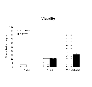
[0016] FIG. 1 depicts the results of an assessment of the relative viability
of
encapsulated and non-encapsulated minced rat kidney tissue.
[0017] FIG. 2 illustrates the amount of Epo released into the culture medium,
as
determined on day 4 post-encapsulation by ELISA; data is included for beads
devoid of tissue
(beads only), non-encapsulated, minced rat kidney tissue (tissue only), and
encapsulated,
minced rat kidney tissue (beads with tissue).
[0018] FIG. 3 depicts data from a study in which the average amount of various
proteins secreted into the medium was measured after four days of culture.
DETAILED DESCRIPTION OF ILLUSTRATIVE EMBODIMENTS
[0019] Despite the increasing interest in cell encapsulation as a method for
delivering therapeutic agents, sparse to no attention has been given to the
encapsulation of
whole tissue fragments. It has presently been discovered that the
encapsulation of minced
kidney tissue provides an opportunity to deliver natural Epo and other
beneficial agents from
endogenous cells, while providing an immunological barrier to prevent tissue
rejection. As
discovered herein, the transplantation of an inducible, beneficial agent-
secreting, implantable
- 4 -
CA 02708959 2010-06-10
WO 2009/085850
PCT/US2008/087211
Docket No. 026038.0222PTUS
device composed of kidney tissue can dramatically alleviate the current
financial, safety, and
medical issues surrounding erythropoiesis-stimulating agents. Kidney tissue
encapsulation
technology may also enable the development of other therapeutic technologies
for the
treatment of various disease states.
[0020] In the present disclosure the singular forms "a," "an," and "the"
include the
plural reference, and reference to a particular numerical value includes at
least that particular
value, unless the context clearly indicates otherwise. When values are
expressed as
approximations, by use of the antecedent "about, "it will be understood that
the particular
value forms another embodiment. Where present, all ranges are inclusive and
combinable.
[0021] Provided are therapeutic implants comprising renal tissue encapsulated
within a polymer bead. The present implants are suitable for introduction in
vivo and for
providing therapeutic effects following implantation. The renal tissue for use
in the present
implants may be autologous tissue, allogeneic tissue, xenogeneic tissue, or
any combination
thereof The renal tissue may be size-processed for use in the present
implants, for example,
by mincing a source of renal tissue into fragments. Such fragments may have a
size of less
than about 1 mm, or they may be larger. The size of the fragments is
preferably measured in
terms of the largest dimension thereof, e.g., lengthwise if the fragments have
an aspect ratio
of greater than 1:1, by the length of a side if the fragments are roughly
cubical, or by diameter
if the fragments are roughly spherical, etc. In addition to mincing, the
fragments may be
further size-processed to reduce the dimensions of the tissue. For example,
the fragments may
be further minced so that the size of substantially all of the fragments are
less than about 300
lam, less than about 150 lam, less than about 100 lam, or less than about 50
lam. The total
quantity of renal tissue in an implant of the present invention may be at
least about 100 mg, at
least about 50 mg, at least about 30 mg, or at least about 10 mg. Various
factors, such as the
desired total surface area of the renal tissue, the type of therapy, the type
of renal tissue, the
characteristics of the subject undergoing therapy, the type and stage of the
disease state
against which therapy is desired, the quantity and type of materials secreted
by the tissue, and
other factors that will be appreciated by those skilled in the art, may be
used to determine the
quantity of renal tissue in the implant, the size of the individual tissue
fragments, or both.
[0022] The polymer bead preferably comprises a biocompatible polymer, such as
a
naturally occurring or synthetically derived biopolymer. The polymer bead may
comprise
such polymers as alginate, hyaluronic acid, carboxymethylcellulose,
polyethylene glycol,
dextran, agarose, poly-L-lysine, carageenan, pectin, tragacanth gum, xanthan
gum, guar gum,
gum arabic, type I collagen, laminin, fibronectin, fibrin, or any combination
thereof A
- 5 -
CA 02708959 2010-06-10
WO 2009/085850
PCT/US2008/087211
Docket No. 026038.0222PTUS
preferred combination of polymers comprises alginate and poly-L-lysine. Such
polymers are
readily commercially available.
[0023] The term "bead" when used in reference to the polymer is intended to
convey that the polymer composition generally assumes a roughly spherical
shape, but may
also be ovoid or oblong. The precise shape of the polymer bead is not
essential to the present
invention; any shape that permits the renal tissue to be substantially
enveloped within the
polymer is acceptable. When measured according to its greatest dimension, a
polymer bead
may have a diameter of about 0.5 mm to about 10 mm, and is preferably about 3
mm to about
6 mm. The size of the polymer bead may be measured according to the
characteristics of the
bead prior to implantation, or following implantation. As polymer beads may
spontaneously
bud, the size of the polymer bead may be measured with respect to an un-budded
bead or
with respect to a bead that results from budding.
[0024] The characteristics of the polymer bead permit the instant implants to
secrete
beneficial agents from within the bead into the ambient environment in which
the bead is
implanted or otherwise contained. In other words, the polymer bead is
permeable to
substances that are secreted by the renal tissue that is encapsulated within
the bead. The renal
tissue may be endogenous, naturally occurring tissue or may include cells that
contain gene
alterations, such as insertions of genes or portions of genes that are not
naturally present.
Renal tissue that includes gene alterations or insertions may be physically
capable of
secreting substances that endogenous or naturally occurring tissue cannot. The
renal tissue
and therefore in turn the implant of the present invention may secrete any
compound that
renal tissue, whether endogenous or altered (e.g., genetically altered) is
physically capable of
producing. For example, the renal tissue may secrete one or more hormones,
prohormones,
proteins, growth factors, trophic factors, or any combination thereof As
additional examples,
and as further described herein, the tissue may secrete one or more of
erythropoietin, MCP-1,
adiponectin, leptin, and MMP-2. The compounds that genetically altered renal
tissue may
secrete are theoretically virtually limitless.
[0025] Also provided are methods for treating a disease state in a subject
comprising implanting within the subject a therapeutic implant comprising
renal tissue
encapsulated within a polymer bead. Because the present implants are capable
of secreting a
number of beneficial agents, the inventive methods can be used to treat a wide
variety of
disease states. As used herein, "treatment" may refer to prophylactic therapy,
or alleviation of
any pathological phenotype. The disease state for which treatment is provided
by the present
invention may be anemia, stroke, cardiovascular disease, or any renal disease,
i.e., any
- 6 -
CA 02708959 2010-06-10
WO 2009/085850
PCT/US2008/087211
Docket No. 026038.0222PTUS
pathology that is directly or indirectly associated with improper kidney
function, for example,
which results in improper kidney function, or which is caused at least in part
by improper
kidney function. Renal disease may be hereditary, congenital, or acquired. Non-
limiting
examples of renal disease include polycystic kidney disease, Alport's
syndrome, hereditary
nephritis, primary hyperoxaluria, cystinuria, nephritis, nephritic syndrome,
hypertension,
diabetes, acute kidney disease, chronic kidney disease (persistent
proteinuria), renal tubular
acidosis, glomerular diseases, and Goodpasture's syndrome. The benefits of
treatment with
Epo, for example has been widely documented with respect to a number of
pathologies, and
is readily appreciated by those skilled in the art. The characteristics of the
polymer beads and
renal tissue for use in the present methods may be as previously described
with respect to the
inventive therapeutic implants.
[0026] The present invention is also directed to methods for making a
therapeutic
implant. The methods for making a therapeutic implant successfully results in
the fabrication
of therapeutic compositions that can be used in accordance with the present
disclosure. The
present methods comprise providing renal tissue; mixing the renal tissue with
a solution
comprising a polymer, thereby forming a tissue-polymer suspension; extruding
the tissue-
polymer suspension into a bead-forming solution, thereby forming a therapeutic
implant
comprising beads of the polymer within which the renal tissue is encapsulated.
[0027] Renal tissue may be prepared in accordance with the previously
disclosed
techniques, including selecting a tissue type and size-processing. The polymer
solution with
which the renal tissue is mixed in accordance with the present invention may
comprise a
combination of a polymer and a growth medium. The characteristics of the
polymer may be
determined as described above. Any acceptable culture medium, nutrient broth,
or the like
may be used for the instant growth medium; the characteristics of an
appropriate growth
medium, which may comprise a mixture of media, are readily determined by those
skilled in
the art. Growth media can vary in pH, glucose concentration, growth factors,
and the
presence of other nutrient components, but the growth medium should fulfill at
least some of
the nutritional requirements of the renal tissue, and preferably fulfills most
or all nutritional
requirements, and should possess pH and other chemical characteristics
necessary to sustain
and nurture the renal tissue. An example of a suitable growth medium is
DULBECCO'S
MODIFIED EAGLES MEDIUM (DMEM; Invitrogen, Carlsbad, CA). Growth media are
commercially available and suitable media are readily recognized by those
skilled in the art.
To prevent infection, antibiotics, such as penicillin, streptomycin, and the
like, may be added
- 7 -
CA 02708959 2010-06-10
WO 2009/085850
PCT/US2008/087211
Docket No. 026038.0222PTUS
to the growth medium. Serum, such as fetal bovine serum, may also be added to
the growth
medium.
[0028] Mixing of the renal tissue and the polymer solution may be achieved by
any
means that are suitable for forming a suspension, i.e., a mixture in which the
renal tissue is
substantially uniformly suspended in the polymer solution, such as agitation,
stirring, or
pouring. For example, the mixing may be achieved by loading the renal tissue
and polymer
solution into a first container, such as a syringe, transferring the solution
into another
container, such as by expelling the contents of a syringe into a second
syringe, transferring
the solution back to the first container, and then repeating this cycle as
necessary until a
suspension is achieved.
[0029] After a suspension is formed, the suspension is extruded into bead-
forming
solution in order to form polymer beads within which the renal tissue is
encapsulated. The
extrusion may comprise ejecting the suspension from a syringe, or otherwise
transferring the
suspension from one container into another in which the bead-forming solution
is contained,
or alternatively, transferring the bead-forming solution into a container in
which the
suspension is held. The bead-forming solution may be ionic, may a cross-
linking solution, or
both. In one embodiment the bead-forming solution comprises CaC12. The polymer
beads
with encapsulated renal tissue may form spontaneously when combined with the
bead-
forming solution. The process of bead-forming may be further assisted, for
example, by
agitating the mixture of the suspension and the bead forming solution,
modifying the
temperature of the mixture (e.g., raising the temperature), or both.
[0030] Following the formation of the polymer beads, chemical cross-linking of
the
beads may be achieved by placing the beads into a cross-linking solution. For
cross-linking,
the beads may be transferred into a dilute solution of polymer, preferably a
different polymer
than the major polymer component of the beads. For example, if the major
component of the
polymer bead comprises alginate, a dilution solution of poly-L-lysine may be
used to cross-
link the alginate beads. Optionally, an additional polymer layer may be added
to the polymer
beads following their "production" in the bead-forming solution, or following
cross-linking.
Preferably, the additional polymer layer comprises the same polymer that makes
up major
component of the polymer beads. For example, an additional alginate layer may
be added to a
bead of which the major component is alginate. Adding another polymer layer to
the polymer
beads may be accomplished by placing the beads in a dilute polymer solution
comprising the
polymer of which the extra layer will be made.
- 8 -
CA 02708959 2015-06-17
[0031] The present invention is further defined in the following Examples. It
should
be understood that these examples, while indicating embodiments of the
invention, arc given
by way of illustration only, and should not be construed as limiting the
appended claims.
From the above discussion and these examples, one skilled in the art can
ascertain the
essential characteristics of this invention, and without departing from the
spirit and scope
thereof, can make various changes and modifications of the invention to adapt
it to various
usages and conditions.
Example I ¨ Formation of Therapeutic Implant
[0032] Four kidneys from female, Long Evans rats (eight weeks old) were
surgically removed, rinsed in ice cold phosphate buffered saline without Ca2'
and Mg2I
(PBS) (Invitrogen, Carlsbad, CA) and then, using a scalpel, were minced into
small pieces (1-
mm3). A 300 uM-steel sieve (Sigma, St Louis, MO) was then used to further
mince the
tissue fragments. Minced tissue was then washed three times with 30-50 mL of
Growth
Medium containing Dulbecco's Modified Eagles Medium (DMEM) (Invitrogen)
containing
1% penicillin/streptomycin (lnvitrogen) and 1% fetal bovine scrum (FBS)
(Hyclonc, Logan,
UT). The final wash was completely removed and the tissue fragments were
loaded into one
1-milliliter syringe of a two syringe mixing system. A 1.8% (w/v) alginate
solution (Sigma)
was prepared in Growth Medium and was loaded into the second 1-milliliter
syringe. The two
solutions were then mixed together by pushing the contents back and forth
through both
syringes. The minced tissue-gel suspension was then extruded into a 100 mM
CaC12 solution.
The resulting encapsulated tissue beads were then incubated at room
temperature in CaCl2
with slow agitation for 5 minutes. The beads were then chemically cross-linked
by
transferring into 0.05% (w/v) poly-L-lysine, molecular weight 24,000 (Sigma)
containing 1%
FBS for 5 minutes and then coated with another layer of 0.1% (w/v) alginate
solution
containing 1% FBS for 5 minutes. Four to ten beads were then transferred to
individual wells
of a 24 well low-cluster, tissue culture dish containing 0.5 mL of Growth
Medium, or Growth
Media containing 100 ng/mL poly-D-glutamic acid (pDGA) (Sigma), and cultured
at 37 C
for four days under either normoxic or hypoxic (5% Oxygen) atmospheric
conditions. Beads
were visually examined and imaged using a digital camera and Eclip2TE2000-U
microscope (Nikon, Japan).
[0033] Visual examination of alginate encapsulated tissue beads showed that
manual extrusion through the two-way syringe system was effective in
generating spherical,
- 9 -
CA 02708959 2015-06-17
tissue containing beads. Tissue fragments within the alginate beads were also
visible,
demonstrating a uniform distribution throughout the alginate gel.
Example 2¨ Bead Diameter Measurements
[0034] Fourteen individual, tissue containing beads were placed into a clean
tissue
culture plate and imaged using a Nikon dissecting microscope fitted with a
digital camera.
TM
The diameter of each bead was then measured using IMAGE PRO PLUS Software.
[0035] Visual examination of alginate encapsulated tissue beads showed that
manual extrusion through the two-way syringe system was effective in
generating spherical,
tissue containing beads. Tissue fragments within the alginate beads were also
visible,
demonstrating a uniform distribution throughout the alginate gel. One-hundred
and fifteen
beads were generated from 3.72g of fragmented kidney tissue resulting in
approximately 32
mg of tissue per bead. Table 1 shows the distribution of bead diameter.
TABLE 1
Diameter
Bead
(mm)
1 4.84
2 4.26
3 4.66
4 4.13
4.37
6 4.13
7 4.83
8 4.59
9 4.36
4.42
11 4.34
12 5.90
13 4.55
14 4.51
Average 4.56
Std 0.44
The average diameter of fourteen individual beads was found to be 4.56 +/-
0.44 mm.
Example 3 ¨ Assessment of Cell Viability
TM
[0036] Minced kidney tissue viability was assessed using ALAMAR BLUE
(Invitrogen), a colorimetric REDOX indicator that is reduced in response to
metabolic
activity. After four days in culture, spent Growth Medium was removed from
samples of
- 10 -
CA 02708959 2015-06-17
non-encapsulated kidney tissue, encapsulated kidney tissue, isopropanol fixed
kidney tissue
and Growth Medium only. One milliliter of Growth Medium, containing 10% ALAMAR
TM
BLUE, was added to the samples and further incubated for 2-4 hours at 37 C, 5%
CO) with
TM
gentle rocking. Spent media was then analyzed spectrophotometrically
(SPECTRAMAX-
190, Molecular devices, Sunnyvale, CA) at 570nm and 600nm. Media from each
sample was
TM
analyzed in triplicate. Percent reduction of ALAMAR BLUE was determined
following the
manufactures instructions and is an indirect measurement of cell viability.
[0037] After four days of culture, tissue viability was evaluated. As compared
to
non-encapsulated tissue, encapsulation maintained greater tissue viability
(FIG. 1). Non-
encapsulated kidney tissue, cultured under either normoxia or hypoxia showed
similar
relative mean viabilities of 20.9% +/- 3.4% and 21.0% +/- 1.9%, respectively.
However,
encapsulated kidney tissue, cultured under normoxic conditions showed an
increase in
relative mean tissue viability. Encapsulated tissue showed a relative mean
tissue viability of
83.5% +/- 4.5%. Encapsulated kidney tissue, cultured under hypoxic conditions
resulted in
reduced tissue viability of 31.3% +/- 3.4%. As expected, tissue fixation
resulted in a
significant decrease in tissue viability to 5.0% +/- 0.084%.
Example 4¨ Epo Secretion Analysis
[0038] After four days of culture, spent media was collected and the amount of
Epo
TM
released into the culture medium was determined using a Quantikine Mouse/Rat
Erythropoietin ELISA kit (R&D systems, MN). The ELISA plate was assayed
TM
spectrophotometrically (SPEC'TRAMAX-190, Molecular devices, Sunnyvale, CA) at
540nm.
Data was analyzed by comparing absorbance values of unknown samples to the
linear
regression of a standard curve.
[0039] The amount of Epo released into the culture medium was determined on
day
4 post-encapsulation by ELISA. Data was normalized to absorbance values
obtained with
Growth Medium only (Corrected Mean). Each measurement was conducted on spent
media
obtained from 8-10 beads. Standard error of the mean (SEM) was also
calculated. Data
shown in Table 2, below, is represented in graphical form in FIG. 2.
- 11 -
CA 02708959 2015-06-17
TABLE 2
TREATMENT GROUP MEAN SEM NORMALIZED
(pg/mL) (pg/mL) MEAN (pg/mL)
Growth Medium only
(background) -26.79 3.73 0.00
Beads only -32.85 2.10 -6.06
Tissue only 19.04 3.34 45.83
Beads with tissue 52.52 28.59 79.31
Beads with tissue and pDGA 34.12 10.50 60.91
[0040] In FIG. 2, data bars represent the average of triplicate measurements,
and
error bars represent SEM. Each measurement was conducted on spent media
obtained from
8-10 beads.
[0041] Results showed that minced kidney tissue produced 45.8 +/- 3.3 pg/mL of
Epo into the surrounding culture media. Likewise, alginate encapsulation did
not impede
Epo release from the minced tissue, producing 79.3 +/- 28.6 pg/mL of Epo. In
order to
determine if Epo production could be chemically enhanced, beads were prepared
and cultured
in pDGA. Results showed that pDGA treatment did not effect Epo production,
generating
60.9 +/- 10.5 pg/mL of Epo. As a negative control, Epo production from beads
devoid of
tissue was determined. As expected, no measurable Epo was detected from these
samples.
Example 5 ¨ Trophic Factor Secretion Analysis
[0042] After four days of culture, spent culture medium was harvested from the
beads. Cell debris was removed from the spent culture medium by centrifugation
and the
culture medium was stored at ¨80 C. At the time of analysis, spent culture
medium was
assayed by ELISA for the following protein factors: interleukin-4 (IL-4),
monocyte
chemotactic protein-1 (MCP-1), RANTES, granulocyte-macrophage colony
stimulating
factor (GMCSF), interleukin-10 (IL-10), adiponectin, leptin, matrix
metalloproteinase-2
TM
(MMP-2) with Searchlight Proteome Arrays (Pierce Biotechnology Inc.).
[0043] As compared to spent culture medium derived from beads without tissue
encapsulation, beads containing kidney tissue fragments secreted elevated
amounts of MCP-1
(50.6 +/- 8.9 pg/mL), adiponcctin (132,060.6 +/- 11,226.7 pg/mL), leptin (10.3
+/- 2.6
pg/mL) and MMP-2 (945.2 +/- 13.3 pg/mL) and low to undetectable amounts of IL-
4,
RANTES, GMCSF and IL-10. As shown in Table 3, below, each treatment group
contained
three samples (1, 2, 3).
- 12 -
CA 02708959 2015-06-17
TABLE 3
IL4 MCP1 RANTES GMCSF 1110
JAdiponectin LetinMMP2
.....
Beads with 'aigigiAiRigigigigigNiagigigaggiagginngigegiMiallatigiggiagnai.i:
tissue 'ggRiiiaiiiipigViiamoQiiiimmii*AiMiiiiiiiikapgiMaiSMONkitienigaiffiiin
1 39.8 68.4 7.6 105.8 20.6 113137.8 14.6
1160.0
2 1.6 77.0 10.4 69.8 1.6 136667.8 19.6
1184.6
3 24.2 47.2 1.6 78.2 1.6 151719.0 10.6
1138.6
AVG 21.9 64.2 6.5 84.6 7.9 133841.5 14.9
1161.1
STD 19.2 15.3 4.5 18.8 11.0 19445.3 4.5 23.0
SEM 11.1 8.9 2.6 10.9 6.3 11226.7 2.6 13.3
1L4 MCP1 RANTES GMCSF IL10 1 Adiponectin Leptin MMP2
Beads without ititiledgettlailkaggaiti;11'n:SaMigragiegtigiataanSSEREM
tissue
Iiigaiiiiiiiiiiigii*Aiiiiiffig:AiigieggiQijiiiigaiOtiiia.O.O6VMM.iniRWittEM.oim
m.
1 32.0 14.4 1.6 63.8 , 13.4 1827.2 7.0
616.4
2 32.8 13.8 3.0 130.4 17.6 1655.2 5.1 15.6
3 32.6 12.6 1.8 118.2 18.8 1860.4 1.8 15.6
AVG 32.5 13.6 2.1 104.1 16.6 1780.9 4.6
215.9
STD 0.4 0.9 0.8 35.5 2.8 110.1 2.6 346.9
SEM 0.2 0.5 0.4 20.5 1.6 63.6 1.5 200.3
IL4 MCP1 RANTES GMCSF 1L10 Adiponectin
Lepttn
Medium only
i'i.iiiaii,gagigkiiitinifiailinnagangiVnginiiiiiaMii;iiiiiMigiagitfigigagdjani'
1 29.0 3.1 1.6 98.8 _ 20.4 939.2 5.1
15.6
2 30.2 22.6 1.6 91.2 16.6 1159.0 5.1 15.6
3 34.8 16.8 1.6 85.0 15.0 1436.4 2.0 441.8
AVG , 31.3 14.2 1.6 91.7 17.3 1178.2 4.1 157.7
STD 3.1 10.0 0.0 6.9 2.8 249.2 1.8 246.1
SEM 1.8 5.8 0.0 4.0 1.6 143.8 1.0 142,1
STD = Standard deviation, SEM = Standard error of the mean. Data shown here is
represented in graphical form in FIG. 3, in which data bars represent the
average amount of
protein secreted into the medium after four days of culture. Background
measurements
obtained from beads without tissue was subtracted from the data shown. Error
bars represent
SEM.
Example 6- Evaluation of Erythropoiesis Stimulating Activity
100441 Rat erythroid CD34+ cells (Lonza, Walkersville MD) are resuspended at
15,000 cells/cm2 in IMDM with 10% FBS. Bead conditioned medium are then added
to
TM
methylcellulose colony forming assay medium (MethoCult OF 114534, StemCell
Technologies, Vancouver BC). Cells arc added to the methylcellulose and plated
with
- 13 -
CA 02708959 2015-06-17
subsequent incubation at 37oC, in a 5% CO2 incubator for 12-14 days. Colonies
containing
over 50 cells arc counted by phase contrast microscopy.
[0045] Conditioned media derived from encapsulated rat kidney tissue has
previously been shown to contain Epo. Conditioned media is presently shown to
have
crythropoicsis stimulating activity (ESA) as measured by BFU-E activity.
Example 7¨ Evaluation of Renoprotecdve Effects of Encapsulated Renal Tissue
Fragments
[0046] The purpose of this study is to evaluate the renoprotective effects of
alginate
encapsulated rat kidney tissue fragments in a rat model of renal disease.
Sprague Dawley rats
(diabetic or non-diabetic) with an initial weight of 200-250g are used for
these experiments.
The rats are anesthetized with an intraperitoneal injection (5mg/kg) of a 4:1
solution of
ketamine hydrochloride and xylazine hydrochloride. Kidney failure is induced
by a two-
stage nephrectomy procedure. The upper and lower parts of the left kidney (two
thirds of one
kidney) are resected using silk ligature while preserving the renal capsule.
Ten days later, the
right kidney is removed, leaving approximately 1/6 of the total kidney mass
(5/6
nephrectomy). Applying soft pressure with methylcellulosc stops bleeding, and
the
M
peritoneum and skin is closed in layers with resorbable 4-0 VicrylT sutures.
[0047] Five weeks after the 5/6-nephrectomy procedure, beads are transplanted
under the capsule As a control, 5/6 nephrectomized rats are injected with
fibrin matrix only.
Serum samples arc obtained on days 0 (prior to 5/6 nephrectomy) and on day 1
(day of cell
transplantation), days 7, 14, 21, 28 and 35 (day of necropsy). Blood urea
nitrogen and
TM
creatinine are quantified using a VETACE CHEMISTRY ANALYZER (Alpha Wassermann
Diagnostic Technologies, LLC, West Caldwell, NJ).
[0048] Animals in all groups are sacrificed five weeks post cell
transplantation by
carbon dioxide asphyxiation. Kidneys are removed for histology and
transcriptional analysis.
Half of each kidney is snap-frozen in liquid nitrogen for RT-PCR analysis.
Messenger RNA
is isolated from the frozen kidney tissue by study coordinator and subjected
to transcriptional
analysis utilizing low-density microarray cards containing pro-fibrotic and
inflammatory
genes. The remaining corneal kidney section is fixed in 10% neutral buffered
formalin for
downstream histological analysis.
[0049] Kidney tissue fixed for histology will be histologically processed,
sectioned
(5 gm-thick) and stained with hematoxylin/cosin. Tubular injury is evaluated
and scored by a
veterinary pathology.
- 14 -
CA 02708959 2015-06-17
[0050] In this study, subcapsular transplantation of alginate encapsulated
kidney
tissue fragments will slow the progression of renal injury in 5/6
nephrectomized rodents or in
rodent models of diabetic nephropathy. Both serum creatinine and blood urea
nitrogen values
are significantly reduced in the hUTC treated animals as compared to the
control animals. In
addition, the bead lowers blood glucose levels in rodent models of diabetic
ncphropathy.
Histological injury assessment reveals a reduction in tubular necrosis and
tubular dilation in
the treated animals.
100511 Despite the increasing interest in cell encapsulation as a method for
delivering therapeutic agents, sparse to no attention has been placed on the
encapsulation of
whole tissue fragments. Tt has herein been demonstrated that encapsulated
kidney tissue
fragments secrete Epo and other beneficial agents into a culture medium.
Therefore, the
disclosed therapeutic implants and therapeutic methods can provide treatment
of numerous
disease states.
- 15 -