Note : Les descriptions sont présentées dans la langue officielle dans laquelle elles ont été soumises.
CA 02712060 2010-07-13
WO 2009/109045
PCT/CA2009/000268
JOINT PROSTHESES
,
TECHNICAL FIELD
[0009] The present invention relates to orthopedic medicine, and more
specifically to
methods and devices for the replacement of joints with artificial joint
prostheses.
BACKGROUND OF THE INVENTION
[0010] Spinal arthroplasty is an emerging field that offers the promise of
restoring and/or
maintaining normal spinal motion. The goal of spinal arthroplasty is to reduce
or eliminate
adjacent segment disease (ASD) by maintaining the normal spinal biomechanics
at the
operative level. To accomplish this, an artificial cervical prosthesis must
duplicate as closely
as possible the natural spinal biomechanics, including maintaining the axial
height of the disc
as well as applying angular adjustment throughout the full range of motion of
the natural
spine.
1
CA 02712060 2010-07-13
WO 2009/109045
PCT/CA2009/000268
100111 The spine plays an integral role in neural protection, load bearing
and motion. The
vertebral column provides a strong, yet mobile central axis for the skeleton
and is composed
of twenty-four vertebral bodies with seventy-five stable articulations. The
intervertebral disc
is a fundamental component of the spinal motion segment, providing cushioning
and
flexibility. Adjacent vertebrae are linked together by three articulations: a)
the vertebral
bodies and disc, which transmit compressive and shear loads and provide
flexibility, and b)
by two facet joints, which protect the disc from translational shear stress
and limit rotation.
This "triple joint complex" allows for flexion, extension, lateral bending and
rotation of the
spine.
[0012] The intervertebral disc is composed of an inner gel-like matrix
called the nucleus
pulposus and an outer surrounding fibrous band called the annulus fibrosus.
When
compressive loads are placed on the spine, increased pressure in the nucleus
pulposus is
transmitted to the annulus, which bulges outwards. The degenerative cascade of
the
intervertebral disc initially involves desiccation of the nucleus pulposus.
With decreased
elasticity and dampening from the nucleus, increased loads are transmitted to
the annulus and
facets. The increased stress on the annulus can lead to fissures and radial
tears in its collagen
fibers. With further degeneration, this can lead to circumferential bulging of
the disc,
contained and uncontained disc herniations, and complete desiccation of the
disc. This
degenerative cascade can result in axial pain, by stimulating pain fibers in
the annulus, or
compression of spinal nerve roots and/or the spinal cord. This can manifest
itself in motor
weakness, pain and/or numbness in the arms or legs or both.
[0013] The structure and function of the discs may be altered by a variety
of factors
including repeated stress, trauma, infection, neoplasm, deformity, segmental
instability and
inflammatory conditions. Degeneration of the intervertebral disc is the most
common
etiology of clinical symptoms referable to the spine. Degeneration of the
spine is a universal
concomitant of human aging. In the cervical spine, neck and arm pain caused by
nerve root
compression has been estimated to affect 51% of the adult population.
Spondylosis of the
spine and aging are intimately related, with spondylosis increasing in both
prevalence and
severity with age. Fortunately, the majority of patients will improve without
surgery. In
approximately 10-15% of cases, spondylosis is associated with persistent nerve
root and
spinal cord compression and/or spinal pain, with a small percentage ultimately
requiring
surgery.
2
CA 02712060 2010-07-13
WO 2009/109045
PCT/CA2009/000268
[0014] The
most common type of surgery used in the United States for the treatment of
degenerative disorders of the spine (spondylosis) is spinal fusion. In an
interbody fusion, the
diseased disc is removed and either a wedge of bone from the patient's hip,
allograft or a
metallic spacer is placed between the vertebrae where the disc was removed.
This
immobilizes the functional spinal unit. While this surgery has been successful
in eliminating
motion, there are disadvantages associated with it. By converting a mobile,
functional spinal
unit into a fixed, nonfunctional one, fusion results in increased strain
patterns at levels
adjacent to the fused segment. When a segment of the spine is fused, there is
elimination of
motion at the level of surgery. Therefore, the stresses that would normally be
absorbed by the
disc at the site of surgery are now transferred to adjacent segments. This can
cause adjacent
segment disease (ASD) to one or several spinal units adjacent to the affected
level. ASD can
be defined as a clinical syndrome of symptomatic degenerative changes
occurring adjacent to
a previously fused motion segment. Retrospective studies have estimated that
ASD can occur
in the cervical spine at a rate as high as 2.9% per year with a projected
survivorship rate of
26% at 10 years (Hilibrand AS, Carlson GD, Palumbo M, Jones PK, Bohlman HH:
Radiculopathy and myelopathy at segments adjacent to the site of a previous
anterior cervical
arthrodesis. J Bone Joint Surg (Am) 81:519-528, 1999).
[0015] In the
cervical spine, thousands of North Americans undergo surgery for cervical
spondylosis each year. The majority of these procedures involve an anterior
discectomy with
decompression of the spinal cord and/or nerve root. The primary indication for
surgery in the
management of cervical spondylosis is radiculopathy, myelopathy and/or neck
pain.
Following the discectomy, an anterior interbody fusion is commonly performed.
Autologous
bone harvested from the iliac crest or cadaveric bone is most commonly used to
fill the space
created by the removal of the disc. A number of other solutions have been
suggested,
including metallic devices such as fusion cages or other types of spacers,
xenografts such as
bovine bone, and biological strategies such as the use of growth factors. The
graft for the
interbody fusion can be shaped to correct underlying deformity of the cervical
spine. By
contouring the graft one can restore lordosis to a straight or kyphotic spine.
[0016] A more
recent alternative to spinal fusion is replacement of the damaged disc with
a motion preservation device, which includes either a nucleus or total disc
replacement
(TDR). The rationale for the development of the artificial disc is to prevent
adjacent segment
disease. Artificial disc devices can be broadly divided into two categories,
those that replace
3
CA 02712060 2010-07-13
WO 2009/109045
PCT/CA2009/000268
the nucleus only, leaving the annulus and vertebral body end plates intact and
those that
involve replacement of the disc and addition of prosthetic end plates. Both
strategies are
directed at restoration of intervertebral disc function. Prosthetic nuclei are
described, for
example, in United States Patent Nos. 5,047,055 and 5,192,326. United States
Patent
application U52002/0183848 also discloses a prosthetic spinal disc nucleus
that has a
hydrogel core surrounded by a constraining jacket.
100171 There are several different types of prosthetic devices for use in
the cervical or
lumbar segments of the spine designed for TDR. For example, the ProdiscTM and
the
ChariteTM disc are composites of cobalt chromium end plates with a
polyethylene core. The
ProdiscTM is described in United States Patent No. 5,314,477 and the ChariteTM
disc is
described in United States Patent Nos. 5,401,269 and 5,556,431. The PrestigeTM
disc is
another type of artificial disc that comprises a metal on metal design with a
ball and trough
articulation. Another type of artificial disc that is gaining popularity in
the cervical spine is
the Bryan disc, described in several United States Patent applications
including
2004/0098131; 2004/00544411; and 2002/0 128715. The Bryan disc is a composite
artificial disc with a low friction, wear resistant, elastic nucleus that
articulates with two
circular metal plates.
[0018] Presently, there are at least four artificial cervical disc
replacement systems
undergoing clinical trials worldwide. These include unconstrained devices,
such as the PCM
cervical disc. These unconstrained devices do not have mechanical stops to
limit their range
of motion. The Bryan Cervical disc, the ProdiscTM C and the PrestigeTM LP
cervical disc
systems limit range of motion to varying degrees. These systems can be
considered semi-
constrained, in that there are mechanical stops outside the normal range of
motion. Thus far,
only the ChariteTM disc has been approved for use in the United States.
[0019] Artificial spinal discs have been implanted for the management of
degenerative
disc disease producing radiculopathy, myelopathy and/or axial spinal pain.
More recently,
artificial discs have been adopted for the treatment of trauma. The aim of TDR
is to
reproduce the biomechanics of the natural disc. Early clinical and
biomechanical studies with
single and multi-level disc replacement have reported favorable clinical
outcomes and
preserved range of motion at the level of surgery. Preservation of range of
motion, however,
while an important feature of an artificial disc, is only a single measure of
spinal
biomechanics. The effect of the disc on angulation at the operative level, the
average disc
4
CA 02712060 2010-07-13
WO 2009/109045
PCT/CA2009/000268
space height, and overall spinal alignment (sagittal and coronal balance) also
needs to be
considered.
[0020] While the introduction of artificial discs has led to many
successful surgeries,
there are still problems associated with the current discs. For example, all
of the current
artificial cervical discs have a fixed height across the entire disc. The
artificial discs presently
available can have issues with focal kyphosis or kyphosis at adjacent segments
of the spine
after the patient post-operatively reassumes an upright position, supporting
the weight of the
head and body. For instance, with the Bryan disc, the end plates are allowed
to move freely
about all axes of rotation, allowing the end plate to assume a position
resulting from the
forces exerted on the implant by the head and neck. At times, this position
may be
significantly different from the positioning of the disc intra-operatively.
Several published
studies with the Bryan cervical disc replacement system have reported a
tendency for the
end plates of the prosthesis and the alignment of the cervical spine to
develop kyphosis
following surgery. [Pickett GE, Mitsis DK, Sekhon LH et al. Effects of a
cervical disc
prosthesis on segmental and cervical spine alignment. Neurosurg Focus
2004;17(E5):30-35;
Johnson JP, Lauryssen C, Cambron HO, et al. Sagittal alignment and the Bryan
cervical
disc. Neurosurg Focus 2004;17(E14):1-4; Sekhon LHS. Cervical arthroplasty in
the
management of spondylotic myelopathy: 18 month results. Neurosurg Focus 2004;
17(E8):55-611 This kyphotic angulation of the prosthesis has been attributed
.to the passive
(unconstrained motion with a mobile nucleus and variable instantaneous axis of
rotation)
design of the implant. None of the current TDR systems addresses this major
complication.
[0021] A significant number of patients with spinal disc disease have a
loss of sagittal
alignment of the spine as a result of the degenerative process. In addition,
varying degrees of
coronal imbalance can also occur. None of the available artificial disc
replacement systems
are designed to restore normal alignment to a spine that is straight, which
have focal/global
kyphosis or corona! deformity. Existing artificial disc replacement systems
that are inserted
into either a straight, kyphotic or angulated segment are likely to take on
the angle and local
biomechanics determined by the facets, ligaments and muscle forces. As such,
patients with a
pre-operative straight spine may develop post-operative kyphosis, and patients
with a pre-
operative kyphosis may have a worsening of the deformity post-operatively.
Kyphosis of the
spine has been implicated in segmental instability and the development of
clinically
significant degenerative disease. Several clinical studies have described that
a change in the
CA 02712060 2010-07-13
WO 2009/109045
PCT/CA2009/000268
sagittal or coronal balance of the spine can result in clinically significant
axial spinal pain as
well the initiation and/or the acceleration of ASD. [Kawakami M, Tamaki T,
Yoshida M, et
al. Axial symptoms and cervical alignment after anterior spinal fusion for
patients with
cervical myelopathy. J Spinal Disord 1999;12:50-60; Harrison DD, Harrison DE,
Janik TJ, et
al. Modeling of the sagittal cervical spine as a method to discriminate
hypolordosis: results of
elliptical and circular modeling in 72 asymptomatic subjects, 52 acute neck
pain subjects, and
70 chronic neck pain subjects. Spine 2004;29:2485-2492; Katsuura A, Hukuda S,
Saruhashi
Y, et al. Kyphotic malalignment after anterior cervical fusion is one of the
factors promoting
the degenerative process in adjacent intervertebral levels. Eur Spine J
2001;10:320-324;
Ferch RD, Shad A, Cadoux-Hudson TA, Teddy PJ. Anterior correction of cervical
kyphotic
deformity: effects on myelopathy, neck pain, and sagittal alignment. J
Neurosurg 2004;
100:S13-S19; Katsuura A, Hukuda S, Imanaka T, Miyamoto K, Kanemoto M. Anterior
cervical plate used in degenerative disease can maintain cervical lordosis. J
Spinal Disord
1996; 9:470-476.]
[0022] Attempting to provide a deformity correction by simply altering the
end plate or
the nucleus of an artificial disc, while still maintaining free movement about
all axes of
rotation, may not be sustainable as the forces exerted by the head and body on
the artificial
disc could counteract the desired correction. To provide a sustainable
correction, some
limitation on the axes of rotation is required. From a design perspective, the
goal is to design
an artificial disc that is able to correct deformity (coronal and sagittal),
has mechanical stops
outside the normal range of motion (semi-constrained), and preferably has
variable
instantaneous axis of rotation (JAR).
[0023] The limits on the axes of rotation can fall into two categories. One
is to provide
correction using a permanent rotation or translation of an axis to support the
correction. This
is accomplished using the geometries of the core and end plates themselves and
is referred to
the Geometric Constraint category. The second is to keep free range of motion
about all axes
but provide the correction using a material support. This type of design
provides the
correction by the imposition of a deformable material in the plane of
correction for normal
rotation in that plane. This is the Material Constraint category of designs.
[0024] Degenerative disc disease is a major source of morbidity in our
society. It can lead
to serious economic and emotional problems for those afflicted. Thus, there is
a need for an
6
CA 02712060 2010-07-13
WO 2009/109045
PCT/CA2009/000268
artificial disc that can alleviate both symptoms and correct deformity
(sagittal or coronal or
both) of the spine.
BRIEF SUMMARY OF THE INVENTION
[0025] There are a number of different strategies that can be used with
disc replacements
to address the need for alignment/deformity correction in the spine. With most
of the
available discs, the angle of disc insertion can significantly alter the
orientation of the
prosthesis. This is related to bone removal and end-plate preparation for the
prosthesis. By
changing the angle of insertion, the disc can be placed either in parallel or
at an angle to the
disc space. Unfortunately, by changing only the angle of insertion, one cannot
correct an
underlying deformity of the spine. Simply changing the angle of insertion is
not adequate to
compensate for a device that does not have sufficient off-center load bearing
support or
structure to maintain the correction of the deformity.
[0026] A strategy to correct lordosis in the lumbar spine has been utilized
by the Link-
ChariteTM and ProdiscTM lumbar disc replacement systems by using wedge-shaped
end
plates. A wedge-shaped end plate has also been used in at least one case with
the Bryan
cervical disc system. However, wedge-shaped end plates are not routinely
available at the
present time for cervical disc replacement systems. The strategy of using
wedge-shaped end
plate(s) involves forming a differential thickness across the end plate. The
articulation
between the ball and socket/trough or the nucleus and end plates is not
altered, which is an
advantage because the complex geometry of how the prosthesis provides motion
is not
altered. The disadvantage, however, is that this strategy is not forgiving if
an error is made
with either an overly corrected end plate or an end plate that is not
corrected enough. The
revision of the end plate can be difficult at the time of surgery and may even
preclude the disc
space from receiving a disc replacement. As most systems have a coating on the
end plates
that promote bony ingrowth, revision at a later date may be extremely
difficult or even
impossible. As there are two surfaces to the end plate, an outer surface that
contacts the bone
and an inner surface that articulates with the nucleus or core, it is
conceivable that by
changing the location or geometry of the inner surface, one could alter the
center of rotation.
This would be most applicable to prostheses that function as a "ball and
socket" articulation.
By changing the location of the "socket" or trough, this could alter how the
prosthesis
impacts alignment at the level of the disc.
7
CA 02712060 2010-07-13
WO 2009/109045
PCT/CA2009/000268
[0027] An alternate method of achieving lordotic correction is by changing
the nucleus or
inner core. The biggest advantage of this approach is that the nucleus or core
can be more
easily interchanged or revised. Intra-operatively, instruments can be used to
gage the need
for and amount of correction and the appropriate nucleus can be inserted. By
designing the
correction into the nucleus, the surgeon is provided with flexibility and ease
of insertion, and
the ability for revision at a later date, which the other methods do not
provide.
[0028] The invention includes a novel artificial disc that provides the
normal range of
motion of the natural intervertebral disc, along with the ability to correct
deformity of the
spine. The proposed disc allows for semi-constrained range of motion of the
functional
spinal unit. It will reproduce the kinematics of the pre-operative normal
spine. It will
possess maximum durability and biocompatibility, and a means for integrating
itself into the
spine bony structure for long-term stability. Its insertion will be safe,
simple, and ideally not
add significantly to surgical time compared with the current procedures. In
contrast to the
existing disc replacement systems, it will allow the surgeon to correct
deformity while
maintaining natural kinematics of the spine.
[0029] A major advantage of this system will be that the nucleus may be
easily revisable.
For instance, in most cases where the Bryan disc needs revision, the entire
disc, including
the end plates, must be removed. In cases where the alignment of the spine
changes with
time, especially in children and young adults, this new disc replacement
system will allow
revision of the nucleus, if needed.
[0030] The present invention addresses the problems associated with the
artificial discs of
the prior art by providing an artificial disc that provides for correction of
spinal alignment
deformity.
[0031] The artificial disc of the present invention is useful for the
treatment of
degenerative disc disease including correcting spinal deformities such as
kyphosis, lordosis,
and scoliosis.
[0032] It is an object of one aspect of the invention to provide an
improved artificial disc
replacement that maintains motion at the operative level and reduces the
incidence of
adjacent segment disease.
[0033] In one aspect of the invention, the artificial disc incorporates an
artificial nucleus
having an asymmetrical maximum vertical axis. The present invention includes a
non-
8
CA 02712060 2010-07-13
WO 2009/109045
PCT/CA2009/000268
spherical nucleus with a maximum point of load-bearing and height in a non-
central location
(a differential in the anterior/posterior height of the nucleus).
[0034] In one embodiment, the nucleus is adapted to provide lordodic
correction to a
damaged spinal segment. In this case, the axis of greatest height is
positioned in the anterior
part of the nucleus.
[0035] In another embodiment, the nucleus is adapted to provide kyphotic
adjustment. In
this case, the maximum height axis is positioned in the posterior part of the
nucleus.
[0036] In yet another embodiment, the asymmetrical nucleus can be used for
the
treatment of scoliosis. To achieve this, the axis of maximum height is lateral
(parasagittal) to
the middle of the disc.
[0037] According to another aspect of the present invention, an artificial
nucleus, or core,
is provided for use in an artificial disc. The nucleus comprises a body of
biocompatible
material, having the greatest vertical height either at the central vertical
axis or at a vertical
axis other than the central vertical axis.
[0038] In another embodiment, the body is spherical or ovoid (egg-shaped),
having
convex upper and lower surfaces and a non-central maximum height vertical
axis. In an
alternative embodiment, the nucleus is in the form of a truncated cylinder
where the top is cut
at a plane that is not parallel to the base. In another preferred embodiment,
the disc is
essentially circular.
[0039] It has been found that nucleus body designs with a completely
rounded surface
(not necessarily spherical) have issues with reliably maintaining correction
when exposed to
the variable forces of the head and neck. To address this issue, a segment or
section that is
flat or which has a contour different from the adjacent surface, can be formed
in the central
region of the nucleus body. This section will be referred to as a flattened
section, which is
meant to refer to any contour that is not the same as the adjacent surface(s)
of the nucleus.
Such a flattened surface can be planar or it can have other shapes such as a
slight convex or
concave shape with a radius of curvature different from the adjacent surface.
Such a flattened
surface could also be in the shape of a compound curve or other complex shape.
In the
example of providing a lordotic correction, the flattened segment can be
angled relative to the
superior end plate of the inferior vertebral body with the height of the
anterior part being
greater than the height of the posterior part. The overall shape of the
nucleus body is still
9
CA 02712060 2010-07-13
WO 2009/109045
PCT/CA2009/000268
asymmetric, but the flattened segment is incorporated to provide a reliable
correction of the
deformity. This
flat segment provides stabilization of the correction by resisting
misalignment moments acting through the nucleus. If the flattened segment is
not of
adequate size, there may be a tendency for the correction to disappear in the
presence of an
anterior load or for a hyper-lordotic over correction in the presence of a
posterior load (during
lordotic correction). An additional advantage of incorporating a flat segment
in the nucleus is
to provide surface contact over that area during small motions about the
resting, neutral
position of the device. This should help reduce wear on the device.
[0040] In
another embodiment, the nucleus or core could be hemispherical in shape with
a flattened inferior surface that fits in an opening or trough formed in the
lower end plate.
Alternatively, the nucleus is asymmetric in that it has a greater vertical
dimension or
thickness on the anterior aspect than on the posterior aspect in order to
provide a lordotic
correction. The superior surface of the nucleus can have a flattened portion.
The flattened
portion may incorporate a concave segment, but can have the other
configurations as
mentioned above. The shape of the trough can be such that it defines the outer
limits of
rotational or translational movement of the nucleus relative to the lower end
plate. This
design allows for greater ease of insertion of the nucleus without undue
distraction of
adjacent vertebrae because the trough could be open at one end to allow for
the nucleus to be
inserted, and then a stop could be inserted in the trough to maintain the
nucleus in the trough.
[0041] In
another embodiment, instead of ovoid shaped nucleus, an elongated or "sausage
type" shape can be used, which has spherical or ovoid end sections and a
flattened or
cylindrical center section. When a nucleus of this shape mates with a
cylindrical bearing
surface on the upper end plate, both surface and line contact are provided
during lateral
bending as well as in flexion and extension. When this type of elongated
nucleus is used, a
corresponding end plate trough in the lower end plate can be provided that
allows for axial
rotation with stops beyond the limits of normal motion. This trough can have
the shape of a
"bow tie," "dog bone" or the like. The trough can be slightly oversized
compared with the
nucleus to allow limited anterior/posterior and medial/lateral translation.
Additionally, the
bearing surface of the end plate trough can be curved upwardly at the outer
limits of
movement of the nucleus. This feature forces the nucleus to rise upwardly when
it rotates
and cause an axial distraction of the device that forces the adjacent
vertebral bodies apart and
loads the tissues between them, resulting in a gradual stop to the motion. The
translation of
CA 02712060 2010-07-13
WO 2009/109045
PCT/CA2009/000268
the core within the trough attempts to preserve the mobile instantaneous axis
of rotation of
the natural disc.
[0042] In another embodiment, an elongated or "sausage type" shape nucleus
is shaped
so that the superior surface of the nucleus possesses a depression or valley
formed in the
flattened section, which extends along the sagittal plane. This can be
accomplished, for
example, by removing material from the central region of the flattened segment
of the
nucleus, creating a valley between the side portions. The side portions are
contiguous with
the remaining elements of the nucleus, and do not protrude in the vertical
plane. The side
portions are preferably symmetrical about the sagittal plane.
[0043] Additionally, the trough can be open at the anterior end to allow
for insertion of
the nucleus without excessive distraction of the adjacent end plates. A
locking mechanism
can be provided to prevent the nucleus from being expelled from the trough
after insertion of
the nucleus.
[0044] In another aspect of the invention, a novel type of end plate is
provided. Unlike
other end plates, which require extensive preparation of the vertebral body
surface, the
present end plates have an essentially flat outer or vertebral-contacting
surface that allows
them to be easily inserted. In a preferred embodiment, the surface is a semi-
round plate
having at least one unidirectional keel for anchoring the plate in position.
The outer surface
of the end plate may be treated in a way that promotes bony ingrowth to
enhance stability of
the end plate in situ. In one embodiment, the outer (vertebral-contacting)
surface and the
inner (nucleus-contacting) surface are essentially parallel to each other. In
another
embodiment, the outer surface and the inner surface are non-parallel thereby
giving the end
plate an essentially wedge-like configuration. The orientation of the wide and
narrow edges
of the wedge can be adjusted to provide various types and degrees of spinal
correction.
[0045] In another aspect of the invention the prosthesis comprises an
artificial nucleus
and at least one end plate. In this embodiment, the prosthesis comprises a
superior end plate
for attachment to an upper vertebral member, an inferior end plate for
attachment to a lower
vertebral member and a nucleus adapted to fit between the two end plates. The
end plate of
the invention has a generally flat surface on the bone contacting side and the
appropriate
geometric receptacle on the other side for articulating with the nucleus. A
central keel can be
formed in the center of the inner surface of the end plate to anchor the
nucleus in position.
The end plate can include a stop member to prevent the prosthesis from moving
toward the
11
CA 02712060 2010-07-13
WO 2009/109045
PCT/CA2009/000268
spinal canal. The nucleus may also have a maximum vertical axis that is not at
the geometric
center.
[0046] In another embodiment, the nucleus has an upper surface with an
upper receptacle
and a lower surface with a lower receptacle. The superior end plate has a
downwardly
projecting protrusion or anchor that engages the upper receptacle and the
inferior end plate
has an upwardly extending protrusion or anchor that engages the lower
receptacle. The
prosthesis maintains an appropriate spatial relationship between adjoining
vertebrae and also
permits normal range of motion of the spine. This embodiment can also include
a receptacle
that comprises a groove open at one end. The anchor on the end plate can
include a central
keel, which slides into position in the groove to secure the nucleus.
[0047] Another embodiment of the invention operates like a universal joint
and
incorporates three anatomical axes of rotation, two of which provide for
flexion/extension
and lateral bending motion, while the other one provides for axial rotation.
These axes of
rotation are accomplished by the use of a pair of two cylinders that can
rotate relative to each
about a central post.
[0048] In another embodiment, one of the plates has a central post that
engages the other
plate, and an annular core positioned around the central post that is formed
of a resilient
material. The core can be asymmetrical and engage both plates to provide
necessary
deformity correction. The core can engage the end plates to provide the
desired angle
between the plates for deformity correction, with the central post engaging
the other plate
when the load exceeds a predetermined limit. Or, the post can engage the other
plate with the
core engaging the other plate to maintain the plates at the desired angle
relative to each other
when applied forces tend to change the relative angle of the plates.
Alternatively, the core
could be replaced by two or more discrete spacers for performing the same
function.
[0049] In another aspect of the invention, the nucleus can utilize material
deformation to
accomplish the desired ranges of motion. The shape of the material can be used
to provide a
restoring force for deformity correction. In order to achieve these results,
material can be
removed from various parts of the core to change the modulus of elasticity of
the core at
selected locations, or material having variable elastic moduli could be used.
In this way,
different forces and motions can be provided though the design of the core.
[0050] The end plates can be provided with features that act as stops
outside of the
desired range of motion, which allow for anatomically-derived gradual
stopping. This result
12
CA 02712060 2010-07-13
WO 2009/109045
PCT/CA2009/000268
can be achieved by forming one or more camming surfaces in or on one of the
end plates and
providing a co-operating member on the other end plate for engaging the
camming surface.
The camming surface has a gradual curve on its inner surface. During relative
movement
between the end plates, the camming surface is engaged by the cooperating
member, which
results in an axial distraction of the end plates and provides a soft tissue
assist to prevent a
hard stop. Alternatively for rotational movement, cooperating camming surfaces
can be
provided so that distraction will occur when one end plate rotates relative to
the other one.
[0051] In another embodiment, the nucleus has a tang or tab protruding in
the posterior
direction from the inferior aspect of the body of the nucleus (core). The tab
interacts with the
inferior endplate to resist "lift off' of the nucleus from the inferior
endplate, thus preventing
posterior migration (expulsion) of the nucleus into the spinal canal. In a
more preferred
embodiment, the underside of the tab is chamfered or beveled.
[0052] In another embodiment, the posterior superior surface of the nucleus
is curved
upward from the medial superior surface to provide an elevated posterior
surface region
relative to the center of the nucleus. In the full extension position of the
prosthesis this
configuration may reposition the instantaneous axis of rotation to a more
superior location
and allows the endplates to resist posterior shear. Posterior shear load is
transmitted through
the nucleus and into the inferior end plate rather than through the facet
joints and related soft
tissue structures.
[0053] In some embodiments, recesses on the anterior portion of the nucleus
provide
access for external instrumentation to facilitate placement and removal. These
recesses are
placed in an area such that they do not substantially interfere with the load
carrying and
transferring capabilities of the nucleus.
[0054] In some embodiments, a polymer is incorporated on one or more of the
articulating surfaces. In one preferred embodiment, a component with an
articulating surface
is molded from the polymer. In a second preferred embodiment the polymer is
incorporated
by insert-molding as a part of the component. A preferred polymer for these
embodiments is
polyetheretherketone (PEEK). In another embodiment, ceramics or alternate
materials such
as zirconium oxide can be utilized.
[0055] The invention also includes a method for implanting spinal
prostheses of the type
described above, and instruments for performing such a method of implantation.
The method
includes the steps of distracting a pair of adjacent vertebral bodies to a
specific disc space
13
CA 02712060 2010-07-13
WO 2009/109045
PCT/CA2009/000268
height, maintaining the height between vertebral bodies with a first
instrument that can
operate to guide subsequent instruments for forming vertebral grooves on the
adjacent
vertebral bodies, forming vertebral grooves on the facing surfaces of the
vertebral bodies that
correspond with keels on the outer surfaces of the prosthesis by using the
second instrument
to guide drill bits; and inserting the prosthesis with the nucleus sandwiched
between the end
plates between the vertebral bodies with the keels being inserted into the
vertebral grooves.
The method also includes the steps of forming starter grooves with the second
instrument and
shaping the starter grooves into grooves with a third instrument, forming the
grooves with a
single instrument, and determining the size, shape and degree of lordosis to
be
accommodated before performing the step of forming grooves.
[0056] The set of instruments includes a first instrument with a pair of
projections
adapted to be inserted between a pair of adjacent vertebral bodies for
maintaining the
vertebral disc height, and a guide surface for guiding one or more other
instruments into a
predetermined position between the adjacent vertebral bodies. A second
instrument includes
a profile for engaging the guide surface of the first instrument for insertion
between the
vertebral bodies into a predetermined position, and a plurality of guide
surfaces for guiding
drill bits for forming grooves in the vertebral bodies that correspond with
the keels formed on
the outer surfaces of the end plates. The instruments can also include a set
of trial
instruments with a profile for engaging the guide surface of the first
instrument for insertion
between the vertebral bodies into a predetermined position, the trial
instruments being sized
and shaped for determining the size of the implant and the degree of lordosis
to be
accommodated. The instruments can include a set of trial instruments that are
gauged to
measure at least 00, 30, 60 and other varying degrees of lordosis.
[0057] A third instrument can be included that includes a profile for
engaging the guide
surface of the first instrument for insertion between the vertebral bodies
into a predetermined
position, and a plurality of cutting surfaces for shaping the grooves to
correspond with the
shapes of the keels when the third instrument is moved back-and-forth relative
to the first
instrument. The guide surfaces in the third instrument can be oblong for
allowing the drill bit
to move superior-inferior, medial-lateral relative to the axis of the grooves.
A plurality of
guide surfaces on the second instrument can be used for guiding drill bits for
forming grooves
in the vertebral bodies that correspond with the keels formed on the outer
surfaces of the end
14
CA 02712060 2016-05-25
plates. The guide surfaces can be shaped to form an unequal number of grooves
in the
adjacent vertebral bodies.
[0058] Additional
embodiments of the invention include implantable joint prostheses
for the replacement of diseased or injured joints. Such prostheses may
include, but are not
limited to: a carpometacarpal joint prosthesis, a metatarsophalangeal joint
prosthesis, a
metacarpophalangeal joint prosthesis, a metatarsophalangeal joint prosthesis,
a distal
interphalangeal joint prosthesis, an ankle joint prosthesis, a knee joint
prosthesis, a hip joint
prosthesis, and a shoulder joint prosthesis. Each joint prosthesis may include
corresponding
flattened sections on opposing bearing surfaces, and the flattened sections
may be
asymmetrically positioned on the bearing surfaces. The flattened sections may
provide for
natural alignment of the joint when in the neutral position.
[0058a] In another
embodiment, the invention provides an implantable joint prosthesis.
The prosthesis comprises: a first component having a first bearing surface and
a second
component having a second bearing surface shaped to articulate with the first
bearing
surface. The first bearing surface has a first planar section positioned
between and
contiguous with first and second curved sections of the first bearing surface,
and the second
bearing surface has a second planar section positioned between and contiguous
with third
and fourth curved sections of the second bearing surface. The second planar
section is
configured to rest against the first planar section in a preferred relative
orientation of the
first and second components. The prosthesis is selected from the group
consisting of a
carpometacarpal joint prosthesis, a metacarpophalangeal joint prosthesis, a
metatarsophalangeal joint prosthesis, a distal interphalangeal joint
prosthesis, a proximal
interphalangeal joint prosthesis, an ankle joint prosthesis, a knee joint
prosthesis, a hip joint
prosthesis, and a shoulder joint prosthesis.
[0058b] In another
embodiment, the invention provides an implantable joint prosthesis.
The prosthesis comprises: a first component having a first bearing surface
with a first curved
section and a first orientation feature comprising a first planar section and
a second
component having a second bearing surface with a second curved section and a
second
orientation feature comprising a second planar section. The second bearing
surface is
shaped to articulate with the first bearing surface. The first and second
orientation features
are configured to cooperate to urge the first and second components toward a
preferred
relative orientation. The prosthesis is
selected from the group consisting of a
carpometacarpal joint prosthesis, a metacarpophalangeal joint prosthesis, a
metatarsophalangeal joint prosthesis, a distal interphalangeal joint
prosthesis, a proximal
CA 02712060 2016-05-25
interphalangeal joint prosthesis, an ankle joint prosthesis, a knee joint
prosthesis, a hip joint
prosthesis and a shoulder joint prosthesis.
[0059] The foregoing has outlined rather broadly the features and technical
advantages
of the present invention in order that the detailed description of the
invention that follows
may be better understood. Additional features and advantages of the invention
will be
described hereinafter which form the subject of the claims of the invention.
It should be
appreciated by those skilled in the art that the conception and specific
embodiment disclosed
may be readily utilized as a basis for modifying or designing other structures
for carrying
out the same purposes of the present invention. It should also be realized by
those skilled in
the art that such equivalent constructions do not depart from the spirit and
scope of the
invention as set fourth in the appended claims. The novel features which are
believed to be
characteristic of the invention, both as to its organization and method of
operation, together
with further objects and advantages will be better understood from the
following description
when considered in connection with the accompanying figures. It is to be
expressly
understood, however, that each of the figures is provided for the purpose of
illustration and
description only and is not intended as a definition of the limits of the
present invention.
BRIEF DESCRIPTION OF THE DRAWINGS
[0060] These and other features of the invention will become more apparent
from the
following description in which reference is made to the appended drawings
wherein:
[0061] FIGURE 1A illustrates an spherical artificial disc nucleus with the
maximum
central axis in the geometric midline of the nucleus;
15a
CA 02712060 2010-07-13
WO 2009/109045
PCT/CA2009/000268
[0062] FIGURE 1B illustrates the nucleus of Figure 1A, with an offset
maximum vertical
axis that provides 3 of correction;
[0063] FIGURE 1C illustrates the nucleus of Figure 1A, with an offset
maximum vertical
axis that provides 6 of correction;
[0064] FIGURE 2A illustrates an asymmetrical artificial disc nucleus with
the maximum
central axis in the geometric midline of the nucleus;
[0065] FIGURE 2B illustrates the nucleus of Figure 2A with an offset
maximum vertical
axis that provides 3 of correction;
[0066] FIGURE 2C illustrates the nucleus of Figure 2A with an offset
maximum vertical
axis that provides 6 of correction;
[0067] FIGURE 3 is a top view of the embodiment of the artificial disc
nucleus shown in
Figure 1A;
[0068] FIGURE 4 is a perspective view of the embodiment of the artificial
nucleus
shown in Figure 1A;
[0069] FIGURE 5 is a perspective view of the embodiment of the artificial
nucleus
shown in Figure 2A;
[0070] FIGURE 6 is a perspective view of an outer surface of an end plate;
[0071] FIGURE 7 is a perspective view of an inner surface of an end plate;
[0072] FIGURE 8 is a front view of an end plate;
[0073] FIGURE 9 is a front view of a spinal disc device with the nucleus
shown in Figure
1A;
[0074] FIGURE 10 is a side view of the spinal disc device of Figure 8;
[0075] FIGURE 11 is a front view of a spinal disc device with the nucleus
shown in
Figure 2A;
[0076] FIGURE 12 is a side view of the spinal disc device of Figure 8;
[0077] FIGURES 13A and 13B illustrate an embodiment of an artificial spinal
disc
prosthesis where the end plates may be adapted for lordotic correction;
16
CA 02712060 2010-07-13
WO 2009/109045
PCT/CA2009/000268
[0078] FIGURES 14A, 14B, and 14C illustrate other embodiments where the end
plates
can be adapted for lordotic correction;
[0079] FIGURE 15 is a side view of another embodiment which provides for
all
directions of movement;
[0080] FIGURES 16A and 16B illustrate the two sections of the nucleus of
the
embodiment of Figure 15;
[0081] FIGURES 17 and 18 illustrate another embodiment of the invention in
which the
nucleus is formed of upper and lower sections with an intermediate section;
[0082] FIGURE 19 illustrates another embodiment of the invention in which
the nucleus
is cut in half and has a flat lower inferior surface;
[0083] FIGURE 20 is a schematic view of the nucleus of Figure 19;
[0084] FIGURE 21 illustrates a modification of the embodiment of Figure 19;
[0085] FIGURE 21A illustrates a nucleus with an asymmetric thickness and a
concave
superior surface that is designed to also provide a lordotic correction.
[0086] FIGURE 22 is a an underside view of the nucleus of Figure 21;
[0087] FIGURE 23 is a schematic view of the nucleus of Figure 21;
[0088] FIGURE 24 illustrates a modification of embodiment of Figure 19;
[0089] FIGURES 25-31 illustrate another embodiment of the invention in
which the
nucleus is elongated with a flattened section in the center;
[0090] FIGURES 32 and 33 illustrate another embodiment of the invention
which utilizes
a universal joint;
[0091] FIGURES 34-36 illustrate another embodiment of the invention in
which a
resilient ring and a post provide for relative motion between the end plates;
[0092] FIGURE 37 illustrates a modification of the embodiment of Figure 34;
[0093] FIGURES 38 and 39 illustrate another embodiment of the invention in
which the
nucleus is shaped to provide medial/lateral correction;
[0094] FIGURES 40-43 illustrate another embodiment of the invention in
which the end
plates are provided with stops outside the normal range of motion;
17
CA 02712060 2010-07-13
WO 2009/109045
PCT/CA2009/000268
[0095] FIGURES
44A, 44B and 44C illustrates another embodiment of the invention in
which the flattened segment of the nucleus contains a central depression;
[0096] FIGURES
45, 45A, 45B and 45C illustrate another embodiment of the invention
in which the posterior superior surface of the nucleus has an elevated surface
region;
[0097] FIGURES
46A, 46B, 46C and 46D illustrate another embodiment of the invention
in which a modified keel configuration is shown;
[0098] FIGURES
47-53 are various views of instruments for implanting spinal prostheses
of the type described above, and for illustrating a preferred method of
implantation;
[0099] FIGURES
54, 55 and 56 illustrate another embodiment of the invention which is a
two piece artificial spinal disc prosthesis;
[00100] FIGURE 57 is an anterior cross-sectional view of a carpometacarpal
joint
prosthesis implanted in a carpometacarpal joint;
[00101] FIGURE 58 is a perspective view of a metacarpal component and a
trapezal
component of the prosthesis of Figure 57;
[00102] FIGURE 59 is an lateral cross-sectional view of a metacarpophalangeal
joint
prosthesis implanted in a metacarpophalangeal joint;
[00103] FIGURE 60 is a lateral cross-sectional view of a distal
interphalangeal joint
prosthesis implanted in a distal interphalangeal joint;
[00104] FIGURE 61 is a perspective view of a distal phalange component and an
intermediate phalange component of the prosthesis of Figure 60;
[00105] FIGURE 62 is a lateral cross-sectional view of a a first
metatarsophalangeal joint
prosthesis implanted in a first metatarsophalangeal joint;
[00106] FIGURE 63 is a perspective view of a metatarsal component and a
phalange
component of the prosthesis of Figure 62;
[00107] FIGURE 64 is a lateral cross-sectional view of an ankle joint
prosthesis implanted
an ankle joint;
[00108] FIGURE 65 is a posterior cross-sectional view of the ankle joint and
ankle joint
prosthesis of Figure 64;
18
CA 02712060 2010-07-13
WO 2009/109045
PCT/CA2009/000268
[00109] FIGURE 66 is a lateral cross-sectional view of an alternative
embodiment of an
ankle joint prosthesis implanted in an ankle joint;
[00110] FIGURE 67 is a perspective view of the a tibial component and a talar
component
of the ankle joint prosthesis of Figure 66;
[00111] FIGURE 68A is a lateral cross-sectional view of a three-part ankle
joint
prosthesis;
[00112] FIGURE 68B is a posterior cross-sectional view of the three-part ankle
joint
prosthesis of Figure 68A;
[00113] FIGURE 69A is a lateral cross-sectional view of an alternative three-
part ankle
joint prosthesis;
[00114] FIGURE 69B is a posterior cross-sectional view of the three-part ankle
joint
prosthesis of Figure 69A;
[00115] FIGURE 70A is a lateral view of a talus with a groove cut into it in
preparation
for implantation of an ankle prosthesis;
[00116] FIGURE 70B is a lateral view of the talus of Figure 70A with a talar
implant in
the groove;
[00117] FIGURE 71 is a coronal view of a knee joint prosthesis;
[00118] FIGURE 72 is a perspective view of a femoral component and a tibial
component
of the knee joint prosthesis of Figure 71;
[00119] FIGURE 73 is an anterior cross-sectional view of a hip joint
prosthesis implanted
in a hip;
[00120] FIGURE 74 is a perspective view of a femoral component and an
acetabular cup
component of the hip joint prosthesis of Figure 73;
[00121] FIGURE 75 is an anterior cross-sectional view of a shoulder joint
prosthesis
implanted in a shoulder; and
[00122] FIGURE 76 is a perspective view of a humeral component and a
glenoid
component of the shoulder joint prosthesis of Figure 75.
DETAILED DESCRIPTION OF THE PREFERRED EMBODIMENTS
19
CA 02712060 2010-07-13
WO 2009/109045
PCT/CA2009/000268
[00123] The present invention relates to systems and methods for partially or
wholly
replacing diseased or injured joints with artificial joint prostheses. Those
of skill in the art
will recognize that the following description is merely illustrative of the
principles of the
invention, which may be applied in various ways to provide many different
alternative
embodiments. This description is made for the purpose of illustrating the
general principles
of this invention and is not meant to limit the inventive concepts in the
appended claims.
[00124] In its proper, healthy alignment, the spine follows natural curves,
which promote
proper sagittal and coronal balance (flexibility) and allow for balanced load
sharing between
the vertebrae. These curves include the cervical, thoracic, lumbar and sacral
regions of the
spine. Naturally, in order to accommodate a curve, there must be some
variation in the angle
of articulation between the functional spinal units and the height of an
intradiscal space. The
cervical and lumbar regions are naturally lordotic, or curved convexly in the
anterior
direction. At different segments along the spine, there are typically
different heights for the
vertebral bodies and the intradiscal space. In addition, the intradiscal space
and vertebral
body height may be different for different people.
[00125] Each intradiscal space has anterior and posterior regions. An
artificial disc in the
cervical, thoracic and lumbar regions that maintain the same height from the
anterior to the
posterior may promote an abnormal alignment, resulting in additional stress at
the anterior or
posterior portions of an adjacent disc. It may also result in an uneven load
distribution across
the device and cause an excessive amount of relative motion, wear debris and
early failure.
[00126] As used herein, the terms, nucleus and core are used interchangeably
to refer to an
artificial intervertebral device that replaces a damaged natural spinal disc.
The artificial core
may be provided alone or in combination with a superior end plate for
attachment to an upper
vertebra or an inferior end plate for attachment to a lower vertebra or both.
[00127] The terms "upper" and "lower" are used herein to refer to the
vertebrae on either
side of the disc to be replaced, or a surface on a part in the position shown
in the referenced
drawing. A "superior" plate is affixed to an upper vertebra and an "inferior"
plate is affixed
to a lower vertebra of a functional spinal unit.
[00128] The terms vertical and horizontal are used herein relative to a
standing human
being in the anatomical position. The term "anterior" refers to the region
towards the front
and the term "posterior" refers to the region towards the back. The term
"sagittal" refers to
regions on either side of the central midline axis of a standing human being.
CA 02712060 2010-07-13
WO 2009/109045
PCT/CA2009/000268
[00129] The term "asymmetrical" is used herein to refer to an axis of maximum
height that
is not placed centrally or to a nucleus or total disc replacement (TDR) not
having its
maximum vertical axis placed centrally. In other words, the maximum height is
not situated
or pivoted at a center line of symmetry so that the TDR comprises regions that
are not exactly
the same in shape or size as other regions on the other side of a line of
symmetry. The
location of maximal load bearing is located in a non-central location. The
term may
analogously apply to joint prostheses in which an axis of maximum height is
not located
centrally on a substantially convex bearing surface, or the axis of maximum
depth of a
depression is not placed centrally on a substantially concave bearing surface.
[00130] The term "normal alignment" is used herein to refer to the natural
positioning of
functional components of a healthy joint, relative to one another and/or the
surrounding
tissues. Normal alignment may refer to the static position of a joint at rest,
wherein no stress
or pressure is placed on the joint, and it may also refer to the dynamic
position of a joint
under natural mechanical stress such as during flexion or extension. Normal
alignment may
also be referred to as natural, healthy, or proper alignment. "Preferred" or
"desired"
alignment are used herein to refer to joint alignment that may be natural, or
corrected, but
places the joint components in a functional or desired position. The terms
"preferred
orientation" or "preferred relative orientation" used herein also refer to
component alignment
that may be natural, or corrected, in which the joint components are in a
functional or desired
position.
[00131] The phrase "preferred relative orientation" may refer to an
orientation about a
single axis, or about multiple axes. For example, an artificial disc implant
may be designed
to establish a preferred relative orientation about an axis extending medial-
laterally to provide
a preferred anterior-posterior angulation that mimics the appropriate lordosis
or kyphosis of
the joint motion segment. Alternatively, an artificial disc implant may be
designed to
establish a preferred relative orientation about an axis extending generally
anterior-
posteriorly to provide a preferred medial-lateral angulation that provides the
desired degree of
lateral bending. Such lateral bending may be zero degrees, reflecting the
straightness of a
healthy spine, or may be nonzero to the left or right to provide correction
for various
pathologies including scoliosis. As another alternative, an artificial disc
implant may be
designed to provide a preferred relative orientation about both of the medial-
lateral and
anterior-posterior axes to encourage proper lordosis or kyphosis while also
encouraging the
21
CA 02712060 2010-07-13
WO 2009/109045
PCT/CA2009/000268
desired lateral bending. A preferred relative orientation is a low energy
point toward which
the joint is naturally encouraged to come, in contrast to a point of
resistance such as a motion
stop.
[00132] An "orientation feature" is a feature present on one or more joint
components that
help the components establish a preferred relative orientation. For example,
opposing bearing
surfaces on joint components may include flattened sections, which are
orientation features
which cooperate to urge the components toward attaining a preferred relative
orientation.
Matching curved surfaces which align better in a preferred relative
orientation may also be
orientation features. Other configurations of orientation features may be
possible in addition
to flat and curved surfaces.
[00133] In one embodiment of the present invention, an artificial disc
comprises a nucleus
that is not geometrically symmetrical. The disc may have a maximum vertical
axis that is not
located at the geometric center of the disc. The maximum vertical axis may be
located toward
the front of the disc, the rear of the disc or on one side of the disc. The
positioning of the
maximum vertical height and load bearing capability is chosen depending on the
type of
deformity that needs to be corrected. The present invention also provides
methods for the
treatment of disc/vertebral body disease, lordosis, kyphosis and scoliosis
using an asymmetric
artificial disc.
[00134] One advantage of the present invention is that the "nucleus" or core
may be
interchanged and revised intra-operatively and post-operatively. Instruments
can be used to
gauge the need for and amount of correction and the appropriate implant can
then be inserted.
By introducing correction into the nucleus, the surgeon benefits from
flexibility, ease of
insertion and revisability that present systems do not provide.
[00135] Artificial discs of the present invention can be provided with various
degrees of
deformity correction. For this aspect of the invention, the surgeon can choose
a disc having
the appropriate correction for the patient. Thus, a method of treating a
spinal deformity is
provided. This method comprises preparing a spinal segment for implantation of
an artificial
disc, determining the desired angle of the intervertebral space, selecting an
artificial nucleus
having the desired dimensions, affixing a superior end plate to the upper
vertebra, affixing an
inferior end plate to the lower vertebra and inserting the selected nucleus
between the
superior and inferior end plates. Alternatively, and the assembled unit of end
plate-nucleus-
end plate may be inserted in unison. The configuration of the nucleus in this
pre-assembled
22
CA 02712060 2010-07-13
WO 2009/109045
PCT/CA2009/000268
construct can be determined by the intra-operative measurement tools, or with
pre-operative
calculations. Pre-operative planning techniques and instruments may also be
able to
determine the size and orientation of this device for insertion.
[00136] A major advantage of the present system is that the artificial disc
can be more
easily and rapidly inserted and the nucleus can be changed or revised in
accordance with the
magnitude of the deformity being corrected. This is especially useful in
children and young
adults where the alignment of the spine changes over time.
[00137] In one embodiment, an asymmetric nucleus adapted for lordotic
correction of the
cervical spine is provided. The surgeon can restore lordosis to the cervical
spine while
maintaining motion. The nucleus may be composed of a low friction elastomer
such as
polyurethane, polycarbonate-polyurethane, a polymer such as polyethylene
(particularly
ultra-high molecular weight polyethylene), a suitable ceramic, metals or metal
alloys such as
titanium or a titanium alloy, chrome-cobalt-molybdenum (CoCrMo), cobalt 28
chromium
molybdenum, cobalt chrome, stainless steel, or other suitable materials. It
has a generally
circular geometric design, with varying degrees of lordosis incorporated into
it by utilizing an
axis of maximum height anterior to the geometric center of the nucleus. The
anterior height
of the nucleus varies, depending on the extent of lordotic correction needed.
The nucleus is
available in various lordotic angles, e.g. 0, 3 and 6 , as well as differing
heights (e.g., 4, 6
and 8 mm). Before deciding on the final nucleus size, a set of instruments or
other means can
be used to gauge the need for lordotic correction.
[00138] The nucleus slides between a superior end plate and an inferior end
plate. The
nucleus can be maintained in position using various types of connectors. For
example, in one
embodiment, the convex surface of the nucleus has a midline groove to allow
the nucleus to
slide into place between the positioned end plates. A central keel on the
concave surface of
the end plate is received in the groove of the nucleus. It is apparent that
other types of
connections can be used to maintain the nucleus in position. For example, a
tooth and lock
system or a pop-in system could be used.
[00139] A number of embodiments of the nucleus and artificial disc of the
present
invention are illustrated in the appended drawings. In one aspect of the
invention, correction
of spinal segment alignment is provided by an artificial nucleus which has the
shape of a
truncated cylinder or which is generally spherical or ovoid in shape, wherein
the two halves
23
CA 02712060 2010-07-13
WO 2009/109045
PCT/CA2009/000268
on the arc on either side of a central axis are not symmetrical. In other
words, the curvature is
not geometrically parallel or symmetric.
[00140] In one embodiment, the implant consists of three pieces. The end
plates will be
made in differing sizes to accommodate differences in anatomy. These may be
fabricated of
titanium or a titanium alloy, chrome-cobalt-molybdenum (CoCrMo), cobalt 28
chromium
molybdenum, cobalt chrome, stainless steel or other materials suitable for
spinal prosthetic
inserts.
[00141] The end plates can have two distinct surfaces. The flat surface of
each end plate,
which contacts the vertebral body end plate, is capable of accommodating bony
ingrowth and
incorporates a suitable coating, such as porous titanium, a calcium phosphate,
or includes
other types of known surfaces that promote bony ingrowth for long-term
stability. The end
plates can also have one or more parasagittal keels that provide immediate
fixation. In one
embodiment of the invention, a pair of parallel keels can be formed on the
outer surface of
one of the end plates, and a single, centrally-located keel can be formed on
the outer surface
of the other end plate. The other (inner) surface of the end plates can have a
contour that
corresponds with the geometric shape of the nucleus to form a bearing surface
that allows for
optimal articulation and wear characteristics with respect to the nucleus. In
the middle of this
bearing surface, there can be a single, central keel, which provides a
constraint for the
nucleus against excessive translation and range of motion. The nucleus can
have a circular
geometric design, with a midline groove to allow the nucleus to slide into
place between the
positioned end plates. A central keel on the concave surface of the end plate
would fit into
the groove of the nucleus. Before deciding on the final nucleus size, a set of
instruments
could be inserted to confirm the lordotic correction, but these may also be
used as
confirmation for other types of pre-surgical planning techniques and
instrumentation.
Alternatively, intra-operative instruments may be used as confirmation for
other types of pre-
surgical planning techniques and instrumentation.
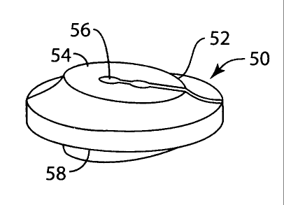
[00142] Figures 1 A to 1C illustrate various examples of artificial disc
nuclei where the
nucleus is symmetrical, with a maximum central axis in the geometric center 20
of a
nucleus 10. The reference letters A and P illustrate the anterior and
posterior orientation,
respectively, of the nuclei 10, 14 and 18. The nucleus 10 is generally
spherical in shape and
is truncated with a flattened portion 22A on the upper side of the nucleus 10
and another
24
CA 02712060 2010-07-13
WO 2009/109045
PCT/CA2009/000268
flattened surface 22B on the lower side. The nucleus also has upper and lower
curved
surfaces 24A and 24B, respectively, and a circumferential wall 26.
[00143] The flattened surfaces, as described above, can be advantageous
because when the
nucleus has a completely rounded surface, it cannot reliably maintain
correction when
exposed to the variable forces of the head and neck. A flattened surface
incorporated into the
central region of the nucleus can be used to solve this problem. The flattened
surfaces have a
contour different from the adjacent surface, and are formed in the nucleus
body. The terms
"flattened section" or "flattened surface" are used interchangeably and are
meant to refer to
any contour that is not the same as the adjacent surface(s) of the nucleus.
Such a flattened
surface can be planar or it be slightly convex or concave and have a radius of
curvature
different from the adjacent surface. Such a flattened surface could also be in
the shape of a
compound curve or other complex shape.
[00144] This flattened surface can be angled relative to the superior end
plate of the
inferior vertebral body (or vice versa, or both), with the height of the
anterior end being
greater than the height of the posterior end when lordotic correction is
sought. The overall
shape of the core can still be asymmetric, but the flattened surface can be
incorporated to
provide a reliable correction of the deformity. This flattened segment
provides stabilization
to resist the moments acting through the nucleus, i.e., if the flat is not of
adequate size, there
may be a tendency for the correction to disappear in the presence of an
anterior load or for a
hyper-lordotic over correction in the presence of a posterior load (during
lordotic correction).
Another advantage of the flattened segment is to provide surface contact over
that area during
small movements about the, neutral position of the device, which could help
reduce wear on
the device.
[00145] Figure lA illustrates a nucleus 10 that has not been adapted for
lordotic correction
because the upper and lower surfaces 22A and 22B are parallel to each other.
In this nucleus,
the axis 20 of greatest height falls in the center of the disc. In Figure 1B,
a nucleus 14 that
provides 3 of correction is illustrated. This nucleus provides for lordotic
correction.
Figure 1C illustrates another artificial disc nucleus 18 having a greater
degree of deformity
correction. When deformity correction is provided as shown in Figures 1B and
1C, the
geometric center of the nucleus may shift to a location that is offset from
the axis 20.
[00146] If the anterior/posterior directions are reversed, it provides a
kyphotic correction.
If the nucleus is rotated 90 degrees, a scoliotic correction is provided. In
the illustration in
CA 02712060 2010-07-13
WO 2009/109045
PCT/CA2009/000268
Figure 1C, the maximum vertical axis 20 is positioned to provide a correction
of 6 . It is
apparent that the nucleus can be adjusted to provide various degrees of
correction and, in
certain cases, if no degree of correction is needed. Alternatively, only one
of the halves of
the nucleus 10 may have a flattened portion, with the other half having an
outer surface that is
curved.
[00147] In Figures 2A through 2C, asymmetrical ovoid embodiments of an
artificial
nucleus are shown. The nucleus comprises upper and lower surfaces 22A and 22B,
which are
"flattened" by virtue of the ovoid shape of the nucleus, upper and lower
curved surfaces 24A
and 24B, and a circumferential center portion 26. In the embodiments shown in
Figures 2B
and 2C, the maximum height axis 16 is asymmetrical with the geometric center
12 of the
disc. In the nucleus shown in Figure 2A, where there is no correction, the
maximum vertical
height is at the central vertical axis 12. In the nucleus shown in Figure 2B,
the maximum
vertical axis 16 is positioned to provide an angle of correction of 3 . In the
nucleus shown in
Figure 2C, the maximum vertical axis 16 is positioned to provide an angle of
correction of 6 .
[00148] Figure 3 is a top view of one example of a nucleus. This nucleus 40
comprises a
central convex or flattened region 42, which includes a groove or slot 44.
This groove or slot
44 enables the nucleus to slide onto the central keel or anchor of an end
plate (not shown).
While the nucleus 40 is shown as essentially circular, it is clearly apparent
that it may take on
other shapes such as an ovoid or ellipsoid shape. It is also clearly apparent
that other types of
anchor receiving means can be used. For example, the shape of the groove may
vary or a
snap-in or bayonet or dog-bone type of receptacle can be provided to anchor
the nucleus in
position. Those practiced in the art can provide additional locking methods
including the
addition of one or more parts to the core that provide an anchor.
[00149] For deformity correction, the nucleus may take the form of a truncated
curved
body as shown in Figure 4. For this embodiment, the nucleus 50 has an upper
surface 52 that
terminates in essentially flattened planar top 54. A slot 56 or a groove or
opening of another
appropriate shape, can be formed in upper surface 52 for receiving an anchor
formed in the
end plate. The lower surface 58 is typically an inverse of the upper surface.
However,
instead of being truncated with a flat surface as shown in Figure 4, the
bottom surface could
be asymmetrically spherical or ovoid in shape.
26
CA 02712060 2010-07-13
WO 2009/109045
PCT/CA2009/000268
[00150] Alternatively, the nucleus may be circular, ovoid or egg-shaped having
a non-
central maximum vertical axis as shown in Fig 5. In another embodiment, the
nucleus could
be essentially circular or asymmetrically spherical.
[00151] Figure 5 illustrates an artificial nucleus 60 where the upper
surface 62 is an
asymmetric convex surface. Again, either the top or the bottom or both
surfaces may be
asymmetric.
[00152] For illustrative purposes, the nuclei in the figures have been shown
adapted for
lordotic correction. It is clearly apparent that the nucleus can have an
asymmetric maximum
height at the front (anterior), the rear (posterior) or the side (lateral).
The asymmetrical
nucleus of the present invention can be used to correct for various types of
spinal
misalignment including sagittal and coronal deformity.
[00153] The novel corrective nucleus of the present invention may be provided
alone or it
may be provided in combination with an upper end plate, a lower end plate or
both an upper
and a lower end plate.
[00154] Figures 6 through 8 illustrate an exemplary artificial end plate 70
that can be used
in conjunction with the nucleus to provide a novel artificial disc unit. An
artificial end plate
according to the present invention comprises an inner surface with a concave
bearing surface
for receiving the convex surface of an artificial disc. The outer, or bone
contacting, surface is
essentially flat.
[00155] To accommodate some previously known end plates, it was necessary to
spend a
significant amount of surgical time to prepare the vertebrae to the
appropriate shape to
accommodate the artificial end plate. Figure 6 shows an end plate 70 with a
flat outer surface
72 that enables the end plate to slide on the surface of the vertebra. One or
more
unidirectional keels 76 are formed on the outer surface 72 to provide for
immediate fixation.
The keels may be placed centrally or parasagittally. Fixation can be enhanced
by
incorporating onto the outer surface 72 a suitable coating 74, such as porous
titanium, a
calcium phosphate or the like, to promote bony ingrowth for long term
stability.
[00156] A stop member 78 can be provided at the anterior edge 80 of the end
plate. The
stop member prevents the prosthesis from migrating posteriorly and possibly
impinging on
the spinal cord. An essentially semi-circular wall 82 joins the outer surface
of the end plate
to the inner surface. The thickness of 82 may vary with increased thickness
anteriorly,
27
CA 02712060 2010-07-13
WO 2009/109045
PCT/CA2009/000268
posteriorly or parasagittally, as discussed further below. The inner surface
84 is shown in
greater detail in Figure 7.
[00157] The inner surface 84 of the end plate articulates with the nucleus. In
the
embodiment shown in Figure 7, this inner surface has a concave region 86,
which receives
the nucleus. An anchor 88 is provided in the center of the concave region 86
for positioning
the nucleus and preventing it from migrating. The anchor 88 can be generally
rectangular in
shape with rounded edges, as shown, avoiding premature wear and cutting into
the nucleus.
Figure 8 illustrates a front view of the end plate showing the outer surface
72 having two
parasagittal keels 76 and the inner surface 84 having a concave region 86 and
a central anchor
88.
[00158] Figures 9-12 illustrate a nucleus and end plates described above
assembled into a
TDR implant. Figures 9 and 10 show the use of a nucleus 96 with a truncated
cylinder shape
and a flattened portion 97 on the superior side of the nucleus as described
above, in
conjunction with Figures 1A-1C, and Figures 11-12 show the same design with a
nucleus 96
having an ovoid shape as shown in Figures 2A-2C. In these figures, a complete
spinal disc
prosthesis 90 comprising a superior end plate 92, an inferior end plate 94 and
an artificial disc
nucleus 96 is provided. The end plates and nucleus can be provided in
different sizes to
accommodate differences in anatomy. The end plates and various nuclei can be
provided in a
kit to the surgeon so that the appropriate sized components can be selected
and used when the
final size is determined. The end plates may be fabricated of titanium or
titanium alloy,
chrome-cobalt-molybdenum (CoCrMo), cobalt 28 chromium molybdenum, cobalt
chrome,
ceramics or other material suitable for spinal prosthetic implants.
[00159] The end plates have two distinct surfaces. The outer surface 98 is the
surface that
contacts the vertebral end plate. The outer surface is essentially flat
enabling it to easily
contact the surface of the natural vertebral end plate. The flat surface can
be porous and
incorporate a suitable treatment, such as porous titanium, a calcium phosphate
or other types
of known treatments such as coatings, plasma sprays, and structural changes to
the surface,
that promote bony ingrowth or ongrowth for long-term stability. At least one
parasagittal
keel 100 is formed on the outer surface of each end plate to provide immediate
fixation.
[00160] As shown in Figs. 9-12, three parasagittal keels 100 are aligned with
each other
and located along both sides of the outer surface of the end plates.
Alternatively, as shown
Figure 9A a similar end plate design with an upper end plate 92 and a lower
end plate 94
28
CA 02712060 2010-07-13
WO 2009/109045
PCT/CA2009/000268
have an offset keel configuration with a pair of aligned parasagittal keels
100A formed on the
outer surface of the upper end plate and a centrally-located row of aligned
keels 100B formed
on the outer surface of the lower end plate 94. This latter arrangement is
believed to be
advantageous because, with the upper and lower keels being offset from each
other, the end
plates should have greater stability and result in less stress on a vertebra
where multiple
implants are used.
[00161] Referring back to Figures 9-12, the inner surface 102 of each of the
end plates has
a concave region 103 or bearing surface that articulates with the nucleus. An
anchoring
protrusion 104 projects outwardly from the concave region, which provides an
anchor for the
nucleus and restricts posterior translation. Both the superior and the
inferior end plates have
flanges 106 for preventing the end plates from migrating into the spinal
canal. The end plates
can have holes 107 for allowing the end plates to be connected to the adjacent
vertebrae
through either metallic or bioabsorbable screws (not shown) that can be
inserted through
holes 107. Figures 9 and 11 illustrate front views of the prosthesis and
Figures 10 and 12
illustrate side views.
[00162] In another aspect of the invention, shown in Figures 13A-13B and 14A-
14C,
spinal deformity can be addressed by providing an artificial spinal disc
prosthesis where
correction is provided in the end plates. Corrective end plates may be
provided alone, in
combination with a symmetrical artificial nucleus that has flattened surfaces
as described
above on both the top and bottom of the nucleus, as shown in Figures 13A-13B,
or in
combination with an asymmetrical nucleus that has flattened surfaces as
described above on
both the top and bottom of the nucleus, as shown in Figures 14A-14C.
[00163] Correctional end plates are shown in Figures 13A-13B and 14A-14C. The
degree
of correction can be achieved by altering the inner (nucleus-contacting) side
of the end plate
or the outer (vertebral-contacting) side of the end plate. As shown in Figures
13A-13B, the
end plate 110 comprises an outer (bone-contacting) surface 112, an inner
surface 114, and a
perimeter wall 116 connecting the outer and inner surfaces. The height of the
perimeter wall
116 may vary according to the degree and type of correction required. For
example, Figure
13B illustrates an end plate adapted for a greater degree of correction than
the end plate of
Figure 13A. The positioning of the variable height can be adjusted to treat
different
conditions such as lordosis, kyphosis or scoliosis. The inner surface may be
shaped to
29
CA 02712060 2010-07-13
WO 2009/109045
PCT/CA2009/000268
receive the nucleus, and the height of the end plate can be adjusted according
to the degree of
correction required.
[00164] Alternatively, as shown in Figures 14A-14C, the outer surface 120 and
the inner
surface 122 may be essentially planar and the height is adjusted as the outer
and inner
surfaces become increasingly non-parallel as a result of variation in the
height of the
perimeter wall 124. Figures 14A through 14C illustrate increasing degrees of
correction,
respectively. An advantage of having an essentially planar outer, or vertebral-
contacting,
surface is that the device is easier to insert and requires less operating
time to prepare the
vertebral surface as compared to traditional artificial disc devices.
[00165] Figures 15,16A and 16B illustrate another embodiment of the invention,
which
provides for all directions of movement, flexion/extension, lateral bending,
and rotation about
the symmetrical axis. In this design, the nucleus 130 is formed in two
sections 130A
and 130B. A post 132 is formed on the inner surface of one section 130A, and
fits in an
opening 134 that is formed on the inner surface of the other section 130B to
provide for
relative rotational movement between the two sections 130A and 130B. The post
132 and
opening 134 can be formed on either section of the nucleus 130. The post and
opening can
be of any suitable size, and can be perpendicular to the opposing surfaces of
the nucleus
sections 130A and 130B, or be tilted at an angle off horizontal to orient the
axis of axial
rotation with the anatomically correct axis and provide a deformity
correction.
[00166] In this configuration, the contact surfaces between the nucleus 130
and end plates
136 and 138, are designed to have the same corresponding asymmetrical contours
at the
preferred angle between them, as shown in Figure 15. Because there is only
relative
movement between the nucleus and the end plates in the anterior/posterior and
medial/lateral
directions, greater surface contact between the nucleus and the respective end
plates is
possible in order to transmit rotations of the end plates to the nucleus so
that the two halves
130A and 130B, of the nucleus 130 will rotate with respect to each other,
rather than having
the end plates 136 and 138, rotate on the outer surface of the nucleus 130.
[00167] Figures 17 and 18 show another embodiment of the invention where
instead of
forming the nucleus 130 of a single piece of material, it can be formed of
upper and lower
sections 130A and 130B, with an intermediate section 140, that is either flat
or wedge-shaped
as shown in Figure 17, fixed to the upper and upper and lower sections. The
intermediate
section 140 can provide the nucleus with the appropriate degree of correction
as shown in
CA 02712060 2010-07-13
WO 2009/109045
PCT/CA2009/000268
Figure 18, instead of providing wedge-shaped end plates as discussed above. In
a related
embodiment of the invention, the nucleus 130 is essentially cut in half and
has a flat inferior
surface. This can be applied to the embodiment seen in Figures 17 and 18,
where the
section 130B is removed, leaving the inferior surface of intermediate section
140 articulating
with the inferior end plate. By varying the configuration of the intermediate
section 140,
deformity correction can be achieved.
[00168] Figures 19 and 20 show another embodiment of the invention where the
nucleus
130 is essentially cut in half and has a flat lower inferior surface. This
shape can be used to
resist expulsion of a nucleus with an ovoid/asymmetric shape, which could
occur when the
ovoid shape of the nucleus causes the end plates to tilt relative to each
other to provide
correction. As shown, the bottom surface of the nucleus 130 is flat and is
formed with a
circular opening 134 that is shaped and positioned to receive a post 136
formed on the
opposing surface of the lower end plate 138 for allowing relative rotational
movement
between the nucleus 130 and the end plate 138. Alternatively, the nucleus
could have the flat
surface and opening 134 on its upper or superior surface, instead of being on
the lower
surface as shown. In this embodiment, the nucleus is preferably asymmetrical
as shown in
Figure 19.
[00169] A modification of the configuration in Figures 19 and 20, is shown in
Figures 21
through 24, where the nucleus 130 is positioned in a slot or trough 142 formed
in the upper
surface of the lower end plate 138. As shown in Figure 21, the undersurface of
the upper end
plate 136 is contoured to match the nucleus. Alternatively, as shown in Figure
24, the
undersurface of the end plate 136 can be flat and engage a flattened upper
surface 144 of the
nucleus 130.
[00170] In another embodiment, shown in Figure 21A, lordotic correction is
achieved in a
prosthesis that replicates the movement of natural vertebrae. A nucleus 130 is
provided that
has a concave configuration on its superior articulating surface 137, as
discussed above. This
concave articulating surface 137 may incorporate a flattened segment as shown
above. The
nucleus 130 is asymmetrical in that its superior surface 137 is formed with an
anterior aspect
145a that has a greater vertical dimension, i.e., is thicker, than the
posterior aspect 145b. The
superior endplate 136 has an undersurface 147 that is similar in shape and
articulates relative
to the superior surface 137 of the nucleus 130. The corresponding articulating
surfaces 137
and 147 can be rounded in the shape of a compound curve or other type of
continuous curve,
31
CA 02712060 2010-07-13
WO 2009/109045
PCT/CA2009/000268
so that they are essentially congruent through a full range of motion. The
superior surface of
the bottom end plate 138 is formed with a defined recess 139 that is highly
polished,
preferably to a mirror finish. The nucleus 130 is positioned for movement in
the recess 139,
with the elevated edges that form the recess limiting movement of the nucleus
relative to the
bottom end plate. The nucleus 130 can be sized and shaped to provide for
controlled motion
between the nucleus and the bottom end plate so that the nucleus 130 can slide
in a gliding
movement from side-to-side and/or from front-to-back relative to the superior
surface 139 of
the bottom end plate 138 to provide for relative translational movement in the
medial-lateral
and/or anterior-posterior directions as indicated by the arrows 146 and 148.
[00171] The trough can be larger than the nucleus in both the
anterior/posterior and
medial/lateral directions to allow for a desired amount of translation in
those directions as
shown by the arrows A and B in Figure 22. The trough can be open on the
posterior or
anterior end to allow the nucleus to be inserted simply by sliding it into the
trough, as shown
by the arrow A in Figure 21. In this way, the nucleus can be inserted without
undue
distraction of adjacent vertebrae. The nucleus can be prevented from moving
out of the
trough by providing a stop 144 of any suitable size and shape. Figure 23 is a
schematic view
of the nucleus 130 that is inserted in the trough in Figure 21.
[00172] Another embodiment of the invention is shown in Figures 25-31, where
the
nucleus 130 is elongated, with a flattened section 150 that is a partial
cylinder with curved
sections 152 and 154 on both sides of the flattened section. It is believed
that this design,
when mated with a cylindrical surface 156 on the interior of the upper end
plate 136, shown
in Figure 29, will provide better wear characteristics because it will have
surface contact
during medial/lateral bending and line contact during flexion/extension.
[00173] The elongated shape of the nucleus 130 is illustrated in Figures 25
and 26, which
show that the nucleus has a round cross section with constant medial-lateral
radius from
anterior to posterior (A-P), with the flat section 150 in the middle being
oriented to provide a
correction angle as described above, for the flatted portions on the other
embodiments of the
nucleus. The interior surface 156 of the upper end plate 136 has a cylindrical
shape with the
same constant radius in the anterior/posterior direction as the nucleus.
[00174] In the neutral position, the cylindrical surface 156 mates with the
flattened section
150 of the nucleus 130, and sits at an angle that provides a deformity
correction as shown in
Figure 31. In this position, there is surface contact between the end plate
136 and the
32
CA 02712060 2010-07-13
WO 2009/109045
PCT/CA2009/000268
nucleus 130. During medial/lateral bending, there is also surface contact
between the end
plate and nucleus. During flexion/extension, with or without lateral bending,
there is line
contact between the end plate and nucleus. This configuration of core and end
plate will
always have line or surface contact, thus reducing the wear potential from
point contact in
some of the previous designs.
[00175] The elongated shape of the nucleus 130 allows for the end plate 138 to
have a
trough 157 in the shape of a "bow tie" as shown in Figures 27 and 28. This
shape allows for
axial rotation with stops beyond the limits of normal motion. The shape is
oversized relative
to the nucleus 130 by an appropriate amount to allow limited
anterior/posterior and
medial/lateral translation. Additionally, the bottom surface of the trough 157
can be rounded
upwardly at the medial/lateral sides in Figure 30 (not shown), so that as the
nucleus 130
rotates it is "cammed" up causing a distraction of the device that forces the
vertebral bodies
apart and loads the tissues between them resulting in a gradual stop to the
motion.
Translation of the nucleus 130 within the trough 157 will tend to preserve the
mobile
instantaneous axis of rotation of the natural disc.
[00176] Figures 32 and 33 show another embodiment of the invention, which
utilizes a
universal joint formed of a pair of cylinders 160 and 162 that rotate relative
to each about a
central post 164 that projects from one of the cylinders 162 and engages an
opening 166 in
the other cylinder 160. The cylinders 160 and 162 are oriented perpendicular
to each other
and engage cylindrical surfaces 168 and 170, respectively, in the adjacent end
plates 136
and 138. This design provides for three anatomical axes of rotation. Because
of the
independence of each axis of rotation, any correction provided by the shape of
the nucleus
that is formed of the two cylinders will result in rotation to compensate for
the correction and
a return to the uncorrected neutral position. Alternatively, the cylinders 160
and 162 may be
shaped similarly to the elongated nucleus 130 shown in Figures 25-27, or
another suitable
shape with a flat inferior surface.
[00177] Another embodiment of the invention is shown in Figures 34-36, where a
resilient
ring 172 and a post 174 that has a rounded top portion 176 provide for
relative motion
between the end plates and for the desired angle of correction. The ring 172
is shown in
detail in Figure 35. The ring 172 can be wedge shaped as shown in order to
provide the
desired amount of correction, or it can be flat (not shown) if no correction
is desired. A
projection 180 can be formed on the upper surface of the lower end plate 138
to mate with an
33
CA 02712060 2010-07-13
WO 2009/109045
PCT/CA2009/000268
opening 182 in the ring 172 in order to prevent the ring 172 from moving
relative to the lower
end plate once the ring is in its desired position.
[00178] The upper end plate 136 has a cavity 178 that can be contoured to
match the shape
of the rounded top portion 176. The ring 172 is shaped so that the end plate
136 will ride on
the ring 172 during "normal" ranges of motion, or through regular activities.
However, when
the normal ranges of motion are exceeded, then the ring 172 will compress and
the upper end
plate 136 will engage the post 174 causing the adjacent vertebrae to distract
and thereby
provide a gradual stopping motion or "anatomically-derived gradual stopping."
Alternatively, the post 174 could be designed to serve as the primary load
carrying part of the
articulation by riding in the cavity 178. In this design, the deformity
correction force is only
provided by compressing the ring 172. This design would have the advantage of
reducing
material stresses in the elastomer ring and creep.
[00179] As shown in Figure 37, instead providing the ring 176, the same result
could be
achieved by providing two or more stops 184A and 184B, formed of a resilient
material,
between the two end plates. The stops 184A and 184B can be mounted on the
upper surface
of the lower end plate 138. One of the stops 184A can project upwardly a
greater distance
than the other stop 184B in order to provide the desired correction.
[00180] Another embodiment of the invention is shown in Figures 38 and 39,
where a
nucleus 186 is provided that is formed of a resilient material that is shaped
so that the nucleus
provides medial/lateral rotation, but requires deformation of the material
during flexion and
extension. This is accomplished by proving a central portion 188 that is
spherical or ovoid in
shape and "flattened" adjacent end portions 190A and 190B that are
cylindrical, which extend
the flattened end portions around the circumference of the nucleus at both
ends. The upper
end plate has a cavity (not shown) that has a contour that is similar in shape
to the nucleus
186. A trough (not shown) similar to the one in Figures 27 and 28 can be
formed in the lower
end plate 138.
[00181] For medial/lateral movement in the direction of the arrows A-A, the
upper and
lower end plates will rotate relative to each other through rotational
movement of the upper
end plate on the nucleus 186. However, flexion/extension in the direction of
arrows B-B will
occur only through deformation of the nucleus 186. Alternatively, the nucleus
186 can be
rotated 900 on the lower end plate 138 so that so that the end plate 136 will
rotate on the
nucleus during flexion/extension and the nucleus will deform during
medial/lateral
34
CA 02712060 2010-07-13
WO 2009/109045
PCT/CA2009/000268
movement. The end portion 190A has a larger diameter than the end portion 190B
to provide
for the desired amount of correction. As shown, the nucleus has been shaped so
the resilience
of the nucleus varies over its length. However, the nucleus could be formed of
materials
having varying degrees of resiliency along its length to achieve the same
results.
[00182] Figures 40-43 illustrate another embodiment of the invention where the
end plates
136 and 138 are provided with stops outside of the normal range of motion,
which also utilize
the concept of "anatomically-derived gradual stopping" discussed above in
conjunction with
Figures 34 and 35. This type of stop can be added to any design that employs
the use of end
plates. This aspect of the invention is based on duplicating how the human
body moves and
then designing the cooperating surfaces to mimic those motions as closely as
possible. As
shown in Figure 40, the end plate 136 has a post 200 on its lower surface that
engages pocket
202 formed in the upper surface of the lower end plate and 138. Preferably, a
pair of posts
and pockets are provided on opposite sides of the nucleus 130.
[00183] As shown in Figures 40-43, the pocket 202 has a slot 204 in it with a
curved
surface 206 that is engaged by the lower end 208 of the post 200. As the end
plates 136 and
138 move in the anterior/posterior direction relative to each other during
extension/flexion,
the lower end 208 of the post rides along the curved surface 206. As the post
reaches the
outer limits of travel the lower end 208 will begin riding up the gradually
curved section of
the surface 208, which causes distraction between adjacent vertebrae as
illustrated by the
arrow A in Figure 43 and loads the tissues between them, resulting in a
gradual stop to the
motion.
[00184] Figures 44a, 44b and 44c illustrate another embodiment of the
invention with a
central depression or valley in the flattened segment on the superior surface
of the core. In
this embodiment the nucleus 130 has first and second flattened sections 150
and 151 that are
formed between curved sections 152 and 154. A shallow depression or valley 330
is formed
in the flattened sections 150 and 151 and the curved section 152 to provide an
indention with
slightly elevated margins 310, 320 that are rounded and contiguous with the
adjacent
surfaces 150, 151 and 152. The depression 330 is elongated in the
anterior/posterior
direction, along the sagittal plane SP. The depression 330 and margins 310 and
320 are
symmetric about the sagittal plane SP. A pair of transition zones 340 and 350
extend
between the depression 330 and the surrounding surfaces. This configuration is
designed to
potentially improve load distribution as the superior endplate moves relative
to the nucleus
CA 02712060 2010-07-13
WO 2009/109045
PCT/CA2009/000268
because contact is distributed along the margins 310 and 320 instead being
concentrated
along the mid-sagittal plane or other portion of the nucleus.
[00185] Another embodiment of the invention is shown in Figures 45, 45a, 45b
and 45c,
where the posterior superior surface 360 of the nucleus 130 is formed to curve
upwardly
toward the superior end plate 136 relative to the superior surface 350 of the
nucleus in order
to provide a relatively elevated posterior surface region 360. The superior
end plate 136 has
a corresponding surface 370 contoured to fit the posterior superior surface
360 of the nucleus.
This configuration is designed to enhance the stability of the prosthesis by
allowing the
endplates 136 and 138 to resist posterior shear when they are in a lordotic
position. Figures
45b and 45c illustrate the relative positions of the components in Figure 45
as the spine
moves in flexion and extension, respectively. The elevated surface region 360
and its
corresponding surface on the end plate 136 causes shear loads to be
transmitted through the
nucleus and into the inferior endplate. Consequently, abnormal loads on facet
joints of the
natural spine and related soft tissue structures are prevented.
[00186] Figure 45b illustrates another feature of the invention where the
nucleus 130 has a
tang or tab 300 that extends posteriorly from the posterior-inferior aspect of
the nucleus 130,
which fits in an undercut portion or recess 302 of the bottom end plate 138.
The tab 300
operates to resist posterior migration (i.e., expulsion) of the nucleus toward
the spinal canal
by preventing "lift off' of the nucleus from the inferior end plate and
possible dislocation of
the nucleus. As shown, the tab 300 can be rounded, chamfered or beveled in
order to
facilitate initial insertion and intra-operative or post-operative replacement
of the nucleus.
[00187] As shown in Figs. 44a and 44c, a pair of shallow cavities or dimples
342 and 344,
are formed on opposite sides of the anterior aspect of the nucleus in order to
allow a
retrieval/insertion instrument to engage the nucleus for insertion and
removal. These
cavities 342 and 344 are formed in a location where they do not interfere with
the load
carrying capabilities of the nucleus.
[00188] Figures 46a, 46b, 46c and 46d show another embodiment of the invention
where a
pair of keels 100a are formed in the outer surface of the top end plate 92,
with a bony
ingrowth surface 101a formed between the keels 100a. The lower surface of the
bottom end
plate 94 has a single keel 100b formed along its anterior/posterior
centerline, or sagittal plane,
with a bony ingrowth surface 101b formed on both sides of the keel 100b. The
design of a
pair of parasagittal keels on the superior endplate and a single central keel
on the inferior
36
CA 02712060 2010-07-13
WO 2009/109045
PCT/CA2009/000268
endplate, or vice versa, prevents "in line" sagittal cuts in the vertebral
body that can increase
the possibility of fracture of the body. Alternatively, a pair of keels could
be formed on the
inferior endplate and a single keel on the superior could be used with the
same beneficial
results. This and other combinations of keels with an unequal number and/or
locations of
keels on the superior and inferior endplates will prevent excessive stress in
the vertebral
bodies when multiple spinal levels undergo device implantation.
[00189] The keels 100a and 100b include a rail 400 attached to the respective
end plates,
with multiple teeth 402 in accordance with the size of the device, on each
rail that projects in
the anterior direction. The rails 400 can include a number of holes 404 to
allow for bony
ingrowth for anchoring the rails in place after implantation. The bony
ingrowth surfaces can
be any known type of surface that allows tissue to grow into or adhere to the
upper end
plate 92. Such a surface can include sintered beads, fibers, or other
materials attached to the
upper end plate, or the surface can be roughened or textured in a known way.
The ingrowth
surfaces 101a and 101b on the top and bottom plates are protected by slight
elevations or lips,
which surround the bony ingrowth surface (not shown). Tabs 406 can be provided
to limit
posterior migration and for tooling locations for insertion instruments. A
pair of shallow
cavities or dimples 408, 410, can be formed on opposite sides of the anterior
aspect of the end
plates 92, 94, respectively, in order to allow a retrieval/insertion
instrument to engage the end
plates for insertion and removal.
[00190] In another aspect of the invention, all of the articulating surfaces
of the prosthesis
can be formed of a polymer. As discussed above, the nucleus can be formed
entirely of a
polymer such as, for example, ultra-high molecular weight polyethylene
("UHMWPE"), a
cross, linked UHMWPE, a ceramic, polyetheretherketone ("PEEK") or other type
of suitable
polymer. The bony ingrowth surfaces can be made from plasma sprayed metals,
hydroxyapatite or similar bone-like coatings, and can include a coating of
bone growth
factors. The articulating surfaces of the end plates can be formed with
inserts of an
appropriate polymer, ceramic or the like. The remaining exterior surfaces of
the end plates
that interface with bone can be formed with bony ingrowth surfaces of the type
discussed
above.
[00191] Figs. 47 and 48 illustrate a frame or scaffold 500 that is used to
distract adjacent
vertebral bodies 502 and 504 of the human spine, looking in the direction of
the anterior
surface of spine with the scaffold 500 in place. The scaffold 500 also
operates as an
37
CA 02712060 2010-07-13
WO 2009/109045
PCT/CA2009/000268
instrument guide for preparing adjacent bone surfaces on the vertebral bodies
502 and 504.
The scaffold 500 is held in place by locking mechanisms 490 that attach to the
vertebral
bodies 502 and 504 through distracting pins 492 that are connected to the
vertebral bodies
502 and 504.
[00192] The vertebral bodies 502 and 504 are distracted in a known way by
means of a
distracting instrument (not shown) that is connected between the distracting
pins 492 that are
connected to the vertebral bodies for distracting them or spreading them apart
to create a
space 509 between them after the natural nucleus (not shown) is removed.
[00193] As the scaffold 500 is inserted between the vertebral bodies 502 and
504, the
locking mechanisms 490 are placed over the distracting pins 492 and screws 491
are
tightened to hold the scaffold 500 in place. As shown in Figure 48, anchoring
spikes 510 on
the inner surface of the scaffold 500 on opposite sides of the space 509
engage the vertebral
bodies 502 and 504 and provide additional support for anchoring the scaffold
500 to the
vertebral bodies.
[00194] A pair of lateral projections 506 on the inner surface of the scaffold
500, extend
posteriorly into the disc space 510, bilaterally, for maintaining the
vertebral bodies 502 and
504 at a specific disc height. Grooves 514 are formed on the facing surfaces
of the
projections 506 for guiding instruments such as a trial, a drill guide and a
keel shaper used
during the implantation process, which are described in greater detail below.
[00195] Figure 49 shows one of a set of trial instruments 516 with a handle
516a, that can
be inserted in the scaffold 500 or directly into the disc space to gauge
AP/medial-lateral
endplate coverage between the vertebral bodies 502 and 504 for determining the
correct
implant size that will be used. The side surfaces of the trial instruments 516
may have
profiles 515 that are shaped to fit into the grooves 514 formed on the
scaffold projections
506. The trial instruments also may have two or more stops 513a and 513b for
preventing the
instrument from being inserted too far posteriorly between the endplates 502
and 504. The
set of trial instruments should be sized and shaped to at least have the
capability of gauging
various degrees of lordotic correction, preferably at least 00, 3 and 6 of
lordotic correction,
and to determine the size of the anterior-posterior and medial-lateral
dimensions of the
endplates for the prosthesis.
[00196] Figure 50 shows a drill guide 518 with a handle 518a, and guide holes
520a, 520b
and 520c formed in tabs 519a, 519b and 519c, respectively, for setting the
trajectory for drill
38
CA 02712060 2010-07-13
WO 2009/109045
PCT/CA2009/000268
bits (not shown) that are used to make starter holes in the vertebral body 502
and 504, for
grooves that will ultimately receive keels formed on the endplates of the
prosthesis that are
described in greater detail above. The tabs 519a, 519b and 519c also operate
as stops for
preventing the drill guide from being inserted too far posteriorly into the
disc space 509
between the vertebral bodies 502 and 504. The drill guide 518 has side
surfaces with profiles
515 that are shaped to fit into the grooves 514 formed in the lateral
projections 506 on the
scaffold 500. The guide holes correspond to the location of the keels formed
on outer
surfaces of the upper and lower end plates of the prosthesis described above,
and are used to
guide drill bits (not shown) for forming starter holes (not shown) that open
onto the facing
surfaces of the vertebral bodies 502 and 504, so that they can be shaped to
receive the keels
by the instrument shown in Figure 52. Figure 51 shows the scaffold 500
positioned between
the vertebral bodies 502 and 504, ready to have the holes drilled in direction
of the arrows.
[00197] Once the starter holes are drilled, a keel cutting instrument 522 of
the type shown
in Figure 52 is inserted in the scaffold 500 for shaping the starter holes
into grooves in the
vertebral bodies 502 and 504. The keel cutting instrument 522 includes side
surfaces with
profiles 515 that are shaped to fit into the grooves 514 in the lateral
projections 506 on the
scaffold 500 so that the location and motion of the instrument can be
precisely controlled.
The keel cutting instrument also includes sharp cutting edges 524a, 524b and
524c that are
used to shape the openings that were drilled into the vertebral bodies 502 and
504, with a
simple back-and-forth movement of the instrument relative to the scaffold 500.
In this way,
grooves of the exact shape are formed in the vertebral bodies for receiving
the keels on the
prosthesis with minimal force being applied to the prosthesis. One or more
stops 526a, 526b
and 526c are provided on the instrument 515 for preventing the instrument from
being
inserted too far posteriorly between the vertebral bodies 502 and 504 as the
grooves are
formed.
[00198] As shown in Figure 52, described above, the keel cutting instrument
522 has one
keel cutter 524a placed on one surface and two keel cutters 524b, 524c on the
opposite
surface, although other configurations can used as long as there is a shape
such that at least
one keel is on one surface and at least two are on the other surface for
providing maximum
stabilization for the prosthesis and resisting torsional forces on the
prosthesis. The trial
instrument also has one or more stops 526a, 526b and 526c for preventing the
instrument
from being inserted too far between the endplates 502 and 504.
39
CA 02712060 2010-07-13
WO 2009/109045
PCT/CA2009/000268
[00199] Alternatively, the drill guide 518 and keel cutter 522 can be
replaced with a drill
guide 600 of the type shown in Figs. 53A-53D. The drill guide 600 can include
side surfaces
with profiles 515 that are shaped to fit into the grooves 514 in the lateral
projections 506 on
the scaffold 500 so that the location and motion of the instrument can be
precisely controlled.
In another technique, the scaffold 500 is not used and the drill guide 600 is
inserted into the
disc space 509 with the aid of intra-operative fluoroscopy. The drill guide
600 also can have
a collar 602 that includes three guide holes 604a, 604b and 604c for guiding
drill bits 606
shown in Figs. 53A and 53B. The collar also operates as a stop to prevent the
drill guide
from moving to far posteriorly between the vertebral bodies 502 and 504. The
guide holes
are oblong in shape so that the drill bits can be moved in the guide holes to
form not only
starter holes, but also to form the grooves for the keels on the prosthesis.
In this way, the keel
cutter 522 described does not have to be used as the groves for receiving the
keels are formed
solely through the use of the drill guide 600.
[00200] The final step of the process of implanting the prosthesis, after the
grooves for the
keels are formed, includes assembling the prosthesis of the selected size and
degree of
lordotic correction, with the nucleus sandwiched between the end plates. The
physician uses
an instrument that is designed to engage the shallow cavities or dimples 408
and 410 on the
end plates of the prosthesis described above. After lining up the keels of the
prosthesis with
the grooves created in the vertebral bodies 502 and 504, the prosthesis is
then gently tapped
into place between the vertebral bodies 502 and 504. The grooves formed as
discussed above
minimize the amount of force needed to position the prosthesis in the proper
location between
the vertebral bodies 502 and 504.
[00201] In the event that a core with an incorrect size or shape is inserted,
the disc space
could be distracted by various means, including vertebral body distracting
pins that are well
known. An instrument with small pins that are designed to engage the small
holes on the
lateral aspect of the nucleus (as described previously) is used to remove the
nucleus so that it
can be replaced with one having the correct size and shape.
[00202] Another embodiment of the invention that is designed to correct
lordosis in the
spine is a 2-piece device of the type shown in Figs. 54, 55 and 56, that
allows normal
kinematics while at the same time has a wedge-shaped geometry. This design
includes an
elongated ball or "sausage shaped" articulating bearing surface, that is
preferably located on
the inferior endplate, that has its greatest diameter either anterior,
posterior or lateral to the
CA 02712060 2010-07-13
WO 2009/109045
PCT/CA2009/000268
mid-portion of the inferior endplate. This design would alter the articulation
between the
superior and inferior endplates to provide a deformity correction.
[00203] As shown in Figs. 54, 55 and 56, an elongated or "sausage type" shaped
articulating surface 500 can be incorporated into the inferior endplate 502
such that the
prosthesis has a 2-piece design, as opposed to the 3-piece design described in
previous
embodiments. The elongated articulating surface 500 includes a flattened
segment 504 and
mates with a bearing surface 506 on the superior endplate 508. As shown in
Fig. 54, the
flattened segment 504 is incorporated onto the inferior bearing surface to
allow the resting
configuration of the engaged endplates to be in a lordotic configuration in
the shape of a
truncated cone. With this design, the inferior endplate 502 will have a
maximum vertical axis
that is not located at the geometric center of the prosthesis, but is located
either toward the
front of the endplate, the rear of the endplate or on one side of the
endplate. The positioning
of the maximum vertical height and load bearing capability is chosen depending
on the type
of deformity that needs to be corrected. In Fig. 54, the geometric center is
located toward the
front of the endplate 502, which will provide lordotic correction.
[00204] The articulating bearing surface 500 of the inferior endplate 502 also
includes
curved sections 510 and 512 on opposite sides of the flattened segment 504 to
provide the full
range of motion and translation in the sagittal and coronal planes.
[00205] The superior endplate 508 attaches to an upper vertebral member (not
shown), and
the inferior end plate 502 attaches to a lower vertebral member (not shown).
The endplates
have an essentially flat outer or vertebral-contacting surface that allows
them to be easily
inserted. There can be keels or teeth 514, having the configuration discussed
in detail above,
to provide acute stability and anchoring of the device. The keels can be
staggered to avoid
stress concentration in the bone. There may be two parasagittal keels on the
superior
endplate and one midline keel on the inferior endplate or vice versa. The
outer surface of the
end plates 502 and 508 may be treated in a way that promotes bony ingrowth to
enhance
stability of the end plate in situ. The end plate can include a stop member
516 to prevent the
prosthesis from moving toward the spinal canal.
[00206] The end plates can have two distinct surfaces. The flat surface of
each end plate,
which contacts the vertebral body end plate, is capable of accommodating bony
ingrowth and
incorporates a suitable coating, such as porous titanium, a calcium phosphate,
or includes
other types of known surfaces mentioned above that promote bony ingrowth for
long-term
41
CA 02712060 2010-07-13
WO 2009/109045
PCT/CA2009/000268
stability. The end plates can also have one or more parasagittal keels that
provide immediate
fixation.
[00207] The two pieces 502 and 508 may be composed of a low friction elastomer
such as
polyurethane, polycarbonate-polyurethane, a polymer such as polyethylene
(particularly
ultra-high molecular weight polyethylene), a suitable ceramic, metals or metal
alloys such as
titanium or a titanium alloy, chrome-cobalt-molybdenum (CoCrMo), cobalt 28,
chromium
molybdenum, cobalt chrome, stainless steel, PEEK or other suitable materials.
The flattened
segment 504 of the inferior endplate 502 has a generally cylindrical geometric
design, with
varying degrees of lordosis incorporated into it by utilizing an axis of
maximum height
anterior to the geometric center of the inferior endplate. The anterior height
of the inferior
endplate varies, depending on the extent of lordotic correction needed. The
inferior endplate
can be made available in various lordotic angles, e.g. 0, 3 and 6 , as well
as differing heights
(e.g., 4, 6 and 8 mm). Before deciding on the final device size, a set of
instruments of the
type described above or other means can be used to gauge the need for lordotic
correction.
[00208] As mentioned above, the articulating bearing surface 500 of the
inferior endplate
502 is elongated, with a flattened section that is either a partial cylinder
or a partial cone, with
curved sections 510 and 512 on both sides of the flattened section. This shape
is designed so
that when the articulating bearing surface 500 of the inferior endplate is
mated with the
cylindrical bearing surface 506 on the superior endplate 508, better wear
characteristics will
result because there will be surface contact during medial/lateral bending and
line contact
during flexion/extension.
[00209] In the neutral position, the bearing surface 506 mates with the
flattened bearing
surface 504 and sits at an angle that provides the desired deformity
correction. In this
position, there is surface contact between the superior end plate 502 and 508
and the inferior
endplate. During medial/lateral bending, there will also be surface contact,
but between the
bearing surface 506 and one of the curved sections 510 and 512.
[00210] In Figures 57-76, various embodiments of joint replacing implants are
depicted,
including a carpometacarpal joint implant, a metatarsophalangeal joint
implant, a
metacarpophalangeal joint, a first metatarsophalangeal joint, a distal and a
proximal
interphalangeal joint implant, an ankle implant, a total knee implant, a hip
implant, and a
shoulder implant. The bearing surfaces of each implant may include an
orientation feature
such as a flattened section similar to those disclosed above. The flattened
sections may be
42
CA 02712060 2010-07-13
WO 2009/109045
PCT/CA2009/000268
contiguous with other portions of the bearing surfaces, meaning they are
immediately
adjacent to those portions.
[00211] Referring to Figure 57, a sagittal view of a carpometacarpal joint is
shown, with a
metacarpal component 610 of a carpometacarpal prosthesis 600 implanted in the
metacarpal
bone, and a trapezal component 650 of the prosthesis implanted in the
trapezium and
potentially extending to the scaphoid. The metacarpal component 610 has a stem
portion 612
joined to a dome-like head portion 614. Positioned opposite the metacarpal
component 610 is
the trapezal component 650, comprising a cup-like receptacle portion 654 and a
stem portion
652. The head portion 614 is configured to mate with and articulate within the
receptacle
portion 654, to replace the motion of the natural carpometacarpal joint.
Alternatively the
dome-like head could be positioned in the trapezium and the cup-like
receptacle on the
metacarpal bone.
[00212] Referring to Figure 58, a perspective view of the metacarpal component
610 and
trapezal component 650 are shown. The stem portion 612 of the metacarpal
component 610
has a generally cylindrical stem body 616, which extends from the head portion
614 to a
tapered, pointed tip 618. The stem body 616 may have a constant diameter along
its length or
may taper from the head portion 614 to the tip 618. Threads 620 encircle the
stem body 616
at its proximal end and continue onto the tip 618. In other embodiments of the
invention,
threads or grooves or porous and/or hydroxyapatite may be present along part
or all of the
stem body, or the stem body may be non-threaded. Additionally, it is
appreciated that the
stem body 616 may not be cylindrical in form with a round cross-sectional
shape, but have
different cross-sectional shapes such as a square, a rectangle, a triangle, a
hexagon, or another
shape.
[00213] Distal to the stem portion 612, the head portion 614 comprises a base
622 and a
dome 624. The base 622 is a flat, circular element from which the dome 624
extends distally.
The base 622 has a proximal-facing bone contacting surface 626 which is
configured to be
seated in the prepared metacarpal. The bone contacting surface 626 may be a
porous or non-
porous tissue ingrowth surface. A thickness of the base may vary from a thin,
bone
conserving profile to a thicker bone replacing profile. The dome 624 has a
generally
hemispherical configuration; however a central section of the dome, extending
from one edge
to the opposite edge is shaped as a portion of a cylinder having a constant
radius, resulting in
a flattened section 630. Alternatively the dome may have an asymmetric
configuration with
43
CA 02712060 2010-07-13
WO 2009/109045
PCT/CA2009/000268
the flattened section placed outside the central section of the dome. The
flattened section 630
has a constant radius, and lies in an anterior-posterior orientation; however
in other
embodiments it may lie in a medial-lateral orientation. The flattened section
may also be
available in varus and valgus and flexion and extension angled implants in
order to correct
preoperative varus and/or valgus deformity and flexion and/or extension
deformity.
Alternatively the flattened section may be positioned to correct coronal or
rotatory deformity.
Contiguous to either side of flattened section 630 are rounded areas, a first
rounded section
632 and a second rounded section 634. The rounded areas 632 and 634 may have
the same or
differing heights and configurations. A continuous substantially convex
bearing surface 636
covers the rounded sections 632, 634 and the flattened section 630.
[00214] The introduction of the flattened section determines the alignment of
the implant
components and either introduces a preferred relative orientation for the
articulation of the
dome-like head and the cup-like receptacle or alternatively reduces a pre-
operative deformity
of the carpometacarpal joint. A prosthesis that allowed a preferred alignment
or orientation of
the carpocarpal, carpometacarpal and intermetacarpal joints would be of
particular
importance in the setting where the palmar and/or dorsal ligature are
compromised. Any
disease process that affected the static alignment of the joints in the
anatomical position
would require an active correction of the deformity by the implant.
[00215] The stem portion 652 of the trapezal component 650 is configured
similarly to the
stem portion 612 set forth previously, with a stem body 656, a tip 658, and
threads 660. In the
emobodiment depicted in Figures 57 and 58, the stem portion 652 is long enough
to engage
only the trapezium; however in alternative embodiments the trapezal stem
portion 652 may
be longer than the metacarpal stem portion 612 in order to extend through the
trapezium and
anchor in the scaphoid. Joined to the stem portion 652 at its proximal end is
the cup-shaped
receptacle portion 654. The receptacle 654 has a distal-facing bone contacting
surface 676
which is configured to be seated in the proximal side of the trapezium. The
bone contacting
surface 676 may be a porous or non-porous tissue ingrowth surface. The inside
of the
receptacle portion 654 is a substantially concave bearing surface 678
configured to mate
concentrically and articulate with the dome 624, and has a flattened section
680 which
extends from one edge of the cup to the other in an anterior-posterior
orientation. The
flattened section 680 may be centered along the antero-posterior axis;
alternately it may
offset from the center in an asymmetrical orientation. In another embodiment,
the flattened
44
CA 02712060 2010-07-13
WO 2009/109045
PCT/CA2009/000268
section 680 may be oriented along the medial-lateral axis. The flattened
section receptacle
portion is available in varus and valgus and flexion and extension angled
implants in order to
correct preoperative varus and/or valgus deformity and flexion and/or
extension deformity.
Contiguous with the flattened section 680 is a first rounded cup section 682,
and a second
rounded cup section 684.
[00216] The carpometacarpal prosthesis 600 is configured to be implanted in
the
metacarpal and trapezium bones such that the dome 624 articulates inside the
cup 654 in a
preferred relative orientation. The dome 624 may rotate partially within the
cup 654, limited
by the flexion/extension and medial/lateral movements of the surrounding
muscles and
ligaments. It may act in an unconstrained or a semi-constrained manner. As the
thumb is
moved medial-laterally, the cup 654 will slide over the dome 624. At a neutral
medial-lateral
position, the flattened section 680 of the cup will be in alignment with the
flattened section
630 of the dome.
[00217] As the thumb flexes and extends, along the dorso-palmar axis, if the
flattened
sections 630, 680 are aligned the movement will be smooth, since the curvature
of the
flattened section 680 of the cup corresponds to the curvature of the flattened
section 630 of
the dome. If the posterior-anterior movement is canted slightly medially or
laterally, the
flattened sections 630, 680 will not be aligned.
[00218] When a joint such as the carpometacarpal joint is diseased or injured,
and the joint
replaced, some surrounding soft tissues are usually irreplaceably lost due to
the disease or
injury, and the replacement procedure. Loss of the soft tissues can result in
the loss of the
natural alignment of the bones in the joint; as tissues provide natural
support and constraint to
the bones. Providing the mateable flattened surfaces 630, 680 may replace some
of the
natural joint alignment formerly provided by the soft tissues. Fixed deformity
of the
carpometacarpal joint is associated with longstanding osteoarthritis,
rheumatoid arthritis,
inflammatory and non- inflammatory arthritis and post traumatic arthritis. The
mateable
flattened surfaces are able to address preoperative deformity in the varus and
valgus plane
and preoperative deformity in all planes.
[00219] Referring to Figure 59, a lateral view of a metacarpophalangeal joint
prosthesis
700 implanted in a metacarpal bone and and a phalangeal bone is shown. The
metacarpophalangeal prosthesis 700 may be generally similar to the
carpometacarpal
prosthesis 600, however it may be sized and shaped slightly differently to
match the
CA 02712060 2010-07-13
WO 2009/109045
PCT/CA2009/000268
particular geometry of the joint. The metacarpophalangeal joint prosthesis 700
comprises a
domed metacarpal component 710 and a cup-like phalangeal component 750.
Alternatively
the dome-like head component could be positioned in the phalangeal component
and the cup-
like receptacle component in the metacarpal bone.
[00220] The metacarpal component 710 has a stem 712, a base 714, and a
substantially
convex head 716. A flattened section 730 extends medial-laterally across the
head 716.
Alternatively the flattened section 730 may extend in a antero-posterior
orientation across the
head 716. The phalangeal component 750 comprises a stem 752 and a
substantially concave
receptacle 754. A flattened section 760, shaped to correspond to the flattened
section 730,
extends medial-laterally across the receptacle 754. When the components are
implanted, the
convex head 716 articulates inside the concave receptacle 754. At a neutral
position, the
corresponding flattened sections 730, 760 align. In this preferred relative
orientation, medial-
lateral movement of the joint is smooth. Alternatively the head 716 may have
an asymmetric
configuration with the flattened section placed outside the central section of
the head.
[00221] Referring to Figure 60, a lateral view depicts a distal
interphalangeal joint
prosthesis 800 implanted in an intermediate phalange and a distal phalange.
The distal
interphalangeal prosthesis 800 comprises an intermediate phalange component
810 which is
shaped to be implanted in the distal end of an intermediate phalange, and a
distal phalange
component 850 which is shaped to be implanted in the proximal end of a distal
phalange.
Alternatively the dome-like head 810 could be positioned in the distal
phalangeal component
and the cup-like receptacle 850 in the intermediate phalangeal bone.
[00222] The distal interphalangeal joint prosthesis may be similar to the
carpometacarpal
and metacarpophalangeal prosthesis, but sized differently to accommodate the
smaller distal
interphalangeal joint. It is appreciated that a substantially comparable
prosthesis could be
made for the proximal interphalangeal joint.
[00223] Referring to Figure 61, the intermediate phalange component 810 and
the distal
phalange component 850 are shown in an enlarged perspective view. The
intermediate
phalange component 810 has a stem 812, a base 814, and a substantially convex
head 816. A
flattened section 830 extends antero-posteriorly across a portion of the head
816, and a
substantially convex bearing surface 832 extends across the distal side of the
head, including
the flattened section 830. In another embodiment of the invention, the
flattened section could
extend medial-laterally across the head, or with another orientation.
Alternatively the dome
46
CA 02712060 2010-07-13
WO 2009/109045
PCT/CA2009/000268
may have an asymmetric configuration with the flattened section placed outside
the central
section of the dome. The flattened section is also available in varus and
valgus and flexion
and extension angled implants in order to correct preoperative varus and/or
valgus deformity
and flexion and/or extension deformity.
[00224] The distal phalangeal component 850 comprises a stem 852 and a concave
receptacle 854. A flattened section 860, shaped to correspond to the flattened
section 830,
extends antero-posteriorly across a portion of the receptacle 854, and a
substantially concave
bearing surface 862 extends across the proximal side of the receptacle 854.
When the
components are implanted, the convex head 816 fits inside the concave
receptacle 854 and
the bearing surfaces 832, 862 articulate. At a neutral position, the
corresponding flattened
sections 830, 860 align. In this preferred relative orientation, antero-
posterior extension and
flexion of the joint is smooth.
[00225] Fixed deformity of the proximal interphalangeal joint is associated
with
longstanding osteoarthritis, rheumatoid arthritis, inflammatory and non
inflammatory arthritis
and post traumatic arthritis. Conditions such as swan neck and boutonniere
deformities of the
interphalangeal joint also require addressing associated soft tissue
contractures at the
metacarpal phalangeal joint. The mateable flattened surfaces are able to
address preoperative
deformity in the varus and valgus plane and preoperative deformity in the
flexion and
extension plane.
[00226] Referring to Figure 62, a lateral view of a "great toe" or first
metatarsophalangeal
joint implant 900 is shown. The first metatarsophalangeal implant 900 has a
metatarsal
component 910 and a phalangeal component 950. When implanted in opposing
metatarsal
and phalangeal bones, the metatarsal 910 and phalangeal 950 components may
engage and
articulate with one another to replace the function of a natural
metatarsophalangeal joint.
[00227] Figure 63 displays enlarged perspective views of the metatarsal 910
and
phalangeal 950 components. The metatarsal component 910 has a plurality of
short spikes
912, which protrude proximally from a substantially circular base 914.
Alternatively, a single
stem may take the place of the spikes. Either spikes or stem may have threads,
grooves or
other features to enhance fixation of the component in the bone. The proximal
side of the
base 914 is a metatarsal bone-contacting surface 918. The bone-contacting
surface 918,
spikes 912, and/or stem may comprise a porous or non-porous tissue ingrowth
surface.
47
CA 02712060 2010-07-13
WO 2009/109045
PCT/CA2009/000268
[00228] A rounded head 916 extends distally from the base 914. One aspect of
the head
916, such as the dorsal aspect, may be built up as a wedge to provide an
angled bearing
surface, to mimic the natural configuration of the first metatarsophalangeal
joint and/or
accommodate deformities of the articular cartilage of the first metatarsal.
The outer surface of
the head 916 is a substantially convex bearing surface 920. Extending from the
dorsal edge of
the head 916 to the opposite anterior edge and forming part of the bearing
surface 920 is a
flattened section 930. The flattened section 930 is substantially shaped as a
portion of a
cylinder; however, since one side of the head may be built up to form an
angled bearing
surface, the radius of the cylinder may not be constant and the flattened
section may therefore
be asymmetrical. The flattened section is also available in varus and valgus
and flexion and
extension angled implants in order to correct preoperative varus and/or valgus
deformity and
flexion and/or extension deformity. A first curved section 932 curves from one
side of the
flattened section 930 to the edge of the head 916, and a second curved section
934 curves
from the opposite side of the flattened section 930 to the opposite edge of
the head 916.
[00229] The phalangeal component comprises a distally extending stem 952, and
a cup-
like receptacle 954 shaped to cooperate with the rounded head 916. The distal
side of the
receptacle 954 is a phalangeal bone-contacting surface 956, and it and the
stem may comprise
a porous or non-porous tissue ingrowth surface. The proximal side of the
receptacle 954 has a
cup-like depression contoured to correspond to the shape of the rounded head
916. A
substantially concave bearing surface 960 covers the proximal side of the
receptacle 954, and
a flattened section 970 extends from the dorsal side of the receptacle 954 to
the opposite
anterior side. Adjacent to the flattened section 970 on one side is a first
cup section 972, and a
second cup section 974 extends from the opposite side of the flattened section
974. The
flattened section receptacle portion is also available in varus and valgus and
flexion and
extension angled implants in order to correct preoperative varus and/or valgus
deformity and
flexion and/or extension deformity.
[00230] The metatarsal 910 and phalangeal 950 components may optimally be
implanted
so that their flattened sections 930, 970 are aligned when in the neutral
position. When
aligned in a preferred relative orientation, flexion/extension of the joint
may be smooth as the
flattened sections 930, 970 slide along one another. As the phalange is moved
laterally to
either side, the flattened sections may no longer align precisely, as the head
flattened section
930 encounters a lateral cup section 972, 974, and the receptacle flattened
section 970
48
CA 02712060 2010-07-13
WO 2009/109045
PCT/CA2009/000268
encounters the opposite curved sections 932 or 934. Fixed deformity of the
metatarsal
phalangeal joint is associated with longstanding osteoarthritis, rheumatoid
arthritis,
inflammatory and non inflammatory arthritis and post traumatic arthritis. The
mateable
flattened surfaces are able to address preoperative deformity in the varus and
valgus plane
and preoperative deformity in the flexion and extension plane.
[00231] Figure 64 is a lateral view of an ankle with an implanted ankle joint
prosthesis
1000 according to one embodiment of the invention. The ankle prosthesis 1000
has a tibial
component 1010 configured to be implanted in the distal end of the tibia, and
a talar
component 1050 configured to be implanted on the talus. Figure 65 is a
posterior view of the
implanted ankle joint prosthesis.
[00232] Returning to Figure 64, the tibial component 1010 has a proximally
extending
stem 1012 which may be fixed with bone cement in the prepared tibia. Threads,
grooves or
other features (not visible in Figure 64) may be present on the stem to assist
in securing the
implantation of the component 1010. Distal to the stem 1012 is the receptacle
1016, which is
cup-like in shape with a generally centrally located depression 1020. The cup
opens distally
or downward when the component is implanted in the tibia. The proximally
facing underside
of the cup is a tibial bone-contacting surface 1018. The tibial bone-
contacting surface 1018,
the stem 1012 and the threads 1014 may comprise a porous or non-porous tissue
ingrowth
surface.
[00233] The depression 1020 may be symmetrical, or may be asymmetrical in
depth and
orientation. That is, the deepest part of the depression 1020 may be located
not at the center
of the receptacle 1016, but removed somewhat anteriorly, posteriorly, medially
or laterally
from the center. A substantially concave bearing surface 1022 lines the
depression 1020. An
orientation feature which is a flattened section 1030 extends antero-
posteriorly across the
bearing surface 1022. Within the flattened section 1030, the surface 1022 is
flat across its
medial-lateral dimension, although it may curve antero-posteriorly. Medially
adjacent to the
flattened section 1030 is a medial cup section 1032, and laterally adjacent to
the flattened
section 1030 is a lateral cup section 1034 (not visible in Figure 64).
[00234] Turning to Figure 65, the talar component 1050 is sized and configured
to
correspond to the tibial component 1010, such that the tibial component 1010
can fit over and
articulate with the talar component 1050 to approximate the motion of the
natural ankle joint.
The talar component 1050 has a plurality of spikes 1052 which protrude
distally from a base
4n
7
CA 02712060 2010-07-13
WO 2009/109045
PCT/CA2009/000268
1054. Generally flat and plate-like, the base 1054 has a distal facing talar
bone-contacting
surface 1056 from which the spikes 1052 extend. The spikes 1052 and talar bone-
contacting
surface 1056 may comprise a porous or non-porous bone ingrowth surface. In
this
embodiment of the invention, the talar bone-contacting surface 1056 is flat,
configured to be
implanted upon a substantially resected section of the talus.
[00235] From the proximal side of the base 1054 rises a dome portion 1058, the
outer
surface of which is a substantially convex bearing surface 1060. The dome
portion 1058 may
not be radially symmetrical as in a true hemisphere but may be asymmetrical to
match the
depression of the tibial component. In the embodiment pictured in Figure 64,
the highest
portion of the dome is shifted anteriorly from the center, although centered
medial-laterally.
This configuration allows the talar component to more closely match the
morphology of the
natural talus. The height and shape of the dome 1058 are designed to mate with
the shape of
the depression 1020 in the receptacle 1010, and to functionally replace the
resected section of
the talus.
[00236] An orientation feature in the form of a flattened section 1070 extends
antero-
posteriorly across the convex bearing surface 1060. Within the flattened
section 1070, the
surface 1060 is flat across its medial-lateral dimension, although it may
curve antero-
posteriorly. Medially adjacent to the flattened section 1070 is a medial
curved section 1072,
and laterally adjacent to the flattened section 1070 is a lateral curved
section 1074. The
flattened section 1070 is also available in varus and valgus and flexion and
extension angled
implants in order to correct preoperative varus and/or valgus deformity and
flexion and/or
extension deformity. The flattened section on the tibial component is also
available in varus
and valgus and flexion and extension angled implants in order to correct
preoperative varus
and/or valgus deformity and flexion and/or extension deformity.
[00237] Once the talar 1050 and tibial 1100 components are implanted and
allowed to
cooperate, the concave bearing surface 1022 comes in contact with the convex
bearing
surface 1060. When the bearing surfaces are aligned at a neutral, low energy
position or
preferred relative orientation, the tibial flattened section 1030 will
correspond to the talar
flattened section 1070. From this position, antero-posterior flexion and
extension of the joint
will be smooth. As the ankle is rotated medial-laterally, the flattened
sections may move out
of alignment as the talar flattened section 1070 encounters a cup section 1032
or 1034, and
the tibial flattened section 1030 encounters the opposite curved sections 1072
or 1074. Fixed
CA 02712060 2010-07-13
WO 2009/109045
PCT/CA2009/000268
deformity of the ankle joint is associated with longstanding osteoarthritis,
rheumatoid
arthritis, inflammatory and non inflammatory arthritis and post traumatic
arthritis. The
mateable flattened surfaces are able to address preoperative deformity in the
varus and valgus
plane and preoperative deformity in the flexion and extension plane.
Additional corrective
procedures at the level of the subtalar joint may be required to address varus
and valgus
deformities.
[00238] Referring to Figure 66, an alternative embodiment of an ankle
prosthesis 1100 is
shown, implanted in the tibia and talus. Ankle prosthesis 1100 is designed to
be less bulky
and to be used when a minimally invasive procedure is preferred. The
prosthesis 1100
comprises a tibial component 1110 and a talar component 1150. The talar
component 1150
may be implanted on the existing talus or alternatively a reamer may be used
to create a
perfect groove for the talar component 1150 such that once seated, component
1150 lies flush
with the surrounding bone.
[00239] Referring to Figure 67, an enlarged perspective view of the ankle
prosthesis 1100
is shown. The talar component 1150 comprises a curved bearing portion 1152,
from which a
plurality of spikes 1154 protrude distally. The curved bearing portion 1152 is
shaped to fit
over a minimally resected section of the talus. Therefore, curved bearing
portion 1152 does
not have a flat base and a dome which varies in thickness to substitute for
the missing
resected bone; instead the curved bearing portion 1152 is curved to fit over
or into the
conserved talus. Alternatively, the talar component 1150 may sit in a groove
created within
the talus with a suitable reamer. A flattened section 1170 extends antero-
posteriorly across a
substantially convex bearing surface 1160 of the talar component 1150. The
flattened section
1170 is available in varus and valgus and flexion and extension angled
implants in order to
correct preoperative varus and/or valgus deformity and flexion and/or
extension deformity.
Within the flattened section 1170, the bearing surface 1160 is flat across its
medial-lateral
dimension, although it may curve antero-posteriorly. Medially adjacent to the
flattened
section 1170 is a medial curved section 1172, and laterally adjacent to the
flattened section
1170 is a lateral curved section 1174. Alternatively the talar component 1150
may not have a
flattened section and purely have a cylindrical configuration with a
corresponding geometry
in the tibial component 1110.
[00240] The tibial component 1110 may have many of the same characteristics
and
features as the tibial component 1010, especially with regards to the stem and
tibial bone
51
CA 02712060 2010-07-13
WO 2009/109045
PCT/CA2009/000268
contacting surface. However the specific morphology of the cup-like depression
1120 may
differ, to conform to the morphology of the trimmer talar component 1150. A
substantially
concave bearing surface 1122 lines the depression 1120, and is shaped to
articulate with the
convex bearing surface 1160 of the talar component. A flattened section 1130
extends antero-
posteriorly across the bearing surface 1122. Within the flattened section
1130, the surface
1122 is flat across its medial-lateral dimension, although it may curve antero-
posteriorly.
Medially adjacent to the flattened section 1130 is a medial cup section 1132,
and laterally
adjacent to the flattened section 1130 is a lateral cup section 1134. The
flattened section
1130 is also available in varus and valgus and flexion and extension angled
implants in order
to correct preoperative varus and/or valgus deformity and flexion and/or
extension deformity.
Fixed deformity of the ankle joint is associated with longstanding
osteoarthritis, rheumatoid
arthritis, inflammatory and non inflammatory arthritis and post traumatic
arthritis. The
mateable flattened surfaces are able to address preoperative deformity in the
varus and valgus
plane and preoperative deformity in the flexion and extension plane.
Additional corrective
procedures at the level of the subtalar joint may be required to address varus
and valgus
deformities.
1002411 Referring to Figures 68A and 68B, lateral and posterior cross-
sectional views of
another embodiment of an ankle implant are presented. Three-part ankle implant
1500
includes a superior tibial endplate 1510, an inferior talar endplate 1530, and
a bearing insert
1550. The tibial and talar endplate may be formed of a biocompatible metal or
metal alloy,
while the bearing insert may be formed of a biocompatible polymer. This
embodiment
provides flattened sections extending both antero-posteriorly and medial-
laterally in the same
implant. The height of the bearing insert may vary and the selection of a
particular height
may depend on the type and severity of deformity.
[00242] Referring to Figure 68A, tibial endplate 1510 has a superior bone-
facing side 1512
with at least one spike 1514 or other feature for securing the endplate to a
prepared tibia. An
inferior bearing side 1516 includes a post 1518 or other feature for securing
the endplate to
the bearing insert 1550, which extends caudally toward the talar implant 1530.
The talar
endplate 1530 has an inferior bone-facing side 1532 with one or a plurality of
spikes 1534 for
securing the endplate to a prepared talus. A superior bearing side 1536 is on
the opposite
cephalad side of the endplate 1530.
52
CA 02712060 2010-07-13
WO 2009/109045
PCT/CA2009/000268
[00243] As seen best in Figure 68B, the bearing insert 1550 includes a
superior bearing
surface 1552, which may be substantially convex, and may be shaped to
articulate with the
inferior bearing side 1516 of the tibial endplate 1510. A pocket 1554 in the
superior
articulating surface 1552 is positioned to receive the post 1518 when the
implant is
assembled. In other embodiments of the invention another type of anchoring
device other
than the post and pocket may be included. The superior bearing surface 1552
includes a
superior flattened section 1560 which is oriented antero-posteriorly across
the surface,
flanked on each side by a curved section 1562. The curved sections allow
eversion and
inversion of the ankle. The superior bearing surface 1552 may be asymmetrical;
preferably an
anterior portion of the superior bearing surface may be higher than a
posterior portion,
although alternatively it may be lower.
[00244] As seen best in Figure 68A an inferior bearing surface 1568, which may
be
substantially convex, is shaped to articulate with the superior bearing side
1536 of the talar
endplate 1530. The bearing surface 1568 includes an inferior flattened portion
1570 which is
oriented medial-laterally across the surface, flanked anteriorly and
posteriorly by a curved
section 1572. The curved sections 1572 allow dorsiflexion and planterflexion
of the ankle.
Alternatively the flat may be introduced into only the antero-posterior
orientation or only the
medial-lateral orientation depending on the desired deformity correction. The
flattened
sections 1560, 1570 may not have identical sizes of flat segments, depending
on the desired
deformity correction.
[00245] Figures 69A and 69B represent lateral and antero-posterior cross-
sectional views
of an alternative three-part ankle implant 1600. Ankle implant 1600 includes a
superior tibial
endplate 1610, inferior talar endplate 1630, and bearing insert 1650. Ankle
implant 1600
differs from three-part ankle implant 1500 in that the orientations of the
flattened sections are
reversed: on the bearing insert a superior flattened section is oriented
medial-laterally and an
inferior flattened section is oriented antero-posteriorly. In addition, the
bearing insert 1650 is
free floating between the endplates 1610, 1650, with no anchoring device. The
talar and/or
tibial endplate may include a depression to match the curvature of the facing
bearing surface
and which may contribute to retention of the bearing insert.
[002461 Referring to Figure 69A, bearing insert 1650 has a substantially
convex superior
bearing surface 1652 which articulates with the superior tibial endplate 1610.
A flattened
section 1660 extends across the bearing surface medial-laterally. Flanking the
flattened
53
CA 02712060 2010-07-13
WO 2009/109045
PCT/CA2009/000268
section 1660 anteriorly and posteriorly is a curved section 1662. The curved
sections 1662
allow dorsiflexion and plantarflexion of the joint. The bearing insert 1650
may be
asymmetrical in that a height of the bearing insert may be higher in the
anterior portion as
seen in Figure 69A. Alternatively the height of the bearing insert may be
constant or thinner
in the anterior portion, depending upon the deformity to be corrected.
Similarly, the height of
the bearing insert can vary medial-laterally, again depending upon the
deformity to be
corrected.
[00247] Referring to Figure 69B, the bearing insert 1650 has a substantially
convex
inferior bearing surface 1668 which articulates with the inferior talar
endplate 1630. The
inferior bearing surface 1668 has a flattened section 1670 which extends
antero-posteriorly
across the insert, flanked on each side by a curved portion 1672. The curved
sections 1672
allow eversion and inversion of the ankle.
[00248] Referring to Figures 70A and 70B, an alternative method for implanting
any of the
ankle implants previously described is shown. A reamer, rasp, or another
suitable tool or
combination of tools, is used to prepare a groove 1702 in the talus, as seen
in Figure 70A.
The groove 1702 is a recess in the bone shaped to receive a talar implant
1704, and may be
deep enough so that the top surface of the implant is substantially flush with
the surrounding
bone when implanted in the groove, as shown in Figure 70B. The talar implant
1704 may be
shaped to mimic the height and general shape of the removed bone surface.
Alternatively, any
of the talar implants described above, or a similar implant may be implanted
in the groove.
By implanting the talar implant in the groove 1702, the total cephalad-caudal
height of the
implanted prosthesis is reduced, since the implant does not extend
significantly above the
surrounding bone surface. A similar groove may be prepared in the tibia to
receive any of the
tibial implants previously presented, or grooves may be prepared in both the
tibia and talus,
further reducing the total height of the joint replacement.
[00249] During an implantation procedure for any of the prostheses described
herein, a
reamer or other suitable tool may be used to prepare a recessed area in the
bone into which
the prosthesis will be implanted. A procedure similar to that described above
for an
implantable ankle prosthesis may also be implemented for other prostheses,
including but not
limited to, thumb, finger, toe, knee, shoulder and hip prostheses. Creating a
recessed groove
into which a component may be implanted may conserve space between joint
components
and reduce the overall height of a joint replacement.
54
CA 02712060 2010-07-13
WO 2009/109045
PCT/CA2009/000268
[00250] Referring to Figure 71, a coronal view of a knee implant 1200
comprising a
femoral component 1210 and tibial component 1250 is shown. The femoral
component 1210
may have three major portions: a stem 1212, a split condylar portion 1214 and
a patellar
portion 1216. A stem 1252, tibial tray 1254 and a meniscal portion 1256 make
up the tibial
component 1250.
[00251] Referring to Figure 72, an enlarged perspective view of the femoral
component
1210 and tibial component 1250 is shown. The stem 1212 extends from a proximal
end 1220
to a distal end 1222, where it is fixed in a base 1224 which is perpendicular
to the stem 1212.
Curving around the base 1224 posteriorly to anteriorly is the condylar portion
1214. The
condylar portion 1214 is split into two condyles, the medial condyle 1226 and
a lateral
condyle 1228. Each condyle 1226, 1228 has a substantially convex bearing
surface 1230,
1232, each of which may curve antero-posteriorly and medial-laterally, to
substantially match
the original geometry of the resected knee. Each convex bearing surface 1230,
1232 may
further have a flattened section 1234, 1236. Within the flattened sections
1234, 1236, the
bearing surfaces 1230, 1232 may not curve antero-posteriorly or medial-
laterally, but may be
flat. Alternatively the flat may be introduced into only the antero-posterior
orientation or only
the medial-lateral orientation depending on the desired deformity correction.
The flattened
sections 1234, 1236 may not have identical sizes of flat segments, depending
on the desired
deformity correction. These flattened sections 1234, 1236 are positioned so
that when the
knee is in extension, the flattened sections 1234, 1236 are oriented distally
and rest upon the
bearing portion 1256.
[00252] The stem 1252 of the tibial component 1250 extends from a distal end
1258 to a
proximal end 1260 which is joined to the distal side of the tibial tray 1254.
On the proximal
side of the tibial tray is the meniscal portion 1256. The meniscal portion
1256 may be a
separate insert or may be formed monolithically with the tibial tray 1254. Two
concavities
extend antero-posteriorly across the meniscal portion 1256, a medial concavity
1262 and a
lateral concavity 1264. The concavities are positioned so that when both
components are
implanted, the medial condyle 1226 contacts and articulates with the medial
concavity 1262,
and the lateral condyle 1228 contacts and articulates with the lateral
concavity 1264. Each
concavity 1262, 1264 has a substantially concave bearing surface 1266, 1268. A
medial
flattened section 1270 may be located on the medial bearing surface 1266, and
a lateral
flattened section 1272 on the lateral bearing surface 1268. Within the
flattened sections 1270,
CA 02712060 2010-07-13
WO 2009/109045
PCT/CA2009/000268
1272, the bearing surface 1266, 1268 are not concave, but flat, configured to
correspond to
the flattened sections 1234, 1236 of the femoral components.
[00253] When the knee is in extension the flattened sections of the femoral
and tibial
components are aligned, as the femoral component rests upon the tibial
component. As the
knee is flexed, the femoral component 1210 will rotate posteriorly with
respect to the tibial
component 1250, and the flattened sections 1234, 1236 will move anteriorly so
that they are
no longer in alignment with the medial 1270 and lateral 1272 flattened
sections on the
meniscal portion 1256. As they move out of alignment, the posterior edges of
the flattened
sections 1234, 1236 will encounter the concave portions of the concavities
1262, 1264.
[00254] Referring to Figure 73, a cross-sectional view of a hip prosthesis
1300 is shown.
The hip prosthesis 1300 comprises an acetabular cup component 1310 and a
femoral
component 1350.
[00255] Referring to Figure 74, a perspective view of the hip prosthesis
components 1310,
1350 is presented. The acetabular cup component 1310 is cup-like in shape,
with a convex
bone-contacting surface 1312 which may be a porous or non-porous bone ingrowth
surface.
Lining the inside of the component is a substantially concave bearing surface
1314, which
may be made of a biocompatible metal, plastic or ceramic. Generally centrally
positioned on
the concave bearing surface 1314 is a flattened section 1316, which may be
circular in shape.
Contiguously encircling the flattened section 1316 is a concave ring section
1318. The
flattened section 1316 may be symmetrically positioned on the concave bearing
surface as in
Figure 74; alternatively it may be asymmetrically placed.
[00256] The femoral component 1350 includes a stem 1352, a neck 1354, and a
head
1356. The stem 1352 may have a porous or non-porous bone ingrowth surface, and
is shaped
to be implanted in the resected proximal end of the femur. The neck 1354 may
be generally
perpendicular to the stem 1352, but can be aligned at a non-perpendicular
angle if required, to
match the particular geometry of the patient's hip. The head 1356 is mounted
to or
contiguous with the proximal end of the neck. The head 1356 is generally
spherical in shape,
and is configured to articulate with the acetabular cup component 1310. A
substantially
convex bearing surface 1358 covers the outer spherical surface of the head. A
section of the
convex bearing surface 1358 may be a flattened section 1360, which is
configured to
potentially incorporate a planar surface. The flattened section 1360 may be
located so that it
corresponds concentrically to the flattened surface 1316 of the acetabular cup
component
56
CA 02712060 2010-07-13
WO 2009/109045
PCT/CA2009/000268
1310, when the components 1310, 1350 are implanted and mated together. As with
the
flattened section 1316 on the acetabular cup, the flattened section 1360 of
the femoral
component may be symmetrically or asymmetrically located on the bearing
surface.
[002571 Referring to Figure 75, a cross-sectional view of a shoulder
prosthesis 1400 is
shown. The shoulder prosthesis 1400 comprises a humeral component 1410
configured to be
implanted in the proximal end of a resected humerus, and a glenoid component
1450
configured to be implanted into the glenoid cavity of the scapula.
[00258] Referring to Figure 76, a perspective view of the shoulder prosthesis
components
1410, 1450 is presented. The glenoid component 1450 depicted comprises a small
stem 1452,
a base 1454, and a socket 1456. In other embodiments of the invention, there
may be a
plurality of stems, or no stem. The stem 1452 is joined to a bone-contacting
side 1458 of the
base 1454. Both the stem 1452 and the bone-contacting side 1458 of the base
may comprise a
porous or non-porous bone ingrowth surface. The concave socket 1456 is joined
to the base
1454 on the opposite side from the stem 1452. A substantially concave bearing
surface 1462
lines the inside of the socket 1456. A flattened section 1460 is generally
centrally located on
the concave bearing surface 1458, forming a substantially circular flat area
on the surface
1458. Contiguously encircling the flattened section 1460 is a concave ring
section 1464.
[00259] The humeral component 1410 comprises a stem 1412 and a head 1414. The
stem
1412 is long, slender and generally cylindrical in shape, and is shaped to be
inserted into the
proximal end of a resected humerus, as seen in Figure 75. A neck 1416 extends
from the
proximal end of the stem 1412 and connects the head 1414 to the stem. The head
1414 is
generally hemispherical in shape, with a flat, distally oriented bone-facing
side 1418 and a
rounded, proximally oriented substantially convex bearing surface 1420. A
flattened section
1430 occupies a portion of the convex bearing surface 1420, forming a
substantially circular
planar segment on the surface.
[00260] When the humeral 1410 and glenoid 1450 components are implanted and
the joint
is in a neutral position or preferred relative orientation, the flattened
sections 1430, 1460 are
aligned, opposing one another in a concentric correspondence. The flattened
segment may
provide a desired alignment that may be particularly relevant in revision or
tumor surgery
where the surrounding soft-tissue structures have been affected. The flattened
sections 1430,
1460 may be located generally centrally on the humeral and glenoid components
as shown, or
may be asymmetrically offset to if necessary correct a deformity.
57
CA 02712060 2010-07-13
WO 2009/109045
PCT/CA2009/000268
[00261] Many of joint replacement implants described above may also include a
third
component designed to cooperate with the first and second components to create
a universal
joint with two axes of rotation. The third component may have two bearing
surfaces, one
configured to cooperate with the first component and a second configured to
cooperate with
the second component. The bearing surfaces on the third component may have
flattened
sections configured to cooperate with the flattened sections on the first and
second
components. For example, a first bearing surface on the third component may
have a
flattened section configured to cooperate with a flattened section on the
first component to
provide a flexion/extension axis of rotation, while the second bearing surface
on the third
component may have a flattened section configured to cooperate with a
flattened section on
the second component to provide a varus/valgus axis of rotation.
[00262] The joint replacement implants depicted in Figures 57-76 may be formed
of
biocompatible materials such as bio-compatible metals or other suitable strong
materials. An
implant may be formed of one biocompatible material while the bearing surface
comprises
another biocompatible material.
[00263] The implant components may be formed wholly or partially of any
biocompatible
metal, such as stainless steel, Titanium, Titanium alloys, Cobalt Chrome, CCM
(Cobalt
Chrome Molybdenum), Aluminum, Zirconium, Nickel Titanium (NiTi / Nitinol),
shape
memory metals, superelastic metals, metal matrix composites such as Titanium
Carbide,
Tungsten Carbide, Tantalum, or Chromium, among others. The implant components
can be
formed wholly or partially of a biocompatible ceramic material such as
alumina, zirconia,
alumina-zirconia blends, or other ceramic composites. The implant components
can be
formed wholly or partially of a biocompatible polymer such as PEEK, carbon or
glass-fiber
reinforced polymers, ABS, polycarbonate, polyethylenes, ultra high molecular
weight
polyethylene (UHMWPE), nylons, polymer composites, acetals, polyesters,
polypropylene,
PTFE, ePTFE, absorbable polymers such as poly I-lactic acid (PLLA), polylactic
acid (PLA),
polyglycolic acid (PGA), TCP, glycolides, lactides, hydrogels, elastomers such
as silicone,
nitrile, butyl, thermoplastic elastomers (TPE's), or ethylene vinyl acetate
(EVA), among
others.
[00264] The implant components can be can be formed wholly or partially of
another
biocompatible material including diamond or dia mond-like materials, carbon,
hyrdogels,
pyrocarbon, pyrolitic carbon, allograft bone, demineralized bone, collagen,
cartilage,
58
CA 02712060 2015-09-11
tricaleium phosphate, calcium phosphate, hydroxyapatite, PMMA ¨ bone cement,
proteins,
amino acids, nucleic acids, or sugars, among others.
1002651 The implant components may also be coated wholly or partially with
specialized
coatings such as Titanium Nitride, Titanium Boride, Titanium Carbide, ion-
based coatings,
ceramic coatings, oxide coatings, plasma, PTFE coatings, low-friction
coatings, hydrophobic
or hydrophilic coatings, or vapor deposition coatings, among others. Bone-
contacting
portions of implant components may comprise porous or non-porous bone ingrowth
surfaces.
1002661 The present invention may be embodied in other specific forms without
departing
from its essential
characteristics. For example, above are described various
alternative examples of joint prostheses. It is appreciated that various
features of the above-
described examples can be mixed and matched to form a variety of other
alternatives, each of
which may have a different bearing surface configuration or preferred relative
orientation
according to the invention. As such, the described embodiments are to be
considered in all
respects only as illustrative and not restrictive. The scope of the invention
is, therefore,
indicated by the appended claims rather than by the foregoing description. All
changes
which come within the meaning and range of equivalency of the claims are to be
embraced
within their scope.
100267) Although the
present invention and its advantages have been described in detail, it
should be understood that various changes, substitutions and alterations can
be made herein
without departing from the scope of the
invention as defined by the appended
claims. Moreover, the scope of the present application is not intended to be
limited to the
particular embodiments of the process, machine, manufacture, composition of
matter, means,
methods and steps described in the specification. As one of ordinary skill in
the art will
readily appreciate from the disclosure of the present invention, processes,
machines,
manufacture, compositions of matter, means, methods, or steps, presently
existing or later to
be developed that perform substantially the same function or achieve
substantially the same
result as the corresponding embodiments described herein may be utilized
according to the
present invention. Accordingly, the appended claims are intended to include
within their
scope such processes, machines, manufacture, compositions of matter, means,
methods, or
steps.
59