Note : Les descriptions sont présentées dans la langue officielle dans laquelle elles ont été soumises.
CA 02749684 2015-01-19
1
COMPOSITE MATERIAL BONE IMPLANT
CROSS-REFERENCES TO RELATED APPLICATIONS
The present application claims priority to U.S. Provisional Patent Application
61/205,160 to Beyar, et al. filed January 16, 2009, and to U.S. Provisional
Patent
Application 61/213,991 to Beyar, et al. filed August 6, 2009.
FIELD OF INVENTION
The present invention in some embodiments thereof, relates to composite
material bone implant devices and to manufacturing methods for such devices.
As used herein, the terms "bone implant devices" and "bone implants" are
intended to encompass hip joints, knee joints, shoulder joints, bone screws,
bone
instruments, bone plates, and intramedullary nails, including proximal femur
nails,
typically including screw holes for receiving bone fixation screws.
BACKGROUND OF THE INVENTION
Intramedullary nails (bone nails) have become a treatment of choice for the
fixation of bone fractures, especially fractures of long bones (e.g., the
humerus, tibia
and femur). Typically, bone nails are rod-shaped devices configured and
constructed to
be secured (interlocked) to a bone using one or more locking elements, such as
transverse screws at one or both ends of the nail.
In many cases, the implant is constructed from metal, such as titanium,
stainless
steel or cobalt chromium. Although metallic implants provide numerous
advantages,
they also have a few drawbacks. Metal construction normally provides adequate
bending strength, thus reducing problems associated with implant fracture and
fatigue.
However, the rigid metal implant creates a relative high degree of stresses in
certain
regions of the bone, while, on the other hand, does not provide for sufficient
load
transfer resulting in stress shielding. Both high stress and stress shielding
can cause
bone deterioration and resorption, leading to areas of bone weakness and loss
of bone
support for the implant (e.g., intramedullary nails and stem components of
joint
replacement systems). In addition, metals may result in artifacts in CT and MR
imaging.
CA 02749684 2015-01-19
2
Furthermore, metals such as stainless steel and cobalt chromium may cause
biocompatibility problems related to corrosion and sensitization reaction
(mainly due to
allergy to nickel).
Non-metal implants made of a lighter and more flexible material, yet having
sufficient strength for load bearing, have been suggested in the past. In
particular,
composite material implants, for example formed of polymer reinforced with
fibers, are
discussed ,in US Patents 4,750,905, 5,181,930, 5,397,358, 5,009,664,
5,064,439,
4,978,360, 7,419,714.
US Patent No. 5,009,664 describes a tubular, curved marrow nail, made of
carbon fibers, which are preferably knit in a crisscross fashion, saturated in
a hardenable
plastic, with a conically tapered distal tip.
US Patent 5,181,930 describes an implant comprising an elongated core formed
of continuous filament fibers embedded in thermoplastic polymer. The core is
encased
within a filler, made of a non-reinforced polymer which is molded around the
core to
proximate the final desired shape of the implant. A sheath, composed of
reinforced
fibers embedded in a polymer, is spiral wound around the filler, at angles
(orientations)
which may vary along the implant axis.
Although composite material implants can provide several advantages, they also
have a few limitations. In contrast to metal, composite material implants are
not visible
under imaging devices (such as fluoroscopy), and hence their implantation as
well as
tracking during follow-up are difficult. US Patents No. 7,419,714 describes a
bone
screw or plate formed of a composite of polymer or ceramic material with
reinforcing
fibers, in which at least part of which are made of an X-ray absorbent
material. For bone
nails or plates, accurate insertion of the screws into the holes in the
nail/plate is crucial
to the success of the operation, especially where no aiming device is used.
The use of
interlocking screws poses a problem in such implants, as the designated holes
at the nail
ends (or at the plate), through which the screws are to be introduced, are not
visible
under fluoroscopy. The addition of fibers made of material that absorbs X-rays
may be
insufficient; as such fibers often do not adequately and accurately mark the
hole. Also,
in order to improve the visualization of implant hole a large quantity of such
fibers
might be required. In addition, with regards to intramedullary nails (or other
implant
construction that may comprise a weakened area), due to the composite material
CA 02749684 2011-07-13
WO 2010/082183
PCT/1B2010/050225
3
construction, the extremities of the nails at the area of the interlocking
screw holes are
more prone to damage.
Further, although such composite materials may have several properties that
are
claimed to be similar to those of bone, the composite material construction
may be less
efficient under torsion loads.
Additionally, the instrumentation that is used with a metal implant, such as
an
insertion handle, is usually connected to the implant via a thread at a
proximal end of
the implant. However, the composite material construction (which is not
isotropic as is
metal), has less resistance to shear forces, and damage (e.g., breakage) may
result at the
thread area.
The present invention addresses improvements in the above-noted areas, and in
other areas of composite bone implant technology.
SUMMARY OF THE INVENTION
There is provided in accordance with an exemplary embodiment of the
invention, a bone implant comprising:
a fiber reinforced polymer matrix body;
a passage through the body, open at opposite ends, and configured to
receive a bone fixation screw; and
a radiopaque marking for location and orientation of the passage.
Optionally, the passage is near a distal end of the body. Optionally or
alternatively, the marking is comprised of at least one peripheral band of
radiopaque material located inside the passage. Optionally or alternatively,
the
radiopaque marking is comprised of a plurality of localized areas of
radiopaque
material around the outside of the passage. Optionally, the radiopaque
material
is in the form of two rods located at each end of the passage.
In an exemplary embodiment of the invention, the radiopaque marking
comprises a metal element extending along a longitudinal axis of the body.
In an exemplary embodiment of the invention, the implant is a bone
plate, and the marking comprises at least one thin metal wire extending in a
plane which is not subject to substantial bending strain.
CA 02749684 2011-07-13
WO 2010/082183
PCT/1B2010/050225
4
In an exemplary embodiment of the invention, the implant is cannulated
and the radiopaque marking is a thin metal layer extending along an inner
surface of a lumen running through the implant body.
In an exemplary embodiment of the invention, the radiopaque marking is present
in a quantity and configuration which results in levels of artifacts upon CT
or
MRI which do not significantly interfere with visualization.
In an exemplary embodiment of the invention, the localized areas are
diametrically opposed, and are equally spaced from a longitudinal axis the
respective passages, whereby correct orientation for insertion of the fixation
screw into the passage is indicated when the rods at each end of a passage
appear as single dots under fluoroscopic imaging.
In an exemplary embodiment of the invention a composite implant, optionally
such as described above, is provided with a guide channel for a guide wire.
Optionally such a channel is formed of a metal tube.
There is provided in accordance with an exemplary embodiment of the
invention, a bone implant comprising:
a fiber reinforced polymer matrix composite body;
a connector at a proximal end of the body configured to be attached to an
implant insertion tool in a single orientation, and adapted for bearing
torsion applied by
the insertion tool.
In an exemplary embodiment of the invention, a kit is provided including a
bone
implant as described above and an insertion tool including a connector
interface having
an alignment element adapted to engage with a complementary element of the
connector
in the single orientation. Optionally, the connector includes an internally
threaded
recess. Optionally or alternatively, a proximal end face of the connector
includes a
plurality of radial slots at extending inwardly from a periphery at the
proximal end.
Optionally or alternatively, the connector has a hexagonal configuration.
In an exemplary embodiment of the invention, the connector includes a bayonet
configuration.
In an exemplary embodiment of the invention, the connector includes a metal
insert configured to receive an implant tool.
CA 02749684 2011-07-13
WO 2010/082183
PCT/1B2010/050225
There is provided in accordance with an exemplary embodiment of the
invention, an end cap for a bone implant wherein the implant comprises a fiber
reinforced polymer body and a connector at a proximal end of the body for
receiving an
insertion tool; wherein the end cap is configured to cover the connector when
the
5 implant is in place to inhibit tissue growth from preventing access to
the connector for
subsequent implant removal. Optionally, the connector is an internal recess
and the end
cap is externally configured to fit in the recess. Optionally or
alternatively, the end cap
includes a radiopaque marking. Optionally or alternatively, the end cap is
formed of the
same material as the implant body.
There is provided in accordance with an exemplary embodiment of the
invention, a bone implant comprising:
a body formed of a reinforced polymer matrix, and
a passage through the body configured to receive a bone fixation screw,
wherein
the passage is configured to resist axial withdrawal of a bone fixation screw
received
therein. Optionally, the passage is a circular hole having a diameter that is
smaller than
an outside diameter of the screw. Optionally or alternatively, the implant
comprises an
elongated longitudinal slot at a proximal end of the body configured to
slidably receive
a bone screw therein, and to resist axial withdrawal of a received bone screw.
Optionally or alternatively, the resistance to axial withdrawal is provided by
a
ridge in an internal surface the circular hole and/or the slot. Optionally or
alternatively,
the passages are unthreaded.
There is provided in accordance with an exemplary embodiment of the
invention, a bone implant comprising:
a body formed of a reinforced polymer matrix; and
a metal element incorporated in the body. Optionally, the metal element is an
insert at a proximal end of the body configured to receive an implant
insertion tool.
Optionally or alternatively, the metal element is a
smooth metal coating on the implant body.
In an exemplary embodiment of the invention, the insert is a coupling element.
Optionally or alternatively, the insert is a structural element.
There is provided in accordance with an exemplary embodiment of the
invention, a bone implant comprising:
CA 02749684 2011-07-13
WO 2010/082183
PCT/1B2010/050225
6
a body having a core constructed and configured to resist mainly bending
forces;
and
a portion enclosing the core constructed and configured to resist mainly
torsional
forces;
wherein the core and the surrounding portion are comprised of substantially
linearly extending comingled long carbon and polymer filaments in a polymer
matrix,
and
wherein at least part of the exterior surface is covered with a layer of
metal. Optionally
or alternatively, the enclosing portion is braided.
In an exemplary embodiment of the invention, the implant is in the form of an
intramedullary nail, and enclosing portion is comprised of two layers of
filaments
helically wound in opposite directions. Optionally, the implant includes an
outer layer
comprised of linearly extending filaments.
In an exemplary embodiment of the invention, a proximal end is comprised of:
a core of linearly extending filaments;
at least two layers of filaments helically wound in opposite directions; and
an outer layer comprised of filaments in a circular spiral configuration.
Optionally, the
helically wound filaments lie at about 45 degrees to a longitudinal axis of
the nail.
In an exemplary embodiment of the invention, the core includes a substantially
central,
axially extending lumen.
In an exemplary embodiment of the invention, the implant is in the form of a
bone plate.
Optionally, the plate further includes a body molded around a plurality of
passages
configured to receive bone fixation screws. Optionally, the plate includes a
radiopaque
marking incorporated into the body.
There is provided in accordance with an exemplary embodiment of the
invention, a bone fixation screw comprising:
a composite core formed of a threaded, reinforced polymer body; and
a metal exterior surface on the core. Optionally, the metal exterior
surface is a plating having a smooth surface which does not promote
integration
with surrounding bone tissue when the screw has been implanted. Optionally or
alternatively, the metal surface is comprised of titanium or a titanium alloy.
Optionally or alternatively, the metal surface is thin enough that it does not
CA 02749684 2011-07-13
WO 2010/082183
PCT/1B2010/050225
7
cause artifacts in CT or MRI images that would interfere significantly with
visualization. Optionally or alternatively, the metal surface is threaded.
In an exemplary embodiment of the invention, the screw threads are oversized
or
mismatched in pitch relative to screw holes in a bone implant configured to
receive the screws. Optionally or alternatively, a portion of the composite
core
penetrates an inner surface of the metal threads.
In an exemplary embodiment of the invention, an interface between the
composite core and the metal surface includes complementary projections and
recesses.
In an exemplary embodiment of the invention, the material comprising the metal
surface is crimped around proximal and/or distal ends of the composite core of
the screw.
There is provided in accordance with an exemplary embodiment of the
invention, a proximal femur (PF) nail assembly comprising:
an elongated stem having a proximal end; and
a passage through the proximal end of the nail oriented at an angle to a
longitudinal axis of the nail, the passage being oriented for anchoring the
nail in
the neck and head of the femur; and
a bone fixation screw received in the passage,
wherein the nail is a composite comprised of a reinforced polymer
matrix. Optionally, the assembly includes a further passage is configured to
receive an anti-rotation pin, wherein the anti-rotation pin passage extends
parallel to the proximal end fixation screw passage. Optionally or
alternatively,
the screw is comprised of the same composite material as the nail, and
includes a
threaded metal shell.
In an exemplary embodiment of the invention, the assembly includes:
an insertion tool connector at the proximal end comprising an axially
extending
bore; and
a cover configured to be received in the bore after the implant is in place
to prevent bone or other tissue regrowth in the bore.
In an exemplary embodiment of the invention, the assembly includes a passage
at a distal end of the body configured to receive a bone fixation screw; and
CA 02749684 2011-07-13
WO 2010/082183
PCT/1B2010/050225
8
a radiopaque marking for the location of the distal passage.
In an exemplary embodiment of the invention, the anti-rotation pin is metal.
In an exemplary embodiment of the invention, the passage for the proximal end
fixation screw includes a holder for the screw.
There is provided in accordance with an exemplary embodiment of the
invention, a tool for removing a bone implant, wherein the implant includes a
body having an axial opening at a proximal end that communicates with a
transverse passage, the tool comprising:
first and second arms;
a first transverse tip at a distal end of the first arm;
a second transverse tip at a distal end of the second arm extending in an
opposite direction from that of the first tip; and
a handle mechanism operable to move the first and second tips between a
retracted position in which the tips are close to each other and an extended
position in which the tips are separated, wherein the
tips are sized and configured such that, in the retracted position, the tool
is
insertable into the axial opening in the implant, and in the extended
position, the
tips are within opposite sides of the screw passage, whereby axial force can
be
applied to withdraw the implant from inside an opening in a bone. Optionally,
the first and second arms are crossed, and are connected at a pivot located
between distal and proximal ends of the arms.
There is provided in accordance with an exemplary embodiment of the
invention, a bone implant drilling assembly comprising:
a power unit; and
a flexible cable connected between the power unit and a drill bit to transfer
torque from
the power unit to the drill bit. Optionally, the flexible cable is contained
in an angled
housing; and including:
couplings at opposite ends of the cable for attachment to the power unit and
the
drill bit. Optionally or alternatively, the power unit contained within the
housing.
Optionally or alternatively, the assembly is constructed for disposal after a
single use.
CA 02749684 2011-07-13
WO 2010/082183
PCT/1B2010/050225
9
There is provided in accordance with an exemplary embodiment of the
invention, a method of forming a bone plate comprised of a fiber reinforced
thermoplastic polymer composite comprising:
pre-forming a bone plate based on average anatomical data;
obtaining specific anatomical data concerning an actual implant site for a
particular patient;
heating the pre-formed bone plate and applying force to bend the pre-
formed bone plate to the required shape; and
cooling the bent bone plate in a manner which allows it to retain its bent
shape without substantial change in its other properties. Optionally, the
specific
anatomical data is obtained by direct measurement of a patient's implant site
during a surgical procedure. Optionally, the specific anatomical data is
obtained
radiologically or by an MRI or CT of a patient's implant site.
There is provided in accordance with an exemplary embodiment of the
invention, a method of forming a bone nail comprised of a fiber reinforced
thermoplastic polymer composite body and including a bend to conform to a
particular implant site comprising:
pre-forming the bone nail without a bend;
heating the pre-formed bone nail while applying force to bend the pre-
formed bone plate to the required shape; and
cooling the bent bone nail in a manner which allows it to retain its bent
shape without substantial change in its other properties.
Unless otherwise defined, all technical and/or scientific terms used herein
have
the same meaning as commonly understood by one of ordinary skill in the art to
which
the invention pertains. Although methods and materials similar or equivalent
to those
described herein can be used in the practice or testing of embodiments of the
invention,
exemplary methods and/or materials are described below. In case of conflict,
the patent
specification, including definitions, will control. In addition, the
materials, methods, and
examples are illustrative only and are not intended to be necessarily
limiting.
CA 02749684 2011-07-13
WO 2010/082183
PCT/1B2010/050225
BRIEF DESCRIPTION OF THE FIGURES
Some embodiments of the invention are herein described, by way of example
only, with reference to the accompanying drawings. With specific reference now
to the
drawings in detail, it is stressed that the particulars shown are by way of
example and for
5 purposes of illustrative discussion of embodiments of the invention. In
this regard, the
description taken with the drawings makes apparent to those skilled in the art
how
embodiments of the invention may be practiced.
In the drawings:
FIG. 1 is a side elevation of bone implant in accordance with some embodiments
10 of the present invention;
FIG. 2A is a cross-sectional view taken along line 2-2 in FIG. 1;
FIG. 2B is a pictorial illustration of an end cap for the proximal end of an
implant according to some embodiments of the invention;
FIG. 3A is an enlarged fragmentary perspective view of the distal end of FIG.
2A showing a radiopaque marking for a screw hole according to some embodiments
of
the invention;
FIG. 3B is an enlarged fragmentary perspective view similar to FIG. 3A
showing an alternative radiopaque marking for a screw hole according to some
embodiments of the invention;
FIG. 3C is an enlarged perspective view of the proximal end of the implant of
FIGs. 1 and 2A certain details of the internal construction of a screw hole
and an
elongated slot according to some embodiments of the invention;
FIG. 4 is perspective view seen from the proximal end of a bone nail showing
details of a connector for an insertion tool according to some embodiments of
the
invention;
FIG. 5 is a perspective view similar to FIG. 4 showing a variation of the
proximal end of a bone nail according to some embodiments of the invention;
FIGs. 6A-6C are respectively schematic illustrations of a bone nail, a blowup
a
proximal end of the bone nail, and a blowup a distal end of the bone nail,
according to
some embodiments of the present invention;
FIG. 7 is a side elevation of a cannulated bone implant in accordance with
some
embodiments of the present invention;
CA 02749684 2011-07-13
WO 2010/082183
PCT/1B2010/050225
11
FIG. 8 is a view rotated 90 degrees from FIG. 7;
FIG. 9A is a cross-sectional view taken along line 9-9 in FIG. 8;
FIG. 9B is an enlarged fragmentary view of the distal end of an implant as
shown in FIGs. 8 and 9;
FIG. 10A is an intramedullary nail including a metal nut to impart added
strength to the connection between the implant, according to some embodiments
of the
present invention;
FIGs. 10B and 10C are illustrations of T shaped nuts according to some
embodiments of the invention;
FIG. 11 is a schematic illustration of a bone plate;
FIGs. 12A-12D are side elevations of bone fixation screws according to some
exemplary embodiments of the invention;
FIG. 13A illustrates a proximal femur (PF) nail, according to some embodiments
of the invention;
FIGs. 13B and 13C illustrate bone screws which may be used as leg screws with
the PF nail of FIG. 13B, according to some embodiments of the invention;
FIG. 14A-14B show an implant removal tool according to some embodiments of
the invention;
FIGs. 15A and 15B are schematic illustrations a bone drill and radiolucent
connector that allows for unobstructed fluoroscopic visualization, according
to some
embodiments of the invention;
FIGs. 16A-16G illustrate a bayonet coupling for the connection between the
implant and an insertion tool, according to some embodiments of the invention;
and
FIGs. 17A and 17B illustrate a tool for bending a bone nail to a desired
configuration, according to some embodiments of the invention;
FIG. 18 shows an example of a drill guide and insertion tool for use with an
implant, according to some of the implant embodiments.
DETAILED DESCRIPTION OF SEVERAL EMBODIMENTS OF THE INVENTION
The present invention, in some embodiments thereof, relates to composite
material bone implant devices and to manufacturing methods for such devices.
More
particularly, but not exclusively, the invention relates to such devices and
methods as
CA 02749684 2011-07-13
WO 2010/082183
PCT/1B2010/050225
12
applied to implant devices formed of fiber-reinforced polymer matrices or self-
reinforcing polymers.
According to an aspect of some embodiments of the invention, implants are
formed of a matrix of polymer material such as polyarylether ketone (PAEK),
polyether
ether ketone (PEEK), or other polyketone based polymers. Implants according to
some
embodiments of the invention may also be formed of a matrix polymer material
such as
but not limited to polyphenylene, polyphenylsulfone, or polysulfone. In all
such
embodiments, reinforcing fibers may included in the matrix. Optionally, these
may be
formed of carbon, ultrahigh density polyethylene (UHDPE), aramid polymers, or
ceramic fibers such as glass. Optionally, two or more of these may be used
together.
According to an aspect of some embodiments of the invention, the implant can
be manufactured of a composite matrix material such as polyphenylene or UHDPE.
According to an aspect of some embodiments of the invention, in a bone implant
having passages for receiving bone fixation screws, radiopaque marking visible
under
fluoroscopy is provided to show the locations of the passages. Optionally, the
marking
is in the form of at least one peripheral band of radiopaque material located
inside each
passage. In some exemplary embodiments, there are two spaced bands. In other
exemplary embodiments, there is a single long band. Optionally, the long band
extends
substantially the length of the passage.
According to an aspect of some embodiments of the invention, the marking is in
the form of a plurality of localized areas of radiopaque material around the
outside of
each passage. In some exemplary embodiments, two rods or pins are located at
each end
of each passage running parallel to the passage. Optionally, the rods are
short compared
to the length of the passage. Optionally, the rods are diametrically located,
and are
equally spaced from a longitudinal axis of the respective passages, whereby
correct
orientation for insertion of the fixation screw into the passage is indicated
when the rods
at each end of a passage appear as single dots when, for example, the X-ray
beam is
parallel to the passage.
.According to an aspect of some embodiments of the invention, the implant is a
bone nail, and a radiopaque marking is formed by at least one metal wire
extending
along a longitudinal axis of the body, in addition to or instead of the
marking described
CA 02749684 2011-07-13
WO 2010/082183
PCT/1B2010/050225
13
above. The wire is interrupted by the fixation screw passages, so that the
locations of
the passages are indicated by the interruptions.
According to an aspect of some embodiments of the invention, the implant is a
bone plate, and the radiopaque marking is formed by at least one metal wire
extending
in a plane which is subject to minimal changes in length during use due to
substantial
bending. The wire may be interrupted by the fixation screw passages, so that
the
locations of the passages are indicated by the interruptions.
According to an aspect of some embodiments of the invention, the implant is a
cannulated bone nail and the radiopaque marking is a thin metal layer
extending along
an inner surface of a lumen running through the implant body. The metal layer
is
interrupted where the fixation screw passages cut through the lumen, so that
the
locations of the passages are indicated by the interruptions.
According to an aspect of some embodiments of the invention, the radiopaque
marking is radiopaque filler, optionally barium, barium sulfate, zircona, etc.
which can
be pre-filled into the polymer matrix material in various concentration from 1-
2 up to
40% by volume or mass, and incorporated in the implant. The filler is
interrupted by the
fixation screw passages, so that the longitudinal locations of the passages
are indicated
by the interruptions.
According to an aspect of some embodiments of the invention, to add hardness
and strength to the implant, a metal or ceramic element is also embedded in
the polymer
implant. In some exemplary embodiments of the invention, the element is a nut
embedded into the implant during manufacturing of the implant.
Alternatively, or additionally, in some exemplary embodiments of the
invention,
a metal layer may be applied to the surface of the implant, for example, as
plating. The
coating is made as smooth as possible to discourage integration with the
surrounding
bone tissue.
Optionally, the embedded elements and the coating are formed of titanium,
titanium alloy or tantalum. Optionally, other suitable metals or metal alloys
may be
used.
According to an aspect of some embodiments of the invention, fixation screws,
for example, for an intramedullary nail or bone plate are formed of the same
composite
material as the nail or bone plate itself. Optionally or additionally, the
threads of the
CA 02749684 2011-07-13
WO 2010/082183
PCT/1B2010/050225
14
fixation screws are plated with a thin coating of metal such as titanium,
titanium alloy
(for example, Ti6A14V), tantalum, gold, or any other biocompatible metal or
metal
alloy to improve shear strength, and surface hardness. The metal plating is
thick enough
to provide the needed additional strength, but thin enough that it does not
cause an
unacceptable level of CT or MRI image artifacts. In case artifacts are caused,
they are
sharply decreased compared to similar implants made of metals. The metal
coating is
made as smooth as possible to prevent attachment of re-grown tissue or bone to
the
threads, or the screw body, which would hinder removal of the screw if the
implant
must later be removed.
Normally, the bone fixation screws are threaded into the bone to anchor an
implant such as a bone nail or plate. However, it is sometimes desirable or
necessary,
for example, in the case of osteoporotic bones which are soft, to lock the
screw also into
the implant to prevent axial withdrawal. According to an aspect of some
embodiments
of the invention, at lest some of the screw holes are slightly smaller than
the outside
diameter of the screw, or conversely, the outside diameter of the screws is
slightly larger
than the screw holes. Optionally, the screw holes may be threaded or
unthreaded.
When the screw holes are unthreaded, during insertion, the screw pushes the
implant material aside, or cuts its own thread, and locks into the surrounding
material.
In embodiments having threaded screw holes, the threads of the holes and the
screws
lock together due to the dimensional disparity.
Alternatively, the thread pitch for the screws and holes may be different. In
such
a case, the screw locks into the hole due to the pitch disparity.
According to an aspect of some embodiments of the invention, when there is a
need for the screw to lock into the implant, at least some of the screw holes
include a
circumferential ring or ridge that reduces the diameter of the hole in a
localized area.
When the screw is inserted, it deforms the material of the ridge or cuts a
thread allowing
it to lock into the implant.
It should also be noted that according to some embodiments of the invention,
bone screws as described herein may be used as standalone implements to attach
two
parts of broken bone, without a nail or plate.
According to an aspect of some embodiments of the invention, a bone nail is
formed with a longitudinal slot at its proximal end. After the nail has been
attached to
CA 02749684 2011-07-13
WO 2010/082183
PCT/1B2010/050225
the broken bone at its distal end by a bone screw, and the broken parts of the
bone have
been aligned, the surgeon can apply compression to the fracture site by
attaching a
screw to the bone through the slot and pulling the nail against the screw in
the slot,
optionally using the implant insertion tool. One or more other screws at the
proximal
5 end may be added to anchor the nail.
According to an aspect of some embodiments of the invention, the slot may
include a ridge or rib to prevent withdrawal of the screw from the slot, as in
the case of
the round screw hole described above.
According to an aspect of some embodiments of the invention, a bone nail
10 implant includes a connector, optionally an internally threaded recess
at its proximal
end, for attachment of an insertion tool having complementary external
threads.
Optionally, the recess is configured with a plurality of radial slots on its
end surface.
Alternatively, the end may have a hexagonal external configuration capable of
bearing
torsion..
15
Optionally, the connection configuration permits only a single manner of
connection, thus assuring connection in the proper orientation
According to an aspect of some embodiments of the invention, a closure cap is
provided for the open end of the connector, optionally formed of the same
material as
the implant body, optionally without the fibers, and includes external threads
which
engage the internal threads of the connector. Closing the connector serves to
inhibit
tissue growth in the open connector end that could hinder access to the
connector by a
removal tool for subsequent implant removal if necessary.
Optionally, a closure cap as described includes radiopaque marking.
Optionally, according to some exemplary embodiments of the invention, the nail
may be cannulated. For such a construction, the core includes a substantially
central,
axially extending lumen. Optionally, according to some embodiments of the
invention,
the inner surface of the lumen has a metal coating which serves as a marking.
According to an aspect of some embodiments of the present invention, an
intramedullary nail is formed with a core constructed and configured to resist
mainly
bending forces (for example, about 75% or more of the forces encountered are
bending
forces), and a sleeve enclosing the core, for resisting mainly torsional
forces (for
example, about 75% or more of the forces encountered are torsional forces). In
some
CA 02749684 2011-07-13
WO 2010/082183
PCT/1B2010/050225
16
exemplary embodiments, the core and an outer layer are formed of substantially
linearly
extending comingled long carbon and polymer filaments in a polymer matrix. The
sleeve is intermediate the core and the outer layer. According to some
embodiments, the
sleeve is braided, i.e., it is formed of two oppositely wound helical layers,
for example,
at 45 degrees. Optionally, the exterior is coated with a layer of metal such
as titanium,
titanium alloy or tantalum.
According to some embodiments of the invention, at the proximal end, the
fibers
in one or more layers are oriented helically with very small pitch, or
optionally,
circularly, around the main axis of the nail. That orientation increases the
strength of the
engagement of the nail and the insertion tool.
Optionally, if the implant is likely to experience high local stresses at the
installation site, or during insertion or removal, an insert may be provided,
optionally in
the form of metal nut
Alternatively, or additionally, the surface of the implant may be provided
with a
metal coating. The net, the metal insert, and the coating are optionally
formed of
titanium or titanium alloy, or any other suitable and desired metal or metal
alloy.
According to an aspect of some embodiments of the present invention, a bone
plate has a woven or braided body formed of substantially linearly extending
comingled
long carbon and polymer filaments in a polymer matrix.
Optionally, passages for receiving bone fixation screws are formed in the
molding process when the plate is fabricated. Optionally, the passages are
formed, for
example, by machining, after the plate has been fabricated.
According to an aspect of some embodiments of the present invention, a bone
plate is preformed of a reinforced thermoplastic polymer, based on average
anatomical
data, and then bent to a final shape before implantation based on specific
anatomical
data concerning the actual implant site for a particular patient. According to
some
exemplary embodiments, the final shaping is done by heating the pre-formed
implant
and applying force to bend it to the required shape, then cooling the bent
implant in a
manner which allows the implant to retain its bent shape without substantial
change in
its other properties.
Optionally, the specific anatomical data is obtained by direct measurement of
the
patient's implant site during a surgical procedure, or even visually.
Optionally, the
CA 02749684 2011-07-13
WO 2010/082183
PCT/1B2010/050225
17
specific anatomical data is obtained radiologically or by an MRI or CT of the
patient's
implant site.
According to an aspect of some embodiments of the invention, a bone fixation
screw may be formed of the same fiber reinforced or self reinforcing polymer
materials
as the implant itself. Optionally, to provide added shear strength, the screw
threads are
coated with a thin layer metal, for example, titanium, titanium alloy,
tantalum, gold, or
any other biocompatible metal or metal alloy. The metal coating should be
thick enough
to provide the needed additional strength, but thin enough that it does not
cause artifacts
in CT images or MRIs.
According to an aspect of some embodiments of the present invention, a
proximal femur (PF) nail includes an elongated stem having a proximal end and
at least
one passages through the proximal end oriented at an angle to a longitudinal
axis of the
nail to receive a proximal end bone fixation screw for anchoring the nail in
the neck and
head of the femur, wherein the nail is comprised of a reinforced polymer
matrix.
Optionally, the PF nail includes a further passage configured to receive an
anti-rotation
pin, which passage extends parallel to the proximal end fixation screw
passage.
Optionally according to some exemplary embodiments of the invention, a PF nail
includes radiopaque markings for at least one passage.
Optionally according to some exemplary embodiments of the invention, a PF
nail includes an insertion tool connector comprising an axially extending bore
at a
proximal end of the nail; and a cover configured to be received in the bore
after the nail
has been implanted to prevent tissue and bone regrowth in the bore.
Optionally, in a PF nail as described above, the reinforced polymer matrix
includes at least one layer of reinforcing fibers extending longitudinally in
the nail
body.
Optionally, in a PF nail as described above, the passage for the proximal end
fixation screw (also called a leg screw) is configured to receive a holder for
the screw.
Optionally, the PF nail is long enough to treat femoral shaft fractures in
addition
to the proximal femur fractures.
According to an aspect of some embodiments of the invention, a bone implant
includes a PF nail as described above, and a leg screw for anchoring the
implant in the
neck and head of the femur. Optionally, the leg screw is formed of the same
material as
CA 02749684 2011-07-13
WO 2010/082183
PCT/1B2010/050225
18
the nail. Optionally, the screw is formed of metal, for example, a titanium
alloy.
Optionally, the implant includes an anti-rotation pin extending parallel to
the leg screw.
According to an aspect of some embodiments of the invention, a bone screw for
a PF nail as described above is formed of a core of the same material as the
nail.
Optionally, the screw includes a metal shell surrounding the reinforced
polymer core.
Optionally, the metal shell is threaded at a distal end. Optionally, a portion
of the
polymer core penetrates an inner surface of the metal threads. Optionally, an
interface
between the polymer core and the shell includes complementary projections and
recesses. Optionally, the metal shell is crimped around proximal and/or distal
ends of
polymer core of the screw.
According to an aspect of some embodiments of the present invention, an
implant removal tool is constructed to engage an installed implant through an
axial
opening at a proximal end of the implant that communicates with a transverse
passage
configured to receive a bone fixation screw.
According to some exemplary embodiments, the tool includes first and second
arms, each having a transverse tip at its distal end, and a lever mechanism
operable to
move the first and second tips between a retracted position in which the tips
are close to
each other and an extended position in which the tips are separated,
According to some exemplary embodiments, the tips are sized and configured
such that, in the retracted position, the tool is insertable into the axial
opening in the
implant, and in the extended position, the tips are within opposite sides of
one of the
screw passages, optionally the slot used to compress the fracture site,
whereby axial
force can be applied to withdraw the implant from inside an opening in a bone.
According to some exemplary embodiments, the first and second arms are
crossed as in a pair of scissors, and are connected at a pivot located between
distal and
proximal ends of the arms.
According to some exemplary embodiments, the first and second arms are
opposed but not crossed, and are connected at a pivot point located at
proximal ends of
the arms. Optionally, the pivot includes a spring which maintains the tips in
the
extended position when the spring is in an uncompressed state, and draws the
tips to
their retracted position when it is compressed.
CA 02749684 2011-07-13
WO 2010/082183
PCT/1B2010/050225
19
According to an aspect of some embodiments of the present invention, a bone
drill for drilling a bone to receive a bone implant includes a power unit and
a
substantially radiolucent angled connector configured to be fitted between the
power
unit and a drill bit, According to some exemplary embodiments, the connector
includes
an angled housing, couplings for attachment to a drill power unit and a drill
bit, and a
flexible cable. Optionally, the connector is constructed for disposal after a
single use.
According to an aspect of some embodiments of the invention, the connection
between the implant and an insertion tool is a bayonet coupling rather than
threaded.
According to an aspect of some embodiments of the invention, a bone nail
which will have a bend as part of its final shape is preformed without a bend,
and then
subjected to heat and a bending force in a mold. The bent nail is then cooled
according
to a protocol which allows it to retain its bent shape and other original
properties.
DETAILED DESCRIPTION OF SOME EMBODIMENTS OF THE INVENTION
Before proceeding with the detailed description of the embodiments of the
invention, it is noted that the devices and parts to be described are all
formed of a matrix
of Thermoplastic polymer material or thermoset polymeric resins, thermoplastic
polymers such as polyarylether ketone (PAEK), polyether ether ketone (PEEK),
other
polyketone based polymers such as OXPEKK , made by Oxford Performance
Materials, Enfield, Connecticut, polyphenylene, polyphenylsulfone, polyamide-
imide,
polyphenylene sufide or polysulfone, or similar. thermoset polymeric resins
such as
epoxy, polyester, polyimide or bismaleimide Reinforcement may be provided by
carbon
and/or ultrahigh density polyethylene (UHDPE) fibers such as Spectra from
Honeywell, of Colonial Heights, Virginia, or Dyneema , from DSM Dyneema of
Heerlin, the Netherlands, aramid fibers, e.g., Kevlar , from DuPont of
Wilmington,
Delaware, quartz, basalt, polyethylene, boron or glass. Optionally, two or
more of these
may be used together. Optionally, the fibers constitute 40 to 80 percent by
volume of
the implant material. In an exemplary embodiment, the fibers constitute 60
percent by
volume of the implant material.
Alternatively, according to some embodiments of the invention, the implant can
be manufactured of a self reinforcing composite material such as Dyneema.
CA 02749684 2011-07-13
WO 2010/082183
PCT/1B2010/050225
Turning now to the drawings, FIGs. 1 and 2A respectively illustrate a side
elevation and a cross-sectional view taken along line 2-2 in FIG. 1 of an
intramedullary
nail in accordance with some embodiments of the invention. The nail, generally
denoted
at 30 is comprised of an elongated body 32 formed of a fiber reinforced
polymer matrix
5 as described above.
At a proximal end 34, body 30 includes one or more generally round screw holes
(one being shown by way of example at 38), extending through body 32, a
longitudinally elongated slot 40, also extending through body 32, and an crown
portion
generally denoted at 42, As shown in FIGs. 4 and 5, proximal end 34 includes a
10 threaded axial bore 44 extending into an connector portion 42,
configured to engage an
insertion tool as described below. Optionally, bore 44 extends axially a
sufficient
distance to communicate with the proximal end of slot 40 to facilitate axial
compression
of the nail to the bone prior to insertion of all the interlocking screws, and
for
connection of an implant removal tool, also as described below.
15 At a
distal end 36, body 32 includes one or more generally round screw holes
(one of which is indicated at 46) extending sidewardly through body 32, and
optionally,
one or more generally round screw holes 48 extending for example at a 90
degree angle
to screw hole 46.
Optionally, some (or all) of the screw holes may be threaded, as indicated by
20 hole 38, or unthreaded, as indicated by holes 46 and 48.
Implants as described above formed of fiber reinforced polymer, may be
fabricated in any of several conventional ways, generally using heat and
pressure such
as compression molding, or injection molding. These are well known to persons
of
ordinary skill in the art, so further description is omitted in the interest
of brevity. In the
case of self reinforcing polymers such as Dyneema, the implant may be
fabricated by
the known technique of holding a bundle of thermoplastic fibers oriented in a
desired
direction, and rapidly heating and cooling the fiber bundle under pressure in
a mold so
the outer the fibers melt together to create the matrix, while the core fibers
do not have
time to melt, and thus keep their very high strength,
According to some embodiments of the invention, radiopaque markings are
provided to assist the surgeon in locating screw holes 46 and 48, etc. and
slot 40 for
accurate insertion of bone fixation screws, not shown, but as described below.
The
CA 02749684 2011-07-13
WO 2010/082183
PCT/1B2010/050225
21
markings may take various forms, as illustrated in FIGs. 1 and 2, FIGs. 3A
through 3C,
and also FIGs. 9A and 9B.
By way of example, screw hole 46 is marked by four short metal rods or pins
50,
two at each end of hole 46, best illustrated in FIGs. 2A and 3A. Rods 50
extend parallel
to screw hole 46 and are equally spaced diametrically from the center of the
hole, As
best seen in FIG. 3A, rods 50a and 50c are located at one end of screw hole
46, and rods
50b and 50d are located at the opposite end. Rods 50a and 50b are aligned on
one side
of screw hole 46, and rods 50c and 50d are aligned on the opposite side.
During the implant procedure, the implant site is visualized fluoroscopically.
As
will be understood, when hole 46 and markings 50 are viewed from the proper
axial
position for insertion of the fixation screws, rods 50a and 50b and 50c and
50d
respectively appear as single dots equally spaced diametrically from the
center of the
hole (see FIG. 1). By inserting the screw at the center thus indicated, and
with the rods
appearing as single dots, proper positioning of the screw is achieved.
Another form of radiopaque marking is illustrated in FIG. 3A. Here, the
markings comprise two thin metal rings 52a and 52b located inside a screw hole
54.
Rings 52 may be formed by plating the surface of hole 54, or may be inserted
into the
hold and radially expanded or may be inserted into the body of the implant as
part of the
molding process, As will be appreciated by those skilled in the art, when
screw hole 54
is visualized fluoroscopically from the proper axial orientation, the rings
52a and 52b
appear as a single circular ring.
FIG. 3B illustrates a variation of the marking arrangement of FIG. 3A, in
which
a single metal tube 58 is provided extending substantially the entire length
of the inside
of a screw hole 56. As will be appreciated, when such a marking is viewed at
the proper
axial orientation for insertion of the fixation screw, it appears as an
undistorted circle.
It should be noted that marking is needed mainly for screw holes at the distal
part of the nail. For the proximal end, an external aiming device may be used
that is
attached to the proximal end of the nail during insertion, according to
conventional
practice
Other forms of radiopaque markings for the screw holes, 46 and 48 are also
possible. For example, body 32 may include one or more longitudinally
extending
wires, such as axial wire 59 (see FIG. 2). In the case of a bone plate, in
some exemplary
CA 02749684 2011-07-13
WO 2010/082183
PCT/1B2010/050225
22
embodiments, the wire is optionally located in a plane which subject to
minimum
change of length due to bending.
In the case of a cannulated nail for use in long bones such as the femur and
tibia,
a marking may optionally take the form of a thin metal tube on the inside of
an internal
lumen (see description below). Another option is to include a quantity of
radiopaque
filler, for example, barium, in the polymer matrix.
It should be understood such alternatives, the screw holes cause
discontinuities
which indicate only longitudinal location, but not provide drilling direction
information.
Suitable metals for use as markings include, tantalum, gold, or other
biocompatible materials having high atomic numbers. In an exemplary
embodiment, the
metal is tantalum.
In all instances, it is to be understood that the size of the markings should
be
sufficient to be clearly visualized fluoroscopically, but not large enough to
cause
significant artifacts in CT images or MRI. In some exemplary embodiments,
wires such
as 59 may have a diameter in the range of 0.05-0.4 mm, for example, 0.2 mm.
Rods 50
may have a diameter in the range of 0.2 ¨ 1 mm, for example, 0.7 mm.
As previously mentioned, the proximal end of nail 30 comprises a connector
including a threaded bore 44 for attachment of an implant insertion tool.
Referring now
to FIG. 2B, there is shown an end cap 60 configured to be threadedly received
within
threaded bore 44 upon completion of the nail implant procedure.
The purpose of end cap 60 is to provide a closure for bore 44 which prevents
regrowth of bone or other tissue inside the bore which would hinder insertion
of an
implant removal tool should removal of the implant later be necessary. End cap
60
includes slots 62 at its end to facilitate its own insertion and removal, but
other
configurations are possible, as will be recognized by persons skilled in the
art.
End cap 60 may optionally be formed of the same matrix material (for example
PEEK) as body 32, without fibers, and may be fabricated in any conventional or
desired
manner. End cap material can include radiopaque marking, for example, spaced
rods or
pins 64.
Normally, an implant such as a bone nail or plate is attached to the
underlying
bone by the fixation screws (not shown) which are threaded into the bone
through holes
in the bone implant. However, in some instances, such as for osteoporotic
bones that are
CA 02749684 2011-07-13
WO 2010/082183
PCT/1B2010/050225
23
soft, it may be desirable or even necessary to lock the screw also into the
implant to
prevent axial withdrawal. In some embodiments of the invention, this is
accomplished
by making at least some of the screw holes slightly smaller than the outside
diameter of
the screw, or conversely, by making the outside diameters of the screws
slightly larger
than the screw holes. Optionally, the screw holes may be threaded or
unthreaded. When
the screw holes are unthreaded, during insertion, the screw pushes the implant
material
aside, or cuts its own thread, and locks into the surrounding material. In
embodiments
having threaded screw holes, the thread pitch may be different on the holes
and the
screws so the two lock together due to the dimensional or pitch disparity.
Alternatively, to provide for locking the screw into the implant, at least
some of
the screw holes such as 38 at the proximal end of implant 30 may include a
ridge or rib
similar to rib 154 shown in FIG. 3C that reduces the diameter of the hole in a
localized
area. When the screw is inserted, it deforms the material of the rib, or cuts
a thread
allowing it to lock into the implant.
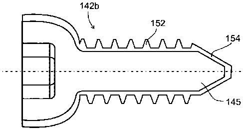
FIG. 3C illustrates an additional feature according to some embodiments of the
invention. As shown, a bone nail is formed with a longitudinal slot 152, for
example, at
its proximal end 34. After the nail has been attached to the broken bone at
its distal end,
for example by a bone screw extending through hole 46 (see FIG. 2), and the
broken
parts of the bone have been aligned, the surgeon can apply compression to the
fracture
site by attaching a screw to the bone through slot 152 and pulling the nail
against the
screw in the slot, optionally using the implant insertion tool. One or more
other screws
at the proximal end may then be added, for example, through hole 38, to anchor
the nail.
According to some embodiments of the invention, slot 152 may include a ridge
or rib 154 to prevent withdrawal of the screw from the slot, as in the case of
the round
screw hole described above.
Referring now to FIGs. 4 and 5, there are shown alternative constructions for
a
connector for an implant insertion tool or handle. In one illustrative
embodiment, a
connector 70 shown in FIG. 4 includes a grouping of radial slots 72 (for
example, three
as illustrated), which are configured to engage a complementary end of a
conventional
implant insertion tool (not shown) in the required orientation, according to
conventional
implant insertion practice. Preferably, more than one slot 72 is employed; as
composite
materials generally provide limited shear strength relative to metal, and
multiple slots
CA 02749684 2011-07-13
WO 2010/082183
PCT/1B2010/050225
24
help assure sharing of the shear load imposed by the torque applied by the
insertion
handle. Alternatively, more than three slots 72 may be employed, provided they
are
arranged at the proper orientation relative to the insertion handle.
In another illustrative embodiment shown in FIG. 5, connector 76 may have a
single position at which it can connect to the insertion tool. Illustratively,
this may be a
generally hexagonal external configuration indicated at 78 capable of bearing
torsion.
In the exemplary embodiments illustrated, connectors 70 and 76 are formed of
the same reinforced polymer material as the rest of the implant body.
Optionally, the
connectors may be formed of a metal end attachment (for example, titanium or
the like)
or ceramics molded into the proximal end of the implant body, provided it does
not
interfere unacceptably with CT or MRI visualization
According to some embodiments of the invention, bone implants as described in
connection with FIGs. 1-5, are formed of fiber layers designed to resist
mainly bending
forces, and mainly torsional forces. (As previously mentioned, the term
"mainly" is
considered to mean that the forces encountered are at least about 75 percent
bending
forces or at least about 75 percent torsional forces.)
FIGs. 6A, 6B, and 6C show some details of a bone nail 89 according to such
embodiments.
Here, core 90 and an outer layer 92 are formed of long substantially linearly
extending fibers parallel to a longitudinal axis 94 within a polymer matrix.
In the embodiments of FIGs. 6A, 6B and 6C, the nail is cannulated for
illustrative purposes. Optionally, an internal lumen 114 is covered with a
metal layer
130, for example, a metal tube, optionally inserted during compression molding
of the
nail.
Alternatively, in some embodiments, the nail is non-cannulated. In such
embodiments, the core may be solid, but may be otherwise the same as core 90
illustrated.
Referring to FIG. 6B, core 90, are multiple layers 100 of filaments in a
polymer
matrix helically wound in opposite directions, example, at 45 degrees. Layers
100 are
optionally wound or braided after manufacturing the longitudinal core 90.
Optionally
this may be formed by winding impregnated strips of composite material. One or
more
layers oriented in opposite direction are employed to resist the torque
applied on the nail
CA 02749684 2011-07-13
WO 2010/082183
PCT/1B2010/050225
in the two directions of rotation. Optionally, at the proximal end 104, layer
100 is
comprised of helically oriented filaments formed by winding multiple layers of
impregnated strips of composite material in opposite directions, for example,
at
approximately +45 and -45 degrees.
5 It
should be noted that some variability in the direction of the fibers is
optional.
For example, the windings 100 may be oriented at angles in the range of 35
to 55
degrees.
Optionally, fibers may braided to combine two neighbor layers.
Optionally, the outer surface may be coated, at least partly, for example by
10 plating, with a layer 110 of titanium, tantalum or similar metal
Optionally metal outer surface 110 may be manufactured by compression
molding the composite into a metal shell.
Referring to FIG. 6C, the distal end 106 may be of the same construction as
the
proximal end. Illustratively, however, it is shown without a metal layer, and
with only
15 two helical layers 112.
As an example of the construction illustrated in FIG. 6A, for an
intramedullary
nail having an outside diameter of 8.5 mm, the inner, linear fiber layer
embedded within
the polymer matrix may have a diameter of up to 7.6 mm. If the nail is
cannulated,
internal lumen diameter may be 2.7mm, metal cover (if any) will be between
diameters
20 2.7 to 2.9 mm. The second layer of helical fibers may have a thickness
of 0.3 mm
between diameter 7.6 mm and diameter 8.2 mm. The third (outer) layer of
linearly
extending fibers embedded in a polymer matrix may have a thickness of 0.15 mm
between inner and outer diameters 8.2 mm and 8.5 mm.
As an example for cannulated nail having a proximal head with a final diameter
25 of 11.6 mm, an inner lumen may have a 2.7 mm diameter, metal cover (if
included) will
be from 2.7 to 2.9 mm in diameter, linear fiber layer may have a diameters
from 2.9 up
to 7 mm. A first helical in -45 deg, orientation may be from 7 to 7.4 mm in
diameter. A
second layer of helical fibers in +45 deg, may be from 7.4 mm to 7.8 mm in
diameter.
One more helical layer in -45 deg. may be from 7.8 to 8.2 mm in diameter, one
more
helical layer in +45 deg may be from 8.2 to 8.6 mm in diameter, and helical
circular
layer may be between 8.6 and 10.8 mm in diameter. An outer layer of
longitudinal
fibers may be between 10.8 mm and 11.6mm. in diameter.
CA 02749684 2011-07-13
WO 2010/082183
PCT/1B2010/050225
26
Optionally, according to some exemplary embodiments, and as shown in FIGs.
6A-9B, a nail 107 may be cannulated. One optional use for a cannulated nail is
repair of
long bones such as the femur, tibia and humerus. As illustrated, in FIGs. 7-
9B, nail 107
includes an elongated body 109 having a proximal end 110, a distal end 113,
and a
substantially central, axially extending lumen 114
Distal end 113 includes a longitudinal slot 116 and a round hole 118 extending
in the same direction through the nail, and round holes 120a and 120b
extending at a 90
degree angle to slot 116 and hole 118. Proximal end 110 includes round screw
holes
122 and 124, and a slot 126.
Each of the screw holes and slots at distal end 113 and proximal end 110 of
nail
107 may include radiopaque location markings. As seen in FIG. 9B, these may
take the
form of rods or pins 128 as described in connection with FIGs. 2A and 3C, or
rings as
described in connection with FIGs. 3A and 3B. Additionally, or alternatively,
a thin
metal tube 130 may be bonded in any suitable manner on the inner surface of
lumen 114
(the distal end of which is best seen in FIG. 9B).
As in the case of the embodiments employing wire 59 shown in FIG. 2, or
employing the radiopaque filler in the matrix, the continuity of tube 130 is
interrupted
by the screw holes and the slots, so that the longitudinal positions of these
passages is
indicated under fluoroscopy by the resulting discontinuities. As will be
appreciated,
tube 130 also serves to mark the location and extent of implant 107 itself.
Cannulated implant 107 is otherwise the same as that previously described in
connection with FIGs. 1, 2, and may include a connector at its proximal end
110 like
that described in connection with FIGs. 4 and 5, and an end cap as described
in
connection with FIG. 2B. Also, it may be formed with the same layer
configuration as
in FIG. 6A. Accordingly, further description is omitted in the interest of
brevity.
Optionally, implants according to some embodiments of the invention may
include additional elements to improve performance, mainly strength. For
example, an
insert can be made of metal or ceramics, or isotropic composite parts. One
such
embodiment is illustrated by way of example, in FIGs. 10A-10C.
In FIG. 10A, there is shown an intramedullary nail 132 including a metal nut
134 to impart added strength to the connection between the implant, and the
insertion
handle. This may be embedded optionally into the implant during molding.
Optionally,
CA 02749684 2011-07-13
WO 2010/082183
PCT/1B2010/050225
27
the nut 134 may by inserted into the proximal slot, and pushed into the
proximal side of
the nail.
In FIGs. 10B and 10C there are illustrated one option of the nut insert 134.
As
shown, nut 134 is generally T-shaped with a body 135 and opposed arms 136.
When not
molded in, nut 134 is oriented as shown in FIGs. 10B and 10C, and placed in
slot 137
near its distal end 138. It is then moved in the proximal direction so that
body is within
the axial bore at the proximal end 139 of the implant.
Alternatively, or additionally, the surface of the implant may be provided
with a
metal coating or plating 141. The metal insert and the coating may be formed
of
titanium, titanium alloy or tantalum, or any other suitable and desired metal
or metal
alloy.
FIG. 11 illustrates the construction of a bone plate 160 according to some
embodiments of the invention. Plate 160 is comprised of a longitudinal fibers
coated
with one, two, three, four layers of 45 longitudinal wires, longitudinal
fibers coated
by one, two, three, four layers of woven or braided 45 layers. As an
example, plate
160 is comprised of a woven or braided body 162 formed of substantially
linearly
extending comingled long carbon and polymer filaments in a thermoplastic
polymer
matrix as previously described. Passages 170 are provided to receive bone
fixation
screws (not shown). Optionally, passages 170 are formed in the molding process
when
plate 160 is fabricated. Alternatively, passages 170 are formed by machining
after the
plate has been fabricated.
Passages 170 may be threaded or non-threaded or a combination of the two.
Optionally only a portion of some or all the passages are threaded with the
other part is
non-threaded, and designed to engage with the screw head.
According to some embodiments of the present invention, bone plate 160 is
preformed based on average anatomical data, and then bent to a final shape
before
implantation based on specific anatomical data concerning the actual implant
site for a
particular patient. According to some exemplary embodiments, the final shaping
is done
by heating the pre-formed implant in a molding press with suitably shaped
inserts.
Force is applied to bend the plate to the required shape, and then the mold is
cooled in a
manner which allows the implant to retain its bent shape without substantial
change in
its other properties. As an example, a bone plate formed of carbon fibers, in
a PEEK
CA 02749684 2011-07-13
WO 2010/082183
PCT/1B2010/050225
28
matrix, is heated to 380-400 Deg C, held at temperature for 5-30 minutes as
needed to
effect proper bending, then cooled at a rate of 5-30 Deg C per minute to 150
Deg C, and
then cooled rapidly to room temperature.
Optionally, specific anatomical data for shaping plate 160 is obtained by
direct
measurement of the patient's implant site during a surgical procedure, or even
visually.
Alternatively, the specific anatomical data is obtained radiologically or by
an MRI or by
CT of the patient's implant site.
FIG. 12A-12D illustrate a bone fixation screw suitable for use with the
various
implant embodiments described above, or as standalone for fixation of
fractures without
an implant. The illustrated screws may be formed of the same fiber reinforced
or self
reinforcing polymer materials as described above.
As illustrated in FIG. 12A, the screws are comprised of a core 145 having long
fibers extending in longitudinal direction, parallel to the screw axis 143
embedded in a
polymer matrix. The thread 144 is made from composite material having long
fibers,
wound with the threads. Optionally some fibers may cross from the core and
interweave
into the thread as shown at 147, to increase the strength of the thread base
149.
Optionally, the thread 144 can be made of composite material with chopped
fibers, optionally molded over the screw core.
The screw connector 148 for engagement with the closing and opening tool, may
be of any conventional shape, for example, an internally or externally
threaded hexagon,
Phillips head, slotted, axial crown, and the like. Optionally the head of the
screw may be
a metal insert.
FIG. 12B illustrates a bone fixation screw 142a, having a helical composite
material layer 150, preferably with long fibers directed in +/- 45 deg
relative to the axis
143. That layer may be included to add resistance to the torque applied on the
screw
during insertion or removal. Optionally layer 150 will comprise a winding only
with
one helical direction. Optionally the two fiber directions +/-45 deg are
braided.
FIG. 12C illustrates a screw 142b providing added shear strength, by having
metal shell 152 outside composite core 145. Shell 152 may be solid, and
comprise the
entire thread with no composite component. Such a structure provides the
strength of
the metal to resist shearing of the thread, and the strength of the composite
core to resist
CA 02749684 2011-07-13
WO 2010/082183
PCT/1B2010/050225
29
bending. Optionally the distal end of the screw will be part of the shell 154,
and
optionally, may be self tapping.
FIG. 12D illustrates a screw 142c having the threads 144 coated with a thin
layer
156 of titanium, or other metal such as titanium alloy Ti6A14V , or any other
biocompatible metal or metal alloy. The metal coating should be thick enough
to
provide the needed additional strength, but thin enough that it does not cause
artifacts in
CT images or MRIs. Coating thicknesses in the range of about 0.02 to 0.2 mm
provide
satisfactory results. As a specific example, the coating may have a thickness
of a 0.1
mm.
The coating layer 156 may be formed in various ways including by
electrochemical coating, physical vapor deposition, plasma spraying, molding
the
composite material into a metal shell etc. Whatever technique is employed, the
coating
should be made a smooth as possible, as a smooth surface is found to prevent
attachment of re-grown tissue or bone to the threads, which would hinder
removal of the
screw if the implant must later be removed.
Optionally, bone screw can be made in any combination of the structural
components described above.
Optionally, bone screw, in any combination can be canullated, with an internal
lumen sized for use with guide wire.
FIG. 13A illustrates the construction of a proximal femur (PF) nail 180 formed
of a reinforced polymer matrix, optionally including an embedded reinforcing
insert as
described above in connection with other embodiments of the invention. PF
nails are
used for repairing fractures involving the femur.
As illustrated, PF nail 180 includes an elongated stem 182 having a proximal
end 184 with at least one passage 186 oriented at an angle to a longitudinal
axis 284 of
the nail. In use, passage 186 receives a proximal end bone fixation screw 286
which
anchors the nail in the neck 188 and head 189 of the femur.
Optionally, PF nail 180 includes a threaded passage 190 to receive an anti-
rotation pin 288. Passage 190 extends parallel to proximal end fixation screw
passage
186.
CA 02749684 2011-07-13
WO 2010/082183
PCT/1B2010/050225
'
Optionally, according to some exemplary embodiments of the invention, passage
186 is also threaded and receives a holder 192 within which leg screw 187 is
slidingly
received.
It should be understood that in addition to passages 186 and 190, a PF nail
5
typically includes additional passages, such as passage 290 at a distal end
292. In use,
passage 290 receives a bone fixation screw for anchoring PF nail 180 to a
lower portion
of the femur. Optionally other passages (not shown) may extend at an angle,
for
example, 90 degrees, to passage 290.
As in previously described embodiments, PF nail 180 may include radiopaque
10 markings for some or all of the passages.
Optionally according to some exemplary embodiments of the invention, PF nail
180 includes an insertion tool connector 294 as described above, and an end
cap 296
configured to be received in connector 294 after PF nail 180 has been
implanted to
prevent bone internal bone or tissue regrowth.
15
Exemplary embodiments of leg screws are shown in FIGs. 13B and 13C.
Optionally, leg screws according to some embodiments of the invention, are
formed of a
core of the same composite material and the nail. Optionally, the screw is
formed of
metal, for example, a titanium alloy such as Ti-6A1-4V.
As shown in FIG. 13B, a leg screw 300 includes a reinforced polymer core 302,
20 and a
surrounding metal shell 304. Optionally, core 302 may include an internal
lumen
306 intended to receive a guide wire (not shown) for assisting the surgeon
during the
implant procedure.
Shell 304 includes threads 308 at least at its distal end 309 for interlocking
with
the surrounding bone. Optionally, the threads are self tapping. Threads 308
may be
25 formed
only in shell 304 or may be internally relieved so that the polymer core 302
penetrates the threads, as best seen at 310 in FIG. 13C. In some instances,
this may
reduce the amount of metal in the shell for improved CT imaging and MRI
visualization, and may help increase the strength of the connection between
the screw
core and the shell. The interface between the core and the shell may also
include
30
recesses and complementary projections of various shapes (not shown) to
provide for
stress sharing.
CA 02749684 2011-07-13
WO 2010/082183
PCT/1B2010/050225
31
Optionally, as also illustrated in FIG. 13C, at 312 and 314, the metal shell
316 is
crimped around proximal and distal ends 318 and 320 of polymer core 322.
FIGs. 14A-14C show an implant removal tool 200 according to an aspect of
some embodiments of the present invention. Removal tool 200 is configured to
engage
an installed implant 202 through the axial connector opening 204 at the
proximal end
206 of the implant. For this purpose, axial opening 204 communicates with a
transverse
slot 208 as previously described.
As seen in FIG. 14B, tool 200 includes first and second arms 210 and 212,
having respective transverse tips 214 and 216 at their distal end, and a
suitable handle
(not shown) for easy manipulation at their proximal ends. Arms 210 and 212 are
connected by a pivot intermediate the proximal and distal ends and thus
provide a
scissor mechanism operable to move tips 214 and 216 between retracted and
extended
positions. In the retracted position, the tips are close to each other so that
the distal end
of tool 200 is easily insertable into opening 204. In the extended position,
tips are
separated, and engage the opposite sides of slot 208.
As will be understood, in the extended position axial, force can be applied to
withdraw implant 202 from inside an opening in a bone.
FIGs. 15A andl5B show a bone drilling assembly 230 used to prepare an
implant site to receive a bone implant according to some embodiments of the
invention.
The illustrated construction is designed to minimize interference with
fluoroscopic
visualization of the drilling site by the surgeon.
Drilling assembly 230 includes a power unit 232 which drives a drill bit 234.
An
angled connector 236 is configured to be fitted between power unit 232 and
drill bit
234. As best seen in FIG. 15B, connector 236 includes a female coupling 238
for
connection to drill bit 234, and a male connector 240 for connection to power
unit 232.
These are mounted in a body 246 formed a polycarbonate or other suitable
radiolucent
material. A flexible cable 242 formed of for example multi-filaments of
stainless steel
or other suitable material is also mounted in body 246 and transfers torque
from coupler
240 to coupler 238.
Still referring to FIG. 15B, connector 236 is angled at 250 so that couplers
238
and 240 are oriented, for example, at 90 degrees to each other. This allows
power unit
CA 02749684 2011-07-13
WO 2010/082183
PCT/1B2010/050225
32
232 to remain outside the fluoroscopy imaging range, and therefore does not
interfere
with visualization by the surgeon of the drilling site.
Couplers 238 and 240 are of conventional design, or of any other suitable and
desired type. According to some embodiments, couplers 238 and 240 include
outer
sleeves 252 and 254 formed of Teflon or the like, which serve as bearings to
minimize
friction during rotation. Cable 242 is sized to rotate freely relative to the
body 246
Optionally, instead of a flexible power transfer connection, rigid elongated
rods
connected together by suitable right-angle gear arrangement, may be employed.
Preferably these parts are also formed of radiolucent material.
According to some embodiments, drill bit 234 may be made of a reinforced
polymer matrix, optionally, including longitudinally extending reinforcing
fibers as
described above, coated with hard metal such as titanium, or diamond.
Power unit 230 may be a standard operating room drill. Optionally angled
connector 234 may include a self-contained, electric motor, gear and battery,
in that
device, a separate power unit 232 is not needed.
Optionally, the connector 236 constructed is provided in sterile packaging,
and
is intended for disposal after a single use.
Optionally, as illustrated in FIGs. 16A-16G, the nail at the proximal end is
connected to the insertion handle with a bayonet connection. Optionally, the
proximal
end nail includes longitudinal grooves 162 in order to insert the bayonet
teeth 165 on
the tube or rod 164 which connects the nail to the handle. Tube 164 rotates
inside the
nail in its radial groove 163. A nut 166 fastens over the tube 164 to tighten
the nail to
the handle. Optionally, the nail proximal end does not include longitudinal
grooves for
insertion of the bayonet teeth. Instead, tube 134 includes expandable bayonet
teeth.
FIG. 17A and FIG. 17B illustrate a way to reduce cost during production of
composite intramedullar nails. The nails are supplied in many lengths and
diameters for
humerus, tibia and femur bones, and are usually curved to follow the anatomic
shape of
the bones. However, it is less costly to manufacture straight nail, with all
or most of the
layers, and add a final manufacturing step of bending the nail.
FIG. 17A illustrates a bending tool 170. As shown, tool 170, includes a cavity
171 to receive a straight nail. The tool has electric heaters 172 heating the
tool with the
nail inside to a suitable plastic deformation temperature. For example for a
nail formed
CA 02749684 2011-07-13
WO 2010/082183
PCT/1B2010/050225
33
of PEEK, a suitable temperature is in the range of 380 to 410 deg C.
Optionally pressure
may be applied to the nail during heating, for example by pressing the nail
axially via
opening 173. The tool has two halves 174 and 175 defining a mold cavity, made
of
material capable to bend without damage, at the process temperature, such as
Nitinol.
At high temperature, the tool bends the nail. FIG. 17B illustrates the tool
after
bending. The nail is cooled within the tool, and the resulting curve 176 is
retained.
PEEK and similar materials can be amorphous or crystalline to some degree, as
determined by the desired heating and cooling treatment. Bending tool 170 has
controller not shown, to establish the desired heating and cooling protocol.
After cooling, the tool opened along surface 177, and the curved nail is
FIG. 18 shows an example of a drill guide and insertion tool 398 for use with
an
implant 400 according to some of the implant embodiments as described above.
As
shown, tool 398 is comprised of a body 402 having a drill guide holes 404 and
a
coupling portion 406 which engages the coupler portion 410 of implant 400.
Included in
coupling portion 406 is an alignment element adapted to engage with a
complementary
element of the connector 410 to permit interconnection of the tool and the
implant in the
single orientation referred to in connection with FIGs. 4 and 5.
As various features of devices and methods have been described. It will be
appreciated by persons skilled in the art that the present invention is not
limited to what
has been particularly shown and described hereinabove. Rather, the scope of
the present
invention includes both combinations and subcombinations of the various
features
described hereinabove, as well as variations and modifications thereof that
are not in the
prior art, which would occur to persons skilled in the art upon reading the
foregoing
description.
It should also be appreciated that some of the embodiments are described only
as
methods or only as apparatus, however the scope of the invention includes both
methods for using apparatus and apparatus for applying methods. The scope of
the
invention also covers machines for creating the apparatus described herein. In
addition,
the scope of the invention also includes methods of using, constructing,
calibrating
and/or maintaining the apparatus described herein. When used in the following
claims
or in the text above, the terms "comprises", "comprising", "includes",
"including" or the
like mean "including but not limited."