Note : Les descriptions sont présentées dans la langue officielle dans laquelle elles ont été soumises.
CA 02771464 2012-02-17
WO 2011/020183 PCT/CA2010/001267
SCREENING OF PROTEIN CANDIDATES
FIELD OF THE INVENTION
The present invention relates to screening of protein candidates. More
particularly, the
invention relates to the screening of expression levels, biophysical
properties, and affinities of
protein candidates.
BACKGROUND OF THE INVENTION
Expression levels, biophysical properties and biological functions are three
key features of an
engineered protein. It is a challenge to preserve or improve expression level
and biophysical
properties of a protein while engineering its biological functions, as any
introduced mutation
may influence the structure of the protein, and this influence is by far still
relatively
unpredictable (Honegger et al, 2009).
Screening for protein candidates (PCs) with good expression levels and higher
affinities has
become more routine. Very high affinity binders are generated in many
laboratories (Jonsson
et al, 2008) and expression screening has made it possible to estimate the
expression levels of
a large number of proteins (Kery et at, 2003).
In contrast, engineering biophysical properties is more challenging.
Strategies have been
designed in all aspects of protein engineering to generate stable PCs. Single
domain
antibodies (sdAbs) derived from camelid heavy chain antibodies (Hamers-
Casterman et al,
1993) are very stable molecules, but introduction of mutations (for
humanization and affinity
maturation) can lower their stabilities (Saerens et al, 2005). Careful design
of libraries can
greatly increase the proportion of PCs with good biophysical properties, but
these libraries
usually still contain significant percentage of proteins that are not
satisfactory (Christ et al,
2007). One of the few exceptions is ankyrin repeats: most if not all reported
protein binders
built on small ankyrin domains seem to have good biophysical properties (Binz
et al, 2004;
Kohl et al, 2003). For evolving individual PCs, strategies such as molecular
evolution based on
sequence consensus (Lehmann et at, 2000) and introduction of potentially
stabilizing residues
(Ewert et al, 2003) have led to more stable proteins. In the selection
process, the addition of
high temperature (Jespers et al, 2004), extreme pH (Famm et al, 2008) and
proteolytic (Ueda
et al, 2004) pressures on PCs as well as selection on higher infectivity of
phage displaying
these PCs (Jespers et al, 2004; (Jespers et al, 2004 et al, 2005) have all led
to successful
selection of satisfactory binders. Despite these efforts, the challenge of
routinely generating
1
CA 02771464 2012-02-17
WO 2011/020183
PCT/CA2010/001267
stable protein variants remains unmet. Another disadvantage of these
approaches is their
requirement for a specific molecular display platform, which is not suitable
for many proteins.
It is noteworthy that the above approaches usually address only one of the
three key features.
In addition to the lack of research tools for generating proteins satisfying
all aspects, PCs have
to be purified in most cases for their characterization. This purification
step renders
characterization, even for less-challenging affinity screening, rather tedious
work. Purifying and
characterizing a large number of PCs thus becomes a significant limitation in
protein
engineering.
Screening methods for either expression levels (Kery et al, 2003), biophysical
properties(Niesen et al, 2008; Woestenenk et al, 2003) or affinities (Leonard
et al, 2007) are
available, but few of the currently known approaches satisfies the requirement
of both
simplicity and high-throughput. Most such selection methods still require some
level of protein
purification, which is time-consuming. Additionally, the art-known methods do
not allow
screening of all key features outlined above.
SUMMARY OF THE INVENTION
The present invention relates to screening of protein candidates. More
particularly, the
invention relates to the screening of expression levels, biophysical
properties, and affinities of
protein candidates.
The present invention provides a method for screening of protein candidates,
comprising:
a) providing fusion proteins, each fusion protein comprising one protein
candidate and a
protein anchor; and
b) evaluating the expression levels of the protein candidates; or
c) evaluating the biophysical properties of the protein candidates; or
d) evaluating the binding kinetics of the protein candidate; or
e) any combination of steps b) to d) above,
wherein, the protein anchor provides a means of capture of the protein
candidates to facilitate
evaluation of expression levels, biophysical characteristics and binding
kinetics. The protein
anchor may accomplish this via binding to a specific coating on a solid
surface.
The present invention further provides a method for screening of protein
candidates,
comprising:
a) providing fusion proteins, each fusion protein comprising one protein
candidate and a
protein anchor; and
2
CA 02771464 2012-02-17
WO 2011/020183
PCT/CA2010/001267
b) evaluating the expression levels of the protein candidates by
i. binding the protein anchor to a specific coating on a solid surface; and
ii. measuring the amount of bound fusion proteins; or
c) evaluating the biophysical properties of the protein candidates by
i. denaturing the fusion proteins;
ii. allowing the denatured fusion proteins to refold;
iii. filtering sample containing the refolded fusion proteins;
iv. binding the protein anchor to a specific coating on a solid surface;
v. measuring the amount of bound fusion proteins; and
vi. comparing the amount of bound fusion proteins to that obtained in step b);
or
d) evaluating the binding kinetics of the protein candidate by
i. binding the protein anchor to a specific coating on a solid surface; and
ii. measuring the binding kinetics of the protein candidates to their
target/antigen
by allowing the target/antigen to bind to the protein candidates and observing
their associations and dissociations; or
e) any combination of steps b) to d) above.
In the method as described above, each of the three screening modules (steps
b) to d)) may
be performed independently, in parallel or in succession. The method as
described generally
does not require purification of the fusion proteins or protein candidates.
In the method described above, the expression levels may be measured by ELISA;
the
denaturation may be accomplished by exposure to heat or extreme pH; and/or the
binding
kinetics may be measured by surface plasmon resonance.
The present invention is also directed to fusion proteins comprising a protein
anchor and
protein candidates. The protein anchor may comprise an antibody or antibody
fragment
comprising a complementarity determining region (CDR) 1 sequence of NYTMA (SEQ
ID
NO:11); a CDR2 sequence of VVSRGGGATDYADSVKG (SEQ ID NO:12); and a CDR3
sequence of GTDLSYYYSTKKWAY (SEQ ID NO:13); the antibody or fragment thereof
may
be based on BSA12 (SEQ ID NOs: 1 and 2), or may comprise BSA12 itself, and the
protein
candidates (PCs) may be any suitable proteins for screening. In these cases,
the specific
coating is bovine serum albumin.
The present invention further provides a vector for expressing the fusion
proteins described
above, as well as a precursor vector into which the nucleic acid molecule
encoding the protein
candidate is cloned. In one non-limiting example, the precursor vector is
pBSA12 (Figure 1,
SEQ ID NO. 3).
3
CA 02771464 2016-10-27
An approach for fast screening of expression, biophysical-properties and
affinities, which allows
the screening of a large number of PCs at the early stage of protein
engineering to exclude or
greatly reduce the number of unsatisfactory candidates, is described herein.
This approach also
allows the ranking of the PCs by their dissociation rates, which are usually
closely related to
their affinities, without protein purification. In one embodiment, the PCs are
fused to a camelid
sdAb BSA12 (Li et al, 2009), which is very stable and has an extreme affinity
to BSA yet this
interaction can be completely disrupted by low pH. The affinity of the sdAb
BSA12 anchors
onto any BSA-coated surface and greatly contributed to the simplicity of the
presently described
method and the accuracy of the generated data.
Another advantage of the present method is that it does not rely on ligand
binding for the
selection of good biophysical properties, which can broaden its application to
practically any
area of protein engineering. For example, the present method may assist in
selecting enzyme
candidates with higher stabilities, or identifying optimum refolding
conditions for various
proteins. The high throughput feature of the present approach also allows for
the selection of a
very large number of PCs to analyze contributions of various residues to
solubility and stability,
and to identify residues with positive contributions to a more stable
structure. As the evidence of
protein folding has become obvious in the development of diseases such as
Alzheimer's
diseases and Parkinson's disease, this approach also allows for investigation
of misfolding
mechanisms and searching for peptidic drug candidates to prevent the formation
of protein
aggregates.
Additional aspects and advantages of the present invention will be apparent in
view of the
following description. The detailed description and examples, while indicating
preferred
embodiments of the invention, are given by way of illustration only, as
various changes and
modifications within the scope of the invention will become apparent to those
skilled in the art in
light of the teachings of this invention.
Brief Description of the Drawings
These and other features of the invention will now be described by way of
example, with
reference to the appended drawings, wherein:
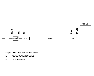
Figure 1 is a schematic presentation of the vector pBSA12. The ompA leader
sequence (ompA) -
will be removed during secretion. Sfil restriction sites are usually used to
fuse protein
candidates with BSA12 linked with ompA signal peptide (MKKTAIAIAVALAGFATVAQA;
SEQ
ID NO:8), the linker (L) sequence (SEQ ID NO:9); and His, histidine
purification tag (H). The 6
Histidine tag (H) is designed for purification of PC-BSA12s by immobilized
metal affinity
chromatography.
4
CA 02771464 2016-10-27
FIGURE 2 is a schematic representation of fast screening of expression-levels,
biophysical
properties and affinities of PCs, using one embodiment of the present
invention. PCs to be
screened are fused directly to a protein anchor (BSA12) by cloning into a
vector (pBSA12) to
make a sub-library. Cell lysates or cell-conditioned media of individual
clones are used to
estimate the expression of PC-BSA12 (left panel) and screen for binders with
good biophysical
properties (the middle panel) as described in the text. For ranking affinities
of the PCs, the
same samples are captured onto an SPA chip surface pre-immobilized with BSA,
and the
antigen is injected to measure its binding to the PCs (right panel). ELISA on
antigen to pre-
screen binders is optional. SP, ompA signal peptide.
FIGURE 3 shows results of screening of expression levels of PCs. FIGURE 3A
depicts the PC-
BSA12 concentrations of 10 out of the approximately 190 constructs in cell-
conditioned media
as measured by ELISA on BSA. Background reading with no BSA coating was
subtracted from
the original data. FIGURE 3B is a Western blot of cell-conditioned media of
the 10 sdAb-BSA12
Clones. FIGURE 3C is a Western blot of pellets (P) and supernatants (5) of six
of the 10 sdAbs
when expressed as monomers.
FIGURE 4 shows results of screening of biophysical properties. FIGURE 4A
depicts the
concentrations of 18 PC-BSA12s as measured by ELISA on BSA with (60 C or 80 C)
or without
(AT) heating and subsequent filtration of the samples. Three clones having
significant signal
reduction after heating and filtration, marked by "x", and three without,
marked by "*", were
selected for further analysis. FIGURE 4B shows SEC profiles of BSA12 and four
sdAbs. Elution
positions of protein standards BSA (67 kDa), ovalalbumin (43 kDa),
chynnotrypsinogen, (25
kDa) and ribonuclease (13.7 kDa) are indicated above the graphs. FIGURE 4C
shows circular
dichroism spectra of purified BSA12 and four sdAbs in 10 mM phosphate buffer,
pH 7Ø
FIGURE 40 shows graphs tracking heat-induced denaturation of BSA12 and three
sdAbs as
measured by CD at 218 nm.
FIGURE 5 shows results of kd ranking. FIGURE 5A shows normalized sensorgram
overlays in
dissociation phase of Fc binding to Fc17-BSA12 of 27 independent
transformants. FIGURE 5B
shows the correlation between amounts of Fc17-BSA12 captured and amounts of Fc
bound to
Fc17. FIGURE 5C depicts the amount of 51 sdAb-BSA12 fusions, (FC1, FC2 FC3 and
FC4)
and BSA12 captured on immobilized BSA. The dashed line represents the level of
BSA12
captured in flow cell 1 in the first round. FIGURE 5D is normalized sensorgram
overlays in
dissociation phase of free Fc bindings to 43 sdAb-BSA12s representing 12
different sdAb
BSA12 clones. Those of Fc7-BSA12, Fc12-BSA12 and FC75-BSA12 are shown in thick
solid,
dotted and dashed lines,
CA 02771464 2012-02-17
WO 2011/020183
PCT/CA2010/001267
respectively. FIGURE 5E is sensorgram overlays of purified Fc7, Fc12 and Fc75
binding to
immobilized Fc.
DETAILED DESCRIPTION OF THE INVENTION
The present invention relates to screening of protein candidates. More
particularly, the
invention relates to the screening of expression levels, biophysical
properties, and affinities of
protein candidates.
The present invention provides a method for screening of protein candidates,
comprising:
a) providing fusion proteins, each fusion protein comprising one protein
candidates and a
protein anchor; and
b) evaluating the expression levels of the protein candidates; or
c) evaluating the biophysical properties of the protein candidates; or
d) evaluating the binding kinetics of the protein candidate; or
e) any combination of steps b) to d) above.
In the method as just described, the protein anchor provides a means of
capture of the protein
candidates to a specific coating to facilitate evaluation of expression
levels, biophysical
characteristics and binding kinetics. The protein anchor may accomplish this
via binding to a
specific coating on a solid surface.
More specifically, the present invention provides a method for screening of
protein candidates,
comprising:
a) providing fusion proteins, each fusion protein comprising one protein
candidates and a
protein anchor; and
b) evaluating the expression levels of the protein candidates by
i. binding the protein anchor to a specific coating on a solid surface; and
ii. measuring the amount of bound fusion proteins; or
c) evaluating the biophysical properties of the protein candidates by
i. denaturing the fusion proteins;
ii. allowing the denatured fusion proteins to refold;
iii. filtering sample containing the refolded fusion proteins;
iv. binding the protein anchor to a specific coating on a solid surface;
v. measuring the amount of bound fusion proteins; and
vi. comparing the amount of bound fusion proteins to that obtained in step b);
or
d) evaluating the binding kinetics of the protein candidate by
i. binding the protein anchor to a specific coating on a solid surface; and
6
CA 02771464 2012-02-17
WO 2011/020183
PCT/CA2010/001267
ii. measuring the binding kinetics of the protein candidates to their
target/antigen by
allowing the target/antigen to bind to the protein candidates and observing
their
associations and dissociations; or
e) any combination of steps b) to d) above.
The method as described herein is designed to provide information on
expression levels,
biophysical properties and affinities of a large number of PCs without
requiring purification of
such molecules (Figure 2). Each of the three screening modules (steps b) to
d)) may be
performed independently, in parallel or in succession.
The method of the present invention allows rapid screening of protein
candidates (PCs). A
"protein candidate" may be any suitable protein of interest, regardless of its
eventual
application. The protein candidates may be based on a naturally-occurring
protein, or may be
an engineered protein; the libraries of protein candidates for screening may
be obtained by any
method known in the art, for example, but not limited to phage-display,
ribosome display, yeast
display, affinity maturation, genomic DNA, cDNA or mutation libraries.
In order to screen the PCs using the method of the present invention, the PCs
are provided as
fusion proteins. The fusion protein may comprise a protein candidate and a
protein anchor.
As described above, the protein candidate is the protein of interest; the
"protein anchor" is a
protein that provides known characteristics to the fusion protein, and it
allows for the capture of
the fusion protein. In order to be useful in the method of the present
invention, the protein
anchor should:
1) have very high affinity to its target or antigen. For example, and without
wishing to
be limiting, the protein anchor may have a KD below about 10 pM; a protein
anchor
with a KD over about 100 pm would start to cause a drifting baseline in kd
ranking
experiments, and therefore would affect the accuracy of collected data and
doesn't
allow ranking of binders with very high affinities in the presently described
method.
Therefore, in a specific, non-limiting example, the protein anchor may have a
KD
below about 100 pm, or below about 10 pm;
2) have an interaction with its target or antigen that may be easily disrupted
despite its
high affinity. The disruption of the interaction between protein anchor and
target
may be disrupted by any suitable method, for example but not limited to
changes in
pH, changes in salt concentration, or changes in buffer;
3) exist in monomeric form and have high thermostability. This can be measured
by
size exclusion chromatography (for its monomer form determination) or circular
7
CA 02771464 2012-02-17
WO 2011/020183
PCT/CA2010/001267
dichroism at various temperatures (for its thermal denaturation curve).
Preferably
the melting temperature of the anchor protein is higher than 65 C;
4) show little non-specific bindings to other targets, antigens, or proteins
in general
(i.e., is highly specific to its target); or
5) any combination of 1) to 4).
Additionally, the target/antigen to which the protein anchor binds should be
resistant to the
reagent that interrupts the protein anchor interaction with the
target/antigen.
As described herein, the protein anchor will allow the characteristics of the
fusion protein, and
thus the protein candidate, to be evaluated without relying on the properties
of the protein
candidate.
The protein anchor may be any suitable protein possessing the characteristic
1) to 5), as
described above. The protein anchor may be an antibody or antibody fragment,
an enzyme, a
structural protein, or any other suitable type of protein. In one non-limiting
example, the
protein anchor may be an antibody or antibody fragment comprising a
complementarity
determining region (CDR) 1 sequence of NYTMA (SEQ ID NO:11); a CDR2 sequence
of
WSRGGGATDYADSVKG (SEQ ID NO:12); and a CDR3 sequence of GTDLSYYYSTKKWAY
(SEQ ID NO:13). In another specific, non-limiting example, the protein anchor
may be an
antibody or antibody fragment based on BSA12, or may comprise BSA12 itself
(SEQ ID NO:2;
as described in PCT/US2009/60495; also in WO 2010/043057) or a mutant or
fragment
thereof. In the case where BSA12 or an antibody based thereon is used as the
protein anchor,
the target or antigen will be bovine serum albumin (BSA). In another non-
limiting example, the
protein anchor may be the affibodies binding to serum albumin (Jonsson et al,
2008).
The fusion protein may additionally comprise additional sequences to aid in
expression,
detection or purification of a recombinant antibody or fragment thereof. For
example, and
without wishing to be limiting, the antibody or fragment thereof may comprise
a targeting or
signal sequence (for example, but not limited to ompA), a detection tag (for
example, but not
limited to c-Myc), a purification tag (for example, but not limited to a
histidine purification tag),
or a combination thereof.
The expression levels of the protein candidates may be evaluated by binding
the protein
anchor of the fusion proteins to a specific coating on a solid surface and
measuring the amount
of bound fusion proteins. The specific coating may comprise the target or
antigen to which the
protein anchor binds. Thus, the protein anchor may bind to the specific
coating on the solid
8
CA 02771464 2012-02-17
WO 2011/020183
PCT/CA2010/001267
surface and the fusion protein may be immobilized on the solid surface. The
solid surface may
be any suitable surface, for example, but not limited to the well surface of a
microtiter plate,
channels of surface plasmon resonance (SPR) sensorchips, membranes etc. The
amount of
the fusion protein on the solid surface may then be measured by any suitable
method, for
example, but not limited to ELISA, SPR, dot blots, Western blots or protein
microarray
technologies. As shown in the examples, the level of expression of the fusion
protein is a
reliable indicator of the expression level of the protein candidate alone.
The biophysical properties of the protein candidate may be evaluated by
denaturing the fusion
protein and allowing it to refold, then binding the protein anchor of the
fusion protein to a
specific coating on a solid surface and measuring the concentration of fusion
protein. The
fusion protein may be denatured by any suitable method. For example, but
without wishing to
be limiting in any manner, the fusion protein may be denatured by exposure to
heat or to
extreme pH. In a non-limiting example, the heat may be temperatures in the
range of about 60
to about 90 C; for example, the denaturing temperature may be about 60, 65,
70, 75, 80, 85,
or 90 C, or any temperature therebetween, or any range of temperature defined
by any two
values just recited. In another non-limiting example, the extreme pH may be in
the range of
about pH 3.5 to about pH 1 (about 3.5 3.0, 2.5, 2.0, 1.5, or 1.0, or any pH
therebetween, or any
range of pH defined by any two values just recited) or about pH 9.5 to about
pH 12 (about 9.5
10, 10.5, 11.0, 11.5, or 12.0, or any pH therebetween, or any range of pH
defined by any two
values just recited). In order to allow the fusion protein to refold, the
temperature and/or pH
may be returned to more normal value. The refolded fusion protein may be
filtered using any
suitable method; for example, and without wishing to be limiting in any
manner, the refolded
fusion protein may be filtered using a membrane filter. Without wishing to be
bound by theory,
protein candidates with undesirable biophysical properties (for example, but
not limited to low
stability, low solubility, oligomerization) will be removed from solution
either by precipitation or
by filtration. The refolded fusion protein is then bound to a specific coating
on a solid surface
by its protein anchor portion and the concentration of refolded fusion protein
is measured. The
concentration of refolded fusion protein may then be compared to that observed
in the step of
evaluating protein expression levels (step b)). If the two concentrations of a
fusion protein
(with and without denaturation and filtration) are similar, then the fusion
protein may said to
possess good biophysical properties. As shown in the examples, the biophysical
properties of
the fusion protein are a relatively good indicator of the biophysical
properties of the protein
candidate alone.
The binding kinetics of the protein candidates may be evaluated by binding the
protein anchor
of the fusion protein to a specific coating on a solid surface such as, but
not limited to the
9
CA 02771464 2012-02-17
WO 2011/020183
PCT/CA2010/001267
sensorchips of a machine which measures surface plasmon resonance (SPR), and
measuring
the binding kinetics of fusion proteins to their targets/antigens. The binding
kinetics may be
measured using any suitable technology, for example but not limited to SPR.
Once the fusion
protein is captured on the solid surface, the ligand that is bound by the PCs
may be used to
measure the PC binding kinetics, for example, but not limited to KD, off-rate,
etc.
As would be understood by a person of skill in the art, the method of the
present invention may
be put into practice using various technologies. In one embodiment of the
present invention,
DNA encoding PCs is first amplified by PCR and cloned into a vector pBSA12 to
generate a
sub-library of PC-BSA12 fusions. Individual clones from this sub-library may
be grown in
microtiter plates, and supernatants of cell lysates containing expressed PC-
BSA12s can be
used for all three screenings. The amount of PC-BSA12s secreted into the
growth media was
presently found sufficient to perform the experiments, and was therefore used.
Expression
level was estimated by ELISA on BSA. In the excess of BSA coated on microtiter
plates and
due to the very high affinity of BSA12 to BSA (KD = 4 pM; Li et al, 2009),
expression levels of
PC-BSA12s can be estimated by measuring the amount of PC-BSA12 bound to BSA
(Figure
2, left panel). Screening of PCs with good biophysical properties was
conducted in the same
way, except that the samples are heated and filtered prior to performing
ELISA. Those PCs
that give similar ELISA results before and after heating were considered to
have good
biophysical properties (Figure 2, middle panel). If binding of the PCs to
their target is of
interest, the same supernatant samples can be used to rank the PCs'
affinities. To rank the
affinity of protein candidates, BSA may first be mobilized on an SPR
sensorchip surface, and a
sample containing PC-BSA12s can be flowed over the chip to capture PC-BSA12s.
The target
antigen is lastly injected to measure its affinity to the binders. The BSA12
chip surface is then
regenerated and can be reused for another round of screening (Figure 2, right
panel).
The present invention is also directed to a fusion protein comprising a
protein anchor and a
protein candidate. The protein anchor may be as described above. In a
specific, non-limiting
example, the protein anchor may be an antibody or antibody fragment comprising
a
complementarity determining region (CDR) 1 sequence of NYTMA (SEQ ID NO:11); a
CDR2
sequence of VVSRGGGATDYADSVKG (SEQ ID NO:12); and a CDR3 sequence of
GTDLSYYYSTKKWAY (SEQ ID NO:13); the protein anchor may be an antibody or
antibody
fragment based on BSA12, or may comprise BSA12 itself or a mutant thereof, and
the protein
candidate may be any suitable protein for screening.
The present invention further provides a vector for expressing the fusion
proteins described
above, as well as a precursor vector into which the nucleic acid molecule
encoding the protein
candidate is cloned. In one non-limiting example, the precursor vector is
pBSA12.
CA 02771464 2012-02-17
WO 2011/020183
PCT/CA2010/001267
The presently described approach for fast screening of expression, biophysical-
properties and
affinities allows for screening of a large number of PCs at the early stages
of protein
engineering. Not only does the present method contribute to reducing the
number of
unsatisfactory candidates, but this approach also allows the ranking of the PC
affinities without
protein purification. Another advantage of the present method is that it is
independent of
ligand binding for the selection of good biophysical properties, which can
broaden its
application to numerous areas of protein engineering. The high throughput
feature of the
present approach also allows for the selection of a very large number of PCs,
not only to
analyze contributions of various residues to solubility and stability, but
also to identify residues
with positive contributions to a more stable structure.
The present invention will be further illustrated in the following examples.
However, it is to be
understood that these examples are for illustrative purposes only and should
not be used to
limit the scope of the present invention in any manner.
Example 1: pBAS12 Vector construction
A vector was constructed to assist in expressing fusion proteins comprising a
protein candidate
fused to BSA12.
Briefly, DNA encoding BSA12 (Li et al, 2009) was amplified using primers:
CGGGATCCGGTGGAGGCGGGTCCGGTGGAGGCGGGTCCGGTGGAGGCGGGTCCCAGG
TAAAGCTGGAGGAGTCTGGG (Forward primer; SEQ ID NO:4);
GAAGATCTGAGGAGACGGTGACCTGGGT (Reverse primer; SEQ ID NO:5);
The PCR product, after digestion with BamHI and Apal, was inserted into pMED2,
a slight
modification of an E. coli expression vector pSJF2 (Tanha et al, 2003),
generating a new
vector pBSA12 (Figure 1, SEQ ID NO. 3), which facilitates fusion of other
proteins to BSA12.
Example 2: Phage Panning and Cloning of Protein Candidates
A human VH sdAb phage display library (kindly provided by Dr. J. Tanha, NRC,
Canada) was
employed to distinguish clones with reasonable and poor expressions in E.
coll. This library
was built on the framework of a stable human VH (To et al, 2005) but was found
to display
many low-expressing and aggregate-prone binders (Arbabi-Ghahroudi et al,
2009a). The
protein antigen used for biopanning was the the ectodomain of matrix protein 2
of human
influenza virus A (M2e, SLLTEVETPIRNEWGCRCNDSSD (SEQ ID NO:6); which was
synthesized and purified to over 90% purity by Genescript (Piscataway, NJ).
Phage display
11
CA 02771464 2012-02-17
WO 2011/020183
PCT/CA2010/001267
biopanning was generally conducted as previously described (Arbabi-Ghahroudi
et al, 2009b),
with the biopanning rounds reduced from four to two. Phage ELISA was performed
with a
small number of individual phage eluted from each round, usually 20-50, to
estimate the
percentage of phage clones binding to the antigens, and the eluted phage
having larger than
50% positive clones were chosen for the construction of sub-libraries of sdAb-
BSA12s. This
established the library to be screened by the method of the present invention.
After the two rounds of biopanning on the M2e peptide antigen, DNA encoding
the sdAbs was
amplified from the eluted phage and cloned into vector pBSA12, described in
Example 1. DNA
encoding PCs was first amplified by PCR and cloned into the pBSA12 vector to
generate a
sub-library of sdAb-BSA12 fusions. DNA encoding potential binders was
amplified with the
addition of Sfil restriction sites to both ends of the fragment and DNA
encoding a peptide linker
GGGGSGGGGSGGGGS (SEQ ID NO:7) at the 3'-end. The PCR fragments were inserted
into
pBSA12. The cloning procedure resulted in over 90 percent, often close to 100
percent, of
individual clones harbouring a binder gene (results not shown).
Example 3: Fusion Protein Expression
Individual clones from the sub-library established in Example 2 were grown in
microtiter plates,
and supernatants of cell lysates containing expressed sdAb-BSA12s can be used
for all three
screenings. Alternatively, as in the present example, fusion protein secreted
into the growth
media may be sufficient to perform the experiments.
Briefly, individual sdAb-BSA12 clones were inoculated in LB medium,
supplemented with 100
g/ml ampicillin, in 96-well microtiter plates and grown at 37 C overnight with
shaking. Cell-
conditioned media were collected after centrifugation of the cell cultures.
(When supernatant of
cell lysates are to be used, the cell pellets may be lysed by adding CelLytic
B (Sigma, St.
Louis, MO) according to product instructions and supernatants of cell lysates
may be collected
after centrifugation. Other methods of obtaining the supernatants of cell
lysates may also be
used.)
Example 4: Assessing Protein Candidate Expression
The expression levels of the protein candidates were evaluated. To do so, the
cell-conditioned
media obtained in Example 3 was submitted to ELISA experiments.
Briefly, the cell-conditioned media of about 190 clones were used to perform
ELISA using BSA
as the antigen. 10 g/m1 of antigens, either BSA or target of the binders,
were coated onto
microtiter plates for overnight at 4 C in 15 mM Na2003, pH9.6. 2% skim milk in
PBS was
12
CA 02771464 2012-02-17
WO 2011/020183
PCT/CA2010/001267
added to the wells to block non-specific binding. 50 p.I of the above-
collected samples, either
cell-conditioned medium or cell lysate supernatants, were added to the wells
and incubated for
1 hr at 37 C. Bound proteins were detected by HRP-labelled anti-His tag
antibody using
standard ELISA procedure.
Some of the collected samples were separated by SDS-PAGE, and His-tagged
proteins were
detected by Western blot using goat anti-llama and alkaline phosphotase-
labelled rabbit anti-
goat antibody (Cedarlane, Burlington, ON).
About 90% of the clones had no detectable expression of the fusion proteins.
ELISA (Figure
3A) and Western blots (Figure 3B) representing 10 of the 190 clones (five with
reasonable and
five with poor expressions) are presented. Without wishing to be bound by
theory, the very
high affinity of BSA12 to BSA may allow near-complete capture of sdAb-BSA12
fusion
proteins, which can resist stringent washes. Advantageously, this approach
makes the capture
of sdAbs independent of their other features, such as affinities to ligands,
solubility and
stability. The ELISA reading is therefore only dependent on the concentration
of sdAb-BSA12s
in the solutions, and may provide a more accurate estimation of the expression
levels than
prior art methods.
To investigate whether the expression levels of sdAb-BSA12 fusions reflect
those of the sdAbs
when expressed alone, three sdAbs with reasonable expression (11A11, IIG3 and
IIG9) and
three with poor expression (103, 11D4 and I1F10), when expressed as BSA-
fusions, were
cloned to express the sdAbs. DNA encoding these six human sdAbs were cloned
into the E.
coil expression vector pMED2.
Clones were then inoculated in 25 ml LB with 200 g/mlampicillin and incubated
at 37 C with
200 rpm shaking overnight. 20 ml of the culture was used to inoculate 1 L of
M9 medium (0.2%
glucose, 0.6% Na2HPO4, 0.3% KH2PO4, 0.1% NH4CI, 0.05% NaCI, 1 mM MgCl2, 0.1 mM
CaCl2) supplemented with 0.4% casamino acids, 5 mg/I of vitamin B1 and 200
lig/m1Ampicillin,
and cultured for 24 hrs. 100 ml of 10 x TB nutrients (12% Tryptone, 24% yeast
extract and 4%
glycerol), 2 ml of 100 mg/ml ampicillin and 1 ml of 1 M isopropyl-beta-D-
Thiogalactopyranoside
(IPTG) were added to the culture and incubation was continued for another 65-
70 hr at 28 C
with 200 rpm shaking. E. coli cells were harvested by centrifugation and lysed
with lysozyme to
release the sdAbs, which were expressed periplasmically. Cell lysates were
centrifuged, and
supernatants were loaded onto High-TrapTm chelating affinity columns (GE
Healthcare, Baie
d'Urfe, QC). After washing the columns with four column volume of 50 mM Tris,
25 mM NaCI,
pH7.4, His-tagged proteins were eluted with a linear gradient (2.5 to 500 mM)
of immidazole,
and the eluted proteins were dialyzed in PBS buffer.
13
CA 02771464 2012-02-17
WO 2011/020183
PCT/CA2010/001267
The results (Figure 3C) demonstrated that expression levels of sdAb-BSA12
fusions were
good indicators of the expression of the sdAbs. This suggests that fusion of
PCs to BSA12 and
estimation of the expression levels of such fusions provide an easy approach
to screen a large
number of PCs for their expression levels.
Example 5: Assessing Protein Candidate Biophysical Properties
BSA12 has a relatively high thermostability with a Tm of -70 C (see Figure
4D). Based on the
hypothesis that less stable proteins would form aggregates upon heating and
the aggregates
can be filtrated out, performing ELISA with denatured and non-denatured
samples would allow
the evaluation of biological properties.
For non-denatured samples, ELISA was performed as described in Example 4. For
the
denatured samples, cell-conditioned media of 18 sdAb-BSA12 clones with
reasonable
expression, as determined in Example 4, were heated (60 C and 80 C, 5 min) and
filtered
before being used for ELISA on BSA. The sdAb-BSA12 clones were expressed as
described
in Example 3. 60 1.1.1 cell-conditioned medium was transferred to PCR tubes,
and the samples
were heated at either 60 C or 80 C for 5 min on a GeneAmp PCR system 9700
(Applied
Biosystems, Foster City, CA) and allowed to slowly cool to room temperature.
The samples
were then transferred to a Multi-Well Filter Plates (Pall Coorportions, Ann
Arbor, MI) and
centrifuged (4000 rpm, 30 min), and the flow-through were collected. ELISA
studies were
then performed as described in Example 4.
When compared to samples processed without the heating and filtration steps, a
significant
reduction in ELISA signals in some samples was observed, whereas little change
was seen in
others (Figure 4A). This suggested that those sdAb-BSA12 samples behaving
similarly before
and after heating can either resist heat denaturation or refold rapidly after
heating is stopped -
a clear indication of good biophysical properties.
To evaluate whether the characteristics of the fusion protein are indicative
of the protein
candidate characteristics, three heat-resistant (I1A11, IIG3, IIG9) and three
heat-sensitive
sdAbs (I1D3, 11D4 and I1F10) when fused to BSA12, were expressed as monomeric
proteins.
Cloning, expression and purification of the sdAbs was performed as described
in Example 4.
Yields of the heat-resistant sdAbs IIG3, IIG9 and 11A11 are 6.0, 2.0 and 1.5
mg/L of culture,
respectively. Relatively pure protein was obtained from only one of the three
heat-sensitive
sdAbs (11F10), with a yield of 3 mg/L of culture; purification of the other
two heat-sensitive
sdAbs failed in repeated efforts.
14
CA 02771464 2012-02-17
WO 2011/020183
PCT/CA2010/001267
The four isolated sdAbs and BSA12 were analyzed by size exclusion
chromatography (SEC)
to determine whether they form oligomers or aggregates (Figure 48).
Separations were
carried out in 10 mM HEPES, pH 7.4, containing 150 mM NaCI, 3.4 mM EDTA and
0.05%
Tween 20 on Superdex 75 (GE Healthcare) SEC on an AKTA FPLC system (GE
Healthcare).
.. Protein standards (GE Healthcare) were run under the same conditions.
BSA12 exists as monomer on a Supderdex75TM column with a measured molecular
mass
(MMM) of 18 kDa (elution volume at 11.8 ml). Similar profiles were observed
from two of the
three heat-resistant human sdAbs, IIG3 and I1A11 with a MMM of 13.7 kDa
(elution volume at
12.5 ml) and 12.7 kDa (elution volume at 12.7 ml), respectively. These MMMs
are very similar
to their calculated MW of ¨13 kDa. No aggregation was observed from IIG3, and
a small
aggregation bump at elution volume of 7.5 ¨ 10 ml can be seen for I1A11. The
third heat-
resistant sdAb IIG9 has a major elution peak (10.4 ml), a minor elution peak
(12.5 ml) and
some shoulders in the range of 7 and 10 ml. This suggests that the majority of
IIG9 exists as a
dimer with a MMM of 31.7 kDa, but monomeric (MMM = 13.7) and higher-valency
oligomeric
protein complex also exist. The only heat-sensitive human sdAb purified
(I1F10) had a major
peak at 7.4 ml representing protein complexes of five sdAbs or higher; some
minor peaks were
also observed, which may represent contamination of unwanted proteins in the
preparation,
based on their elution volumes.
The CD spectra (Figure 4C) of the sdAbs were determined using a circular
dichroism (CD)
spectrometer. To provide substantially pure protein for CD, the proteins were
collected at their
major SEC peaks for BSA12, IIA3, IIG9 and I1F10 and at the 10.4 ml (dimer) and
12.5 ml
(monomer) peaks for IIG9. Briefly, proteins were separated in a Superdex75 SEC
in 10 mM
phosphate buffer, pH 7.0, and peaks representing major formats of proteins
were collected and
used in CD analysis. CD from 250 to 200 nm was measured with the protein
concentrations of
¨ 2.5 IAM in a 10 mm path-length cuvette with a J-850 CD spectrometer (JASCO).
Data were
collected at a band width of 1.0 nm and scanning speed of 50 nm/min with two
data
accumulations and subtracted with buffer control. Molar ellipiticity was
calculated as previously
described (Schmid, 1997); above parameters, with the exception of only one
accumulation,
were used in determining thermal denaturation of proteins, which was measured
at every two
degrees from 30 to 90 C at a temperature shift speed of 1 C/min. CD values at
218 nm were
plotted to temperature in GraphPadPrism and Boltzmann Sigmoidal modal was used
to
calculate the T,õ of the proteins.
11G3 and I1A11 have similar CD spectrometry profiles, which in turn are
similar to that of
BSA12. For IIG9, which exists as a mixture of monomer, dimer and other
oligomers, the
CA 02771464 2012-02-17
WO 2011/020183
PCT/CA2010/001267
monomeric portion and dimeric peaks were analyzed separately; their CD
profiles were found
nearly identical (only that of the monomeric peak is shown in Figure 4C). This
CD profile is
different from those of BSA12, IIG3 and I1A11, which all exist mainly as
monomeric proteins.
The CD spectrum also suggested that IIG9 has a significantly higher portion of
a-helices,
which is usually not seen in variable domains of antibodies. The CD spectrum
of I1F10
suggested that it has an even higher proportion of a-helices.
To better evaluate protein stability, temperature-induced denaturation of the
proteins was also
investigated using CD (Figure 4D) as described above. Plotting CD values of
BSA12 at 218
nm gave a calculated Tm of 70 C, inline with camelid sdAbs reported by others
(Dumoulin et al,
2002). The two monomeric heat-resistant human sdAbs, 11A3 and 11G11, have a Tm
of 68 C.
The third human sdAb IIG9, which exists in multiple forms (Figure 4B),
selected by the heating
process has a much lower Tm of 55 C. Interestingly, the CD spectrum of the
only available
heat-sensitive human sdAb, I1F10, showed little change during heating (data
not shown).
Without wishing to be bound by theory, possible explanations include: the
I1F10 aggregates
provide an ultra-stable structure, or the CD spectrum (Figure 4C) represents
an unstructured
format.
An effort was made to distinguish proteins with good biophysical properties
from those with
less desirable properties using EL1SA. One of the three heat-resistant sdAbs
exists as pure
monomer, the second predominantly as monomer and the third as a mixture of
dimer,
monomer and other type of oligomers. In contrast, the only heat sensitive sdAb
obtained exists
mainly, if not entirely, as aggregates. Despite the fact that one of the three
heat-resistant
sdAbs did not meet the biophysical property standards set, the screening
method is still
regarded as very useful as it excluded most PCs with unsatisfactory features.
Notably, little
protein was obtained from two of the three clones that were sensitive to heat
treatment, even
though expression screening suggested that they would express reasonably well.
It is not
unusual that scaling-up of protein expression leads to poor yields for some
proteins. The
benefit to the present method is its ability to screen these clones out.
Example 6: Assessing Protein Candidate Binding Kinetics
If the PCs are also potential binders, their binding kinetics can be
investigated using cell-
conditioned media or cell lysates containing PC-BSA12s. Since the majority of
human sdAbs
obtained from the M2e biopanning had poor expression in E. coli, this portion
of the present
method was evaluated using an anti-human IgG1 llama sdAb library. As camelid
sdAbs are
known to have very good stability in general, use of this library would allow
analysis of affinities
of a large number of binders without consideration of their expression and
stability.
16
CA 02771464 2012-02-17
WO 2011/020183
PCT/CA2010/001267
An immune llama sdAb library was constructed after a llama was immunized with
human IgG
and other antigens, as previously described (Li et al, 2009). After two rounds
of biopanning, a
sub-library of llama sdAb-BSA12s was constructed and cell-conditioned media
were used to
study the dissociation of potential binders.
The binding kinetics of human Fc to llama sdAb-BSA12s captured on immobilized
BSA were
determined by SPR using Biacore 3000 (GE Healthcare). Approximately 8000 RUs
of BSA
were immobilized on research grade Sensorchip CMS (GE Healthcare).
lmmobilizations were
carried out at a protein concentration of 50 g/ml in 10 mM acetate buffer,
pH4.5, using amine
coupling kit supplied by the manufacturer. Typically 40 I of culture
supernatants were added
to 96 well-microtiter plates manually and covered by self-adhesive foils (GE
Healthcare). 60 I
of the running buffer was added to the wells to dilute culture supernatants.
40 1,LI of the diluted
culture supernatants were then injected to flow cells 2, 3 & 4 alternatively
at a flow rate of 5
I/min. For the reference surface, 20 j.d of 80 nM BSA12 was injected to flow
cell 1. 60 pd of
buffer blank and then 1 IAM human Fc was injected over all 4 flow cells at a
flow rate of 20
j.i.1/min and the dissociations were monitored for 3 min followed by surface
regeneration with 15
s injection of 10mM Glycine/HCI pH 2Ø The same BSA surfaces were repeatedly
used to
collect all data sets. In all instances, analyses were carried out at 25 C in
10 mM HEPES,
pH7.4 containing 150 mM NaCI, 3mM EDTA and 0.01% surfactant P20. Data were
analyzed
with BlAevaluation 4.1 software. The collected data were aligned and buffer
blanks were
subtracted from each sensorgrams prior to normalization. When the data fitted
1:1 binding
model, kd was calculated as described (Zhang et al, 2004).
To rank the dissociations of binders in the unavailability of their kd data,
dissociation diagrams
of the binders are normalized to 100 at the start of their dissociations. This
analysis allows
easy visual identification of fast, medium and slow associations of the
bindings, which
represent low, medium and high affinities for the binders.
The accuracy and reproducibility of such measurements were first investigated
using samples
from 27 independent transformants of the same clone FC17-BSA12 (Figure 5A). 23
of the 27
dissociation profiles are nearly identical (Figure 5A, upper group).
Dissociation profiles of four
sdAb-BSA12s (Figure 5A, lower group) have slightly different profiles. This is
very likely
because these four isolates have lower concentrations than the others, and
errors caused by
switching from antigen injection to dissociation made a bigger impact on the
data. Although Fc
is a dimeric antigen, the dissociation data during the first 30 s fitted 1:1
binding model nicely,
and initial kds for the 27 Fc17-BSA12s were calculated as 8 x 10⁻³ 1/s SD
6.7%. The small
SD value strongly suggests that this approach of affinity determination can
provide reliable and
17
CA 02771464 2012-02-17
WO 2011/020183
PCT/CA2010/001267
reproducible data. Furthermore, the amounts of Fc bound to Fc17-BSA12 at the
end of
injection were linear-correlated to the amounts by Fc17-BSA12 captured on BSA
(Figure 5B).
Another set of 51 transformants representing 12 different sdAb clones were
then subjected to
analysis of their dissociation profiles using Biacore 3000 with a Sensorchip
CM5 which can
monitor four flow cells simultaneously. This was achieved through 17 rounds of
capturing
sdAb-BSA12 on pre-immobilized BSA surfaces, measuring bindings of human Fc to
sdAb-
BSA12 and subsequent regeneration of the BSA surfaces. In each round one flow
cell was
used to capture purified BSA12 to investigate the stability of the BSA
surface, which is very
important if automation of affinity ranking is required. The other three flow
cells were used to
capture sdAb-BSA12s and subsequent determination of their dissociation
profiles.
The immobilized BSA was very resilient to the employed regeneration buffer.
The amounts of
BSA12 captured in all 17 rounds were practically identical (Figure 50). This
provides a solid
basis for ranking kds of a large number of clones in an automated manner.
More than 500 RUs of sdAb-BSA12s were captured for the majority of the
constructs, yet only
less than 40 RUs were observed for eight of the clones (Figure 50).
Dissociation data of the
eight binders were poor, probably because of the low surface capacity, and
were not further
analyzed.
All of the rest 43 sdAb-BSA12s showed specific bindings to Fc (Figure 5D). 22
of them
reached equilibrium or near equilibrium within the injection time of 3 min
(data not shown). The
data were normalized to facilitate comparison of their dissociation patterns.
Although an
accurate kd can not be obtained for most of the interactions, normalization of
the dissociation
profiles still provided an easy way to rank the rates of the dissociations.
Different isolates from
the same clone again had near identical profiles (data not shown), reaffirming
the
reproducibility of the data generated through this approach. The majority of
the constructs had
a dissociation profiles similar to that of Fc12-BSA12 (highlighted in thick
solid line). One of the
constructs, Fc7-BSA12 (thick dotted line), had an obviously slower
dissociation than others.
Some constructs, such as Fc75-BSA12 (thick dashed line), had relatively fast
dissociations.
To assess whether ranking of the dissociations by injecting an antigen onto
sdAb-BSA12
surfaces reflects ranking of their real affinities, three sdAbs Fc7, Fc12 and
Fc75 were
expressed and purified as monomeric sdAbs and their affinities measured by
injecting them
onto an Fc surface. The affinities of Fc7, Fc12 and Fc75 were calculated as 2
x 10-9M, 7 x 10⁻⁸
M and 6 x 10⁻⁷M, respectively, and their fittings into the 1:1 biding model
are good. The order
of the affinities (Figure 5E) was the same as that obtained from dissociation
ranking using
18
CA 02771464 2012-02-17
WO 2011/020183
PCT/CA2010/001267
FASEBA (Figure 5D), suggesting that injecting an antigen to its Binder-BSA12
surface after
the latter being captured by BSA allows ranking of the affinities of the
binders. Combination of
FASEBA with SPR instrument allowing injection of multiple concentrations of
ligands (available
in the market) would generate accurate K0 data, if the antigen is monomeric.
The embodiments and examples described herein are illustrative and are not
meant to limit the
scope of the invention as claimed. Variations of the foregoing embodiments,
including
alternatives, modifications and equivalents, are intended by the inventors to
be encompassed
by the claims. Furthermore, the discussed combination of features might not be
necessary for
the inventive solution.
Sequences
SEQ ID NO:1
CAGGTAAAGCTGGAGGAGTCTGGGGGAGGACTGGTGCAGGTTGGGGACTCTCTGAGAC
TCTCCTGTGCAGCCTCCGGACGCACCTTCAGTAACTATACCATGGCCTGGTTCCGCCAGT
TTCCAGGGAAGGAGCGTGAGTTTGTAGCAGTAGTTAGTCGGGGGGGTGGCGCCACAGAC
TATGCAGACTCCGTGAAGGGCCGATTCACCATCTCCAGAGACAACGCCAAGAACACCATG
TATCTGCAAATGAACAGCCTGAAAACTGAGGACACGGCCGTCTATTACTGTGCAGCGGGT
ACAGACCTAAGTTACTATTACAGCACAAAAAAATGGGCCTACTGGGGCCAGGGGACCCAG
GTCACCGTCTCCTCA
SEQ ID NO:2
QVKLEESGGGLVQVGDSLRLSCAASGRTFSNYTMAWFRQFPGKEREFVAVVSRGGGATDY
ADSVKGRFTISRDNAKNTMYLQMNSLKTEDTAVYYCAAGTDLSYYYSTKKWAYWGQGTQVT
VSS
SEQ ID NO:3
TAGAGGGTAGAATTCATGAAAAAAACCGCTATCGCGATCGCAGTTGCACTGGCTGGTTTC
GCTACCGTTGCGCAGGCCCAGCCGGCCCAGGTGCACCTGCAGTCTGCGGCCGCGGGCC
AGGCCGGCCAGGGATCCGGTGGAGGCGGGTCCGGTGGAGGCGGGTCCGGTGGAGGCG
19
CA 02771464 2016-10-27
GGTCCCAGGTAAAGCTGGAGGAGTCTGGGGGAGGACTGGTGCAGGTTGGGGACTCTCT
GAGACTCTCCTGTGCAGCCTCCGGACGCACCTTCAGTAACTATACCATGGCCIGGITCCG
CCAGITTCCAGGGAAGGAGCGTGAGTTTGTAGCAGTAGTTAGTCGGGGGGGTGGCGCC
ACAGACTATGCAGACTCCGTGAAGGGCCGATTCACCATCTCCAGAGACAACGCCAAGAA
CACCATGTATCTGCAAATGAACAGCCTGAAAACTGAGGACACGGCCGTCTATTACTGTGC
AGCGGGTACAGACCTAAGTTACTATTACAGCACAAAAAAATGGGCCTACTGGGGCCAGG
GGACCCAGGTCACCGTCTCCTCAGATCTGAACCATCACCATCACCATCACTAGTGAAAGC
TTGGCACTGGCCGTCGTTTTACAACGTCGTGACTGGGAAAACCCTGGCGTTACCCAACTT
AATCGCCTTGCAGCACATCCCCCTTTCGCCAGCTGGCGTAATAGCGAAGAGGCCCGCAC
CGATCGCCCTTCCAACAGTTGCGCAGCCTGAATGGCGAATGGCGCCTGATGCGGTATTT
TCTCCTTACGCATCTGTGCGGTATTTCACACCGCATATGGTGCACTCTCAGTACAATCTGC
TCTGATGCCGCATAG
References
Arbabi-Ghahroudi, M.,Tanha, J. & MacKenzie, R. (2009b). Isolation of
monoclonal
antibody fragments from phage display libraries. Methods Mol Blot 502, 341-64.
Arbabi-Ghahroudi, M., To, R., Gaudette, N., Hirama, T., Ding, W., MacKenzie,
R. &
Tanha, J. (2009a). Aggregation-resistant VHs selected by in vitro evolution
tend to have
disulfide-bonded loops and acidic isoelectric points. Protein Eng Des Se! 22,
59-66.
Arbabi-Ghahroudi, M., To, R., Gaudette, N., Hiranna, T., Ding, W., Mackenzie,
R. &
Tanha, J. (2008). Aggregation-resistant VHs selected by in vitro evolution
tend to have
= disulfide-bonded loops and acidic isoelectric points. Protein Eng Des
Se!.
Binz, H. K., Amstutz, P., Kohl, A., Stumpp, M. T., Briand, C., Forrer, P.,
Grutter, M. G. &
Pluckthun, A. (2004). High-affinity binders selected from designed ankyrin
repeat protein
libraries. Nat Biotechnol 22, 575-82.
Christ, D., Famm, K. & Winter, G. (2007). Repertoires of aggregation-resistant
human
antibody domains. Protein Eng Des Se! 20, 413-6.
CA 02771464 2012-02-17
WO 2011/020183
PCT/CA2010/001267
Dumoulin, M., Conrath, K., Van Meirhaeghe, A., Meersman, F., Heremans, K.,
Frenken, L. G.,
Muyldermans, S., Wyns, L. & Matagne, A. (2002). Single-domain antibody
fragments with high
conformational stability. Protein Sci 11, 500-15.
Ewert, S., Honegger, A. & Pluckthun, A. (2003). Structure-based improvement of
the
biophysical properties of immunoglobulin VH domains with a generalizable
approach.
Biochemistry 42, 1517-28.
Famm, K., Hansen, L., Christ, D. & Winter, G. (2008). Thermodynamically stable
aggregation-
resistant antibody domains through directed evolution. J Mol Biol 376, 926-31.
Hamers-Casterman, C., Atarhouch, T., Muyldermans, S., Robinson, G., Hamers,
C., Songa, E.
B., Bendahman, N. & Hamers, R. (1993). Naturally occurring antibodies devoid
of light chains.
Nature 363, 446-8.
Honegger, A., Malebranche, A. D., Rothlisberger, D. & Pluckthun, A. (2009).
The influence of
the framework core residues on the biophysical properties of immunoglobulin
heavy chain
variable domains. Protein Eng Des Se! 22, 121-34.
Jespers, L., Schon, 0., Famm, K. & Winter, G. (2004). Aggregation-resistant
domain
antibodies selected on phage by heat denaturation. Nat Biotechnol 22, 1161-5.
Jonsson, A., Dogan, J., Herne, N., Abrahmsen, L. & Nygren, P. A. (2008).
Engineering of a
femtomolar affinity binding protein to human serum albumin. Protein Eng Des
Se! 21, 515-27.
Kery, V., Savage, J. R., Widjaja, K., Blake, B. K., Conklin, D. R., Ho, Y. S.,
Long, X., von
Rechenberg, M., Zarembinski, T. I. & Boniface, J. J. (2003). Expression screen
by enzyme-
linked immunofiltration assay designed for high-throughput purification of
affinity-tagged
proteins. Anal Biochem 317, 255-8.
Kohl, A., Binz, H. K., Forrer, P., Stumpp, M. T., Pluckthun, A. & Grutter, M.
G. (2003).
Designed to be stable: crystal structure of a consensus ankyrin repeat
protein. Proc Nat! Acad
Sci S A 100, 1700-5.
Lehmann, M., Kostrewa, D., Wyss, M., Brugger, R., D'Arcy, A., Pasamontes, L. &
van Loon, A.
P. (2000). From DNA sequence to improved functionality: using protein sequence
comparisons
to rapidly design a thermostable consensus phytase. Protein Eng 13, 49-57.
21
CA 02771464 2012-02-17
WO 2011/020183
PCT/CA2010/001267
Leonard, P., Safsten, P., Hearty, S., McDonnell, B., Finlay, W. & O'Kennedy,
R. (2007). High
throughput ranking of recombinant avian scFv antibody fragments from crude
lysates using the
Biacore A100. J lmmunol Methods 323, 172-9.
Li, S., Zheng, W., Kuolee, R., Hirama, T., Henry, M., Makvandi-Nejad, S.,
Fjallman, T., Chen,
W. & Zhang, J. (2009). Pentabody-mediated antigen delivery induces antigen-
specific mucosal
immune response. Mo/ Immunol 46, 1718-1726.
Niesen, F. H., Koch, A., Lenski, U., Harttig, U., Roske, Y., Heinemann, U. &
Hofmann, K. P.
(2008). An approach to quality management in structural biology: biophysical
selection of
proteins for successful crystallization. J Struct Biol 162, 451-9.
Saerens, D., Pellis, M., Loris, R., Pardon, E., Dumoulin, M., Matagne, A.,
Wyns, L.,
Muyldermans, S. & Conrath, K. (2005). Identification of a universal VHH
framework to graft
non-canonical antigen-binding loops of camel single-domain antibodies. J Mol
Biol 352, 597-
607.
Schmid, F. X. (1997). Optical Spetroscopy to Characterize Protein Conformation
and
Conformational Change. In Protein Structure, A Practical Approach (Creighton,
T. E., ed.), pp.
266-297. Oxford University Press.
Tanha, J., Muruganandam, A. & Stanimirovic, D. (2003). Phage display
technology for
identifying specific antigens on brain endothelial cells. Methods Mol Med 89,
435-49.
To, R., Hirama, T., Arbabi-Ghahroudi, M., MacKenzie, R., Wang, P., Xu, P., Ni,
F. & Tanha, J.
(2005). Isolation of monomeric human V(H)s by a phage selection. J Biol Chem
280, 41395-
403.
Ueda, H., Kristensen, P. & Winter, G. (2004). Stabilization of antibody V-H-
domains by
proteolytic selection. Journal of Molecular Catalysis B Enzymatic 28, 173-179.
Woestenenk, E. A., Hammarstrom, M., Hard, T. & Berglund, H. (2003). Screening
methods to
determine biophysical properties of proteins in structural genomics. Anal
Biochem 318, 71-9.
Zhang, J., Tanha, J., Hirama, T., Khieu, N. H., To, R., Tong-Sevinc, H.,
Stone, E., Brisson, J.
R. & MacKenzie, C. R. (2004). Pentamerization of single-domain antibodies from
phage
libraries: a novel strategy for the rapid generation of high-avidity antibody
reagents. J Mol Biol
335, 49-56.
22
CA 02771464 2012-02-17
WO 2011/020183
PCT/CA2010/001267
PCT/US2009/60495, entitled Induction of Mucosal Immune Responses by Mucosal
Delivery
Pentabody Complex (MDPC), filed on October 13, 2009.
WO 2010/043057
23