Note : Les descriptions sont présentées dans la langue officielle dans laquelle elles ont été soumises.
1
X-RAY SYSTEM AND METHOD OF USING THEREOF
100011 (This paragraph is intentionally left blank)
BACKGROUND
[00021 This application and its disclosure generally relate to the field of
taking X-ray images,
in particular, the images of a person's spine using X-ray technology.
100031 Various kinds of illnesses can be traced to deformations in the spines
of patients. In
order to obtain a prognosis for such illnesses, for many years standard
practice has been to
obtain images of the spines of patients and the visually inspect these images
and review the
patients' medical histories. Typically, deformations of the spine can be a
result of a congenital
condition, or can result from a severe trauma suffered during an automotive
accident, a fall, a
physical altercation, etc. It is a directive of the American Medical
Association (AMA) that an
evaluator must assess spinal segments for abnormal motion during a routine
evaluation of
spine. In addition, the AMA publishes data mandating a specific protocol of
quantification
and ranges of such evaluation. Unfortunately, until now there was very little
practice of
quantitative analysis from such images due to technical difficulties and
distortions during X-
ray taking procedures, as also noted by the AMA. Therefore a physician had to
rely on
anecdotal evidence and his years of experience to make a reasonably accurate
prognosis, or
quantifications. X-rays have been used for more than a hundred years for
generating images
showing human anatomical structures, e.g., the components of the spinal
column. However,
since existing systems for this purpose have many disadvantages in generating
accurate X-
rays for purposes of generating intelligible quantification reports from the X-
rays images, it
CA 2846224 2018-11-01
2
became a time consuming and erroneous process subjected to a number of human
errors
making the end result, i.e., the quantification report, highly inaccurate.
[0004] In an earlier application by the present inventor
an X-ray system is disclosed for capturing X-ray images of a portion of a
patient's spine, the images including an L-shaped target of known dimensions
which is
attached to the patient's body. The X-rays are produced with the image of the
target and
analyzed using the image of the target as scaling indicator and a process is
discussed for
automatically, or semi-automatically analyzing the X-ray images and generating
quantification data that assists a doctor in establishing of a diagnosis and a
prognosis of the
patient.
[0005] While the system described in the earlier application works well and
provides a great
improvement in the state of the art, it still has some shortcomings. One of
them is that it is
specific only to the newer X-ray systems (such as the ones made by GE) and may
not work
for others older systems. A further disadvantage is that it does not address
reliably the
problems associated with errors and uncertainties associated with magnitude of
systems
generators which generate variations of intensities of energy produced, and
therefore making
the target non-visible in the image created.
SUMMARY
[0006] In one general aspect, the present invention is an X-ray system
including an X-ray
source generating X-rays, an X-ray receptor receiving the X-rays and
generating X-ray
images, a patient satellite and a server connected to the X-ray source. The
patient satellite is
secured to a patient positioned between the X-ray source and the X-ray
receptor and includes
an angular orientation sensor, X-ray radiation sensor and a distance sensor.
The angular
orientation sensor detects an angular orientation of the patient relative to
the direction of X-
rays (for proper performance, this orientation must be close to 90 degrees)
and outputs a
CA 2846224 2018-11-01
CA 02846224 2014-02-21
WO 2013/028219 3 PCT/1JS2012/000344
signal to an operator allowing the operator to position the patient with
respect to the X-ray
source at a correct angle so as to eliminate an angular distortion on the X-
ray receptor of the
X-ray images. The distance sensor measures a distance between the patient and
the X-ray
receptor for magnification adjustment purposes. The X-ray radiation (or diode)
sensor is
utilized for the purposes of quantifying and documenting a cumulative X-ray
dose for the
human body for the purposes of documentation which will be included in the
quantification
radiology reports, and also for dose monitoring purposes of X-ray generators.
It is this
inventor's observation and further conclusion based on significant number of
testing of
different X-ray equipment that, not only different generators output different
amounts of X-
Ray radiation, but also the same generator may output different doses during
the same kind of
X-ray procedure depending on various factors like temperature of the X-ray
head, electricity
load, age of the X-ray unit, etc. It has been observed on a number of X-ray
units registered in
New York City that with a "cold" X-ray head the equipment may emit a lesser
dose of
radiation than a dose emitted after a few of the same kinds of X-rays has been
taken. These
outputs vary significantly and this phenomenon poses significant public health
risk since X-
rays are invasive and their effect is latent and cumulative. Further, it is
this inventor's
observation that different equipment generators within the same model category
of the same
manufacturer during same type of exposures may output different X-ray
radiation doses. In
the present invention, the X-ray radiation/diode sensor records all cumulative
doses during X-
ray procedures for the purposes of keeping the record for patient's and
provider's safety,
further analysis and control of the dose.
[0007] In another general aspect, the present invention is a method of taking
X-ray images.
The method includes providing an X-ray source generating X-rays and providing
an X-ray
receptor receiving the X-rays and generating X-ray images. The method also
includes
securing a patient satellite to a patient positioned between the X-ray source
and the X-ray
CA 02846224 2014-02-21
WO 2013/028219 4 PCT/1JS2012/000344
receptor, the patient satellite including an angular orientation sensor, X-ray
radiation sensor
or diode measuring the X-ray intensity and a distance sensor for
magnification/ scaling factor
adjustments, detecting an angular orientation of the patient using the angular
orientation
sensor, and outputting a signal to an operator allowing the operator to
position the patient
with respect to the X-ray source and the X-ray receptor so as to eliminate an
angular
distortion in the X-ray images. The distance sensor measures a distance
between the patient
and the X-ray receptor, and the X-ray source and the X-ray receptor. Knowing
the exact
distances the scaling factor is mathematically calculated using simple
mathematical
calculations. In accordance with the invention, a server is provided and
connected to the X-
ray source, the X-ray receptor and the patient satellite via a microprocessor
and a bluetooth
connection, the X-ray images being transmitted from the X-ray receptor to the
server via a
known process called parsing.
[0008] The above aspects, advantages and features are of representative
embodiments only. It
should be understood that they are not to be considered limitations on the
invention as
defined by the claims. Additional features and advantages of the invention
will become
apparent in the following description, from the drawings, and from the claims.
BRIEF DESCRIPTION OF THE DRAWINGS
[0009] The invention is illustrated by way of examples which are not a
limitation, and the
figures of the accompanying drawings in which references denote corresponding
parts, and in
which:
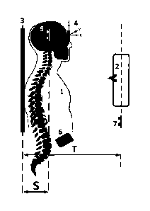
[0010] Fig. 1 is a schematic diagram of the preferred embodiment of the
invention showing
preferred locations and positioning of the sensors in the frontal positioning
of the patient;
[0011] Fig. 2 is a schematic diagram of the preferred embodiment of the
invention showing
preferred locations and positioning of the sensors in the lateral positioning
of the patient; and
[0012] Fig. 3 is a schematic diagram of the X-ray system.
5
DETAILED DESCRIPTION OF THE PREFERRED EMBODIMENT AND THE
DRAWINGS
[0013] The spine consists of a series of vertebrae and interconnecting tissues
disposed and
arranged along the length of the skeleton mammals. In humans, the cord assumes
several
curvatures and is partitioned along these curvatures into four regions,
cervical, thoracic,
lumbar, and lumbar-sacral. The vertebrae of the different regions (and
sometimes, even
within the same region), have different shapes and sizes.
[0014] Damage caused either by sudden impact to the spine either vertically
along its axis or
laterally, congenital defects, or certain diseases can cause the vertebrae to
deform or even
portions thereof to break off, causing discomfort or pain to the patient, and
impairing his
ability to bend and move his body. Moreover, lateral translational (rather
than rotational)
traumatic forces between adjacent vertebrae may cause the internal channels of
adjacent
vertebrae to be offset to the point where the spinal cord passing therethrough
can be
damaged, or even severed, resulting in major health problems to the patient,
such as loss of
the ability to move or sense the body part's.
[0015] (This paragraph is intentionally left blank)
[0016] More specifically, the shape and position of the vertebrae are
determined from X-ray
images. Once each vertebra is identified on an image, and processed within the
device, the
automated software that is a part of a device is used to mathematically
analyze the spine or at
least a region thereof, and, using this analysis, to generate a diagnosis for
the patient. A
problem plaguing this analysis until now has been that each vertebrae is
specific to the size of
CA 2846224 2018-11-01
6
the patient and images taken have magnification and orientation distortions
occurring because
of the relative positions of the X-ray beam source, the patient and the X-ray
image recorder
(film), and as a result, the exact shape, size and position relative to
another vertebra is
difficult to determine accurately for quantification purposes from
conventional X-ray images.
Obviously, any errors in determining the shape, orientation and size of a
vertebra may result
in an erroneous diagnosis, treatment and a prognosis of a disease.
[0017] A further problem in detecting the shape, size and position of
vertebrae exactly is that
the spinal vertebra and the actual shape of the whole spine can look quite
different and can
change from person to person based on a large number of factors such as age,
sex, injuries
and pathological changes in the vertebra and the spine itself.
[0018] Another problem is that the existing systems, like DX Analyzer which
does not solve
the distortion problems due to magnification and orientation. Although an
operator is
preselecting the source of X-rays and film distance, it does not specify the
position of the
patient in relation to the source of X-rays and the film. If the patient is
standing closer to the
X-ray source the image on the film will appear larger than actual, and if the
patient is
standing closer to the film, the image will appear closer to the actual size.
Moreover, if he
does not stand completely straight and/or not facing in a direction that is
exactly
perpendicular to the direction of the X-ray beam, the orientation (angular
optical) distortion
of an X-ray image becomes an issue since the 2 axial geometry of a shadow of
the 3 axial
vertebra is changing when the vertebra is relocated relative to the three
axial space. Because
of these flaws the accuracy of measurements is not attainable with the method
used by the
DX Analyzer.
[0019] (This paragraph is intentionally left blank)
CA 2846224 2018-11-01
7
[0020] To eliminate the above problems and in accordance with the preferred
embodiment, a
novel system is presented for taking X-rays which functions with any X-ray
equipment. The
present system utilizes sensors which control 3-dimensional distortions during
an X-ray
image taking procedure and assist in adjustment of any angular distortion
which was
registered during taking of the X-ray image. Once the information is recorded
during the X-
ray taking procedure the information is transferred to the server system via
Bluetooth
electronic board or any other suitable wireless connection.
[0021] As shown in Figs. 1 and 2, the system includes an X-ray source 2
generating X-rays
directed at a patient 1.The X-rays pass through the patient and are
intercepted by a receptor.
The receptor is used to generate a raw image. While there are many different
kinds of
receptors on the market, in one embodiment an x-ray sensor array 3 is used.
Such arrays are
available from GE, Naomi, and other well-known sources.
[0022] In the preferred embodiment, the locator sensors system includes two
wirelessly
accessible sets of sensors: a primary set of sensors; and a secondary set of
sensors. Figs. 1
and 2 show preferred locations and positioning of the sensors. The primary set
of sensors
preferably includes a main board device 6; a frontal radiation sensor module 4
measuring
effective skin input radiation dose in x-ray examinations for the frontal
view; a distance
sensor module 5 measuring distance S, i.e., the distance from the distance
sensor module 5 to
CA 2846224 2018-11-01
CA 02846224 2014-02-21
WO 2013/028219 8 PCT/1JS2012/000344
the X-Ray sensor array 3; and a lateral radiation sensor module 8 measuring
effective skin
input radiation dose in x-ray examinations for the lateral view. In the
preferred embodiment,
distance sensor module 5 is connected to the frontal radiation sensor module 4
via a cable.
The frontal radiation sensor module 4 is, in turn, connected to the main board
device 6 via a
cable. The primary set of sensors and the angular orientation sensor
(including the digital
compass) are connected together into a patient satellite 10. Patient satellite
10 also preferably
includes a Bluetooth transceiver for communicating with the local server as
described below.
[0023] The secondary set of sensors preferably includes a main board device
(not shown) and.
a second distance sensor module 7 measuring distance T, i.e., the distance
between the X-Ray
source 2 and X-Ray sensor array 3. The main board device and the second
distance sensor
module 7 are preferably formed in a unitary housing as an X-Ray source
satellite 12.
[0024] The system further includes a local server 20 (shown in Fig. 3). The X-
ray source
satellite 12 and the patient satellite 10 are in communication with the local
server via a
conventional wireless communication channel, such as Bluetooth. Moreover, as
schematically shown in Fig. 3, raw images collected by the X-ray sensor array
3 are also sent
to the local server.
[0025] Preferably, the patient satellite is small enough (less than about
1"x1") and is light-
weight, preferably less than 100 grams. If necessary, the above-described
components and
others can be provided in two or more cases. The satellite is attached to the
patient, at a
convenient location closest to a respective body part to be imaged.
[0026] In one embodiment, a single ECG electrode-type pad is attached to the
skin of the
patient using an adhesive and a button which is attached to the patient's
satellite. The satellite
is then snapped to the ECG electrode by means of the button.
[0027] The system operates as follows. First, the patient's satellite is
calibrated toward the X-
ray tube of the X-ray system, so that the positioning of the satellite would
be at the third axis
CA 02846224 2014-02-21
WO 2013/028219 9 PCT/US2012/000344
at zero degrees enabling correction for the third axis to produce a pure
lateral X-ray view
without the 3rd axial interferences. The calibration angles are saved in the
system. The
patient receives the patient satellite component, which is then attached to
the ECG electrode
by means of the button. The patient is then positioned between the source of
the X-rays and
the sensor array.
[0028] The angular positioning of patient's parts is adjusted as required for
a particular kind
of X-ray image utilizing the 3-axial angular sensor by moving the patient to a
specific
positioning guided by the angular sensor to provide a pure lateral view. The
angular
positioning of the patient is monitored by the angular sensor. The sensor
provides an
indication to a technician as to its position. In one embodiment, the
indication is dynamic
(and is visual and/or audible) to assist the technician in positioning the
patient to a
predetermined angular orientation. Thus, the angles can be defined in three
dimensions.
10029] Once the patient is positioned properly, the X-ray source is turned on,
and it starts
generating X-rays. They pass through the patient and the patient's satellite
and reach the X-
ray sensor array. When the X-rays are sensed by the X-ray sensor in the
patient satellite, the
data from the angular sensor and the ultrasound sensors is saved and a message
is sent to the
local server to connect to the X-ray sensor array and detect the X-ray image
(parsing).
100301 The images are studied in a DICOM viewer 22 connected to the local
server 20. The
DICOM viewer displays the received X-ray images with all of the corrections
(as described
in this specification) allowing a radiologist to place proper markings on the
images. Once the
image markings are finished by the radiologist, they are saved at the local
server, and the
coordinates of the markings from the DICOM viewer are sent to a remote
location for
quantifications and a report (preferably in a PDF format) is produced with
specific X-ray
intensities which were registered by the X-ray radiation sensor.
CA 02846224 2014-02-21
WO 2013/028219 10
PCT/US2012/000344
[0031] In one embodiment, when the X-ray image is received by the local server
20, the
image is processed and several corrections are made. One correction is a
magnification
correction that takes into consideration the distances between the X-ray
source, the patient
and the X-ray sensor array. In order to correct a magnification error, the
value of the
magnification is calculated for each particular instance of taking X-ray
images. The value of
the magnification M is calculated using the following formula: M = , T
where T is the
¨ S
distance between the X-ray source and the sensor array, as measured by the
second distance
sensor module 7, and S is the distance between the patient and the sensor
array, as measured
by the distance sensor module 5. Knowing the value of the magnification, each
image can be
uniformly adjusted.
[0032] Another correction takes into consideration, angular offsets of the
various visual
elements in the x-ray image.
[0033] Finally, a correction for an X-ray exposure may also be necessary. It
is this inventor
research conclusion that the variations in X-ray exposures produced by the
same X-ray unit
depend on the temperature of the X-ray beam. Once the X-ray system's output is
monitored
an unnecessary X-ray exposure to the patient can be avoided by constantly
monitoring
performances of the X-ray unit. As soon as the intensity of the X-ray beam is
not consistent
proper machine and image adjustment can be made and the inconsistency noticed
in the
report.
[0034] In the preceding specification, the invention has been described with
reference to
specific exemplary embodiments thereof. It will however, be evident that
various
modifications and changes may be made thereto without departing from the
broader spirit and
scope of the invention as set forth in the claims that follow. The
specification and drawings
are accordingly to be regarded in an illustrative manner rather than a
restrictive sense.