Note : Les descriptions sont présentées dans la langue officielle dans laquelle elles ont été soumises.
CA 02915572 2015-12-15
WO 2014/206856
PCT/EP2014/062939
1
MEDICAL DEVICE COMPRISING COLLAGEN-VI
Field of the invention
The invention relates to a non-biodegradable medical device intended
for insertion into a living body, the medical device comprising a tissue
contact
surface coated with collagen VI. The invention also relates to methods of
manufacturing such devices.
Background
Implantable medical devices may be used for treatment, curing or
remedy of many diseases and conditions in a patient's body. Implantable
medical devices may be used for replacing a part of the body (e.g. dental and
orthopaedic implants, intraocular lenses), or may be used to correct or
restore
the structure of an internal tissue or organ (e.g. vascular stents).
Implantable
medical devices may also be used as drug delivery vehicles.
For example, dental implant systems are widely used for replacing
damaged or lost natural teeth. In such implant systems, a dental fixture
(screw), usually made of titanium or a titanium alloy, is fixed in the jawbone
of
the patient in order to replace the natural tooth root. An abutment structure
is
then attached to the fixture in order to build up a core for the part of the
prosthetic tooth protruding from the bone tissue, through the soft gingival
tissue and into the mouth of the patient. On said abutment, the prosthesis or
crown may finally be seated.
For any type of medical device intended for contact with living tissue,
biocompatibility is a crucial issue. The risk for foreign body reaction, clot
formation and infection, among many other things, must be addressed and
minimized in order to avoid adverse effects, local as well as systemic, which
may otherwise compromise the health of the patient and/or lead to failure of
the device. This is particularly the case for permanent implants. Furthermore,
healing or regeneration of tissue around an implant is often vital in order to
secure the implant and its long-term functionality. This is especially
important
for load-bearing implants such as dental or orthopedic implants.
CA 02915572 2015-12-15
WO 2014/206856
PCT/EP2014/062939
2
For dental fixtures, a strong attachment between the bone tissue and
the implant is necessary. For implants intended for contact with soft tissue,
such as abutments which are to be partially located in the soft gingival
tissue,
also the compatibility with soft tissue is vital for total implant
functionality.
Typically, after implantation of a dental implant system, an abutment is
partially or completely surrounded by gingival tissue. It is desirable that
the
gingival tissue should heal quickly and firmly around the implant, both for
medical and aesthetic reasons. A tight sealing between the oral mucosa and
the dental implant serves as a barrier against the oral microbial environment
and is crucial for implant success. This is especially important for patients
with poor oral hygiene and/or inadequate bone or mucosal quality. Poor
healing or poor attachment between the soft tissue and the implant increases
the risk for infection and peri-implantitis, which may ultimately lead to bone
resorption and failure of the implant.
There are several strategies for increasing the chances of a successful
implantation of a medical device, for example enhancing the rate of new
tissue formation and/or, in instances where tissue-implant bonding is desired,
enhancing the rate of tissue attachment to the implant surface, or by
reducing the risk for infection. Enhancement of new tissue formation may be
achieved for example by various surface modifications and/or deposition of
bioactive agents on the surface. The risk of infection in connection with
dental
implants is today primarily addressed by preventive measures, such as
maintaining good oral hygiene. Once a biofilm is formed on the surface of a
dental implant, it is difficult to remove it by applying antibacterial agents.
In
the case of infection in the bone or the soft tissue surrounding a dental
implant (peri-implantitis), mechanical debridement is the basic element,
sometimes in combination with antibiotics, antiseptics, and/or ultrasonic or
laser treatment.
There is a need in the art for better ways of preventing infections at the
site of an implanted medical device.
CA 02915572 2015-12-15
WO 2014/206856
PCT/EP2014/062939
3
Summary of the invention
It is an object of the present invention to at least partially overcome this
problem, and to provide a medical device, such as an implant, having a
surface which reduces the risk for infection upon contact of the medical
device with living tissue.
According to a first aspect of the invention, this and other objects are
achieved by a medical device intended for insertion into a living body, the
medical device comprising a non-biodegradable substrate having a tissue
contact surface, wherein the tissue contact surface is at least partially
coated
with microfibrils of collagen VI.
Collagen is a protein that forms a major component of the extracellular
matrix of many tissues and organs. There are at least 28 different types of
collagen found in various tissues. Collage type VI (also denoted "collagen
VI",
"collagen-VI" or "type VI collagen") is a ubiquitous component of the
mammalian extracellular matrix. As used herein "collagen VI microfibril" or
"microfibrils of collagen VI" refers to a filament structure formed of
collagen VI
molecule tetramers aggregated end-to-end. Such microfibrils may have a
width in the range of from 1 to 50 nm, for example from 5 to 20 nm, and a
length in the range of 0.1 to 10 pm, for example from 0.5 to 5 pm.
The inventors have found that a biomaterial coated with collagen VI
microfibrils provides antimicrobial properties against aerobic and anaerobic
human pathogens. . Notably, the antimicrobial effect was exerted by collagen
VI present on the surface and likely not involving any significant release of
collagen VI from the surface. Hence, the antimicrobial surface may better
resist attenuation compared to devices releasing antimicrobial substances.
Experimental results suggest that the surface may retain an antimicrobial
effect over an extended period of time. The medical device according to
embodiments of the invention thus offers better prevention of infections
following surgical implantation of an implant or medical device into a
patient.
Furthermore, a synergistic effect between the antimicrobial properties
of collagen VI and the innate immune system, especially neutrophils, was
seen. This was surprising; at most the recruitment of polymorphonuclear
neutrophils (PMNs) to remove the already dead bacteria had been expected,
CA 02915572 2015-12-15
WO 2014/206856
PCT/EP2014/062939
4
since the main biological function of PMNs is to fagocyte, destroy and discard
bacteria. Instead, it was unexpectedly found that PMNs were stimulated to
produce neutrophil extracellular traps (NETs) to entrap the bacteria on the
surface, such that the bacteria were prevented from further spreading and
also rapidly killed by collagen VI. Additionally, bacterial killing by
collagen VI
is surprisingly enhanced by the presence of PMN and their released
proteases. Not wishing to be bound by any particular theory, it is believed
that
this PMN stimulating effect may, at least partly, be due to the microfibrillar
structure of the collagen VI, which includes intact N- and C-terminal domains,
is believed to imitate the natural biological environment of a wound, and
might
also benefit from yet unknown immune and inflammatory processes.
Hence, in situations where neutrophils would come in contact with a
collagen VI coated medical device, such as during surgery or any procedure
that causes bleeding and/or inflammation at the site of an implant or a
medical device, the antibacterial effect will be further enhanced by using a
medical device according to the present invention.
Typically, the collagen VI may be present as native microfibrils, with
preserved N- and C-terminal globular domains.
In embodiments of the invention, the non-biodegradable substrate
comprises a biocompatible material selected from metallic, ceramic or plastic
materials. For example, the non-biodegradable substrate comprises a metallic
material selected from the group consisting of titanium, zirconium, hafnium,
vanadium, niobium, tantalum, cobalt and iridium, and alloys thereof. In other
embodiments, the substrate may comprise a ceramic material, for example
zirconia.
In embodiments of the invention, the medical device may be a surgical
implant, intended for implantation into hard tissue such as bone, and/or soft
tissue..
In embodiments of the invention, the medical device may be a load-
bearing implant.
For instance, the medical device may be a dental implant or a part
thereof, such as a dental fixture, a dental abutment or a one-piece dental
implant. In other instances, the medical device may be a bone anchored
CA 02915572 2015-12-15
WO 2014/206856
PCT/EP2014/062939
hearing device. Alternatively, the medical device may be an orthopaedic
implant.
In embodiments of the invention, the medical device may be a stent or
a shunt.
5 In embodiments of the invention, the tissue contact surface of the
medical device may be coated with a layer of collagen VI. Optionally, the
collagen VI may be attached to the surface via linker molecules. A layer of
collagen VI may have a layer thickness in the range of from 1 nm to 50 nm,
for example from 5 nm to 50 nm. The layer may be discontinuous, i.e., not
completely covering the underling surface.
In another aspect, the invention provides a method of coating a surface
of a medical device, comprising
(i) providing a non-biodegradable substrate having a tissue contact
surface;
(ii) optionally attaching linker molecules to said tissue contact surface;
and
(iii) contacting at least part of said tissue contact surface with a solution
comprising microfibrils of collagen VI to attach said microfibrils of collagen
VI
to said surface and/or said linker molecules.
The linker molecules may comprise poly-L-lysine (PLL).
In embodiments of the invention, step ii) may be performed by
(ii-a) applying a solution comprising the linker molecules and a solvent
onto the surface of the article, and
(ii-b) removing said solvent.
Alternatively or additionally, step iii) may be performed by
(iii-a) applying a solution comprising microfibrils of collagen VI and a
solvent to said surface;
(iii-b) incubating the substrate having said solution applied to said
surface; and
(iii-c) removing said solvent.
Step iii-b) may be performed by keeping the article at a temperature in the
range of 4 to 40 C for at least 10 minutes.
CA 02915572 2015-12-15
WO 2014/206856
PCT/EP2014/062939
6
In embodiments of the invention, the solution comprising microfibrils of
collagen VI may have a concentration of collagen fibrils in the range of from
nM (150 ng/ml) to 10 pM (150 jig/m1), for example from 0.5 to 5 pM, such
as from 1 to 2 pM.
5 Also in the method forming the second aspect of the invention, the
collagen VI may preferably be present as native microfibrils.
It is noted that the invention relates to all possible combinations of
features recited in the claims.
10 Brief description of the drawings
Figure 1 shows scanning electron micrographs of different surfaces
incubated with Streptococcus mitis for 0 hours (left column), 24 hours (middle
column) and 48 hours (right column), respectively. The surfaces were the
following: a titanium surface (Ti; top row), a Ti surface coated with collagen
IV
(Ti/cVI; second row from the top), a Ti surface coated with poly-L-lysine, PLL
(Ti/PLL; third row from the top) and a Ti surface coated with poly-L-lysine
and
collagen VI (Ti/PLL/cVI; bottom row). The scale bar represents 10 pm (same
scale in all images).
Figure 2 shows scanning electron micrographs of different surfaces
incubated with Actinomyces naeslundii for 0 hours (left column), 24 hours
(middle column) and 48 hours (right column), respectively. The surfaces were
the following: a titanium surface (Ti; top row), a Ti surface coated with
collagen IV (Ti/cVI; second row from the top), a Ti surface coated with poly-L-
lysine, PLL (Ti/PLL; third row from the top) and a Ti surface coated with poly-
L-lysine and collagen VI (Ti/PLL/cVI; bottom row). The scale bar represents
10 pm (same scale in all images).
Figure 3 shows scanning electron micrographs of different surfaces
incubated with Fusobacterium nucleatum for 0 hours (left column), 24 hours
(middle column) and 48 hours (right column), respectively. The surfaces were
the following: a titanium surface (Ti; top row), a Ti surface coated with
collagen IV (Ti/cVI; second row from the top), a Ti surface coated with poly-L-
lysine, PLL (Ti/PLL; third row from the top) and a Ti surface coated with poly-
CA 02915572 2015-12-15
WO 2014/206856
PCT/EP2014/062939
7
L-lysine and collagen VI (Ti/PLL/cVI; bottom row). The scale bar represents
pm (same scale in all images).
Figure 4 shows scanning electron micrographs of different surfaces
incubated with Prevotella intermedia for 0 hours (left column), 24 hours
5 (middle column) and 48 hours (right column), respectively. The surfaces
were
the following: a titanium surface (Ti; top row), a Ti surface coated with
collagen IV (Ti/cVI; second row from the top), a Ti surface coated with poly-L-
lysine, PLL (Ti/PLL; third row from the top) and a Ti surface coated with poly-
L-lysine and collagen VI (Ti/PLL/cVI; bottom row). The scale bar represents
10 10 pm (same scale in all images).
Figure 5 shows scanning electron micrographs of the surfaces Ti/PLL
(top row) and Ti/PLL/cVI (bottom row), respectively after 48 hours of
incubation with S. mitis (left column), A. naeslundii (second to left column),
Fusobacterium nucleatum (second to right column) and Prevotella intermedia
(right column), respectively. As can be seen, in the presence of collagen VI
(Ti/PLL/cVI) bacterial killing is visible as membrane disruption and
cytoplasmic exudation, indicated by white arrowheads. The scale bars
represent 2 pm (same scale in all images).
Figure 6 shows scanning electron micrographs of S. mitis incubated on
a collagen VI coated Ti surface (denoted "collagen VI", top row) and a Ti
surface ("control", bottom row), respectively. Every day a fresh 0.1% solution
of bacteria was added to the surfaces. In the presence of collagen VI
bacterial
growth was significantly inhibited. The scale bar represents 10 pm.
Figure 7 shows scanning electron micrographs of A. naeslundii
incubated on a collagen VI coated Ti surface (denoted "collagen VI", top row)
and a Ti surface ("control", bottom row), respectively. Every day a fresh 0.1%
solution of bacteria was added to the surfaces. In the presence of collagen VI
bacterial growth was significantly inhibited. The scale bar represents 10 pm.
Figure 8 shows S scanning electron micrographs of entrapment of
bacteria (S. mitis) in Neutrophil Extracellular Traps (NETs) produced by
polymorphonuclear neutrophil (PMNs) on a ceramic dental abutment (top
row) and a titanium screw (bottom row). The SEM images show at increasing
CA 02915572 2015-12-15
WO 2014/206856
PCT/EP2014/062939
8
magnification (scale bars indicating 1 mm, 100 pm, 20 pm and 5 pm) how
bacteria are entrapped in NETs ejected by PMN.
Figure 9 presents a scanning electron micrograph showing entrapment
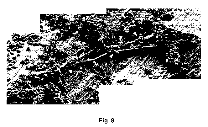
by PMN NETs on a Ti surface coated with collagen VI (Ti/PLL/cVI). The PMN
eject NETs to which the bacteria adhere and become entrapped (as indicated
by white arrowheads). The scale bar represents 5 pm.
Figures 10A and 10B each shows scanning electron micrographs of
bacterial entrapment and killing in PMN NETs on the surface of a titanium
screw (Fig. 10 A) or a ceramic abutment (Fig. 10B), coated with collagen VI
using PLL as linker (cVI; right column)) or without coating (control; left
column)) incubated with S. mitis for 0 minutes (top row) and 120 minutes
(bottom row), respectively. In the presence of collagen VI the structural
integrity of the bacteria is rapidly compromised as evidenced by membrane
blebbing (indicated by white arrowheads). Without the collagen VI coating the
bacteria remain trapped in NETs but are not killed. The scale bar represents
2 pm.
Figure 11 schematically depicts the various structures of collagen VI,
including polypeptide chains, collagen VI monomers, and a native collagen VI
microfibril.
Detailed description
The present inventors have found that a medical device having a tissue
contact surface at least partially coated with microfibrils of collagen VI, in
particular native microfibrils, may provide a significant antibacterial
effect,
which is very desirable, in particular for implantable medical devices.
Additionally, it was found that collagen VI microfibril coated surfaces
exhibited
an immune stimulating effect beyond expectation.
As used herein, "collagen VI microfibril" or "microfibrils of collagen VI"
refers to a filament structure formed of collagen VI molecule tetramers
aggregated end-to-end. The present invention preferably uses native
microfibrils, meaning that the microfibil structure corresponds to the native
form of collagen VI found in living tissue. In contrast, a non-native
microfibril
may be partially degraded, e.g. at the N- and/or C-terminal globular domains.
CA 02915572 2015-12-15
WO 2014/206856
PCT/EP2014/062939
9
Native microfibrils may be isolated from tissue samples using a method as
described in Spissinger T, Engel J, Matrix Biol 1995; 14:499-505, using
bovine corneal collagenase. Advantageously, this method preserves the
globular domains. In contrast, a method using e.g. pepsin cleaves the
microfibrils in the globular domains, thus resulting in a partially degraded,
non-native collagen VI microfibril.
Directive 2007/47/ec defines a medical device as: "any instrument,
apparatus, appliance, software, material or other article, whether used alone
or in combination, including the software intended by its manufacturer to be
used specifically for diagnostic and/or therapeutic purposes and necessary for
its proper application, intended by the manufacturer to be used for human
beings". In the context of the present invention, only medical devices
intended
for contact with living tissue are considered, that is, any instrument,
apparatus
appliance, material or other article of physical character that is intended to
be
applied on, inserted into, implanted in or otherwise brought into contact with
the body, a body part or an organ. Furthermore, said body, body part or organ
may be that of a human or animal, typically mammal, subject. Preferably
however the medical device is intended for human subjects. Medical devices
included within the above definition are for example implants, catheters,
shunts, tubes, stents, intrauterine devices, and prostheses.
In particular, the medical device may be a medical device intended for
implantation into living tissue or for insertion into the body or a body part
of a
subject, including insertion into a bodily cavity.
The present medical device may be intended for short-term, prolonged
or long-term contact with living tissue. By "short-term" is meant a duration
of
less than 24 hours, in accordance with definitions found in ISO 10993-1 for
the biological evaluation of medical devices. Furthermore, "prolonged",
according to the same standard, refers to a duration of from 24 hours up to
days. Accordingly, by the same standard, by "long-term" is meant a
30 duration of more than 30 days. Thus, in some embodiments the medical
device of the invention may be a permanent implant, intended to remain for
months, years, or even life-long in the body of a subject.
CA 02915572 2015-12-15
WO 2014/206856
PCT/EP2014/062939
As used herein the term "implant" includes within its scope any device
of which at least a part is intended to be implanted into the body of a
vertebrate animal, in particular a mammal, such as a human. Implants may be
used to replace anatomy and/or restore any function of the body. Generally,
5 an implant is composed of one or several implant parts. For instance, a
dental
implant usually comprises a dental fixture coupled to secondary implant parts,
such as an abutment and/or a restoration tooth. However, any device, such
as a dental fixture, intended for implantation may alone be referred to as an
implant even if other parts are to be connected thereto.
10 By "biocompatible" is meant a material which upon contact with living
tissue does not as such elicit an adverse biological response (for example
inflammation or other immunological reactions) of the tissue.
By "soft tissue" is meant any tissue type, in particular mammalian
tissue types, that is not bone or cartilage. Examples of soft tissue for which
the medical device is suitable include, but are not limited to, connective
tissue, fibrous tissue, epithelial tissue, vascular tissue, muscular tissue,
mucosa, gingiva, and skin.
Collagen is a protein that forms a major component of the extracellular
matrix of many tissues and organs. There are at least 28 different types of
collagen found in various tissues; collagen type I (collagen I) being the most
abundant form in bone and connective tissue; collagen type II being
predominant in cartilage, collagen III being a major constituent of the blood
vessel wall but also present in cartilage, and collagen type IV being a
constituent of the basement membrane. An individual collagen molecule
consists of three polypeptide chains (also referred to as pro a-chains), each
forming an a-helix, closely intertwined in a triple helix configuration.
Different
types of collagen differ in the amino acid sequences of the polypeptide
chains, and also with respect to secondary structure and/or tertiary
structure.
Collage type VI (also denoted "collagen VI", "collagen-VI" or "type VI
collagen") is a ubiquitous component of the mammalian extracellular matrix. It
is present in connective tissues, often associated with basement membranes.
As shown in Fig. 11, to form a collagen VI monomer 10, al , a2, and a3
polypeptide chains assemble in a heterotrimer formation, where additional
CA 02915572 2015-12-15
WO 2014/206856
PCT/EP2014/062939
11
tissue-specific chains may substitute for the a3 chain in some instances. Four
monomers align to a tetramer by lateral association, and a plurality of
tetramers aggregate end-on-end to form a microfibril 20 having the shape of a
thin, beaded filament. Such microfibrils, also referred to as native
microfibrils,
typically have a length in the range of from 0.5 to 5 pm and a width of about
to 15 nm.
Interestingly, in their bological environment native collagen microfibrils
are typically not sensitive to enzymatic degradation. This may be due to the
biological role of collagen VI as a biomechanical tissue stabilizer, being
10 important for tissue volume, vascularization and immune cell
infiltration.
The N- and C-terminal globular domains of collagen VI share homology
with von Willebrand factor type A domains (Specks U, Mayer U, Nischt R,
Spissinger T, Mann K, Timpl R, Engel J, Chu ML, EMBO J 1992; 11:4281-
4290), and collagen VI in solution has been shown to possess an
antimicrobial activity against A, C and G streptococci, which are Gram-
positive bacteria (Abdillahi S. M., Balvanovic S., Baumgarten M, Morgelin M.,
J Innate Immun 2012; 4:371-3762). The present inventors have now shown
that collagen VI is not only effective against bacteria when coated onto a
device surface, but also has an innate immunomodulating effect, which is
likely to be highly beneficial for healing and tissue regeneration. During
infection, bacteria stimulate PMN cells to secrete Neutrophil Extracellular
Traps (NETs) which entrap and immobilize the bacteria. The recent results
show that bacterial killing in NETs is considerably more effective on surfaces
which are coated with collagen VI (Fig. 9, 10A-B). Moreover, bacterial killing
by collagen VI is surprisingly enhanced by the presence of PMN and their
released proteases. Taken together, the results suggest that collagen VI may
be beneficial for wound healing in the clinical situation by minimizing the
occurrence of bacterial colonization.
In view of these insights and results, the present inventors propose a
medical device intended for insertion into a living body, the medical device
comprising a non-biodegradable substrate having a tissue contact surface,
wherein said tissue contact surface is at least partially coated with collagen
VI.
CA 02915572 2015-12-15
WO 2014/206856
PCT/EP2014/062939
12
The medical device according to embodiments of the invention may be
made of any suitable biocompatible material, e.g. materials used for
implantable devices. Typically the medical device comprises a substrate
having a tissue contact surface.
By "tissue contact surface" is meant a surface intended for contact
(short-term, prolonged, or long-term) with living tissue.
The substrate may for example be made of a biocompatible metal or
metal alloy, including one or more materials selected from the group
consisting of titanium, zirconium, hafnium, vanadium, niobium, tantalum,
cobalt and iridium, and alloys thereof. Alternatively, the substrate of the
medical device may be made of a biocompatible ceramic, such as zirconia,
titania, shape memory metal ceramics and combinations thereof. In
embodiments where the medical device is used as or forms part of a dental
abutment, the substrate is preferably made of a metallic material.
In contact with oxygen, the metals titanium, zirconium, hafnium,
tantalum, niobium and their alloys instantaneously react to form an inert
oxide. Thus, the surfaces of articles of these materials are virtually always
covered with a thin oxide layer. The native oxide layer of a titanium
substrate
mainly consists of titanium(IV) dioxide (Ti02) with minor amounts of Ti203,
TiO and Ti304.
Thus, in embodiments where the medical device comprises one or
more of titanium, zirconium, hafnium, tantalum, niobium or an alloy of any one
thereof, the medical device typically has a native metal oxide surface layer.
Such a native metal oxide layer may, in turn, be at least partially covered by
a
layer of collagen VI microfibrils.
In other embodiments of the present invention, the medical device, in
particular the substrate, may be made of a biocompatible polymer, typically
selected from the group consisting of polyether ether ketone (PEEK), poly
methyl methacrylate (PMMA), poly lactic acid (PLLA) and polyglycolic acid
(PGA) and any combinations and copolymers thereof.
In embodiments of the invention, the medical device is intended for
short-term, prolonged or long-term contact with living tissue. For example,
the
medical device of the invention may be an implant, typically intended to
CA 02915572 2015-12-15
WO 2014/206856
PCT/EP2014/062939
13
temporarily or permanently replace or restore a function or structure of the
body.
Typically, at least part of the surface of the medical device is intended
for contact with soft tissue, and at least part of this soft tissue contact
surface
has a coating of collagen VI microfibrils. For example, the medical device may
be an implant intended for contact primarily or exclusively with soft tissue,
for
example a dental abutment. Alternatively, the medical device may be an
implant to be inserted partially in bone and partially in soft tissue.
Examples of
such implants include one-piece dental implants and bone-anchored hearing
devices (also referred to as bone anchored hearing aids). Where only part of
the implant is intended for contact with soft tissue, it is preferred that the
coating comprising collagen-VI is provided at least on a part of a soft tissue
contact surface.
The medical device may also be suitable for contact with cartilage.
In other embodiments, the medical device may be intended for contact
with bone tissue, e.g. the jawbone, the femur or the skull of a mammal, in
particular a human. Examples of such medical devices include dental fixtures
and orthopedic implants.
In embodiments of the invention, the tissue contact surface may be a
rough surface. The substrate surface roughness, and hence optionally also
the surface of the medical device formed by coating with collagen-VI, may
have an average surface roughness Ra of at least 0.05 i.tm, typically at least
0.1 i.tm, for example at least 0.2 i.tm. Since surfaces having an average
surface roughness (Ra) of at least 0.2 i.tm are believed to be more
susceptible
of biofilm formation, a coating of collagen VI as described herein may be
particularly advantageous for medical devices having a surface roughness of
at least 0.2 i.tm, and may be increasingly useful for preventing biofilm
formation on medical devices having even higher surface roughness. As an
example, a dental abutment comprising a titanium substrate may have a
surface roughness of about 0.2-0.3 i.tm. A coating layer of collagen VI
microfibrils may preserve an underlying surface roughness.
CA 02915572 2015-12-15
WO 2014/206856
PCT/EP2014/062939
14
Furthermore, in embodiments of the invention, the tissue contact
surface of the medical device may comprise at least one additional
biomolecule.
The medical device of the invention may be produced by coating
the surface with collagen VI microfibrils directly onto the surface or via
linker
molecule. In embodiments using a linker molecule, the linker molecule is first
attached to the surface, and subsequently the collagen microfibrils are
attached to said linker molecules. In embodiments using no linker molecule,
the surface may however optionally be treated chemically or physically, e.g.
in
order to clean the surface or to impart a net electrical charge, to enhance
attachment of the collagen VI microfibrils. For example, the surface may be
subjected to a surface treatment that increases the hydrophilicity of the
surface.
After attaching the collagen fibrils, the medical device may optionally
be subjected to a mild sterilizing treatment, before use e.g. as an implant or
a
part thereof.
The collagen VI microfibrils according to embodiments of the present
invention may assume any orientation when coated onto a surface, with or
without the use of a linker molecule.
For example, the collagen VI microfibrils may be applied to the surface
of medical device by applying a solution comprising the collagen microfibrils
to the surface. The solution may be applied to the surface by any
conventional technique that leaves at least a thin film of solution covering
the
surface to be coated with collagen VI. Such methods include spraying,
pouring and dripping the solution onto the surface, and immersing the surface
into the solution.
The solution may be an aqueous solution of collagen VI microfibrils at
a concentration in the range of from 10 nM to 10 pM, for example from 0.5 to
5 pM, such as from 1 to 2 pM.
After applying a thin film of a collagen VI solution to the surface, the
medical device may be allowed to incubate for a time period of at least 10
minutes, typically at least 30 minutes, for example about 45 minutes, and up
to several hours, typically up to 1 hour. Incubation may be carried out at a
CA 02915572 2015-12-15
WO 2014/206856
PCT/EP2014/062939
temperature of 40 C or less, typically in the range of 4 to 40 C, for example
at
room temperature (15-25 C). The medical device may be incubated in a
humid chamber. Incubating the device in a humid atmosphere is
advantageous because it ensures that the solvent does not evaporate too fast
5 from the surface. A humid chamber as used in embodiments of the invention
typically means a closed chamber in which the component is placed, and in
which is also present a pool of sterile water or a tissue soaked with sterile
water. In an industrial setting the humid chamber may be a controlled
chamber with 75-100 % humidity. However, it should be noted that a humid
10 chamber is not necessary, and too fast drying of the applied solution
may be
avoided also at ambient humidity.
After incubation (evaporation of the solvent), the surface is typically
washed, e.g. in sterile water or a suitable buffer solution, to remove
remaining
solution, and may optionally be subjected to a suitable sterilizing treatment,
15 e.g. UV or gamma irradiation or chemical sterilization using ethylene
oxide
gas.
In embodiments of the invention using a linker molecule, the linker is
typically attached to the surface before applying the collagen VI
microfibrils.
The linker may be attached to the surface by any suitable means, including
for example electrostatic interaction, hydrophobic interaction, or covalent
binding. In particular, the linker molecule may be attached to the surface via
electrostatic interaction. For example, on a surface having a negative
electric
charge, such as a titanium oxide surface of a titanium article, a positively
charged linker molecule such as poly-L-lysine may be attached. If necessary,
the surface may be treated or modified by known methods to obtain an
electric charge.
The linker molecule may be attached to the surface by applying a
solution of the linker molecule onto the surface, preferably so as to
completely
cover the surface with said solution. Typically, the surface is previously
washed e.g. with ethanol, and dried. The solution of linker molecule may be
applied by any conventional techniques, such as spraying, pouring or dripping
the solution onto the surface or immersing the surface into the solution.
CA 02915572 2015-12-15
WO 2014/206856
PCT/EP2014/062939
16
In the case of poly-L-lysine, the solution applied to the surface may be
a solution of PLL having a concentration may be in the range of 0.01 to
1 mg/ml, typically about 0.2 mg/ml.
After applying the linker solution to the surface of the medical device,
the solvent is removed, leaving the linker molecules attached to the surface.
For example, the solvent may be evaporated by treating the medical device at
elevated temperature, e.g. in the range of 40 to 60 C, and typically about
60 C. The time required for allowing the solvent to evaporate may be in the
range of 10 minutes to 2 hours, typically from 30 minutes to 1 hour.
Optionally, in embodiments of the invention, after applying the linker
solution to the surface of the article, the medical device is incubated for a
few
minutes, e.g. 1-10 minutes, and the linker solution, except those linker
molecules that have already bound to the surface, is subsequently washed off
by rinsing with a rinsing agent e.g. sterile water, before the article is
subjected
to elevated temperature as described above. After evaporation or the solvent
or the rinsing agent, the surface having attached linker molecules is
optionally
washed, e.g. with sterile water and dried or allowed to dry.
The collagen microfibrils may be attached to the linker molecules by
applying a solution comprising collagen fibrils to the surface coated with the
linker molecules according to the description above. The solution comprising
collagen fibrils may be applied to the surface by any conventional technique
that leaves at least a thin film of solution covering the surface to be coated
with collagen fibrils. Such methods include spraying, pouring and dripping the
solution onto the surface, and immersing the surface into the solution.
The solution comprising collagen fibrils may be an aqueous solution of
collagen VI microfibrils at a concentration in the range of from 10 nM to
10 pM, for example from 0.5 to 5 pM, such as from 1 to 2 pM.
After applying a thin film of collagen solution to the surface, the medical
device may be allowed to incubate for a time period of at least 10 minutes,
typically at least 30 minutes, for example about 45 minutes, and up to several
hours, typically up to 1 hour. Incubation may be carried out at a temperature
of 40 C or less, typically in the range of 4 to 40 C, for example at room
temperature (15-25 C). The medical device may be incubated in a humid
CA 02915572 2015-12-15
WO 2014/206856
PCT/EP2014/062939
17
chamber. Incubating the device in a humid atmosphere is advantageous
because it ensures that the solvent does not evaporate too fast. A humid
chamber as used in embodiments of the invention typically means a closed
chamber in which the component is placed, and in which is also present a
pool of sterile water or a tissue soaked with sterile water. In an industrial
setting the humid chamber may be a controlled chamber with 75-100%
humidity. However, it should be noted that a humid chamber is not necessary,
and too fast drying of the applied solution may be avoided also at ambient
humidity.
After incubation (evaporation of the solvent), the surface is typically
washed, e.g. in sterile water or a suitable buffer solution, to remove
remaining
solution, and may optionally be subjected to a suitable sterilizing treatment,
e.g. UV or gamma irradiation or chemical sterilization using ethylene oxide
gas.
It is envisaged that further modifications could be made to the collagen
fibril coating obtained by the method according to the invention. For example,
a bioactive substance as described above could be applied to the collagen
fibril coating. Additionally of alternatively, the collagen fibrils could be
cross-
linked after being attached to the surface, e.g. in order to reduce the rate
of
fibril degradation in vivo after implantation of the medical device.
Experiments
A. Material and Methods
1. Bacteria
Streptococcus mitis, Actinomyces naeslundii, Fusobacterium
nucleatum and Prevotella intermedia were kindly provided by Julia Davies
and Gunnel Svensater (Department of Oral Biology, Faculty of Odontology,
Malmo University, Malmo, Sweden). S. mitis and A. naeslundii were grown
overnight in Todd-Hewitt broth (THB) at 37 C in humid atmosphere containing
5 % 002. F. nucleatum and P. intermedia were grown in Peptone Yeast
Glucose (PYG) medium at 37 C in humid atmosphere under anaerobic
conditions.
CA 02915572 2015-12-15
WO 2014/206856
PCT/EP2014/062939
18
2. Collagen VI
Collagen VI was isolated from bovine cornea by collagenase digestion
as described by Abdillahi et al. (2012). Calf eyes were received from the
local
slaughterhouse. Corneas were cut into pieces and extracted with
collagenase, followed by gel filtration with Sepharose CL-2B.
3. Coating titanium
Titanium circles with a diameter of 5 mm were punched out from a foil.
First the circles are washed with chloroform and followed by distilled water.
After air drying, 50 pL poly-L-lysine (0.2 mg/mL) was applied on desired
titanium pieces. Then the pieces have to be incubated at 60 C for two hours.
Afterwards the titanium is washed again in distilled water and air dried. In
meantime, 1 mL collagen VI solution is applied into wells of a 12-well plate.
Finally desired titanium circles are plunged into the collagen VI solution and
incubated over night at 4 C. Following morning, the titanium was air dried and
assays were performed.
4. Bacterial adhesion
For adhesion assays S. mitis and A. naeslundii were grown overnight
in 10 mL THB, F. nucleatum and P. intermedia were grown in PYG medium
under anaerobic conditions and pelleted down at 3500 rpm for 10 minutes at
4 C on the next day. For the anaerobic species, Fusobacterium nucleatum
and Prevotella intermedia, the procedure was performed inside an anaerobic
box. Then the pellet was diluted in 10 mL PBST and the 0D600 was
measured. The 0D600 has to be adjusted to 1 and diluted 1:2 in PBST.
500 pL of the bacteria solution were applied into each well with prepared
titanium circles inside and incubated at 37 C and 5 (:)/0 CO2 for 0, 30 and
240 minutes.
The samples were washed with 500 pL PBS three times. PBS was
removed and replaced by 500 pL of EM-fix consisting of 2.5 (:)/0
glutaraldehyde
in 0.15 M sodium-cacodylate. Samples were incubated in EM-fix overnight.
Following steps were performed by an experienced technician. Washing
steps with Cacodylate-buffer were performed, followed by a dehydration
CA 02915572 2015-12-15
WO 2014/206856
PCT/EP2014/062939
19
series. Therefore the samples were incubated for five minutes twice with
50 %, 70 (:)/0 and 95 (:)/0 ethanol and with absolute ethanol for 30 minutes
and
one hour. For drying the samples, ethanol was carried to its critical point to
turn into gas by using liquid 002. This step was performed three times for ten
minutes. Afterwards samples were mounted and coated with gold/palladium
20 nm Agar. Samples were investigated at a scanning electron microscope
XL 30 FEG and images were processed by AnalySIS ITEM software.
5. Bacterial killing assays
5.1 BacLight for FACS
Flow cytometry analysis enables to determine the nature of single
cells. In this study BacLight staining was used to stain bacterial cells. That
way, it is possible to distinguish between the amount of living and dead
bacteria in one population.
Bacteria were grown over night in THB under standard conditions.
1 mL of the overnight culture was transferred to 9 mL fresh THB the next day.
Bacteria were incubated under standard conditions, until an 0D600 of 0.4
was reached. Bacteria were pelleted down at 3500 rpm and 4 C for 10
minutes. The supernatant was discarded and the pellet diluted in 10 mL cold
TG buffer. The OD was measured and the bacterial solution was pelleted
down a second time. After discarding the supernatant the bacterial amount
was adjusted to 1 (:)/0 with cold TG buffer. The solution was diluted 1:10 in
TG
and 20 pL of the solution were transferred to 1.5 mL reaction tubes. 80, 160,
200 or 500 pL collagen VI was applied to the 1.5 mL reaction tubes. For the
negative control 80 pL TG buffer were added. For the positive control 300 pL
LL-37 were added. Samples were incubated for 0, 2, 24 and 48 hours and
stained with 5 pL of a PI and STY09 diluted 1:100. Samples were measured
using BD Accuri 06 flow cytometer.
5.2 Scanning electron microscopy (SEM)
With SEM it is possible to investigate the adhesion of bacteria to
coated titanium surfaces.
CA 02915572 2015-12-15
WO 2014/206856
PCT/EP2014/062939
For SEM bacteria were grown overnight to an 0D600 of 1.
F. nucleatum and P. intermedia were grown in PYG medium under anaerobic
conditions, whereas S. mitis and A. naeslundii were grown in THB. The
anaerobic species were treated inside an anaerobic box. The cultures were
5 pelleted down and diluted in 10 mL PBST. After the 0D600 was adjusted to
1,
the bacteria suspension was diluted 1:2 with PBST. 500 pL of this suspension
were applied on each well of a 24-well-plate with a coated titanium circle
inside. The bacteria were incubated for two hours at 37 C to permit adhesion
on the titanium surface. After this time samples were washed with PBS three
10 times and 500 pL THB were added to allow bacterial growth. Bacteria were
incubated for 0 minutes, 4, 24 and 48 hours at 37 C. After the incubation,
wells were washed three times with 500 pL PBS and bacteria were fixed with
EM-fix consisting of 2.5% glutaraldehyde in 0.15 M sodium-cacodylate.
Samples were incubated with EM-fix overnight. Following steps were
15 performed by an experienced technician. Washing steps with Cacodylate-
buffer were performed, followed by a dehydration series. Therefor the
samples were incubated for five minutes twice with 50 %, 70 (:)/0 and 95 (:)/0
ethanol and with absolute ethanol for 30 minutes and one hour. For drying the
samples, ethanol was carried to its critical point to turn into gas by using
liquid
20 CO2. This step was performed three times for ten minutes. Afterwards
samples were mounted and coated with gold/palladium 20 nm Agar. Samples
were investigated at a scanning electron microscope XL 30 FEG and images
are processed by AnalySIS ITEM software.
6. Long-term activity of collagen VI
To find out how long collagen VI is active on titanium surfaces new
bacteria were applied to the experimental setting every day new bacteria.
For the experiment titanium was coated as usual. Bacteria were grown
over night in THB at standard conditions. The next morning 1 mL of the
overnight culture is transferred to 9 mL fresh THB and incubated until the
0D600 of the bacteria solution reached 0.4. Bacteria were pelleted down at
3500 rpm for 10 minutes at 4 C. The pellet was resuspended in 10 mL cold
TG-buffer and OD as measured. Bacteria were pelleted down again and the
CA 02915572 2015-12-15
WO 2014/206856
PCT/EP2014/062939
21
bacterial amount was adjusted to 1 %. This solution was then diluted 1:10
(0.1 (:)/0 solution). To each well with a titanium slice inside, 500 pL of the
bacteria solution was applied and incubated for 0 minutes, 4 h, 24 h, 48 h,
72 h and 96 hours. Then the samples were fixed with EM-fix and prepared for
electron microscopy. Every day the bacteria solution was replaced by fresh
0.1 (:)/0 solution.
7. Effect of y -radiation on collagen VI
Dental implants are industrially sterilized by y-radiation. To see, if the
radiation has an effect on collagen VI, a long-term study was performed.
Titanium was coated as usual and treated with y-radiation.
8. Neutrophil extra cellular trap (NET) activation by oral bacteria
To investigate how the innate immune system reacts to oral bacteria,
growing on dental implants, titanium screw and abutments were coated with
either, pLL or pLL/cVI and incubated with S. mitis or A. naeslundii.
8.1 Coating of dental implants
Screws and abutments were washed firstly with chloroform and
subsequently with deionized water and applied to a 24-well plate. 500 pL of
poly-L-Lysine was added until the implants were covered. The implants were
incubated at 60 C until the pLL was dried. Then the implants were washed
with deionized water to remove unbound pLL. Screws and abutments that
were coated with collagen VI, were applied into an 1.5 mL reaction tube.
Collagen VI was added until the implants were covered completely and then
incubated at 4 C overnight. The next day, collagen VI was removed and the
implants were air dried.
8.2 Preparation of bacteria
Bacteria were grown overnight in THB under standard conditions. The
next day, 1 mL of bacterial solution was added to 9 mL fresh THB. Bacteria
were grown until they reached an 0D600 of 0.4. Then they were pelleted
down at 3500 rpm and 4 C for 10 minutes. The supernatant was discarded
and the pellet diluted in 10 mL cold TG buffer. The OD was measured and the
CA 02915572 2015-12-15
WO 2014/206856
PCT/EP2014/062939
22
bacterial solution was pelleted down a second time. After discarding the
supernatant the bacterial amount was adjusted to 1 % with cold TG buffer.
Bacteria were stored on ice until neutrophils were isolated.
8.3 Neutrophil isolation
For the isolation of neutrophils, 20 mL polymorph-prepTM was pipetted
into a 50 mL Falcon tube. Blood from healthy donors was collected in Heparin
6 mL tubes and incubated at room temperature for 30 minutes. The
polymorph-prep was over layered with 20 mL blood without mixing the
fractions. To separate the different blood contents, the falcon tubes were
centrifuged for 60 minutes at 500 x g and 20 C. After the centrifugation,
different layers were visible. The neutrophil layer was removed and
transferred into a new falcon tube. Neutrophils are then washed with the
double volume of 1 x PBS and centrifuged for 10 minutes at 500 x g and
20 C. Contaminating erythrocytes are then lysed with 2.7 mL sterile Millipore
water for 10 seconds. The reaction was stopped with 300 pL 10 x PBS.
Volume was adjusted to 15 mL and the samples were centrifuged for
5 minutes at 250 xg and 20 C. This step has to be repeated until all
erythrocytes are lysed. The pellet was diluted in 500 mL Sodium-medium
(containing 5.6 mM glucose, 127 mM NaCI, 10.8 mM KCI, 2.4 mM KH2PO4,
1.6 mM Mg504, 10 mM Hepes, and 1.8 mM CaC12; the pH was adjusted to
7.3 with NaOH) and cells were counted using LunaTM automated cell
counter. Amount of cells per sample was calculated and the correspondent
volume of neutrophil suspension was added to every 1.5 mL reaction tube
containing screws, abutments and bacterial solution. Samples were incubated
for 0, 30, 60 and 120 minutes and transferred into 1 mL EM-fix (2.5 %
glutaraldehyde in 0.15 M sodium-cacodylate). Preparation for SEM was
conducted by an experienced technician.
9. Statistical analysis
Data were analyzed by using Excel and Graph Pad Prism 6Ø
Experiments were performed at least twice independently. Bacterial killing
assays using crystal violet were performed in triplets, whereas BacLight
CA 02915572 2015-12-15
WO 2014/206856
PCT/EP2014/062939
23
viability count for fluorescence microscopy was conducted in duplets. For
data of bacterial killing assays using crystal violet and BacLight for
fluorescence microscopy a one way analysis of variance (ANOVA) was used.
Data received from bacteria incubated in uncoated wells were specified as
positive control. The significance is indicated as **** for p<0.0001.
B. Results
1. Bacterial adhesion
For testing the level of adhesion, bacteria were incubated on coated
titanium surfaces and analyzed by scanning electron microscopy. There was
some increase of bacterial amount of both Streptococcus mitis and
Actinomyces naeslundii detectable after 4 hours of incubation. Between
different coatings there is no difference in bacterial adhesion visible. The
anaerobic bacterial species Fusobacterium nucleatum and Prevotella
intermedia showed a similar degree of adhesion on the surfaces In view of
these results, bacteria were incubated for two hours for bacterial killing
assays to allow an appropriate level of adhesion.
2. Bacterial killing assays
2.1 BacLight for FACS
Viability analysis using FAGS was used to determine the amount of
living bacteria. Propidium iodide (PI) was used to stain dead bacteria. A low
STY09 signal was detected for S. mitis treated with collagen VI. Only after
0 min of incubation approximately 15 % of bacteria treated with 160 pL
collagen are alive. For the PI signals of bacteria treated with collagen VI
increased over time to a maximum of approximately 80 % in bacteria treated
with 160, 200 and 500 pL.
For A. naeslundii a slight increase of STY09 stained bacteria can be
observed at 48 hours of incubation. For PI the signals of all surface
treatment
were higher. The highest signals for A. naeslundii treated with collagen VI
was be detected after 2 hours of incubation for bacteria treated with 160 pL
of
cVl. Afterwards the signals decrease again. Hence, bacterial killing caused by
collagen VI was stably proved by FAGS. The signals for PI staining dead
CA 02915572 2015-12-15
WO 2014/206856
PCT/EP2014/062939
24
bacteria increased as expected in S. mitis. Within two hours collagen VI
killed
the pathogens dose independently. The same effect was observed when A.
naeslundii was incubated with different amounts of collagen VI. After 2 hours
of incubation the maximal bacterial killing was detected. Afterwards the few
bacteria that survived start growing again and the signal for PI decreased
whereas the signal for STY09 increased. These results are consistent with
the viability results by SEM.
2.2 Scanning electron microscopy
Bacterial killing on titanium surfaces with different coatings was
analyzed by scanning electron microscopy during 48 hours. Representative
images for the different bacterial species are shown in Fig.1 to Fig.4.
In SEM images an increase of bacterial amount was seen for S. mitis
(Fig. 1) and A. naeslundii (Fig. 2) incubated on Ti and Ti/pLL during 48 hours
of incubation. Compared to images of S. mitis, A. naeslundii showed a
stronger growth. Multilayers were appearing after 24 hours of incubation, and
increasing until 48 hours. In contrast, a decrease of bacterial number and an
increase in the number of dead, or at least blebbing, bacteria was found when
these bacteria were incubated on ti/cVI and ti/pLL/cVI (Fig. 1, Fig. 2). From
24
to 48 hours of incubation, bacterial number increased little. Some colonies
were forming during this time, by settling down of bacteria on a layer of dead
cells.
Healthy bacteria were observed after four hours of incubation on
titanium or titanium coated with pLL. Under these conditions A. naeslundii
already starts building multilayer colonies. When this species was treated
with
collagen VI, the amount of bacteria was decreased after four hours of
incubation. Blebbing of vesicular membrane compounds was observed, as
well as the ejection of bacterial interior contents.
Bacterial killing for Fusobacterium nucleatum and Prevotella intermedia
is shown in Fig. 3 and Fig. 4, respectively. For samples not treated with
collagen VI, an increase in bacterial amount was seen in both bacteria during
48 hours of incubation. After 24 hours, both species start building massive
biofilms similar to those seen for A. naeslundii. In comparison, treatment
with
CA 02915572 2015-12-15
WO 2014/206856
PCT/EP2014/062939
collagen VI leads to a strong decrease of bacterial number after already four
hours of incubation. Only few bacteria can be detected on the surface. The
few bacteria that survived started dividing again, which was seen as increase
of bacterial number during 48 hours of incubation. Bacterial growth was
5 decreased compared to the bacteria not treated with collagen VI.
This experiment showed that when S. mitis was treated with collagen
VI, surfaces appeared to be empty after 4 hours of incubation compared to
S. mitis not treated with collagen VI. At a higher magnification the effects
of
bacterial killing can be seen for all tested bacterial species (Fig. 5, left
10 column). Bacterial membranes form vesicles and the individual cells seem
to
be swollen compared to untreated bacteria. Finally, bacteria eject their
interior
contents, including their DNA. When A. naeslundii in not treated with collagen
VI, it forms massive biofilms after 24 hours of incubation. However, when this
species is incubated with collagen VI, the surfaces are almost empty after 4
15 hours of incubation with collagen VI which is related to bacterial
killing. After
24 hours few colonies can be detected, which grow until 48 hours of
incubation. Compared to untreated A. naeslundii the effect of collagen VI on
this pathogens can be seen clearly. In a higher magnification it was clear
that
A. naeslundii was disrupted by treatment with collagen VI compared to
20 untreated bacteria (Fig. 5, second to left column). The membrane
disruption
did not occur to equal extent as for S. mitis, but still ejection of bacterial
interior contents was observed.
In the anaerobic pathogens the collagen VI coated surfaces appear to
be almost empty of bacteria after 24 hours on incubation (Fig. 3, Fig. 4).
25 In summary,
Figures 1 to 4 show that during 48 hours of incubation,
increasing amounts of growing bacteria are observed in all cases on Ti and
Ti/PLL surfaces. After 48 hours, all bacteria have grown to such an extent
that
they cover the whole surface with a thick layer of biofilm. In contrast, after
only four hours of incubation on titanium coated with cVI or pLL/cVI a large
amount of dead bacterial as well as blebbing of membrane vesicles and
bleeding bacteria was detected, an effect which is even more clearly visible
after 24 hours. Bacteria started to eject their interior contents. In
contrast,
CA 02915572 2015-12-15
WO 2014/206856
PCT/EP2014/062939
26
bacteria not incubated with collagen VI looked healthy and had started
forming coccids.
Similar observations were made during a long term study using S.mitis
and A. naeslundii. Every day new bacteria were applied to coated titanium
surfaces during 5 days. For S. mitis (Fig. 6) as well as for A. naeslundii
(Fig. 7) bacterial growth was inhibited when the pathogens were applied on
titanium surfaces treated with collagen VI. Untreated bacteria could grow
much faster than bacteria growing on collagen VI coated surfaces. Taken
together, these experiments demonstrate that the antimicrobial effect of
collagen VI is stable at least for 5 days. For dental implants this would mean
that coating the implants with collagen VI might prevent infections during the
first wave of oral pathogens after surgery.
3. Long-term activity and y-radiation of collagen VI coatings
Long-term activity of collagen VI was observed during five days on test
surfaces also treated by y-radiation. Every day, a fresh 0.1% bacterial
solution was applied. Without a treatment with collagen VI, bacterial amount
increases dramatically in both, S. mitis and A. naeslundii, during 96 hours of
incubation. In comparison to that the presence of collagen VI leads to
bacterial killing after only two hours of incubation. Some bacteria which
survived start dividing afterwards, but not in the same manner as bacteria not
treated with collagen VI. If the same experiment is conducted on coated
titanium, industrially sterilized by y-radiation, no difference was observed
compared to normal coated titanium. In settings containing collagen VI,
bacterial killing was observed after two hours.
4. Neutrophil extra cellular trap (NET) activation by oral bacteria
S. mitis and A. naeslundii were incubated on titanium screws (Fig. 8,
bottom row) and abutments, respectively (Fig. 8, top row) in independent
experiments in the presence of neutrophils. In Figs. 9 and 10A-B the effect of
neutrophils on the oral pathogens can be observed in detail. Fig. 10A (screw)
and Fig. 10B (abutment) show killing of S. mitis and NET-formation (NETs
indicated by arrows in the figures). Dying bacteria were immediately (0
CA 02915572 2015-12-15
WO 2014/206856
PCT/EP2014/062939
27
minutes of incubation) visible in the presence of collagen VI. The effect is
enhanced during 120 minutes of incubation, visualized by extensive
membrane blebbing and cytoplasmic exudation. When the pathogens are not
treated with collagen VI, killing through NETosis can be observed after 120
minutes to a considerably lesser extent (Fig. 10). Similar effects of collagen
VI
coating can be seen for A. naeslundii. No difference between screw and
abutment surfaces was seen.
The innate immune system seems to support and enhance the function
of collagen VI or vice versa. During the application of dental implants, the
innate immune system gets in contact with the implants and the oral
pathogens via the bleeding. For dentistry this means that patients treated
with
collagen VI coated implants may be considerably better protected against
bacterial infections.
References
1. Spissinger T, Engel J, Matrix Biol 1995; 14:499-505
2. Specks U, Mayer U, Nischt R, Spissinger T, Mann K, Tim p1 R, Engel J,
Chu ML, EMBO J 1992; 11:4281-4290
3. Abdillahi S. M., Balvanovic S., Baumgarten M, Morgelin M., J Innate
Immun 2012; 4:371-376