Note : Les descriptions sont présentées dans la langue officielle dans laquelle elles ont été soumises.
SELECTIVE DELIVERY OF MATERIAL TO CELLS
[0001]
[0002]
TECHNICAL FIELD
[0003] The field of the invention relates to size-selective
delivery of
material to cells.
BACKGROUND
[0004] Intracellular delivery of materials is a challenge.
Existing
technologies that rely on nanoparticles, electrical fields, pore-forming
chemicals, etc.
are capable of delivering some materials to certain cell types but often in an
indiscriminant fashion with regards to the physical properties of the target
cell. By
developing selective delivery methods dependent on the physical properties of
the
target cells, one could exert more robust control in delivery activity for
research,
diagnostic or therapeutic applications. For example, Circulating tumor cells
(CTCs)
1
Date Recue/Date Received 2020-09-24
CA 02921579 2016-02-16
WO 2015/023982
PCMJS2014/051343
are tumor cells found in the bloodstream, believed to mediate metastasis, or
the spread
of cancer, to distant sites in the body. Approximately 90% of human deaths
from
cancer are due to metastasis. Identification and characterization of CTCs
could be the
key to understanding, treating, or preventing metastatic cancer. Moreover
these cells
are known to have different physical properties compared to the surrounding
blood
cells.
SUMMARY
[0005] The current subject matter provides devices, systems, and
methods
for selectively delivering material to one or more cells based on their
physical
properties, such as size, volume, diameter, cytosol viscosity, or membrane
stiffness.
For example materials can be delivered in a cell size dependent manner. A cell
suspension containing differentially sized cells can be run through a device
in the
presence of the target delivery material (e.g., a dye, a protein, nucleic
acid, and the
like) and these materials can be selectively delivered to the larger cells
within the
population. The mechanism of delivery in the data being through selective
disruption
of the cell membrane of larger cells as they are deformed in a channel
constriction
while smaller cells are not deformed enough to cause membrane disruption.
[0006] In some example implementations, labelling tumor cells relative
to
non-tumor cells can be achieved. Cells are run through a device for size
selective
tagging using fluorescent dyes or other detectable markers. The cells are
optionally
stained with an antibody, e.g., a tumor cell selective antibody, e.g.,
antibodies against
CD45 to provide further contrast between cancer cells and blood cells (most
blood
cells are CD45+). The samples are run through a cell sorter, e.g. a standard
fluorescence-activated cell sorter (FACS).
2
CA 02921579 2016-02-16
WO 2015/023982
PCMJS2014/051343
[0007] In some example implementations, labeling of cells based on
their
cell cycle can be achieved because cells within a population that are closer
to division
are larger than those that have just undergone division. Delivery of a dye to
the bigger
cells within a population can be used to identify the individual cells that
are in a later
stage of their cell cycle.
[0008] In some example implementations, therapeutics for blood cancers
(e.g. lymphomas) can be achieved because lymphoma cells are often bigger than
the
surrounding blood cells thus an intracellular toxin can be delivered to
lymphoma cells
but not the healthy surrounding blood cells. This can induce selective death
of
diseased cells.
[0009] Tagged cells can be isolated by fluorescence or magnetic
purification techniques. Flow cytometry or microarrays with robotic
manipulators can
be used to select cells based on fluorescence, while magnetic columns,
microfluidic
magnetic separation systems, or magnetic sweepers can be used to isolate
magnetically tagged particles.
[0010] Cells can be identified based on relative size or diameter.
Thus,
relatively larger cells selectively or preferentially take up markers, because
the extent
of cell membrane disruption is relatively greater in larger cells, i.e.,
larger cells are
deformed to a greater extent compared to smaller cells. Due to the greater
degree of
membrane disruption of larger cells, at least 10%, 25%, 50%, 2-fold, 5-fold,
10-fold,
100-fold or more of a payload molecule gains access to the inside (cytoplasm)
of a
larger cell compared to a smaller cell. As a result of the uptake of
detectable markers
in this manner and subsequent sorting based on uptake of the marker, the
purity of
tumor cells is enhanced by 100 times: 1,000 times, and up to 10,000 times or
more
compared to the level of purity in peripheral blood. Purity is assessed by an
antibody
3
CA 02921579 2016-02-16
WO 2015/023982
PCMJS2014/051343
that targets/binds to a known marker that is expressed/overexpressed by tumor
cells.
Alternatively, antibodies against markers that are not expressed by tumor
cells but are
expressed/overexpressed by blood cells (CD45 is an example). Either approach
helps
provide increased contrast to sort out the cells of interest.
[0011] Samples with high size-tag fluorescence and low CD45
fluorescence are captured as candidate/potential CICs. FACS outputs are
inherently
relative. A "high" signal is minimum one decade (ten times higher level) of
fluorescence intensity above the baseline control signal, and a "low" is one
decade
below the positive control population.
[0012] The device and methods of the invention provide a solution to
the
long-standing problem of how to identify and/or isolate approximately lor more
(2, 5,
10, 100, 1,000 or more) CTCs per 1-10 million leukocytes in a patient-derived
sample of blood. For example, l CTC per ml of blood is clinically relevant in
a
cancer patient. Accordingly, a method for isolating or identifying a
circulating tumor
cell comprises the steps of providing a cell suspension; passing the solution
through a
microfluidic channel that includes a constriction, the constriction being
sized to
preferentially deform a circulating tumor cell compared to a leukocyte;
passing the
cell suspension through the constriction; and contacting the cell suspension
solution
with a detectable marker. The suspension can be passed through a microfluidic
channel that includes a constriction, the constriction being sized to
preferentially
deliver a compound to a group of cells having a relatively different physical
property
than another group of cells. The physical property can include cell size,
diameter,
cytosol viscosity, and/or membrane stiffness (e.g., as measured by transit
time assays,
stiffer cells pass through specialized microchannels more slowly than less
stiff cells,
e.g., as described in Sharei et al., 2012, Anal. Chem. 84(15):6438-6443; Cross
et al.,
4
CA 02921579 2016-02-16
WO 2015/023982
PCMJS2014/051343
2007, Nature Nanotechnology 2:780-783). The contact can happen after
deformation
treatment. Or the material can be premixed with the cells before defoimation
treatment. Both CTCs and leukocytes are defouned; however larger cells are
deformed to a greater degree and therefore, molecules are selectively
delivered to
such cells, thereby treating or tagging them.
[0013] For example, the marker comprises a detectably labeled, e.g.,
fluorescently or magnetically labeled material, such as a dye or particle. The
dyes or
particles need not be tumor specific. Optionally, they differentially bind to
tumor
cells (e.g., at least 20%, 50%, 2 times, 5 times, or more compared to non-
tumor cells).
However, the specificity of the method is based on the discovery that tumor
cells are
slightly larger than leukocytes and the device is highly size selective. This
size
difference depends on the tumor type. For example, tumor cells are generally
from
50%-400% larger than the leukocytes. Therefore, the delivery material
preferentially
enters into cells that are large enough to be tagged via size-specific
defoimation of
cells.. The delivered tag is then in turn detected to identify the CTC.
[0014] In one example, the suspension comprises whole blood.
Alternatively, the cell suspension is a mixture of cells in a physiological
saline
solution other than blood. Typically, the cell suspension comprises whole
blood of a
subject at risk of or diagnosed as comprising a tumor. For example, the
patient is
suspected of having, has been diagnosed as having, or is suspected or
diagnosed as
having metastatic disease of melanoma, colon, prostate, breast, liver, lung,
pancreatic,
brain, or blood. CTCs can be present before the patient has developed
metastatic
disease. Therefore, early detection of CTCs is clinically important, because
such
detection represents an early identification of patients likely to progress to
develop
metastatic disease.
CA 02921579 2016-02-16
WO 2015/023982
PCMJS2014/051343
[0015] Optionally, erythrocyte lysis is carried out as a pretreatment
step
prior to flowing cells through the device.
[0016] The device is characterized by physical parameters that
distinguish
tumor cells from non-tumor cells, e.g., normal erythrocytes or leukocytes. For
example, the constriction comprises a width from 4ittm-101.tm, length of 1lim-
100m,
and 1-10 constrictions in series. The estimated speed of the cells can range
from
10min/s to 10m/s. To push or propel the cell suspension through the device,
the
method further comprises applying a pressure to cells. Pressure is used to
drive the
cell suspension through the device, and the transit through the constriction
point is
what deforms the cells and leads to membrane disruption, and therefore
delivery.
[0017] The method involves introducing into the tumor cell a
detectable
compound. Thus, the cell suspension comprises a payload or the method further
comprises a step of incubating said cell suspension in the solution containing
a
payload for a predetemiined time after it passes through the constriction. For
example,
the payload comprises a magnetic particle such as a nanoparticle, a
fluorescent
particle, such as a quantum dot or carbon nanotube, or a fluorescent dye or
protein, or
genetic material (DNA or RNA) that codes for a fluorescent protein or other
compound that enables detection (e.g., luciferase). Alternatively one could
deliver a
combination of the aforementioned materials to enable detection and
simultaneous
manipulation of the cells. For example, one could deliver a fluorescent
particle to
enable sorting and co-deliver DNA, RNA or a protein to facilitate subsequent
tumor
cell survival and encourage its growth and proliferation post-sorting to
enable further
studies of cultured metastatic cells.
[0018] Also within the invention is a microfluidic system for
distinguishing tumor cells from non-tumor cells, comprising a microfluidic
channel
6
CA 02921579 2016-02-16
WO 2015/023982
PCMJS2014/051343
defining a lumen and being configured such that a tumor cell suspended in a
buffer
can pass therethrough and is constricted compared to a non-tumor cell. Non
tumor
cells may be deformed to some extent; however, the key is that the tumor cells
are
deformed enough to cause a cell membrane disruption whereas the non-tumor
cells
are not deformed enough to result in membrane disruption due to their smaller
relative
size. The membranes of smaller cells are not disrupted or disrupted less than
larger
cells, e.g., in some cases, both smaller and larger cells are disrupted but
smaller cells
receive less material than the larger cells. The microfluidic channel includes
a cell-
deforming constriction, wherein a diameter of the constriction is a function
of the
diameter of the cell. The constriction is sized to preferentially deform a
tumor cell
compared to a non-tumor cell. This preferential deformation is designed to
selectively
facilitate the delivery of the target material to tumor cells vs. non tumor
cells.
Selective delivery enables one to enrich the desired tumor population through
sorting/enrichment methods such as flow cytometery (FACS), micromanipulation,
magnetic separation, cell culture.
[0019] The method is carried out at physiological temperature, e.g.,
37 C,
room temperature, e.g., 20 C, or alternatively, at 0-4 C. In some cases, the
latter is
preferred, because it can yield better delivery performance due to delayed
membrane
repair and minimize background from endocytosis by reducing the endocytotic
activity of cells. As described above, the cell suspension is whole blood or
any
mammalian cell suspension in a physiological buffer solution such as phosphate
buffers saline (PBS) or tissue culture media as a delivery buffer. In some
examples,
PBS is preferred due to reduced effects from Ca or Mg in tissue culture media.
[0020] In an aspect, isolating or identifying a cell based on a
physical
property of the cell can include providing a cell suspension; passing the
suspension
7
CA 02921579 2016-02-16
WO 2015/023982
PCMJS2014/051343
through a microfluidic channel that includes a constriction; passing the cell
suspension through the constriction; and, contacting the cell suspension
solution with
a compound. The constriction can be sized to preferentially deform a
relatively larger
cell compared to a relatively smaller cell.
[0021] In another aspect, a microfluidic system for distinguishing
tumor
cells from non-tumor cells can include a microfluidic channel defining a lumen
and
being configured such that a tumor cell suspended in a buffer can pass
theretlarough
and is constricted compared to a non-tumor cell. The microfluidic channel can
include
a cell-deforming constriction. A diameter of the constriction can be a
function of the
diameter of the cell.
[0022] One or more of the following features can be included. For
example, the physical property can be one or more of size and diameter. The
cell
suspension can include one or more of: peripheral blood cells; and at least
two
different cell types having different physical properties. The cell suspension
can
include an erythrocyte-depleted population of peripheral blood cells. The
larger cell
can include a circulating tumor cell and the smaller cell can include a
leukocyte. The
compound can include a molecular mass of 0.5 kDa to 5 MDa. The compound can
include a molecular mass of 3 kDa to 10 kDa. The compound can include a
detectable marker (e.g., quantum dots, cyanine, fluorescein, rhodamine, and
derivatives thereof such as fluorescein isothiocyanate (FITC) or
Tetramethylrhodamine isothiocyanate (TRITC) or NHS-Rhodamine, maleimide
activated fluorophores such as fluorescein-5-maleimide, as well as Alexa
Fluors), an
active biomolecule, and/or a toxin, (e.g.. Pseudomonas exotoxin, Diphtheria
toxin,
and ricin, caspase proteins, antibodies that interfere with essential cell
functions (e.g.
antibodies against tubulin)) for selectively killing target cells. The
compound can
8
CA 02921579 2016-02-16
WO 2015/023982
PCMJS2014/051343
influence cell function (e.g. transcription factors, siRNA, DNA, mRNA,
antibodies,
small molecule drugs) and/or can induce cell death. The compound can enter the
cell
after the cell has passed through the constriction. The suspension can include
whole
blood. The suspension can include whole blood of a subject at risk of or
diagnosed as
comprising a tumor. The tumor can include melanoma, colon, prostate, lung,
pancreatic, breast, liver, brain, or blood cancer. The constriction can
include a width
from 4p tn-10 m, length of 1pm-100pm, and 1-10 constrictions in series. A
speed of
the cells traversing a constriction can range from lOmm/s to 10m/s. A pressure
can be
applied to the cell suspension to drive cells through the constriction of a
microfluidic
channel.
[0023] The cell suspension can include a payload or the cell
suspension
can be incubated in the solution containing a payload for a predetermined time
after it
passes through the constriction. The payload can include a magnetic particle a
fluorescent particle, such as a quantum dot or carbon nanotube, or a
fluorescent dye or
protein, or genetic material (DNA or RNA) that codes for a fluorescent protein
or
other compound that enables detection (e.g. luciferase).The constriction can
be sized
to preferentially deform a tumor cell more than a non-tumor cell.
[0024] These and other capabilities of the invention, along with the
invention itself, will he more fully understood after a review of the
following figures,
detailed description, and claims.
BRIEF DESCRIPTION OF THE DRAWINGS
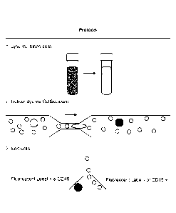
[0025] Fig. 1 is a diagram of a system for size selective tagging of
CTCs
by rapid mechanical deformation.
[0026] Fig. 2 is a bar graph showing that combining size selective
delivery
of the microfluidic platform with antibody staining for CD45 produces a sample
9
CA 02921579 2016-02-16
WO 2015/023982
PCMJS2014/051343
enrichment factor over an order of magnitude better than either technique
independently.
[0027] Fig. 3A is a schematic diagram of cell labeling. Red blood
cells
(RBCs) were depleted from whole blood by RBC lysis using standard erythrocyte
lysis reagents such as eBioscience RBC lysis buffer (Cat. No. 00-4333). The
resulting
suspension flowed through the constriction channel microfluidics device
incubated
with a fluorescent dye (and optionally other compounds). The suspension was
then
labeled for CD45 and processed on a fluorescence-activated cell sorter (FACS)
machine to collect the non-CD45+ cells that have been labeled with the
fluorescent
dye.
[0028] Fig. 3B is a series of flow cytometry plots of cascade blue
conjugated 3klla dextran delivered by CellSqueeze devices to PBMCs (30-6 chip
at
50psi), HT-29 (30-6 chip at 50psi), SK-MEL-5 (10-7 chip at 50psi), and PANC-
1(10-
7 chip at 50psi).
[0029] Fig. 3C is a series of transmitted light and fluorescence
micrographs of Panc-1 tumor cells and blood cells before and after passing
through
the constriction channel. The pre-delivery cells are incubated in the presence
of dye to
correct for background endocytosis. The post-delivery images were taken 24 h
after
delivery to demonstrate retention of dye and ability of the cells to adhere
and grow
following delivery. Although large blood cells can also get labeled in the
process,
these data demonstrate selective labeling of tumor cells.
[0030] Figure 4 is a plot of PBMC delivery versus percent PBMC in
PBMC and lymphoma mixture showing selective delivery of dyes to lymphoma cells
vs. healthy PBMCs. Even when the suspension is 99.9% healthy PBMCs by number,
CA 02921579 2016-02-16
WO 2015/023982
PCMJS2014/051343
in some implementations up to 8 times specificity in delivery can be acheived.
In
other implementations, greater specificity can be achieved.
[0031] Figure 5A is a FACS plot of tetramethylrhodamine dextran-
labeled
Panc-1-GFP cells spiked into whole blood (40 cells/nil) and processed with a
CD45
counter stain (APC).
[0032] Figure 5B is a FACS plot of MI) versus CD45, demonstrating how
PANC-1 GFP tagging could be verified independently based on GFP fluorescence.
The PS gate would be used as a basis for sorting candidate CTCs, P4 is used to
verify
the identity of PANC-1 GFP cells. Green dots are accurate hits (P4 & P5), red
dots are
false positives (PS only), blue dots are misses (P4 only).
[0033] Figure SC is an image of histopathology of HTB1760's primary
tumor confirms pancreatic ductal adenocarcinoma.
DETAILED DESCRIPTION
[0034] CTCs are tumor cells that are found in the bloodstream, and are
believed to be responsible for the dissemination of cancer to distant organs.
CTCs are
regarded as minimally-invasive, "liquid biopsies" for cancer patients and are
useful as
prognostic indicators for patient outcome and treatment efficacy.
Comprehensive
characterizations of these single cells provide a better understanding of
metastatic
dissemination, treatment resistance, and tumor biology.
[0035] A typical human erythrocyte has a disk diameter of
approximately
6.2-8.2 p m and a thickness at the thickest point of 2-2.5 pm and a minimum
thickness
in the center of 0.8-1 gm, being much smaller than most other human cells.
Leukocytes (white blood cells) include neutrophils (12-14 pm diameter),
eosinophils
(12-17p m diameter), basophils (14-16 um diameter), lymphocytes (average 6-9
um in
diameter for resting. and 10-14 um diameter for activated), and monocytes, the
largest
11
CA 02921579 2016-02-16
WO 2015/023982
PCMJS2014/051343
type of white blood cells that can be up to 201am in diameter. As shown in
Fig. 1, the
size difference between CTCs and hematologic cells generally permits
distinguishing
CTCs from other cells in circulating blood (CTCs ¨9-20 i.tm; RBC ¨8 jim
discoid;
leukocytes ¨7-12 lam). See Fig. 1. Subsequent tumor cell specific labeling
using
antibodies (or cell-specific fragments thereof) or other tumor cell specific
ligands
increase the selectivity of the method.
[0036] Since CTCs are present as one in 106-107 mononuclear cells in
the
bloodstream, high-sensitivity enrichment techniques are used that rely on
immunological or morphological differences in CTCs from the blood cells.
Immunological approaches often target epithelial cell surface markers (such as
EpCAM) and tumor-specific proteins (such as Her2-neu, MUC I/MUC2,
carcinoembryonic antigen (CEA), mammaglobulin, and alpha-fetoprotein) or aim
to
deplete CD45+ cells. Microfilters, density-gradient separations, and
microfluidics
platforms are examples of morphology-based methods. All of these approaches
have
inherent biases, suffer from low enrichment efficiencies and a significant
number of
CTCs may down-regulate surface antigens or exhibit varying morphological
features.
These biases pose a significant challenge in the field as it is still largely
unknown
which subset of CTCs are responsible for metastasis or are reliable prognostic
markers. Thus, it is important to develop techniques that can ensure high
sensitivity
isolation of all candidate CTC sub-types to screen for the most clinically
relevant
candidates. The devices and methods described herein permit the isolation and
enumeration of the CTC subtype of interest.
[0037] A combined enrichment method integrates both immunological and
morphologic-based approaches to tag and isolate pure CTCs with less bias and
based
on tunable parameters. The method combines microfluidic intracellular delivery
(Fig.
CA 02921579 2016-02-16
WO 2015/023982
PCMJS2014/051343
1) and antibody staining to yield robust, high sensitivity purification of
circulating
tumor cells from whole blood (Fig. 2) comprises a width from 4 -10gm, length
of
1p m-100gm, and 1-10 constrictions in series. The estimated speed of the cells
can
range from lOmm/s to 10m/s. The specific device parameters chosen are dictated
by
the target tumor cell type, e.g., a different device design is used to select
CTCs for a
melanoma patient vs. a colon cancer patient. Examples of tumor cell
sizes/diameters
include; melanoma ¨15um, colon cancer ¨hum, and pancreatic cancer ¨15um.
[0038] In this approach, a rapid mechanical deformation delivery
system
exploits the inherent size difference between many CTCs and the surrounding
blood
cells to selectively deliver fluorescent, magnetic and/or other distinguishing
materials
to the tumor cells. In further processing, antibody-based fluorescent and/or
magnetic
tagging is used to enhance the contrast between the candidate CTCs and the
surrounding blood cells. By uniquely combining size-based and immunological
approaches to CTC isolation, this technology has demonstrated utility for the
non-
biased isolation of candidate tumor cells from patient samples for analysis.
In some
implementations, both smaller and larger cells are deformed but the smaller
cells
membrane is not deformed to the point that the membrane becomes compromised.
For example, to selectively delivering to 15 gm tumor cells in whole blood
where
most healthy white blood cells are ¨8 gm in size, a 6um width constriction can
be
used. Such a constriction would deform both cell types but would very
preferentially
disrupt the membrane of the 15 gm tumor cells not the 8 gm blood cells.
[0039] Clinical/Translation Relevance
[0040] CTCs are being explored as surrogates for tumor biopsies for
understanding mechanisms of resistance and guiding the selection of targeted
therapies. Measures of the number and composition of CTCs before and after
13
CA 02921579 2016-02-16
WO 2015/023982
PCMJS2014/051343
treatment indicate treatment efficacy and prognosis. The approach utilizes a
robust,
high-throughput, disposable device for the tagging of CTCs based on cell size
and
surface antigens. Moreover, the ability to deliver a diversity of
macromolecules also
enables one to deliver molecular probes (such as antibodies, quantum dots,
carbon
nanotubes, and molecular beacons) that respond to the intracellular
environment and
thus provide further information on the intracellular properties of the target
cell.
This combinatorial approach provides a robust platform capable of enriching
CTC
populations that would have been missed by alternative methods that rely
solely on
immunological or morphological separation. The technique is useful to isolate
patients' CTCs.
[0041] Example 1
[0042] Whole blood or other cell suspensions are processed using both
unlabeled and/or antibody-coated magnetic beads. These cells are then isolated
using a high-fidelity, magnetic enrichment system for rare cells. A nanowell
technology may also be used to achieve high purity isolations by imaging and
robotically-retrieving single cells of interest from an elastomeric array of
84,672
subnanoliter wells.
[0043] Obtaining single, live, pure, intact CTCs of diverse phenotypes
allows a host of characterization efforts from the genomic to functional
levels with
immediate clinical and translational relevance. The methods permit a highly
sensitive and specific enrichment of live, diverse CTCs with reduced bias.
[0044] Example 2
[0045] Magnetic nanoparticles are delivered to tumor cell lines &
PBMCs. Nanoparticle delivery to EpCAM-expressing, epithelial cancer cell
lines,
14
CA 02921579 2016-02-16
WO 2015/023982
PCMJS2014/051343
e.g., HT-29, LNCaP, and SK-BR-3, is compared to bulk peripheral blood
mononuclear cell (PBMC) suspensions derived from human blood.
[0046] lOnm iron-oxide nanoparticles with a polyethylene glycol (PEG)
surface coating are delivered to cancer cells mixed with whole blood, and the
resulting mixture of tagged cells are processed using the cell separation
system
described above. For example, the microfluidic delivery system was used to
induce
a rapid mechanical deformation of a cell to generate transient pores in the
cell
membrane (Fig. 1). The approach has demonstrated an ability to deliver a range
of
materials, including proteins, RNA, DNA and nanoparticles to a variety of cell
types
and works with whole blood, a medium that often poses problems for
microfluidic
systems.
[0047] Exemplary tagging molecules, e.g., 3kDa and 70kDa,
fluorescently-labeled, dextran polymers as model molecules, were used to
discriminate between PBMCs and two different cancer cell lines based on size
alone. The results also indicate the utility of the system for the selective
delivery of
magnetic particles to tumor cells in the blood. PEG coated iron-oxide
particles are
used to magnetically tag colon cancer (e.g., as exemplified by the cell line
HT-29).
Further enrichment is accomplished using conjugation of FITC to the iron-oxide
nanoparticle surface to directly measure nanoparticle uptake.
[0048] PEG coated lOnm iron-oxide nanoparticles are delivered to cell
suspensions that are suspected of containing or are known to contain CTCs,
e.g., a
patient-derived blood sample, or cell lines HT-29, I,NCaP, and SK-BR-3 cells,
separately mixed with whole blood. The resulting mixture of tagged cells are
then
purified, e.g., using a high fidelity magnetic separator. The separator
accurately
discriminates between the model CTCs with high iron-oxide content and less-
effectively labeled PBMCs. Optionally, red blood cells are lyscd prior to
treatment,
nanoparticle concentration increased, their size altered, or incorporating
multiple
treatment steps.
[0049] Example 3
[0050] A combined immunological and morphologic-based method is
can-ied out as follows. After cell size-based processing by the device, cells
are
treated with an antibody or other tumor cell specific ligand such as
fluorescently
labeled anti-CD45 antibodies. The sensitivity and specificity of three
different
separation approaches were compared:: 1) device only 2) anti-CD45 antibody
only
3) device+ anti-CD45 antibody. Morphologic tagging (device) + immunological
tagging (e.g., anti-CD45 antibodies) was found to show superior sensitivity
(and
specificity) relative to either of the individual techniques (Fig. 2). For
example, a 2-
5x increase in sensitivity and/or a 2-5x increase in specificity relative to
anti-CD45
antibodies alone is observed. Enrichment factor of over an order of magnitude
was
observed (Fig. 2).
[0051] Exampk A
[0052] In one example, the devices are fabricated out of
silicon and
glass. Alternatively, the device is fabricated using a polymer such as
silicone,
PDMS, polycarbonate, acrylic, polypropylene, polystyrene. Either device is
sterilized (heat or gamma radiation) and disposable. Performance of the
devices is
validated for various cell types using materials and parameters. For example,
performance at a range of flow speeds (10Ornm/s-10,000mm/s) using PEG coated
quantum dots (ranging from 10-50nm in size) is used to determine if the
delivery
efficiency of nanoparticles and cell viability. Exemplary device are described
in
PCT/US20 12/060646,
16
Date Recue/Date Received 2020-09-24
CA 02921579 2016-02-16
WO 2015/023982
PCMJS2014/051343
[0053] Advantages
[0054] When compared to existing approaches this method has the
following advantages. Relative to antibody-based methods, this approach
provides a
non-biased isolation procedure that is generalizable to most cancer types and
is
independent of any particular cell surface marker. The device and method
accomplishes the identification of CTCs that could not be isolated by existing
markers
and thus, has significant diagnostic and prognostic implications.
[0055] Relative to existing size-based isolation methods, the device
and
methods described herein provide far higher throughput and are tunable by
varying
"W" (Fig. 1) to capture specific CTC size ranges. For example, a 6pm width
constriction is suitable for the capture of colon cancer cells whereas a 7p m,
and 8p m
width are suitable for the capture of pancreatic cancer and melanoma cells
respectively. Moreover, unlike existing technologies, this system is combined
with
antibody-based technologies to enhance isolation sensitivity and/or enable
multi-
parametric isolation of subsets of CTCs (for example by isolating CTCs of a
certain
size + surface marker).
[0056] By enabling the effective, robust isolation of CTCs from a
range of
cancer types this technology would be a valuable platform in the fight against
cancer.
The prognostic and diagnostic potential of this technology could enable the
identification of new genes that are critical to cancer progression and thus
enable the
development of novel therapeutics. It may also provide a more accurate
prediction of
patient life-expectancy and treatment efficacy.
[0057] The CTC isolation methods described herein combines
immunological and size-based isolation to yield a high enrichment
factor/recovery
rate and adjustable bias (marker specific vs. size specific).
17
CA 02921579 2016-02-16
WO 2015/023982
PCMJS2014/051343
[0058] Although a few variations have been described in detail above,
other modifications are possible. For example, the implementations described
above
can be directed to various combinations and subcombinations of the disclosed
features
and/or combinations and subcombinations of several further features disclosed
above.
In addition, the logic flows described herein do not require the particular
order
described, or sequential order, to achieve desirable results. Other
embodiments may
be within the scope of the following claims.
18