Note : Les descriptions sont présentées dans la langue officielle dans laquelle elles ont été soumises.
OPHTHALMIC DEVICES, SYSTEM AND METHODS
THAT IMPROVE PERIPHERAL VISION
[0001] This application claims benefit under 35 U.S.C. 119(e) of U.S.
Provisional Application No. 61/982,135, filed on April 21, 2014, titled
"OPHTHALMIC
DEVICES, SYSTEM AND METHODS FOR IMPROVING PERIPHERAL VISION." This
application also claims benefit under 35 U.S.C. 119(e) of U.S. Provisional
Application No.
62/038,667, filed on August 18, 2014, titled "OPHTHALMIC DEVICES, SYSTEM AND
METHODS FOR IMPROVING PERIPHERAL VISION."
BACKGROUND
Field
[0002] This disclosure generally relates to devices, systems and
methods that
improve peripheral vision.
Description of Related Art
[0003] Intraocular Lenses (IOLs) may be used for restoring visual
performance
after a cataract or other ophthalmic procedure in which the natural
crystalline lens is replaced
with or supplemented by implantation of an IOL. When such a procedure changes
the optics
of the eye, generally a goal is to improve vision in the central field. Recent
studies have
found that, when a monofocal IOL is implanted, peripheral aberrations are
changed, and that
these aberrations differ significantly from those of normal, phakic eyes. The
predominant
change is seen with respect to peripheral astigmatism, which is the main
peripheral
aberration in the natural eye, followed by sphere, and then higher order
aberrations. Such
changes may have an impact on overall functional vision, on myopia
progression, and for
newborns and children on eye development.
[0004] There are also certain retinal conditions that reduce central
vision, suell as
AMD or a central scot-efna. Other diseases may impact central vision, even at
a very young
-1-
Date Recue/Date Received 2021-08-26
CA 02946356 2016-10-19
WO 2015/177651 PCT/IB2015/001588
age, such as Stargardt disease, Best disease, and inverse retinitis
pigmentosa. The visual
outcome for patients suffering from these conditions can be improved by
improving
peripheral vision.
SUMMARY
[0005] The systems, methods and devices of the disclosure each have
several
innovative aspects, no single one of which is solely responsible for the
desirable attributes
disclosed herein.
[0006] Various systems, methods and devices disclosed herein are
directed
towards intraocular lenses (IOLs) including, for example, posterior chamber
IOLs, phakic
IOLs and piggyback IOLs, which are configured to improve peripheral vision.
For normal
patients, e.g., uncomplicated cataract patients, peripheral vision may be
balanced with good
central vision in order to improve or maximize overall functional vision. For
those patients
having a pathological loss of central vision, peripheral vision may be
improved or
maximized, taking into account the visual angle where the retina is healthy.
[0007] In some embodiments, an IOL can be configured to reduce
peripheral
aberrations by tailoring parameters of the IOL according to stop-shift
equations, which are
discussed in greater detail herein. The IOL can be configured to position its
principal plane
posterior (relative to the pupil) to a standard 10L's principal plane by
tailoring the shape
factor of the lens, the axial displacement (physical or virtual) of the lens,
the index of
refraction of the lens, the asphericity of one or more surfaces of the lens,
by adding an extra
aperture, or any combination of these techniques. In various embodiments, the
principal
place can be shifted by a distance between about 0.1 mm and about 4.5 mm by
movement of
haptics. In some embodiments, the shape factor of the IOL can be altered by
altering the
geometry (e.g., radius of curvature and/or thickness) or changing the
refractive index of the
material of the 10L. Altering the shape factor of the IOL can shift the
principal plane by
about a few hundred microns. In various embodiments, shifting the principal
plane by
movement of haptics and by altering the shape factor of the lens can
advantageously reduce
peripheral astigmatism.
[0008] In one embodiment, the principal plane of the lens is moved
posteriorly,
further from the iris, which is the natural aperture at the eye, or closer to
the nodal point of
-2-
CA 02946356 2016-10-19
WO 2015/177651 PCT/IB2015/001588
the eye as compared to standard IOLs. This effectively changes the field
curvature in the
image plane, to better align with the shape of the retina. In some
embodiments, the axial
position of the IOL is between about 1.5 mm and about 2.5 mm behind the iris.
For example,
the axial position of the IOL may be about 1.9 mm behind the iris. In certain
embodiments,
the axial position of the IOL is between about 2.5 mm and about 3.5 mm behind
the iris. For
example, the axial position of the IOL may be about 2.9 mm behind the iris.
[0009] In some embodiments, the axial position of the IOL may be between
about
3.5 mm and about 4.1 mm behind the iris. For example, the axial position of
the IOL may be
about 3.9 mm behind the iris. For regular eye dimensions, the position of the
lens may be
limited by the vitreous body, to not exceed about 4.5 mm behind the iris. For
some
embodiments of the lenses used in this example, the principal plane is about
0.4 mm
posterior to the anterior lens surface. The location of the principal plane
posterior to the
anterior lens surface can be altered by modifying the shape factor. For
example, depending
on the shape factor of the lens, the principal planes can be located placed at
different
distances, such as, for example, greater than or equal to 0.1 mm posterior to
the anterior lens
surface, greater than or equal to 0.5 mm posterior to the anterior lens
surface, greater than or
equal to 0.8 mm posterior to the anterior lens surface, greater than or equal
to 1.0 mm
posterior to the anterior lens surface, greater than or equal to 1.5 mm
posterior to the anterior
lens surface, greater than or equal to 1.8 mm posterior to the anterior lens
surface, greater
than or equal to 2.1 mm posterior to the anterior lens surface, greater than
or equal to 2.5 mm
posterior to the anterior lens surface, greater than or equal to 3.0 mm
posterior to the anterior
lens surface, greater than or equal to 3.5 mm posterior to the anterior lens
surface and greater
than or equal to 4.0 mm posterior to the anterior lens surface. Therefore,
when the example
refers to a distance of the lens of, e.g., 1.5 mm behind the iris, it means
the principal plane of
the lens is about 1.9 mm behind the iris.
[0010] Instead of moving the lens posteriorly relative to a conventional
position
in the eye, a lens configuration may be applied that moves the principal plane
of the lens
posteriorly, while the physical lens is still in the conventional position in
the eye. One way
to achieve this is to change the shape factor of the lens, e.g., to a meniscus
lens having a
concave anterior surface and a convex posterior surface. The meniscus lens can
also
advantageously reduce astigmatism. Without subscribing to any particular
theory, a
-3-
modification of shape factor can be achieved by changing the geometry (e.g.,
radius of
curvature, thickness) of the lens, refractive index of the material of the
lens or a combination
of both. Accordingly, in some embodiments, the location of the principal place
can be
altered by increasing or decreasing the thickness of the lens. In some
embodiments, the
location of the principal place can be altered by increasing or decreasing the
radius of
curvature of the lens. In some embodiments, an intraocular lens system of 2
lenses is used,
e.g., having a negative power anterior lens and a positive power posterior
lens. Those skilled
in the art will appreciate that other combinations are possible.
[0011] The lens may be a multifocal lens, a lens including a prism, or
a telescope
lens, having the principal plane moved posteriorly by one of the methods
described above.
In a multifocal lens, the position of the principal plane may be determined
based on analysis
using one focal point, several of the focal points, or all focal points of the
multifocal lens. In
a preferred embodiment, a multifocal IOL has at least 2 zones, wherein the at
least 2 zones
have about the same optical power. The inner zone may be a spherical lens
producing a good
central focus. The outer zone(s) comprise of a spherical lens combined with a
prism,
producing a good focus at a predetermined spot in the periphery. A similar
affect may be
achieved if the outer zone(s) are aspheric. Alternatively, a bag-filling lens
with a gradient
refractive index may be used. Such lenses can also advantageously reduce age
related
macular degeneration (AMD).
[0012] In some embodiments, an artificial pupil may be implanted
between the
lenses of a dual lens system or posterior to an IOL or lens combination. Such
an artificial
pupil may have a similar impact as moving the IOL posteriorly.
[0013] In some embodiments, a singular circular surface structure,
which acts as a
phase shifting profile extends the depth of focus in the peripheral field.
Implementations of
such structures are described in U.S. Patent No. 8,430,508. An implementation
of a single
ring IOL includes an anterior face and a posterior face. A profile can be
imposed on the
anterior or posterior surface or face. The profile can have an inner portion
and an outer
portion. The inner portion typically presents a parabolic curved shape. The
inner
portion may also be referred to as a microstructure, an isolated echelette, or
a central
echelette. Between the inner portion and the outer portion, there may be a
transition zone
that connects the inner and outer portions.
-4-
Date Recue/Date Received 2021-08-26
CA 02946356 2016-10-19
WO 2015/177651 PCT/IB2015/001588
An IOL with such a structure provides for a reduction in peripheral
aberrations, including
astigmatism and other higher order aberrations. In certain embodiments, a
multifocal IOL is
used to induce multiple foci. While traditional multifocal 10Ls utilize
multiple foci at
multiple powers, in this embodiment, the multiple foci are of the same optical
power. In
addition, the multiple foci focus images on different parts of the retina,
thus producing
optimal optical quality at those regions of the retina that are healthy.
[0014] In some embodiments, characteristics of the retina are considered
for the
IOL design. In particular, a geographical map of retinal functionality and/or
the retinal shape
are combined with other ocular geometry, such as pupil size and location,
axial positions of
the pupil, lens, and retina, anterior and/or posterior corneal aberrations,
tilts and
decentrations within the eye, and angle kappa. A metric function can be used
to improve or
optimize the IOL, accounting for both central and peripheral optical quality.
In some
embodiments, the IOL power distribution at the periphery can be related with
retinal shape.
Therefore, while measuring retinal shape it might be possible to select the
IOL with the
peripheral power distribution that matches patient's retina.
[0015] In some embodiments, a dual-optics IOL system can be used to
improve
natural vision by reducing peripheral aberrations. The dual-optics lens can
comprise an
anterior lens and a posterior lens. The dual-optics lens can have a shape
factor based on the
optical powers of the anterior and posterior lenses, the shape factor being
tailored to reduce
peripheral aberration. The shape factor can be modified for each lens while
maintaining the
total optical power relatively constant. The shape factors can be modified by
adjusting the
anterior and posterior radii of curvature of each lens, e.g., the anterior
lens and the posterior
lens. The shape factors can be tailored to reduce astigmatism and spherical
equivalent in the
periphery of the retina while maintaining on-axis optical quality on the
retina.
100161 In some embodiments, one or more IOLs can be used which have one
or
more aspherical surfaces configured to improve peripheral vision by reducing
peripheral
aberrations. The asphericity of the surfaces can be tailored to improve off-
axis contrast,
thereby improving peripheral vision relative to IOLs with typical surface
geometries.
[0017] In some embodiments, a method is provided for improving vision
using an
intraocular lens which reduces peripheral aberrations. The method includes
determining a
principal plane of an intraocular lens; determining a value of at least one
peripheral
-5-
CA 02946356 2016-10-19
WO 2015/177651 PCT/IB2015/001588
aberration at the retina of an eye based at least in part on an initial
proposed placement of the
principal plane of the intraocular lens in the eye and based at least in part
on a computer
model of an eye; modifying a parameter of the intraocular lens, wherein the
parameter
consists of at least one of a shape factor of the intraocular lens, an axial
displacement of the
intraocular lens, an index of refraction of the intraocular lens, or an
asphericity of the
intraocular lens; comparing the value of the at least one peripheral
aberration with a value of
the at least one peripheral aberration after modification of the parameter;
and incorporating
the modified parameter into the intraocular lens if the modification improves
the vision of the
patient by reducing the at least one peripheral aberration.
[0018] Various implementations of the method can comprise determining a
modified value of the at least one peripheral aberration after modification of
the parameter
using at least one stop-shift equation. The at least one peripheral aberration
can include
coma or astigmatism. The method can further comprise determining a constraint
on the
parameter of the intraocular lens. The intraocular lens designed using the
method above can
include a lens element which has an aspherical surface. The asphericity of the
surface of the
intraocular lens can be further modified to increase an off-axis contrast
produced by the
intraocular lens. In various implementations of the method modifying a
parameter of the
intraocular lens can include providing an additional aperture. The method can
include
determining a target position of the intraocular lens in an eye of a patient,
wherein the target
position of the intraocular lens is such that the principal plane of the
intraocular lens is
between 1.9 mm and 4.5 mm behind the iris.
[0019] In some embodiments, a method is provided for improving vision
using a
dual-optic intraocular lens comprising an anterior lens element having an
anterior optical
power and a posterior lens element having a posterior optical power. The
method includes
calculating a shape factor of the intraocular lens where the shape factor is
equal to the sum of
the anterior optical power and the posterior optical power divided by the
difference between
the posterior optical power and the anterior optical power; determining a
value of at least one
peripheral aberration at the retina of an eye based at least in part on the
shape factor of the
intraocular lens and based at least in part on a computer model of an eye;
modifying an
anterior shape factor of the anterior lens element by modifying an anterior
radius of the
anterior lens element or the posterior radius of the anterior lens element;
modifying a
-6-
CA 02946356 2016-10-19
WO 2015/177651 PCT/IB2015/001588
posterior shape factor of the posterior lens element by modifying an anterior
radius of the
posterior lens element or the posterior radius of the posterior lens element;
determining a
modified value of the at least one peripheral aberration at the retina of the
eye based at least
in part on the shape factor of the intraocular lens and based at least in part
on the computer
model of an eye; comparing the value of the at least one peripheral aberration
with the
modified value of the at least one peripheral aberration; and incorporating
the modified
anterior lens element and the posterior lens element into the intraocular lens
if the
modification improves the vision of the patient by reducing the at least one
peripheral
aberration, wherein a total optical power of the intraocular lens remains
substantially
unchanged after modification of the anterior shape factor and the posterior
change factor. In
various implementations of the dual-optic intraocular lens designed by the
method described
above, a surface of the anterior lens element or a surface of the posterior
lens element can be
aspheric. The asphericity of the surface of the anterior lens element or the
surface of the
posterior lens element can be modified to increase an off-axis contrast
produced by the
intraocular lens.
[0020] One aspect of the innovative aspect disclosed herein can be
implemented
in a dual-optic intraocular lens comprising an anterior optic and a posterior
optic. The
anterior optic can have an anterior optical power. The anterior optic can
include a first
surface with a first radius of curvature and a second surface opposite the
first surface with a
second radius of curvature. The anterior optic can have an anterior shape
factor that is
associated with the first and the second radius of curvature. The posterior
optic can have a
posterior optical power. The posterior optic can include a third surface with
a third radius of
curvature and a fourth surface opposite the third surface with a fourth radius
of curvature.
The posterior optic can have a posterior shape factor that is associated with
the third and the
fourth radius of curvature. A shape factor of the intraocular lens given by
the sum of the
anterior optical power and the posterior optical power divided by the
difference between the
posterior optical power and the anterior optical power can be optimized by
optimizing the
anterior shape factor or the posterior shape factor such that a degradation in
the visual
information obtained from a peripheral retinal location is below a threshold
degradation. A
total optical power of the intraocular lens can remain substantially unchanged
after
modification of the anterior shape factor or the posterior shape factor.
-7-
CA 02946356 2016-10-19
WO 2015/177651 PCT/IB2015/001588
[0021] In various implementations, the posterior optic can be disposed
in the
capsular bag of the eye of a patient. The anterior optic can be disposed in
the capsular bag of
the eye of a patient or at a location between the iris and the capsular bag.
In various
implementations, at least one of the first, second, third or fourth surface
can be aspheric.
[0022] In some embodiments, a method is provided for increasing contrast
sensitivity function (CSF) for peripheral vision. The method includes
providing a first JUL
for implanting into a first eye of the patient, the first JUL configured to
increase acuity of a
sagittal image; and providing a second intraocular lens (TOL) for implanting
into a second
eye of a patient, the second JUL configured to increase CSF of a tangential
image.
[0023] In various implementations of the method, the first JUL can be
configured
to increase contrast of the sagittal image when implanted at a first distance
from the pupil.
The second JUL can be configured to increase contrast of the tangential image
when
implanted at a second distance from the pupil. The first JUL can be configured
to be
implanted in the first eye at a first distance from the pupil and the second
JUL can be
configured to be implanted in the second eye at a second distance from the
pupil. The first
distance can be lesser than the second distance. A difference between the
first distance and
the second distance can be between about 0.5 mm and about 5 mm.
[0024] In some embodiments, an JUL is provided that is configured to
increase
CSF in the horizontal field of view without increasing CSF in the vertical
field of view to
improve peripheral vision. The IOL includes at least one tone portion and at
least one non-
toric portion. In various implementations of the JUL, the at least one tonic
portion can have a
higher optical power along the vertical axis than the horizontal axis. The at
least one toric
portion can be disposed in a central region of the JUL. The at least one tonic
portion can be
disposed in a peripheral region of the JUL. The IOL can include features that
induce
spherical aberrations. The JUL can include features that induce aspherical
aberrations. The
IOL can include diffractive features. The JUL can be configured to provide
astigmatic
correction The JUL can be configured to provide extended depth of focus. The
JUL can be
configured to provide acuity for peripheral vision. The tonic portion can
improve acuity for
peripheral vision along a horizontal direction.
[0019] In some embodiments, an JUL is provided that is configured to
compensate for peripheral aberrations, such as, for example, peripheral
astigmatism and
-8-
CA 02946356 2016-10-19
WO 2015/177651 PCT/IB2015/001588
horizontal coma arising from light incident at oblique angles. Various
embodiments of IOL
that compensate for peripheral aberrations can reduce coma and/or astigmatism
in the
peripheral field of view. Due to the oblique incidence of the light in the
eye, astigmatism
increases with eccentricity. The increase in astigmatism with eccentricity
follows a fixed
trend. As previous studies have found, this dependence does not change with
age and/or
foveal refractive errors, either for foveal sphere or astigmatism. Therefore
patients can
benefit from embodiments of IOLs having an arrangement of optical features
(e.g. optical
elements, grooves, volume or surface diffractive features, regions of varying
refractive index,
etc.) that results in a peripheral astigmatism that decreases with
eccentricity. The decrease in
astigmatism with eccentricity for the IOL can follow an opposite trend.
[0025] Recent studies indicate that similar to peripheral astigmatism,
horizontal
coma is also independent of the patient's age and/or foveal refractive errors,
axial length of
the cornea, corneal curvature, etc. and depends on the eccentricity or field
of view according
to a fixed trend. Accordingly, errors in peripheral vision can be compensated
by an IOL
having an arrangement of optical features (e.g. optical elements, grooves,
volume or surface
diffractive features, regions of varying refractive index, etc.) such that the
dependence of
horizontal coma for the IOL on the eccentricity or field of view has an
opposite trend.
[0026] For example, in various implementations, an IOL configured to
correct for
peripheral aberrations in a patient can include an arrangement of a first set
of optical features
and an arrangement of a second set of optical features that compensate for
peripheral
aberrations. The arrangement of the first set of optical features can be
determined based on
the direction of incident light and independent of the spherical power
required to achieve
emmetropia. The arrangement of the second set of optical features can be
determined based
on the spherical power required to achieve emmetropia. The arrangement of the
first set of
optical features can compensate for peripheral astigmatism and/or horizontal
coma. The
arrangement of the second set of optical features can compensate for
peripheral defocus. The
arrangement of the second set of optical features can be determined based on
an axial length
of the patient's eye or a curvature of the cornea.
[0027] Generally, peripheral defocus changes as a function of the foveal
refractive state. Accordingly, in various embodiments of IOLs, the amount of
defocus can
vary based on the refractive power of the IOL, which ultimately depends on the
preoperative
-9-
CA 02946356 2016-10-19
WO 2015/177651 PCT/IB2015/001588
refractive state or preoperative biometry of the patient. For example, since
patients with
hypermetropia have a different defocus distribution as compared to patients
with myopia the
arrangement of optical features that compensates for peripheral defocus will
be different in
both cases. As a way of example, patients with hypermetropia have relative
peripheral
myopia. In such patients, a higher central power of the IOL can be associated
with a lower
peripheral power distribution, as compared to the central power. On the other
hand, patients
with myopia tend to have relative peripheral hyperopia. In such patients, a
lower central
power of the IOL can be associated with a higher peripheral power
distribution, relative to
the central power.
[0028] Thus, the present disclosure provides a lens apparatus, system
and method
that improve peripheral visual acuity at least in part by reducing aberrations
arising from
light directed to peripheral or high field angle retinal areas (sometimes
referred to herein as
peripheral aberration) relative to standard IOLs while maintaining good vision
arising from
light directed to most sensitive or low field angle or central retinal areas
(sometimes referred
to herein as central vision).
[0029] Various implementations of an IOL configured to correct for
peripheral
aberrations in a patient's eye can include a first optical power distribution
that compensates
for peripheral astigmatism; a second optical power distribution that
compensates for
horizontal coma in peripheral regions; and a third optical power distribution
that
compensates for defocus in the peripheral regions. The first and second
optical power
distribution can be independent of foveal refractive state of the patient's
eye and the third
optical power distribution can depend on the foveal refractive state of the
patient's eye and/or
the IOL power required for the patient to achieve foveal emmetropia. The first
power
distribution can vary nonlinearly with visual field angle. The first power
distribution can
vary quadratically with visual field angle. The first power distribution can
have a higher
absolute value of cylinder power at visual field angle having an absolute
value greater than
or equal to 10 degrees than the absolute value of cylinder power at visual
field angle having
an absolute value less than 10 degrees. The first power distribution can have
increased
astigmatic correcting power in the peripheral regions and decreased astigmatic
correcting
power in a central region. The second power distribution can vary linearly
with visual field
angle. The second power distribution can linearly decrease from a first
peripheral region
-10-
CA 02946356 2016-10-19
WO 2015/177651 PCT/IB2015/001588
oriented temporally to a second peripheral region oriented nasally for left
eyes and increase
from a first peripheral region oriented temporally to a second peripheral
region oriented
nasally in right eyes. The third power distribution can be configured to
provide myopic
correction power in the peripheral regions for a patient with emmetropia,
hyperopia or low
myopia. The third power distribution can be configured such that an absolute
magnitude of
spherical optical power for visual field angles having an absolute value
greater than or equal
to 10 degrees is greater than the absolute magnitude of spherical optical
power for visual
field angles having an absolute value less than 10 degrees for a patient with
emmetropia,
hyperopia or low myopia. The third power distribution can be configured such
that an
absolute magnitude of spherical optical power for visual field angles having
an absolute
value greater than or equal to 10 degrees is smaller than the absolute
magnitude of spherical
optical power for visual field angles having an absolute value less than or
equal to 10 degrees
for a patient with moderate or high myopia.
[0030] An innovative aspect of the subject matter disclosed herein can
be
implemented in a method of compensating for peripheral aberrations. The method
comprises
determining a first optical power distribution that compensates for peripheral
aberrations
resulting from oblique incidence of light; and determining a second optical
power
distribution that compensates for peripheral aberrations based on the
patient's ocular
characteristics. In various implementations, the second optical power
distribution can be
determined based on at least one of foveal refractive power, an axial length
of the eye or a
curvature of the cornea. The second power distribution can be configured to
provide myopic
correction power in the peripheral regions for a patient with emmetropia,
hyperopia or low
myopia. The second optical power distribution can be determined based on the
required IOL
power to achieve foveal emmetropia. The second power distribution can be
configured such
that an absolute magnitude of spherical optical power for visual field angles
having an
absolute value greater than or equal to 10 degrees is greater than the
absolute magnitude of
spherical optical power for visual field angles having an absolute value less
than 10 degrees
for a patient with emmetropia, hyperopia or low myopia. The second power
distribution can
be configured to provide hyperopic correction in the peripheral regions for a
patient with
moderate or high myopia. The second power distribution can be configured such
that an
absolute magnitude of spherical optical power for visual field angles having
an absolute
-11-
CA 02946356 2016-10-19
WO 2015/177651 PCT/IB2015/001588
value greater than or equal to 10 degrees is smaller than the absolute
magnitude of spherical
optical power for visual field angles having an absolute value less than 10
degrees for a
patient with moderate or high myopia.
[0031] Various implementations disclosed herein include an IOL
configured to
compensate for peripheral astigmatism in a patient. The IOL can be configured
to provide a
cylinder power with an absolute magnitude of at least 0.5 Diopter for at least
one visual field
angle having an absolute value greater than or equal to 10 degrees. The IOL
can be
configured to have a horizontal coma coefficient of at least 0.01 microns for
at least one
visual field angle greater than or equal to 10 degrees in the nasal visual
field for right eyes
and temporal visual field for left eyes. Alternately, the IOL can be
configured to have a
horizontal coma coefficient of at least -0.01 microns for at least one visual
field angle greater
than or equal to +10 degrees in the temporal visual field for right eyes and
nasal visual field
for left eyes. Such implementations of an IOL can be configured to compensate
for
horizontal coma.
[0032] Various implementations of an IOL described herein can be
configured to
compensate for peripheral defocus. Implementations of an IOL configured to
compensate for
peripheral defocus can provide defocus between about -0.1 Diopter and about
+1.0 Diopter
for a patient with spherical equivalent power between about -0.5 Diopter and
about +0.5
Diopter for at least one visual field angle having an absolute value greater
than or equal to 10
degrees is. Implementations of an IOL configured to compensate for peripheral
defocus can
provide defocus between about -0.1 Diopter and about +2.0 Diopter for a
patient with
spherical equivalent power between about -0.5 Diopter and about -1.5 Diopter
for at least one
visual field angle having an absolute value greater than or equal to 10
degrees.
Implementations of an IOL configured to compensate for peripheral defocus can
provide
defocus between about +1.0 Diopter and about +3.0 Diopter for a patient with
spherical
equivalent power between about -1.5 Diopter and about -2.5 Diopter for at
least one visual
field angle having an absolute value greater than or equal to 10 degrees.
Various
implementations of an IOL configured to compensate for peripheral defocus can
provide
defocus between about +2.5 Diopter and about +6.0 Diopter for a patient with
spherical
equivalent power between about -2.5 Diopter and about -6.0 Diopter for at
least one visual
field angle having an absolute value greater than or equal to 10 degrees.
-12-
CA 02946356 2016-10-19
WO 2015/177651 PCT/IB2015/001588
[0033] Various implementations of an JUL described herein can include a
plurality of optical features that have an overall optical power distribution
that compensates
for peripheral rotationally and non-rotationally symmetric aberrations. The
non-rotationally
symmetric aberrations can include peripheral astigmatism and horizontal coma.
The
rotationally symmetric aberration can include defocus. An arrangement of some
of the
plurality of optical features that compensate for non-rotationally symmetric
aberrations can
be independent of the spherical power of the JUL. The arrangement of some of
the plurality
of optical features that compensate for non-rotationally symmetric aberrations
can depends
on whether the eye to be implanted is right or left. An arrangement of some of
the plurality
of optical features that compensates for rotationally symmetric aberrations
can depend on the
spherical power of the JUL. An arrangement of some of the plurality of optical
features that
compensates for rotationally symmetric aberrations can be different for
optical powers
between 0.0D and 10.0D, 10.0D and 25.0D and 25.0D and 34.0D.
[0034] Various implementations of IOLs described herein can include
markings
showing the orientation of the JUL and the eye to be implanted.
[0035] Various embodiments of an JUL include a first surface that
receives
incident light entering through the cornea and the natural pupil and a second
surface opposite
the first surface through which incident light exits the JUL and propagates
towards the retina.
In some such embodiments, an extra aperture can be provided after (e.g., at
the second
surface or posterior to) the second surface of the JUL. This extra aperture
can reduce the
peripheral aberrations arising from the cornea. The shape of the cornea and
the distance
between the cornea and the posterior surface of the IOL, which can be large in
some
embodiments, can enhance the extra aperture's capability of reducing
peripheral aberrations
arising from the cornea. The natural pupil can reduce the peripheral
aberrations from the
JUL itself.
[0036] An innovative aspect of the subject matter disclosed herein can
be
implemented in an intraocular lens configured to improve vision for a
patient's eye. The
intraocular lens comprises an optic comprising a first surface and a second
surface opposite
the first surface. The first surface and the second surface are intersected by
an optical axis.
The optic is symmetric about the optical axis. The first or the second surface
of the optic can
be aspheric. The optic is configured to focus light incident along a direction
parallel to the
-13-
CA 02946356 2016-10-19
WO 2015/177651 PCT/IB2015/001588
optical axis at the fovea to produce a functional foveal image. The optic is
further
configured to focus light incident on the patient's eye at an oblique angle
with respect to the
optical axis at a peripheral retinal location disposed at a distance from the
fovea. Light can
be incident at one or more oblique angles between about 1 degree and about 30
degrees. In
various implementations, light can be incident at an oblique angle greater
than 30 degrees
from example, at oblique angle upto 45 degrees, upto 60 degrees, upto 75
degrees or greater.
The peripheral retinal location can have an eccentricity between 1 and 30
degrees with
respect to the optical axis. In various implementations, the peripheral
retinal location can
have an eccentricity greater than 30 degrees. For example, the peripheral
retinal location can
have an eccentricity upto 45 ¨ 50 degrees with respect to the optical axis of
the eye.
[0037] The optic is configured to improve image quality at the
peripheral retinal
location by reducing at least one optical aberration at the peripheral retinal
location. The at
least one optical aberration can be selected from the group consisting of
defocus, peripheral
astigmatism and coma. The foveal image can have a modulation transfer function
(MTF) of
at least 0.5 at a spatial frequency of 100 cycles/mm for both the tangential
and the sagittal
foci in green light for a pupil size between 3 ¨ 5 mm. An image formed at the
peripheral
retinal location can have a figure of merit of at least 0.5. In various
implementations, the
figure of merit can be an average MTF for a range of spatial frequencies
between 0
cycles/mm and 30 cycles/mm obtained at different eccentricities between 1 and
30 degrees.
The first or the second surface of the optic can comprise a plurality of
optical features that
are configured to reduce the at least one optical aberration.
[0038] In various implementations, the optic can be a meniscus lens with
a vertex
curving inwards from edges of the optic. The optic can have a maximum
thickness between
about 0.3 mm and about 2.0 mm. In various implementations, the lens can be a
dual-optic
IOL further comprising a second optic separated from the optic by a fixed or a
variable
distance. In implementations of the dual-optic IOL, wherein the distance
between the two
optics is variable, the distance can be varied by application of ocular
forces. A first optic of
the dual-optic IOL can be disposed in the capsular bag of the patient's eye,
and the second
optic can be disposed between the iris and the patient's eye. Alternately,
both the optics of
the dual-optic IOL can be disposed in the capsular bag of the patient's eye.
-14-
CA 02946356 2016-10-19
WO 2015/177651 PCT/IB2015/001588
[0039] The
optic can be configured to improve image quality at the peripheral
retinal location by adjusting a shape factor of the optic such that the at
least one peripheral
optical aberration is reduced. The shape factor of the optic can be adjusted
by adjusting a
parameter of the optic. The parameter can be selected from the group
consisting of a
curvature of the first or the second surface, an axial position of the optic
with respect to the
retina and a thickness of the optic.
[0040] Another
innovative aspect of the subject matter disclosed herein can be
implemented in a method of selecting an intraocular lens (TOL) configured to
be implanted in
a patient's eye. The method comprises obtaining at least one physical or
optical
characteristic of the patient's eye using a diagnostic instrument; and
selecting an IOL having
a shape factor that is configured to focus light incident along a direction
parallel to the
optical axis at the fovea to produce a functional foveal image and is further
configured to
improve image quality at a peripheral retinal location disposed at a distance
from the fovea
by reducing at least one optical aberration at the peripheral retinal
location. The peripheral
retinal location can have an eccentricity between 1 and 30 degrees. In
various
implementations, the peripheral retinal location can have an eccentricity
greater than 30
degrees (e.g., upto 45 ¨ 50 degrees). The shape factor of the IOL can be
selected based on
the at least one physical or optical characteristic of the patient's eye. The
shape factor of the
IOL can be adjusted by adjusting a parameter of the optic. The parameter of
the optic can
include a curvature of the first or the second surface, an axial position of
the optic with
respect to the retina and/or a thickness of the optic. At least one surface of
the IOL can be
aspheric. The optic can be configured to provide a foveal image having a
modulation
transfer function (MTF) of at least 0.5 at a spatial frequency of 100
cycles/mm for both the
tangential and the sagittal foci in green light for a pupil size between 3 ¨ 5
mm. An image
formed at the peripheral retinal location by the optic can have a figure of
merit of at least 0.5.
In various implementation, the figure of merit can be an average MTF for a
range of spatial
frequencies between 0 cycles/mm and 30 cycles/mm obtained at different
eccentricities
between 1 and 30 degrees.
-15-
[0040A] In one embodiment there is provided an intraocular lens configured to
improve vision for a patient's eye. The intraocular lens includes an optic
comprising a first
surface and a second surface opposite the first surface, the first surface and
the second
surface intersected by an optical axis, the optic being symmetric about the
optical axis,
wherein the first or the second surface of the optic is aspheric. The optic is
configured to
focus light incident along a direction parallel to the optical axis at the
fovea to produce a
functional foveal image. The optic is configured to focus light incident on
the patient's eye
at an oblique angle with respect to the optical axis at a peripheral retinal
location disposed at
a distance from the fovea, the peripheral retinal location having an
eccentricity between 1
and 30 degrees. The optic is a meniscus lens having a concave anterior surface
with
negative optical power, a convex posterior surface with positive optical
power, and a shape
factor between minus 4 and minus 1.5, wherein the shape factor is equal to the
sum of the
anterior optical power and the posterior optical power divided by the
difference between the
posterior optical power and the anterior optical power. The image quality at
the peripheral
retinal location is improved by reducing astigmatism at the peripheral retinal
location.
-15 a-
Date Recue/Date Received 2021-08-26
CA 02946356 2016-10-19
WO 2015/177651 PCT/IB2015/001588
BRIEF DESCRIPTION OF THE DRAWINGS
[0041] The systems, methods and devices may be better understood from
the
following detailed description when read in conjunction with the accompanying
schematic
drawings, which are for illustrative purposes only. The drawings include the
following
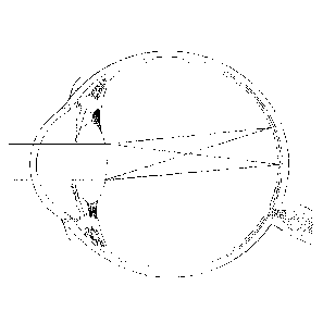
figures:
[0042] FIG. 1 is a cross-sectional view of a phakic eye containing a
natural
crystalline lens.
[0043] FIG. 2 is a cross-sectional view of a pseudophakic eye containing
an
intraocular lens.
[0044] FIG. 3 is a graph illustrating peripheral astigmatism with the
field angle in
degrees and cylinder in diopters.
[0045] FIG. 4 is a graph illustrating peripheral astigmatism with the
field angle in
degrees and sphere in diopters.
[0046] FIG. 5 is a graph illustrating peripheral astigmatism with the
field angle in
degrees and higher order aberrations in micrometers.
[0047] FIG. 6 shows aspects of a lens including a ring microstructure.
[0048] FIG. 7 illustrates aspects of a diffractive lens.
[0049] FIG. 8 is a graph illustrating through-focus MTF at different
axial focus
positions.
[0050] FIG. 9 is a graph illustrating through-focus MTF at different
axial focus
positions.
[0051] FIG. 10 shows aspects of a multifocal IOL in an eye.
[0052] FIG. 11 depicts the astigmatism in the natural lens and an
implementation
of an artificial IOL as a function of eccentricity in degrees.
[0053] FIG. 12 is a graph illustrating astigmatism and coma as a
function of
displacement of an IOL with a shape factor of 0.15.
[0054] FIG. 13 is a graph illustrating astigmatism and coma as a
function of
displacement of an IOL with a shape factor of -1.5.
[0055] FIG. 14 is a graph illustrating the influence of shape factor on
astigmatism
and position of an IOL with respect to the pupil.
-16-
CA 02946356 2016-10-19
WO 2015/177651 PCT/IB2015/001588
[0056] FIGS. 15A-D are graphs illustrating spherical equivalent,
cylinder,
spherical aberration, and coma as a function of field angle for a variety of
IOL
displacements.
[0057] FIG. 16 is a graph illustrating astigmatism and coma as a
function of
displacement from the cornea of an additional aperture.
[0058] FIG. 17 illustrates a flow chart of an example method for
tailoring IOL
properties to reduce peripheral aberrations using stop-shift equations.
[0059] FIG. 18 is a graph illustrating relative refraction at 30 degrees
eccentricity
as a function of shape factor for a dual optic configuration.
[0060] FIG. 19 is a graph illustrating relative refraction as a function
of
eccentricity for a dual optic configuration.
[0061] FIG. 20A-B are graphs illustrating the impact of a global shape
factor and
asphericity on relative refraction for astigmatism and spherical equivalent.
[0062] FIG. 21A-B are graphs illustrating the impact of a global shape
factor and
asphericity on contrast as a function of eccentricity for tangential and
sagittal directions.
[0063] FIG. 22 illustrates a flow chart of an example method for
tailoring a global
shape factor of a dual-optics IOL to reduce peripheral aberrations.
[0064] FIG. 23 shows the substantial part of the peripheral field of
view that is
visible to both eyes for an implementation of an IOL implanted in the eye.
[0065] FIG. 24A is a graph illustrating the modulation transfer function
(or MTF)
as a function of eccentricity for sagittal and tangential vision for an
implementation of an
IOL at a first axial focus position. FIG. 24B is a graph illustrating MTF as a
function of
eccentricity for sagittal and tangential vision for an implementation of the
IOL at a second
axial focus position.
[0066] FIG. 25 is a graph illustrating contrast sensitivity function in
four different
field directions.
[0067] FIG. 26 illustrates a comparison of the optical image quality
(horizontal
astigmatism) in the periphery of phakic and pseudophakic eyes.
[0068] FIG. 27 is a graph illustrating the variation of cylinder power
along the
axis oriented at 0-degrees with respect to the equator (Jo) as a function of
visual field for
patients with different refractive error on axis.
-17-
CA 02946356 2016-10-19
WO 2015/177651 PCT/IB2015/001588
[0069] FIG. 28 is a graph illustrating the variation of horizontal coma
as a
function of visual field.
[0070] FIG. 29 is a graph illustrating the variation of defocus as a
function of
visual field for patients with different refractive error on axis.
[0071] FIG. 30A illustrates the through-focus MTF for an implementation
of a
lens having a cylindrical error of about 8.4 Diopter for an image formed at a
location of the
peripheral retina centered at 25 degrees eccentricity in green light at 10
cycles/mm. FIG.
30B illustrates the through-focus MTF for an implementation of a lens having a
cylindrical
error of about 1.2 Diopter for an image formed at a location of the peripheral
retina centered
at 25 degrees eccentricity in green light at 10 cycles/mm. FIG. 30C
illustrates the through-
focus MTF for an implementation of a lens having a cylindrical error of about
0.75 Diopter
for an image formed at a location of the peripheral retina centered at 25
degrees eccentricity
in green light at 10 cycles/mm.
[0072] Figure 31 illustrates a flowchart depicting an implementation of
a method
to obtain a metric used to evaluate the peripheral image quality provided by
an
implementation of a lens.
[0073] Figure 32 illustrates the spatial frequency that is achievable
based on the
ganglion cell density at different eccentricities.
[0074] Figure 33 shows the MTF curve for tangential and sagittal rays at
an
eccentricity of 20 degrees for spatial frequencies between 0 cycles/mm and 20
cycles/mm for
an implementation of a lens in green light.
[0075] Figure 34A illustrates the surface sag of a first surface of an
implementation of a standard IOL and Figure 34B illustrates the surface sag of
a second
surface of the standard IOL. Figure 34C illustrates the through-focus MTF at a
spatial
frequency of 100 cycles/mm in green light for a 5mm pupil provided by the
standard IOL.
[0076] Figure 35A illustrates the surface sag of a first surface of an
implementation of a meniscus IOL and Figure 35B illustrates the surface sag of
a second
surface of the meniscus IOL. Figure 35C illustrates the through-focus MTF at a
spatial
frequency of 100 cycles/mm in green light for a 5mm pupil provided by the
meniscus IOL.
[0077] Figure 36A illustrates the surface sag of a first surface of an
implementation of a double aspheric IOL and Figure 36B illustrates the surface
sag of a
-18-
CA 02946356 2016-10-19
WO 2015/177651 PCT/IB2015/001588
second surface of the double aspheric IOL. Figure 36C illustrates the through-
focus MTF at
a spatial frequency of 100 cycles/mm in green light for a 5mm pupil provided
by the double
aspheric 10L.
[0078] Figure 37A illustrates the surface sag of a first surface of an
implementation of a thick IOL and Figure 37B illustrates the surface sag of a
second surface
of the thick 10L. Figure 37C illustrates the through-focus MTF at a spatial
frequency of 100
cycles/mm in green light for a 5mm pupil provided by the thick IOL.
[0079] Figure 38A illustrates the surface sag of a first surface of an
implementation of a shifted aspheric IOL and Figure 38B illustrates the
surface sag of a
second surface of the shifted aspheric IOL. Figure 38C illustrates the through-
focus MTF at
a spatial frequency of 100 cycles/mm in green light for a 5mm pupil provided
by the shifted
aspheric IOL.
[0080] Figure 39A illustrates the surface sag of a first surface of a
first optic of a
dual optic 10L and Figure 39B illustrates the surface sag of a second surface
of the first
optic. Figure 39C illustrates the surface sag of a first surface of a second
optic of a dual optic
IOL and Figure 39D illustrates the surface sag of a second surface of the
second optic.
Figure 39E illustrates the through-focus MTF at a spatial frequency of 100
cycles/mm in
green light for a 5mm pupil provided by the dual optic IOL.
[0081] Figure 40A illustrates the surface sag of a first surface of a
first optic of an
accommodating dual optic IOL and Figure 40B illustrates the surface sag of a
second surface
of the first optic. Figure 40C illustrates the surface sag of a first surface
of a second optic of
the accommodating IOL and Figure 40D illustrates the surface sag of a second
surface of the
second optic. Figure 40E illustrates the through-focus MTF at a spatial
frequency of 100
cycles/mm in green light for a 5mm pupil provided by the accommodating dual
optic IOL.
[0082] FIG. 41 is a flow chart of a method of designing an IOL to
compensate for
peripheral aberrations.
[0083] FIG. 42 is a flow chart of an implementation of a method to
estimate the
position of an IOL or an optic implanted in the eye.
[0084] FIG. 43 is a graphical representation of the elements of
computing system
for selecting an ophthalmic lens.
-19-
DETAILED DESCRIPTION
[0085] The present disclosure generally provides devices, systems, and
methods
for improving or optimizing peripheral vision by reducing peripheral
aberrations. Peripheral
aberrations is a broad term and is intended to have its plain and ordinary
meaning, including,
for example, aberrations which occur outside of the central visual field, such
as from light
directed to peripheral or high field angle retinal areas. Peripheral
aberrations can include, for
example and without limitation, spherical aberrations, astigmatism, coma,
field curvature,
distortion, defocus, and/or chromatic aberrations. As disclosed herein,
improving or
optimizing peripheral vision includes reducing peripheral aberrations while
maintaining good
on-axis visual quality, or good visual quality at or near the central visual
field.
[0086] Although, the implementations described herein are directed
towards
implantable intraocular lenses; it is understood that embodiments disclosed
herein may be
applied directly, or indirectly, to other types of ophthalmic lenses
including, but not limited
TM TM
to, corneal implants, corneal surgical procedures such as LASIK or PRK,
contact lenses, and
other such devices. In some embodiments, various types of ophthalmic devices
are
TM
combined, for example, an intraocular lens and a LASIK procedure may be used
together to
provide a predetermined visual outcome. Embodiments disclosed herein may also
find
particular use with multifocal or accommodating intraocular lenses.
[0087] The terms "power" or "optical power" are used herein to
indicate the
ability of a lens, an optic, an optical surface, or at least a portion of an
optical surface, to
focus incident light for the purpose of forming a real or virtual focal point.
Optical power
may result from reflection, refraction, diffraction, or some combination
thereof and is
generally expressed in units of Diopters. One of ordinary skill in the art
will appreciate that
the optical power of a surface, lens, or optic is generally equal to the
refractive index of the
medium (n) of the medium that surrounds the surface, lens, or optic divided by
the focal
length of the surface, lens, or optic, when the focal length is expressed in
units of meters.
[0088] The angular ranges that are provided for eccentricity of the
peripheral
retinal location in this disclosure refer to the visual field angle in object
space between an
object with a corresponding retinal image on the fovea and an object with a
corresponding
retinal image on a peripheral retinal location.
-20-
Date Recue/Date Received 2021-08-26
CA 02946356 2016-10-19
WO 2015/177651 PCT/IB2015/001588
Phakic and Pseudophakic Eyes
[0089] Embodiments disclosed herein may be understood by reference to
FIG. 1,
which is a cross-sectional view of a phakic eye with the natural crystalline
lens, an eye 10
comprises a retina 12 that receives light in the form of an image that is
produced by the
combination of the optical powers of a cornea 14 and a natural crystalline
lens 16, both of
which are generally disposed about an optical axis OA. As used herein, an
"anterior
direction" is in the direction generally toward the cornea 14 relative to the
center of the eye,
while a "posterior direction" is generally in the direction toward the retina
12 relative to the
center of the eye.
[0090] The natural lens 16 is contained within a capsular bag 20, which
is a thin
membrane that completely encloses the natural lens 16 and is attached to a
ciliary muscle 22
via zonules 24. An iris 26, disposed between the cornea 14 and the natural
lens 16, provides
a variable pupil that dilates under lower lighting conditions (mesopic or
scotopic vision) and
contracts under brighter lighting conditions (photopic vision). The ciliary
muscle 22, via the
zonules 24, controls the shape and position of the natural lens 16, which
allows the eye 10 to
focus on both distant and near objects. Distant vision is provided when the
ciliary muscle 22
is relaxed, wherein the zonules 24 pull the natural lens 16 so that the
capsular bag 20 is
generally flatter and has a longer focal length (lower optical power). Near
vision is provided
as the ciliary muscle contracts, thereby relaxing the zonules 24 and allowing
the natural lens
16 to return to a more rounded, unstressed state that produces a shorter focal
length (higher
optical power).
[0091] The optical performance of the eye 10 also depends on the
location of the
natural lens 16. This may be measured as the spacing between the cornea 14 and
the natural
lens which is sometimes referred to as the anterior chamber depth prior to an
ocular surgical
procedure, ACDpre.
[0092] Referring additionally to FIG. 2, which is a cross-sectional view
of a
pseudophakic eye 10, the natural crystalline 16 lens has been replaced by an
intraocular lens
100. The intraocular lens 100 comprises an optic 102 and haptics 104, the
haptics 104 being
generally configured to position the optic 102 within the capsular bag 20,
where ALP refers
to the actual lens position. Numerous configurations of haptics 104 relative
to optic 102 are
well-known within the art and embodiments disclosed herein may be applied to
any of these.
-21-
For purposes of the embodiments disclosed herein, the location of the
intraocular lens is
measured as the spacing between the iris and the anterior surface of the lens.
For example, a
lens can have a principal plane that is posterior to the anterior lens
surface, e.g., a distance P.
For such an example lens, where the disclosure refers to a distance of the
lens of behind the
iris, e.g., a distance L, the principal plane of the lens is a distance P+L
behind the iris. To
provide example values, where the principal plane is about 0.4 mm behind the
anterior lens
surface and the lens is about 1.5 mm behind the iris, the principal plane of
the lens would
then be about 1.9 mm behind the iris. As discussed above, the location of the
principal plane
of the lens can vary depending on the shape factor of the IOL. Accordingly,
for
embodiments of lenses with different shape factors, the principal plane can be
located at a
distance different from 0.4 mm from the anterior surface of the lens.
Placement of the Principal Plane of an IOL
[0093] In one
embodiment, the principal plane of the lens is moved posteriorly or
closer to the nodal point of the eye as compared to standard IOLs. As seen in
FIGS. 3-5,
placing the IOL posteriorly improves peripheral vision. For purposes of the
calculations
detailed in FIGS. 3-5 an eye model described in the non-patent literature "Off-
axis
aberrations of a wide-angle schematic eye model," by Escudero-Sanz, I., &
Navarro, R. "Off-
axis aberrations of a wide-angle schematic eye model, J. Opt. Soc. Am. A. Opt.
Image Sci.
Vis., vol. 16 (8), pp. 1881-1891, 1999 was used.
[0094] The
peripheral aberrations of the natural eye were calculated according to
this reference and are disclosed in FIGS. 3-5 as the "natural lens." The
natural lens was
replaced by a standard monofocal IOL. For an average eye, the axial position
of the
principal plane of the lens is typically about 0.9 mm behind the iris. The
peripheral refraction (sphere and cylinder) were then calculated for different
axial positions
of the IOL ( as measured from the iris). As used herein, the term peripheral
refraction
includes spherical and cylindrical aberrations or errors.
[0095] The
graphs show that the peripheral astigmatism is reduced considerably
when the lens is placed further posteriorly in the eye (FIG. 3), while having
limited impact
on peripheral sphere (FIG. 4), and higher order aberrations (FIG. 5). As used
herein, the
term higher order aberrations is a RMS value of higher order aberrations, such
as, for
-22-
Date Recue/Date Received 2021-08-26
CA 02946356 2016-10-19
WO 2015/177651 PCT/IB2015/001588
example, coma and trefoil. The graphs also show that when the lens is placed
about 2.9 mm
behind the iris (which is about 2.0 mm posterior to the current normal
position of an IOL),
the peripheral refraction (sphere and astigmatism) is about the same as that
of the natural eye.
As current IOLs are located more or less at the equator of the capsular bag, a
position of 2.0
mm more posteriorly means that the lens is positioned about against the
vitreous. Since the
natural lens is about 4.5 mm thick, there is space to place the IOL further
posteriorly.
[0096] Various lens haptic/optic configurations may be implemented in
order to
place the optic further posteriorly. For example the haptics may be anteriorly
angled such
that when the IOL is placed in the eye, the optic portion is vaulted
posteriorly. "Virtual"
posterior placement of the IOL may be achieved by changing the shape factor of
the IOL
such that the distribution power of the lens is such that more power is on the
posterior side.
For a single optic, for example, this can be done using a meniscus lens,
having negative
power at the anterior surface and positive power at the posterior surface. For
a dual optic
design, for example, this can be achieved by having an anterior lens with a
negative power,
and a posterior lens with a positive power. Increasing the lens thickness is
another option
disclosed herein. As will be described in greater detail herein, moving the
principal plane
relative to the pupil, which acts as a stop in the eye's optical system,
affects peripheral
aberrations based on a framework which can be used to tailor parameters of IOL
optics to
reduce peripheral aberrations while maintaining good on-axis optical quality.
[0097] Yet another option is to provide an optical system making use of
3 lenses.
Such lens systems are capable of optimizing field curvature, as well as
astigmatism.
[0098] In another embodiment, an artificial pupil may be implanted
between the
lenses of a dual lens system, or posterior to an IOL or lens combination. Such
an artificial
pupil can advantageously reduce peripheral aberrations arising from the
cornea.
[0099] In some embodiments, peripheral vision is improved by employing
binocular summation. To optimize peripheral vision using binocular summation
one eye is
implanted with an IOL that improves or optimizes sagittal image quality in the
periphery,
and the other is implanted with an IOL that improves or optimizes tangential
image quality.
Various approaches of the sagittal/tangential image quality improvement or
optimization are
described below. One approach to improve sagittal/tangential image quality
includes
-23-
configuring the IOL such that the modulus of the optical transfer function
(MTF) for sagittal
rays and tangential rays is above a threshold.
[0100] In some embodiments, peripheral vision is improved by
implanting an
IOL with a toric component. In various embodiments, the toric component can be
included
even when the patient has good central vision and does not need an astigmatic
or tonic
correction and. The IOL with the toric component has a higher optical power
along the
vertical axis corresponding to an axis of 90-degrees using the common negative
cylinder sign
convention than the horizontal axis corresponding to an axis of 180-degrees
using the
common negative cylinder sign convention. Such a lens can improve image
quality in the
horizontal field of view. This can be beneficial to patients, as most relevant
visual tasks are
carried out in the horizontal field of view.
[0101] Additionally, the IOL can be configured to provide an
astigmatic
correction along the vertical and/or the horizontal axis. An astigmatic
correction when
combined with the correct higher order aberrations can provide a good on-axis
depth of
focus, which can advantageously reduce the need for glasses to improve near
distance vision.
Extended Depth of Focus
[0102] In another embodiment, peripheral vision is improved by an IOL
design
having an extended depth of focus in the periphery. There are several methods
to extend the
depth of focus that can be applied. Below is a specific example, based on
extending the depth
of focus with a single ring microstructure.
[0103] FIG. 6 discloses a single ring microstructure for extending
depth of focus
as detailed in U.S. patent application Ser. No, 12/971,506 (now U.S. Patent
No. 8,430,508).
Only half of an optical surface profile 200 of the lens is shown in FIG. 6,
although since
the single ring microstructure is rotationally symmetric, the other half is a
mirror image that
complements the lens at the left side of FIG. 6. Profile 200 of the single
ring surface
includes an inner portion or single ring 210, a step or transition 220, and an
outer portion
230. Inner portion 210 extends between a central location 270 of profile 200
and transition
220, and outer portion 230 extends between transition 220 and a peripheral
location 280 of
profile 200. Central location 270 is typically disposed at the optical axis.
Transition 220 is
disposed at a distance of about 1.5 mm from the optical axis, and peripheral
location 280 is
disposed at the diameter of the clear aperture
-24-
Date Recue/Date Received 2021-08-26
of the lens, here at a distance of about 3.0 mm from the optical axis. In some
cases,
transition 220 can be disposed at a distance from the optical axis that is
within a range from
about 0.5 mm to about 2.0 mm, and peripheral location 280 can be disposed at a
distance
from the optical axis that is within a range from about 2.0 to about 3.5 mm,
or bigger (for
example, for contact lenses, the ranges would be scaled due to the larger
sizes of the contact
lens compared to an IOL).
[0104] As shown in FIG. 6, the surface height or sag (d) from a
reference plane
perpendicular to the optical axis, of each point on the lens profile is
plotted against the radial
distance (r) from the optical axis of the lens. As shown here, the value of
displacement or
total sag (d) can have a value within a range from about 0 mm to about 0.07
mm. The total
sag can depend on the refractive shape of the surface and can have a value,
for an IOL, of
typically between 0 mm and about 2 mm, or to about minus 2 mm, in cases where
the surface
is concave.
Extended Depth of Focus ¨ Inner Portion
[0105] Inner portion or echelette 210 includes a center 210a and a
peripheral edge
210b. At center or central section 210a of inner portion 210, the sag (d) of
inner portion 210
is substantially equivalent to the displacement or sag (d) of peripheral curve
260. At
peripheral edge 210b, the sag (d) of inner portion 210 is substantially
equivalent to the sag
(d) of diffractive base curve 240. Where radial distance (r) is zero, sag (d)
of inner portion
210 is equivalent to the value of the peripheral curve 260. The value of sag
(d) between
radial distance zero and radial distance at the peripheral edge 210b, for
example at 1.5 mm,
gradually and smoothly changes from the value of peripheral curve 260 (at r=0)
to diffractive
base curve 240 (at 1-1.5 mm) in a parabolic fashion. As shown here, inner
portion 210 can
present a parabolic shape, for example as described in Equation 4a of Cohen,
Applied Optics,
31:19, pp. 3750-3754 (1992).
Extended Depth of Focus ¨ Transition
[0106] At the peripheral edge 210b, where the radial distance (r) is
1.5 mm, the
value of sag (d) steps or changes from the value of diffractive base curve 240
to the value of
peripheral curve 260. Where radial distance (r) corresponds to transition 220,
sag (d) of
inner portion 210 is equivalent to the value of the diffractive base curve
240. Relatedly, the
displacement of the profile 200 approaches that of the peripheral curve 260 as
the radial
-25 -
Date Recue/Date Received 2021-08-26
CA 02946356 2016-10-19
WO 2015/177651 PCT/IB2015/001588
distance increases from a value of zero to a value of about 1.5 mm. The value
of the offset
can be determined along the vertical axis. The offset value may be selected
depending on the
amount of phase delay. According to one embodiment, the inner portion 210 and
the outer
portion 230 may not end up at the same vertical height at position 210b/230a.
One way to
connect these two endpoints is by using a straight vertical line. As shown
here, the
diffractive transition step provides a sharp step in the profile. In some
cases the transition is
characterized by a step height having a value within a range from about 0.5
microns and
about 4 microns.
Extended Depth of Focus ¨ Outer Portion
[0107] Outer portion 230 includes an inner or central edge 230a and a
peripheral
edge 230b. At inner edge 230a, the sag (d) of outer portion 230 is
substantially equivalent to
the sag (d) of peripheral curve 260. At peripheral edge 230b, the sag (d) of
outer portion 230
remains substantially equivalent to the sag (d) of peripheral curve 260. The
value of sag (d)
for the outer portion 230 of profile 100 between radial distance 1.5 mm and
radial distance
3.0 mm is equivalent to the value of peripheral curve 260. The sag of the
profile 200 and the
peripheral curve 260 are approximately equivalent between radial distance
values of 1.5 mm
and 3.0 mm.
Extended Depth of Focus ¨ Example Embodiments
[0108] In addition to a single ring, limited ring extended depth of
focus
embodiments, as disclosed in application Ser. No. 12/971,607, can be achieved
by adding a
limited number of echelettes to the above detailed single ring microstructure.
In general such
limited ring embodiments comprise a limited number of echelettes that are
either adjacent or
non-adjacent to the inner central echelette and may or may not be separated by
a refractive
region. It should be appreciated that any variation of single and limited ring
embodiments
falls within the scope of embodiments disclosed herein.
[0109] FIG. 7 provides a graphical representation of a portion of a lens
diffractive
profile with a central echelette and one peripheral adjacent echelette
according to some
embodiments. In FIG. 7, the height of the surface relief profile (from a plane
perpendicular
to the light rays) of each point on the echelettes surface is plotted against
the distance from
the optical axis of the lens. The echelettes can have a characteristic optical
zone 930 and
transition zone 931. Optical zone 930 can have a shape or downward slope that
may be
-26-
linear when plotted against p as shown in FIG. 7. When plotted against radius
r, optical zone
930 can have a shape or downward slope that is parabolic. Central and
peripheral echelettes
can have a surface area that is between 0.7 and 7 mm2. For example, the
echelettes may have
a surface area that is 0.85 mm2. An outer (refractive) zone can follow the
base radius with a
fixed offset. Example embodiments include peripheral echelette(s) that are
similar in shape
(e.g., elliptical) and variable step height as the central echelette. Of
course, this disclosure
includes those embodiments where the peripheral echelette(s) differ in shape
and/or variable
step height as compared to the central echelette.
Extended Depth of Focus ¨ Peripheral Aberrations
[0110] The aforementioned structures can extend the depth of focus and
reduce
aberrations in the peripheral field. As seen in FIGS. 8 and 9, the extended
depth of focus
IOL has no significant peripheral astigmatism as compared to a standard
monofocal IOL.
For the purpose of analysis, a standard monofocal chromatic IOL was used in a
schematic
eye model, based on the following Liou & Brennan publication: Liou, H. L., &
Brennan, N.
A., "Anatomically accurate, finite model eye for optical modeling," J. Opt.
Soc. Am. A, 14
(8), 1684-1695 1997, with a retinal radius of curvature of 12 mm, a pupil
diameter of 3
mm. The through focus white light MTF at 50 c/mm was calculated at the
periphery
and at 15 degrees eccentricity in 2 perpendicular orientations (tangential and
sagittal).
As seen in FIG. 8, the peak MTF value for tangential rays and the peak MTF
value for
sagittal rays do not occur at the same axial position. In fact, as observed
from FIG. 8, the
monofocal IOL has a reduced sagittal MTF at the tangential peak, and vice
versa. This
can be attributed to peripheral astigmatism. As seen in FIG. 9, the single
ring extended
depth of focus IOL, at zero defocus, had approximately equal MTF in both
orientations,
indicating a reduction in astigmatism. Thus, the monofocal IOL has greater
astigmatism
in the periphery as compared to the extended depth of focus IOL.
[0111] While other solutions may have a very specific influence on a
particular
peripheral wavefront aberration, an extended depth of focus in the periphery
is relatively
insensitive to specific aberrations and dimensions of the eye of a particular
patient.
Additionally, such an extended depth of focus solution also has an increased
tolerance to
possible issues related to surgically induced changes of aberrations, as well
as IOL placement
issues. Therefore, it can be used as a one-size-fits-all solution.
-27-
Date Recue/Date Received 2021-08-26
CA 02946356 2016-10-19
WO 2015/177651 PCT/IB2015/001588
[0112] Analogously, movement of the IOL posteriorly or closer to the
nodal point
also provides for a more general solution as opposed to an IOL which has a
particular design
to address particular aberrations.
Multifocal 10Ls
[0113] In another embodiment, a multifocal IOL is used to induce
multiple foci of
the same optical power. In other words, unlike traditional multifocal IOLs,
the add power for
the particular embodiment described herein is about zero. Instead, the
multiple foci focus
images on different parts of the retina, thus producing optimal optical
quality at those regions
of the retina that are healthy, or alternatively in a ratio that optimizes
vision.
[0114] In some embodiments, a multifocal IOL has at least 2 zones,
wherein the
at least 2 zones have about the same optical power. The inner zone may be a
spherical lens
producing a good central focus on the central fovea. The outer zone(s) consist
of a spherical
lens combined with a prism, producing a good focus at a predetermined spot in
the periphery
as seen in FIG. 10. One skilled in the art will appreciate that many zone
variations are
possible including, but not limited to concentric or non-concentric
variations. Additionally
more than two images may be formed, and the light distribution may be varied
in order to
optimize visual acuity. The multifocal lens has a small add power, typically
smaller than
about 6 diopters. Preferably, the multifocal lens has an add power of less
than about 4
diopters. In another preferred embodiment, the multifocal lens has an add
power of less than
about 2 diopters. Preferably the add power is about equal to zero.
[0115] Similar effects may be achieved through the use of outer zone(s)
which
are aspheric. Alternatively, diffractive optics may be used to induce multiple
foci on
different parts of the retina with the same optical power. This disclosure
also contemplates
implementations of IOLs including a bag-filling lens with a gradient
refractive index to
achieve results similar to the results discussed above.
Consideration of Retina Characteristics
[0116] In another embodiment, characteristics of the retina are
considered for the
IOL design. In particular, a geographical map of retinal functionality and/or
the retinal shape
are combined with other ocular geometry, such as pupil size and location,
axial positions of
the pupil, lens, and retina, anterior and/or posterior corneal aberrations,
tilts and
decentrations within the eye, and angle kappa. The shape of the retina may be
measured
-28-
CA 02946356 2016-10-19
WO 2015/177651 PCT/IB2015/001588
using MRI, tomography, or other techniques apparent to those skilled in the
art. A metric
function can be used to improve or optimize the IOL design, where the metric
function
includes both central and peripheral optical quality. Optical quality is
measured taking into
account any particular damage to the fovea or other region of the retina. For
example, the
size and location of a possible retinal seotoma may be determined. If the
patient has a central
scotoma which covers the entire fovea, then maximizing visual acuity in the
peripheral
region would be included into the optical design.
[0117] Such maximization of peripheral vision would be dependent on the
peripheral threshold MTF, which depends ganglion cell size and spacing. For
example, the
large ganglion cell size seen in the periphery limits the spatial resolution.
Thus, improving
the optical quality at spatial frequencies beyond the sampling limit cutoff
imposed by the
ganglion cells would not improve resolution acuity. Therefore, any
optimization procedure
for resolution can be limited to be below that cutoff frequency.
[01181 However, if detection acuity is considered, optimization beyond
the retinal
cutoff frequency is beneficial for peripheral vision.
[0119] Additionally, recent data suggests that peripheral optics in
myopes differs
from that in emmetropes. For example, myopes can have relative peripheral
hyperopia,
whereas emmetropes can have relative peripheral emmetropia or relative
peripheral myopia.
Thus, customizing an IOL to account for particular peripheral aberrations
while balancing
peripheral MTF may lead to improved overall vision.
Improving Peripheral Vision Provided by IOLs
[0120] As discussed above, a human eye can suffer from many impairments,
such
as, for example presbyopia, myopia, hypermetropia, degraded peripheral vision,
etc. A
patient suffering from presbyopia has reduced ability to focus on objects at
near distance.
Patients implanted with IOLs to correct for various impairments can have
degraded
peripheral vision (relative to a natural eye) caused by the IOL due to off-
axis astigmatism,
peripheral defocus, and higher order aberrations such as coma. As used herein,
on- and off-
axis refer respectively to being on (e.g., along or near) or off (e.g., away
from) the optical
axis of the eye or the center of vision (e.g., the fovea). FIG. 11 depicts the
astigmatism in the
natural lens and an implementation of a typical IOL as a function of
eccentricity in degrees.
As used herein, eccentricity refers to the angular distance from the center of
the visual field,
-29-
CA 02946356 2016-10-19
WO 2015/177651 PCT/IB2015/001588
such as for example, the central fovea. The curve represented by the reference
numeral 1105
depicts the astigmatism in the natural lens as a function of eccentricity and
the curve
represented by the reference numeral 1110 depicts the astigmatism in an
implementation of a
typical IOL as a function of eccentricity. As observed from FIG. 11, the curve
1110 has
lower optical power at higher values of eccentricity as compared to the curve
1105 indicating
that implementing a typical IOL degrades peripheral vision of the recipient as
compared to
the natural lens. Degraded peripheral vision can result in optical errors on
detection acuity,
low contrast resolution acuity, and contrast sensitivity function in the
periphery. Degraded
peripheral vision may adversely affect daily tasks where good peripheral
vision is needed,
such as scene gist recognition, car driving and locomotion. Accordingly, there
is a need for
improving the peripheral vision provided by typical IOLs.
[0121] Several methods of improving peripheral vision provided by 10Ls
are
discussed herein. For example, peripheral vision can be improved by an IOL
design having
an extended depth of focus in the periphery as discussed above. As another
example,
peripheral vision can be improved by tailoring parameters of the IOL based on
calculations
of peripheral aberrations using stop-shift equations (discussed below) which
can be used to
calculate aberrations resulting from lens modifications which alter the
relative displacement
of an aperture (e.g., the pupil) and the principal plane of the lens. As
another example,
peripheral vision can be improved by modifying a shape factor and/or
asphericity of a dual-
optic lens IOL to reduce peripheral aberrations while maintaining
substantially constant the
total optical power of the 10L. As another example, peripheral vision can be
improved
through the use of binocular summation by implanting an IOL that optimizes
sagittal image
quality in the periphery in one eye and implanting another IOL that optimizes
tangential
image quality in the other eye. Peripheral vision can also be improved by
configuring the
implanted IOL to be at least partially toric so as to provide an astigmatic
correction in the
horizontal visual field. These approaches are discussed in further detail
below.
Using Stop-Shift Equations to Tailor 10Ls
[0122] Image quality produced by artificial IOLs, and particularly off-
axis image
quality, can be improved or optimized by varying different parameters of the
IOL. The
variation of such parameters can improve off-axis image quality by reducing
peripheral
aberrations while maintaining good on-axis image quality. Examples of
parameters that can
-30-
CA 02946356 2016-10-19
WO 2015/177651 PCT/IB2015/001588
be tailored to improve peripheral vision after implantation of an IOL include,
for example, a
shape factor of the lens, through geometrical radius and material refractive
index or indexes,
axial displacement of the lens or the lens' principal plane, additional
apertures, or any
combination of these. Modifying one or more of these parameters can
consequently modify
a position of the principal plane of the lens with respect to the aperture.
Displacement of the
principal plane of the lens relative to an aperture affects aberrations in the
optical system.
These effects on the aberrations can be modeled and predicted using a set of
equations called
stop-shift equations which provide a theoretical framework for predicting
changes to
aberrations when distances change between apertures (e.g., the pupil) and
refractive surfaces
(e.g., IOL lens elements, cornea, etc.). Accordingly, modifications can be
tailored to
improve or optimize one or more peripheral aberrations to improve peripheral
vision relative
to a typical IOL while accounting for other visual tradeoffs such as on-axis
image quality.
[0123] The stop-shift equations provide a framework for calculating
aberrations
caused by relative movements of apertures and refractive surfaces (which
includes movement
of a principal plane of a lens). Without subscribing to any particular theory,
the Seidel
aberrations provided in Table 1 describe aberrations for a single thin lens
placed in air.
Wave Aberration Coefficient Seidel Aberrations Name
W040 s,1 Spherical Aberration
W131 1/2S'11 Coma
W222 1/2 ,111 Astigmatism
W220 1/4(S III+S Field Curvature
W311 1/2 S' v Distortion
e2\Vo2o 1/2 Long. Chromatic Aberration
Lat. Chromatic Aberration
Table 1
[0124] Table 2 expresses the Seidel aberrations as a function of
structural
coefficients of a lens (ai'). The structural coefficients change when a lens
is displaced with
respect to an aperture. This change is described by the stop-shift equations,
listed in Table 3.
-31-
CA 02946356 2016-10-19
WO 2015/177651 PCT/IB2015/001588
Seidel Aberration Structural Coefficient
S'1 h4K3&1
S'll 1/2 Lh2K2a'11
S'nt L2Ka'jjj
S'IV L2,IV
S'y (2/h2)L36'v
CL h21(cs'L
C'T 2LG'T
Table 2
Remote Stop Stop at Lens
a't GI
G'Il GII + XGI
G'III GM + 2XGII + X2GI
G'IV GIV
2 3
G'V GV + X(GIV + 3 _L _LGIII) 3X all x GI
G'L GL
G'T GT + xuL
Table 3
[0125] Table 4 includes structural coefficients for a thin lens in air
when the stop
is at the lens. In Table 4, X is the shape factor of the lens (generally
calculated as
(Rp+Ra)/(Rp-Ra) where R, is the radius of curvature of the anterior surface of
the lens and Rp
is the radius of curvature of the posterior surface of the lens), Y is the
conjugate factor
(generally calculated as (1/L1 - 1/L2)/(1/L 1+1/L2), where Li is the distance
to the object and
L2 is the distance to the image. In most cases considered, the object is
assumed to be at
infinity, which simplifies the equation so that Y=-1, n is the index of
refraction of the lens.
Table 5 expresses certain material coefficients in terms of the index of
refraction of the lens.
Structural Coefficient Value
AX2 + BXY + CY2 + D
-32-
CA 02946356 2016-10-19
WO 2015/177651 PCT/IB2015/001588
CYTI EX + FY
Ofli 1
1/n
(Tv 0
c5L 1/V
0
Table 4
Material Coefficient Value
A (n+2)/[n(n- 1)2]
[4(n+ 1)]/[n(n- I)]
(3n+2),/n
n )2 ¨
E (n+ 1 )/[n(n- 1 )]
(2n+ 1 ),/n
Table 5
[0126] The stop shift factor, x, is given by the equations below, where
s is the
distance between the surface and the aperture stop, which can be shifted. Ks
is the power of
the surface, cornea or IOL under consideration. It is noted that s>0 refers to
the case of the
aperture being placed behind the refracting surface or lens.
= -Ks/[(1 + Y)Ks + 2] for s < 0
x = Ks/[(1 - Y)Ks - 2] for s > 0
[0127] These equations can be adapted for use in mediums other than air.
For
example, optical properties of a system with multiple refracting surfaces can
be tailored or
optimized when it is immersed in another medium other than air. For the change
of medium,
the following substitution can be made: n = ntens ¨ naqueous + 1. For the
multiple surfaces, the
cornea can also be taken into account.
[0128] As shown by Tables 1-3, spherical aberration is unaffected by
shifts in the
stop position. As shown in Tables 4 and 5, spherical aberration depends on the
relevant
structural parameters. Coma is affected by movement when the lens has
spherical aberration
-33-
CA 02946356 2016-10-19
WO 2015/177651 PCT/IB2015/001588
in the non-displaced state. Astigmatism is affected, provided there is coma or
spherical
aberration in the non-displaced state. Accordingly, there can be coma present
which is
eliminated by a shift in the relative stop position, which still supports a
reduction in
peripheral astigmatism. As such, both coma and astigmatism can potentially be
removed by
tailoring the structural parameters and/or relative stop position of an IOL.
[0129] As can be seen from the tables and the stop-shift equations, the
shape
factor, X, is a parameter which affects many aberrations. As a reference, the
shape factor for
a symmetrical lens is 0, a piano-convex lens has a shape factor of -1 or 1
(depending on its
orientation), and a meniscus lens has a shape factor that is less than -1 or
greater than 1.
Accordingly, it can be advantageous to determine which shape factors provide
greater
reductions in peripheral aberrations.
[0130] Referring now to FIGS. 12 and 13, the effects of the displacement
of IOLs
with different shape factors are illustrated. The graphs were produced using
computer
simulations based on the stop-shift concepts described herein. The graphs 1200
and 1300
show astigmatism and coma as a function of displacement of an JUL for an JUL
having a
shape factor of 0.15 (graph 1200) and a shape factor of -1.5 (graph 1300).
[0131] A typical JUL has a shape factor of about 0.15, so FIG. 12
illustrates the
behavior of a typical IOL when it is displaced from an aperture. Astigmatism,
line 1205, is
plotted against the left-hand axis 1210, and coma, line 1215, is plotted
against the right-hand
axis 1220. As displacement increases, both astigmatism and coma approaches
zero. As
stated herein, the lens cannot be displaced much more than 5 mm with respect
to the iris in a
typical patient.
[0132] An JUL with a meniscus shape (e.g., shape factor of -1.5)
improves
performance with respect to astigmatism and coma relative to the typical IOL,
as shown in
FIG. 13, when requiring a smaller displacement for reducing astigmatism and
coma.
Astigmatism, line 1305, and coma, line 1315, both pass zero at particular
values of
displacement. By reaching negative astigmatism, the JUL can be configured to
reduce or
remove astigmatism caused by the cornea.
[0133] An improved or optimal displacement would be one where peripheral
aberrations, such as astigmatism and coma, are reduced the most or eliminated,
or where a
combined aberration factor is reduced or minimized. The combined aberration
factor can be
-34-
CA 02946356 2016-10-19
WO 2015/177651 PCT/IB2015/001588
a weighted sum or average of the various aberrations that are discussed
herein. In some
embodiments, finding an improved or optimized displacement includes accounting
for both
off- and on-axis image quality. The considerations for finding or calculating
an improved or
optimal displacement presented in this paragraph apply for finding improved or
optimal
shape factors or any other parameters of the IOL discussed herein.
[0134] As evidenced by FIGS. 12 and 13, the shape factor affects the
optimal
displacement which reduces or eliminates one or more peripheral aberrations.
To investigate
the effect the shape factor has on optimal displacement, FIG. 14 shows a graph
1400 of the
influence of shape factor on astigmatism and an optimal position of an IOL.
The optimal
displacement of the IOL can be defined, in the graph 1400, as the displacement
which
maximizes astigmatism correction (or minimizes astigmatism induction) for a
given shape
factor. In some embodiments, the optimal displacement can be defined as the
displacement
which most reduces the induction of coma, spherical aberration, field
curvature, distortion,
chromatic aberration, or any combination of these. There are constraints on
the amount of
improvement or optimization based on constraints of the system, such as a
maximal
displacement due to the geometry of the eye. For example, an IOL can have a
maximum
displacement of about 5 mm in a typical eye, as described herein. With that
constraint
imposed, there is a limit on the amount of astigmatism correction that can be
achieved.
However, making the shape factor more negative reduces the need for
displacement from the
iris to maintain astigmatism correction. Accordingly, FIG. 14 illustrates that
as the shape
factor increases from -4 to zero, the optimal displacement, shown as curve
1415 plotted
against axis 1420, increases from 0 mm to 5 mm. When the shape factor becomes
greater
than -1, the astigmatism, shown as curve 1405 plotted against axis 1410,
begins to increase.
This indicates that a meniscus lens with a negative shape factor (e.g., a
shape factor of less
than -1) can provide astigmatism correction while being displaced less than 5
mm from the
iris.
[0135] Ray tracing simulations were performed using eye models implanted
with
either lenses representing a typical IOL (e.g., a shape factor of about 0.15)
and with a reverse
meniscus IOL (e.g., a shape factor of 1.5). Table 6 shows the spherical
equivalent (SE),
cylinder (CYL), coma, and spherical aberration (SA) calculated for the
complete eye model
at 20 degrees eccentricity for different IOL displacements with respect to the
pupil. The first
-35-
CA 02946356 2016-10-19
WO 2015/177651 PCT/IB2015/001588
distance was set to represent a typical IOL position with respect to the pupil
(e.g., about
0.9 mm), while an additional 2 mm displacement with respect to the pupil was
also
considered. For both shape factors, a displacement of 2 mm reduced ocular
cylinder (CYL)
and coma with respect to the original IOL position.
20 degrees 0.9mm (displacement) 2.9mm (displacement)
X=0.15 SE=-1D SE-0
CYL=-2D CYL=-1D
coma=0.25um coma=0um
SA-0 SA=0.07um
X=-1.5 SE=OD SE=2D
CYL=-1 .2D CYL=-0.4D
coma=-0.7um coma=-0.66um
SA=0.08um SA=0.02um
Table 6
[0136] Simulations were performed on the impact of the physical
displacement of
a typical IOL when implanted in a model eye. The ocular aberrations for
different field
angles and IOL positions with respect to the pupil are shown in FIGS. 15A-D.
With a
displacement of about 2 mm with respect to a typical IOL position (e.g., about
0.9 mm with
respect to the pupil), the IOL design with a shape factor of 0.15 provides a
similar peripheral
cylinder as the typical crystalline lens, but without inducing sphere or coma.
This illustrates
that, even for a non-meniscus IOL (e.g., an IOL with a shape factor close to
0), physical
displacement of the lens from the iris or pupil can be tailored to reduce or
eliminate
peripheral aberrations relative to the typical placement.
[0137] A range of lens characteristics can be varied to improve or
optimize the
resulting image quality for both on- and off-axis images. For example, to
reduce astigmatism
and coma, lens displacement, lens shape factor, spherical aberration or
asphericity of the
lens, index of refraction of the lens, lens thickness, or any combination of
these can be
configured to improve or optimize peripheral vision. Generally, lens
displacement improves
astigmatism and coma as it increases (e.g., away from the iris). Similarly,
some specific lens
-36-
CA 02946356 2016-10-19
WO 2015/177651 PCT/IB2015/001588
shape factors improve astigmatism and coma as it decreases (e.g., a more
negative shape
factor is better). Likewise, having either a high positive spherical
aberration (e.g., to provide
an increase in the stop-shift effect) or a high negative spherical aberration
(e.g., to
compensate corneal spherical aberration) is preferable. For the index of
refraction, generally
a lower value reduces the need for increased lens displacement. In addition, a
gradient
index-type lens with several indices of refraction improves peripheral
aberrations. Finally, a
thicker lens generally gives a better peripheral effect. Implementations of
different lens
designs that can reduce at least one peripheral optical aberration are
discussed in detail
below.
[0138] In some embodiments, a customized procedure for each patient can
be
implemented which changes one or more lens characteristics (e.g., peripheral
power,
peripheral astigmatism), to suit the shape of the patient's retina. Such a
procedure is
described with greater detail herein with reference to FIG. 42.
[0139] In some embodiments, an additional aperture is inserted at the
plane of the
IOL. The introduction of the additional aperture does not necessarily reduce
the astigmatism
and coma of the JUL itself. However, it can decrease the astigmatism and coma
that arises
by oblique incidence on the cornea itself. This effect increases with distance
between the
cornea and the additional aperture. In certain embodiments, a maximum or
optimal effect is
achieved when the additional aperture is between about 5 mm and 6 mm from the
cornea.
This value for optimal displacement depends on the optical power of the
cornea. This is
illustrated in FIG. 16 which shows a graph 1600 of astigmatism, curve 1605
plotted against
axis 1610, and coma, curve 1615 plotted against axis 1620, as a function of
displacement of
the additional aperture where the JUL has a shape factor of 1.3. As
illustrated by the graph
1600, astigmatism is at a minimum and coma is closest to 0 at a displacement
of about 5 mm
to 6 mm. This demonstrates that it may be beneficial to introduce an
additional aperture after
the JUL because the pupil position which provides increased performance in
reducing
astigmatism and/or coma is about 5-6 mm behind the cornea, different from the
position of
the natural pupil.
[0140] The stop-shift equations, placement of additional apertures, and
related
concepts described herein can be applied to other JUL types, including, for
example and
without limitation, bi- or multi-focal IOLs, accommodative IOLs, or 10Ls with
filters.
-37-
CA 02946356 2016-10-19
WO 2015/177651 PCT/IB2015/001588
[0141] In some embodiments, an additional aperture can be introduced in
the
middle of a dual optical system comprising two lenses with the same absolute
value of shape
factor but with opposite signs (i.e., one positive and one negative, or both
0). For such a
configuration, coma, distortion, and transversal chromatic aberration are zero
based at least
in part on the symmetry of the optical system. Accordingly, in certain
embodiments
elements behind an aperture stop can be configured to be mirror images of
those ahead of the
stop, where the optical system functions in unit magnification. In some
embodiments, the
IOL optical system can be designed to be symmetrical when placed in a
patient's eye, e.g.,
being symmetric with the cornea, the shape factor (e.g., about -1.3), and the
position with
respect to the pupil.
[0142] FIG. 17 illustrates a flow chart of an example method 1700 for
tailoring
IOL properties to reduce peripheral aberrations using the stop-shift
equations. The method
1700 can be performed using a computer configured to execute instructions, as
described
herein with reference to FIG. 43. A patient's peripheral contrast sensitivity
can be improved
or optimized when the patient receives an IOL tailored according to the method
1700, where
the improvement is relative to a typical JUL (e.g., a shape factor of about
0.15) implanted at a
typical distance from the iris (e.g., about 0.9 mm).
[0143] In block 1705, a computer model can be used to simulate or
determine
peripheral aberrations at a retina of a patient with the JUL. The peripheral
aberrations can be
considered for different eccentricities, field angles, and the like. The
peripheral aberrations
can be one or more of the aberrations chose from the group consisting of
spherical
aberrations, coma, astigmatism, defocus, field curvature, distortion,
longitudinal chromatic
aberration, or lateral chromatic aberration. In some embodiments, a
combination of
peripheral aberrations can be computed which comprises a weighted sum or
weighted
average of aberrations. The weighting of the aberrations can be done based at
least in part on
its contribution to loss of peripheral contrast sensitivity.
[0144] Various parameters of the JUL such as the shape factor and/or the
placement of the principal plane can be varied to reduce the determined
peripheral
aberrations. This can be accomplished in several ways. For example, in the
illustrated
method 1700, various constraints on the placement and shape factor of the JUL
are
determined as shown in block 1710 and the stop-shift equations described
herein are used to
-38-
CA 02946356 2016-10-19
WO 2015/177651 PCT/IB2015/001588
determine a combination of placement and shape factor that reduces peripheral
aberrations as
shown in block 1715.
[0145] Another example of determining parameters of the JUL that reduce
peripheral aberrations can include starting with an initial shape factor of
the JUL and an
initial position of the principal plane. Keeping the position of the principal
plane fixed, the
initial shape factor of the IOL given by the stop-shift equations can be
changed to a new
shape factor that reduces the peripheral aberrations. Various parameters of
the lens such as
radius of curvature of the lens, thickness of the lens, refractive indices of
the material of the
lens can be varied to obtain the final shape factor for the JUL. The principal
plane can be
shifted to a new position and the shape factor of the JUL can be varied until
a new
combination of the position of the principal plane and shape factor of the JUL
is obtained that
further reduces the peripheral aberrations. This process can be repeated
iteratively until a
combination of position of the principal plane and shape factor of the JUL is
obtained that
reduces the peripheral aberrations to a threshold or acceptable value or range
(e.g., minimizes
the peripheral aberrations).
[0146] Yet another example of determining parameters of the JUL that
reduce
peripheral aberrations can include starting with an initial shape factor of
the IOL given by the
stop-shift equations and an initial position of the principal plane. Keeping
the initial shape
factor of the JUL fixed, the initial position of the principal plane can be
changed to a new
position of the principal plane that reduces the peripheral aberrations. The
position of the
principal plane can be varied in a range around the initial position of the
principal plane. The
principal plane can be shifted to the new position and the shape factor of the
JUL can be
varied until a new combination of the position of the principal plane and
shape factor of the
JUL is obtained that further reduces the peripheral aberrations. This process
can be repeated
iteratively until a combination of position of the principal plane and shape
factor of the JUL
is obtained that reduces the peripheral aberrations to a threshold or
acceptable value or range
(e.g., minimizes the peripheral aberrations).
[0147] If image quality improves based at least in part on a reduction
of
peripheral aberrations, then the modified JUL can be used in place of the
previous JUL. This
process can be iterated any number of times and/or until an optimal or
acceptable JUL is
produced. An optimal JUL can be an JUL which minimizes one or more peripheral
-39-
CA 02946356 2016-10-19
WO 2015/177651 PCT/IB2015/001588
aberrations (or a weighted combination of aberrations). An acceptable IOL can
be an IOL
which improves visual acuity based on a determined, selected, or desired
threshold of
performance, where the threshold of performance can be based at least in part
on one or more
peripheral aberrations (or a weighted combination of aberrations).
Dual-Optics IOL and Asphericity
[0148] In some embodiments, a dual-optics IOL design can be configured
to
reduce astigmatism and spherical equivalent in a periphery while maintaining
good on-axis
optical quality. The dual-optics IOL comprises an anterior lens and a
posterior lens, where
anterior and posterior are relative to the position of the iris. The anterior
lens includes an
anterior surface and a posterior surface and the posterior lens includes an
anterior surface and
a posterior surface. In some embodiments, one or more of the surfaces of the
anterior and/or
posterior lens can be modified to be aspherical, which may also reduce
peripheral refraction.
Accommodating and non-accommodating implementations of dual-optic 10Ls
including one
or more aspheric surfaces are described below.
[0149] In the dual-optics IOL design, the global shape factor of the IOL
can be
modified to reduce peripheral refraction. Analogous to a single lens where the
shape factor
is equal to (Rp+Ra)/(Rp-Ra) (described herein above), the global shape factor
of a dual-optics
IOL can be defined as (Pp+Pa)/(Pp-Pa) where Pp is the power of the posterior
lens and Pa is the
power of the anterior lens. The shape factor for each individual lens can be
modified while
keeping the total optical power constant. The shape factor for each lens can
be modified by
adjusting the anterior and/or posterior surface of the respective lens.
[0150] Computer simulations utilizing eye models and ray tracing, as
described
herein, can be used to determine the effects of the shape factor on both
astigmatism and
spherical equivalent in periphery. With reference to FIG. 18, graph 1800
illustrates relative
refraction at 30 degrees eccentricity as a function of shape factor. The box
1805 labeled
"original design" includes a single lens design with a typical shape factor.
The box 1810 on
the left is for an IOL with a modified shape factor of -1.268 and with the
same optical power
as the "original design" which results in a reduction of astigmatism of 2.6D
and spherical
equivalent of 0.4D. The astigmatism and spherical equivalent calculations are
based on a
maximum of a modulation transfer function utilizing a focusing frequency of 50
c/mm. Each
point on the graph 1800 is for an IOL with the same optical power as the
"original design."
-40-
CA 02946356 2016-10-19
WO 2015/177651 PCT/IB2015/001588
This illustrates that modifying the shape factor while maintaining the same
optical power
provides an improvement in peripheral refraction.
[0151] For a dual-optics IOL design, the results are similar to those
for the single
lens design in graph 1800. For example, FIG. 19 shows graphs 1900a and 1900b
of the
relative refraction as a function of eccentricity for a dual-optics design.
Graph 1900a
illustrates the effect on astigmatism for two shape factors, the first shape
factor is -0.4342
represented by curve 1905a, and the second shape factor is -8.3487 represented
by curve
1910a. As the eccentricity moves away from 0 degrees, the dual lens design
with the second
shape factor demonstrates an improved astigmatism because the absolute value
of the relative
refraction is less than that of the IOL with the first shape factor. Similar
behavior is observed
for spherical equivalent where the first shape factor represented by curve
1905b has a
spherical equivalent that is further from 0 when compared to the JUL with the
second shape
factor represented by curve 1910b as the eccentricity moves away from 0
degrees. As seen
in the graphs 1900a and 1900b, the modified shape factor for the dual lens
design reduces
astigmatism by 2.2D and spherical equivalent by 0.6D at 30 degrees
eccentricity (e.g., when
taking the difference of the absolute values of the relative refraction
values). As with the
single lens, the total optical power of the dual lens designs is configured to
remain the same
with the modification of the shape factor. Maintaining the total optical power
to be
substantially same for one or more configurations of the dual lens design
provides good on-
axis visual quality.
[0152] In addition to modifying the shape factor to reduce or minimize
peripheral
aberrations, either surface of each of the anterior and/or posterior lens can
be aspherical with
asphericity terms tailored to improve the contrast off-axis. FIGS. 20A-B
demonstrate
comparative results when asphericity terms are assigned to the anterior
surface of each lens
(anterior and posterior lenses, each with similar asphericity in this
example). FIG. 20A
contains a graph 2000a of astigmatism and FIG. 20B contains a graph 2000b of
spherical
equivalent, both of which demonstrate the effect of shape factor and
asphericity on relative
refraction. The astigmatism is reduced by 2.5D and the spherical equivalent is
reduced by
0.6D at 30 degrees eccentricity compared to the original design. The
respective lines 2010a
and 2010b represent the "original design," having a shape factor of -0.4342
and no
asphericity. The respective lines 2005a and 2005b represent a modified design
with a
-41-
modified shape factor of -8.3487 and asphericity where the aspheric surfaces
of each
individual lens is given by the equation:
r-
,ar) ______________________ + liseD + = r6
r2(cv +
f. -
Ai
where R is the anterior radius of curvature, Z is the direction of the optical
axis, r is
perpendicular to the Z-axis, cc is the conical constant, and AD and AE are
coefficients for
higher order terms. For the modified design, cc is -1.0228, AD is -7.26e-4,
and AE
is -9.26e-6.
101531 Similarly, FIGS. 21A-B demonstrate the impact of shape factor
and
asphericity on contrast, where contrast is expressed as the maximum of a
modulation transfer
function. Whereas the modulation transfer function is similar for on-axis
values, the
maximal off-axis values are higher in both tangential (graph 2100a) and
sagittal (graph
2100b) directions when the shape factor is tailored to improve contrast and
the asphericity
terms are added on both lenses of the dual-optics IOL, as described herein.
The respective
lines 2110a and 2110b represent the "original design," having a shape factor
of -0.4342 and
no asphericity. The respective lines 2105a and 2105b represent a modified
design with a
modified shape factor of -8.3487 and asphericity where the aspheric surfaces
of each
individual lens is given by the equation and coefficients above. For the
graphs in FIGS.
2000A-B and 2100A-B, the focusing frequency used in the modulation transfer
function
calculations is 10 c/mm using a 5 mm aperture.
[0154] FIG. 22 illustrates a flow chart of an example method 2200 for
tailoring a
shape factor of a dual-optics IOL to reduce peripheral aberrations. The method
2200 can be
performed using a computer configured to execute instructions, as described
herein with
reference to FIG. 43. A patient's peripheral contrast sensitivity can be
improved or
optimized when the patient receives a dual-optics IOL with a shape factor
tailored according
to the method 2200, where the improvement is relative to a typical IOL or a
dual-optic IOL
with a typical shape factor.
[0155] The step 2205 includes calculating a shape factor of the IOL.
The shape
factor of the IOL depends on the optical power of the anterior and posterior
lenses in the
dual-optic IOL, as described herein.
Date Recue/Date Received 2021-08-26 -42-
CA 02946356 2016-10-19
WO 2015/177651 PCT/IB2015/001588
[0156] In step 2210, a computer model can be used to simulate or
determine
peripheral aberrations at a retina of a patient with the dual-optic IOL of
step 2205. The
peripheral aberrations can be considered for different eccentricities, field
angles, and the like.
The peripheral aberrations can be one or more of the aberrations chose from
the group
consisting of spherical aberrations, coma, astigmatism, field curvature,
distortion,
longitudinal chromatic aberration, or lateral chromatic aberration. In some
embodiments, a
combination of peripheral aberrations can be computed which comprises a
weighted sum or
weighted average of aberrations. The weighting of the aberrations can be done
based at least
in part on its contribution to loss of visual acuity.
[0157] In step 2215, the shape factor of the dual-optics IOL are
modified to
change the peripheral aberrations. As tested in step 2225, the total optical
power of the dual-
optic IOL can be configured to remain constant when changes are made to the
shape factor.
[0158] In step 2220, the performance of the modified dual-optic IOL (as
modified
in step 2215), is compared to the dual-optic IOL of the previous iteration. If
image quality
improves based at least in part on a reduction of peripheral aberrations, then
the modified
IOL can be used in place of the previous IOL. This process can be iterated any
number of
times and/or until an optimal or acceptable IOL is produced. An optimal IOL
can be an IOL
which minimizes one or more peripheral aberrations (or a weighted combination
of
aberrations). An acceptable IOL can be an IOL which improves peripheral
contrast
sensitivity based on a determined, selected, or desired threshold of
performance, where the
threshold of performance can be based at least in part on one or more
peripheral aberrations
(or a weighted combination of aberrations).
[0159] In step 2225, the total optical power of the dual-optic IOL is
computed. If
the total optical power changes, the method 2200 returns to step 2215 to
modify the shape
factor to maintain a constant total optical power.
[0160] In step 2230, when an acceptable or optimized dual-optic IOL has
been
determined, the dual-optic IOL can be implanted into a patient's eye to
improve the patient's
vision by reducing peripheral aberrations relative to a typical IOL.
[0161] In some embodiments, the method 2200 can be implemented for an
IOL
with more than two lenses. In some embodiments, the method 2200 can include an
additional step of modifying the asphericity of one or more surfaces of the
anterior and/or
-43-
CA 02946356 2016-10-19
WO 2015/177651 PCT/IB2015/001588
posterior lenses. With such modifications, a similar procedure is followed
where the effects
on the peripheral aberrations are verified to improve peripheral refraction
effects and the
total optical power is verified to remain constant.
Binocular Summation to Improve Peripheral Vision
[0162] Image quality produced by artificial IOLs can be optimized by
varying
different design parameters such as index of refraction of the material of the
IOL, radii of
curvature, asphericity, etc. In various implementations of artificial IOLs it
may not be
practical to simultaneously optimize the acuity for central vision as well
contrast sensitivity
for peripheral vision due to the limited available degrees of freedom.
Accordingly, in such
implementations, optimizing the acuity for central vision could degrade the
acuity for
peripheral vision. Furthermore, due to limited available degrees of freedom,
it may not be
practical to remove all peripheral astigmatism, coma and peripheral defocus
even when
performing optimization procedures solely for peripheral vision. The methods
and systems
described herein can use binocular summation to overcome visual losses caused
by
peripheral aberrations.
[0163] Without subscribing to any particular theory, humans have a
forward
facing horizontal field of view of approximately 190 degrees with two eyes,
approximately
120 degrees of which makes up the binocular field of view (i.e., seen by both
eyes). FIG. 23
represents the forward facing horizontal field of view. The forward facing
horizontal field of
view includes a central region 2305 that represents the binocular field of
view and edge
regions 2310 that represents the monocular field of view (i.e., seen by one
eye). In general
the binocular field of view includes the peripheral field of view used for
most daily tasks.
Thus optimizing visual acuity in the binocular field of view can increase the
contrast
sensitivity for peripheral vision as well.
[0164] One approach to increase contrast sensitivity in the binocular
field of view
is to implant a first JUL in one eye that is adapted to view tangential
targets (targets in the
tangential plane) better than the sagittal targets and a second JUL in another
eye that is
adapted to view sagittal targets (targets in the sagittal plane) better than
the tangential targets.
For example, in the horizontal visual field, the left eye could be implanted
with an JUL that
is better at seeing vertical lines whereas the right eye could be implanted
with an JUL that is
better at seeing horizontal lines. The brain combines the information received
from the first
-44-
CA 02946356 2016-10-19
WO 2015/177651 PCT/IB2015/001588
and the second eyes through binocular summation such that the combined vision
has more
contrast sensitivity than the vision provided by one eye alone.
[0165] In various embodiments, visual acuity in the binocular field of
view can be
increased by implanting an IOL that is optimized to provide increased contrast
sensitivity in
the sagittal plane in a first eye and by implanting an IOL that is optimized
to provide
increased contrast sensitivity in the tangential plane in a second eye. In
various
embodiments, the increased contrast sensitivity in the sagittal plane can come
at the expense
of decreased visual acuity in the tangential plane and vice versa. The various
embodiments,
the IOL configured to provide increased visual acuity in the sagittal and
tangential planes can
include a single ring microstructure as discussed above. In various
embodiments, the JUL
configured to provide increased contrast sensitivity in the sagittal and
tangential planes can
also provide increased visual acuity at near or far distances. Without any
loss of generality,
the tangential plane is the plane that contains the principal ray and the
optical axis of the JUL
and the sagittal plane is the plane that contains only the principal ray and
is oriented
perpendicular to the tangential plane.
[0166] One approach to optimize the visual image in the
sagittal/tangential planes
is to implant an JUL at a first distance from the pupil in a first eye and
implant an JUL at a
second distance from the pupil in a second eye. The first and second distance
can be
different. For example, in various embodiments, a difference between the first
and second
distance can be approximately 0.5 mm to approximately 10 mm.
[0167] FIG. 24A is a graph illustrating MTF as a function of
eccentricity for
sagittal and tangential vision for an implementation of an JUL implanted at a
first distance
from the pupil in a first eye such that incident light is focused at a first
axial focus position.
FIG. 24B is a graph illustrating MTF as a function of eccentricity for
sagittal and tangential
vision for an implementation of the JUL implanted at a second distance from
the pupil in a
second eye such that incident light is focused at a second axial focus
position. The second
distance is about 2 mm further from the pupil as compared to the first
distance. As noted
from FIG. 24A, for the JUL implanted at the first distance, the sagittal
vision (represented by
solid line) has almost uniform visual acuity at different values of
eccentricity from about 0 to
about 30 degrees. However, the visual acuity for tangential vision
(represented by dashed
-45-
CA 02946356 2016-10-19
WO 2015/177651 PCT/IB2015/001588
line) decreases sharply as the eccentricity increases from about 0 to about 30
degrees for the
IOL implanted at the first distance.
[0168] It is noted from FIG. 24B that optical quality for tangential
vision
(represented by dashed line) for the JUL implanted at the second distance is
better than the
optical quality for tangential vision (represented by dashed line) for the JUL
implanted at the
first distance for higher values of eccentricity. It is further noted from
FIG. 24B that optical
quality for sagittal vision (represented by solid line) for the JUL implanted
at the second
distance is lower than the optical quality for sagittal vision (represented by
solid line) for the
JUL implanted at the first distance for higher values of eccentricity.
Accordingly, due to
binocular summation, the combined image produced by the two IOLs implanted at
different
distances will give better vision for both tangential and sagittal vision as
compared to two
IOLs implanted at the same distance.
[0169] In various embodiments, other parameters of the IOLs such as
coma,
radius of curvature, focal length, etc. can be optimized separately
monocularly such that
visual acuity in the periphery for the image produced by combining information
from each
eye by employing binocular summation can be increased. A binocular visual
simulator can
be used to optimized different parameters of the IOLs such as coma, radius of
curvature,
focal length, implant distance, etc. for each eye of a patient to obtain
increased visual acuity
in the entire binocular field of view that includes the central visual zone
and the peripheral
visual zone. In some embodiments, the stop-shift equations and associated
methods
described herein can be used to improve or optimize the individual IOLs in the
binocular
system, where each side is improved or optimized to achieve an appropriate
performance
standard (e.g., by reducing peripheral aberrations along an appropriate
direction).
JUL that Provides Astigmatic Correction to Improve Peripheral Vision
[0170] The field of view can be vertically divided into a first vertical
hemi-field
that is oriented nasally (referred to as nasal field of view) and a second
vertical hemi-field
oriented temporally (referred to as temporal field of view). The field of view
can be
horizontally divided into an upper horizontal hemi-field oriented upwards
towards the brow
(referred to as superior field of view) and a lower horizontal hemi-field
oriented downwards
towards the cheek (referred to as inferior field of view). The nasal and
temporal fields of
view correspond to a view along the horizontal direction while the superior
and inferior
-46-
CA 02946356 2016-10-19
WO 2015/177651 PCT/IB2015/001588
fields of view correspond to a view along the vertical direction. FIG. 25 is a
graph illustrating
contrast sensitivity function (CSF) in the nasal, temporal, superior and
inferior fields of view.
The CSF values plotted in the graph are corrected for optical errors using an
adaptive optics
system. Thus, the CSF values depend only on the neural limits. Curve 2505
depicts the CSF
20 degrees in the periphery of the nasal field of view. Curve 2505 depicts the
CSF 20 degrees
in the periphery of the temporal field of view. Curve 2515 depicts the CSF 20
degrees in the
periphery of the inferior field of view. Curve 2520 depicts the CSF 20 degrees
in the
periphery of the superior field of view. It is noted from FIG. 25 that the CSF
values 20
degrees in the periphery of the nasal and temporal fields of view are larger
than the CSF
values 20 degrees in the periphery of the inferior and superior fields of
view. This indicates
that the vision is less limited by neural factors along the horizontal
direction corresponding
to the nasal and temporal fields of view than along the vertical direction
corresponding to
inferior and superior fields of view. Thus, providing optical correction along
the horizontal
direction may be more advantageous than providing optical correction along the
vertical
direction.
[0171] In various embodiments, optical correction along the horizontal
direction
can be provided by implanting an IOL with a toric component. In various
embodiments, the
tonic component can be included even when the patient has good central vision
and does not
need an astigmatic or toric correction and. The IOL with the tonic component
has a higher
optical power along the vertical axis corresponding to an axis of 90-degrees
using the
common negative cylinder sign convention than the horizontal axis
corresponding to an axis
of 180-degrees using the common negative cylinder sign convention. Such a lens
can
improve image quality in the horizontal field of view. This can be beneficial
to patients, as
most relevant visual tasks are carried out in the horizontal field of view.
[0172] Additionally, the IOL can be configured to provide an astigmatic
correction along the vertical and/or the horizontal axis. An astigmatic
correction when
combined with the correct higher order aberrations can provide a good on-axis
depth of
focus, which can advantageously reduce the need for glasses to improve near
distance vision.
For example, as discussed above, vision is less limited by neural factors in
the horizontal
direction, thus providing optical correction along the horizontal direction is
beneficial as
compared to providing optical correction along the vertical direction.
-47-
CA 02946356 2016-10-19
WO 2015/177651 PCT/IB2015/001588
[0173] Moreover, for most daily activities peripheral vision along the
horizontal
direction is more common and relevant than peripheral vision along the
vertical direction.
For example, when driving the objects in the peripheral vision along the
vertical direction
includes portions of the sky and the interior of the car which are relatively
less important to
monitor as compared to objects in the peripheral vision along the horizontal
direction which
include portions of the street, street lights, incoming traffic, traffic
signs, pedestrians, etc. To
drive safely, objects in the peripheral vision along the horizontal direction
should be
monitored, detected, identified and resolved with sufficient acuity. Thus
providing optical
correction that improves visual acuity for objects in the peripheral vision
along the horizontal
direction can be beneficial for accomplishing most daily activities.
[0174] In various embodiments, the optical correction provided to
increase visual
acuity along the horizontal direction can include a refractive IOL configured
such that a part
of an anterior or posterior surface of the IOL is a toric surface and a part
of the same anterior
or posterior surface of the IOL is a non-toric surface. In various
embodiments, the non-toric
part of the IOL can be a spherical surface or an aspheric surface. The toric
surface can
provide astigmatic correction. In various embodiments, the tonic surface can
have higher
optical power along the vertical axis than the horizontal axis. The view
through such an IOL
can increase the contrast sensitivity along the horizontal field of view to a
larger extent than
the contrast sensitivity along the vertical field of view.
[0175] In various embodiments, the tonic surface of the IOL can be
configured to
provide a single add power. In some embodiments, the tonic surface of the IOL
can be
configured to provide multiple add powers. In various embodiments, the IOL can
include
more than one tonic surface. In various embodiments, one or more tonic surface
of the IOL
can either be sectorial or concentric.
[0176] In various embodiments, the contrast sensitivity of the field of
view can be
optimized by selecting the add power provided by the toric surface and the
position and/or
orientation of the tore surface to satisfy the individual needs of the
patient. For example, a
patient who desires a good image quality indoors could be provided with an IOL
that
includes the non-toric portions in the center of the IOL and tonic portions
toward the edges of
the IOL. Another patient may desire to have increased visual acuity in the
peripheral vision
-48-
CA 02946356 2016-10-19
WO 2015/177651 PCT/IB2015/001588
along with increased depth of focus. Such patients can be provided with an IOL
that includes
tonic portions in the center of the 10L.
[0177] The optical correction provided to increase contrast sensitivity
along the
horizontal direction can include corrections for astigmatism (e.g., with the
rule and/or against
the rule astigmatism) and other spherical and/or non-spherical aberrations
(e.g., coma, trefoil,
etc.). In various embodiments, the optical correction provided to increase
visual acuity along
the horizontal direction can also increase on-axis depth of focus. In various
embodiments,
aberrations can be included in the IOL to provide on-axis depth of focus. The
aberrations
included to provide on-axis depth of focus can be a combination of spherical
aberration and
coma or other higher order aberrations. In various embodiments, the IOL can
include
diffractive features to extend depth of focus. In various embodiments, one eye
can be
implanted with an IOL having a first amount of astigmatic correction and a
first amount of
aberrations and the second eye can be implanted with an IOL having a second
amount of
astigmatic correction and a second amount of aberrations such that increased
visual acuity
for peripheral vision and increased depth of focus is obtained due to
binocular summation.
IOLs that compensate for peripheral refractive errors
[0178] FIG. 26 illustrates a comparison of the cylinder in the periphery
of phakic
(having a natural lens) eyes (represented by curve 2603) and pseudophakic
(implanted with
an IOL) eyes (represented by curve 2605). The data represented by curves 2603
and 2605
were obtained using a scanning aberrometer on 12 subjects in the age group
between 64 and
80 years in phakic and pseudophakic eyes with a pupil size of 3 mm. Using
Curves 2603 and
2605 are reproduced from the article "Comparison of the Optical Image Quality
in the
Periphery of Phakic and Pseudophakic Eyes," by Bart Jaeken, Sandra Mirabet,
Jose Maria
Mann and Pablo Antal that was published in the journal Investigative
Ophthalmology &
Visual Science, Vol. 54, No. 5, pages 3594-3599, May 2013. A scanning
aberrometer (e.g., a
Hartmann-Shack (HS) vvavefront sensor) was used to obtain values for defocus
(M),
cylindrical power along two axes oriented at 0 degrees and 45 degrees (Jo and
J45) and higher
order aberrations (e.g. spherical aberrations and coma) included in curves
2603 and 2605.
FIG. 26 also illustrates data obtained from 14 phakic eyes in the age group
less than 30 years
old (represented by curve 2601). The data includes values for defocus (M),
cylindrical
power along two axes oriented at 0 degrees and 45 degrees (Jo and J45) with
respect to the
-49-
CA 02946356 2016-10-19
WO 2015/177651 PCT/IB2015/001588
equator of the IOL and higher order aberrations (e.g. spherical aberrations
and coma),
obtained using a complete ophthalmic analysis system (COAS). FIG. 26
illustrates the
variation of the power along the cylinder axis oriented at 0 degrees with
respect to the
equator (Jo) as a function of the visual field. The visual field is measured
in degrees and
varies between about +40 degrees as a patient's gaze shifts from temporal
vision to nasal
vision along the natural convergence path. The terms visual field angle and
eccentricity can
be used interchangeably in the context of this application.
[0179] Comparison of curves 2601 and 2603 indicates a good agreement
between
the COAS and the scanning aberrometer systems. Comparison of curves 2601 and
2603
further indicates that the cylinder power is independent of age. It is also
observed from FIG.
26 that the cylindrical power component Jo increases in the temporal and nasal
peripheral
visions in pseudophakic eyes as compared with phakic eyes indicating a
possible increase in
peripheral refractive errors in patients implanted with IOLs. This increase in
peripheral
refractive errors can have a measurable impact on visual function and could
affect day-to-day
tasks such as driving, locomotion, gist recognition etc. Various systems and
methods to
improve peripheral vision disclosed herein are based on the recognition that
certain
peripheral aberrations can depend not only on the visual field angle but also
on the foveal
refractive correction and therefore in the IOL power. Thus, it would be
advantageous if
embodiments of IOLs take into account the effect of refractive power of the
IOL on
peripheral vision and optimize the refractive characteristics in the periphery
of the IOL
accordingly.
[0180] Recent data indicates that peripheral astigmatism and/or
horizontal coma
can be patient independent. For example, peripheral astigmatism and/or
horizontal coma can
be independent of the age of the patient and on its geometrical and optical
properties. FIG.
27 is a graph illustrating the variation of cylinder power along the axis
oriented at 0-degrees
with respect to the equator (Jo) as a function of visual field for young
subjects with different
visual conditions (e.g., emmetropia, low myopia, moderate myopia and high
myopia). Curve
2701 illustrates the variation of Jo with visual field angle for an eye in an
emmetropic state.
Without any loss of generality, an eye in an emmetropic state has a foveal
spherical
equivalent power between about -0.5 Diopter and about +0.5 Diopter. Curve 2703
illustrates
the variation of Jo with visual field angle for patients with low amounts of
myopia. Without
-50-
CA 02946356 2016-10-19
WO 2015/177651 PCT/IB2015/001588
any loss of generality, patients with low amounts of myopia have a foveal
spherical
equivalent power between about -0.5 Diopter and about -1.5 Diopter. Curve 2705
illustrates
the variation of Jo with visual field angle for patients suffering from
moderate amounts of
myopia. Without any loss of generality, patients with moderate amounts of
myopia have a
foveal spherical equivalent power between about -1.5 Diopter and about -2.5
Diopter. Curve
2707 illustrates the variation of Jo with visual field angle for patients
suffering from high
amounts of myopia. Without any loss of generality, patients with high amounts
of myopia
have a foveal spherical equivalent power between about -2.5 Diopter and -6.0
Diopter. It is
noted from FIG. 27 that there is no significant difference in cylinder power
Jo for an
emmetropic eye and patients with low, moderate and high amounts of myopia. It
is further
noted from FIG. 27 that cylinder power Jo varies with visual field angle for
the different
groups of patients with increased astigmatism in the peripheral regions (e.g.,
at visual field
angles with absolute value greater than about 10 degrees) as compared to the
central region
(e.g., at visual field angles between -10 degrees and +10 degrees).
[0181] Recent studies indicate that the amount of peripheral astigmatism
is
approximately the same for emmetropes, hypertoropes, low myopes, moderate
myopes and
high myopes. Thus, peripheral astigmatism can be considered to be independent
of the
foveal refractive state of the patient. Accordingly, the optical refractive
characteristics of an
IOL that is configured to correct for peripheral astigmatism can be determined
without taking
the foveal refractive state of the patient's eye. For example, in various
embodiments of the
IOL, the optical refractive characteristics of an IOL that is configured to
correct for
peripheral astigmatism can be determined by considering only the oblique
incidence of light
without taking into consideration any other ocular characteristics of the
patient, such as for
example, foveal refractive data, axial length of the eye, curvature of the
cornea, etc.
[0182] As discussed above and noted from FIG. 27, the cylinder power
varies
nonlinearly with visual field angle. This nonlinear variation of the cylinder
power with
visual field angle can be quadratic with higher magnitude of cylindrical power
in the
peripheral regions (e.g., at visual field angles having an absolute value
greater than or equal
to 10 degrees) as compared to the central region (e.g., at visual field angles
between -10
degrees and +10 degrees). In FIG. 27, the variation of the cylinder power
decreases from a
lower cylinder power in the central region, such as, for example, within a
visual field angle
-51-
CA 02946356 2016-10-19
WO 2015/177651 PCT/IB2015/001588
of about 10 degrees to a higher negative cylinder power in the peripheral
regions, such as,
for example, at visual field angle greater than or equal to about +10 degrees
and/or less than
or equal to -10 degrees. Accordingly, an IOL that is configured to correct for
peripheral
astigmatism can have an optical power distribution that varies inversely with
the variation of
the cylinder power such that the combination of the eye and the IOL reduces
peripheral
astigmatism. For example, the cylinder power of an IOL that compensates for
peripheral
astigmatism can increase nonlinearly from a lower cylinder power in the
central region to a
higher positive cylinder power in the peripheral regions such that the
combination of the eye
and the IOL has negligible astigmatic power in the peripheral regions. Various
embodiments
of the IOL can be configured to provide peripheral astigmatic correction at
all visual field
angles. Some embodiments of the IOL can be configured to provide astigmatic
correction at
certain specific visual field angles (e.g., +15 degrees, 20 degrees, +25
degrees, 30
degrees). An IOL configured to correct for peripheral astigmatism can include
an
arrangement of optical features (e.g. optical elements, grooves, volume or
surface diffractive
features, regions of varying refractive index, regions of varying curvatures,
etc.) that results
in the peripheral astigmatism having a desired dependence on eccentricity or
field of view.
Other methods of compensating peripheral astigmatism discussed above (such as
correcting
peripheral astigmatism using IOLs different shape factors, lens displacement
or binocular
summation) can be used simultaneously with designing an IOL having a cylinder
power that
varies nonlinearly with visual field angle (e.g., quadratic as discussed
herein).
[01831 Another peripheral aberration that can be compensated to improve
peripheral vision is horizontal coma. Recent studies indicate that similar to
peripheral
astigmatism, horizontal coma is also independent of the patient's ocular data,
such as, for
example, foveal refractive state, axial length of the cornea, corneal
curvature, etc. FIG. 28 is
a graph illustrating the variation of horizontal coma as a function of visual
field. It is
observed from FIG. 28 that horizontal coma increases linearly from a negative
value at a
visual field angle of about -30 degrees to a positive value at a visual field
angle of about +30
degrees. Accordingly, an IOL configured to compensate for horizontal coma can
have a
horizontal coma that decreases linearly from a positive value at a visual
field angle of about -
30 degrees to a negative value at a visual field angle of about +30 degrees
such that a
combination of the eye and the IOL has negligible horizontal coma in the
peripheral regions.
-52-
CA 02946356 2016-10-19
WO 2015/177651 PCT/IB2015/001588
Various embodiments of the IOL can be configured to compensate for horizontal
coma at all
visual field angles. Alternately, some embodiments of the IOL can be
configured to
compensate for horizontal coma at certain specific visual field angles (e.g.,
+15 degrees, +20
degrees, +25 degrees, 30 degrees). An IOL configured to correct for
horizontal coma can
include an arrangement of optical features (e.g. optical elements, grooves,
volume or surface
diffractive features, regions of varying refractive index, etc.) that results
in the horizontal
coma having a desired dependence on eccentricity or field of view. Other
methods of
compensating horizontal coma discussed above (such as correcting horizontal
using 10Ls
different shape factors, lens displacement, etc.) can be used simultaneously
with designing an
IOL having a horizontal coma that varies linearly (e.g., decreases linearly)
with visual field
angle. It is advantageous to consider binocular mirror symmetry in higher
order aberrations
in IOLs configured to correct coma. For example, due to binocular mirror
symmetry,
horizontal coma in right and left eye have same magnitude but opposite sign.
Thus, for the
right eye, horizontal coma increases from negative values in the nasal
peripheral region to
positive values in the temporal peripheral region and for the left eye,
horizontal coma
increases from negative values in the temporal peripheral region to positive
values in the
nasal peripheral region. Accordingly, embodiments of IOL configured to correct
horizontal
coma can be designed by adopting appropriate sign conventions for right and
left eye.
Alternately, embodiments of IOL configured to correct horizontal coma can
include
markings that indicate the orientation for placement in right and left eyes.
[0184] Another peripheral aberration that can be compensated to improve
peripheral vision is defocus. Unlike peripheral astigmatism and coma,
peripheral defocus
depends on the foveal refractive state of the patient. The effect of foveal
refractive state on
peripheral defocus is shown in FIG. 29 which illustrates the variation of
defocus as a
function of visual field angle for patients with different foveal refractive
state (e.g.,
emmetropic eye, low myopia, moderate myopia and high myopia). Referring to
FIG. 29,
curve 2901 shows the variation of defocus versus visual field angle for an
emmetropic eye as
measured by COAS. In FIG. 29, curve 2903 shows the variation of defocus versus
visual
field angle for patients with low amount of myopia as measured by COAS. In
FIG. 29, curve
2905 shows the variation of defocus versus visual field angle for patients
with moderate
amount of myopia as measured by COAS. In FIG. 29, curve 2907 shows the
variation of
-53-
CA 02946356 2016-10-19
WO 2015/177651 PCT/IB2015/001588
spherical optical power versus visual field angle for patients with high
amount of myopia as
measured by COAS.
[0185] As noted from FIG. 29, peripheral defocus changes from a relative
myopic
shift characterized by higher negative optical power in the peripheral regions
(e.g., at visual
field angles having an absolute value greater than 10 degrees) as compared to
the central
region (e.g., at visual field angles between -10 degrees and + 10 degrees) for
an emmetropic
eye or patients with low amount of myopia to a relative hyperopic shift
characterized by
lower negative optical power in the peripheral regions as compared to the
central region for
patients with moderate to high amounts of myopia. Accordingly, an IOL
configured to
compensate for peripheral defocus can have a greater amount of optical power
in the
peripheral regions as compared to the amount of optical power in the central
region for an
emmetropic eye or patients with low myopia and a smaller amount of optical
power in the
peripheral regions as compared to the optical power in the central region for
patients with
moderate to high myopia.
[0186] Various embodiments of an IOL configured to compensate for
peripheral
defocus in an emmetropic eye or patients with low amount of myopia can have a
defocus
power distribution that increases nonlinearly from the central region to the
peripheral
regions. In various embodiments, the optical power distribution can be
symmetric about the
central region such that the defocus power distribution for various
embodiments of an IOL
configured to compensate for peripheral defocus in an emmetropic eye or in
patients with
low amount of myopia is an increasing parabola. Various embodiments of an IOL
configured to compensate for peripheral defocus in patients with moderate to
high amount of
myopia can have a defocus power distribution that decreases nonlinearly from
the central
region to the peripheral regions. In various embodiments, the defocus power
distribution can
be symmetric about the central region such that the defocus power distribution
for various
embodiments of an IOL configured to compensate for peripheral defocus in
patients with
moderate to high amount of myopia is a decreasing parabola.
[0187] In various implementations, the optical power distribution that
can correct
peripheral aberrations (e.g., astigmatism, coma, defocus, etc.) can depend on
the refractive
power of the IOL. In various embodiments, the refractive power of the IOL can
be spherical
and/or cylindrical power that can achieve emmetropia, Thus, patients with high
myopia can
-54-
CA 02946356 2016-10-19
WO 2015/177651 PCT/IB2015/001588
benefit from low IOL powers while emmetropes can benefit from optical powers
around 20-
24D and patients with hyperopia can benefit from high cylinder powers.
Therefore, the
optical power distribution that can reduce peripheral defocus can depend on
the refractive
power of the IOL. An example embodiment of an IOL configured to compensate for
peripheral defocus in an emmetropic eye (e.g., an eye with spherical
equivalent error
between about -0.5 Diopter and about +0.5 Diopter) having a peripheral defocus
power
distribution similar to the distribution illustrated by curve 2901 has an
optical defocus
between about -0.1 ¨ +1.0 Diopter at visual field angles between about +10
degrees to +30
degrees and/or between about -10 degrees to -30 degrees. Another example
embodiment of
an IOL configured to compensate for peripheral defocus in patients with low
myopia (e.g.,
with spherical equivalent power between about -0.5 Diopter and about -1.5
Diopter) having a
defocus power distribution similar to the distribution illustrated by curve
2903 has an optical
defocus between about -0.1 ¨ +2.0 Diopter at visual field angles between about
+10 degrees
to +30 degrees and/or between about -10 degrees to -30 degrees. Yet another
example
embodiment of an IOL configured to compensate for peripheral defocus in
patients with
moderate myopia (e.g., with spherical equivalent power between about -1.5
Diopter and
about -2.5 Diopter) having a defocus power distribution similar to the
distribution illustrated
by curve 2905 has an optical defocus between about +1.0 ¨ +3.0 Diopter at
visual field
angles between about +10 degrees to +30 degrees and/or between about -10
degrees to -30
degrees. Another example embodiment of an IOL configured to compensate for
peripheral
defocus in patients with high myopia (e.g., with spherical equivalent power
between about -
2.5 Diopter and about -6.0 Diopter) having a defocus power distribution
similar to the
distribution illustrated by curve 2907 has an optical defocus between about
+2.5 ¨ +6.0
Diopter at visual field angles between about +10 degrees to +30 degrees and/or
between
about -10 degrees to -30 degrees. Various embodiments of the IOL can be
configured to
compensate for defocus at all visual field angles. Alternately, some
embodiments of the IOL
can be configured to compensate for defocus at certain specific visual field
angles (e.g., +15
degrees, 20 degrees, +25 degrees, 30 degrees). Other methods of compensating
peripheral
defocus discussed above (such as correcting horizontal using IOLs different
shape factors,
lens displacement, etc.) can be used simultaneously with designing an IOL
having a defocus
power distribution that is based on the foveal refractive state of the
patient.
-55-
CA 02946356 2016-10-19
WO 2015/177651 PCT/IB2015/001588
[0188] Because peripheral defocus is related to axial length and corneal
power,
and these parameters are the basic input for IOL power determination, the IOL
designs to
correct peripheral defocus also depends on the IOL spherical power to achieve
emmetropia.
Emmetropes (e.g., eyes with spherical equivalent error between about +0.5
Diopter and about
-0.5 Diopter) have IOL powers around 20-24D. Therefore, previous embodiments
to correct
peripheral defocus in emmetropes can be extended to lens with IOL powers
around 20-24D.
Myopes require lower IOL powers than emmetropes (the higher the myopic error,
the lower
the IOL power) and hyperopes require higher IOL powers than emmetropes (the
higher the
hyperopic error, the higher the IOL power). Therefore, IOL designs described
herein to
correct peripheral defocus can depend on the spherical power of the IOL.
Metrics for Evaluating the Peripheral Image Quality of IOLs
[0189] Various implementations of IOLs described herein can improve
peripheral
image quality by correcting for peripheral errors. One method of designing
implementations
of IOLs that can improve peripheral image quality includes optimizing the
image quality at
multiple regions of the retina such as, for example, the fovea, and additional
points in the
region of the retina surrounding the fovea. While, it may be possible to
optimize the image
quality at every point of the central and peripheral visual field, this
approach may be time
intensive and/or computationally intensive. Accordingly, it is conceived that
algorithms that
determine the image quality at fewer points along the retina are employed to
design
implementations of IOLs that can improve peripheral image quality without
degrading fovcal
image quality. Different metrics can be used to evaluate the peripheral image
quality of
various lens designs. The presence of large amounts of peripheral aberrations,
such as coma,
in the population can render the traditional metrics that have been developed
to evaluate the
foveal image quality of existing IOLs insufficient to evaluate the peripheral
image quality of
an IOL that is configured to improve peripheral image quality. For example, a
frequently
used metric to characterize the image quality at the fovea of existing IOLs,
the visual Strchl
OTF ratio, depends on fovea] neural sensitivity which may not be suitable to
evaluate
peripheral image quality.
[0190] It has been proposed to use spherical and cylindrical errors in
the
peripheral visual field as a metric for evaluating the optical performance of
different
peripheral optics. This approach may be reasonable for phakic eyes, although
some accuracy
-56-
CA 02946356 2016-10-19
WO 2015/177651 PCT/IB2015/001588
can be gained if higher order aberrations are included as well. However, when
modeling lens
designs that can improve peripheral image quality, metrics based on only
spherical and/or
cylindrical errors or single aberration coefficients can be inadequate. This
is explained with
reference to Figures 30A ¨ 30C which illustrate the through-focus MTF curves
for three lens
designs evaluated at 25 degrees eccentricity in green light at 10 cycles/mm.
All the three
lens designs have a spherical error of 0. The first lens design whose
performance is
illustrated in Figure 30A has a cylindrical (or astigmatic) error JO = 8.4
Diopters. The peak
MTF for tangential rays focused by the first lens design is about 0.78 and the
peak MTF for
sagittal rays focused by the first lens design is about 0.7. The second lens
design whose
performance is illustrated in Figure 30B has a cylindrical (or astigmatic)
error JO = 1.2
Diopter. The peak MTF for tangential rays focused by the second lens design is
about 0.55
and the peak MTF for sagittal rays focused by the first lens design is about
0.8. The third
lens design whose performance is illustrated in Figure 30C has a cylindrical
(or astigmatic)
error JO = 0.75 Diopter. The peak MTF for tangential rays focused by the third
lens design is
about 0.35 and the peak MTF for sagittal rays focused by the first lens design
is about 0.4. It
is observed from the MTF curves that while the astigmatic error for the third
lens design is
the lowest of the three lens designs, the peak MTF values for tangential rays
and sagittal rays
focused by the third lens design are lower than the peak MTF values for the
first and the
second lens. Thus, if only the refractive and cylindrical errors were
considered to evaluate
the different lens designs, then the third lens design would be selected over
the first and
second lens designs, even though the image quality provided the first and
second lens
designs at the peripheral retinal location is better than the third lens
design. Therefore, it can
advantageous to develop a new metric that can evaluate the image quality
provided by
different lens designs at the peripheral retinal location.
[0191] This disclosure contemplates utilizing a metric based on the
Modulation
Transfer Function (MTF) to evaluate the peripheral image quality for different
lens designs
that are configured to improve peripheral image quality. An example of a
metric to evaluate
the peripheral image quality can be a weighted average of MTF values at
different spatial
frequencies and at different eccentricities. Another example of a metric to
evaluate the
peripheral image quality can be an area under the through focus MTF curve
obtained for
multiple spatial frequencies and different eccentricities.
-57-
CA 02946356 2016-10-19
WO 2015/177651 PCT/IB2015/001588
[0192] The metrics described herein can be obtained from pre-clinical
measurements of an IOL design performed by a bench-top optical system or by
performing
simulations using an eye model. The metrics can be used to predict visual
performance at
different eccentricities for a range of spatial frequencies. For example, the
metrics discussed
herein can predict the image quality of a lens design when implanted in the
eye for different
eccentricities (e.g. 5 degrees, 10 degrees, 15 degrees, 20 degrees, 25
degrees, 30 degrees, or
values therebetween) and a for a range of spatial frequencies between about 0
cycles per mm
and about 50 cycles per mm, or about 0 cycles per mm and about 100 cycles per
mm, or
about 0 cycles per mm and about 200 cycles per mm
[0193] The metrics described herein can be used to rank the visual
performance
of different lens designs and thus can be used to select lenses that would
provide optical
performance that would best suit the needs of a patient when implanted in the
eye of the
patient. The metrics described herein can also be used to preform pre-clinical
assessment of
safety and efficacy of new lens designs and select which among the new IOL
designs can be
used in clinical trials. The metrics described herein can also be used as a
design tool to
improve the performance of new and existing IOLs. The metrics described herein
can be
used for development and optimization of monofocal lenses, enhanced monofocal
lenses,
extended depth of focus lenses, multifocal lenses, extended range of vision
lenses. The
metrics described herein can be used to develop new categories of lenses.
[0194] Figure 31 illustrates a flowchart 3100 depicting an
implementation of a
method to obtain a metric (also referred to as a Figure of Merit (FoM)) that
can be used to
evaluate the peripheral image quality provided by a lens design. The method
comprises
identifying the visual field of interest as shown in block 3105. Identifying
the visual field of
interest can include, determining which part of the visual field should be
considered to
evaluate the optical performance of a lens design. For example, the visual
field of interest
can include the foveal region as well as the peripheral retinal region. As
another example,
the visual field of interest can include only the peripheral portions of the
retina. In various
implementations, the visual field of interest can include a region having
eccentricity greater
than or equal to about 0 degrees (corresponding to the foveal location) and
less than or equal
to about 30 degrees. For example, the visual field of interest can include
regions having
eccentricity greater than or equal to about 0 degrees (corresponding to the
foveal location)
-58-
CA 02946356 2016-10-19
WO 2015/177651 PCT/IB2015/001588
and less than or equal to about 10 degrees, greater than or equal to about 0
degrees
(corresponding to the foveal location) and less than or equal to about 15
degrees, greater than
or equal to about 0 degrees (corresponding to the foveal location) and less
than or equal to
about 20 degrees, greater than or equal to about 0 degrees (corresponding to
the foveal
location) and less than or equal to about 30 degrees, greater than or equal to
about 5 degrees
(corresponding to the foveal location) and less than or equal to about 30
degrees, greater than
or equal to about 10 degrees (corresponding to the foveal location) and less
than or equal to
about 30 degrees, etc. Without any loss of generality, a retinal location
having an eccentricity
of 0 degrees can lie on a circle that is centered about the fovea and oriented
such that a
tangential line to the circle forms an angle of about 0-degrees with respect
to the optical axis
of the eye.
[0195] The method further comprises identifying the spatial frequencies
of
interest for which the MTF is to be calculated, as shown in block 3110. The
spatial
frequencies of interest can be between greater than or equal to 0 cycles/mm
and less than or
equal to 200 cycles/mm. For example, the spatial frequencies of interest can
be greater than
or equal to 0 cycles/mm and less than or equal to 30 cycles/mm, greater than
or equal to 0
cycles/mm and less than or equal to 50 cycles/mm, greater than or equal to 0
cycles/mm and
less than or equal to 100 cycles/mm, greater than or equal to 10 cycles/mm and
less than or
equal to 200 cycles/mm, greater than or equal to 50 cycles/mm and less than or
equal to 200
cycles/mm, greater than or equal to 0 cycles/mm and less than or equal to 100
cycles/mm,
etc. The MTF can be calculated for different illumination conditions, such as,
for example,
illumination provided by a white light source or a green light source.
[0196] The method further comprises calculating a metric based on the
MTF
values obtained for the identified spatial frequencies within the identified
visual field of
interest, as shown in block 3115. The metric can be calculated by taking an
average of the
obtained MTF values. For example, the metric can be a weighted average of the
obtained
MTF values wherein different weights are assigned to the MTF values obtained
for different
spatial frequencies and different eccentricities.
[0197] The identification of the visual field of interest and the
spatial frequencies
can be based on the ocular anatomy and functional tasks that are desired to be
improved.
The functional tasks can include pattern detection, pattern recognition,
luminance detection,
-59-
CA 02946356 2016-10-19
WO 2015/177651 PCT/IB2015/001588
car driving, walking, navigation, reading, tasks performed in photopic
conditions, tasks
performed in scotopic conditions, etc. The ocular anatomy can include
photoreceptor
density, iris structure, ganglion cell density, pupil size, shape and size of
retina, etc. The
metrics described herein can be calculated for an entire population or a group
of patients
based on average population statistic. Alternately, the metric can be
calculated for a specific
patient based on the patient's specific eye geometry and the specific
functional requirements
of the patient.
Example Metric to Evaluate Peripheral Image Quality
[0198] An example of a metric that can be used to evaluate peripheral
image
quality is described below. The visual field of interest is identified. As
discussed above, the
visual field of interest can be selected based on the functional tasks to be
performed and/or
the ocular anatomy. For the purpose of the illustrative example, the visual
field of interest is
selected to be a circular region of the retina having an eccentricity up to 30
degrees. The
retinal region having an eccentricity up to 30 degrees is advantageous for
driving. Different
eccentricities can be selected for other tasks. For example, the visual field
of interest
selected for pattern detection and/or pattern recognition may be smaller than
30 degrees. In
the illustrative example, MTF curves can be obtained for eccentricities in
increments of 5
degrees between 0 degrees and 30 degrees. For example, MTF curves can be
obtained at
eccentricities of 0 degrees (corresponding to the foveal region), 5 degrees,
10 degrees, 15
degrees, 20 degrees, 25 degrees and 30 degrees. In other implementations, MTF
curves can
be obtained for more or less eccentricities in the selected visual field of
interest.
[0199] As discussed above, the spatial frequencies of interest can be
selected
based on the ocular anatomy and the functional tasks that are to be performed.
The ganglion
cell density in the peripheral retina is less than the ganglion cell density
in the central retina.
Accordingly, the contrast ratio of an image formed on the peripheral retina
can be lower than
the contrast ratio of an image formed on the central retina. Additionally,
tasks (such as
driving, walking, etc.) that can benefit from improved peripheral image
quality can be
performed at low spatial frequencies and low contrast ratios. Thus, it may not
be necessary
to evaluate lens designs for higher spatial frequencies (e.g., 50 cycles/mm,
100 cycles/mm or
higher). Instead, it may be advantageous to evaluate lens designs for lower
spatial
frequencies. Since the ganglion cell density limits the maximum peripheral
resolution that
-60-
CA 02946356 2016-10-19
WO 2015/177651 PCT/IB2015/001588
can be achieved if all peripheral errors and aberrations are corrected, the
range of spatial
frequencies can be selected using the distribution of ganglion cell density in
the visual field
of interest. Figure 32 illustrates the spatial frequency that is achievable
based on the
ganglion cell density at different eccentricities. It is observed from Figure
32 that the
ganglion cell density limits the maximum achievable spatial frequency to about
50
cycles/mm at an eccentricity of about 5 degrees and to about 15 cycles/mm at
an eccentricity
of about 15 degrees. In the illustrative example, the selected range of
spatial frequencies is
from 0 cycles/mm to 20 cycles/mm. In other example, the upper limit on the
range of spatial
frequencies can be greater than 20 cycles/mm. For example, the selected range
of spatial
frequencies can be from 0 cycles/mm to 25 cycles/mm, 0 cycles/mm to 30
cycles/mm, 0
cycles/mm to 35 cycles/mm, 0 cycles/nun to 40 cycles/mm, 0 cycles/mm to 45
cycles/mm or
0 cycles/mm to 50 cycles/mm.
[0200] In the illustrative example, to calculate the metric MTF curves
for
tangential and sagittal rays are obtained at different eccentricity values
from 5 degrees to 30
degrees in increments of 5 degrees for different spatial frequencies between 0
cycles/mm and
20 cycles/mm. Figure 33 shows the MTF curve for tangential and sagittal rays
at an
eccentricity of 20 degrees for spatial frequencies between 0 cycles/mm and 20
cycles/mm for
a lens design in green light. A metric can be obtained for each eccentricity
to evaluate the
image quality obtained at each eccentricity. A metric for the entire range of
eccentricities
can be obtained by averaging the metric obtained for each eccentricity.
[0201] In the illustrative example, the metric obtained at each
eccentricity is
based on the MTF values for tangential and sagittal rays at a spatial
frequency of 10
cycles/mm and 20 cycles/mm. For example, the metric can be an arithmetic
average or a
geometric average of the MTF values for tangential and sagittal rays at a
spatial frequency of
cycles/mm and 20 cycles/mm. As another example, the metric can be a weighted
average
of the MTF values for tangential and sagittal rays at a spatial frequency of
10 cycles/mm and
cycles/mm.
[0202] In other examples, the metric obtained at each eccentricity can
be equal or
proportional to the area under the MTF curve for all spatial frequencies in
the selected range.
[0203] For the purpose of the illustrative example, the metric for each
eccentricity
is obtained by taking a geometric average of the MTF values for tangential and
sagittal rays
-61-
CA 02946356 2016-10-19
WO 2015/177651 PCT/IB2015/001588
at a spatial frequency of 10 cycles/mm and 20 cycles/mm. Selecting geometric
average as a
metric can simplify the optimization process such that it converges toward a
lens design in
which the MTF values for both tangential and sagittal rays are above a
threshold value
thereby reducing the dependence of image quality on the orientation of the
lens.
[0204] With reference to Figure 33, the metric FoM20 for an eccentricity
of 20
degrees is given by:
FaV120 = V0.86x0.43x0.55 x0.31 =0.5
[0205] Once the metric for each eccentricity (e.g., 5-degrees, 10-
degrees, 15-
degrees, 20-degrees, 25-degrees and 30-degrees in the illustrative example) is
obtained, the
overall (also referred to as total) metric for the peripheral retina can be
calculated. The
overall metric can be an arithmetic average a geometric average or a weighted
average of the
metric obtained at each eccentricity.
[0206] With reference to Figure 33, the overall metric FoMtotai is given
by:
l ¨
¨ FoM5 x FoM x FoM15 x 20 x FoM25 x FoM30 ¨ ¨ 0.64
tota 10
[0207] The metrics described above can be used to compare and evaluate
different lens designs. The foveal performance can be evaluated separately for
each lens
design. Alternately, the foveal performance can be included in the metric
directly. For
example, in various implementations, the overall metric can be calculated by
including a
figure of merit at 0 degree eccentricity (FoM0) can be obtained for one or
more spatial
frequencies to include foveal performance. When included directly in the
metric, the foveal
performance can be weighted with an appropriate factor.
[0208] In various implementations, the range of spatial frequencies can
be
calculated based on photoreceptor data instead of ganglion cell density.
Lenses optimized
based on photoreceptor data instead of ganglion cell density can be suitable
for detection
tasks rather than tasks that require resolution. The pupil size can vary
depending on the task.
Accordingly, the variation of the pupil size can be taken into account when
calculating the
metrics to evaluate the optical performance of different lens designs.
Chromatic effects can
also be included into the metric. For example, transverse chromatic aberration
can be larger
in the periphery retina than in the fovea. Accordingly, correction of
transverse chromatic
aberration can be advantageous in improving the peripheral image quality.
Other existing
-62-
CA 02946356 2016-10-19
WO 2015/177651 PCT/IB2015/001588
metrics adapted to foveal conditions can also be adapted to the peripheral
conditions. For
example, foveal metrics that take into consideration the elliptical pupil
shape and reduced
neural sensitivity can also be adapted to evaluate the peripheral image
quality of various lens
designs.
Lens Designs for Improving Peripheral Image Quality in IOLs
[0209] This disclosure contemplates a range of lens designs that can
improve
peripheral image quality while maintaining foveal image quality. The lens
designs discussed
herein can be applied to 10Ls and other optical solutions (e.g.,
contact/spectacle lenses, laser
ablation patterns, etc.). The implementations of lens designs described below
include a lens
with a first surface and a second surface intersected by an optical axis. The
optical axis can
pass through the geometric center of the lens and joins the centers of
curvature of the first
and the second surface. Various implementations of lenses discussed herein
that are
configured to improve peripheral image quality can be configured to be
symmetric about the
optical axis. .An advantage of having symmetric lenses is that image quality
in different
visual fields can substantially equal. For example, if a symmetric lens is
configured to
provide good image quality in a left visual field, then it can also provide
good image quality
in a right visual field. Similarly, if a symmetric lens is configured to
provide good image
quality in a visual field upward with respect to an axis perpendicular to the
optical axis, then
it can also provide good image quality downward with respect to that axis.
Another
advantage of having symmetric lenses is that the image quality in a region
around the optical
axis is uniform. Accordingly, the image quality can be insensitive to the
orientation of the
lens. This may make the implantation process easier for the surgeon. Symmetric
lenses may
also have manufacturing advantages over the asymmetric lenses. The first and
second
surface of the implementations of lenses described herein can be spheric,
aspheric, conic or
any other shape. In various implementations of the lenses, one or both
surfaces can be a
higher order asphere described by the second, fourth, sixth, eight, tenth and
12th order
coefficients. Higher order aspheric surfaces can advantageously provide a
plurality of
degrees of freedom when designing the lens. Having plurality of degrees of
freedom can be
useful in designing lenses that provide sufficient image quality at the fovea
as well as a
peripheral retinal location.
-63-
CA 02946356 2016-10-19
WO 2015/177651 PCT/IB2015/001588
[0210] The implementations of lenses described herein are configured to
improve
peripheral image quality without sacrificing foveal image quality. The
implementations of
lenses described below can be designed using the principles discussed above.
For example,
stop shift equations can be used to optimize the surfaces of the lenses based
on their
placement in the eye to reduce at least one optical aberration (e.g., defocus,
astigmatism,
coma, etc.) at the peripheral retinal location. As another example, the shape
factor of the
lenses described below can be optimized to reduce degradation of visual
information
obtained from the peripheral retinal location. As yet another example, the
principal plane of
the lenses described below can be shifted by modifying the shape factor of the
lenses and/or
by axially displacing the lenses to improve image quality at the peripheral
retinal location.
Additionally, the implementations of lenses described herein are configured to
improve
peripheral image quality without sacrificing foveal image quality in bright
light (photopic
conditions) as well as dim light (scotopic conditions). The peripheral image
quality of each
implementation of a lens is evaluated using a metric as described above, while
the foveal
image quality is evaluated by MTF at a spatial frequency of 100 cycles/mm in
green light.
Various implementations of lenses described herein have a through-focus MTF of
at least 0.5
for a spatial frequency of 100 cycles/mm in green light for a large pupil
having a diameter of
mm as well as a small pupil having a diameter of 3 mm. The surface profiles of
the various
lenses described below correspond to a base optical power of 20 Diopters.
Implementation of a lens currently available in the market including an
aspheric surface
(Standard Lens)
[0211] The peripheral image quality of an implementation of a lens
currently
available in the market (also referred to as a standard lens) was evaluated
using the metric
discussed above as a baseline for comparing the different optical solutions.
The standard
lens can be similar to a standard tonic IOL (e.g., TECNISO). The overall
figure of merit
given by a geometric average of figures of merit obtained at different
eccentricities between
5 degrees and 30 degrees in increments of 5 degrees as discussed above for the
implementation of the standard lens was 0.40.
[0212] The implementation of the standard lens has a first surface and a
second
surface intersected by an optical axis that passes through the geometric
center of the standard
lens and joins the center of curvatures of the first and second surfaces. The
first surface and
-64-
CA 02946356 2016-10-19
WO 2015/177651 PCT/IB2015/001588
second surface are both convex as noted from the surface sag profiles shown in
Figures 34A
and 34B. Figure 34A illustrates the surface sag of the first surface on which
light from the
object is incident. The first surface of the lens can be referred to as an
anterior surface which
will face the cornea when the lens is implanted in the eye. Figure 34B
illustrates the surface
sag of the second surface from which light incident on the lens exits the
lens. The second
surface of the lens can be referred to as a posterior surface which will face
the retina when
the lens is implanted in the eye. At least one of the first or the second
surface of the lens is
aspheric such that the lens is configured to enhance foveal image quality.
[0213] Figure 34C illustrates the through-focus MTF at a spatial
frequency of 100
cycles/mm in green light for a 5mm pupil, which can be used to measure of the
foveal image
quality. As noted from Figure 34C, the through-focus MTF at a spatial
frequency of 100
cycles/mm in green light is about 0.76 indicating sufficient image quality at
the fovea.
Optical performance of the lens at different eccentricities between 0 to 30-
degrees in
increments of 5 degrees can be deduced from the data provided in Table 7.1
below. With
reference to Table 7.1, M is the spherical defocus and JO is the astigmatic
error. It is
observed from Table 7.1 that at an eccentricity of about 30-degrees, the
implementation of
the standard lens has an astigmatic error of -1.89 Diopters and a spherical
defocus value of
about -1.28 Diopters.
[0214] 0209] The maximum distance between the two surfaces of the
standard lens along the optical axis (also referred to as thickness of the
standard lens) can be
between 0.5 mm and 1 mm. The standard lens can be placed in the capsular such
that the
distance between the pupil and the anterior surface of the lens is small. For
example, the
implementation of lenses disclosed above can be implanted such that the
distance between
the pupil and the anterior surface of the lens is between 0.9 mm and 1.5 mm
(e.g., 0.75 mm).
[0215]
Angle M JO
0 0 0
-0.05 -0.06
-0.18 -0.24
-0.39 -0.53
-65-
CA 02946356 2016-10-19
WO 2015/177651 PCT/IB2015/001588
20 -0.66 -0.92
25 -0.96 -1.38
30 -1.28 -1.89
Table 7.1
Meniscus Lens
[0216] An implementation of a meniscus lens was designed according to
the
concepts discussed above to improve peripheral image quality without
sacrificing foveal
image quality. The implementation of the meniscus lens has a first surface and
a second
surface intersected by an optical axis that passes through the geometric
center of the
meniscus lens and joins the center of curvatures of the first and second
surfaces. Figure 35A
illustrates the surface sag of the first surface on which light from the
object is incident. The
first surface of the lens can be referred to as an anterior surface which will
face the cornea
when the lens is implanted in the eye. Figure 35B illustrates the surface sag
of the second
surface from which light incident on the lens exits the lens. The second
surface of the lens
can be referred to as a posterior surface which will face the retina when the
lens is implanted
in the eye. The first surface is concave and the second surface is convex as
noted from the
surface sag profiles shown in Figures 35A and 35B. in other words, the first
surface and the
second surface bend the same way with a vertex of the lens curving inwards
from the edges
of the lens. The thickness and the placement of the meniscus lens can be
similar to the
thickness and the placement of the standard lens discussed above. The meniscus
lens is
designed based on an assumption that a distance between the pupil and the lens
will, when
combined with the right shape factor, substantially decrease the peripheral
astigmatism. The
overall figure of merit given by a geometric average of figures of merit
obtained at different
eccentricities between 5 degrees and 30 degrees in increments of 5 degrees as
discussed
above for the implementation of meniscus lens was 0.41.
[0217] The foveal image quality for the implementation of the meniscus
lens can
be evaluated using the through-focus MTF at a spatial frequency of 100
cycles/mm in green
light for a 5mm pupil, illustrated in Figure 35C. As noted from Figure 35C,
the through-
focus MTF at a spatial frequency of 100 cycles/mm in green light is about 0.75
indicating
sufficient image quality at the fovea. Optical performance of the lens at
different
-66-
CA 02946356 2016-10-19
WO 2015/177651 PCT/IB2015/001588
eccentricities between 0 to 30-degrees in increments of 5 degrees can be
deduced from the
data provided in Table 7.2 below. With reference to Table 7.2, M is the
spherical defocus
and JO is the astigmatic error. It is observed from Table 7.2 that at an
eccentricity of about
30-degrees, the implementation of the meniscus lens has an astigmatic error of
-1.14 Diopters
and a spherical defocus value of about -0.31 Diopters.
Angle M JO
0 0 0
0 -0.04
0 -0.15
0 -0.33
0.04 -0.57
0.13 -0.84
0.31 -1.14
Table 7.2
[0218] A comparison of the optical performance of the implementation of
the
meniscus lens and the standard lens shows that the implementation of the
meniscus lens
configured to improve peripheral image quality has a foveal image quality that
is
substantially equal to or within a margin of error of the foveal image quality
of the standard
lens. Additionally, the implementation of the meniscus lens has a depth of
focus, represented
by the full width of the through-focus MTF peak at a threshold MTF value
(e.g., 0.2, 0.3, 0.4
or 0.5) that is substantially equal to or within a margin of error of the
depth of focus provided
by the standard lens. Spherical defocus (M) and the astigmatic error (JO)
provided by the
implementation of the meniscus lens is lower than the spherical defocus (M)
and the
astigmatic error (JO) provided by the standard lens. Accordingly, the
implementation of the
meniscus lens can reduce peripheral refraction errors without degrading the
foveal image
quality. Various physical and optical characteristics of the meniscus lens
described herein
can be similar to the physical and optical characteristics of the various
lenses that are
configured to focus obliquely incident light at a peripheral retinal location
as described in
-67-
U.S. Application No. 14/644,101 filed on March 10, 2015, titled 'Dual-Optic
Intraocular
Lens that Improves Overall Vision where there is a Local Loss of Retinal
Function;" U.S.
Application No. 14/644,110 filed on March 10, 2015, titled 'Enhanced Toric
Lens that
Improves Overall Vision where there is a Local Loss of Retinal Function;" U.S.
Application
No. 14/644,107 filed on March 10, 2015, titled 'Piggyback Intraocular Lens
that Improves
Overall Vision where there is a Local Loss of Retinal Function;" and U.S.
Application No.
14/644,082 filed on March 10, 2015, titled `Intraocular Lens that Improves
Overall Vision
where there is a Local Loss of Retinal Function."
Double aspheric lens
[0219] An implementation of a double aspheric lens was designed
according to
the concepts discussed above to improve peripheral image quality without
sacrificing foveal
image quality. The implementation of the double aspheric lens has a first
surface and a
second surface intersected by an optical axis that passes through the
geometric center of the
double aspheric lens and joins the center of curvatures of the first and
second surfaces.
Figure 36A illustrates the surface sag of the first surface on which light
from the object is
incident. The first surface of the lens can be referred to as an anterior
surface which will face
the cornea when the lens is implanted in the eye. Figure 36B illustrates the
surface sag of the
second surface from which light incident on the lens exits the lens. The
second surface of the
lens can be referred to as a posterior surface which will face the retina when
the lens is
implanted in the eye. Both the first and the second surface are higher order
aspheric surfaces
including upto twelfth (12th) order aspheric terms. For example, the first
surface and/or the
second surface can be described mathematically by the equation below:
cr2 6
Z ____________________
1- 41¨(1 1C)C2r2
where z is the sag of the surface, c is the curvature of the surface, r the
radial distance from
the optical axis, k the conic constant and al, ..., a12, are the aspheric
coefficients. Without
any loss of generality, the curvature of the surface can be correlated to the
inverse of the
radius of curvature R. The surface described by the above equation is
symmetric about the
-68-
Date Recue/Date Received 2021-08-26
CA 02946356 2016-10-19
WO 2015/177651 PCT/IB2015/001588
optical axis and thus does not have any angular dependency. Accordingly, the
optical effect
(and/or image quality) is independent of angular location.
[0220] The values of the surface parameters such as radius of curvature,
aspheric
coeffiicents, conic constant, etc. can be different for the first and the
second surface. For
example, the surface that faces the cornea can have a high conic constant
(e.g., between 10
and 1000) and the surface that faces the retina can have a low conic constant
(e.g., between 0
and 10). The curvature of the second surface can be greater than or lesser
than the curvature
of the first surface. In the particular implementation described herein, other
parameters of
the lens (e.g., thickness and placement) are similar to the standard lens.
[0221] The foveal image quality for the implementation of the double
aspheric
lens can be evaluated using the through-focus MTF at a spatial frequency of
100 cycles/mm
in green light for a 5mm pupil, illustrated in Figure 36C. As noted from
Figure 36C, the
through-focus MTF at a spatial frequency of 100 cycles/mm in green light is
about 0.74
indicating sufficient image quality at the fovea. Optical performance of the
double aspheric
lens at different eccentricities between 0 to 30-degrees in increments of 5
degrees can be
deduced from the data provided in Table 7.3 below. With reference to Table
7.3, M is the
spherical defocus and JO is the astigmatic error. It is observed from Table
7.3 that at an
eccentricity of about 30-degrees, the implementation of the double aspheric
lens has an
astigmatic error of -1.39 Diopters and a spherical defocus value of about -
0.27 Diopters. The
overall figure of merit given by a geometric average of figures of merit
obtained at different
eccentricities between 5 degrees and 30 degrees in increments of 5 degrees as
discussed
above for the implementation of double aspheric lens was 0.50 corresponding to
an average
contrast ratio increase of about 25% as compared to the standard lens.
Angle M JO
0 -0.02 0
-0.04 -0.05
-0.09 -0.19
-0.17 -0.41
-0.24 -0.70
-69-
25 -0.28 -1.04
30 -0.27 -1.39
Table 7.3
[0222] A comparison of the optical performance of the implementation
of the
double aspheric lens and the standard lens shows that the implementation of
the double
aspheric lens configured to improve peripheral image quality has a foveal
image quality that
is substantially equal to or within a margin of error of the foveal image
quality of the
standard lens. Additionally, the implementation of the double aspheric lens
has a depth of
focus, represented by the full width of the through-focus MTF peak at a
threshold MTF value
(e.g., 0.2, 0.3, 0.4 or 0.5) that is substantially equal to or within a margin
of error of the depth
of focus provided by the standard lens. Spherical defocus (M) and the
astigmatic error (JO)
provided by the implementation of the double aspheric lens is lower than the
spherical
defocus (M) and the astigmatic error (JO) provided by the standard lens.
Additionally, the
implementation of the double aspheric lens provides about 25% increase in the
contrast ratio
as compared to the standard lens. Accordingly, the implementation of the
double aspheric
lens can reduce peripheral refraction errors and improve peripheral image
quality without
degrading the foveal image quality. Various physical and optical
characteristics of the
double aspheric lens described herein can be similar to the physical and
optical
characteristics of the various lenses that are configured to focus obliquely
incident light at a
peripheral retinal location as described in U.S. Application No. 14/644,101
filed on March
10, 2015, titled 'Dual-Optic Intraocular Lens that Improves Overall Vision
where there is a
Local Loss of Retinal Function;" U.S. Application No. 14/644,110 filed on
March 10, 2015,
titled 'Enhanced Toric Lens that Improves Overall Vision where there is a
Local Loss of
Retinal Function;" U.S. Application No. 14/644,107 filed on March 10, 2015,
titled
'Piggyback Intraocular Lens that Improves Overall Vision where there is a
Local Loss of
Retinal Function;" and U.S. Application No. 14/644,082 filed on March 10,
2015, titled
Intraocular Lens that Improves Overall Vision where there is a Local Loss of
Retinal
Function."
-70-
Date Recue/Date Received 2021-08-26
CA 02946356 2016-10-19
WO 2015/177651 PCT/IB2015/001588
Thick lens
[0223] An implementation of a thick lens was designed according to the
concepts
discussed above to improve peripheral image quality without sacrificing fovea]
image
quality. The implementation of the thick lens has a first surface and a second
surface
intersected by an optical axis that passes through the geometric center of the
thick lens and
joins the center of curvatures of the first and second surfaces. Figure 37A
illustrates the
surface sag of the first surface on which light from the object is incident.
The first surface of
the lens can be referred to as an anterior surface which will face the cornea
when the lens is
implanted in the eye. Figure 37B illustrates the surface sag of the second
surface from which
light incident on the lens exits the lens. The second surface of the lens can
be referred to as a
posterior surface which will face the retina when the lens is implanted in the
eye. Both the
first and the second surface are higher order aspheric surfaces including upto
eighth (8t)
order aspheric terms. In the particular implementation described herein, the
placement of the
thick lens in the eye is similar to the standard lens. However, the thickness
of the thick lens
is increased to 1.5 mm.
[0224] The foveal image quality for the implementation of the thick lens
can be
evaluated using the through-focus MTF at a spatial frequency of 100 cycles/mm
in green
light for a 5mm pupil, illustrated in Figure 37C. As noted from Figure 37C,
the through-
focus MTF at a spatial frequency of 100 cycles/mm in green light is about 0.73
indicating
sufficient image quality at the fovea. Optical performance of the double
aspheric lens at
different eccentricities between 0 to 30-degrees in increments of 5 degrees
can be deduced
from the data provided in Table 7.4 below. With reference to Table 7.4, M is
the spherical
defocus and JO is the astigmatic error. It is observed from Table 7.4 that at
an eccentricity of
about 30-degrees, the implementation of the double aspheric lens has an
astigmatic error of -
1.19 Diopters and a spherical defocus value of about -0.15 Diopters. The
overall figure of
merit given by a geometric average of figures of merit obtained at different
eccentricities
between 5 degrees and 30 degrees in increments of 5 degrees as discussed above
for the
implementation of thick lens was 0.48 corresponding to an average contrast
ratio increase of
about 25% as compared to the standard lens.
Angle M JO
-71-
CA 02946356 2016-10-19
WO 2015/177651 PCT/IB2015/001588
0 -0.02 0
-0.03 -0.04
-0.04 -0.16
-0.06 -0.35
-0.05 -0.60
-0.01 -0.89
-0.15 -1.19
Table 7.4
[0225] A comparison of the optical performance of the implementation of
the
thick lens and the standard lens shows that the implementation of the thick
lens configured to
improve peripheral image quality has a fovea! image quality that is
substantially equal to or
within a margin of error of the foveal image quality of the standard lens.
Additionally, the
implementation of the thick lens has a depth of focus, represented by the full
width of the
through-focus MTF peak at a threshold MTF value (e.g., 0.2, 0.3, 0.4 or 0.5)
that is
substantially equal to or within a margin of error of the depth of focus
provided by the
standard lens. Spherical defocus (M) and the astigmatic error (JO) provided by
the
implementation of the thick lens is lower than the spherical defocus (M) and
the astigmatic
error (JO) provided by the standard lens. Additionally, the implementation of
the thick lens
provides about 25% increase in the contrast ratio as compared to the standard
lens.
Accordingly, the implementation of the thick lens can reduce peripheral
refraction errors and
improve peripheral image quality without degrading the foveal image quality.
[0226] It is further noted from a comparison of the double aspheric lens
and the
thick lens that while the extra thickness of the thick lens decreases
spherical and cylindrical
errors, it does not substantially affect the overall figure of merit. Various
physical and
optical characteristics of the thick lens described herein can be similar to
the physical and
optical characteristics of the various lenses that are configured to focus
obliquely incident
light at a peripheral retinal location as described in U.S. Application No.
14/644,101 filed on
March 10, 2015, titled 'Dual-Optic Intraocular Lens that Improves Overall
Vision where
there is a Local Loss of Retinal Function;" U.S. Application No. 14/644,110
filed on March
-72-
10, 2015, titled 'Enhanced Tonic Lens that Improves Overall Vision where there
is a Local
Loss of Retinal Function;" U.S. Application No. 14/644,107 filed on March 10,
2015, titled
'Piggyback Intraocular Lens that Improves Overall Vision where there is a
Local Loss of
Retinal Function;" and U.S. Application No. 14/644,082 filed on March 10,
2015, titled
Intraocular Lens that Improves Overall Vision where there is a Local Loss of
Retinal
Function."
Shifted or (Moved) aspheric lens
102271 As
discussed above, the implementations of lenses discussed above can be
implanted in the eye such that the distance between the pupil and the anterior
surface of the
lens is small. For example, the implementation of lenses disclosed above can
be implanted
such that the distance between the pupil and the anterior surface of the lens
is between 0.9
mm and 1.5 mm. However, it is also conceived that the implementations of the
lens
discussed above can be implanted as far back in the eye as possible. For
example, in some
implementations, the lens can be implanted such that it is still in the
capsular bag but is
closer to the retina. In such implementations, the distance between the pupil
and the anterior
surface of the lens can be between distance between 1.5 mm and 3.2 mm. As
discussed
above, axially displacing the lens can modify the principal plane of the lens
which in turn
can affect the peripheral aberrations. Accordingly, parameters (e.g., the
asphericity) of
the various surfaces of an aspheric lens can change if the lens is placed
closer to the
retina.The surface profiles of an aspheric lens that is placed closer to the
retina that
would reduce peripheral aberrations can be obtained using the stop-shift
equations
described above. The surface profiles of an aspheric lens that is configured
to be placed
at a distance of about 2 mm from the position of the standard lens when
implanted is shown
in Figures 38A and 38B. The implementation of the shifted aspheric lens was
designed
according to the concepts discussed above to improve peripheral image quality
without sacrificing foveal image quality. Figure 38A illustrates the surface
sag of the
first surface on which light from the object is incident also referred to as
the anterior
surface and Figure 38B illustrates the surface sag of the second surface from
which light
incident on the lens exits the lens also referred to as the posterior surface.
Both the first and
the second surface are configured as higher order aspheric surfaces including
upto tenth
(10th) order aspheric terms. In the particular
-73-
Date Recue/Date Received 2021-08-26
CA 02946356 2016-10-19
WO 2015/177651 PCT/IB2015/001588
implementation described herein, the thickness of the aspheric lens is similar
to the standard
lens. However, the aspheric lens is displaced by about 2.0 mm towards the
retina when
implanted in the eye as compared to the standard lens.
[0228] The foveal image quality for the implementation of the shifted
aspheric
lens can be evaluated using the through-focus MTF at a spatial frequency of
100 cycles/mm
in green light for a 5mm pupil, illustrated in Figure 38C. As noted from
Figure 38C, the
through-focus MTF at a spatial frequency of 100 cycles/mm in green light is
about 0.73
indicating sufficient image quality at the fovea. Optical performance of the
double aspheric
lens at different eccentricities between 0 to 30-degrees in increments of 5
degrees can be
deduced from the data provided in Table 7.5 below. With reference to Table
7.5, M is the
spherical defocus and JO is the astigmatic error. It is observed from Table
7.5 that at an
eccentricity of about 30-degrees, the implementation of the double aspheric
lens has an
astigmatic error of -1.87 Diopters and a spherical defocus value of about -
0.75. The overall
figure of merit given by a geometric average of figures of merit obtained at
different
eccentricities between 5 degrees and 30 degrees in increments of 5 degrees as
discussed
above for the implementation of shifted aspheric lens was 0.56 corresponding
to an average
contrast ratio increase of about 40% as compared to the standard lens.
Angle M JO
0 -0.01 0
0 -0.03
0.02 -0.12
0.06 -0.28
0.08 -0.52
-0.05 -0.94
-0.75 -1.87
Table 7.5
[0229] A comparison of the optical performance of the implementation of
the
thick lens and the standard lens shows that the implementation of the shifted
aspheric lens
-74-
configured to improve peripheral image quality has a foveal image quality that
is
substantially equal to or within a margin of error of the foveal image quality
of the standard
lens. Additionally, the implementation of the thick lens has a depth of focus,
represented by
the full width of the through-focus MTF peak at a threshold MTF value (e.g.,
0.2, 0.3, 0.4 or
0.5) that is substantially equal to or within a margin of error of the depth
of focus provided
by the standard lens. The shifted aspheric lens provides some reduction in the
spherical
defocus (M) over the standard lens but does not provide significant
improvement in the
astigmatic error (JO) over the standard lens. Additionally, the implementation
of the shifted
aspheric lens provides about 50% increase in the contrast ratio as compared to
the standard
lens. Accordingly, the implementation of the shifted aspheric lens can reduce
peripheral
refraction errors and improve peripheral image quality without degrading the
foveal image
quality. Various physical and optical characteristics of the shifted aspheric
lens described
herein can be similar to the physical and optical characteristics of the
various lenses that are
configured to focus obliquely incident light at a peripheral retinal location
as described in
U.S. Application No. 14/644,101 filed on March 10, 2015, titled 'Dual-Optic
Intraocular
Lens that Improves Overall Vision where there is a Local Loss of Retinal
Function;" U.S.
Application No. 14/644,110 filed on March 10, 2015, titled 'Enhanced Toric
Lens that
Improves Overall Vision where there is a Local Loss of Retinal Function;" U.S.
Application
No. 14/644,107 filed on March 10, 2015, titled 'Piggyback Intraocular Lens
that Improves
Overall Vision where there is a Local Loss of Retinal Function;" and U.S.
Application No.
14/644,082 filed on March 10, 2015, titled Intraocular Lens that Improves
Overall Vision
where there is a Local Loss of Retinal Function."
Dual Optic lens
[0230] An
implementation of a dual optic aspheric lens was designed according
to the concepts discussed above to improve peripheral image quality without
sacrificing
foveal image quality. The implementation of the dual optic lens includes two
optics that are
separated from each other by a distance of 1.5 mm. The distance between the
two optics of
the dual optic lens is fixed in the particular implementation described
herein. Each optic of
the dual optic lens has a first surface and a second surface intersected by an
optical axis that
-75-
Date Recue/Date Received 2021-08-26
CA 02946356 2016-10-19
WO 2015/177651 PCT/IB2015/001588
passes through the geometric center of the lens and joins the center of
curvatures of the first
and second surfaces. The optical axis of each of the two optics can coincide
with each other,
be tilted with respect to each other or be offset from each other. Figure 39A
illustrates the
surface sag of the first surface of the first optic of the dual optic lens,
the first surface of the
first optic can be the surface on which light from the object is incident and
can be referred to
as an anterior surface which will face the cornea when the dual optic lens is
implanted in the
eye. Figure 39B illustrates the surface sag of the second surface of the first
optic of the dual
optic lens from which light exits the first optic. Figure 39C illustrates the
surface sag of the
first surface of the second optic of the dual optic lens which receives light
that exits the first
optic. Figure 39D illustrates the surface sag of the second surface of the
second optic of the
dual optic lens from which light exits the dual optic lens. The second surface
of the second
optic can be referred to as a posterior surface which will face the retina
when the dual optic
lens is implanted in the eye. The first and the second surfaces of the first
and second optics
can be aspheric surfaces including upto eighth (8t5) order aspheric terms. In
the particular
implementation described herein, the thickness of the first optic is 0.557 mm
and the
thickness of the second optic is 0.916 mm.
[0231] The foveal image quality for the implementation of the dual optic
lens can
be evaluated using the through-focus MTF at a spatial frequency of 100
cycles/mm in green
light for a 5mm pupil, illustrated in Figure 39C. As noted from Figure 39C,
the through-
focus MTF at a spatial frequency of 100 cycles/mm in green light is about 0.74
indicating
sufficient image quality at the fovea. Optical performance of the dual optic
lens at different
eccentricities between 0 to 30-degrees in increments of 5 degrees can be
deduced from the
data provided in Table 7.6 below. With reference to Table 7.6, M is the
spherical defocus
and JO is the astigmatic error. It is observed from Table 7.6 that at an
eccentricity of about
30-degrees, the implementation of the double aspheric lens has an astigmatic
error of -0.66
Diopters and a spherical defocus value of about -1.03 Diopters. The overall
figure of merit
given by a geometric average of figures of merit obtained at different
eccentricities between
degrees and 30 degrees in increments of 5 degrees as discussed above for the
implementation of the dual optic lens was 0.56 corresponding to an average
contrast ratio
increase of about 40% as compared to the standard lens.
-76-
CA 02946356 2016-10-19
WO 2015/177651 PCT/IB2015/001588
Angle M JO
0 0.01 0
0.05 -0.03
0.07 -0.10
0.17 -0.22
0.33 -0.39
0.58 -0.55
1.03 -0.66
Table 7.6
[0232] A comparison of the optical performance of the implementation of
the
dual optic lens and the standard lens shows that the implementation of the
dual optic lens
configured to improve peripheral image quality has a foveal image quality that
is
substantially equal to or within a margin of error of the foveal image quality
of the standard
lens. Additionally, the implementation of the dual optic lens has a depth of
focus,
represented by the full width of the through-focus MTF peak at a threshold MTF
value (e.g.,
0.2, 0.3, 0.4 or 0.5) that is substantially equal to or within a margin of
error of the depth of
focus provided by the standard lens. Spherical defocus (M) and the astigmatic
error (JO)
provided by the implementation of the dual optic lens is lower than the
spherical defocus (M)
and the astigmatic error (JO) provided by the standard lens. Additionally, the
implementation
of the dual optic lens provides about 50% increase in the contrast ratio as
compared to the
standard lens. Accordingly, the implementation of the dual optic lens can
reduce peripheral
refraction errors and improve peripheral image quality without degrading the
foveal image
quality. Various physical and optical characteristics of the dual optic lens
described herein
can be similar to the physical and optical characteristics of the various
lenses that are
configured to focus obliquely incident light at a peripheral retinal location
as described in
U.S. Application No. 14/644,101 filed on March 10, 2015, titled 'Dual-Optic
Intraocular
Lens that Improves Overall Vision where there is a Local Loss of Retinal
Function;" U.S.
Application No. 14/644,110 filed on March 10, 2015, titled 'Enhanced Tonic
Lens that
Improves Overall Vision where there is a Local Loss of Retinal Function;" U.S.
Application
-77-
No. 14/644,107 filed on March 10, 2015, titled 'Piggyback Intraocular Lens
that Improves
Overall Vision where there is a Local Loss of Retinal Function;" and U.S.
Application No.
14/644,082 filed on March 10, 2015, titled Intraocular Lens that Improves
Overall Vision
where there is a Local Loss of Retinal Function."
Accommodating Dual Optic lens
[0233] An implementation of an accommodating dual optic lens was
designed
according to the concepts discussed above to improve peripheral image quality
without
sacrificing foveal image quality. Without any loss of generality, an IOL that
is configured to
change the axial position of the optic and/or shape and size of the optic in
response to ocular
forces applied by the capsular bag and/or ciliary muscles can be referred to
as an
accommodating lens. The implementation of the accommodating dual optic lens
includes
two optics that are separated from each other by a variable distance. The
distance between
the two optics of the accommodating dual optic lens can be varied in response
to ocular
forces exerted by the capsular bag, the zonules and/or the cillary muscles.
The dual optic
lens can be configured to provide upto about 1.0 Diopter of additional optical
power when
the distance between the two optics is varied.
[0234] Each optic of the dual optic lens has a first surface and a
second surface
intersected by an optical axis that passes through the geometric center of the
lens and joins
the center of curvatures of the first and second surfaces. The optical axis of
each of the two
optics can coincide with each other, be tilted with respect to each other or
be offset from each
other. In the particular implementation described herein, a first optic of the
accommodating
dual optic lens which is configured to receive incident light from the object
(also referred to
as an anterior optic) can be a spherical lens having an optical power of about
25 Diopter. In
the particular implementation described herein, a second optic of the
accommodating dual
optic lens from which light exits the dual optic lens (also referred to as a
posterior optic)
includes two aspheric surfaces. The surfaces of the posterior optic can
include upto eighth
(8th) order aspheric terms. In the particular implementation described herein,
the thickness of
the first and the second optic is about 0.9 mm. Various physical and optical
characteristics of
the accommodating dual optic lens described herein can be similar to the
physical and optical
-78-
Date Recue/Date Received 2021-08-26
characteristics of the various lenses that are configured to focus obliquely
incident light at a
peripheral retinal location as described in U.S. Application No. 14/644,101
filed on March
10, 2015, titled 'Dual-Optic Intraocular Lens that Improves Overall Vision
where there is a
Local Loss of Retinal Function;" U.S. Application No. 14/644,110 filed on
March 10, 2015,
titled 'Enhanced Tonic Lens that Improves Overall Vision where there is a
Local Loss of
Retinal Function;" U.S. Application No. 14/644,107 filed on March 10, 2015,
titled
'Piggyback Intraocular Lens that Improves Overall Vision where there is a
Local Loss of
Retinal Function;" and U.S. Application No. 14/644,082 filed on March 10,
2015, titled
Intraocular Lens that Improves Overall Vision where there is a Local Loss of
Retinal
Function."
[0235] Figure
40A illustrates the surface sag of the first surface of the first optic
of the dual optic lens, the first surface of the first optic can be the
surface on which light from
the object is incident and can be referred to as an anterior surface which
will face the cornea
when the dual optic lens is implanted in the eye. Figure 40B illustrates the
surface sag of the
second surface of the first optic of the dual optic lens from which light
exits the first optic.
Figure 40C illustrates the surface sag of the first surface of the second
optic of the dual optic
lens which receives light that exits the first optic. Figure 40D illustrates
the surface sag of
the second surface of the second optic of the dual optic lens from which light
exits the dual
optic lens. The second surface of the second optic can be referred to as a
posterior surface
which will face the retina when the dual optic lens is implanted in the eye.
[0236] The
foveal image quality for the implementation of the accommodating
dual optic lens can be evaluated using the through-focus MTF at a spatial
frequency of 100
cycles/mm in green light for a 5mm pupil, illustrated in Figure 40C. As noted
from Figure
40C, the through-focus MTF at a spatial frequency of 100 cycles/mm in green
light is about
0.57 which is lesser than the MTF of the standard lens described above.
However, the MTF
of the accommodating dual optic lens at a spatial frequency of 100 cycles/mm
in green light
is similar to the MTF achieved by a standard spherical lens having
accommodating
capabilities.
[0237]
Optical performance of the dual optic lens at different eccentricities
between 0 to 30-degrees in increments of 5 degrees can be deduced from the
data provided in
-79-
Date Recue/Date Received 2021-08-26
CA 02946356 2016-10-19
WO 2015/177651 PCT/IB2015/001588
Table 7.7 below. With reference to Table 7.7, M is the spherical defocus and
JO is the
astigmatic error. It is observed from Table 7.7 that at an eccentricity of
about 30-degrees, the
implementation of the double aspheric lens has an astigmatic error of ¨13.7
Diopters and a
spherical defocus value of about -21.15 Diopters. Although, the refractive
errors are larger
as compared to the standard lens, the overall figure of merit given by a
geometric average of
figures of merit obtained at different eccentricities between 5 degrees and 30
degrees in
increments of 5 degrees as discussed above for the implementation of the
accommodating
dual optic lens was 0.53 corresponding to an average contrast ratio increase
of about 40% as
compared to the standard lens.
Angle M JO
0 -0.13 0
-0.29 0.12
-0.92 -0.56
-0.246 -1.63
-5.61 -3.73
-11.36 -7.47
-21.15 -13.70
Table 7.7
Summary of Various Optical Lens Designs
[0238] The peripheral and the foveal image quality for the various lens
designs
discussed above are summarized in Tables 7.8 and 7.9 below. Table 7.8 provides
the
summary of the optical performance for a 5 mm pupil and table 7.9 provides the
summary of
the optical performance for a 3 mm pupil. As discussed above, the various lens
designs
represent different optical surface configurations that through the use of
optimization
algorithms and metrics, as described above, arc configured to provide improved
peripheral
image quality. From Tables 7.8 and 7.9, it is noted that the different lens
designs with the
exception of the meniscus lens design, gives a figure of merit increase
corresponding to an
average MTF gain in a peripheral image between about 25% - 50% as compared to
the
standard lens, with the more complex designs providing a higher MTF gain. It
is also noted
-80-
CA 02946356 2016-10-19
WO 2015/177651 PCT/IB2015/001588
that it is advantageous to use MTF based metrics to evaluate the peripheral
image quality
instead of the optical errors (e.g., spherical defocus or astigmatic error) in
the peripheral
image. For example, although the meniscus lens design significantly reduced
optical errors
in the peripheral image as compared to the standard lens, the overall figure
of merit for the
meniscus lens design was equal to the overall figure of merit of the standard
lens. The lack
of improvement in the overall MTF of the meniscus lens design can be
attributed to a
combination of higher order aberrations. As another example, the accommodating
dual optic
lens had a higher overall figure of merit as compared to the standard lens
despite having
large optical errors.
Design Overall Figure of Foveal MTF 100 c/mm
merit
Standard lens 0.40 0.76
Meniscus lens 0.41 0.75
Double aspheric lens 0.50 0.74
Thick lens 0.48 0.73
Shifted aspheric lens 0.56 0.72
Dual optic lens 0.56 0.74
Accommodating Dual optic 0.53 0.57
lens
Table 7.8: Optical Performance of Various Lens designs for 5mm pupil
Design Overall Figure of Fovea' MTF 100 c/mm
merit
Standard lens 0.44 0.61
Meniscus lens 0.61 0.59
Double aspheric lens 0.66 0.61
Thick lens 0.70 0.61
Shifted aspheric lens 0.69 0.60
Dual optic lens 0.68 0.62
Accommodating Dual optic 0.67 0.61
lens
Table 7.9: Optical Performance of Various Lens designs for 3mm pupil
[0239] A comparison of the optical performance of the different lens
designs in
bright conditions (pupil size of 3 mm) tabulated in Table 7.9 indicates that
the overall figures
of merits are increased for the different lens designs as compared to the
standard lens
whereas the foveal MTF remains substantially equal to the foveal MTF provided
by the
standard lens. It is further noted that the optical performance of the thick
lens thickness is
-81-
comparable to the optical performance of the other lens designs. Furthermore,
the meniscus
lens has a higher overall figure of merit as compared to the overall figure of
merit of the
standard lens since higher order aberrations introduced by the meniscus less
are less relevant
when the pupil size is small. It is also observed that the foveal image
quality of the
accommodating dual optic lens is comparable to the other lens designs.
[0240] The various lens designs discussed above can be implemented in
an IOL
including an optic having surfaces similar to the surface profiles described
above and an
haptic that holds the IOL in place when implanted in the eye. The haptic can
comprise a
biocompatible material that is suitable to engage the capsular bag of the eye,
the iris, the
sulcus and/or the ciliary muscles of the eye. For example, the haptic can
comprise materials
such as acrylic, silicone, polymethylmethacrylate (PMMA), block copolymers of
styrene-
ethylene-butylene-styrene (C-FLEX) or other styrene-base copolymers, polyvinyl
alcohol
(PVA), polystyrene, polyurethanes, hydrogels, etc. In various implementations,
the haptic
can include a one or more arms that are coupled to the optic of the IOL. For
example, the
haptic can be configured to have a structure similar to the structure of the
biasing elements
disclosed in U.S. Publication No. 2013/0013060. In various implementations,
the haptic
can include one or more arms that protrude into the optic. In various
implementations,
the haptic can be configured to move the optic along the optical axis of the
eye in response to
ocular forces applied by the capsular bag and/or the ciliary muscles. For
example, the
haptic can include one or more hinges to facilitate axial movement of the
optic. As
another example, the haptic can include springs or be configured to be spring-
like to effect
movement of the optic. In this manner, the axial position of the optic can be
varied in
response to ocular forces to provide vision over a wide range of distances. In
various
implementations, the haptic can also be configured to change a shape of the
optic in
response to ocular forces. As discussed above, varying the axial position of
the optic
or the shape of the optic can shift the principal plane which can affect
(e.g., reduce) one or
more peripheral optical aberrations. Thus, the haptic can be configured to
reduce at least
one optical aberration in an image formed at a peripheral retinal location.
[0241] The optic of the lens can be configured such that the
refractive properties
of the optic can be changed in response to the eye's natural process of
accommodation. For
example, the optic can comprise a deformable material that can compress or
expand in
-82-
Date Recue/Date Received 2021-08-26
response to ocular forces applied by the capsular bag and/or the ciliary
muscles. As another
example, the optic can be configured to change their shape in response to
ocular forces in the
range between about 1 gram to about 10 grams, 5 to 10 grams, 1 to 5 grams,
about 1 to 3
grams or values therebetween to provide an optical power change between about
0.5
Diopters and about 6.0 Diopters. In various implementations, the optic can
comprise
materials such as acrylic, silicone, polymethylmethacrylate (PMMA), block
copolymers of
styrene-ethylene-butylene-styrene (C-FLEX) or other styrene-base copolymers,
polyvinyl
alcohol (PVA), polystyrenes, polyurethanes, hydrogels, etc. The optic can
comprise
structures and materials that are described in U.S. Publication No.
2013/0013060.
[0242] The
lens designs discussed above can be configured such that light
incident on the cornea parallel to the optical axis of the eye is focused on
the central portion
of the retina so as to produce a functional foveal image having sufficient
image quality. For
example, the foveal image can have a MTF of at least 0.5 at a spatial
frequency greater than
or equal to 50 cycles/mm in green light for a pupil size of 3 ¨ 5 mm.
Additionally, light
incident at oblique angles from is focused at a location of the peripheral
retina away from
the fovea so as to produce a functional peripheral image with sufficient image
quality. The
light can be incident obliquely from the vertical field of view or the
horizontal field of view.
For example, the implementations of lenses discussed herein can be configured
to focus
light incident at oblique angles between about 5 degrees and about 30 degrees
with respect to
the optical axis of the eye, between about 10 degrees and about 25 degrees
with respect to
the optical axis of the eye, between about 15 degrees and about 20 degrees
with respect to
the optical axis of the eye, or there between at a location on the peripheral
retina away from
the fovea. Additionally, the lenses discussed herein can also be configured to
accommodate to focus objects located at different distances on to the retina
(e.g., at a
location on the periphery of the retina and/or the fovea) in response to
ocular forces exerted
by the capsular bag and/or ciliary muscles. Portions of the first or second
surface of the
lenses described above can be toric so as to provide corneal astigmatic
correction. The
first or the second surface of the lenses described above can include
diffractive features
to provide a larger depth of field. The first or the second surface of the
lenses described
above can include extra apertures to further enhance peripheral image quality.
The first or
the second surface of the
-83 -
Date Recue/Date Received 2021-08-26
CA 02946356 2016-10-19
WO 2015/177651 PCT/IB2015/001588
lenses described above can include asymmetric parts to selectively improve
parts of the
visual field. For example as discussed above, the first or second surface of
the lenses
described above can include a toric component having a higher optical power
along the
vertical axis corresponding to an axis of 90-degrees using the common negative
cylinder sign
convention than the horizontal axis corresponding to an axis of 180-degrees
using the
common negative cylinder sign convention. Such a lens can improve image
quality in the
horizontal field of view which can be beneficial to patients, as most relevant
visual tasks are
carried out in the horizontal field of view. The various lens designs
discussed above can be
implemented as add-on lenses to existing 10Ls to improve peripheral image
quality of
existing IOLs.
[0243] The
implementations of lenses described in this disclosure can be
configured to correct lower order errors (e.g. sphere and cylinder), higher
order aberrations
(e.g., coma, trefoil) or both resulting from the oblique incidence of light in
the image formed
at a location of the peripheral retina. The geometry of the various surfaces
of the lenses
described in this disclosure, the thickness of the lenses described in this
disclosure, the
placement of the various implementations of lenses described in this
disclosure and other
parameters can be configured such that the lenses can focus light incident
parallel to the
optical axis at the fovea with sufficient visual contrast and light incident
at a plurality of
oblique angles (e.g., between about -25 degree and about +25 degrees with
respect to the
optical axis of the eye) in an area around a location on the peripheral retina
spaced away
from the fovea with sufficient visual contrast. The various lens designs
discussed above can
be implemented as an add-on lens to improve image quality at a peripheral
location by
reducing one or more optical aberrations at the peripheral location in
patients who have been
fitted with a standard intraocular lens currently available in the market.
Example Method of Designing an IOL to Compensate for Peripheral Aberrations
[0244] An
example method of designing an IOL to compensate for peripheral
aberrations is illustrated in FIG. 41. The
method 3000 includes obtaining ocular
measurements for a patient as shown in block 3005. The ocular measurements can
be
obtained using a COAS and any biometer which is currently available in
ophthalmology
practice. The ocular measurements can include obtaining axial length of the
eye, corneal
power and the spherical power that achieves emmetropia. The ocular
measurements can
-84-
CA 02946356 2016-10-19
WO 2015/177651 PCT/IB2015/001588
include obtaining the variation of the peripheral astigmatism, horizontal coma
and spherical
optical power as a function of visual field angle.
[0245] The method 3000 is configured to determine an JUL design
including a
plurality of optical features that compensates for peripheral astigmatism,
horizontal coma and
peripheral defocus as shown in the block 3025. The plurality of optical
features can include
one or more optical elements (e.g., focusing elements, diffracting elements),
grooves, volume
or surface diffractive features, etc. In various embodiments, the plurality of
optical features
can include regions of varying refractive index and/or regions with varying
curvatures. In
various embodiments, some of the plurality of optical features can be arranged
regularly to
form a pattern. In various embodiments, some of the plurality of optical
features can be
arranged in a random manner. The plurality of optical features can include or
be based on a
first set of optical features configured to compensate for peripheral
astigmatism as shown in
block 3010, a second set of optical features configured to compensate for
horizontal coma, as
shown in block 3015 and a third set of optical features configured to
compensate for
peripheral defocus as shown in block 3020.
[0246] As discussed above, peripheral astigmatism is independent of the
patient's
biometric inputs. Accordingly, the determination of the first set of optical
features that result
in an optical power distribution that corrects for peripheral astigmatism can
be independent
of the patient's biometric inputs. In various embodiments, the arrangement of
the first set of
optical features can provide greater cylinder power in the peripheral regions
at visual field
angles having an absolute value greater than about 10 degrees as compared to
the cylinder
power provided in the central region at visual field angles between about -10
degrees and
about +10 degrees. In various embodiments, the arrangement of the first set of
optical
features can provide cylinder power that continuously increases from the
central region to the
peripheral regions such that peripheral astigmatism is compensated at most or
all visual field
angles. This variation can be nonlinear in different embodiments. For example,
in various
embodiments, the cylinder power resulting from the arrangement of the first
set of optical
features can increase quadratically from the central region to the peripheral
regions. In some
embodiments, the arrangement of the first set of optical features can provide
additional
cylinder power that compensates for peripheral astigmatism only at certain
specific visual
field angles (e.g., 15 degrees, 20 degrees, 25 degrees, 30 degrees).
-85-
CA 02946356 2016-10-19
WO 2015/177651 PCT/IB2015/001588
[0247] As discussed above, horizontal coma is independent of the
patient's
biometric inputs. Accordingly, the determination of the second set of optical
features that
results in an optical power distribution that corrects for horizontal coma can
be independent
of the patient's biometric inputs. In various embodiments, the amount of
horizontal coma
provided by the arrangement of the second set of optical features can decrease
linearly from
positive values at a visual field angle of about -40 degrees to negative
values at a visual field
angle of about +40 degrees. In various embodiments, the arrangement of the
second set of
optical features can provide a horizontal coma value that varies continuously
(e.g., increasing
for right eyes and decreasing for left eyes) from the temporal peripheral
region to the nasal
temporal region such that horizontal coma is compensated at most or all visual
field angles.
Alternately, in some embodiments, the JUL can be configured to compensate for
horizontal
coma only at certain specific visual field angles (e.g., 15 degrees, 20
degrees, 25 degrees,
30 degrees).
[0248] As discussed above, peripheral defocus is related to a patient's
biometric
inputs, such as, for example, axial length and corneal power. Since, these
paramaters are
also used to calculate the spherical power of an IOL, IOLs configured to
correct peripheral
defocus also depend on the foveal refractive state or the JUL spherical power
to achieve
emmetropia. In various embodiments of an JUL configured to compensate for
peripheral
defocus in an emmetropic eye or in patients with low amounts of myopia, the
arrangement of
the third set of optical features can provide greater amount of optical
defocus in the
peripheral regions as compared to the central region. In various embodiments
of an JUL
configured to compensate for peripheral defocus, in patients with moderate to
high amounts
of myopia, the arrangement of the third set of optical features can provide
lesser amount of
optical defocus in the peripheral regions as compared to the central region.
In various
embodiments, the arrangement of the third set of optical features can result
in an optical
power distribution that is symmetric about the central region. In various
embodiments, the
the arrangement of the third set of optical features can result in an optical
power distribution
that is nonlinear with eccentricity. In various embodiments, the arrangement
of the third set
of optical features can result in an optical power distribution that varies
continuously from
the central region to the peripheral regions such that defocus is compensated
at most or all
visual field angles. Alternately, in some embodiments, the arrangement of the
third set of
-86-
CA 02946356 2016-10-19
WO 2015/177651 PCT/IB2015/001588
optical features can be configured to compensate for defocus only at certain
specific visual
field angles (e.g., +15 degrees, +20 degrees, +25 degrees, +30 degrees). The
various
operations illustrated in method 3000 can be performed sequentially or
simultaneously. In
various embodiments, the first, second and third sets of optical features can
be disposed on
an IOL having a base optical power. In various embodiments, the IOL can be
designed
considering the variation of the peripheral astigmatism, peripheral defocus
and horizontal
coma with respect to field of view simultaneously. In various embodiments, the
method
3000 can be iterative wherein the operations in blocks 3010, 3015, 3020 and
3025 can be
repeated several times to obtain an optimized IOL power distribution that
corrects for
peripheral errors, such as, for example, peripheral astigmatism, horizontal
coma and
peripheral defocus.
[0249] Referring to FIG. 42, in certain embodiments, a method 200 for
optimizing peripheral vision comprises an element 205 of determining one or
more physical
and/or optical properties of the eye 100 including a geographical map of
retinal functionality
and/or the retinal shape.
[0250] The method 200 additionally comprises an element 210 of either
designing or determining the type of intraocular lens 100 suitable for
optimizing visual
acuity, including peripheral visual acuity. The design of the lens may be of
any detailed
herein, as well as modifications and alternate constructions that are apparent
to a person
having ordinary skill in the art.
[0251] The method 200 also comprises an element 215 of calculating a
desired
position of the intraocular lens 100 or the optic 102 after an ocular surgical
procedure.
[0252] Referring to FIG. 43, in certain embodiments, a computer system
300 for
improving or optimizing peripheral vision comprises a processor 302 and a
computer
readable memory 304 coupled to the processor 302. The computer readable memory
304 has
stored therein an array of ordered values 308 and sequences of instructions
310 which, when
executed by the processor 302, cause the processor 302 to perform certain
functions or
execute certain modules. For example, a module can be executed that is
configured to
calculate a postoperative lens position within an eye and/or for selecting an
ophthalmic lens
or an optical power thereof As another example, a module can be executed that
is
configured to perform one or more of the steps in method 1700, 2200, 3100,
3000 or 200 as
-87-
CA 02946356 2016-10-19
WO 2015/177651 PCT/IB2015/001588
described with reference to FIGS. 17, 22, 31, 41 and 42 respectively. As
another example, a
module can be executed that is configured to determine an improved or optimal
IOL design
through the evaluation of aberrations after a shift in the relative positions
of a stop and a lens,
by using the stop-shift equations as described herein. As another example, a
module can be
executed which is configured to determine binocular IOL properties for
improving peripheral
contrast sensitivity. As another example, a module can be executed which is
configured to
determine an optical correction which is provided to increase contrast
sensitivity along the
horizontal direction which can include corrections for astigmatism and other
spherical and/or
non-spherical aberrations.
[0253] The array of ordered values 308 may comprise, for example, one or
more
ocular dimensions of an eye or plurality of eyes from a database, a desired
refractive
outcome, parameters of an eye model based on one or more characteristics of at
least one
eye, and data related to an IOL or set of 10Ls such as a power, an aspheric
profile, and/or a
lens plane. In some embodiments, the sequence of instructions 310 includes
determining a
position of an IOL, performing one or more calculations to determine a
predicted refractive
outcome based on an eye model and a ray tracing algorithm, comparing a
predicted refractive
outcome to a desired refractive outcome, and based on the comparison,
repeating the
calculation with an IOL having at least one of a different power, different
design, and/or a
different IOL location.
[0254] The computer system 300 may be a general purpose desktop or
laptop
computer or may comprise hardware specifically configured performing the
desired
calculations. In some embodiments, the computer system 300 is configured to be
electronically coupled to another device such as a phacoemulsification console
or one or
more instruments for obtaining measurements of an eye or a plurality of eyes.
In other
embodiments, the computer system 300 is a handheld device that may be adapted
to be
electronically coupled to one of the devices just listed. In yet other
embodiments, the
computer system 300 is, or is part of, refractive planner configured to
provide one or more
suitable intraocular lenses for implantation based on physical, structural,
and/or geometric
characteristics of an eye, and based on other characteristics of a patient or
patient history,
such as the age of a patient, medical history, history of ocular procedures,
life preferences,
and the like.
-88-
CA 02946356 2016-10-19
WO 2015/177651 PCT/IB2015/001588
[0255] Generally, the instructions of the system 300 will include
elements of the
method 200, 1700, 2200, 3000, 3100 and/or parameters and routines for
performing
calculations of one or more of Equations above, such as the stop-shift
equations or the
metrics.
[0256] In certain embodiments, the system 300 includes or is part a
phacoemulsification system, laser treatment system, optical diagnostic
instrument (e.g,
autorefractor, aberrometer, and/or corneal topographer, or the like). For
example, the
computer readable memory 304 may additionally contain instructions for
controlling the
handpiece of a phacoemulsification system or similar surgical system.
Additionally or
alternatively, the computer readable memory 304 may additionally contain
instructions for
controlling or exchanging data with an autorefractor, aberrometer,
tomographer, and/or
topographer, or the like.
[0257] In some embodiments, the system 300 includes or is part of a
refractive
planner. The refractive planner may be a system for determining one or more
treatment
options for a subject based on such parameters as patient age, family history,
vision
preferences (e.g., near, intermediate, distant vision), activity type/level,
past surgical
procedures.
Conclusion
[0258] The above presents a description of the best mode contemplated of
carrying out the concepts disclosed herein, and of the manner and process of
making and
using it, in such full, clear, concise, and exact terms as to enable any
person skilled in the art
to which it pertains to make and use the concepts described herein. The
systems, methods
and devices disclosed herein are, however, susceptible to modifications and
alternate
constructions from that discussed above which are fully equivalent.
Consequently, it is not
the intention to limit the scope of this disclosure to the particular
embodiments disclosed. On
the contrary, the intention is to cover modifications and alternate
constructions coming
within the spirit and scope of the present disclosure as generally expressed
by the following
claims, which particularly point out and distinctly claim the subject matter
of the
implementations described herein.
[0259] Although embodiments have been described and pictured in an
example
form with a certain degree of particularity, it should be understood that the
present disclosure
-89-
CA 02946356 2016-10-19
WO 2015/177651 PCT/IB2015/001588
has been made by way of example, and that numerous changes in the details of
construction
and combination and arrangement of parts and steps may be made without
departing from the
spirit and scope of the disclosure as set forth in the claims hereinafter.
[0260] As used herein, the term "processor" refers broadly to any
suitable device,
logical block, module, circuit, or combination of elements for executing
instructions. For
example, the processor 302 can include any conventional general purpose single-
or multi-
chip microprocessor such as a Pentium processor, a MIPSO processor, a Power
PC
processor, AMDO processor, ARM processor, or an ALPHA processor. In addition,
the
processor 302 can include any conventional special purpose microprocessor such
as a digital
signal processor. The various illustrative logical blocks, modules, and
circuits described in
connection with the embodiments disclosed herein can be implemented or
performed with a
general purpose processor, a digital signal processor (DSP), an application
specific integrated
circuit (ASIC), a field programmable gate array (FPGA), or other programmable
logic
device, discrete gate or transistor logic, discrete hardware components, or
any combination
thereof designed to perform the functions described herein. Processor 302 can
be
implemented as a combination of computing devices, e.g., a combination of a
DSP and a
microprocessor, a plurality of microprocessors, one or more microprocessors in
conjunction
with a DSP core, or any other such configuration.
[0261] Computer readable memory 304 can refer to electronic circuitry
that
allows information, typically computer or digital data, to be stored and
retrieved. Computer
readable memory 304 can refer to external devices or systems, for example,
disk drives or
solid state drives. Computer readable memory 304 can also refer to fast
semiconductor
storage (chips), for example, Random Access Memory (RAM) or various forms of
Read
Only Memory (ROM), which are directly connected to the communication bus or
the
processor 302. Other types of memory include bubble memory and core memory.
Computer
readable memory 304 can be physical hardware configured to store information
in a non-
transitory medium.
[0262] Methods and processes described herein may be embodied in, and
partially or fully automated via, software code modules executed by one or
more general
and/or special purpose computers. The word "module" can refer to logic
embodied in
hardware and/or firmware, or to a collection of software instructions,
possibly having entry
-90-
CA 02946356 2016-10-19
WO 2015/177651 PCT/IB2015/001588
and exit points, written in a programming language, such as, for example, C or
C++. A
software module may be compiled and linked into an executable program,
installed in a
dynamically linked library, or may be written in an interpreted programming
language such
as, for example, BASIC, Peri, or Python. It will be appreciated that software
modules may
be callable from other modules or from themselves, and/or may be invoked in
response to
detected events or interrupts. Software instructions may be embedded in
firmware, such as
an erasable programmable read-only memory (EPROM). It will be further
appreciated that
hardware modules may comprise connected logic units, such as gates and flip-
flops, and/or
may comprised programmable units, such as programmable gate arrays,
application specific
integrated circuits, and/or processors. The modules described herein can be
implemented as
software modules, but also may be represented in hardware and/or firmware.
Moreover,
although in some embodiments a module may be separately compiled, in other
embodiments
a module may represent a subset of instructions of a separately compiled
program, and may
not have an interface available to other logical program units.
[0263] In certain embodiments, code modules may be implemented and/or
stored
in any type of computer-readable medium or other computer storage device. In
some
systems, data (and/or metadata) input to the system, data generated by the
system, and/or
data used by the system can be stored in any type of computer data repository,
such as a
relational database and/or flat file system. Any of the systems, methods, and
processes
described herein may include an interface configured to permit interaction
with users,
operators, other systems, components, programs, and so forth.
-91-