Note : Les descriptions sont présentées dans la langue officielle dans laquelle elles ont été soumises.
CA 02965308 2017-04-20
WO 2016/064379 PCMJS2014/061597
1
STRONTIUM PHOSPHATE MICROPARTICLE FOR RADIOLOGICAL
IMAGING AND THERAPY
[ 0001 ] The field of the present invention is radiomicroparticles for medical
therapy and imaging, and particularly radioactive microparticles comprising
strontium
phosphate, for radiological imaging and radioisotope therapy.
[ 0002 ] In the treatment of patients with certain kinds of cancer or
rheumatoid
arthritis, methods are known in which radioactive particles are introduced
intravascularly to a tumor site (radioembolization) or locally into the
synovial fluid in a
joint in order to trap the radioactive particle at a particular site for its
radiation effect.
Similar methods are used for imaging parts of the body, organs, tumors, and so
forth.
[0003] According to this technique, a quantity of the radioactive particles
are
injected into a localized area of the patient to be imaged and/or treated. For
imaging,
gamma emitting materials are commonly used to label carriers that provide
imaging of
a tissue area, tumor or organ. Some of these carriers have a specific affinity
for certain
binding sites or biochemical targets allowing target specific or location
specific uptake
of the labelled carrier.
[ 0004 ] Radiological imaging of various tissues in the human body is
commonly accomplished using Technetium-99m, typically by single photon
emission
99
computed tomography (SF'ECT). m-Tc is a well-known radioactive isotope used
for
radio diagnostics such as SPECT. It emits detectable low level 140 keV gamma
rays,
has a half life of 6 hours and decays to Tc-99 in 24 hours (93.7%). It is used
for
imaging and function studies of the brain, myocardium, thyroid, lungs, liver,
gallbladder, kidneys, bone, blood, and tumors. It is reported to be used in
over 20
million diagnostic nuclear medicine procedures each year. Positron emission
tomography (PET) employs radionuclides that emit positrons, a beta-like
nuclear
particle that travels a few millimetres from its nucleus, collides with an
electron leading
to annihilation resulting in creating two photons of 511 KeV that travel in
1800 opposite
direction. The PET imaging system captures and registers the photons arising
from the
collision precisely at the same time thereby providing exceptional imaging
sensitivity.
PET imaging has become a valuable diagnostic imaging procedure, particularly
in the
oncology area and it has been reported that in the US approximately two
million PET
scans are performed annually. Radioisotopes commonly employed for PET imaging
CA 02965308 2017-04-20
WO 2016/064379 PCT/US2014/061597
2
include fluorine-18 (18F) that has a half-life of 109.8 minutes and gallium-68
(68Ga) that
has a half-life of 68 minutes.
[0005] Targeted radiation therapy using microparticles or microspheres is
also a well developed field radioisotope therapy. Radionuclides such as
Yttrium-90
and Holmium-166 are commonly used radioactive beta emitters in microsphere
radiotherapy. Polymer microspheres such as albumin, poly-lactic acid
derivatives, and
so forth, and glass microspheres, are both generally known in the medical arts
for use
in delivering both phartnaceuticals and radiopharmaceuticals to specific
tissue sites.
These microspheres are usually provided preloaded with a single radionuclide
and so
lack the flexibility to control dose or radionuclide depending on the
patient's needs.
Further, when radio microparticles are prepared in bulk, off-site by third
party
providers, the selection of radionuclide available for use may be limited, by
the time
involved in preparation and delivery.
[0006] Thus a need remains for an improved radioactive microparticle for
delivery of one or more radiophaimaceuticals and which have characteristics
which
will permit the microparticles to be suitable for injection into a patient for
localized
imaging or therapy.
[0007] In accordance with the present invention, novel, porous microparticle
carriers have been devised for use in the imaging and/or treatment of certain
cancers,
tumor bearing tissue, rheumatoid arthritis, or other diseases where nuclear
medicine
imaging or treatment is indicated. These carriers constitute microparticles
that
comprise a porous strontium-phosphate material having one or more
radiopharmaccuticals bound to the surface. Radio diagnostic gamma and positron
emitting agents and radio therapeutic alpha and beta emitting agents are
contemplated.
[0008] The invention provides a radio microparticle comprising crystalline
strontium phosphate and at least one radioisotope suitable for radio imaging
and/or
radiotherapy bonded oradsorbed to the surface thereof
[0009] The radio microparticle of the present invention is prepared by
reacting a strontium-containing borate glass microparticle with a phosphate
solution in
amounts and for a sufficient time under suitable conditions (such as time,
temperature,
phosphate concentration etc.) to convert at least a portion strontium-
containing borate
glass at the surface to crystalline strontium phosphate, and bonding at least
one
CA 02965308 2017-04-20
WO 2016/064379 PCT/US2014/061597
3
radioisotope suitable for radio imaging and/or radiotherapy to the surface of
said
microparticle. This results in radiopaque, porous particles capable of being
loaded with
one o rmore radioisotopes and therefore being able to deliver radiation in a
dose
suitable for radiotherapy, or depending on the isotope chosen, for use in
radiodiagnostics.
[0010] The invention also provides processes for preparing such
radiomicroparticles and methods of medical treatment using such
microparticles.
[0011] The invention therefore also provides a process for the preparation of
a
strontium, phosphate radio microparticle, comprising: contacting a strontium-
containing borate glass microparticle with a phosphate solution such as to
convert at
least a portion of the strontium-containing borate glass to strontium
phosphate; and
bonding or adsorbing at least one radioisotope suitable for radio imaging
and/or
radiotherapy to said microparticle.
[0012] Conveniently the radioisotope is contacted with the strontium
phosphate microparticle in the form of a solution, parfticulalry and aqueous
solution.
The radio isiotope is conveniently in the form of a soluble salt. It is
preferred that the
salt should be soluble at least to the degree to allow the radioisotope to
become bound
to the surface of the microparticle and the reaction to proceed at a
reasonable rate to
prepare a therapeutically or diagnostically useful microparticle.
[0013] In other preferred embodiments, there are provided additional features
available singularly and in combination.
[0014] Some of the advantages provided by this approach include: the ability
to adsorb a radioisotope or combination of different radioisotopes onto a
porous
microparticle, allowing adaptability of the treatment, the ability to
customize dose to
the patient, customize imaging of the tissue, reducing time-related
degradation of the
activated radiopharmaceutical, and reducing exposure to medical personnel.
Further
advantages include the ability to use radioisotopes that have a short half-
life and to
avoid using microparticle manufacturing processes that would vaporize certain
radioisotopes such as Technetium and Rhenium. These microparticles also
display an
improved radiopacity over previous calcium apatite particles which provides
clearer
radiographic images.
[0015] The phosphate solution conversion process converts solid strontium
borate
glass into a porous strontium phosphate material of the Bevolite Srio (PO4 )6
(OH)2 type.
CA 02965308 2017-04-20
WO 2016/064379 PCT/US2014/061597
4
[0016] By manufacturing a non-radioactive solid strontium-containing borate
glass microparticle of a specific diameter, the conversion results in a porous
strontium
phosphate microparticle of a specific diameter. Due to the substantially
thorough
chemical action of the phosphate solution on the borate glass, a substantially
pure
porous strontium phosphate material having a high surface area is achieved
where the
phosphate solution has reacted with the borate glass.
[0017] The strontium-containing borate glass microparticle is preferably a
microsphere and preferably has a diameter of about 5 to about 1000 m. The
strontium
phosphate microparticle is also preferably a microsphere. The strontium-
containing
borate glass microparticle may be fully converted to a strontium phosphate
microparticle, or it may be partially converted to create a layer comprising
strontium
phosphate covering the surface of the particle and an unconverted strontium
containing
borate glass core. The strontium phosphate containing portion is preferably
porous and
may be amorphous or crystalline, but is preferably crystalline. Preferably the
strontium
phosphate containing surface layer is at least about 0.5um thick, but it may
be at least 1,
2, 3, 4, 5, 7 or 10 um thick depending on the properties, such as binding
capacity,
density etc. required. The degree of conversion will depend on the reaction
conditions,
such as temperature, pH, concentration of the phosphate solution, time of
reaction and
so on and optimum conditions can readily be determined by those skilled in the
art. In
some embodiments, the strontium-containing borate glass microparticle may be
substantially calcium-free.
[0018] The porosity of the resulting strontium phosphate material and the
controllable size and number of the microparticles provide an excellent
delivery
platform for delivering compounds of interest to specific locations. Thus, the
process
of manufacturing of the microsphere has been divorced from the process of
adding the
radiolabel or radio therapeutic. This provides nuclear medicine professionals
the ability
to control the radio diagnostic and radio therapeutic regimen by allowing, in
the
clinical setting, at or near the time of delivery, the decision of the type
and quantity of
radiopharmaceutical(s) to be incorporated into the delivery vehicle.
[0019] Due to the porous nature of the strontium phosphate component, the
microparticle has a greatly increased surface area. Preferably the surface
area of the
strontium phosphate microparticle is greater than about 90 square meters per
gram and
may be up to about 200 square meters per gram or greater.
CA 02965308 2017-04-20
WO 2016/064379 PCT/US2014/061597
[ 0020] The strontium phosphate microparticle comprises at least one
radioisotope, suitable for radio imaging and/or radiotherapy. The
microparticle may
advantageously comprise one, two, three or more different radioisotopes. In
one
preferred embodiment, the radioisotope is, or comprises a therapeutic alpha or
beta-
emitting radioisotope or a diagnostic gamma- or positron-emitting
radioisotope. The
radioisotope may also be, or comprise a combination of therapeutic alpha or
beta-
emitting radioisotope or diagnostic gamma or positron-emitting radioisotopes.
[0021] In a preferred embodiment, the radioisotopes / radionuclides are
chosen so that, when administered to the patient, the microparticles emit
either beta
radiation, gamma radiation, or both. The beta emitter is chosen to deliver a
therapeutic
intensity and therapeutic amount of short-range (e.g., a penetration of the
tissue in the
order of about several millimetres or less) beta radiation but does not emit a
significant
amount of unwanted beta radiation which could have a negative impact on
healthy
tissue surrounding the cancerous or tumor bearing tissue. The gamma emitter is
chosen to deliver a diagnostic intensity and diagnostic amount of longer-range
(e.g.,
capable of external detection) gamma radiation but does not emit a significant
amount
of unwanted gamma radiation.
[0022] Since the radioisotopes / radionuclides are bonded or prepared in situ
just prior to delivery by a radiology professional, the type of
radioisotope(s) may be
chosen based on each patient's needs and diagnosis, in a manner less limited
by
considerations of half-life. By providing a patient-specific dosing, patient
outcome is
improved and side-effects are minimized.
[0023] Patient data such as age, gender, weight, and pre-existing conditions
are considered when determining a radio therapeutic and/or radio diagnostic
profile.
Cancer data such as tumor size, tumor type, tumor location, degree of surgical
intervention and success, vascular structures within and adjacent to the area
being
treated, and organ involvement are also considered when determining a radio
therapeutic and/or radio diagnostic profile.
[0024] The radioisotope Yttrium-90, which forms radioisotopes having a
half-life greater than about two days and less than about 30 days, is one
particularly
preferred therapeutic radioisotope which emit therapeutic beta radiation. For
radio
imaging techniques such as SPECT the radioisotope Technetium-99m is
particularly
preferred and for PET imaging Fluorine-18 or Gallium-68 are preferred.
CA 02965308 2017-04-20
WO 2016/064379 PCT/US2014/061597
6
[0025] The radioisotope is preferably of radiophamtaceutical grade and is
selected from the group consisting of: Actinium-225, Antimony-127, Arsenic-74,
Barium-140, Bismuth-2I0, Califomium-246, Calcium-46, Calcium-47, Carbon-l1,
Carbon-14, Cesium-131, Cesium-137, Chromium-51, Cobalt-57, Cobalt-58, Cobalt-
60,
Copper-64, Copper-67, Dysprosium-165, Erbium-169, Fluorine-18, Gallium-67,
Gallium-68, Gold-198, Holmium-1 66, Hydrogen-3, Indium-111, Indium-113m,
Iodine-123, Iodine- 1 24, Iodine-125, Iodine-131 (which may be used either as
a
diagnostic or a therapeutic isotope), Iridium-192, Iron-59, Iron-82, Krypton
81m,
Lanthanum-140, Lutetium-177, Molybdenum-99, Nitrogen-13, Oxygen-15, Paladium-
103, Phosphorus-32, Radon-222, Radium 223, Radium-224, Rhenium-186, Rhenium-
188, Rubidium-82, Samarium-153, Selenium-75, Sodium-22, Sodium-24, Strontium-
89, Technetium-99m, Thallium-201 , Xenon-127, Xenon-133, Yttrium-90, Zirconium-
89
and combinations thereof.
[0026] Where combinations of radioisotopes are used with the microparticles,
preferred combinations of radioisotopes include the combination of one or more
beta
emitters with one or more gamma emitters. Examples of preferred combinations
include, but are not limited, to Y--90/In-111 Y-90/Tc-99m, Y-9 0/G a- 6 8 , Y-
90/F -
18, Y-90/Cu-64, Cu-67/Cu-64, Y-90/Lu-177, P-32/In-111, P-32/Tc-99m, P-
32/Ga-68, Ho-166/In-111, Ho-166/Tc-99m, Srn-153/1n-111, and Sm-153/ Tc-99m.
[0027] Particularly preferred radioisotopes include Technetium-99m and
Indium-111 (radio diagnostic gamma emitters), Lutetium-177 (being both a beta
and
gamma emitter), Samarium-153 and Yttrium-90 (radio therapeutic beta emitters)
and
Fluorine-18 and Galium-68 (diagnostic positron emitters). Tc-99m has been used
for
imaging and function studies of the brain, myocardium, thyroid, lungs, liver,
gallbladder, kidneys, bone, blood, and tumors. Indium-111 pentetreotide has
been used
in imaging of neuroendocrine tumors that overexpress somatostatin receptors
and has
become standard for localization of these tumors. This radio ligand is
internalized into
the cell and can induce receptor-specific cytotoxicity by emission of Auger
electrons.
Lutetium-177 having both gamma and beta properties enables its use in imaging
as
well as treatment. It has a shorter radius of penetration than Y-90 which
makes it an
ideal candidate for radiotherapy of small tumors. Samarium-153 Lexidronam
(chemical
name Samarium-153-ethylene diamine tetramethylene phosphonate, abbreviated
Samarium-153 EDTMP, trade name Quadramet) is a complex of a radioisotope of
the
CA 02965308 2017-04-20
WO 2016/064379 PCT/US2014/061597
7
lanthanide element samarium with the chelator EDTMP. It has been used to treat
cancer pain when cancer has spread to the bone. Once injected into a vein, it
distributes
throughout the body and localizes in areas where cancer has invaded the bone,
allowing the beta particles (electrons) to destroy the nearby cancer cells. It
is also
commonly used in lung cancer, prostate cancer, breast cancer, and
osteosarcoma.
Yttrium-90 has been used in the treatment of various cancers including
lymphoma,
leukaemia, ovarian, colorectal, pancreatic, and bone cancers, and in treatment
of
rheumatoid arthritis by radionuclide synovectomy.
[0028] Although an attempt is made to provide an exhaustive list, it is well-
known to nuclear medicine specialists that radioisotopes may be produced using
a
generator system like Mo-Tc or Sn/In systems, a thermal neutron reactor, a
cyclotron,
or fission produced. Accordingly, any radioisotopes with functional
equivalents to
those listed are intended to be encompassed wherever appropriate within the
scope of
the present invention.
[0029] In one preferred embodiment, the radioisotope is selected from the
group consisting of Technetium-99m, Indium-111, Lutetium-177, Samarium-153,
Yttrium-90, gallium-68 and fluorine 18.
[0030] Because the microparticle binds radio isotopes in a very simple
reaction, the
step of bonding the radioisotope to the strontium phosphate microparticle may
be
carried out just prior to the time of administration to a patient (e.g. within
6, 12 or
24hrs).
[0031] The invention also provides a strontium phosphate radio
microparticle, prepared according to the process described herein.
[0032] The strontium phosphate microparticles achieve a porosity, or surface
area, that allows for a significant amount of radioisotope to be bound. It is
contemplated that surface area values of 90 square meters per gram or greater
are
within the scope of the present invention. The surface area may be up to to
200 square
meters per gram, or greater.
[0033] Thus the present invention also provides microparticles, particularly
micro spheres, comprising crystalline strontium phosphate and having a
diameter of between 5
and 1000 um (preferably 5, 10 or 20 to 200um). Preferably such microspheres
have a surface
area of 90 meters square per gram or greater.
CA 02965308 2017-04-20
WO 2016/064379 PCT/US2014/061597
8
[0034] Significantly, prior art attempts using calcium apatite to create the
microparticles have provided much lower surface area values of only 40sq.
meters per
gram, or less. Further, these calcium-only microparticles demand complex
manufacturing that includes a two-step process to adsorb the isotope requiring
a
binder, a heating step that destroys the surface area, and chemical
precipitation.
[0035] These radioactive substantially spherical strontium phosphate
radiomicroparticles are made by reacting a pre-made strontium-containing
borate glass
microparticle with a phosphate solution in amounts and for a sufficient time
under
suitable conditions to convert, partially or fully, the strontium-containing
borate glass
microparticle to an amorphous or crystalline strontium phosphate
microparticle. Once
the glass has been converted and the porous material is made, a radioisotope-
bearing
radi pharmaceutical is then adsorbed or bonded to the substantially pure
strontium
phosphate microparticle and is then suitable for radio imaging and/or
radiotherapy in a
mammal.
[0036] The phosphate solution conversion process converts a solid strontium
borate glass microparticle into a porous strontium phosphate material that can
be either
amorphous or crystalline; amorphous strontium phosphate converting to
crystalline
strontium phosphate with time. The glass can be converted completely thus
forming a
completely porous or even hollow microparticle. The glass can also be
partially
converted thus resulting in a glass core and an outer porous strontium
phosphate layer
surrounding the glass core, to which radio isotopes may be bound or adsorbed.
The
conversion of the borate glass is performed by exposing it to an aqueous
phosphate
solution. Many different phosphate solutions are contemplated as within the
scope of
the present invention. One non-limiting example includes phosphate buffered
saline
(PBS). PBS may be prepared in many different ways. Some formulations do not
contain potassium, while others contain calcium or magnesium. Generally, PBS
contains the following constituents: 137 mM NaCl, 2.7 mM KC1, 10 mM sodium
phosphate dibasic, 2 mM potassium phosphate monobasic and a pH of 7.4. Another
non-limiting example is a 0.25 M K2PO4 solution. Non-saline phosphate
solutions may
be prepared using monosodium phosphate (NaH2PO4), disodium phosphate
(Na2HPO4), and water, with phosphoric acid or sodium hydroxide to adjust the
pH as
desired. Other concentrations and types of aqueous phosphate solutions are
contemplated as within the scope of the invention.
CA 02965308 2017-04-20
WO 2016/064379 PCT/US2014/061597
9
[ 0 0 3 7 ] Conversion of the strontium borate glass results, at the molecular
level, in a high surface-area porous material, that itself, is an
agglomeration containing
the strontium phosphate compound. The pores of the surface provide access to
the
strontium phosphate compound. When a radioisotope is mixed with the
microparticles,
a strong chemical bond is made with the exposed strontium phosphate compound.
Without being held to any particular chemical reaction or theory, it is
believed that the
isotope can bind in a substitution reaction removing a phosphate (PO4) group,
or it may
be bound into a void space or it may substitute for a strontium ion (Sr2').
[0 0 3 8 ] Porosity may be determined by a number of methods well known in the
art,
however the preferred method is nitrogen absorption. These particles are
preferably not
biodegradable. A biodegradable particle will not be present in the body after
2, 4, or
preferably 6 months.
[ 0 0 3 9 ] By manufacturing a non-radioactive solid strontium-containing
borate
glass microparticle of a specific diameter, the size and shape of the
strontium
phosphate microparticle can be controlled; conversion results in a porous
strontium
phosphate microparticle of a specific diameter. Since the starting material is
a solid
strontium-containing borate glass microparticle and it becomes fully (or
partially)
converted to a porous strontium phosphate microparticle, the physical
parameters of
shape, size, diameter are dictated by the glass microparticle manufacturing
process.
Importantly, the size and dimension of the converted strontium microparticle
are
substantially the same as the size and dimension of the starting strontium
borate glass
microparticle. This feature provides the significant advantage of being able
to control
the size and dimension of the delivery vehicle itself, the porous strontium
phosphate
microparticle.
[ 0 0 4 0 ] "Microparticle", as used herein, generally refers to a particle of
a
relatively small size, but not necessarily in the micron size range; the term
is used in
reference to particles of sizes that can be less than 50 nm to 1000 microns or
greater.
"Radio microparticle" refers to the microparticles of the present invention
with one or
more radioisotopes adsorbed thereon. The microparticles are preferably round
spheroids having a preferred diameter of about 20 1..im and above. In other
preferred
embodiments, the microparticles range from about 20 pm to about 200 m, from
about
30-80 ittm, from about 20-40 p.m, and from about 25 IAM to 38 ptm. In another
embodiment, the diameter of the particles is from about 5 to about 100
microns,
CA 02965308 2017-04-20
WO 2016/064379 PCT/1JS2014/061597
preferably from about 10 to about 50 microns. As used herein, the
microparticle
encompasses microspheres, microcapsules, ellipsoids, fibres, and
microparticles,
unless specified otherwise.
[0041] The porosity of the resulting strontium phosphate material and the
controllable size and number of the microparticles provide an excellent
delivery
platform for delivering radiation to specific locations.
[0042] Importantly, no radioisotope is incorporated in the borate glass
microparticle, thus, the process of manufacturing of the microsphere has been
divorced
from the process of adding the radioisotope label or the radio therapeutic.
Prior radio
microspheres must be manufactured as glass or biopolymer particles with the
radioisotope as a homogeneous integral component of the glass or biopolymer.
The
present inventive approach provides a medical radiology professional the
ability to
control the radio diagnostic and radio therapeutic regimen by allowing them,
in the
clinical setting, to decide the type and quantity of radiopharmaceutical(s) to
incorporate into the delivery vehicle.
[0043] The combination of the significantly increased surface area and the
electrical attraction of the radioisotope to the porous microparticle provides
for
bonding multiple radioisotopes to the microparticle. In preferred embodiments,
two
radioisotopes are bound. Binding a first isotope to the porous microparticle
is
performed using simple mixing in an appropriate solution over a pre-determined
time,
and then washing and eluting out the unbound isotope. This provides a
composition
where a radioisotope is bound to the microparticles.
[0044] Binding a second isotope to the porous microparticle is performed by
simple mixing in a solution having the second isotope. The second isotope does
not
displace the first isotope since the microparticles have a large surface area,
and a
nuclear pharmacist or other professional can take advantage of the different
binding
capacities of various radioisotopes to the microparticles. Thus, three and
even four
different radioisotopes can be bound within a single dose, or batch, of
microparticles.
[0045] The invention also provides a method of administering strontium-
phosphate radiomicroparticles to a patient in need thereof, comprising
delivering, for
example by catheter or injection, to a tissue target or organ of the patient,
a
composition comprising strontium-phosphate radio microparticles and a
physiologically acceptable carrier.
CA 02965308 2017-04-20
WO 2016/064379 PCT/US2014/061597
11
[0046] In one preferred embodiment, the method is a method of treatment of a
tumor, particularly a hyper vascular tumour. Such treatments, as described
further
below, may be by delivery into a blood vessel such as to lodge the particles
in the
vasculature (radio embolization) or by direct injection into the site.
Radiomicroparticles
of the invention for use in such treatments are also provided by the
invention.
[0047] In other preferred methods, there are provided additional features
available singularly and in combination.
[0048] In one preferred method, the tissue target or organ is selected from
the
group consisting of: brain, myocardium, thyroid, lung, liver, spleen, gall
bladder,
kidney, bone, blood, and head and neck, prostate, breast, ovarian and uterine.
[0049] The invention also provides a method of obtaining a radiologic image of
a
tissue or organ of a patient, comprising administering to a tissue target or
organ of the patient a
composition containing strontium-phosphate radiomicroparticles, as described
herein, and
obtaining a radiologic image of the tissue or organ, typically by capturing
the gamma radiation
emitted by the radiomicroparticles.
[0050] Diagnostic agents comprising the radiomicroparticles of the invention
are thus also provided by the invention.
[0051] In this embodiment, the radioisotope is typically a radio diagnostic
agent and
in one embodiment is preferably Technetium-99m for SPECT imaging and fluorine
18 or
Gallium 68 for PET imaging. Typically for these methods, the radio
microspheres will comprise
spheres in the between about 20 and about 40 microns in diameter.
[0052] The radiologic image may be captured using any suitable nuclear
imaging technique.
[0053] Further, by using radiation dosimeters which show the keV peaks of
various radioisotopes, activity can be tested, and tailored to a specific
therapy. For
example, treatment could in one non-limiting example consist of 100 units of
isotope
#1 and 50 units of isotope #2.
[0054] The microparticles may be administered to the patient in a variety
of ways such as via catheters or needles and may be delivered alone or in
combination with vaso constricting agents or by any other means of
administration that
effectively causes the microparticles to become embedded in the cancerous or
tumor
bearing tissue. For purposes of administration, the microparticles are
preferably
suspended in a pharmacologically acceptable suspension medium. The medium
CA 02965308 2017-04-20
WO 2016/064379 PCT/US2014/061597
12
advantageously has a sufficient density or viscosity in order to prevent the
microparticles from settling out of suspension during the administration
procedure.
Presently preferred liquid vehicles for suspension of the microparticles
include
polyvinylpyrrolidone (PVP such as that sold under the trade designation
Plasdone K-
30 and Povidone by GAF Corp), contrast media (such as that sold under the
trade
designation Metrizamide by Nyegard & Co. of Oslo, Norway or under the trade
designation Renografin 76 by E. R. Squibb & Co.) and saline or mixtures
thereof
Although many contrast media provide for the adjustment of specific gravity,
the
addition of specific gravity adjusting components, such as dextrans (e.g. 50%
dextran)
may also be useful.
[0055] The strontium containing borate glass microspheres described herein
represent a further aspect of the present invention. These microspheres are
preferably non biodegradable. The microspheres may be prepared from a
homogenous
mixture of powders (i.e., the batch) that is melted to form the desired glass
composition. The exact chemical compounds or raw materials used for the batch
is not
critical so long as they provide the necessary oxides in the correct
proportion for the
melt composition being prepared. For instance, for making a strontium borate
glass,
then strontium, borate, and/or soda, powders may be used as some the batch raw
materials.
[0056] Typically the strontium containing borate glass will comprise 10 or
more mol% strontium oxide, but preferably 15mol% or greater. The strontium
containing borate glass may comprise up to 25mo1% strontium oxide, but more
preferably 20 5mo1% strontium oxide.
[0057] The present invention therefore provides a strontium containing
borate glass microsphere, comprising 10 or more mol% strontium oxide and
having a
diameter of between 5 and 1000microns.
[0058] Typically the strontium containing borate glass will comprise 10mol%
or more sodium oxide, preferably 15mole% or more. Preferably the strontium
containing borate glass will comprise up to 30mo1% sodium oxide. Preferably
the the
strontium containing borate glass will comprise 20+5 or 20 10mol% sodium
oxide. Up
to one quarterof the sodium oxide portion of the glass may be replaced by
lithium oxide
or potassium oxide (or a combination of the two).
CA 02965308 2017-04-20
WO 2016/064379 PCT/US2014/061597
13
[0059] Typically the strontium containing borate glass will comprise at least
50% boron oxide, preferably the strontium containing borate glass will
comprise at
least 60% boron oxide, preferably the strontium containing borate glass will
comprise
up to 70% boron oxide. Preferably the strontium containing borate glass will
comprise
60 1 Omol% boron oxide.
[0060] Typical compositions suitable for preparing the batch are given
below, although it is to be understood that these should not be considered to
be limiting;
starting glasses having a wide range of compositions can be used so long as
they
contain a source of strontium oxide (from the glass) and phosphate, which may
be from
the starting glass or may be present in the solution in which the glass is
being reacted.
The general compositions given below assume that the starting glass
composition is
being reacted in a solution that is the source of phosphate to form the
strontium
phosphate particle.
[0061] A typical composition comprises:
20 10mol% Na2O which may contain up to 25% of Li2O or K20 or a
combination thereof, on a molar basis,
20 5mo1% Sr0, up to one quarter of which may be substituted by an alternative
radiopaque oxide, such as barium, calcium manganese or cobalt oxides.
60 1 Omol% B203; and
0-5mo1% of other oxides.
[0062] An example composition is 20:20:60 mol% Na20:SrO:B203.
[0063] Thus the composition can be modified by varying the soda content by
up to plus/minus 5 or 10mol%, and or the boron oxide content by plus/minus 10
mol%.
It is also possible to substitute up to 5mo1% Li2O or K20 (or a combination of
the two)
for a portion of the soda. It is also possible to substitute small amounts of
P205 (say up
to 5m01%) for a portion of the B203. A few (up to 5) mol% of a wide range of
other
oxides can be substituted into the base glass composition for various specific
purposes
such as varying the melting temperature or the conversion reaction rate,
modifying the
radio-opacity, etc. If needed, the Sr0 could be replaced partially (up to
5mo1% of the
20) by barium oxide, calcium oxide, manganese oxide or cobalt oxide (eg CO) or
a
combination thereof.
[0064] All percentages herein are mol% unless indicated or inherently
otherwise. While the materials are described as containing various oxides and
other
CA 02965308 2017-04-20
WO 2016/064379 PCT/US2014/061597
14
components by molar %, those skilled in the art understand that in the final
glass or
crystalline composition, the compounds are dissociated, and the specific
compounds are
not separately identifiable or even necessarily separately present.
Nonetheless, it is
conventional in the art to refer to the final composition as containing a
given % of the
individual compounds, so that is done here. So from this perspective, the
compositions
herein are on an equivalent basis.
[0065] The purity of each raw material is preferably greater than 99.9%.
After either dry or wet mixing of the powders to achieve a homogeneous
mixture, the
mixture may be placed in a platinum crucible for melting. High purity alumina
crucibles can also be used if at least small amounts of alumina can be
tolerated in the
glass being made. The crucibles containing the powdered batch are then placed
in an
electric furnace which is heated 1000 C to 1600 C, depending upon the
composition.
In this temperature range, the batch melts to faun a liquid which is stirred
several
times to improve its chemical homogeneity. The melt should remain at 1000 C to
1600 C until all solid material in the batch is totally dissolved, usually 4-
10 hours
being sufficient. Significantly, by not incorporating the radioisotope into
the melt, no
radioisotope can be vaporized, thus avoiding a radiation hazard.
[0066] Another advantage of the invention is the ability to use radioisotopes
that have a shorter half-life. For example, Tc-99m (Technetium-99m) cannot be
made
part of the glass, i.e. the half-life may often be too short to be useful when
it is
incorporated as part of certain homogeneous glass-radioisotope compositions.
Additionally, the ability to use radioisotopes that would otherwise be
destroyed or
degrade by the glass-melt process. For example, trying to incorporate
Technetium or
Rhodium into a melt would vaporize the Technetium or Rhodium.
[0067] When melting and stirring is complete, the crucible is removed from
the furnace and the melt is quickly quenched to a glass by pouring the melt
onto a cold
steel plate or into clean, distilled water. This procedure breaks the glass
into fragments,
which aids and simplifies crushing the glass to a fine powder. The powder is
then sized
and spheroidized for use.
[0068] To obtain spheroid microparticles having a diameter in the desired
range of micrometres, the glass is processed using varying techniques such as
grinding
and passing through mesh sieves of the desired size, where the glass particles
may be
formed into spheroids by passing the sized particles through a gas/oxygen
flame where
CA 02965308 2017-04-20
WO 2016/064379 PCT/US2014/061597
they are melted and a spherical liquid droplet is formed by surface tension. A
vibratory
feeder located above the gas/oxygen burner slowly vibrates the powder into a
vertical
tube that guides the falling powder into the flame at a typical rate of 5 - 25
gm/hr. The
flame is directed into a metal container which catches the spheroidized
particles as
they are expelled from the flame. The droplets are rapidly cooled before they
touch
any solid object so that, their spherical shape is retained in the solid
product.
[0069] After spheroidization, the glass spheres are preferably collected and
rescreened based upon size. As a non-limiting example, when the microparticles
are
intended to be used in the treatment of liver cancer, the fraction less than
30 and
greater than 20 micrometres in diameter is recovered since this is a desirable
size for
use in the human liver. After screening, the -30/+20 microparticles are
examined with
an optical microscope and are then washed with a weak acid (HCI, for example),
filtered, and washed several times with reagent grade acetone. The washed
spheres are
then heated in a furnace in air to 500 C to 600 C for 2-6 hours to destroy any
organic
material.
[0070] The final step is to examine a representative sample spheres in a
scanning electron microscope to evaluate the size range and shape of the
spheres. The
quantity of undersize spheres is determined along with the concentration of
non-
spherical particles. The composition of the spheres can be checked by energy
dispersive x-ray analysis to confirm that the composition is correct and that
there is an
absence of chemical contamination. The glass microparticles are then ready for
phosphate conversion, bonding with radionuclide, and subsequent administration
to the
patient.
[0071] In accordance with the present invention, the above processing steps
are merely exemplary and do not in any way limit the present invention.
Similarly, the
present invention is not limited to glass microparticles having a size
described above;
the size of the microparticles of the present invention may be varied
according to the
application.
[0072] The microparticles of the present invention may be used in a variety of
clinical situations, and treatments for which internal radiation therapy is
indicated.
These include, for example, the treatment of tumors and the treatment of
arthritis.
[0073] The treatment of both benign and cancerous tumors is contemplated.
Tumors may be treated, for example, by selective internal radiation therapy
(SIRT) for
CA 02965308 2017-04-20
WO 2016/064379 PCT/US2014/061597
16
i.a., hypervascular tumors or other tumors of areas that have favorable
vasculature,
including the liver (liver tumors include, for example, hepatocellular
carcinomas or
metastatic disease arising from other tissues, such as colorectal cancers),
spleen, brain,
kidney, head & neck, uterine, ovarian and prostate. The microparticles may be
delivered via the vasculature, such as by catheter, to provide a
radioembolization of the
tissue, or they may be directly injected to provide a local depot. The
microparticles may
also be used for imaging, including a Liver/ Spleen scan - for tumors, cysts
or
hepatocellular disease; a brain scan- for tumors, trauma, or dementia; a Tumor
Scan for
malignant tumors or metastatic disease of the Kidney, Head & Neck,
Uterine/Gynaecological; and any Scan or Therapy having favourable vasculature
for
this approach.
[0074] Since most organs, besides the liver, have only one blood vessel that
feeds them, administration may be performed by delivery to that main feeder
artery
and allowing the microparticles to lodge in the capillary bed since they are
too large to
move through the capillary. The liver may require a specialized delivery
regimen. In
another embodiment, the vessel that feeds the tumor may be identified, and
this artery
is used to deliver the microparticles in a more local fashion.
Figures
[0075] Figure 1 illustrates the conversion of a strontium containing borate
glass microsphere into a crystalline strontium phosphate microsphere.
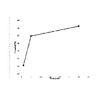
[0076] Figure 2 shows the up-take of 90Y from a solution of 90Yttrium
chloride into strontium phosphate microspheres of the invention over a period
of 30
minutes.
Examples
Example 1: Preparation of Strontium Phosphate Microspheres
[0077] Strontium containing borate glass microspheres were prepared from a
batch
comprising 20 mol%Na20, 20 mol%Sr0 and 60 mol% Ba203 as described above. The
microspheres ranged in size from 44 to 105 microns.
[0078] The microspheres were reacted with 0.25mo1ar K2HPO4 solution (pH=12)
for
lh, 6h, and 72 hrs at 85 C. After rinsing and drying, the surface area was
measured by nitrogen
WO 2016/064379 PCT/US2014/061597
17
absorption using a Micromeretics Tristar 3000 device. Multiple samples were
measured and
averaged. The measured surface area was 10, 93, and 72 m2/gm, respectively.
Example 2: Loading Strontium Phosphate Microspheres with radioisotopes
[ 0079 ] A solution of 5ug yttrium (Y-90) chloride in 0.5m1 of 0.05N HCI was
added
to 15mg of microspheres and incubated for 30 minutes. The amount of yttrium
loaded into the
microspheres was determined at 1, 5 and 30 minutes in 5 replicates and
averaged. Up-take of the
yttrium with time is shown in fig. 1.
[ 0080 ] As various changes could be made in the above methods and
products, without departing from the scope of the invention, it is intended
that all
matter contained in the above description or shown in any accompanying
drawings
shall be interpreted as illustrative and not in a limiting sense.
It will be clear to a
person of ordinary skill in the art that the above embodiments may be altered
or that
insubstantial changes may be made without departing from the scope of the
invention.
Accordingly, the scope of the invention is determined by the scope of the
following
claims and their equitable equivalents.
Date Recue/Date Received 2020-12-29