Note : Les descriptions sont présentées dans la langue officielle dans laquelle elles ont été soumises.
CA 03007582 2018-06-06
WO 2017/129480 PCT/EP2017/051152
Collagen type VII alpha 1 Assay
Technical Field
The present invention relates to antibodies which are
reactive with fragments of collagen type VII alpha 1
comprising an N- or C-terminal neo-epitope, the use of said
antibodies in an assay for detecting and quantifying said
fragments of collagen type VII alpha 1, and the use of said
assay for evaluating chronic obstructive pulmonary disease
(COPD) or systemic sclerosis.
Background Art
Collagen type VII is the main component of the anchoring
fibrils that connects the basement membrane to the underlying
interstitial matrix. It consists of three identical alpha-1
chains with two non-collagenous (NC) domains and a central
collagenous triple helical domain. It has been identified in
the basement membranes of skin and mucous membranes [1].
Collagen type VII has mainly been investigated for its role
in dystrophic epidermolysis bullosa, a severe skin disease.
Mutations in the collagen type VII alpha-1 chain leads to the
formation of abnormal, diminished or absent anchoring fibrils
which causes separation of epidermis from dermis and thus
skin blistering [1]. Collagen type VII has also been
identified as the protein at fault in epidermolysis bullosa
acquisita, an autoimmune disease causing blistering of the
skin and mucous membranes. It is caused by IgG autoantibodies
directed at the collagen type VII NC1 domain [2].
Autoimmunity to collagen type VII has also been associated
with inflammatory bowel disease and bullous systemic lupus
erythematosus [3-4].
1
CA 03007582 2018-06-06
WO 2017/129480 PCT/EP2017/051152
An up-regulation of collagen type VII level in the skin of
patients suffering from systemic sclerosis has been
identified [5]. Patients with systemic sclerosis have skin
fibrosis and may present with fibrosis of internal organs
including the lungs. One study has also identified a reduced
level of collagen type VII protein in the anchoring fibrils
in the airways in a monkey model of asthma [6].
In order to evaluate a pathogenic condition linked to
collagen type VII it is necessary to produce assays capable
of detecting and quantifying species related to the
pathogenic condition.
Chen et al., Saleh et al. and Kim et al. independently
developed ELISAs for detecting autoantibodies against the NC1
or NC2 domain of collagen type VII [7-9]. The methods
comprise coating a microtiter plate with recombinant NC1
and/or NC2 domain of collagen type VII alpha-1, adding serum
samples of interest, and using anti-human IgG antibody to
detect autoantibodies present in the serum sample.
Recke et al. describes the generation of autoantibodies
against the collagen type VII NC1 domain and investigated the
pathogenic potential in human ex vivo models of epidermolysis
bullosa acquisita [10]. They proposed the use of this as a
diagnostic tool.
Sakai et al. raised a monoclonal antibody against collagen
type VII [11]. The mAb was reactive only with intact collagen
type VII.
Monoclonal and polyclonal antibodies targeting collagen type
VII can be obtained commercially from several vendors.
2
CA 03007582 2018-06-06
WO 2017/129480
PCT/EP2017/051152
It has now been found that fragments of collagen type VII
alpha 1 are detectable in circulation and could serve as
potential biomarkers for evaluating pathological conditions
linked to collagen type VII. Specifically, a link between
collagen type VII alpha 1 fragments and the pathological
conditions COPD and systemic sclerosis has been identified.
Summary of the Invention
Accordingly, in a first aspect the present invention relates
to an antibody reactive with a fragment of collagen type VII
alpha 1 comprising an N- or C-terminal neo-epitope, wherein
said antibody binds to the N- or C-terminal neo-epitope.
The antibody is preferably a monoclonal antibody, but may
also be a polyclonal antibody or an antibody fragment
exhibiting the desired biological activity.
Preferably, the antibody does not recognise or bind intact
collagen type VII alpha 1.
In a preferred embodiment, the antibody may bind to an N- or
C-terminal neo-epitope comprised in a non-collagenous amino-
terminal domain of collagen type VII alpha 1 (NC1) or
comprised in a central collagenous domain of collagen type
VII alpha 1.
In another preferred embodiment, the antibody may bind to a
C-terminal neo-epitope comprised in the central collagenous
domain of collagen type VII alpha 1. Preferably, the
antibody binds to a C-terminal neo-epitope comprised in the
amino acid sequence GPPGPPGRLV-COOH (SEQ ID NO: 1).
Preferably, the antibody binds to a C-terminal neo-epitope
comprising or consisting of the amino acid sequence PPGRLV-
COOH (SEQ ID NO: 2). This C-terminal neo-epitope may be
3
CA 03007582 2018-06-06
WO 2017/129480 PCT/EP2017/051152
formed by cleavage of human collagen type VII alpha 1 at the
Val-Asp bond between amino acids V1709-D1710 in the central
collagenous domain of collagen type VII alpha 1. Preferably,
the antibody does not recognise or bind elongated amino acid
sequence GPPGPPGRLVX-COOH (SEQ ID NO: 3), wherein X is one or
more amino acids of the sequence of collagen type VII alpha
1.
In another preferred embodiment, the antibody may bind to an
N-terminal neo-epitope comprised in the non-collagenous
amino-terminal domain of collagen type VII alpha 1 (NC1).
Preferably, the antibody may bind to an N-terminal neo-
epitope comprised in the amino acid sequence H2N-EAPRVRAQHR
(SEQ ID NO: 4). Preferably, the antibody binds to an N-
terminal neo-epitope comprising or consisting of the amino
acid sequence H2N-EAPRVR (SEQ ID NO: 5). This N-terminal
neo-epitope may be formed by cleavage of human collagen type
VII alpha 1 at the Ala-Glu bond between amino acids A16-E17
in the non-collagenous amino-terminal domain of collagen type
VII alpha 1. Preferably, the antibody does not recognise or
bind elongated amino acid sequence H2N-XEAPRVRAQHR (SEQ ID
NO: 6), wherein X is one or more amino acids of the sequence
of collagen type VII alpha 1.
Antibodies described herein may be raised against a synthetic
peptide corresponding to the N- or C-terminal neo-epitope
amino acid sequence.
In a second aspect the present invention relates to a method
of immunoassay for detecting in a biological sample a
fragment of collagen type VII alpha 1 comprising an N- or C-
terminal neo-epitope, said method comprising contacting said
biological sample comprising said fragment of collagen type
VII alpha 1 comprising said N- or C-terminal neo-epitope with
4
CA 03007582 20113--06
WO 2017/129480 PCT/EP2017/051152
an antibody as described herein, and determining the amount
of binding of said antibody.
The method may be used to quantify the amount of the fragment
of collagen type VII alpha 1 comprising said N- or C-terminal
neo-epitope in biofluids. The biofluid may be, but is not
limited to, serum, plasma, bronchoalveolar lavage fluid,
sputum, saliva, exhaled breath or urine.
The immunoassay may be, but is not limited to, a competition
assay or a sandwich assay. The immunoassay may be, but is not
limited to, a radioimmunoassay or an enzyme-linked
immunosorbent assay.
The method may further comprise the step of correlating the
quantity of the fragment of collagen type VII alpha 1
comprising an N- or C-terminal neo-epitope determined by said
method with standard collagen type VII related disease
samples of known disease severity to evaluate the severity of
a collagen type VII related disease.
Such collagen type VII related diseases may be, but are not
limited to, chronic obstructive pulmonary disease (COPD) or
systemic sclerosis.
It is envisaged that the method of the invention may be
utilised in the quantitation, diagnosis and/or prognosis of
such collagen type VII related diseases.
In a third aspect the present invention relates to a peptide,
wherein the peptide has an N-terminal amino acid sequence
corresponding to an amino acid sequence of an N-terminal neo-
epitope of a fragment of collagen type VII alpha 1 comprising
said N-terminal neo-epitope, or wherein the peptide has a C-
terminal amino acid sequence corresponding to an amino acid
5
CA 03007582 20113--06
WO 2017/129480 PCT/EP2017/051152
sequence of an C-terminal neo-epitope of a fragment of
collagen type VII alpha 1 comprising said C-terminal neo-
epitope. Preferably, the peptide is ten amino acid residues
in length, more preferably nine amino acid residues, more
preferably eight amino acid residues, more preferably seven
amino acid residues, and most preferably six amino acid
residues in length. The peptide may be biotinylated.
In a preferred embodiment, the peptide has the amino acid
sequence EAPRVRAQHR (SEQ ID NO: 4) or EAPRVR (SEQ ID NO: 5).
In another preferred embodiment, the peptide has the amino
acid sequence GPPGPPGRLV (SEQ ID NO: 1) or PPGRLV (SEQ ID NO:
2).
In a fourth aspect the present invention relates to an assay
kit for determining the quantity of a fragment of collagen
type VII alpha 1 comprising an N- or C-terminal neo-epitope
in a biological sample, the kit comprising an antibody as
described herein and at least one of:
- a streptavidin coated 96 well plate
- a biotinylated peptide corresponding to the amino
acid sequence of the N- or C-terminal neo-epitope,
with an optional linker located between the biotin
residue and the peptide
- a biotinylated secondary antibody for use in a
sandwich immunoassay
- a calibrator peptide corresponding to the amino
acid sequence of the N- or C-terminal neo-epitope
- an antibody HRP labeling kit
- an antibody radiolabeling kit
- an assay visualization kit
6
CA 03007582 20113--06
WO 2017/129480 PCT/EP2017/051152
Preferably, the assay kit comprises a biotinylated peptide
Biotin-L-GPPGPPGRLV (SEQ ID NO: 7), wherein L is an optional
linker, and a calibrator peptide comprising the C-terminal
sequence GPPGPPGRLV-COOH (SEQ ID NO: 1).
Preferably, the assay kit comprises a biotinylated peptide
EAPRVRAQHR-L-Biotin (SEQ ID NO: 8), wherein L is an optional
linker, and a calibrator peptide comprising the N-terminal
sequence H2N-EAPRVRAQHR (SEQ ID NO: 4).
Definitions
The term "antibody" is used according to the invention in the
broadest sense and specifically covers intact monoclonal
antibodies, polyclonal antibodies, and antibody fragments, so
long as they exhibit the desired biological activity.
"Antibody fragments" according to the invention comprise a
portion of an intact antibody, preferably comprising the
antigen-binding or variable region thereof. Examples of
antibody fragments include Fab, Fab', F(ab')2, Fv and Fc
fragments.
"A fragment of Collagen type VII alpha 1" according to the
invention means a peptide fragment produced by protease
cleavage of collagen type VII.
"N- or C-terminal neo-epitope" according to the invention
means an N- or C-terminal epitope formed at a protease
cleavage site of Collagen type VII alpha 1. For example, the
following sequence of Collagen type VII alpha 1
_PGPPGPPGRLVtDTGPGAREKGE_
would produce the N-terminal neo-epitope H2N-DTGPGAREKGE_ and
the C-terminal neo-epitope _PGPPGPPGRLV-COOH when cleaved by
7
CA 03007582 2018-06-06
WO 2017/129480 PCT/EP2017/051152
a protease at the site between the V1-709-D1710 peptide bond, as
denoted by the symbol "t".
"C7" as used herein refers to fragments of collagen type VII
alpha 1 comprising the C-terminal neo-epitope GPPGPPGRLV-COOH
(SEQ ID NO: 1).
"NB677" as used herein refers to fragments of collagen type
VII alpha 1 comprising the N-terminal neo-epitope H2N-
EAPRVRAQHR (SEQ ID NO: 4).
Figures
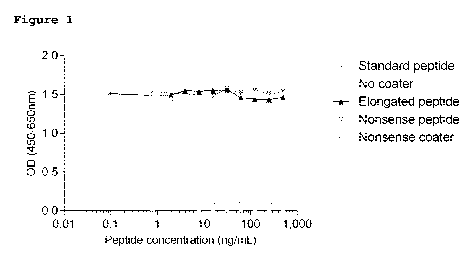
Figure 1 shows a calibration curve for the "C7" assay.
Figure 2 shows the correlation between "C7" and COPD.
Figure 3 shows the correlation between "C7" and systemic
sclerosis.
Figure 4 shows a calibration curve for the "NB677" assay.
Figure 5. Clinical evaluation of serum C7 in systemic
sclerosis. Serum C7 levels were assessed in healthy donors
(n=70) and a cohort of patients with systemic sclerosis (SSc;
n=119). Data are presented as Tukey's box plots. Statistical
significance was evaluated by Mann-Whitney test. ***p<0.0001.
Examples
Example 1 - COPD Biomarker ("C7" Assay)
Rationale
Mass spectrometry was performed on serum samples from a
patient with COPD, a patient with idiopathic pulmonary
fibrosis (IPF), and a healthy donor.
8
CA 03007582 2018-06-06
WO 2017/129480 PCT/EP2017/051152
The initial mass spectrometry analyses identified peptides
derived from collagen type VII in serum. Peptides were
isolated from serum using IMAC Cu beads. Identity
significance threshold for individual peptides were 51. Serum
samples were analyzed using an orbitrap (OrbiB) instrument.
Fragments of collagen type VII alpha 1 comprising the C-
terminal neo-epitope GPPGPPGRLV-COOH ("C7") (SEQ ID NO: 1)
were found in the COPD sample but not in the IPF or healthy
donor samples. The neo-epitope corresponds to the cleavage
site located between amino acids Val-Asp at positions 1709-
1710 of human collagen type VII. The protease responsible for
this cleavage is as yet unknown. The sequence was analyzed
using BLAST and was found to be unique for the collagen type
VII alpha-1 chain.
Antibody
A monoclonal antibody was raised against the C-terminal neo-
epitope amino acid sequence GPPGPPGRLV-COOH (SEQ ID NO: 1).
Briefly, four to six-week-old Balb/C mice were immunized
subcutaneously with 200 pL emulsified antigen and 50 pg of a
C7 synthetic peptide (KLH-CGG-GPPGPPGRLV, SEQ ID NO: 9) using
Freund's incomplete adjuvant. Immunizations were performed
every 2'd week until stable sera titer levels were reached.
The mouse with highest serum titer was selected for fusion.
The mouse was rested for one month and then boosted
intravenously with 50 pg C7 peptide in 100 pL 0.9% sodium
chloride solution three days before isolation of the spleen
for cell fusion. Mouse spleen cells were fused with 5P2/0
myeloma fusion partner cells. The resulting hybridoma cells
were cloned using a semi-solid medium method, transferred
into 96-well microtiter plates for further growth and
9
CA 03007582 2018-06-06
WO 2017/129480 PCT/EP2017/051152
incubated in a CO2 incubator. Standard limited dilution was
used to promote monoclonal growth.
ELISA
A competitive ELISA using the monoclonal antibody raised
against C7 was performed using the following procedure:
Streptavidin-coated plates were coated with 100 pL/well of
2.5 ng/mL biotin-labeled peptide (Biotin-KKGPPGPPGRLV, SEQ ID
NO: 10) diluted in assay buffer (50mM TBS-BTB, 2g/L NaC1, pH
8.0) and incubated at 20 C, 300 rpm shaking for 30 minutes.
Plates were washed five times in washing buffer (20 nM TRIS,
50 mM NaC1, pH 7.2). Sample or standard peptide (20 pL) was
added in double determinations and followed immediately by
addition of 100 pL/well of 200 ng/mL HRP-labeled monoclonal
antibody diluted in assay buffer and plates were incubated at
20 C, 300 rpm shaking for 3 hours. The standard peptide was a
synthetic peptide (GPPGPPGRLV, SEQ ID NO: 1) with a starting
concentration of 125 ng/mL and diluted 2-fold to create an 11
points calibration curve (Figure 1). After incubation, plates
were washed five times in washing buffer. A volume of 100 pL
3,3',5,5'-tetramethylbenzidine (TMB) was added and incubated
for 15 min at 20 C in the dark. To stop the enzyme reaction
of TMB, 100 mL 0.1% sulphuric acid was added and the
absorbance was measured at 450nm with 650nm as the reference
using an ELISA reader. A calibration curve was plotted using
a 4-parametric mathematical fit model. Each ELISA plate
included both kit control and in-house quality control
samples to monitor inter-assay variation. All samples were
measured within the range of the assay. All samples below the
lower limit of detection (LLOD) were assigned the value of
LLOD.
CA 03007582 2018-06-06
WO 2017/129480 PCT/EP2017/051152
The technical characteristics of the C7 ELISA are as follows:
Technical characteristics Results
Human serum
Biological matrix
(undiluted measurements)
Measurement range 1.5 - 105.6 ng/mi,
Normal range of healthy
3.5 ng/mL
serum
Inter-assay variation 13% (accepted if <15%)
Intra-assay variation 9% (accepted if <10%)
Accepted
Dilution recovery
(undiluted to 1 :8)
166% (peptide in serum)
Spiking recovery
131% (serum in serum)
Accepted
Analyte stability
(freeze/thaw and storage)
The ELISA was shown to be specific for the cleavage site as
reactivity was seen towards the standard peptide but not to
an elongated peptide (GPPGPPGRLVD, SEQ ID NO: 11), indicating
that the assay does not recognize intact collagen type VII
protein (Figure 1).
Clinical Utility
COPD
Serum levels of C7 were significantly elevated in a cohort of
68 patients with COPD when compared to healthy donors (Figure
2).
These results show the utility of the C7 assay in identifying
COPD, and may prove useful in evaluating COPD, for example as
a diagnostic or prognostic tool.
Systemic Sclerosis
11
CA 03007582 20113--06
WO 2017/129480 PCT/EP2017/051152
Serum levels of C7 were significantly elevated in 20 patients
with early diffuse systemic sclerosis when compared to
healthy control (p=0.022) (Figure 3). The early stage of
systemic sclerosis is associated with high disease activity,
whereas the late stage patients are progressing slowly. In
the group of patients with early diffuse disease, a
subpopulation with intermediate progression rate (defined by
the skin thickness progression rate) had significantly
elevated levels when compared to controls (p=0.016).
The elevated level of C7 in early stage systemic sclerosis
when compared to late stage systemic sclerosis suggests that
the C7 assay may be capable of differentiating between early
and late stages of systemic sclerosis, thereby providing a
potentially useful diagnostic and/or prognostic tool for
evaluating systemic sclerosis.
Example 2 ("NB677" Assay)
The signal peptide in collagen type VII alpha-1 is found at
amino acids 1-16 [12]. The N-terminal neo-epitope sequence
that is formed by cleavage of the signal peptide
(17'.EAPRVRAQHR'26) was analyzed using BLAST and was found to
be unique for the collagen type VII alpha-1 chain.
Following the success of the "C7" assay for COPD, it is
postulated that this unique collagen type VII alpha-1 neo-
epitope may also be useful in the identification and/or
evaluation of COPD and/or systemic sclerosis.
Antibody
Accordingly, a monoclonal antibody was raised against the N-
terminal neo-epitope amino acid sequence H2N-EAPRVRAQHR (SEQ
ID NO: 4).
12
CA 03007582 2018-06-06
WO 2017/129480 PCT/EP2017/051152
Briefly, four to six-week-old Balb/C mice were immunized
subcutaneously with 200 pL emulsified antigen and 50 pg of a
NB677 synthetic peptide (EAPRVRAQHR-GGC-KLH, SEQ ID NO: 12)
using Freund's incomplete adjuvant. Immunizations were
performed every 2'd week until stable sera titer levels were
reached. The mouse with highest serum titer was selected for
fusion. The mouse was rested for one month and then boosted
intravenously with 50 pg NB677 peptide in 100 pL 0.9% sodium
chloride solution three days before isolation of the spleen
for cell fusion. Mouse spleen cells were fused with SP2/0
myeloma fusion partner cells. The resulting hybridoma cells
were cloned using a semi-solid medium method, transferred
into 96-well microtiter plates for further growth and
incubated in a 002 incubator. Standard limited dilution was
used to promote monoclonal growth.
ELISA
A competitive ELISA using the monoclonal antibody was
performed using the following procedure:
Streptavidin-coated plates were coated with 100 pL/well of
2.0 ng/mL biotin-labeled peptide (EAPRVRAQHR-Lys-Biotin, SEQ
ID NO: 13) diluted in coating buffer (50 mM PBS-BTE, 8g/L
NaC1, 10% sorbitol) and incubated at 20 C, 300 rpm shaking
for 30 minutes. Plates were washed five times in washing
buffer (20 nM TRIS, 50 mM NaC1, pH 7.2). Standard peptide (20
pL) was added in double determinations and followed
immediately by addition of 100 pL/well of 120 ng/mL
monoclonal antibody diluted in assay buffer (25 mM PBS-BTB,
8g/L NaC1) and plates were incubated at 4 C, 300 rpm shaking
for 20 hours. The standard peptide was a synthetic peptide
(EAPRVRAQHR, SEQ ID NO: 4) with a starting concentration of
100 ng/mL and diluted 2-fold to create a calibration curve
13
CA 03007582 2018-06-06
WO 2017/129480 PCT/EP2017/051152
(Figure 4). After incubation, plates were washed five times
in washing buffer. 100 pL/well of secondary HRP-labeled
antibody (rabbit anti-mouse IgG) was added diluted 1:3000 in
assay buffer and plates were incubated at 20 C, 300 rpm
shaking for 1 hour. A volume of 100 pL 3,3',5,5'-
tetramethylbenzidine (TMB) was added and incubated for 15 min
at 20 C in the dark. To stop the enzyme reaction of TMB, 100
mL 0.1% sulphuric acid was added and the absorbance was
measured at 450nm with 650nm as the reference using an ELISA
reader. A calibration curve was plotted using a 4-parametric
mathematical fit model. The monoclonal antibody directed to
the collagen type VII alpha 1 N-terminal neo-epitope has been
confirmed to recognize the desired sequence, assessed by the
reactivity to the standard peptide.
Example 3
The C7 ELISA was evaluated in a second, larger cohort of
patients with systemic sclerosis (SSc). The C7 ELISA was re-
calibrated (compared to the previous example) to improve
accuracy of the assessments in human serum.
Results: The biological relevance of the C7 ELISA was
evaluated by comparing serum levels in healthy donors (n=70)
with patients with SSc (n=119). Data are shown in Figure 5.
Median serum C7 level was significantly elevated in patients
with SSc (9.3 ng/mL [IQR 6.7-13.2]) as compared with healthy
donors (3.9 ng/mL [IQR 2.3-8.3 ng/mL]; p<0.0001).
The clinical data support that serum C7 levels are elevated
in patients with SSc
14
CA 03007582 2018-06-06
WO 2017/129480 PCT/EP2017/051152
In conclusion, the novel assays described herein utilise
antibodies specific for an N- or C-terminal neo-epitope of
collagen VII alpha 1. To the best of our knowledge, this is
the first time that collagen type VII has been associated
with COPD. Accordingly, it is envisaged that these assays may
be used for assessing COPD as well as systemic sclerosis.
In this specification, unless expressly otherwise
indicated, the word 'or' is used in the sense of an operator
that returns a true value when either or both of the stated
conditions is met, as opposed to the operator 'exclusive or'
which requires that only one of the conditions is met. The
word 'comprising' is used in the sense of 'including' rather
than in to mean 'consisting of'. All prior teachings
acknowledged above are hereby incorporated by reference. No
acknowledgement of any prior published document herein should
be taken to be an admission or representation that the
teaching thereof was common general knowledge in Australia or
elsewhere at the date hereof.
15
CA 03007582 20113--06
WO 2017/129480 PCT/EP2017/051152
References
[1] Chung, H. J. and J. Uitto. 2010. Type VII collagen:
the anchoring fibril protein at fault in dystrophic
epidermolysis bullosa. Dermatol.Clin. 28:93-105.
[2] Chen, M., G. H. Kim, L. Prakash, and D. T. Woodley.
2012. Epidermolysis bullosa acquisita: autoimmunity to
anchoring fibril collagen. Autoimmunity 45:91-101.
[3] Hundorfean, G., M. F. Neurath, and C. Sitaru. 2010.
Autoimmunity against type VII collagen in inflammatory bowel
disease. J.Cell Mol.Med. 14:2393-2403.
[4] Gammon, W. R., D. T. Woodley, K. C. Dole, and R. A.
Briggaman. 1985. Evidence that anti-basement membrane zone
antibodies in bullous eruption of systemic lupus
erythematosus recognize epidermolysis bullosa acquisita
autoantigen. J.Invest Dermatol. 84:472-476.
[5] Rudnicka, L., J. Varga, A. M. Christiano, R. V.
Iozzo, S. A. Jimenez, and J. Uitto. 1994. Elevated expression
of type VII collagen in the skin of patients with systemic
sclerosis. Regulation by transforming growth factor-beta.
J.Clin.Invest 93:1709-1715.
[6] Evans, M. J., M. V. Fanucchi, L. A. Miller, M. A.
Carlson, S. J. Nishio, and D. M. Hyde. 2010. Reduction of
collagen VII anchoring fibrils in the airway basement
membrane zone of infant rhesus monkeys exposed to house dust
mite. Am.J.Physiol Lung Cell Mol.Physiol 298:L543-L547.
16
CA 03007582 2018-06-06
WO 2017/129480
PCT/EP2017/051152
[7] Chen, M., L. S. Chan, X. Cai, E. A. O'Toole, J. C.
Sample, and D. T. Woodley. 1997. Development of an ELISA for
rapid detection of anti-type VII collagen autoantibodies in
epidermolysis bullosa acquisita. J.Invest Dermatol. 108:68-
72.
[8] Saleh, M. A., K. Ishii, Y. J. Kim, A. Murakami, N.
Ishii, T. Hashimoto, E. Schmidt, D. Zillikens, Y. Shirakata,
K. Hashimoto, et al. 2011. Development of NC1 and NC2
domains of type VII collagen ELISA for the diagnosis and
analysis of the time course of epidermolysis bullosa
acquisita patients. J.Dermatol.Sci 62:169-175.
[9] Kim, J. H., Y. H. Kim, S. Kim, E. B. Noh, S. E.
Kim, A. Vorobyev, E. Schmidt, D. Zillikens, and S. C. Kim.
2013. Serum levels of anti-type VII collagen antibodies
detected by enzyme-linked immunosorbent assay in patients
with epidermolysis bullosa acquisita are correlated with the
severity of skin lesions. J.Eur.Acad Dermatol.Venereol.
27:e224-e230.
[10] Recke, A., C. Sitaru, G. Vidarsson, M. Evensen, M.
T. Chiriac, R. J. Ludwig, and D. Zillikens. 2010.
Pathogenicity of IgG subclass autoantibodies to type VII
collagen: induction of dermal-epidermal separation.
J.Autoimmun. 34:435-444.
[11] Sakai, L. Y., D. R. Keene, N. P. Morris, and R. E.
Burgeson. 1986. Type VII collagen is a major structural
component of anchoring fibrils. J.Cell Biol. 103:1577-1586.
[12] Uniprot. http://www.uniprot.org/uniprot/Q02388.
Accessed 2015.
17