Note : Les descriptions sont présentées dans la langue officielle dans laquelle elles ont été soumises.
CA 03055539 2019-09-05
WO 2018/165228 PCT/US2018/021249
IMMUNE CELL COMPOSITIONS AND METHODS OF USE
1. CROSS-REFERENCE TO RELATED APPLICATIONS
[0001] This application claims the benefit of United States Provisional
application No.
62/468,887, filed March 8, 2017, and United States Provisional application No.
62/469,366, filed
March 9, 2017, each of which is incorporated by reference herein in its
entirety.
2. REFERENCE TO SEQUENCE LISTING SUBMITTED ELECTRONICALLY
[0002] This application incorporates by reference a Sequence Listing with
this application as
an ASCII text file entitled "13542-044-228 SL.TXT" created on March 2, 2018,
and having a
size of 82,681 bytes.
3. FIELD
[0003] The present invention relates generally to cancer treatment and
pathogen infection
treatment, and more specifically to immunotherapy for cancer treatment and
pathogen infection
treatment.
4. BACKGROUND
[0004] Recent years have provided tremendous advancements in the treatment
of cancer and
pathogen infections (e.g., viral infections). Among these advancements are the
use of
immunotherapy, where a patient's immune response is harnessed to treat cancer
or infection.
Such immunotherapy treatment methods include the use of cell-based
immunotherapy, where
cells of the immune system are utilized for therapeutic treatment. Immune
system cells such as
T cells and other immune cells can be modified to target tumor antigens.
[0005] In response to immune attack, solid tumors upregulate PD-Li in
response to immune
attack, which in turn binds PD-1 receptor expressed on T cells, resulting in T-
cell inhibition (see
Pardoll, Nat. Rev. Cancer 12(4):252-64 (2012)). Upregulation of PD-Li on T
cells and antigen
presenting cells (APCs) was described as well, resulting in inhibition of
activated T cells (Talay
et al., Proc. Natl. Acad. Sci. USA 106(8):2741-2746 (2009); Latchman et al.,
Proc. Natl. Acad.
1
CA 03055539 2019-09-05
WO 2018/165228 PCT/US2018/021249
Sci. USA 101(29):10691-10696 (2004); Liu etal., I Cell. Mol. Med. 19(6):1223-
1233 (2015)).
PD-1/PD-L1 checkpoint blockade therapy counteracts this inhibition, thereby
leading to
activated T cells. Various strategies to inhibit the immune checkpoint
blockade mediated by PD-
1 have been described, including the use of PD-1 or PD-Li antibodies (Burga et
al., Cancer
Immunol. Immunother. 64(7):817-829 (2015); Moon etal., Cl/n. Cancer Res.
20(16):4262-4273
(2014); John et al., Cl/n. Cancer Res. 19(20):5636-5646 (2013)), RNA
interference (Borkner et
al., Cancer Immunol. Immunother. 59(8):1173-1183 (2010)), and co-stimulatory
molecules
(Prosser et al., Mol. Immunol. 51(3-4):263-272 (2012); Ankri etal., I Immunol.
191(8):4121-
4129 (2013)).
[0006] Similarly, functional impairment of T cells is characteristic of
many human
pathogenic infections, such as viral infections (see Day et al., Nature
443:350-354 (2006) and
references cited therein). PD-1 is a negative regulator of activated T cells,
and is markedly
upregulated on the surface of exhausted virus-specific CD8+ T cells (Ishida et
al., EMBO
11:3887-3895 (1992); Noshimura etal., Immunity 11:141-151 (1999); Sharpe
etal., Nat. Rev.
Immunol. 2:116-126 (2002); Che. Nat. Rev. Immunol. 4:336-347 (2004); Barber et
al., Nature
439:682-687 (2006)). Blockade of this pathway using antibodies against the PD
ligand 1 (PD-
L1, also known as CD274) restores CD8+ T-cell function and reduces viral load
(Barber et al.,
Nature 439:682-687 (2006)). It was found that PD-1 is significantly
upregulated on T cells, and
expression correlates with impaired HIV-specific CD8+ T-cell function as well
as predictors of
disease progression: positively with plasma viral load and inversely with CD4+
T-cell count
(Day etal., Nature 443:350-354 (2006)). PD-1 expression on CD4+ T cells
likewise showed a
positive correlation with viral load and an inverse correlation with CD4+ T-
cell count, and
blockade of the pathway augmented HIV-specific CD4+ and CD8+ T-cell function
(Day et al.,
Nature 443:350-354 (2006)). The results described by Day et al. (supra, 2006)
indicate that the
immunoregulatory PD-1/PD-L1 pathway is operative during a persistent viral
infection in
humans, and define a reversible defect in HIV-specific T-cell function (Day et
al., Nature
443:350-354 (2006)).
[0007] While immunotherapy methods have provided new modalities for cancer
and
infection treatment, including antibody therapies and cell-based therapies
using immune cells
such as T cells, limitations have been found for the effectiveness of such
treatments. For
2
CA 03055539 2019-09-05
WO 2018/165228 PCT/US2018/021249
example, malignant cells and infected cells can adapt to generate an
immunosuppressive
microenvironment that protects the cells from immune recognition and
elimination. This
microenvironment poses a challenge to methods of treatment involving
stimulation of an
immune response, including immunotherapy methods such as targeted T cell
therapies.
Furthermore, solid tumors or infections can be restricted within anatomical
compartments such
that access of therapeutic immune cells to the tumors or infected cells is
limited. In addition, an
immunosuppressive microenvironment must be overcome so that the immunotherapy
is
effective. The successful elimination of cancer cells and the successful
elimination of infected
cells thus both require overcoming tumor-induced or infection-induced
immunosuppression.
[0008] Thus, there exists a need for therapies to provide improved
treatment of cancer and
pathogen infections that overcome microenvironments associated with malignant
cells or
infected cells that inhibit effective immunotherapies. The present invention
satisfies this need
and provides related advantages as well.
5. SUMMARY OF INVENTION
[0009] The invention can be summarized by the claims appended hereto and as
described
below.
[0010] In one aspect, provided herein is a T cell comprising in one or more
transgenes: (a) a
first nucleotide sequence encoding a dominant negative form of an inhibitor of
a cell-mediated
immune response of the T cell, and (b) a second nucleotide sequence encoding
an
immunomodulatory agent, wherein the immunomodulatory agent is a single chain
variable
fragment (scFv) or peptide antibody, which immunomodulatory agent binds to and
inhibits an
immune checkpoint inhibitor, and wherein the immune checkpoint inhibitor is
different from the
inhibitor of a cell-mediated immune response. In certain embodiments, the
dominant negative
form of the inhibitor of a cell-mediated immune response is expressed as a
membrane protein on
the T cell surface. In certain embodiments, the inhibitor of a cell-mediated
immune response is
an immune checkpoint inhibitor.
[0011] In certain embodiments of a T cell, the inhibitor of a cell-mediated
immune response
is selected from the group consisting of programmed death 1 (PD-1), cytotoxic
T lymphocyte
3
CA 03055539 2019-09-05
WO 2018/165228 PCT/US2018/021249
antigen-4 (CTLA-4), B- and T-lymphocyte attenuator (BTLA), T cell
immunoglobulin mucin-3
(TIM-3), lymphocyte-activation protein 3 (LAG-3), T cell immunoreceptor with
Ig and ITIM
domains (TIGIT), leukocyte-associated immunoglobulin-like receptor 1 (LAIR1),
natural killer
cell receptor 2B4 (2B4), and CD160. In a particular embodiment, the inhibitor
of a cell-
mediated immune response is PD-1.
[0012] In certain embodiments of a T cell, the inhibitor of a cell-mediated
immune response
is TGF-I3 receptor.
[0013] In certain embodiments of a T cell, the immunomodulatory agent is
secreted from the
T cell. In certain embodiments of a T cell comprising in one or more
transgenes, the
immunomodulatory agent is a scFv. In certain embodiments of a T cell
comprising in one or
more transgenes, the immunomodulatory agent is a peptide antibody.
[0014] In certain embodiments of a T cell, the immune checkpoint
inhibitor to which the
immunomodulatory agent binds is selected from the group consisting of
programmed death 1
(PD-1), cytotoxic T lymphocyte antigen-4 (CTLA-4), B- and T-lymphocyte
attenuator (BTLA),
T cell immunoglobulin mucin-3 (TIM-3), lymphocyte-activation protein 3 (LAG-
3), T cell
immunoreceptor with Ig and ITIM domains (TIGIT), leukocyte-associated
immunoglobulin-like
receptor 1 (LAIR1), natural killer cell receptor 2B4 (2B4), and CD160. In a
particular
embodiment, the immune checkpoint inhibitor to which the immunomodulatory
agent binds is
TIM-3. In another particular embodiment, the immune checkpoint inhibitor to
which the
immunomodulatory agent binds is LAG-3.
[0015] In certain embodiments of a T cell, the T cell comprises a transgene
comprising (a)
the first nucleotide sequence encoding a dominant negative form of an
inhibitor of a cell-
mediated immune response of the T cell, and (b) the second nucleotide sequence
encoding an
immunomodulatory agent, wherein a nucleotide sequence encoding a cleavable
linker is present
in between the first nucleotide sequence encoding a dominant negative form and
the second
nucleotide sequence encoding an immunomodulatory agent, and wherein expression
of the
transgene is under control of a promoter such that the transgene is
expressible in the T cell to
produce the dominant negative form and the immunomodulatory agent. In certain
embodiments,
the promoter is constitutive. In certain embodiments, the transgene further
comprises a third
4
CA 03055539 2019-09-05
WO 2018/165228 PCT/US2018/021249
nucleotide sequence encoding a reporter, wherein a nucleotide sequence
encoding a cleavable
linker is present in between any adjacent occurrences in the transgene of the
first nucleotide
sequence encoding a dominant negative form of an inhibitor of a cell-mediated
immune response
of the T cell, the second nucleotide sequence encoding an immunomodulatory
agent, and the
third nucleotide sequence encoding a reporter, and wherein the transgene is
expressible in the T
cell to produce the reporter.
[0016] In certain embodiments of a T cell, the T cell recognizes and is
sensitized to a target
antigen associated with a mammalian disease or disorder.
[0017] In certain embodiments of a T cell, the T cell further comprises a
fourth nucleotide
sequence encoding a chimeric antigen receptor (CAR), wherein the CAR binds to
a target
antigen that is associated with a mammalian disease or disorder.
[0018] In certain embodiments of a T cell, the transgene further comprises
a fourth
nucleotide sequence encoding a chimeric antigen receptor (CAR), wherein the
CAR binds to a
target antigen that is associated with a mammalian disease or disorder, and
wherein a nucleotide
sequence encoding a cleavable linker is present in between any adjacent
occurrences in the
transgene of the first nucleotide sequence encoding a dominant negative form
of an inhibitor of a
cell-mediated immune response of the T cell, the second nucleotide sequence
encoding an
immunomodulatory agent, and the fourth nucleotide sequence encoding a CAR, and
wherein the
transgene is expressible in the T cell to produce the CAR.
[0019] In certain embodiments of a T cell, the transgene further comprises
a fourth
nucleotide sequence encoding a chimeric antigen receptor (CAR), wherein the
CAR binds to a
target antigen that is associated with a mammalian disease or disorder, and
wherein a nucleotide
sequence encoding a cleavable linker is present in between any adjacent
occurrences in the
transgene of the first nucleotide sequence encoding a dominant negative form
of an inhibitor of a
cell-mediated immune response of the T cell, the second nucleotide sequence
encoding an
immunomodulatory agent, the third nucleotide sequence encoding a reporter, and
the fourth
nucleotide sequence encoding a CAR, and wherein the transgene is expressible
in the T cell to
produce the CAR.
CA 03055539 2019-09-05
WO 2018/165228 PCT/US2018/021249
[0020] In another aspect, provided herein is a T cell comprising a
transgene, which transgene
comprises a first nucleotide sequence encoding a dominant negative form of an
inhibitor of a
cell-mediated immune response of the T cell, wherein expression of the
transgene is under
control of an inducible promoter, which inducible promoter is induced upon
activation of the T
cell. In certain embodiments, the inducible promoter is induced by nuclear
factor of activated T
cells (NFAT) binding. In certain embodiments, the dominant negative form of
the inhibitor of a
cell-mediated immune response is expressed as a membrane protein on the T cell
surface.
[0021] In certain embodiments of a T cell comprising a transgene, the
inhibitor of a cell-
mediated immune response is an immune checkpoint inhibitor.
[0022] In certain embodiments, the inhibitor of a cell-mediated immune
response is selected
from the group consisting of programmed death 1 (PD-1), cytotoxic T lymphocyte
antigen-4
(CTLA-4), B- and T-lymphocyte attenuator (BTLA), T cell immunoglobulin mucin-3
(TIM-3),
lymphocyte-activation protein 3 (LAG-3), T cell immunoreceptor with Ig and
ITIM domains
(TIGIT), leukocyte-associated immunoglobulin-like receptor 1 (LAIR1), natural
killer cell
receptor 2B4 (2B4), and CD160. In a particular embodiment, the inhibitor of a
cell-mediated
immune response is PD-1.
[0023] In certain embodiments, the inhibitor of a cell-mediated immune
response is TGF-
receptor.
[0024] In certain embodiments of a T cell comprising a transgene, the
transgene further
comprises a second nucleotide sequence encoding a reporter, wherein a
nucleotide sequence
encoding a cleavable linker is present in between the first nucleotide
sequence encoding a
dominant negative form of an inhibitor of a cell-mediated immune response of
the T cell and the
second nucleotide sequence encoding a reporter, and wherein the transgene is
expressible in the
T cell to produce the reporter.
[0025] In certain embodiments of a T cell comprising a transgene, the T
cell recognizes and
is sensitized to a target antigen associated with a mammalian disease or
disorder.
[0026] In another aspect, provided herein is a T cell comprising a
transgene, which transgene
comprises a first nucleotide sequence encoding an immunomodulatory agent,
wherein expression
6
CA 03055539 2019-09-05
WO 2018/165228 PCT/US2018/021249
of the transgene is under control of an inducible promoter, which inducible
promoter is induced
upon activation of the T cell, wherein the immunomodulatory agent is a single
chain variable
fragment (scFv) or peptide antibody, which immunomodulatory agent binds to and
inhibits an
immune checkpoint inhibitor. In certain embodiments, the inducible promoter is
induced by
nuclear factor of activated T cells (NFAT) binding.
[0027] In certain embodiments of a T cell comprising a transgene, the
immunomodulatory
agent is secreted from the T cell.
[0028] In certain embodiments, the immunomodulatory agent is a scFv. In
certain
embodiments, the immunomodulatory agent is a peptide antibody.
[0029] In certain embodiments of a T cell comprising a transgene, the
immune checkpoint
inhibitor to which the immunomodulatory agent binds is selected from the group
consisting of
programmed death 1 (PD-1), cytotoxic T lymphocyte antigen-4 (CTLA-4), B- and T-
lymphocyte
attenuator (BTLA), T cell immunoglobulin mucin-3 (TIM-3), lymphocyte-
activation protein 3
(LAG-3), T cell immunoreceptor with Ig and ITIM domains (TIGIT), leukocyte-
associated
immunoglobulin-like receptor 1 (LAIR1), natural killer cell receptor 2B4
(2B4), and CD160. In
a particular embodiment, the immune checkpoint inhibitor to which the
immunomodulatory
agent binds is TIM-3. In another particular embodiment, the immune checkpoint
inhibitor to
which the immunomodulatory agent binds is LAG-3.
[0030] In certain embodiments of a T cell comprising a transgene, the
transgene further
comprises a second nucleotide sequence encoding a reporter, wherein a
nucleotide sequence
encoding a cleavable linker is present in between the first nucleotide
sequence encoding an
immunomodulatory agent and the second nucleotide sequence encoding a reporter,
and wherein
the transgene is expressible in the T cell to produce the reporter.
[0031] In certain embodiments of a T cell comprising a transgene, the T
cell recognizes and
is sensitized to a target antigen associated with a mammalian disease or
disorder.
[0032] In certain embodiments, the mammalian disease or disorder is a
cancer and the target
antigen is a cancer antigen. In certain embodiments, the cancer antigen is
selected from the
group consisting of mesothelin, prostate specific membrane antigen (PSMA),
prostate stem cell
7
CA 03055539 2019-09-05
WO 2018/165228 PCT/US2018/021249
antigen (PCSA), carbonic anhydrase IX (CAIX), carcinoembryonic antigen (CEA),
CD5, CD7,
CD10, CD19, CD20, CD22, CD30, CD33, CD34, CD38, CD41, CD44, CD49f, CD56, CD74,
CD123, CD133, CD138, epithelial glycoprotein2 (EGP 2), epithelial glycoprotein-
40 (EGP-40),
epithelial cell adhesion molecule (EpCAM), folate-binding protein (FBP), fetal
acetylcholine
receptor (AChR), folate receptor-a and I (FRa and 13), Ganglioside G2 (GD2),
Ganglioside G3
(GD3), human Epidermal Growth Factor Receptor 2 (HER-2/ERB2), Epidermal Growth
Factor
Receptor vIII (EGFRvIII), ERB3, ERB4, human telomerase reverse transcriptase
(hTERT),
Interleukin-13 receptor subunit alpha-2 (IL-13Ra2), x-light chain, kinase
insert domain receptor
(KDR), Lewis A (CA19.9), Lewis Y (LeY), Li cell adhesion molecule (L1CAM),
melanoma-
associated antigen 1 (melanoma antigen family Al, MAGE-A1), Mucin 16 (Muc-16),
Mucin 1
(Muc-1), NKG2D ligands, cancer-testis antigen NY-ES0-1, oncofetal antigen
(h5T4),
tumor-associated glycoprotein 72 (TAG-72), vascular endothelial growth factor
R2 (VEGF- R2),
Wilms tumor protein (WT-1), type 1 tyrosine-protein kinase transmembrane
receptor (ROR1),
B7-H3 (CD276), B7-H6 (Nkp30), Chondroitin sulfate proteoglycan-4 (CSPG4), DNAX
Accessory Molecule (DNAM-1), Ephrin type A Receptor 2 (EpHA2), Fibroblast
Associated
Protein (FAP), Gp100/HLA-A2, Glypican 3 (GPC3), HA-1H, HERK-V, IL-11Ra, Latent
Membrane Protein 1 (LMP1), Neural cell-adhesion molecule (N-CAM/CD56), and
Trail
Receptor (TRAIL R). In a particular embodiment, the cancer antigen is
mesothelin.
[0033] In certain embodiments, the mammalian disease or disorder is an
infection with a
pathogen and the target antigen is an antigen of the pathogen. In certain
embodiments, the
pathogen is a human pathogen. In certain embodiments, the pathogen is a virus,
a bacterium, a
fungus, a protozoan, a helminth, or a protist.
[0034] In certain embodiments, the target antigen is a viral antigen. In
certain embodiments,
the viral antigen can elicit an immune response in a human subject infected
with the virus.
[0035] In certain embodiments, the viral antigen is selected from the group
consisting of a
human immunodeficiency virus (HIV) antigen, a hepatitis B virus (HBV) antigen,
a hepatitis C
virus (HCV) antigen, a herpes simplex virus (HSV) antigen, a varicella zoster
virus (VZV)
antigen, an adenovirus antigen, a cytomegalovirus (CMV) antigen, and an
Epstein-Barr virus
(EBV) antigen.
8
CA 03055539 2019-09-05
WO 2018/165228 PCT/US2018/021249
[0036] In certain embodiments, the viral antigen is a HIV antigen selected
from the group
consisting of group-specific antigen (gag) protein, p55, p24, p18, envelope
glycoprotein (env),
gp160, gp120, gp41, reverse transcriptase (pol), p66, and p31.
[0037] In certain embodiments, the viral antigen is a HBV antigen selected
from the group
consisting of HBV envelope protein S, HBV envelope protein M, HBV envelope
protein L, and
the S domain of HBV envelope protein S, M or L.
[0038] In certain embodiments, the viral antigen is a HCV antigen selected
from the group
consisting of core protein, envelope protein El, envelope protein E2, NS2,
NS3, NS4, and NS5.
[0039] In certain embodiments, the viral antigen is a HSV antigen selected
from the group
consisting of gE, gI, gB, gD, gH, gL, gC, gG, gK, gM, and the extracellular
domain of gE.
[0040] In certain embodiments, the viral antigen is a VZV antigen selected
from the group
consisting of gE and gI.
[0041] In certain embodiments, the viral antigen is an adenovirus antigen
selected from the
group consisting of hexon protein and penton protein.
[0042] In certain embodiments, the viral antigen is a CMV antigen selected
from the group
consisting of pp65, immediate early (IE) antigen, and 'El.
[0043] In certain embodiments, the viral antigen is an EBV antigen selected
from the group
consisting of latent membrane protein 2 (LMI32), Epstein¨Barr nuclear antigen
1 (EBNA1), and
BZLF1.
[0044] In certain embodiments of a T cell, the T cell further recombinantly
expresses a
suicide gene. In certain embodiments, the suicide gene comprises inducible
Caspase 9.
[0045] In certain embodiments of a T cell, the T cell is a cytotoxic T
lymphocyte (CTL). In
certain embodiments, the T cell is CD4+. In certain embodiments, the T cell is
CD8+.
[0046] In certain embodiments of a T cell, the T cell is derived from a
human.
9
CA 03055539 2019-09-05
WO 2018/165228 PCT/US2018/021249
[0047] In another aspect, provided herein is an immunostimulatory cell
comprising in one or
more transgenes: (a) a first nucleotide sequence encoding a chimeric antigen
receptor (CAR),
wherein the CAR binds to a target antigen associated with a mammalian disease
or disorder, (b)
a second nucleotide sequence encoding a dominant negative form of an inhibitor
of a cell-
mediated immune response of the immunostimulatory cell, and (c) a third a
nucleotide sequence
encoding a membrane bound form of interleukin 12 (membrane IL-12).
[0048] In certain embodiments, the immunostimulatory cell comprises a
transgene
comprising: (a) the first nucleotide sequence encoding a chimeric antigen
receptor (CAR), (b)
the second nucleotide sequence encoding a dominant negative form of an
inhibitor of a cell-
mediated immune response of the immunostimulatory cell, and (c) the third
nucleotide sequence
encoding a membrane bound form of interleukin 12 (membrane IL-12), wherein a
nucleotide
sequence encoding a cleavable linker is present in between any adjacent
occurrences in the
transgene of the first nucleotide sequence encoding a CAR, the second
nucleotide sequence
encoding a dominant negative form of an inhibitor of a cell-mediated immune
response of the
immunostimulatory cell, and the third nucleotide sequence encoding a membrane
IL-12, and
wherein expression of the transgene is under control of a promoter such that
the transgene is
expressible in the immunostimulatory cell to produce the CAR, the dominant
negative form and
the membrane IL-12. In certain embodiments, the promoter is constitutive.
[0049] In certain embodiments, the immunostimulatory cell comprises: (1) a
first transgene,
which first transgene comprises: (a) the first nucleotide sequence encoding a
chimeric antigen
receptor (CAR), and (b) the second nucleotide sequence encoding a dominant
negative form of
an inhibitor of a cell-mediated immune response of the immunostimulatory cell,
and (2) a second
transgene, which second transgene comprises (c) the third nucleotide sequence
encoding a
membrane bound form of interleukin 12 (membrane IL-12), wherein a nucleotide
sequence
encoding a cleavable linker is present in between the first nucleotide
sequence encoding a CAR
and the second nucleotide sequence encoding a dominant negative form of an
inhibitor of a cell-
mediated immune response of the immunostimulatory cell, and wherein expression
of the first
transgene is under control of a promoter such that the first transgene is
expressible in the
immunostimulatory cell to produce the CAR and the dominant negative form, and
wherein
expression of the second transgene is under control of an inducible promoter,
which inducible
CA 03055539 2019-09-05
WO 2018/165228 PCT/US2018/021249
promoter is induced upon activation of the immunostimulatory cell. In certain
embodiments, the
promoter is constitutive.
[0050] In certain embodiments, the immunostimulatory cell comprises: (1) a
first transgene,
which first transgene comprises (a) the nucleotide sequence encoding a
chimeric antigen receptor
(CAR), (2) a second transgene, which second transgene comprises (b) the
nucleotide sequence
encoding a dominant negative form of an inhibitor of a cell-mediated immune
response of the
immunostimulatory cell, and (3) a third transgene, which third transgene
comprises (c) the
nucleotide sequence encoding a membrane bound form of interleukin 12 (membrane
IL-12),
wherein expression of the third transgene is under control of an inducible
promoter, which
inducible promoter is induced upon activation of the immunostimulatory cell.
[0051] In certain embodiments of an immunostimulatory cell, the first and
the second
transgenes are under control of a constitutive promoter.
[0052] In certain embodiments of an immunostimulatory cell, the inducible
promoter is
induced by nuclear factor of activated T cells (NFAT) binding.
[0053] In another aspect, provided herein is an immunostimulatory cell
comprising: (1) in
one or more transgenes: (a) a first nucleotide sequence encoding a chimeric
antigen receptor
(CAR), wherein the CAR binds to a target antigen associated with a mammalian
disease or
disorder, (b) a second nucleotide sequence encoding a dominant negative form
of an inhibitor of
a cell-mediated immune response of the immunostimulatory cell, that is a
receptor-synthetic
Notch fusion protein comprising (i) an extracellular domain of the inhibitor
of a cell-mediated
immune response of the immunostimulatory cell, (ii) the transmembrane core
domain of Notch
C-terminal to the extracellular domain, and (iii) a transcription factor C-
terminal to the
transmembrane core domain of Notch, and (2) in a different transgene (c) a
third nucleotide
sequence encoding a membrane bound form of interleukin 12 (membrane IL-12),
wherein
expression of the membrane IL-12 is under control of an inducible promoter,
which inducible
promoter is induced upon binding of the transcription factor, and wherein the
transcription factor
is cleaved from the receptor-synthetic Notch fusion protein intracellularly
upon binding of the
extracellular domain to its ligand.
11
CA 03055539 2019-09-05
WO 2018/165228 PCT/US2018/021249
[0054] In certain embodiments, the immunostimulatory cell comprises: (1) a
first transgene,
which first transgene comprises: (a) the first nucleotide sequence encoding a
chimeric antigen
receptor (CAR), and (b) the second nucleotide sequence encoding a receptor-
synthetic Notch
fusion protein, and (2) a second transgene, which second transgene comprises
(c) the third
nucleotide sequence encoding a membrane IL-12, wherein a nucleotide sequence
encoding a
cleavable linker is present in between the first nucleotide sequence encoding
a CAR and the
second nucleotide sequence encoding a receptor-synthetic Notch fusion protein,
and wherein
expression of the first transgene is under control of a promoter such that the
first transgene is
expressible in the immunostimulatory cell to produce the CAR and the receptor-
synthetic Notch
fusion protein, and wherein expression of the second transgene is under
control of an inducible
promoter, which inducible promoter is induced upon binding of the
transcription factor, and
wherein the transcription factor is cleaved from the receptor-synthetic Notch
fusion protein
intracellularly upon binding of the extracellular domain to its ligand. In
certain embodiments,
the promoter is constitutive.
[0055] In certain embodiments, the immunostimulatory cell comprises: (1) a
first transgene,
which first transgene comprises: (a) the first nucleotide sequence encoding a
chimeric antigen
receptor (CAR), (2) a second transgene, which second transgene comprises: (b)
the second
nucleotide sequence encoding a receptor-synthetic Notch fusion protein, and
(3) a third
transgene, which third transgene comprises (c) the third nucleotide sequence
encoding a
membrane IL-12, wherein the first and second transgenes are expressible in the
immunostimulatory cell to produce the CAR and the receptor-synthetic Notch
fusion protein, and
wherein expression of the second transgene is under control of an inducible
promoter, which
inducible promoter is induced upon binding of the transcription factor, and
wherein the
transcription factor is cleaved from the receptor-synthetic Notch fusion
protein intracellularly
upon binding of the extracellular domain to its ligand. In certain
embodiments, the first and
second transgenes are under control of constitutive promoters.
[0056] In certain embodiments of an immunostimulatory cell, the membrane IL-
12
comprises a p40 subunit and a p35 subunit separated by a linker, and wherein
the p35 subunit is
fused to a transmembrane domain.
12
CA 03055539 2019-09-05
WO 2018/165228 PCT/US2018/021249
[0057] In another aspect, provided herein is an immunostimulatory cell
comprising in one or
more transgenes: (a) a first nucleotide sequence encoding a dominant negative
form of an
inhibitor of a cell-mediated immune response of the immunostimulatory cell,
and (b) a second
nucleotide sequence encoding interleukin 12 (IL-12), wherein the IL-12 when
expressed by the
immunostimulatory cell is secreted from the immunostimulatory cell.
[0058] In certain embodiments, the immunostimulatory cell comprises a
transgene
comprising: (a) the first nucleotide sequence encoding a dominant negative
form of an inhibitor
of a cell-mediated immune response of the immunostimulatory cell, and (b) the
second
nucleotide sequence encoding interleukin 12 (IL-12), wherein the first
nucleotide sequence
encoding the dominant negative form and the second nucleotide sequence
encoding the IL-12 are
separated by an internal ribosome entry site (TRES), wherein expression of the
transgene is under
control of a promoter such that the transgene is expressible in the
immunostimulatory cell to
produce the dominant negative form and the IL-12. In certain embodiments, the
promoter is
constitutive.
[0059] In certain embodiments, the immunostimulatory cell comprises: (1) a
first transgene
comprising (a) the first nucleotide sequence encoding a dominant negative form
of an inhibitor
of a cell-mediated immune response of the immunostimulatory cell, and (2) a
second transgene
comprising (b) the second nucleotide sequence encoding interleukin 12 (IL-12),
wherein
expression of the dominant negative form is under control of a promoter such
that the first
transgene is expressible in the immunostimulatory cell to produce the dominant
negative form,
wherein expression of the IL-12 is under control of an inducible promoter,
which inducible
promoter is induced upon activation of the immunostimulatory cell, and wherein
the IL-12 when
expressed by the immunostimulatory cell is secreted from the immunostimulatory
cell. In certain
embodiments, the promoter is constitutive.
[0060] In certain embodiments of an immunostimulatory cell, the inducible
promoter is
induced by nuclear factor of activated T cells (NFAT) binding.
[0061] In certain embodiments, the immunostimulatory cell recognizes and is
sensitized to a
target antigen associated with a mammalian disease or disorder.
13
CA 03055539 2019-09-05
WO 2018/165228 PCT/US2018/021249
[0062] In certain embodiments of an immunoinhibitory cell, the mammalian
disease or
disorder is a cancer and the target antigen is a cancer antigen.
[0063] In certain embodiments, the cancer antigen is selected from the
group consisting of
mesothelin, prostate specific membrane antigen (PSMA), prostate stem cell
antigen (PCSA),
carbonic anhydrase IX (CAIX), carcinoembryonic antigen (CEA), CD5, CD7, CD10,
CD19,
CD20, CD22, CD30, CD33, CD34, CD38, CD41, CD44, CD49f, CD56, CD74, CD123,
CD133,
CD138, epithelial glycoprotein2 (EGP 2), epithelial glycoprotein-40 (EGP-40),
epithelial cell
adhesion molecule (EpCAM), folate-binding protein (FBP), fetal acetylcholine
receptor (AChR),
folate receptor-a and l (FRa and 13), Ganglioside G2 (GD2), Ganglioside G3
(GD3), human
Epidermal Growth Factor Receptor 2 (HER-2/ERB2), Epidermal Growth Factor
Receptor vIII
(EGFRvIII), ERB3, ERB4, human telomerase reverse transcriptase (hTERT),
Interleukin-13
receptor subunit alpha-2 (IL-13Ra2), x-light chain, kinase insert domain
receptor (KDR), Lewis
A (CA19.9), Lewis Y (LeY), Li cell adhesion molecule (L1CAM), melanoma-
associated antigen
1 (melanoma antigen family Al, MAGE-A1), Mucin 16 (Muc-16), Mucin 1 (Muc-1),
NKG2D
ligands, cancer-testis antigen NY-ESO-1, oncofetal antigen (h5 T4), tumor-
associated
glycoprotein 72 (TAG-72), vascular endothelial growth factor R2 (VEGF- R2),
Wilms tumor
protein (WT-1), type 1 tyrosine-protein kinase transmembrane receptor (ROR1),
B7-H3
(CD276), B7-H6 (Nkp30), Chondroitin sulfate proteoglycan-4 (CSPG4), DNAX
Accessory
Molecule (DNAM-1), Ephrin type A Receptor 2 (EpHA2), Fibroblast Associated
Protein (FAP),
Gp100/HLA-A2, Glypican 3 (GPC3), HA-1H, HERK-V, IL-11Ra, Latent Membrane
Protein 1
(LMP1), Neural cell-adhesion molecule (N-CAM/CD56), and Trail Receptor (TRAIL
R). In a
particular embodiment, the cancer antigen is mesothelin.
[0064] In certain embodiments of an immunostimulatory cell, the mammalian
disease or
disorder is an infection with a pathogen and the target antigen is an antigen
of the pathogen. In
certain embodiments, the pathogen is a human pathogen. In certain embodiments,
the pathogen
is a virus, a bacterium, a fungus, a protozoan, a helminth, or a protist.
[0065] In certain embodiments of an immunostimulatory cell, the target
antigen is a viral
antigen. In certain embodiments, the viral antigen can elicit an immune
response in a human
subject infected with the virus.
14
CA 03055539 2019-09-05
WO 2018/165228 PCT/US2018/021249
[0066] In certain embodiments of an immunostimulatory cell, the viral
antigen is selected
from the group consisting of a human immunodeficiency virus (HIV) antigen, a
hepatitis B virus
(HBV) antigen, a hepatitis C virus (HCV) antigen, a herpes simplex virus (HSV)
antigen, a
varicella zoster virus (VZV) antigen, an adenovirus antigen, a cytomegalovirus
(CMV) antigen,
and an Epstein-Barr virus (EBV) antigen.
[0067] In certain embodiments of an immunostimulatory cell, the viral
antigen is a HIV
antigen selected from the group consisting of group-specific antigen (gag)
protein, p55, p24, p18,
envelope glycoprotein (env), gp160, gp120, gp41, reverse transcriptase (pol),
p66, and p31.
[0068] In certain embodiments of an immunostimulatory cell, the viral
antigen is a HBV
antigen selected from the group consisting of HBV envelope protein S, HBV
envelope protein
M, HBV envelope protein L, and the S domain of HBV envelope protein S, M or L.
[0069] In certain embodiments of an immunostimulatory cell, the viral
antigen is a HCV
antigen selected from the group consisting of core protein, envelope protein
El, envelope protein
E2, NS2, NS3, NS4, and NS5.
[0070] In certain embodiments of an immunostimulatory cell, the viral
antigen is a HSV
antigen selected from the group consisting of gE, gI, gB, gD, gH, gL, gC, gG,
gK, gM, and the
extracellular domain of gE.
[0071] In certain embodiments of an immunostimulatory cell, the viral
antigen is a VZV
antigen selected from the group consisting of gE and gI.
[0072] In certain embodiments of an immunostimulatory cell, the viral
antigen is an
adenovirus antigen selected from the group consisting of hexon protein and
penton protein.
[0073] In certain embodiments of an immunostimulatory cell, the viral
antigen is a CMV
antigen selected from the group consisting of pp65, immediate early (IE)
antigen, and "El.
[0074] In certain embodiments of an immunostimulatory cell, the viral
antigen is an EBV
antigen selected from the group consisting of latent membrane protein 2
(LMP2), Epstein¨Barr
nuclear antigen 1 (EBNA1), and BZLF1.
CA 03055539 2019-09-05
WO 2018/165228 PCT/US2018/021249
[0075] In certain embodiments of an immunostimulatory cell, the dominant
negative form of
the inhibitor of a cell-mediated immune response is expressed as a membrane
protein on the
immunostimulatory cell surface. In certain embodiments, the inhibitor of a
cell-mediated
immune response is an immune checkpoint inhibitor.
[0076] In certain embodiments of an immunostimulatory cell, the inhibitor
of a cell-mediated
immune response is selected from the group consisting of programmed death 1
(PD-1), cytotoxic
T lymphocyte antigen-4 (CTLA-4), B- and T-lymphocyte attenuator (BTLA), T cell
immunoglobulin mucin-3 (TIM-3), lymphocyte-activation protein 3 (LAG-3), T
cell
immunoreceptor with Ig and ITIM domains (TIGIT), leukocyte-associated
immunoglobulin-like
receptor 1 (LAIR1), natural killer cell receptor 2B4 (2B4), and CD160. In a
particular
embodiment, the inhibitor of a cell-mediated immune response is PD-1.
[0077] In certain embodiments of an immunostimulatory cell, he inhibitor
of a cell-
mediated immune response is TGF-I3 receptor.
[0078] In certain embodiments, the immunostimulatory cell is a T cell. In
certain
embodiments, the T cell is a cytotoxic T lymphocyte (CTL). In certain
embodiments, the T cell
is CD4+. In certain embodiments, the T cell is CD8+.
[0079] In certain embodiments, the immunostimulatory cell is a Natural
Killer (NK) cell.
[0080] In certain embodiments, the immunostimulatory cell further
recombinantly expresses
a suicide gene. In certain embodiments, the suicide gene comprises inducible
Caspase 9.
[0081] In certain embodiments, the immunostimulatory cell is derived from a
human.
[0082] In another aspect, provided herein is a pharmaceutical composition
comprising a
therapeutically effective amount of a T cell or an immunostimulatory cell as
described above.
[0083] In another aspect, provided herein is a method of treating a cancer
in a subject in need
thereof, comprising administering to the subject a therapeutically effective
amount of a T cell or
an immunostimulatory cell as described above.
16
CA 03055539 2019-09-05
WO 2018/165228 PCT/US2018/021249
[0084] In another aspect, provided herein is a method of treating an
infection with a
pathogen in a subject in need thereof, comprising administering to the subject
a therapeutically
effective amount of a T cell or an immunostimulatory cell as described above.
[0085] In another aspect, provided herein is a method of treating a cancer
in a subject in need
thereof, comprising administering to the subject pharmaceutical composition
comprising a T cell
or an immunostimulatory cell as described above.
[0086] In another aspect, provided herein is a method of treating an
infection with a
pathogen in a subject in need thereof, comprising administering to the subject
a pharmaceutical
composition comprising a T cell or an immunostimulatory cell as described
above.
[0087] In certain embodiments of the methods, the cancer is selected from
the group
consisting of mesothelioma, lung cancer, pancreatic cancer, ovarian cancer,
breast cancer, colon
cancer, pleural tumor, glioblastoma, esophageal cancer, gastric cancer, and
synovial sarcoma.
[0088] In certain embodiments of the methods, the infection with a pathogen
is an infection
with a virus, a bacterium, a fungus, a protozoan, a helminth, or a protist. In
a particular
embodiment, the infection with a pathogen is an infection with a virus.
[0089] In certain embodiments of the methods, the infection with a pathogen
is an infection
with HCV, HIV, HBV, HSV, VZV, adenovirus, CMV or EBV.
[0090] In certain embodiments of the methods, the subject is a human.
[0091] In certain embodiments of the methods, the administering is by
intrapleural
administration, intravenous administration, subcutaneous administration,
intranodal
administration, intratumoral administration, intrathecal administration,
intraperitoneal
administration, intracranial administration, or direct administration to the
thymus.
[0092] In certain embodiments of the methods, the T cell or the
immunostimulatory cell is
administered in a dose in the range of 104 to 1010 cells per kilogram of body
weight. In certain
embodiments, the dose is in the range of 3x10⁵ to 3x10⁶ cells per kilogram of
body weight.
17
CA 03055539 2019-09-05
WO 2018/165228 PCT/US2018/021249
6. DESCRIPTION OF THE DRAWINGS
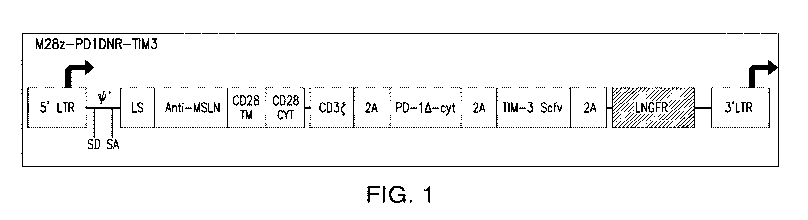
[0093] FIG. 1. Illustration of a construct that contains a nucleotide
sequence encoding a
mesothelin (MSLN)-specific chimeric antigen receptor (CAR), a nucleotide
sequence encoding a
dominant negative form of PD-1, a nucleotide sequence encoding a single chain
variable
fragment (scFv) that binds to TIM-3, and a nucleotide sequence encoding a
LNGFR reporter.
The adjacent nucleotide sequences encoding different proteins are separated by
a nucleotide
sequence encoding a 2A peptide. The CAR contains a CD3t endodomain, a CD28
transmembrane (TM) domain, and a CD28 cytoplasmic (CYT) domain (as the
costimulatory
domain). LTR, long terminal repeat; LS, leader sequence; SA, splice acceptor;
SD, splice donor.
[0094] FIG. 2. Illustration of a construct that contains a nucleotide
sequence encoding a
mesothelin-specific CAR, a nucleotide sequence encoding a dominant negative
form of PD-1, a
nucleotide sequence encoding an scFv that binds to LAG-3, and a nucleotide
sequence encoding
a LNGFR reporter. The adjacent nucleotide sequences encoding different
proteins are separated
by a nucleotide sequence encoding a 2A peptide. The CAR contains a CD3t
endodomain, a
CD28 transmembrane domain, and a CD28 cytoplasmic domain (as the costimulatory
domain).
LTR, long terminal repeat; LS, leader sequence; SA, splice acceptor; SD,
splice donor.
[0095] FIG. 3. Illustration of a construct that contains a nucleotide
sequence encoding a
mesothelin-specific CAR, a nucleotide sequence encoding a dominant negative
form of PD-1, a
nucleotide sequence encoding an scFv that binds to TIM-3, and a nucleotide
sequence encoding
a LNGFR reporter. The adjacent nucleotide sequences encoding different
proteins are separated
by a nucleotide sequence encoding a 2A peptide. The CAR contains a CD3t
endodomain, a
CD28 transmembrane domain, and a 4-1BB cytoplasmic domain (as the
costimulatory domain).
LTR, long terminal repeat; LS, leader sequence; SA, splice acceptor; SD,
splice donor.
[0096] FIG. 4. Illustration of a construct that contains a nucleotide
sequence encoding a
mesothelin-specific CAR, a nucleotide sequence encoding a dominant negative
form of PD-1, a
nucleotide sequence encoding an scFv that binds to LAG-3, and a nucleotide
sequence encoding
a LNGFR reporter. The adjacent nucleotide sequences encoding different
proteins are separated
by a nucleotide sequence encoding a 2A peptide. The CAR contains a CD3t
endodomain, a
18
CA 03055539 2019-09-05
WO 2018/165228
PCT/US2018/021249
CD28 transmembrane domain, and a 4-1BB cytoplasmic domain (as the
costimulatory domain).
LTR, long terminal repeat; LS, leader sequence; SA, splice acceptor; SD,
splice donor.
[0097] FIG.
5. Illustration of a construct that contains a nucleotide sequence encoding a
dominant negative form of PD-1, a nucleotide sequence encoding an scFv that
binds to TIM-3,
and a nucleotide sequence encoding an mCherry reporter. The adjacent
nucleotide sequences
encoding different proteins are separated by a nucleotide sequence encoding a
2A peptide. LTR,
long terminal repeat; LS, leader sequence; SA, splice acceptor; SD, splice
donor.
[0098] FIG.
6. Illustration of a construct that contains a nucleotide sequence encoding a
dominant negative form of PD-1, a nucleotide sequence encoding an scFv that
binds to LAG-3,
and a nucleotide sequence encoding an mCherry reporter. The adjacent
nucleotide sequences
encoding different proteins are separated by a nucleotide sequence encoding a
2A peptide. LTR,
long terminal repeat; LS, leader sequence; SA, splice acceptor; SD, splice
donor.
[0099] FIG.
7. Illustration of a construct that contains a nucleotide sequence encoding a
dominant negative form of PD-1, whose expression is under control of an
inducible promoter
containing NFAT responsive elements, and a nucleotide sequence encoding an
mCherry reporter.
The two nucleotide sequences are separated by a nucleotide sequence encoding a
2A peptide.
LTR, long terminal repeat; LS, leader sequence; SA, splice acceptor; SD,
splice donor.
[00100] FIG. 8. Illustration of a construct that contains a nucleotide
sequence encoding an
scFv that binds to TIM-3, whose expression is under control of an inducible
promoter containing
NFAT responsive elements, and a nucleotide sequence encoding an mCherry
reporter. The two
nucleotide sequences are separated by a nucleotide sequence encoding a 2A
peptide. LTR, long
terminal repeat; LS, leader sequence; SA, splice acceptor; SD, splice donor.
[00101] FIG. 9. Illustration of a construct that contains a nucleotide
sequence encoding an
scFv that binds to LAG-3, whose expression is under control of an inducible
promoter containing
NFAT responsive elements, and a nucleotide sequence encoding an mCherry
reporter. The two
nucleotide sequences are separated by a nucleotide sequence encoding a 2A
peptide. LTR, long
terminal repeat; LS, leader sequence; SA, splice acceptor; SD, splice donor.
19
CA 03055539 2019-09-05
WO 2018/165228 PCT/US2018/021249
[00102] FIG. 10. Illustration of a construct that contains a nucleotide
sequence encoding a
mesothelin-specific CAR, a nucleotide sequence encoding a dominant negative
form of PD-1,
and a nucleotide sequence encoding a membrane IL-12. The membrane IL-12
contains p40 and
p35 subunits separated by a linker, and the p35 subunit is fused to the
transmembrane domain of
CD8. The adjacent nucleotide sequences encoding different proteins are
separated by a
nucleotide sequence encoding a P2A peptide. The CAR contains a CD3t
endodomain, a CD28
transmembrane domain, and a CD28 or 4-1BB cytoplasmic domain (as the
costimulatory
domain).
[00103] FIG. 11. Illustration of a first construct that contains a nucleotide
sequence encoding
a mesothelin-specific CAR, and a nucleotide sequence encoding a dominant
negative form of
PD-1, and a second construct containing a nucleotide sequence encoding a
membrane IL-12,
whose expression is under control of an inducible promoter containing NFAT
responsive
elements. The membrane IL-12 contains p40 and p35 subunits separated by a
linker, and the p35
subunit is fused to the transmembrane domain of CD8. The two nucleotide
sequences on the
first construct are separated by a nucleotide sequence encoding a P2A peptide.
The CAR
contains a CD3t endodomain, a CD28 transmembrane domain, and a CD28 or 4-1BB
cytoplasmic domain (as the costimulatory domain).
[00104] FIG. 12A ¨ FIG. 12B. FIG. 12A Illustration of a first construct that
contains a
nucleotide sequence encoding a mesothelin-specific CAR, and a nucleotide
sequence encoding a
receptor-synthetic Notch fusion protein (which contains an extracellular
domain of PD-1, the
transmembrane core domain of Notch C-terminal to the extracellular domain, and
a transcription
factor C-terminal to the transmembrane core domain of Notch), and a second
construct
containing a nucleotide sequence encoding a membrane IL-12, whose expression
is under control
of an inducible promoter containing responsive elements of the transcription
factor. The
membrane IL-12 contains p40 and p35 subunits separated by a linker, and the
p35 subunit is
fused to the transmembrane domain of CD8. The two nucleotide sequences on the
first construct
are separated by a nucleotide sequence encoding a P2A peptide. The CAR
contains a CD3
endodomain, a CD28 transmembrane domain, and a CD28 or 4-1BB cytoplasmic
domain (as the
costimulatory domain). FIG. 12B Scheme illustrating expression of membrane IL-
12 induced
by the transcription factor released from the receptor-synthetic Notch fusion
protein.
CA 03055539 2019-09-05
WO 2018/165228 PCT/US2018/021249
[00105] FIG. 13. Illustration of a construct that contains a nucleotide
sequence encoding a
dominant negative form of PD-1, an internal ribosome entry site (TRES), and a
nucleotide
sequence encoding soluble IL-12. LTR, long terminal repeat.
[00106] FIG. 14. Illustration of a construct that contains a nucleotide
sequence encoding a
dominant negative form of PD-1, and a nucleotide sequence encoding soluble IL-
12. The
expression of soluble IL-12 is under control of an inducible promoter
containing NFAT
responsive elements. LTR, long terminal repeat.
[00107] FIG. 15. Illustration of a construct that contains a nucleotide
sequence encoding a
mesothelin-specific CAR, a nucleotide sequence encoding a dominant negative
form of TGF-f3
receptor, a nucleotide sequence encoding an scFv that binds to TIM-3, and a
nucleotide sequence
encoding a LNGFR reporter. The adjacent nucleotide sequences encoding
different proteins are
separated by a nucleotide sequence encoding a 2A peptide. The CAR contains a
CD3
endodomain, a CD28 transmembrane domain, and a CD28 cytoplasmic domain (as the
costimulatory domain). LTR, long terminal repeat; LS, leader sequence; SA,
splice acceptor;
SD, splice donor.
[00108] FIG. 16. Illustration of a construct that contains a nucleotide
sequence encoding a
mesothelin-specific CAR, a nucleotide sequence encoding a dominant negative
form of TGF-f3
receptor, a nucleotide sequence encoding an scFv that binds to LAG-3, and a
nucleotide
sequence encoding a LNGFR reporter. The adjacent nucleotide sequences encoding
different
proteins are separated by a nucleotide sequence encoding a 2A peptide. The CAR
contains a
CD3t endodomain, a CD28 transmembrane domain, and a CD28 cytoplasmic domain
(as the
costimulatory domain). LTR, long terminal repeat; LS, leader sequence; SA,
splice acceptor;
SD, splice donor.
[00109] FIG. 17. Illustration of a construct that contains a nucleotide
sequence encoding a
mesothelin-specific CAR, a nucleotide sequence encoding a dominant negative
form of TGF-f3
receptor, a nucleotide sequence encoding an scFv that binds to TIM-3, and a
nucleotide sequence
encoding a LNGFR reporter. The adjacent nucleotide sequences encoding
different proteins are
separated by a nucleotide sequence encoding a 2A peptide. The CAR contains a
CD3
endodomain, a CD28 transmembrane domain, and a 4-1BB cytoplasmic domain (as
the
21
CA 03055539 2019-09-05
WO 2018/165228 PCT/US2018/021249
costimulatory domain). LTR, long terminal repeat; LS, leader sequence; SA,
splice acceptor;
SD, splice donor.
[00110] FIG. 18. Illustration of a construct that contains a nucleotide
sequence encoding a
mesothelin-specific CAR, a nucleotide sequence encoding a dominant negative
form of TGF-f3
receptor, a nucleotide sequence encoding an scFv that binds to LAG-3, and a
nucleotide
sequence encoding a LNGFR reporter. The adjacent nucleotide sequences encoding
different
proteins are separated by a nucleotide sequence encoding a 2A peptide. The CAR
contains a
CD3t endodomain, a CD28 transmembrane domain, and a 4-1BB cytoplasmic domain
(as the
costimulatory domain). LTR, long terminal repeat; LS, leader sequence; SA,
splice acceptor;
SD, splice donor.
7. DETAILED DESCRIPTION OF THE INVENTION
[00111] The present invention relates to compositions and methods for treating
cancer and
pathogen infections (for example, viral infections). It is known that
malignant cells and infected
cells can adapt to generate an immunosuppressive microenvironment to protect
the cells from
immune recognition and elimination. The immunosuppressive microenvironment
provides a
mechanism for cancer cells, tumors, and infected cells to inhibit the effects
of a patient's immune
system to avoid their elimination. This microenvironment poses a challenge to
methods of
treatment involving stimulation of an immune response, including immunotherapy
methods such
as targeted T cell therapies. Although inhibition of certain inhibitors of
cell-mediated immune
response, such as immune checkpoint inhibitors, has been explored to overcome
the
immunosuppressive microenvironment, the overcoming effect is usually transient
because
inhibition of one inhibitor of cell-mediated immune response can result in
upregulation of
another inhibitor of cell-mediated immune response. According to the present
invention, the
effectiveness of cell-based immunotherapy methods can be enhanced by modifying
the cells used
in immunotherapy to express certain combinations of proteins to enhance or
prolong the effect of
overcoming the immunosuppressive microenvironment. As described herein,
immunotherapy
cells can be genetically engineered to intrinsically express a dominant
negative form of an
inhibitor of a cell-mediated immune response and an immunomodulatory agent
that inhibits an
immune checkpoint inhibitor. In addition, immunotherapy cells can be
genetically engineered to
22
CA 03055539 2019-09-05
WO 2018/165228 PCT/US2018/021249
intrinsically express a dominant negative form of an inhibitor of a cell-
mediated immune
response or an immunomodulatory agent that inhibits an immune checkpoint
inhibitor, and an
interleukin-12 (IL-12) protein (in particular, a membrane-bound IL-12 protein,
which has
reduced side effects as compared with secreted IL-12). Furthermore, the
expression of the
dominant negative form of an inhibitor of a cell-mediated immune response, the
immunomodulatory agent that inhibits an immune checkpoint inhibitor, and/or
the IL-12 protein
(for example, a membrane-bound IL-12 protein) can be under the control of an
inducible
promoter to limit their immunostimulatory effects to activated immune cells.
By expressing the
combination of proteins, immune cells can provide a more effective immune
response against the
cancer or the infection. By limiting the immunostimulatory effects to
activated immune cells,
the side effects associated with immunostimulation can be reduced or avoided.
7.1 Cells
[00112] In one aspect, the invention provides immunostimulatory cells
comprising in one or
more transgenes: (a) a nucleotide sequence encoding a dominant negative form
of an inhibitor of
a cell-mediated immune response of the immunostimulatory cell, and (b) a
nucleotide sequence
encoding an immunomodulatory agent, wherein the immunomodulatory agent is a
single chain
variable fragment (scFv) or peptide antibody, which immunomodulatory agent
binds to and
inhibits an immune checkpoint inhibitor, and wherein the immune checkpoint
inhibitor is
different from the inhibitor of a cell-mediated immune response.
[00113] The nucleotide sequence encoding a dominant negative form and the
nucleotide
sequence encoding an immunomodulatory agent can be present in two different
transgenes or
preferably in one single transgene. In specific embodiments, they are present
in one single
transgene (i.e., the immunostimulatory cells comprise a transgene comprising:
the nucleotide
sequence encoding a dominant negative form of an inhibitor of a cell-mediated
immune response
of the immunostimulatory cell, and the nucleotide sequence encoding an
immunomodulatory
agent, wherein expression of the transgene is under control of a promoter (for
example, a
constitutive promoter) such that the transgene is expressible in the
immunostimulatory cell to
produce the dominant negative form and the immunomodulatory agent).
23
CA 03055539 2019-09-05
WO 2018/165228 PCT/US2018/021249
[00114] In various embodiments of the aspect, the immunostimulatory cell
further comprises a
nucleotide sequence encoding a reporter. The nucleotide sequence encoding a
reporter, the
nucleotide sequence encoding a dominant negative form, and the nucleotide
sequence encoding
an immunomodulatory agent can be present in two different transgenes, in three
different
transgenes, or preferably in one single transgene. In specific embodiments,
they are present in
one single transgene (i.e., the immunostimulatory cell comprises a transgene
comprising: the
nucleotide sequence encoding the dominant negative form of an inhibitor of a
cell-mediated
immune response of the immunostimulatory cell, the nucleotide sequence
encoding an
immunomodulatory agent, and the nucleotide sequence encoding a reporter,
wherein the
transgene is expressible in the immunostimulatory cell to produce the dominant
negative form,
the immunomodulatory agent, and the reporter).
[00115] In various embodiments of the aspect, the immunostimulatory cell
further comprises a
nucleotide sequence encoding a chimeric antigen receptor (CAR), wherein the
CAR binds to a
target antigen associated with a mammalian disease or disorder (i.e., a cancer
or an infection with
a pathogen). In some embodiments, the immunostimulatory cell further comprises
a nucleotide
sequence encoding a chimeric antigen receptor (CAR), wherein the CAR binds to
a target
antigen that is a cancer antigen. In other embodiments, the immunostimulatory
cell further
comprises a nucleotide sequence encoding a chimeric antigen receptor (CAR),
wherein the CAR
binds to a target antigen of a pathogen. The nucleotide sequence encoding a
CAR, the nucleotide
sequence encoding a dominant negative form, and the nucleotide sequence
encoding an
immunomodulatory agent can be present in two different transgenes, in three
different
transgenes, or preferably in one single transgene. In specific embodiments,
they are present in
one single transgene (i.e., the immunostimulatory cell comprises a transgene
comprising: the
nucleotide sequence encoding the dominant negative form of an inhibitor of a
cell-mediated
immune response of the immunostimulatory cell, the nucleotide sequence
encoding an
immunomodulatory agent, and the nucleotide sequence encoding a chimeric
antigen receptor
(CAR), wherein the transgene is expressible in the immunostimulatory cell to
produce the
dominant negative form, the immunomodulatory agent, and the CAR). In specific
embodiments,
the immunostimulatory cell further comprises a nucleotide sequence encoding a
reporter. The
nucleotide sequence encoding a reporter, the nucleotide sequence encoding a
CAR, the
nucleotide sequence encoding a dominant negative form, and the nucleotide
sequence encoding
24
CA 03055539 2019-09-05
WO 2018/165228 PCT/US2018/021249
an immunomodulatory agent can be present in two different transgenes, in three
different
transgenes, in four different transgenes, or preferably in one single
transgene. In specific
embodiments, they are present in one single transgene (i.e., the
immunostimulatory cell
comprises a transgene comprising: the nucleotide sequence encoding the
dominant negative
form of an inhibitor of a cell-mediated immune response of the
immunostimulatory cell, the
nucleotide sequence encoding an immunomodulatory agent, the nucleotide
sequence encoding a
chimeric antigen receptor (CAR), and the nucleotide sequence encoding a
reporter, wherein the
transgene is expressible in the immunostimulatory cell to produce the dominant
negative form,
the immunomodulatory agent, the CAR and the reporter).
[00116] In preferred embodiments of the aspect, adjacent occurrences of
nucleotide sequences
encoding different proteins that are present in the same transgene are
separated from each other
by a nucleotide sequence encoding a cleavable linker. Nucleotide sequences
encoding different
cleavable linkers may be used to separate different pairs of adjacent
occurrences of nucleotide
sequences encoding different proteins that are present in the same transgene.
In a specific
embodiment, adjacent occurrences of nucleotide sequences encoding different
proteins that are
present in the same transgene are separated from each other by an internal
ribosomal entry site
(IRES). In another specific embodiment, adjacent occurrences of nucleotide
sequences encoding
different proteins that are present in the same transgene are separated from
each other by a
nucleotide sequence encoding a 2A peptide.
[00117] When the nucleotide sequences encoding the different proteins are
present in different
transgenes, the different transgenes can be present on different vectors or
the same vector.
[00118] In another aspect, the invention provides immunostimulatory cells
comprising a
transgene, which transgene comprises a nucleotide sequence encoding a dominant
negative form
of an inhibitor of a cell-mediated immune response of the immunostimulatory
cell, wherein
expression of the transgene is under control of an inducible promoter, which
inducible promoter
is induced upon activation of the immunostimulatory cell (thus, allowing
expression of the
inhibitor of a cell-mediated immune response only in an activated
immunostimulatory cell). In
specific embodiments, the inducible promoter is induced by nuclear factor of
activated T cells
CA 03055539 2019-09-05
WO 2018/165228 PCT/US2018/021249
(NFAT) binding. In a specific embodiment, the immunostimulatory cell is a T
cell and the
promoter is induced by nuclear factor of activated T cells (NFAT) binding.
[00119] In various embodiments of the aspect, the immunostimulatory cell
further comprises a
nucleotide sequence encoding a reporter. The nucleotide sequence encoding a
reporter and the
nucleotide sequence encoding a dominant negative form can be present in two
different
transgenes, or preferably in one single transgene. In specific embodiments,
they are present in
one single transgene (i.e., the immunostimulatory cell comprises a transgene
comprising: the
nucleotide sequence encoding a dominant negative form of an inhibitor of a
cell-mediated
immune response of the immunostimulatory cell, and the nucleotide sequence
encoding a
reporter, wherein the transgene is expressible in the immunostimulatory cell
to produce the
dominant negative form and the reporter).
[00120] In various embodiments of the aspect, the immunostimulatory cell
further comprises a
nucleotide sequence encoding a chimeric antigen receptor (CAR), wherein the
CAR binds to a
target antigen associated with a mammalian disease or disorder (i.e., a cancer
or an infection with
a pathogen). In some embodiments, the immunostimulatory cell further comprises
a nucleotide
sequence encoding a chimeric antigen receptor (CAR), wherein the CAR binds to
a target
antigen that is a cancer antigen. In other embodiments, the immunostimulatory
cell further
comprises a nucleotide sequence encoding a chimeric antigen receptor (CAR),
wherein the CAR
binds to a target antigen of a pathogen. The nucleotide sequence encoding a
CAR and the
nucleotide sequence encoding a dominant negative form can be present in two
different
transgenes, or preferably in one single transgene. In specific embodiments,
they are present in
one single transgene (i.e., the immunostimulatory cell comprises a transgene
comprising: the
nucleotide sequence encoding a dominant negative form of an inhibitor of a
cell-mediated
immune response of the immunostimulatory cell and the nucleotide sequence
encoding a
chimeric antigen receptor (CAR), wherein the transgene is expressible in the
immunostimulatory
cell to produce the dominant negative form and the CAR). In specific
embodiments, the
immunostimulatory cell further comprises a nucleotide sequence encoding a
reporter. The
nucleotide sequence encoding a reporter, the nucleotide sequence encoding a
CAR, and the
nucleotide sequence encoding a dominant negative form can be present in two
different
transgenes, in three different transgenes, or preferably in one single
transgene. In specific
26
CA 03055539 2019-09-05
WO 2018/165228 PCT/US2018/021249
embodiments, they are present in one single transgene (i.e., the
immunostimulatory cell
comprises a transgene comprising: the nucleotide sequence encoding a dominant
negative form
of an inhibitor of a cell-mediated immune response of the immunostimulatory
cell, the nucleotide
sequence encoding a chimeric antigen receptor (CAR), and the nucleotide
sequence encoding a
reporter, wherein the transgene is expressible in the immunostimulatory cell
to produce the
dominant negative form, the CAR and the reporter).
[00121] In preferred embodiments of the aspect, adjacent occurrences of
nucleotide sequences
encoding different proteins that are present in the same transgene are
separated from each other
by a nucleotide sequence encoding a cleavable linker. Nucleotide sequences
encoding different
cleavable linkers may be used to separate different pairs of adjacent
occurrences of nucleotide
sequences encoding different proteins that are present in the same transgene.
In a specific
embodiment, adjacent occurrences of nucleotide sequences encoding different
proteins that are
present in the same transgene are separated from each other by an internal
ribosomal entry site
(IRES). In another specific embodiment, adjacent occurrences of nucleotide
sequences encoding
different proteins that are present in the same transgene are separated from
each other by a
nucleotide sequence encoding a 2A peptide.
[00122] When the nucleotide sequences encoding the different proteins are
present in different
transgenes, the different transgenes can be present on different vectors or
the same vector.
[00123] In another aspect, the invention provides immunostimulatory cells
comprising a
transgene, which transgene comprises a nucleotide sequence encoding an
immunomodulatory
agent, wherein expression of the transgene is under control of an inducible
promoter, which
inducible promoter is induced upon activation of the immunostimulatory cell
(thus, allowing
expression of the immunomodulatory agent only in an activated
immunostimulatory cell),
wherein the immunomodulatory agent is a single chain variable fragment (scFv)
or peptide
antibody, which immunomodulatory agent binds to and inhibits an immune
checkpoint inhibitor.
In specific embodiments, the inducible promoter is induced by nuclear factor
of activated T cells
(NFAT) binding. In a specific embodiment, the immunostimulatory cell is a T
cell and the
inducible promoter is induced by nuclear factor of activated T cells (NFAT)
binding.
27
CA 03055539 2019-09-05
WO 2018/165228 PCT/US2018/021249
[00124] In various embodiments of the aspect, the immunostimulatory cell
further comprises a
nucleotide sequence encoding a reporter. The nucleotide sequence encoding a
reporter and the
nucleotide sequence encoding an immunomodulatory agent can be present in two
different
transgenes, or preferably in one single transgene. In specific embodiments,
they are present in
one single transgene (i.e., the immunostimulatory cell comprises a transgene
comprising: the
nucleotide sequence encoding an immunomodulatory agent and the nucleotide
sequence
encoding a reporter, wherein the transgene is expressible in the
immunostimulatory cell to
produce the immunomodulatory agent and the reporter).
[00125] In various embodiments of the aspect, the immunostimulatory cell
further comprises a
nucleotide sequence encoding a chimeric antigen receptor (CAR), wherein the
CAR binds to a
target antigen associated with a mammalian disease or disorder (i.e., a cancer
or an infection with
a pathogen). In some embodiments, the immunostimulatory cell further comprises
a nucleotide
sequence encoding a chimeric antigen receptor (CAR), wherein the CAR binds to
a target
antigen that is a cancer antigen. In other embodiments, the immunostimulatory
cell further
comprises a nucleotide sequence encoding a chimeric antigen receptor (CAR),
wherein the CAR
binds to a target antigen of a pathogen. The nucleotide sequence encoding a
CAR and the
nucleotide sequence encoding an immunomodulatory agent can be present in two
different
transgenes, or preferably in one single transgene. In specific embodiments,
they are present in
one single transgene (i.e., the immunostimulatory cell comprises a transgene
comprising: the
nucleotide sequence encoding an immunomodulatory agent and the nucleotide
sequence
encoding a chimeric antigen receptor (CAR), wherein the transgene is
expressible in the
immunostimulatory cell to produce the immunomodulatory agent and the CAR). In
specific
embodiments, the immunostimulatory cell further comprises a nucleotide
sequence encoding a
reporter. The nucleotide sequence encoding a reporter, the nucleotide sequence
encoding a
CAR, and the nucleotide sequence encoding an immunomodulatory agent can be
present in two
different transgenes, in three different transgenes, or preferably in one
single transgene. In
specific embodiments, they are present in one single transgene (i.e., the
immunostimulatory cell
comprises a transgene comprising: the nucleotide sequence encoding an
immunomodulatory
agent, the nucleotide sequence encoding a chimeric antigen receptor (CAR), and
the nucleotide
sequence encoding a reporter, wherein the transgene is expressible in the
immunostimulatory cell
to produce the immunomodulatory agent, the CAR and the reporter).
28
CA 03055539 2019-09-05
WO 2018/165228 PCT/US2018/021249
[00126] In preferred embodiments of the aspect, adjacent occurrences of
nucleotide sequences
encoding different proteins that are present in the same transgene are
separated from each other
by a nucleotide sequence encoding a cleavable linker. Nucleotide sequences
encoding different
cleavable linkers may be used to separate different pairs of adjacent
occurrences of nucleotide
sequences encoding different proteins that are in the same transgene. In a
specific embodiment,
adjacent occurrences of nucleotide sequences encoding different proteins that
are present in the
same transgene are separated from each other by an internal ribosomal entry
site (IRES). In
another specific embodiment, adjacent occurrences of nucleotide sequences
encoding different
proteins that are present in the same transgene are separated from each other
by a nucleotide
sequence encoding a 2A peptide.
[00127] When the nucleotide sequences encoding the different proteins are
present in different
transgenes, the different transgenes can be present on different vectors or
the same vector.
[00128] In another aspect, the invention provides immunostimulatory cells
comprising: (a) a
nucleotide sequence encoding a dominant negative form of an inhibitor of a
cell-mediated
immune response of the immunostimulatory cell, and (b) a nucleotide sequence
encoding an
immunomodulatory agent, wherein the immunomodulatory agent is a single chain
variable
fragment (scFv) or peptide antibody, which immunomodulatory agent binds to and
inhibits an
immune checkpoint inhibitor, wherein the immune checkpoint inhibitor is
different from the
inhibitor of a cell-mediated immune response, and wherein expression of the
dominant negative
form and/or the immunomodulatory agent is under control of an inducible
promoter, which
inducible promoter is induced upon activation of the immunostimulatory cell
(thus, allowing
expression of the dominant negative form and/or the immunomodulatory agent
only in an
activated immunostimulatory cell). In specific embodiments, the inducible
promoter is induced
by nuclear factor of activated T cells (NFAT) binding. In a specific
embodiment, the
immunostimulatory cell is a T cell and the inducible promoter is induced by
nuclear factor of
activated T cells (NFAT) binding.
[00129] The nucleotide sequence encoding a dominant negative form and the
nucleotide
sequence encoding an immunomodulatory agent can be present in two different
transgenes or
preferably in one single transgene. In specific embodiments, they are present
in one single
29
CA 03055539 2019-09-05
WO 2018/165228 PCT/US2018/021249
transgene (i.e., the immunostimulatory cells comprise a transgene comprising:
the nucleotide
sequence encoding a dominant negative form of an inhibitor of a cell-mediated
immune response
of the immunostimulatory cell, and the nucleotide sequence encoding an
immunomodulatory
agent).
[00130] In various embodiments of the aspect, the immunostimulatory cell
further comprises a
nucleotide sequence encoding a reporter. The nucleotide sequence encoding a
reporter, the
nucleotide sequence encoding a dominant negative form, and the nucleotide
sequence encoding
an immunomodulatory agent can be present in two different transgenes, in three
different
transgenes, or preferably in one single transgene. In specific embodiments,
they are present in
one single transgene (i.e., the immunostimulatory cell comprises a transgene
comprising: the
nucleotide sequence encoding a dominant negative form of an inhibitor of a
cell-mediated
immune response of the immunostimulatory cell, the nucleotide sequence
encoding an
immunomodulatory agent, and the nucleotide sequence encoding a reporter,
wherein the
transgene is expressible in the immunostimulatory cell to produce the dominant
negative form,
the immunomodulatory agent, and the reporter).
[00131] In various embodiments of the aspect, the immunostimulatory cell
further comprises a
nucleotide sequence encoding a chimeric antigen receptor (CAR), wherein the
CAR binds to a
target antigen associated with a mammalian disease or disorder (i.e., a cancer
or an infection with
a pathogen). In some embodiments, the immunostimulatory cell further comprises
a nucleotide
sequence encoding a chimeric antigen receptor (CAR), wherein the CAR binds to
a target
antigen that is a cancer antigen. In other embodiments, the immunostimulatory
cell further
comprises a nucleotide sequence encoding a chimeric antigen receptor (CAR),
wherein the CAR
binds to a target antigen of a pathogen. The nucleotide sequence encoding a
CAR, the nucleotide
sequence encoding a dominant negative form, and the nucleotide sequence
encoding an
immunomodulatory agent can be present in two different transgenes, in three
different
transgenes, or preferably in one single transgene. In specific embodiments,
they are present in
one single transgene (i.e., the immunostimulatory cell comprises a transgene
comprising: the
nucleotide sequence encoding the nucleotide sequence encoding a dominant
negative form of an
inhibitor of a cell-mediated immune response of the immunostimulatory cell,
the nucleotide
sequence encoding an immunomodulatory agent, and the nucleotide sequence
encoding a
CA 03055539 2019-09-05
WO 2018/165228 PCT/US2018/021249
chimeric antigen receptor (CAR), wherein the transgene is expressible in the
immunostimulatory
cell to produce the dominant negative form, the immunomodulatory agent, and
the CAR). In
specific embodiments, the immunostimulatory cell further comprises a
nucleotide sequence
encoding a reporter. The nucleotide sequence encoding a reporter, the
nucleotide sequence
encoding a CAR, the nucleotide sequence encoding a dominant negative form, and
the
nucleotide sequence encoding an immunomodulatory agent can be present in two
different
transgenes, in three different transgenes, in four different transgenes, or
preferably in one single
transgene. In specific embodiments, they are present in one single transgene
(i.e., the
immunostimulatory cell comprises a transgene comprising: the nucleotide
sequence encoding a
dominant negative form of an inhibitor of a cell-mediated immune response of
the
immunostimulatory cell, the nucleotide sequence encoding an immunomodulatory
agent, the
nucleotide sequence encoding a chimeric antigen receptor (CAR), and the
nucleotide sequence
encoding a reporter, wherein the transgene is expressible in the
immunostimulatory cell to
produce the dominant negative form, the immunomodulatory agent, the CAR and
the reporter).
[00132] In preferred embodiments of the aspect, adjacent occurrences of
nucleotide sequences
encoding different proteins that are present in the same transgene are
separated from each other
by a nucleotide sequence encoding a cleavable linker. Nucleotide sequences
encoding different
cleavable linkers may be used to separate different pairs of adjacent
occurrences of nucleotide
sequences encoding different proteins that are present in the same transgene.
In a specific
embodiment, adjacent occurrences of nucleotide sequences encoding different
proteins that are
present in the same transgene are separated from each other by an internal
ribosomal entry site
(IRES). In another specific embodiment, adjacent occurrences of nucleotide
sequences encoding
different proteins that are present in the same transgene are separated from
each other by a
nucleotide sequence encoding a 2A peptide.
[00133] When the nucleotide sequences encoding the different proteins are
present in different
transgenes, the different transgenes can be present on different vectors or
the same vector.
[00134] In another aspect, the invention provides immunostimulatory cells in
one or more
transgenes comprising: (a) a nucleotide sequence encoding a chimeric antigen
receptor (CAR),
wherein the CAR binds to a target antigen associated with a mammalian disease
or disorder (i.e.,
31
CA 03055539 2019-09-05
WO 2018/165228 PCT/US2018/021249
a cancer or an infection with a pathogen), (b) a nucleotide sequence encoding
a dominant
negative form of an inhibitor of a cell-mediated immune response of the
immunostimulatory cell,
and (c) a nucleotide sequence encoding a membrane bound form of interleukin 12
(membrane
IL-12). Membrane IL-12 has reduced side effects as compared with secreted IL-
12. In a specific
embodiment, the membrane IL-12 is as described in Pan et at., Molecular
Therapy 20(5): 927-
937 (2012). In certain embodiments, the membrane IL-12 comprises a p40 subunit
and a p35
subunit separated by a linker, wherein the p35 subunit is fused to a
transmembrane domain (for
example, a transmembrane domain of CD8). The linker can by any peptide
sequence known in
the art to be used to link a heavy chain variable region and a light chain
variable region in a scFv.
In specific embodiments, the immunostimulatory cell is a T cell, a macrophage,
a dendritic cell,
or a Natural Killer (NK) cell.
[00135] The nucleotide sequence encoding a CAR, the nucleotide sequence
encoding a
dominant negative form, and the nucleotide sequence encoding a membrane IL-12
can be present
in two different transgenes, in three different transgenes, or in one single
transgene.
[00136] In specific embodiments of the aspect, the immunostimulatory cells
comprise a
transgene comprising: (a) the nucleotide sequence encoding a chimeric antigen
receptor (CAR),
(b) the nucleotide sequence encoding a dominant negative form of an inhibitor
of a cell-mediated
immune response of the immunostimulatory cell, and (c) the nucleotide sequence
encoding a
membrane bound form of interleukin 12 (membrane IL-12), wherein expression of
the transgene
is under control of a promoter such that the transgene is expressible in the
immunostimulatory
cell to produce the CAR, the dominant negative form and the membrane IL-12.
[00137] In specific embodiments of the aspect, the immunostimulatory cells
comprise: (1) a
first transgene, which first transgene comprises: (a) the nucleotide sequence
encoding a chimeric
antigen receptor (CAR), and (b) the nucleotide sequence encoding a dominant
negative form of
an inhibitor of a cell-mediated immune response of the immunostimulatory cell,
and (2) a second
transgene, which second transgene comprises (c) the nucleotide sequence
encoding a membrane
bound form of interleukin 12 (membrane IL-12), wherein expression of the first
transgene is
under control of a promoter (for example, a constitutive promoter) such that
the first transgene is
expressible in the immunostimulatory cell to produce the CAR and the dominant
negative form,
32
CA 03055539 2019-09-05
WO 2018/165228 PCT/US2018/021249
and wherein expression of the second transgene is under control of an
inducible promoter, which
inducible promoter is induced upon activation of the immunostimulatory cell.
In specific
embodiments, the inducible promoter is induced by nuclear factor of activated
T cells (NFAT)
binding. In a specific embodiment, the immunostimulatory cell is a T cell and
the inducible
promoter is induced by nuclear factor of activated T cells (NFAT) binding.
[00138] In specific embodiments of the aspect, the immunostimulatory cells
comprise: (1) a
first transgene, which first transgene comprises (a) the nucleotide sequence
encoding a chimeric
antigen receptor (CAR), (2) a second transgene, which second transgene
comprises (b) the
nucleotide sequence encoding a dominant negative form of an inhibitor of a
cell-mediated
immune response of the immunostimulatory cell, and (3) a third transgene,
which third transgene
comprises (c) the nucleotide sequence encoding a membrane bound form of
interleukin 12
(membrane IL-12), wherein expression of the third transgene is under control
of an inducible
promoter, which inducible promoter is induced upon activation of the
immunostimulatory cell.
In specific embodiments, the inducible promoter is induced by nuclear factor
of activated T cells
(NFAT) binding. In a specific embodiment, the immunostimulatory cell is a T
cell and the
inducible promoter is induced by nuclear factor of activated T cells (NFAT)
binding. In a
specific embodiment, the first and the second transgenes are under control of
constitutive
promoters.
[00139] In various embodiments of the aspect, the immunostimulatory cell
further comprises a
nucleotide sequence encoding a reporter. The nucleotide sequence encoding a
reporter can be
present in the same transgene as or on a different transgene from the
transgene(s) comprising the
nucleotide sequence encoding a CAR, the nucleotide sequence encoding a
dominant negative
form, and/or the nucleotide sequence encoding a membrane IL-12. If the
nucleotide sequence
encoding a CAR, the nucleotide sequence encoding a dominant negative form, and
the
nucleotide sequence encoding a membrane IL-12 are present in one single
transgene, preferably
the nucleotide sequence encoding a reporter is also present in the same
transgene. If the
nucleotide sequence encoding a CAR, the nucleotide sequence encoding a
dominant negative
form, and the nucleotide sequence encoding a membrane IL-12 are present in two
or three
transgenes (in particular if they are each present on a different vector),
preferably each of the two
or three transgenes comprises a nucleotide sequence encoding a different
reporter (for example, a
33
CA 03055539 2019-09-05
WO 2018/165228 PCT/US2018/021249
red fluorescence reporter on one transgene and a green fluorescence reporter
on a different
transgene).
[00140] In preferred embodiments of the aspect, adjacent occurrences of
nucleotide sequences
encoding different proteins that are present in the same transgene are
separated from each other
by a nucleotide sequence encoding a cleavable linker. Nucleotide sequences
encoding different
cleavable linkers may be used to separate different pairs of adjacent
occurrences of nucleotide
sequences encoding different proteins that are present in the same transgene.
In a specific
embodiment, adjacent occurrences of nucleotide sequences encoding different
proteins that are
present in the same transgene are separated from each other by an internal
ribosomal entry site
(IRES). In another specific embodiment, adjacent occurrences of nucleotide
sequences encoding
different proteins that are present in the same transgene are separated from
each other by a
nucleotide sequence encoding a 2A peptide.
[00141] When the nucleotide sequences encoding the different proteins are
present in different
transgenes, the different transgenes can be present on different vectors or
the same vector.
[00142] In another aspect, the invention provides immunostimulatory cells
comprising: (1) in
one or more transgenes: (a) a nucleotide sequence encoding a chimeric antigen
receptor (CAR),
wherein the CAR binds to a target antigen associated with a mammalian disease
or disorder, (b)
a nucleotide sequence encoding a dominant negative form of an inhibitor of a
cell-mediated
immune response of the immunostimulatory cell, that is a receptor-synthetic
Notch fusion
protein comprising (i) an extracellular domain of the inhibitor of a cell-
mediated immune
response of the immunostimulatory cell, (ii) the transmembrane core domain of
Notch (which
mediates control of proteolysis) C-terminal to the extracellular domain, and
(iii) a transcription
factor C-terminal to the transmembrane core domain of Notch, and (2) in a
different transgene
(c) a nucleotide sequence encoding a membrane bound form of interleukin 12
(membrane IL-12),
wherein expression of the membrane IL-12 is under control of an inducible
promoter, which
inducible promoter is induced upon binding of the transcription factor, and
wherein the
transcription factor is cleaved from the receptor-synthetic Notch fusion
protein intracellularly
upon binding of the extracellular domain to its ligand. The receptor-synthetic
Notch fusion
protein can be generated as described in, for example, Example 1, Morsut et
at., Cell, 164(4):
34
CA 03055539 2019-09-05
WO 2018/165228 PCT/US2018/021249
780-791 (2016), or Roybal et al., Cell, 167: 419-432 (2016). Membrane IL-12
has reduced side
effects as compared with secreted IL-12. In a specific embodiment, the
membrane IL-12 is as
described in Pan et al., Molecular Therapy 20(5): 927-937 (2012). In certain
embodiments, the
membrane IL-12 comprises a p40 subunit and a p35 subunit separated by a
linker, wherein the
p35 subunit is fused to a transmembrane domain (for example, the transmembrane
domain of
CD8). The linker can by any peptide sequence known in the art to be used to
link a heavy chain
variable region and a light chain variable region in a scFv. In specific
embodiments, the
immunostimulatory cell is a T cell, a macrophage, a dendritic cell, or a
Natural Killer (NK) cell.
[00143] The nucleotide sequence encoding a CAR and the nucleotide sequence
encoding a
receptor-synthetic Notch fusion protein can be present in two different
transgenes, or preferably
in one single transgene.
[00144] In specific embodiments of the aspect, the immunostimulatory cells
comprise: (1) a
first transgene, which first transgene comprises: (a) the nucleotide sequence
encoding a chimeric
antigen receptor (CAR), and (b) the nucleotide sequence encoding a receptor-
synthetic Notch
fusion protein, and (2) a second transgene, which second transgene comprises
(c) the nucleotide
sequence encoding a membrane bound form of interleukin 12 (membrane IL-12),
wherein
expression of the first transgene is under control of a promoter (for example,
a constitutive
promoter) such that the first transgene is expressible in the
immunostimulatory cell to produce
the CAR and the receptor-synthetic Notch fusion protein, and wherein
expression of the
membrane IL-12 is under control of an inducible promoter, which inducible
promoter is induced
upon binding of the transcription factor, and wherein the transcription factor
is cleaved from the
receptor-synthetic Notch fusion protein intracellularly upon binding of the
extracellular domain
to its ligand.
[00145] In specific embodiments of the aspect, the immunostimulatory cells
comprise: (1) a
first transgene, which first transgene comprises (a) the nucleotide sequence
encoding a chimeric
antigen receptor (CAR), (2) a second transgene, which second transgene
comprises (b) the
nucleotide sequence encoding a receptor-synthetic Notch fusion protein, and
(3) a third
transgene, which third transgene comprises (c) the nucleotide sequence
encoding a membrane
bound form of interleukin 12 (membrane IL-12), wherein expression of the
membrane IL-12 is
CA 03055539 2019-09-05
WO 2018/165228 PCT/US2018/021249
under control of an inducible promoter, which inducible promoter is induced
upon binding of the
transcription factor, and wherein the transcription factor is cleaved from the
receptor-synthetic
Notch fusion protein intracellularly upon binding of the extracellular domain
to its ligand.
[00146] In various embodiments of the aspect, the immunostimulatory cell
further comprises a
nucleotide sequence encoding a reporter. The nucleotide sequence encoding a
reporter can be
present in the same transgene as or on a different transgene from the
transgene(s) comprising the
nucleotide sequence encoding a CAR, the nucleotide sequence encoding a
receptor-synthetic
Notch fusion protein, and/or the nucleotide sequence encoding a membrane IL-
12. If the
nucleotide sequence encoding a CAR and the nucleotide sequence encoding a
receptor-synthetic
Notch fusion protein are present in one single transgene, preferably the
nucleotide sequence
encoding a reporter is also present in the same transgene. If the nucleotide
sequence encoding a
CAR, the nucleotide sequence encoding a receptor-synthetic Notch fusion
protein, and the
nucleotide sequence encoding a membrane IL-12 are present in three transgenes
(in particular if
they are each present on a different vector), preferably each of the three
transgenes comprises a
nucleotide sequence encoding a different reporter (for example, a red
fluorescence reporter on
the first transgene, a green fluorescence reporter on the second transgene,
and a blue
fluorescence reporter on the third transgene).
[00147] In preferred embodiments of the aspect, adjacent occurrences of
nucleotide sequences
encoding different proteins that are present in the same transgene are
separated from each other
by a nucleotide sequence encoding a cleavable linker. Nucleotide sequences
encoding different
cleavable linkers may be used to separate different pairs of adjacent
occurrences of nucleotide
sequences encoding different proteins that are present in the same transgene.
In a specific
embodiment, adjacent occurrences of nucleotide sequences present in the same
transgene are
separated from each other by an internal ribosomal entry site (IRES). In
another specific
embodiment, adjacent occurrences of nucleotide sequences present in the same
transgene are
separated from each other by a nucleotide sequence encoding a 2A peptide.
[00148] When the nucleotide sequences encoding the different proteins are
present in different
transgenes, the different transgenes can be present on different vectors or
the same vector.
36
CA 03055539 2019-09-05
WO 2018/165228 PCT/US2018/021249
[00149] In another aspect, the invention provides immunostimulatory cells
comprising in one
or more transgenes: (a) a nucleotide sequence encoding a dominant negative
form of an inhibitor
of a cell-mediated immune response of the immunostimulatory cell, and (b) a
nucleotide
sequence encoding interleukin 12 (IL-12), wherein IL-12 when expressed by the
immunostimulatory is secreted from the immunostimulatory cell. In specific
embodiments, the
immunostimulatory cell is a T cell, a macrophage, a dendritic cell, or a
Natural Killer (NK) cell.
[00150] The nucleotide sequence encoding a dominant negative form and the
nucleotide
sequence encoding IL-12 can be present in two different transgenes or in one
single transgene.
[00151] In specific embodiments of the aspect, the immunostimulatory cell
comprises a
transgene comprising: (a) the nucleotide sequence encoding a dominant negative
form of an
inhibitor of a cell-mediated immune response of the immunostimulatory cell,
and (b) the
nucleotide sequence encoding interleukin 12 (IL-12), wherein the nucleotide
sequence encoding
the dominant negative form and the nucleotide sequence encoding the IL-12 are
separated by an
internal ribosome entry site (TRES), wherein expression of the transgene is
under control of a
promoter (for example, a constitutive promoter) such that the transgene is
expressible in the
immunostimulatory cell to produce the dominant negative form and the IL-12.
[00152] In specific embodiments of the aspect, the immunostimulatory cell
comprises: (1) a
first transgene comprising (a) the nucleotide sequence encoding a dominant
negative form of an
inhibitor of a cell-mediated immune response of the immunostimulatory cell,
and (2) a second
transgene comprising (b) the nucleotide sequence encoding interleukin 12 (IL-
12), wherein
expression of the dominant negative form is under control of a promoter (for
example, a
constitutive promoter) such that the first transgene is expressible in the
immunostimulatory cell
to produce the dominant negative form, wherein expression of the IL-12 is
under control of an
inducible promoter, which inducible promoter is induced upon activation of the
immunostimulatory cell, and wherein the IL-12 when expressed by the
immunostimulatory is
secreted from the immunostimulatory cell. In specific embodiments, the
inducible promoter is
induced by nuclear factor of activated T cells (NFAT) binding. In a specific
embodiment, the
immunostimulatory cell is a T cell and the inducible promoter is induced by
nuclear factor of
activated T cells (NFAT) binding.
37
CA 03055539 2019-09-05
WO 2018/165228 PCT/US2018/021249
[00153] In various embodiments of the aspect, the immunostimulatory cell
further comprises a
nucleotide sequence encoding a reporter. The nucleotide sequence encoding a
reporter can be
present in the same transgene as or on a different transgene from the
transgene(s) comprising the
nucleotide sequence encoding a dominant negative form and/or the nucleotide
sequence
encoding a secreted IL-12. If the nucleotide sequence encoding a dominant
negative form and
the nucleotide sequence encoding a secreted IL-12 are present in one single
transgene, preferably
the nucleotide sequence encoding a reporter is also present in the same
transgene. If the
nucleotide sequence encoding a dominant negative form and the nucleotide
sequence encoding a
secreted IL-12 are present in two transgenes (and in particular if the two
transgenes are present
on two vectors), preferably each of the two transgenes comprises a nucleotide
sequence encoding
a different reporter (for example, a red fluorescence reporter on one
transgene and a green
fluorescence reporter on the other transgene).
[00154] In preferred embodiments of the aspect, adjacent occurrences of
nucleotide sequences
encoding different proteins that are present in the same transgene are
separated from each other
by a nucleotide sequence encoding a cleavable linker. Nucleotide sequences
encoding different
cleavable linkers may be used to separate different pairs of adjacent
occurrences of nucleotide
sequences encoding different proteins that are present in the same transgene.
In a specific
embodiment, adjacent occurrences of nucleotide sequences present in the same
transgene are
separated from each other by an internal ribosomal entry site (IRES). In
another specific
embodiment, adjacent occurrences of nucleotide sequences present in the same
transgene are
separated from each other by a nucleotide sequence encoding a 2A peptide.
[00155] When the nucleotide sequences encoding the different proteins are
present in different
transgenes, the different transgenes can be present on different vectors or
the same vector.
[00156] In a preferred embodiment of the invention, the immunomodulatory agent
is a scFv
that binds to an immune checkpoint inhibitor. Preferably, the scFv binds to
and inhibits (e.g., by
blocking ligand binding or inhibiting signaling of) the immune checkpoint
inhibitor.
[00157] In a specific embodiment of the invention, the immunomodulatory agent
is a peptide
antibody that binds to an immune checkpoint inhibitor. Preferably, the peptide
antibody binds to
and inhibits (e.g., by blocking ligand binding or inhibiting signaling of) the
immune checkpoint
38
CA 03055539 2019-09-05
WO 2018/165228 PCT/US2018/021249
inhibitor. A peptide antibody that binds to an immune checkpoint inhibitor can
be an antigen-
binding fragment (for example, a variable region or part of a variable region)
of an antibody that
binds to and inhibits the immune checkpoint inhibitor, or a ligand that binds
to and inhibits the
immune checkpoint inhibitor (when the immune checkpoint inhibitor is a
receptor).
[00158] In specific embodiments of the invention, the immunomodulatory agent
is secreted
from the immunostimulatory cell.
[00159] In specific embodiments of the invention, the immune checkpoint
inhibitor to which
the immunomodulatory agent binds is selected from the group consisting of
programmed death 1
(PD-1), cytotoxic T lymphocyte antigen-4 (CTLA-4), B- and T-lymphocyte
attenuator (BTLA),
T cell immunoglobulin mucin-3 (TIM-3), lymphocyte-activation protein 3 (LAG-
3), T cell
immunoreceptor with Ig and ITIM domains (TIGIT), leukocyte-associated
immunoglobulin-like
receptor 1 (LAIR1), natural killer cell receptor 2B4 (2B4), and CD160. In a
specific
embodiment, the immune checkpoint inhibitor to which the immunomodulatory
agent binds is
TIM-3. In another specific embodiment, the immune checkpoint inhibitor to
which the
immunomodulatory agent binds is LAG-3.
[00160] In some embodiments of the invention, the inhibitor of a cell-mediated
immune
response is an immune checkpoint inhibitor. In specific embodiments, the
inhibitor of a cell-
mediated immune response is selected from the group consisting of programmed
death 1 (PD-1),
cytotoxic T lymphocyte antigen-4 (CTLA-4), B- and T-lymphocyte attenuator
(BTLA), T cell
immunoglobulin mucin-3 (TIM-3), lymphocyte-activation protein 3 (LAG-3), T
cell
immunoreceptor with Ig and ITIM domains (TIGIT), leukocyte-associated
immunoglobulin-like
receptor 1 (LAIR1), natural killer cell receptor 2B4 (2B4), and CD160. In a
preferred
embodiment, the inhibitor of a cell-mediated immune response is PD-1.
[00161] In other embodiments of the invention, the inhibitor of a cell-
mediated immune
response is transforming growth factor 0 (TGF- (3) receptor.
[00162] In specific embodiments of the invention, the dominant negative form
of the inhibitor
of a cell-mediate immune response is expressed as a membrane protein on the
surface of the
immunostimulatory cell.
39
CA 03055539 2019-09-05
WO 2018/165228 PCT/US2018/021249
[00163] The reporter as described herein can be any protein that can be used
as an indication
of gene expression in the immunostimulatory cell, and makes the cells
expressing it easily
identified, measured, and/or selected. Preferably, the reporter is non-toxic
to the
immunostimulatory cell, and its expression can be detected in live cells. In
specific
embodiments, the reporter is a fluorescent protein, such as red fluorescent
protein (for example,
mCherry, RFP, tdTomato, or DsRed), green fluorescent protein (for example, GFP
or EGFR),
green fluorescent protein (for example, EBFP, EBFP2, Azurite, or mKalamal),
cyan fluorescent
protein (for example, ECFP, Cerulean, or CyPet), or yellow fluorescent protein
(for example,
YFP, Citrine, Venus, or YPet). In a specific embodiment, the reporter is
mCherry. In a specific
embodiment, the reporter is low-affinity nerve growth factor receptor (LNGFR).
In a specific
embodiment, the reporter is a truncated mutant of LNGFR (for example, a mutant
LNGFR
lacking the cytoplasmic domain, such as one described in Lauer et at., Cancer
Gene Ther. 7:430-
437 (2000)).
[00164] The "cleavable linker" as used herein includes any linker sequence
that allows for
bicistronic or multicistronic expression from the same promoter, such as a 2A
peptide, by
providing for separation of the multiple proteins encoded by the RNA molecule
transcribed
under control of the promoter. Such separation can occur regardless of
mechanism, e.g., by
cleavage, ribosome skipping, separate ribosome entry site, etc. Non-limiting
exemplary 2A
peptides that can be used in the invention include P2A peptides, T2A peptides,
E2A peptides,
and F2A peptides (see, for example, Szymczak et at., Expert Op/n. Biol.
Therapy 5(5):627-638
(2005), Ibrahimi et al., Hum Gene Ther. 20(8):845-860 (2009), Kim et al., PLoS
One
6(4):e18556 (2011)). Alternatively, an internal ribosomal entry site (IRES)
can be used as the
nucleotide sequence encoding the cleavable linker to allow for bicistronic or
multicistronic
expression from the same promoter. As will be clear, an IRES as used herein in
the context of a
transgene or any DNA nucleic acid sequence is the DNA sequence corresponding
to an RNA
IRES element.
[00165] The immunostimulatory cells can be used to enhance or provide an
immune response
against a target antigen such as a cancer antigen or an antigen of a pathogen.
Preferably, the
immunostimulatory cells are derived from a human (are of human origin prior to
being made
CA 03055539 2019-09-05
WO 2018/165228 PCT/US2018/021249
recombinant) (and human-derived immunostimulatory cells are particularly
preferred for
administration to a human in the methods of treatment of the invention).
[00166] The immunostimulatory cells of the invention are immune cells that
stimulate or
promote immune response or their precursor cells, such as cells of the
lymphoid lineage. Non-
limiting examples of cells of the lymphoid lineage that can be used as
immunostimulatory cells
include T cells and Natural Killer (NK) cells. In a preferred embodiment, the
immunostimulatory cells are T cells. T cells express the T cell receptor
(TCR), with most cells
expressing a and l chains and a smaller population expressing y and 6 chains.
T cells useful as
immunostimulatory cells of the invention can be CD4+ or CDS+ and can include,
but are not
limited to, T helper cells (CD4+), cytotoxic T cells (also referred to as
cytotoxic T lymphocytes,
CTL; CDS+ T cells), and memory T cells, including central memory T cells, stem-
cell-like
memory T cells (or stem-like memory T cells), and effector memory T cells, for
example, TEM
cells and TEMRA (CD45RA+) cells, natural killer T cells, mucosal associated
invariant T cells
(MATT), and y6 T cells. In a specific embodiment, the T cell is CD4+. In
another specific
embodiment, the T cell is CDS+. In another specific embodiment, the T cell is
a CTL. In a
specific embodiment, the immunostimulatory cell is a NK cell. Other exemplary
immunostimulatory cells include, but are not limited to, macrophages, antigen
presenting cells
(APCs) such as dendritic cells, or any immune cell that expresses an inhibitor
of a cell-mediated
immune response, for example, an immune checkpoint inhibitor pathway receptor,
e.g., PD-1 (in
such instance expression of the dominant negative form in the cell inhibits
the inhibitor of the
cell-mediated immune response to promote sustained activation of the cell).
Precursor cells of
immune cells that can be used according to the invention are, by way of
example, hematopoietic
stem and/or progenitor cells. Hematopoietic stem and/or progenitor cells can
be derived from
bone marrow, umbilical cord blood, adult peripheral blood after cytokine
mobilization, and the
like, by methods known in the art. Particularly useful precursor cells are
those that can
differentiate into the lymphoid lineage, for example, hematopoietic stem cells
or progenitor cells
of the lymphoid lineage.
[00167] Immune cells and precursor cells thereof can be isolated by methods
well known in
the art, including commercially available isolation methods (see, for example,
Rowland-Jones et
al., Lymphocytes: A Practical Approach, Oxford University Press, New York
(1999)). Sources
41
CA 03055539 2019-09-05
WO 2018/165228 PCT/US2018/021249
for the immune cells or precursor cells thereof include, but are not limited
to, peripheral blood,
umbilical cord blood, bone marrow, or other sources of hematopoietic cells.
Various techniques
can be employed to separate the cells to isolate or enrich for desired immune
cells. For instance,
negative selection methods can be used to remove cells that are not the
desired immune cells.
Additionally, positive selection methods can be used to isolate or enrich for
desired immune cells
or precursor cells thereof, or a combination of positive and negative
selection methods can be
employed. Monoclonal antibodies (MAbs) are particularly useful for identifying
markers
associated with particular cell lineages and/or stages of differentiation for
both positive and
negative selections. If a particular type of cell is to be isolated, for
example, a particular type of
T cell, various cell surface markers or combinations of markers, including but
not limited to,
CD3, CD4, CD8, CD34 (for hematopoietic stem and progenitor cells) and the
like, can be used to
separate the cells, as is well known in the art (see Kearse, T Cell Protocols:
Development and
Activation, Humana Press, Totowa NJ (2000); De Libero, T Cell Protocols, Vol.
514 of Methods
in Molecular Biology, Humana Press, Totowa NJ (2009)).
[00168] Procedures for separation of cells include, but are not limited to,
density gradient
centrifugation, coupling to particles that modify cell density, magnetic
separation with antibody-
coated magnetic beads, affinity chromatography; cytotoxic agents joined to or
used in
conjunction with a monoclonal antibody (mAb), including, but not limited to,
complement and
cytotoxins, and panning with an antibody attached to a solid matrix, for
example, a plate or chip,
elutriation, flow cytometry, or any other convenient technique (see, for
example, Recktenwald et
al., Cell Separation Methods and Applications, Marcel Dekker, Inc., New York
(1998)).
[00169] The immune cells or precursor cells thereof can be autologous or non-
autologous to
the subject to which they are administered in the methods of treatment of the
invention.
Autologous cells are isolated from the subject to which the immunostimulatory
cells are to be
administered. Optionally, the cells can be obtained by leukapheresis, where
leukocytes are
selectively removed from withdrawn blood, made recombinant, and then
retransfused into the
donor. Alternatively, allogeneic cells from a non-autologous donor that is not
the subject can be
used. In the case of a non-autologous donor, the cells are typed and matched
for human
leukocyte antigen (HLA) to determine an appropriate level of compatibility, as
is well known in
the art. For both autologous and non-autologous cells, the cells can
optionally be cryopreserved
42
CA 03055539 2019-09-05
WO 2018/165228 PCT/US2018/021249
until ready to be used for genetic manipulation and/or administration to a
subject using methods
well known in the art.
[00170] Various methods for isolating immune cells that can be used for
recombinant
engineering have been described previously, and can be used, including but not
limited to, using
peripheral donor lymphocytes (Sadelain et al., Nat. Rev. Cancer 3:35-45
(2003); Morgan et al.,
Science 314:126-129 (2006), using lymphocyte cultures derived from tumor
infiltrating
lymphocytes (TILs) in tumor biopsies (Panelli et al., I Immunol. 164:495-504
(2000); Panelli et
al., Immunol. 164:4382-4392 (2000)), and using selectively in vitro-expanded
antigen-specific
peripheral blood leukocytes employing artificial antigen-presenting cells
(AAPCs) or dendritic
cells (Dupont et al., Cancer Res. 65:5417-5427 (2005); Papanicolaou et al.,
Blood 102:2498-
2505 (2003)). In the case of using stem cells, the cells can be isolated by
methods well known in
the art (see, for example, Klug et al., Hematopoietic Stem Cell Protocols,
Humana Press, New
Jersey (2002); Freshney et al., Culture of Human Stem Cells, John Wiley & Sons
(2007)).
[00171] The immunostimulatory cells can be genetically engineered for
recombinant
expression to introduce the one or more nucleotide sequences described above
by methods well
known in the art.
[00172] In certain embodiments of the invention, the immunostimulatory cells
according to
the invention (for example, T cells) recognize and are sensitized to a target
antigen associated
with a mammalian disease or disorder (i.e., cancer or infection with a
pathogen). In some
embodiments, the immunostimulatory cells according to the invention (for
example, T cells)
recognize and are sensitized to a target antigen that is a cancer antigen. In
other embodiments,
the immunostimulatory cells according to the invention (for example, T cells)
recognize and are
sensitized to a target antigen of a pathogen. Such immunostimulatory cells
(for example, T cells)
can but need not express a CAR that binds to a target antigen, since the cells
already are target
antigen-specific so that their immune response (for example, cytotoxicity) is
stimulated
specifically by such target antigen (generally in the form of a cell
expressing the target antigen
on its cell surface). Such immunostimulatory cells (for example, T cells) that
recognize and are
sensitized to a target antigen can be obtained by known methods, by way of
example, in vitro
sensitization methods using naive T cells (see, for example, Wolfl et al.,
Nat. Protocols 9:950-
43
CA 03055539 2019-09-05
WO 2018/165228 PCT/US2018/021249
966 (2014)) or hematopoietic progenitor cells (see van Lent et al., I Immunol.
179:4959-4968
(2007)); or obtained from a subject that has been exposed to and is mounting
an immune
response against the target antigen. Methods for isolating an antigen-specific
T cell from a
subject are well known in the art. Such methods include, but are not limited
to, a cytokine
capture system or cytokine secretion assay, which is based on the secretion of
cytokines from
antigen stimulated T cells that can be used to identify and isolate antigen-
specific, and expansion
of cells in vitro (see Assenmacher et al., Cytometric Cytokine Secretion
Assay, in Analyzing T
Cell Responses: How to Analyze Cellular Immune Responses Against Tumor
Associated
Antigens, Nagorsen et al., eds., Chapter 10, pp. 183-195, Springer, The
Netherlands (2005);
Haney et al., I Immunol. Methods 369:33-41 (2011); Bunos et al., Vox Sanguinis
DOT:
10.1111/vox.12291 (2015); Montes et al., Clin. Exp. Immunol. 142:292-302
(2005); Adusumilli
et al., Sci Transl Med. 6:261ra151 (2014)). Such cytokines include, but are
not limited to
interferon--y and tumor necrosis factor-a. The antigen-specific T cells can be
isolated using well
known techniques as described above for isolating immune cells, which include,
but are not
limited to, flow cytometry, magnetic beads, panning on a solid phase, and so
forth. Antigen-
specific T cell isolation techniques are also commercially available, which
can be used or
adapted for clinical applications (see, for example, Miltenyi Biotec,
Cambridge, MA;
Proimmune, Oxford, UK; and the like).
[00173] In an embodiment where target antigen sensitized immunostimulatory
cells (for
example, T cells) are used, and wherein such cells are obtained by in vitro
sensitization, the
sensitization can occur before or after the immunostimulatory cells (for
example, T cells) are
genetically engineered to introduce the one or more nucleotide sequences
described above. In an
embodiment where the sensitized immunostimulatory cells (for example T cells)
are isolated
from in vivo sources, it will be self-evident that genetic engineering occurs
of the already-
sensitized immunostimulatory cells (for example, T cells).
[00174] The immune cells or precursor cells thereof can be subjected to
conditions that favor
maintenance or expansion of the immune cells or precursor cells thereof (see
Kearse, T Cell
Protocols: Development and Activation, Humana Press, Totowa NJ (2000); De
Libero, T Cell
Protocols, Vol. 514 of Methods in Molecular Biology, Humana Press, Totowa NJ
(2009);
Parente-Pereira et al., I Biol. Methods 1(2) e7 (doi 10.14440/jbm.2014.30)
(2014); Movassagh
44
CA 03055539 2019-09-05
WO 2018/165228 PCT/US2018/021249
etal., Hum. Gene Ther. 11:1189-1200 (2000); Rettig et al., Mol. Ther. 8:29-41
(2003); Agarwal
et al., I Virol. 72:3720-3728 (1998); Pollok et al., Hum. Gene Ther. 10:2221-
2236 (1999);
Quinn etal., Hum. Gene Ther. 9:1457-1467 (1998); see also commercially
available methods
such as DynabeadsTm human T cell activator products, Thermo Fisher Scientific,
Waltham,
MA)). The immunostimulatory cells, or target antigen sensitized
immunostimulatory cells (for
example, T cells), can optionally be expanded prior to or after ex vivo
genetic engineering.
Expansion of the cells is particularly useful to increase the number of cells
for administration to a
subject. Such methods for expansion of immune cells are well known in the art
(see Kaiser et
al., Cancer Gene Therapy 22:72-78 (2015); Wolfl et al., Nat. Protocols 9:950-
966 (2014)).
Furthermore, the cells can optionally be cryopreserved after isolation and/or
genetic engineering,
and/or expansion of genetically engineered cells (see Kaiser et al., supra,
2015)). Methods for
cyropreserving cells are well known in the art (see, for example, Freshney,
Culture of Animal
Cells: A Manual of Basic Techniques, 4th ed., Wiley-Liss, New York (2000);
Harrison and Rae,
General Techniques of Cell Culture, Cambridge University Press (1997)).
[00175] In a preferred embodiment, a transgene as described herein includes
the necessary
elements providing for expression of the protein(s) encoded by the transgene
in an
immunostimulatory cell of the invention.
[00176] The invention also provides one or more transgenes as described above.
In a specific
embodiment, the transgene comprises the nucleotide sequences as described
above. In another
embodiment, the transgene consists essentially of the nucleotide sequences as
described above.
In another embodiment, the transgene consists of the nucleotide sequences as
described above,
plus any additional nucleotide sequences that may be necessary for the protein
coding
sequence(s) of the transgene to be expressible or expressed in the
immunostimulatory cell. The
invention further provides a vector comprising a transgene as described above.
The invention
further provides one or more vectors comprising one or more transgenes as
described above.
Preferably, the vectors are purified.
[00177] The one or more nucleotide sequences described above can be introduced
into the
immunostimulatory cell using one or more suitable expression vectors (i.e.,
transgenes), for
example, by transduction. The nucleotide sequences can be on separate vectors
or on the same
CA 03055539 2019-09-05
WO 2018/165228 PCT/US2018/021249
vector, as desired. For example, a nucleotide sequence described herein can be
cloned into a
suitable vector, such as a retroviral vector, and introduced into the
immunostimulatory cell using
well known molecular biology techniques (see Ausubel et al., Current Protocols
in Molecular
Biology, John Wiley and Sons, Baltimore, MD (1999)). Any vector suitable for
expression in a
cell of the invention, particularly a human immunostimulatory cell, can be
employed. The
vectors contain suitable expression elements such as promoters that provide
for expression of the
encoded nucleic acids in the immunostimulatory cell. In the case of a
retroviral vector, cells can
optionally be activated to increase transduction efficiency (see Parente-
Pereira et al., I Biol.
Methods 1(2) e7 (doi 10.14440/jbm.2014.30) (2014); Movassagh et al., Hum. Gene
Ther.
11:1189-1200 (2000); Rettig et al., Mol. Ther. 8:29-41 (2003); Agarwal et al.,
I Virol. 72:3720-
3728 (1998); Pollok et al., Hum. Gene Ther. 10:2221-2236 (1998); Quinn et al.,
Hum. Gene
Ther. 9:1457-1467 (1998); see also commercially available methods such as
DynabeadsTM
human T cell activator products, Thermo Fisher Scientific, Waltham, MA).
[00178] In a specific embodiment, one or more nucleic acids are used to
transduce both CD4+
and CD8+ T cells. In such an embodiment, administration of the transduced T
cells to a subject
should generate both helper and cytotoxic T lymphocyte (CTL) responses in the
subject,
resulting in a sustained anti-tumor or anti-infection response.
[00179] In one embodiment, the vector is a retroviral vector, for example, a
gamma retroviral
or lentiviral vector, which is employed for the introduction of one or more
nucleotide sequences
into the immunostimulatory cell. For genetic modification of the cells, a
retroviral vector is
generally employed for transduction. However, it is understood that any
suitable viral vector or
non-viral delivery system can be used. Combinations of a retroviral vector and
an appropriate
packaging line are also suitable, where the capsid proteins will be functional
for infecting human
cells. Various amphotropic virus-producing cell lines are known, including,
but not limited to,
PA12 (Miller et al., Mol. Cell. Biol. 5:431-437 (1985)); PA317 (Miller et al.,
Mol. Cell. Biol.
6:2895-2902(1986)); and CRIP (Danos et al., Proc. Natl. Acad. Sci. USA 85:6460-
6464 (1988)).
Non-amphotropic particles are suitable too, for example, particles pseudotyped
with VSVG,
RD114 or GALV envelope and any other known in the art (Relander et al., Mol.
Therap. 11:452-
459 (2005)). Possible methods of transduction also include direct co-culture
of the cells with
producer cells (for example, Bregni et al., Blood 80:1418-1422 (1992)), or
culturing with viral
46
CA 03055539 2019-09-05
WO 2018/165228 PCT/US2018/021249
supernatant alone or concentrated vector stocks with or without appropriate
growth factors and
polycations (see, for example, Xu et al., Exp. Hemat. 22:223-230 (1994);
Hughes, et al. I Cl/n.
Invest. 89:1817-1824 (1992)).
[00180] Generally, the chosen vector exhibits high efficiency of infection and
stable
integration and expression (see, for example, Cayouette et al., Human Gene
Therapy 8:423-430
(1997); Kido et al., Current Eye Research 15:833-844 (1996); Bloomer et al., I
Virol. 71:6641-
6649 (1997); Naldini et al., Science 272:263 267 (1996); and Miyoshi et al.,
Proc. Natl. Acad.
Sci. U.S.A. 94:10319-10323 (1997)). Other viral vectors that can be used
include, for example,
adenoviral, lentiviral, and adeno-associated viral vectors, vaccinia virus, a
bovine papilloma
virus derived vector, or a herpes virus, such as Epstein-Barr Virus (see, for
example, Miller,
Hum. Gene Ther. 1(1):5-14 (1990); Friedman, Science 244:1275-1281 (1989);
Eglitis et al.,
BioTechniques 6:608-614 (1988); Tolstoshev et al., Current Opin. Biotechnol.
1:55-61(1990);
Sharp, Lancet 337:1277-1278 (1991); Cornetta et al., Prog. Nucleic Acid Res.
Mol. Biol. 36:311-
322 (1989); Anderson, Science 226:401-409 (1984); Moen, Blood Cells 17:407-416
(1991);
Miller et al., Biotechnology 7:980-990 (1989); Le Gal La Salle et al., Science
259:988-990
(1993); and Johnson, Chest 107:77S- 83S (1995)). Retroviral vectors are
particularly well
developed and have been used in clinical settings (Rosenberg et al., N. Engl.
I Med. 323:370
(1990); Anderson et al., U.S. Pat. No. 5,399,346).
[00181] Particularly useful vectors for use according to the invention include
vectors that have
been used in human gene therapy. In one non-limiting embodiment, a vector is a
retroviral
vector. The use of retroviral vectors for expression in T cells or other
immune cells, including
engineered CART cells, has been described (see Scholler et al., Sci. Transl.
Med. 4:132-153
(2012; Parente-Pereira et al., I Biol. Methods 1(2):e7 (1-9)(2014); Lamers et
al., Blood
117(1):72-82 (2011); Reviere et al., Proc. Natl. Acad. Sci. USA 92:6733-6737
(1995)). In one
embodiment, the vector is an SGF retroviral vector such as an SGF y-retroviral
vector, which is
Moloney murine leukemia-based retroviral vector. SGF vectors have been
described previously
(see, for example, Wang et al., Gene Therapy 15:1454-1459 (2008)).
[00182] The vectors of the invention employ suitable promoters for expression
in a particular
host cell. The promoter can be an inducible promoter or a constitutive
promoter. In a particular
47
CA 03055539 2019-09-05
WO 2018/165228 PCT/US2018/021249
embodiment, the promoter of an expression vector provides expression in an
immunostimulatory
cell, such as a T cell. Non-viral vectors can be used as well, so long as the
vector contains
suitable expression elements for expression in the immunostimulatory cell.
Some vectors, such
as retroviral vectors, can integrate into the host genome (thus, such vectors
can be used to
integrate the nucleotide sequences or transgenes described herein into the
genome of the
immunostimulatory cells described above). If desired, targeted integration can
be implemented
using technologies such as a nuclease, transcription activator-like effector
nucleases (TALENs),
Zinc-finger nucleases (ZFNs), and/or clustered regularly interspaced short
palindromic repeats
(CRISPRs), by homologous recombination, and the like (Gersbach et al., Nucl.
Acids Res.
39:7868-7878 (2011); Vasileva, et al. Cell Death Dis. 6:e1831. (Jul 232015);
Sontheimer, Hum.
Gene Ther. . 26(7):413-424 (2015)).
[00183] The vectors and constructs can optionally be designed to include a
reporter. For
example, the vector can be designed to express a reporter protein, which can
be useful to identify
cells comprising the vector or nucleic acids provided on the vector, such as
nucleic acids that
have integrated into the host chromosome. In one embodiment, the reporter can
be expressed
from a bicistronic or multicistronic expression construct with the CAR,
dominant negative form,
immunomodulatory agent, and/or IL-12. Exemplary reporter proteins include, but
are not
limited to, fluorescent proteins, such as mCherry, green fluorescent protein
(GFP), blue
fluorescent protein, for example, EBFP, EBFP2, Azurite, and mKalamal, cyan
fluorescent
protein, for example, ECFP, Cerulean, and CyPet, and yellow fluorescent
protein, for example,
YFP, Citrine, Venus, and YPet. In an additional embodiment, a vector construct
can comprise a
P2A sequence, which provides for optional co-expression of a reporter
molecule. P2A is a self-
cleaving peptide sequence, which can be used for bicistronic or multicistronic
expression of
protein sequences (see Szymczak et al., Expert Op/n. Biol. Therapy 5(5):627-
638 (2005)).
[00184] Assays can be used to determine the transduction efficiency using
routine molecular
biology techniques. If a marker has been included in the construct, such as a
fluorescent protein,
gene transfer efficiency can be monitored by FACS analysis to quantify the
fraction of
transduced (for example, GFP+) immunostimulatory cells, such as T cells,
and/or by quantitative
PCR. Using a well-established cocultivation system (Gade et al., Cancer Res.
65:9080-9088
(2005); Gong et al., Neoplasia 1:123-127 (1999); Latouche et al., Nat.
Biotechnol. 18:405-409
48
CA 03055539 2019-09-05
WO 2018/165228 PCT/US2018/021249
(2000)) it can be determined whether fibroblast AAPCs expressing cancer
antigen (vs. controls)
direct cytokine release from transduced immunostimulatory cells, such as T
cells, expressing a
CAR (cell supernatant LUMINEX (Austin TX) assay for IL-2, IL-4, IL-10, IFN-y,
TNF-a, and
GM-CSF), T cell proliferation (by carboxyfluorescein succinimidyl ester (CF
SE) labeling), and
T cell survival (by Annexin V staining). The influence of CD80 and/or 4-1BBL
on T cell
survival, proliferation, and efficacy can be evaluated. T cells can be exposed
to repeated
stimulation by target antigen positive target cells, and it can be determined
whether T cell
proliferation and cytokine response remain similar or diminished with repeated
stimulation. The
target antigen CAR constructs can be compared side by side under equivalent
assay conditions.
Cytotoxicity assays with multiple E:T ratios can be conducted using chromium-
release assays.
[00185] In addition to providing a nucleic acid encoding a polypeptide in a
vector for
expression in an immunostimulatory cell, a nucleic acid encoding the
polypeptide can also be
provided in other types of vectors more suitable for genetic manipulation,
such as for expression
of various constructs in a bacterial cell such as E. coil. Such vectors can be
any of the well
known expression vectors, including commercially available expression vectors
(see in
Sambrook et al., Molecular Cloning: A Laboratory Manual, Third Ed., Cold
Spring Harbor
Laboratory, New York (2001); and Ausubel et al., Current Protocols in
Molecular Biology, John
Wiley and Sons, Baltimore, MD (1999).
[00186] If desired, a nucleic acid encoding a polypeptide for genetic
engineering of a cell of
the invention, can be codon optimized to increase efficiency of expression in
an
immunostimulatory cell thereof Codon optimization can be used to achieve
higher levels of
expression in a given cell. Factors that are involved in different stages of
protein expression
include codon adaptability, mRNA structure, and various cis-elements in
transcription and
translation. Any suitable codon optimization methods or technologies that are
known to one
skilled in the art can be used to modify the polynucleotides encoding the
polypeptides. Such
codon optimization methods are well known, including commercially available
codon
optimization services, for example, OptimumGeneTM (GenScript; Piscataway, NJ),
Encor
optimization (EnCor Biotechnology; Gainseville FL), Blue Heron (Blue Heron
Biotech; Bothell,
WA), and the like. Optionally, multiple codon optimizations can be performed
based on
49
CA 03055539 2019-09-05
WO 2018/165228 PCT/US2018/021249
different algorithms, and the optimization results blended to generate a codon
optimized nucleic
acid encoding a polypeptide.
[00187] Further modification can be introduced to the immunostimulatory cells
of the
invention. For example, the cells can be modified to address immunological
complications
and/or targeting by the CAR to healthy tissues that express the same target
antigens as the tumor
cells or infected cells. For example, a suicide gene can be introduced into
the cells to provide for
depletion of the cells when desired. In a specific embodiment, the
immunostimulatory cell
further recombinantly expresses a suicide gene. Suitable suicide genes
include, but are not
limited to, Herpes simplex virus thymidine kinase (hsv-tk), inducible Caspase
9 Suicide gene
(iCasp-9), and a truncated human epidermal growth factor receptor (EGFRt)
polypeptide. In a
specific embodiment, the suicide gene comprises inducible Caspase 9. Agents
are administered
to the subject to which the cells containing the suicide genes have been
administered, including
but not limited to, gancilovir (GCV) for hsv-tk (Greco et al., Frontiers
Pharmacol. 6:95 (2015);
Barese et al., Mol. Therapy 20:1932-1943 (2012)), AP1903 for iCasp-9 (Di Stasi
et al., N. Engl.
Med. 365:1673-1683 (2011), and cetuximab for EGFRt (U.S. Patent No.
8,802,374), to
promote cell death. In one embodiment, administration of a prodrug designed to
activate the
suicide gene, for example, a prodrug such as AP1903 that can activate iCasp-9,
triggers
apoptosis in the suicide gene-activated cells. In one embodiment, iCasp9
consists of the
sequence of the human FK506-binding protein (FKBP12; GenBank number, AH002818
(AH002818.1, M92422.1, GI:182645; AH002818.2, GI:1036032368)) with an F36V
mutation,
connected through a Ser-Gly-Gly-Gly-Ser linker (SEQ ID NO:48) to the gene
encoding human
caspase 9 (CASP9; GenBank number, NM001229 (NM 001229.4, GI:493798577)), which
has
had its endogenous caspase activation and recruitment domain deleted. FKBP12-
F36V binds
with high affinity to an otherwise bioinert small-molecule dimerizing agent,
AP1903. In the
presence of AP1903, the iCasp9 promolecule dimerizes and activates the
intrinsic apoptotic
pathway, leading to cell death (Di Stasi et al., N. Engl. I Med. 365:1673-1683
(2011)). In
another embodiment, the suicide gene is an EGFRt polypeptide. The EGFRt
polypeptide can
provide for cell elimination by administering anti-EGFR monoclonal antibody,
for example,
cetuximab. The suicide gene can be expressed on a separate vector or,
optionally, expressed
within the vector encoding one or more nucleotide sequences described herein,
and can be a
CA 03055539 2019-09-05
WO 2018/165228 PCT/US2018/021249
bicistronic or multicistronic construct joined to one or more nucleotide
sequences described
herein.
[00188] In a specific embodiment of the invention, the immunostimulatory cell
comprises a
transgene(s) as illustrated in any of the FIGS. 1- 18.
[00189] In a specific embodiment, the invention provides an immunostimulatory
cell as
described herein, wherein the cancer antigen is mesothelin and the inhibitor
of a cell-mediated
immune response is PD-1.
[00190] In a specific embodiment, the invention provides an immunostimulatory
cell as
described herein, wherein the cancer antigen is mesothelin, the inhibitor of a
cell-mediated
immune response is PD-1, and the immune checkpoint inhibitor to which the
immunomodulatory
agent binds is TIM-3.
[00191] In a specific embodiment, the invention provides an immunostimulatory
cell as
described herein, wherein the cancer antigen is mesothelin, the inhibitor of a
cell-mediated
immune response is PD-1, and the immune checkpoint inhibitor to which the
immunomodulatory
agent binds is LAG-3.
[00192] In a specific embodiment, the invention provides an immunostimulatory
cell as
described herein, wherein the cancer antigen is mesothelin and the inhibitor
of a cell-mediated
immune response is transforming growth factor l (TGF-I3) receptor.
[00193] In a specific embodiment, the invention provides an immunostimulatory
cell as
described herein, wherein the cancer antigen is mesothelin, the inhibitor of a
cell-mediated
immune response is TGF-I3 receptor, and the immune checkpoint inhibitor to
which the
immunomodulatory agent binds is TIM-3.
[00194] In a specific embodiment, the invention provides an immunostimulatory
cell as
described herein, wherein the cancer antigen is mesothelin, the inhibitor of a
cell-mediated
immune response is TGF-I3 receptor, and the immune checkpoint inhibitor to
which the
immunomodulatory agent binds is LAG-3.
51
CA 03055539 2019-09-05
WO 2018/165228 PCT/US2018/021249
[00195] The invention provides immunostimulatory cells comprising the
nucleotide
sequence(s), transgene(s), construct(s), or vector(s) described herein. In
specific embodiments of
the invention, the immunostimulatory cells express the transgene(s) or
protein(s) encoded by the
nucleotide sequence(s) described herein.
7.2 Chimeric Antigen Receptors (CARs)
[00196] The CAR that is recombinantly expressed by a cell of the invention has
an antigen
binding domain that binds to a target antigen associated with a mammalian
disease or disorder
(i.e., a cancer or an infection with a pathogen). In specific embodiments, the
CAR can be a "first
generation," "second generation" or "third generation" CAR (see, for example,
Sadelain et al.,
Cancer Discov. 3(4):388-398 (2013); Jensen et al., Immunol. Rev. 257:127-133
(2014); Sharpe et
al., Dis. Model Mech. 8(4):337-350 (2015); Brentj ens et al., Cl/n. Cancer
Res. 13:5426-5435
(2007); Gade et al., Cancer Res. 65:9080-9088 (2005); Maher et al., Nat.
Biotechnol. 20:70-75
(2002); Kershaw et al., I Immunol. 173:2143-2150 (2004); Sadelain et al.,
Curr. Op/n. Immunol.
21(2):215-223 (2009); Hollyman et al., I Immunother. 32:169-180 (2009)).
[00197] "First generation" CARs are typically composed of an extracellular
antigen binding
domain, for example, a single-chain variable fragment (scFv), fused to a
transmembrane domain,
which is fused to a cytoplasmic/intracellular domain of the T cell receptor
chain. "First
generation" CARs typically have the intracellular domain from the CD3-chain,
which is the
primary transmitter of signals from endogenous T cell receptors (TCRs). "First
generation"
CARs can provide de novo antigen recognition and cause activation of both CD4+
and CD8+ T
cells through their CD3 chain signaling domain in a single fusion molecule,
independent of
HLA-mediated antigen presentation. "Second-generation" CARs for use in the
invention
comprise a target antigen-binding domain fused to an intracellular signaling
domain capable of
activating immune cells such as T cells and a co-stimulatory domain designed
to augment
immune cell, such as T cell, potency and persistence (Sadelain et al., Cancer
Discov. 3:388-398
(2013)). CAR design can therefore combine antigen recognition with signal
transduction, two
functions that are physiologically borne by two separate complexes, the TCR
heterodimer and
the CD3 complex. "Second generation" CARs include an intracellular domain from
various co-
stimulatory molecules, for example, CD28, 4-1BB, ICOS, 0X40, and the like, in
the cytoplasmic
52
CA 03055539 2019-09-05
WO 2018/165228 PCT/US2018/021249
tail of the CAR to provide additional signals to the cell. "Second generation"
CARs provide
both co-stimulation, for example, by CD28 or 4-1BB domains, and activation,
for example, by a
CD3 signaling domain. Preclinical studies have indicated that "Second
Generation" CARs can
improve the anti-tumor activity of T cells. For example, robust efficacy of
"Second Generation"
CAR modified T cells was demonstrated in clinical trials targeting the CD19
molecule in patients
with chronic lymphoblastic leukemia (CLL) and acute lymphoblastic leukemia
(ALL) (Davila et
al., Oncoimmunol. 1(9):1577-1583 (2012)). "Third generation" CARs provide
multiple co-
stimulation, for example, by comprising both CD28 and 4-1BB domains, and
activation, for
example, by comprising a CD3t activation domain.
[00198] In the embodiments disclosed herein, the CARs generally comprise an
extracellular
antigen binding domain, a transmembrane domain and an intracellular domain, as
described
above, where the extracellular antigen binding domain binds to a target
antigen. In a particular
non-limiting embodiment, the extracellular antigen-binding domain is an scFv.
[00199] The extracellular antigen-binding domain of a CAR is usually derived
from a
monoclonal antibody (mAb) or from receptors or their ligands.
[00200] The design of CARs is well known in the art (see, for example, reviews
by Sadelain
et al., Cancer Discov. 3(4):388-398 (2013); Jensen et al., Immunol. Rev.
257:127-133 (2014);
Sharpe et al., Dis. Model Mech. 8(4):337-350 (2015), and references cited
therein). A CAR
directed to a desired target antigen can be generated using well known methods
for designing a
CAR, including those as described herein. A CAR, whether a first, second or
third generation
CAR, can be readily designed by fusing a target antigen binding activity, for
example, an scFv
antibody directed to the target antigen, to an immune cell signaling domain,
such as a T cell
receptor cytoplasmic/intracellular domain. As described above, the CAR
generally has the
structure of a cell surface receptor, with the target antigen binding
activity, such as an scFv, as at
least a portion of the extracellular domain, fused to a transmembrane domain,
which is fused to
an intracellular domain that has cell signaling activity in an
immunostimulatory cell, such as a T
cell. The target antigen CAR can include co-stimulatory molecules, as
described herein. One
skilled in the art can readily select appropriate transmembrane domains, as
described herein and
53
CA 03055539 2019-09-05
WO 2018/165228 PCT/US2018/021249
known in the art, and intracellular domains to provide the desired signaling
capability in the
immunostimulatory cell, such as a T cell.
[00201] A CAR for use in the present invention comprises an extracellular
domain that
includes an antigen binding domain that binds to a target antigen. The antigen
binding domain
binds to an antigen on the target cancer or infected cell or tissue. Such an
antigen binding
domain is generally derived from an antibody. In one embodiment, the antigen
binding domain
can be an scFv or a Fab, or any suitable antigen binding fragment of an
antibody (see Sadelain et
al., Cancer Discov. 3:388-398 (2013)). Many antibodies or antigen binding
domains derived
from antibodies that bind to a target antigen are known in the art.
Alternatively, such antibodies
or antigen binding domains can be produced by routine methods. Methods of
generating an
antibody are well known in the art, including methods of producing a
monoclonal antibody or
screening a library to obtain an antigen binding polypeptide, including
screening a library of
human Fabs (Winter and Harris, Immunol. Today 14:243-246 (1993); Ward et al.,
Nature
341:544-546 (1989); Harlow and Lane, Antibodies: A Laboratory Manual, Cold
Spring Harbor
Laboratory Press (1988); Hilyard et al., Protein Engineering: A practical
approach (IRL Press
1992); Borrabeck, Antibody Engineering, 2nd ed. (Oxford University Press
1995); Huse et al.,
Science 246:1275-1281 (1989)). For the CAR, the antigen binding domain derived
from an
antibody can be human, humanized, chimeric, CDR-grafted, and the like, as
desired. For
example, if a mouse monoclonal antibody is a source antibody for generating
the antigen binding
domain of a CAR, such an antibody can be humanized by grafting CDRs of the
mouse antibody
onto a human framework (see Borrabeck, supra, 1995), which can be beneficial
for
administering the CAR to a human subject. In a preferred embodiment, the
antigen binding
domain is an scFv. The generation of scFvs is well known in the art (see, for
example, Huston,
et al., Proc. Nat. Acad. Sci. USA 85:5879-5883 (1988); Ahmad et al., Clin.
Dev. Immunol. 2012:
ID980250 (2012); U.S. Patent Nos. 5,091,513, 5,132,405 and 4,956,778; and U.S.
Patent
Publication Nos. 20050196754 and 20050196754)).
[00202] With respect to obtaining a target antigen binding activity, one
skilled in the art can
readily obtain a suitable target antigen binding activity, such as an
antibody, using any of the
well known methods for generating and screening for an antibody that binds to
a desired antigen,
as disclosed herein, including the generation of an scFv that binds to a
target antigen, which is
54
CA 03055539 2019-09-05
WO 2018/165228 PCT/US2018/021249
particularly useful in a CAR. In addition, a number target antigen antibodies,
in particular
monoclonal antibodies, are commercially available and can also be used as a
source for a target
antigen binding activity, such as an scFv, to generate a CAR.
[00203] Alternative to using an antigen binding domain derived from an
antibody, a CAR
extracellular domain can comprise a ligand or extracellular ligand binding
domain of a receptor
(see Sadelain et al., Cancer Discov. 3:388-398 (2013); Sharpe et al., Dis.
Model Mech. 8:337-
350 (2015)). In this case, the ligand or extracellular ligand binding domain
of a receptor
provides to the CAR the ability to target the cell expressing the CAR to the
corresponding
receptor or ligand. The ligand or extracellular ligand binding domain is
selected such that the
cell expressing the CAR is targeted to a cancer cell, tumor, or infected cell
(see Sadelain et al.,
Cancer Discov. 3:388-398 (2013); Sharpe et al., Dis. Model Mech. 8:337-350
(2015), and
references cited therein). In an embodiment of the invention, the ligand or
extracellular ligand
binding domain is selected to bind to a target antigen that is the
corresponding receptor or ligand
(see Sadelain et al, Cancer Discov. 3:388-398 (2013)).
[00204] As described above, a CAR also contains a signaling domain that
functions in the
immunostimulatory cell, expressing the CAR. Such a signaling domain can be,
for example,
derived from CDC or Fc receptor y (see Sadelain et al., Cancer Discov. 3:388-
398 (2013)). In
general, the signaling domain will induce persistence, trafficking and/or
effector functions in the
transduced immunostimulatory cells such as T cells (Sharpe et al., Dis. Model
Mech. 8:337-350
(2015); Finney et al., I Immunol. 161:2791-2797 (1998); Krause et al., I Exp.
Med. 188:619-
626 (1998)). In the case of CDC or Fc receptor y, the signaling domain
corresponds to the
intracellular domain of the respective polypeptides, or a fragment of the
intracellular domain that
is sufficient for signaling. Exemplary signaling domains are described below
in more detail.
[00205] Exemplary polypeptides are described herein with reference to GenBank
numbers, GI
numbers and/or SEQ ID NOS. It is understood that one skilled in the art can
readily identify
homologous sequences by reference to sequence sources, including but not
limited to GenBank
(ncbi.nlm.nih.gov/genbank/) and EMBL (embl.org/).
CA 03055539 2019-09-05
WO 2018/165228 PCT/US2018/021249
[00206] CD3. In a non-limiting embodiment, a CAR can comprise a signaling
domain
derived from a CD3 polypeptide, for example, a signaling domain derived from
the intracellular
domain of CD3, which can activate or stimulate an immune cell, for example, a
T cell, or
precursor cell thereof. CD3 comprises 3 Immune-receptor-Tyrosine-based-
Activation-Motifs
(ITAMs), and transmits an activation signal to the cell, for example, a cell
of the lymphoid
lineage such as a T cell, after antigen is bound. A CD3 polypeptide can have
an amino acid
sequence corresponding to the sequence having GenBank No. NP 932170 (NP
932170.1,
GI:37595565; see below), or fragments thereof. In one embodiment, the CD3
polypeptide has
an amino acid sequence of amino acids 52 to 164 of the CD3t polypeptide
sequence provided
below, or a fragment thereof that is sufficient for signaling activity. An
exemplary CAR is Mz,
which has an intracellular domain comprising a CD3t polypeptide comprising
amino acids 52 to
164 of the CD3t polypeptide sequence provided below. Another exemplary CAR is
M28z,
which has an intracellular domain comprising a CD3t polypeptide comprising
amino acids 52 to
164 of the CD3t polypeptide provided below. Still another exemplary CAR is
MBBz, which has
an intracellular domain comprising a CD3t polypeptide comprising amino acids
52 to 164 of the
CD3t polypeptide provided below. Yet another exemplary CAR is P28z, which has
an
intracellular domain derived from a CD3t polypeptide. See GenBank NP 932170
for reference
to domains within CD3, for example, signal peptide, amino acids 1 to 21;
extracellular domain,
amino acids 22 to 30; transmembrane domain, amino acids 31 to 51;
intracellular domain, amino
acids 52 to 164.
1 MKWKALFTAA ILQAQLPITE AQSFGLLDPK LCYLLDGILF IYGVILTALF LRVKFSRSAD
61 APAYQQGQNQ LYNELNLGRR EEYDVLDKRR GRDPEMGGKP QRRKNPQEGL YNELQKDKMA
121 EAYSEIGMKG ERRRGKGHDG LYQGLSTATK DTYDALHMQA LPPR (NP 932170; SEQ ID
NO: 1)
[00207] It is understood that a "CD3 nucleic acid molecule" refers to a
polynucleotide
encoding a CD3t polypeptide. In one embodiment, the CD3t nucleic acid molecule
encoding
the CD3t polypeptide comprised in the intracellular domain of a CAR, including
exemplary
CARs Mz, M28z, or MBBz, comprises a nucleotide sequence as set forth below.
AGAGTGAAGTTCAGCAGGAGCGCAGACGCCCCCGCGTACCAGCAGGGCCAGAACCAGCTC
TATAACGAGCTCAATCTAGGACGAAGAGAGGAGTACGATGTTTTGGACAAGAGACGTGGC
CGGGACCCTGAGATGGGGGGAAAGCCGAGAAGGAAGAACCCTCAGGAAGGCCTGTACAAT
56
CA 03055539 2019-09-05
WO 2018/165228 PCT/US2018/021249
GAACTGCAGAAAGATAAGATGGCGGAGGCCTACAGTGAGATTGGGATGAAAGGCGAGCGC
CGGAGGGGCAAGGGGCACGATGGCCTTTACCAGGGTCTCAGTACAGCCACCAAGGACACC
TACGACGCCCTTCACATGCAGGCCCTGCCCCCTCGCTAA (SEQ ID NO: 2)
[00208] In certain non-limiting embodiments, an intracellular domain of a CAR
can further
comprise at least one co-stimulatory signaling domain. Such a co-stimulatory
signaling domain
can provide increased activation of an immunostimulatory cell. A co-
stimulatory signaling
domain can be derived from a CD28 polypeptide, a 4-1BB polypeptide, an 0X40
polypeptide, an
ICOS polypeptide, a DAP10 polypeptide, a 2B4 polypeptide, and the like. CARs
comprising an
intracellular domain that comprises a co-stimulatory signaling region
comprising 4-1BB, ICOS
or DAP-10 have been described previously (see U.S. 7,446,190, which is
incorporated herein by
reference, which also describes representative sequences for 4-1BB, ICOS and
DAP-10). In
some embodiments, the intracellular domain of a CAR can comprise a co-
stimulatory signaling
region that comprises two co-stimulatory molecules, such as CD28 and 4-1BB
(see Sadelain et
al., Cancer Discov. 3(4):388-398 (2013)), or CD28 and 0X40, or other
combinations of co-
stimulatory ligands, as disclosed herein.
[00209] CD28. Cluster of Differentiation 28 (CD28) is a protein expressed on T
cells that
provides co-stimulatory signals for T cell activation and survival. CD28 is
the receptor for CD80
(B7.1) and CD86 (B7.2) proteins. In one embodiment, a CAR can comprise a co-
stimulatory
signaling domain derived from CD28. For example, as disclosed herein, a CAR
can include at
least a portion of an intracellular/cytoplasmic domain of CD28, for example an
intracellular/cytoplasmic domain that can function as a co-stimulatory
signaling domain. A
CD28 polypeptide can have an amino acid sequence corresponding to the sequence
having
GenBank No. P10747 (P10747.1, GI:115973) or NP 006130 (NP 006130.1,
GI:5453611), as
provided below, or fragments thereof If desired, CD28 sequences additional to
the intracellular
domain can be included in a CAR of the invention. For example, a CAR can
comprise the
transmembrane of a CD28 polypeptide. In one embodiment, a CAR can have an
amino acid
sequence comprising the intracellular domain of CD28 corresponding to amino
acids 180 to 220
of CD28, or a fragment thereof. In another embodiment, a CAR can have an amino
acid
sequence comprising the transmembrane domain of CD28 corresponding to amino
acids 153 to
179, or a fragment thereof. M28z is an exemplary CAR, which comprises a co-
stimulatory
signaling domain corresponding to an intracellular domain of CD28. M28z also
comprises a
57
CA 03055539 2019-09-05
WO 2018/165228 PCT/US2018/021249
transmembrane domain derived from CD28. Thus, M28z exemplifies a CAR that
comprises two
domains from CD28, a co-stimulatory signaling domain and a transmembrane
domain. In one
embodiment, a CAR has an amino acid sequence comprising the transmembrane
domain and the
intracellular domain of CD28 and comprises amino acids 153 to 220 of CD28. In
another
embodiment, a CAR is exemplified by M28z CAR and comprises amino acids 117 to
220 of
CD28. Another exemplary CAR having a transmembrane domain and intracellular
domain of
CD28 is P28z. In one embodiment, a CAR can comprise a transmembrane domain
derived from
a CD28 polypeptide comprising amino acids 153 to 179 of the CD28 polypeptide
provided
below. See GenBank NP 006130 for reference to domains within CD28, for
example, signal
peptide, amino acids 1 to 18; extracellular domain, amino acids 19 to 152;
transmembrane
domain, amino acids 153 to 179; intracellular domain, amino acids 180 to 220.
It is understood
that sequences of CD28 that are shorter or longer than a specific delineated
domain can be
included in a CAR, if desired.
1 MLRLLLALNL FPSIQVTGNK ILVKQSPMLV AYDNAVNLSC KYSYNLFSRE FRASLHKGLD
61 SAVEVCVVYG NYSQQLQVYS KTGFNCDGKL GNESVTFYLQ NLYVNQTDIY FCKIEVMYPP
121 PYLDNEKSNG TIIHVKGKHL CPSPLFPGPS KPFWVLVVVG GVLACYSLLV TVAFIIFWVR
181 SKRSRLLHSD YMNMTPRRPG PTRKHYQPYA PPRDFAAYRS (NP 006130; SEQ ID NO:3)
[00210] It is understood that a "CD28 nucleic acid molecule" refers to a
polynucleotide
encoding a CD28 polypeptide. In one embodiment, the CD28 nucleic acid molecule
encoding
the CD28 polypeptide of M28z comprising the transmembrane domain and the
intracellular
domain, for example, the co-stimulatory signaling region, comprises a
nucleotide sequence as set
forth below.
ATTGAAGTTATGTATCCTCCTCCTTACCTAGACAATGAGAAGAGCAATGGAACCATTATC
CATGTGAAAGGGAAACACCTTTGTCCAAGTCCCCTATTTCCCGGACCTTCTAAGCCCTTT
TGGGTGCTGGTGGTGGTTGGTGGAGTCCTGGCTTGCTATAGCTTGCTAGTAACAGTGGCC
TTTATTATTTTCTGGGTGAGGAGTAAGAGGAGCAGGCTCCTGCACAGTGACTACATGAAC
ATGACTCCCCGCCGCCCCGGGCCCACCCGCAAGCATTACCAGCCCTATGCCCCACCACGC
GACTTCGCAGCCTATCGCTCC (SEQ ID NO:4)
[00211] 4-1BB. 4-1BB, also referred to as tumor necrosis factor receptor
superfamily
member 9, can act as a tumor necrosis factor (TNF) ligand and have stimulatory
activity. In one
embodiment, a CAR can comprise a co-stimulatory signaling domain derived from
4-1BB. A
58
CA 03055539 2019-09-05
WO 2018/165228 PCT/US2018/021249
4-1BB polypeptide can have an amino acid sequence corresponding to the
sequence having
GenBank No. P41273 (P41273.1, GI:728739) or NP 001552 (NP 001552.2,
GI:5730095) or
fragments thereof. In one embodiment, a CAR can have a co-stimulatory domain
comprising the
intracellular domain of 4-1BB corresponding to amino acids 214 to 255, or a
fragment thereof
In another embodiment, a CAR can have a transmembrane domain of 4-1BB
corresponding to
amino acids 187 to 213, or a fragment thereof. An exemplary CAR is MBBz, which
has an
intracellular domain comprising a 4-1BB polypeptide (for example, amino acids
214 to 255 of
NP 001552, SEQ ID NO:5). See GenBank NP 001552 for reference to domains within
4-1BB,
for example, signal peptide, amino acids 1 to 17; extracellular domain, amino
acids 18 to 186;
transmembrane domain, amino acids 187 to 213; intracellular domain, amino
acids 214 to 255.
It is understood that sequences of 4-1BB that are shorter or longer than a
specific delineated
domain can be included in a CAR, if desired. It is also understood that a "4-
1BB nucleic acid
molecule" refers to a polynucleotide encoding a 4-1BB polypeptide.
1 MGNSCYNIVA TLLLVLNFER TRSLQDPCSN CPAGTFCDNN RNQICSPCPP NSFSSAGGQR
61 TCDICRQCKG VFRTRKECSS TSNAECDCTP GFHCLGAGCS MCEQDCKQGQ ELTKKGCKDC
121 CFGTFNDQKR GICRPWTNCS LDGKSVLVNG TKERDVVCGP SPADLSPGAS SVTPPAPARE
181 PGHSPQIISF FLALTSTALL FLLFFLTLRF SVVKRGRKKL LYIFKQPFMR PVQTTQEEDG
241 CSCRFPEEEE GGCEL (NP 001552; SEQ ID NO:5)
[00212] 0X40. 0X40, also referred to as tumor necrosis factor receptor
superfamily member
4 precursor or CD134, is a member of the TNFR-superfamily of receptors. In one
embodiment,
a CAR can comprise a co-stimulatory signaling domain derived from 0X40. An
0X40
polypeptide can have an amino acid sequence corresponding to the sequence
having GenBank
No. P43489 (P43489.1, GI:1171933) or NP 003318 (NP 003318.1, GI:4507579),
provided
below, or fragments thereof In one embodiment, a CAR can have a co-stimulatory
domain
comprising the intracellular domain of 0X40 corresponding to amino acids 236
to 277, or a
fragment thereof In another embodiment, a CAR can have an amino acid sequence
comprising
the transmembrane domain of 0X40 corresponding to amino acids 215 to 235 of
0X40, or a
fragment thereof See GenBank NP 003318 for reference to domains within 0X40,
for
example, signal peptide, amino acids 1 to 28; extracellular domain, amino
acids 29 to 214;
transmembrane domain, amino acids 215 to 235; intracellular domain, amino
acids 236 to 277.
It is understood that sequences of 0X40 that are shorter or longer than a
specific delineated
59
CA 03055539 2019-09-05
WO 2018/165228 PCT/US2018/021249
domain can be included in a CAR, if desired. It is also understood that an
"0X40 nucleic acid
molecule" refers to a polynucleotide encoding an 0X40 polypeptide.
1 MCVGARRLGR GPCAALLLLG LGLSTVTGLH CVGDTYPSND RCCHECRPGN GMVSRCSRSQ
61 NTVCRPCGPG FYNDVVSSKP CKPCTWCNLR SGSERKQLCT ATQDTVCRCR AGTQPLDSYK
121 PGVDCAPCPP GHFSPGDNQA CKPWTNCTLA GKHTLQPASN SSDAICEDRD PPATQPQETQ
181 GPPARPITVQ PTEAWPRTSQ GPSTRPVEVP GGRAVAAILG LGLVLGLLGP LAILLALYLL
241 RRDQRLPPDA HKPPGGGSFR TPIQEEQADA HSTLAKI (NP 003318; SEQ ID NO:6)
[00213] ICOS. Inducible T-cell costimulator precursor (ICOS), also referred to
as CD278, is
a CD28-superfamily costimulatory molecule that is expressed on activated T
cells. In one
embodiment, a CAR can comprise a co-stimulatory signaling domain derived from
ICOS. An
ICOS polypeptide can have an amino acid sequence corresponding to the sequence
having
GenBank No. NP 036224 (NP 036224.1, GI:15029518), provided below, or fragments
thereof.
In one embodiment, a CAR can have a co-stimulatory domain comprising the
intracellular
domain of ICOS corresponding to amino acids 162 to 199 of ICOS. In another
embodiment, a
CAR can have an amino acid sequence comprising the transmembrane domain of
ICOS
corresponding to amino acids 141 to 161 of ICOS, or a fragment thereof See
GenBank
NP 036224 for reference to domains within ICOS, for example, signal peptide,
amino acids 1 to
20; extracellular domain, amino acids 21 to 140; transmembrane domain, amino
acids 141 to
161; intracellular domain, amino acids 162 to 199. It is understood that
sequences of ICOS that
are shorter or longer than a specific delineated domain can be included in a
CAR, if desired. It is
also understood that an "ICOS nucleic acid molecule" refers to a
polynucleotide encoding an
ICOS polypeptide.
1 MKSGLWYFFL FCLRIKVLTG EINGSANYEM FIFHNGGVQI LCKYPDIVQQ FKMQLLKGGQ
61 ILCDLIKTKG SGNTVSIKSL KFCHSQLSNN SVSFFLYNLD HSHANYYFCN LSIFDPPPFK
121 VTLIGGYLHI YESQLCCQLK FWLPIGCAAF VVVCILGCIL ICWLTKKKYS SSVHDPNGEY
181 MFMRAVNTAK KSRLTDVTL (NP 036224; SEQ ID NO:7)
[00214] DAP10. DAP10, also referred to as hematopoietic cell signal
transducer, is a
signaling subunit that associates with a large family of receptors in
hematopoietic cells. In one
embodiment, a CAR can comprise a co-stimulatory domain derived from DAP10. A
DAP10
polypeptide can have an amino acid sequence corresponding to the sequence
having GenBank
CA 03055539 2019-09-05
WO 2018/165228 PCT/US2018/021249
No. NP 055081.1 (GI:15826850), provided below, or fragments thereof. In one
embodiment, a
CAR can have a co-stimulatory domain comprising the intracellular domain of
DAP10
corresponding to amino acids 70 to 93, or a fragment thereof In another
embodiment, a CAR
can have a transmembrane domain of DAP10 corresponding to amino acids 49 to
69, or a
fragment thereof See GenBank NP 055081.1 for reference to domains within
DAP10, for
example, signal peptide, amino acids 1 to 19; extracellular domain, amino
acids 20 to 48;
transmembrane domain, amino acids 49 to 69; intracellular domain, amino acids
70 to 93. It is
understood that sequences of DAP10 that are shorter or longer than a specific
delineated domain
can be included in a CAR, if desired. It is also understood that a "DAP10
nucleic acid molecule"
refers to a polynucleotide encoding an DAP10 polypeptide.
1 MIHLGHILFL LLLPVAAAQT TPGERSSLPA FYPGTSGSCS GCGSLSLPLL AGLVAADAVA
61 SLLIVGAVFL CARPRRSPAQ EDGKVYINMP GRG (NP 055081.1; SEQ ID NO:8)
[00215] The extracellular domain of a CAR can be fused to a leader or a signal
peptide that
directs the nascent protein into the endoplasmic reticulum and subsequent
translocation to the
cell surface. It is understood that, once a polypeptide containing a signal
peptide is expressed at
the cell surface, the signal peptide has generally been proteolytically
removed during processing
of the polypeptide in the endoplasmic reticulum and translocation to the cell
surface. Thus, a
polypeptide such as a CAR is generally expressed at the cell surface as a
mature protein lacking
the signal peptide, whereas the precursor form of the polypeptide includes the
signal peptide. A
signal peptide or leader can be essential if a CAR is to be glycosylated
and/or anchored in the
cell membrane. The signal sequence or leader is a peptide sequence generally
present at the N-
terminus of newly synthesized proteins that directs their entry into the
secretory pathway. The
signal peptide is covalently joined to the N-terminus of the extracellular
antigen-binding domain
of a CAR as a fusion protein. In one embodiment, the signal peptide comprises
a CD8
polypeptide comprising amino acids MALPVTALLLPLALLLHAARP (SEQ ID NO:9). It is
understood that use of a CD8 signal peptide is exemplary. Any suitable signal
peptide, as are
well known in the art, can be applied to a CAR to provide cell surface
expression in an
immunostimulatory cell (see Gierasch Biochem. 28:923-930 (1989); von Heijne, I
Mol. Biol.
184 (1):99-105 (1985)). Particularly useful signal peptides can be derived
from cell surface
proteins naturally expressed in the immunostimulatory cell, including any of
the signal peptides
61
CA 03055539 2019-09-05
WO 2018/165228 PCT/US2018/021249
of the polypeptides disclosed herein. Thus, any suitable signal peptide can be
utilized to direct a
CAR to be expressed at the cell surface of an immunostimulatory cell.
[00216] In certain non-limiting embodiments, an extracellular antigen-binding
domain of a
CAR can comprise a linker sequence or peptide linker connecting the heavy
chain variable
region and light chain variable region of the extracellular antigen-binding
domain. In one non-
limiting example, the linker comprises amino acids having the sequence set
forth in
GGGGSGGGGSGGGGS (SEQ ID NO:10).
[00217] In certain non-limiting embodiments, a CAR can also comprise a spacer
region or
sequence that links the domains of the CAR to each other. For example, a
spacer can be
included between a signal peptide and an antigen binding domain, between the
antigen binding
domain and the transmembrane domain, between the transmembrane domain and the
intracellular domain, and/or between domains within the intracellular domain,
for example,
between a stimulatory domain and a co-stimulatory domain. The spacer region
can be flexible
enough to allow interactions of various domains with other polypeptides, for
example, to allow
the antigen binding domain to have flexibility in orientation in order to
facilitate antigen
recognition. The spacer region can be, for example, the hinge region from an
IgG, the CH2CH3
(constant) region of an immunoglobulin, and/or portions of CD3 (cluster of
differentiation 3) or
some other sequence suitable as a spacer.
[00218] The transmembrane domain of a CAR generally comprises a hydrophobic
alpha helix
that spans at least a portion of the membrane. Different transmembrane domains
result in
different receptor stability. After antigen recognition, receptors cluster and
a signal is
transmitted to the cell. In an embodiment, the transmembrane domain of a CAR
can be derived
from another polypeptide that is naturally expressed in the immunostimulatory
cell. In one
embodiment, a CAR can have a transmembrane domain derived from CD8, CD28, CD3c
CD4,
4-1BB, 0X40, ICOS, CTLA-4, PD-1, LAG-3, 2B4, BTLA, or other polypeptides
expressed in
the immunostimulatory cell having a transmembrane domain, including others as
disclosed
herein. Optionally, the transmembrane domain can be derived from a polypeptide
that is not
naturally expressed in the immunostimulatory cell, so long as the
transmembrane domain can
function in transducing signal from antigen bound to the CAR to the
intracellular signaling
62
CA 03055539 2019-09-05
WO 2018/165228 PCT/US2018/021249
and/or co-stimulatory domains. It is understood that the portion of the
polypeptide that
comprises a transmembrane domain of the polypeptide can include additional
sequences from the
polypeptide, for example, additional sequences adjacent on the N-terminal or C-
terminal end of
the transmembrane domain, or other regions of the polypeptide, as desired.
[00219] CD8. Cluster of differentiation 8 (CD8) is a transmembrane
glycoprotein that serves
as a co-receptor for the T cell receptor (TCR). CD8 binds to a major
histocompatibility complex
(MHC) molecule and is specific for the class I MHC protein. In one embodiment,
a CAR can
comprise a transmembrane domain derived from CD8. A CD8 polypeptide can have
an amino
acid sequence corresponding to the sequence having GenBank No. NP 001139345.1
(GI:225007536), as provided below, or fragments thereof In one embodiment, a
CAR can have
an amino acid sequence comprising the transmembrane domain of CD8
corresponding to amino
acids 183 to 203, or fragments thereof In one embodiment, an exemplary CAR is
Mz, which
has a transmembrane domain derived from a CD8 polypeptide. In another
embodiment, an
exemplary CAR is MBBz, which has a transmembrane domain derived from a CD8
polypeptide.
In one non-limiting embodiment, a CAR can comprise a transmembrane domain
derived from a
CD8 polypeptide comprising amino acids 183 to 203. In addition, a CAR can
comprise a hinge
domain comprising amino acids 137-182 of the CD8 polypeptide provided below.
In another
embodiment, a CAR can comprise amino acids 137-203 of the CD8 polypeptide
provided below.
In yet another embodiment, a CAR can comprise amino acids 137 to 209 of the
CD8 polypeptide
provided below. See GenBank NP 001139345.1 for reference to domains within
CD8, for
example, signal peptide, amino acids 1 to 21; extracellular domain, amino
acids 22 to 182;
transmembrane domain amino acids, 183 to 203; intracellular domain, amino
acids 204 to 235.
It is understood that additional sequence of CD8 beyond the transmembrane
domain of amino
acids 183 to 203 can be included in a CAR, if desired. It is further
understood that sequences of
CD8 that are shorter or longer than a specific dilineated domain can be
included in a CAR, if
desired. It also is understood that a "CD8 nucleic acid molecule" refers to a
polynucleotide
encoding a CD8 polypeptide.
1 MALPVTALLL PLALLLHAAR PSQFRVSPLD RTWNLGETVE LKCQVLLSNP TSGCSWLFQP
61 RGAAASPTFL LYLSQNKPKA AEGLDTQRFS GKRLGDTFVL TLSDFRRENE GYYFCSALSN
121 SIMYFSHFVP VFLPAKPTTT PAPRPPTPAP TIASQPLSLR PEACRPAAGG AVHTRGLDFA
63
CA 03055539 2019-09-05
WO 2018/165228 PCT/US2018/021249
181 CDIYIWAPLA GTCGVLLLSL VITLYCNHRN RRRVCKCPRP VVKSGDKPSL SARYV
(NP 001139345.1; SEQ ID NO:11)
[00220] CD4. Cluster of differentiation 4 (CD4), also referred to as T-cell
surface
glycoprotein CD4, is a glycoprotein found on the surface of immune cells such
as T helper cells,
monocytes, macrophages, and dendritic cells. In one embodiment, a CAR can
comprise a
transmembrane domain derived from CD4. CD4 exists in various isoforms. It is
understood that
any isoform can be selected to achieve a desired function. Exemplary isoforms
include isoform
1 (NP 000607.1, GI:10835167), isoform 2 (NP 001181943.1, GI:303522479),
isoform 3
(NP 001181944.1, GI:303522485; or NP 001181945.1, GI:303522491; or NP
001181946.1,
GI:303522569), and the like. One exemplary isoform sequence, isoform 1, is
provided below.
In one embodiment, a CAR can have an amino acid sequence comprising the
transmembrane
domain of CD4 corresponding to amino acids 397 to 418, or fragments thereof.
See GenBank
NP 000607.1 for reference to domains within CD4, for example, signal peptide,
amino acids 1 to
25; extracellular domain, amino acids 26 to 396; transmembrane domain amino
acids, 397 to
418; intracellular domain, amino acids 419 to 458. It is understood that
additional sequence of
CD4 beyond the transmembrane domain of amino acids 397 to 418 can be included
in a CAR, if
desired. It is further understood that sequences of CD4 that are shorter or
longer than a specific
dilineated domain can be included in a CAR, if desired. It also is understood
that a "CD4 nucleic
acid molecule" refers to a polynucleotide encoding a CD4 polypeptide.
1 MNRGVPFRHL LLVLQLALLP AATQGKKVVL GKKGDTVELT CTASQKKSIQ FHWKNSNQIK
61 ILGNQGSFLT KGPSKLNDRA DSRRSLWDQG NFPLIIKNLK IEDSDTYICE VEDQKEEVQL
121 LVFGLTANSD THLLQGQSLT LTLESPPGSS PSVQCRSPRG KNIQGGKTLS VSQLELQDSG
181 TWTCTVLQNQ KKVEFKIDIV VLAFQKASSI VYKKEGEQVE FSFPLAFTVE KLTGSGELWW
241 QAERASSSKS WITFDLKNKE VSVKRVTQDP KLQMGKKLPL HLTLPQALPQ YAGSGNLTLA
301 LEAKTGKLHQ EVNLVVMRAT QLQKNLTCEV WGPTSPKLML SLKLENKEAK VSKREKAVWV
361 LNPEAGMWQC LLSDSGQVLL ESNIKVLPTW STPVQPMALI VLGGVAGLLL FIGLGIFFCV
421 RCRHRRRQAE RMSQIKRLLS EKKTCQCPHR FQKTCSPI (NP 000607.1; SEQ ID NO:12)
[00221] As disclosed herein, mesothelin CARs exemplify CARs that can target a
cancer
antigen, and CARs directed to other cancer antigens or antigens of pathogens
can be generated
using similar methods and others well known in the art, as described above. It
is understood that
domains of the polypeptides described herein can be used in a cancer antigen
or pathogen
64
CA 03055539 2019-09-05
WO 2018/165228 PCT/US2018/021249
antigen-specific CAR, as useful to provide a desired function such as a signal
peptide, antigen
binding domain, transmembrane domain, intracellular signaling domain and/or co-
stimulatory
domain. For example, a domain can be selected such as a signal peptide, a
transmembrane
domain, an intracellular signaling domain, or other domain, as desired, to
provide a particular
function to a CAR of the invention. Possible desirable functions can include,
but are not limited
to, providing a signal peptide and/or transmembrane domain.
[00222] In one embodiment, the invention provides CARs directed to mesothelin.
In certain
non-limiting embodiments, MSLN is human mesothelin having the sequence with an
NCBI
Reference No: AAV87530.1 (GI:56406362), or fragments thereof, as provided
below:
MALPTARPLL GSCGTPALGS LLFLLFSLGW VQPSRTLAGE TGQEAAPLDG VLANPPNISS
LSPRQLLGFP CAEVSGLSTE RVRELAVALA QKNVKLSTEQ LRCLAHRLSE PPEDLDALPL
DLLLFLNPDA FSGPQACTHF FSRITKANVD LLPRGAPERQ RLLPAALACW GVRGSLLSEA
DVRALGGLAC DLPGRFVAES AEVLLPRLVS CPGPLDQDQQ EAARAALQGG GPPYGPPSTW
SVSTMDALRG LLPVLGQPII RSIPQGIVAA WRQRSSRDPS WRQPERTILR PRFRREVEKT
ACPSGKKARE IDESLIFYKK WELEACVDAA LLATQMDRVN AIPFTYEQLD VLKHKLDELY
PQGYPESVIQ HLGYLFLKMS PEDIRKWNVT SLETLKALLE VNKGHEMSPQ VATLIDRFVK
GRGQLDKDTL DTLTAFYPGY LCSLSPEELS SVPPSSIWAV RPQDLDTCDP RQLDVLYPKA
RLAFQNMNGS EYFVKIQSFL GGAPTEDLKA LSQQNVSMDL ATFMKLRTDA VLPLTVAEVQ
KLLGPHVEGL KAEERHRPVR DWILRQRQDD LDTLGLGLQG GIPNGYLVLD LSVQEALSGT
PCLLGPGPVL TVLALLLAST LA (GenBank AAV87530.1; SEQ ID NO:13)
[00223] In certain embodiments, the extracellular antigen-binding domain of
the anti-
mesothelin CAR comprises a human anti-mesothelin antibody or an antigen-
binding portion
thereof described in U.S. Patent No. 8,357,783, which is herein incorporated
by reference in its
entirety. In some embodiments, the extracellular antigen-binding domain is
derived from a
heavy chain variable region and a light chain variable region of an antibody
that binds to human
mesothelin, for example, antibody m912 as disclosed in Feng et al., Mol.
Cancer Therapy
8(5):1113-1118 (2009), which is herein incorporated by reference in its
entirety. Antibody m912
was isolated from a human Fab library by panning against recombinant
mesothelin. In other
embodiments, the extracellular antigen-binding domain is derived from an Fab,
for example,
from human or mouse Fab libraries.
CA 03055539 2019-09-05
WO 2018/165228 PCT/US2018/021249
[00224] In certain embodiments, the extracellular antigen-binding domain or an
MSLN CAR
comprises a heavy chain variable region comprising amino acids having the
sequence set forth
below.
QVQLQESGPGLVKPSETLSLTCTVSGGSVSSGSYYWSWIRQPPGKGLE
WIGYIYYSGSTNYNPSLKSRVTISVDTSKNQFSLKLSSVTAADTAVYY
CAREGKNGAFDIWGQGTMVTVSSASTKGPSVFPLAPSSKSTSGGTAAL
GCLVKDYFPEPVTVSWNSGALTSGVHTFPAVLQSSGLYSLSSVVTVPS
SSLGTQTYICNVNHKPSNTKVDKKVEPKSCDKTSGQAG (SEQ ID NO: 14)
[00225] The nucleic acid sequence encoding the amino acid sequence above is
set forth
below.
caggtgcagctgcaggagtccggcccaggactggtgaagccttcggagaccctgtccctc 60
acctgcactgtctctggtggctccgtcagcagtggtagttactactggagctggatccgg 120
cagcccccagggaagggactggagtggattgggtatatctattacagtgggagcaccaac 180
tacaacccctccctcaagagtcgagtcaccatatcagtagacacgtccaagaaccagttc 240
tccctgaagctgagctctgtgaccgctgcggacacggccgtgtattactgtgcgagagag 300
gggaagaatggggcttttgatatctggggccaagggacaatggtcaccgtctcttcagcc 360
tccaccaagggcccatcggtcttccccctggcaccctcctccaagagcacctctgggggc 420
acagcggccctgggctgcctggtcaaggactacttccccgaaccggtgacggtgtcgtgg 480
aactcaggcgccctgaccagcggcgtgcacaccttcccggctgtcctacagtcctcagga 540
ctctactccctcagcagcgtggtgaccgtgccctccagcagcttgggcacccagacctac 600
atctgcaacgtgaatcacaagcccagcaacaccaaggtggacaagaaagttgagcccaaa 660
tcttgtgacaaaactagtggccaggccggccac 693 (SEQ ID NO:15)
[00226] In some embodiments, the extracellular antigen-binding domain
comprises a light
chain variable region comprising amino acids having the sequence set forth
below.
DIQMTQSPSSLSASVGDRVTITCRASQSISSYLNWYQQKPGKAPKLLI
YAASSLQSGVPSGFSGSGSGTDFTLTISSLQPEDFATYYCQQSYSTPL
TEGGGTKVEIKRTVAAPSVFIFPPSDEQLKSGTASVVCLLNNEYPREA
KVQWKVDNALQSGNSQESVTEQDSKDSTYSLSSTLTLSKADYEKHKVY
ACEVTHQGLSSPVTKSFNRGEC (SEQ ID NO: 16)
[00227] The nucleic acid sequence encoding the amino acid sequence above is
set forth
below.
66
CA 03055539 2019-09-05
WO 2018/165228 PCT/US2018/021249
gacatccagatgacccagtctccatcctccctgtctgcatctgtaggagacagagtcacc 60
atcacttgccgggcaagtcagagcattagcagctatttaaattggtatcagcagaaacca 120
gggaaagcccctaagctcctgatctatgctgcatccagtttgcaaagtggggtcccatca 180
gggttcagtggcagtggatctgggacagatttcactctcaccatcagcagtctgcaacct 240
gaagattttgcaacttactactgtcaacagagttacagtaccccgctcactttoggcgga 300
gggaccaaggtggagatcaaacgaactgtggctgcaccatctgtcttcatcttcccgcca 360
tctgatgagcagttgaaatctggaactgcctctgttgtgtgcctgctgaataacttctat 420
cccagagaggccaaagtacagtggaaggtggataacgccctccaatcgggtaactcccag 480
gagagtgtcacagagcaggacagcaaggacagcacctactgcctcagcagcaccctgacg 540
ctgagcaaagcagactacgagaaacacaaactctacgcctgcgaagtcacccatcagggc 600
ctgagctcgcccgtcacaaagagcttcaacaggggagagt (SEQ ID NO: 17)
[00228] In some embodiments, the extracellular antigen-binding domain
comprises a light
chain variable region comprising amino acids having the sequence set forth
below.
RHQMTQSPSSLSASVGDRVTITCRASQSISSYLNWYQQKPGKAPKLLIYAASSLQSG
VPSGESGSGSGTDFTLTISSLQPEDFATYYCQQSYSTPLTEGGGTKVEIKRTVAAPS
VET FP P SDEQLKS GTASVVCLLNNFYPREAKVQWKVDNALQS GNSQESVTEQDSKDS
TYSLSSTLTLSKADYEKHKVYACEVTHQGLSSPVTKSFNRGEC ( SEQ ID NO: 18)
[00229] In certain embodiments, the extracellular antigen-binding domain of an
MSLN CAR
comprises a single-chain variable fragment (scFv). In one specific embodiment,
the extracellular
antigen-binding domain of a CAR comprises a human scFv. In one embodiment, the
human
scFv comprises a heavy chain variable region comprising amino acids 1-119 of
the MSLN CAR
described above (SEQ ID NO:14). In another embodiment, the human scFv of an
MSLN CAR
comprises a heavy chain variable region comprising amino acids having the
sequence set forth
below.
QVQLQESGPGLVKPSETLSLTCTVSGGSVSSGSYYWSWIRQPPGKGLEWIGYI
YYSGSTNYNPSLKSRVTISVDTSKNQFSLKLSSVTAADTAVYYCAREGKNGAFD
IWGQGTMVTVSSS (SEQ ID NO:19)
[00230] In one embodiment, the human scFv comprises a light chain variable
region
comprising amino acids 1-107 of SEQ ID NO:16. In one embodiment, the human
scFv
comprises alight chain variable region comprising amino acids 1-107 of SEQ ID
NO:18.
67
CA 03055539 2019-09-05
WO 2018/165228 PCT/US2018/021249
[00231] In certain embodiments, the human scFy comprises amino acids having
the sequence
set forth below.
QVQLQESGPGLVKPSETLSLTCTVSGGSVSSGSYY
WSWIRQPPGKGLEWIGYIYYSGSTNYNPSLKSRVT
ISVDTSKNQFSLKLSSVTAADTAVYYCAREGKNGA
FDIWGQGTMVTVSSSGGGGSGGGGSGGGGSRHQMT
QSPSSLSASVGDRVTITCRASQSISSYLNWYQQKP
GKAPKLLIYAASSLQSGVPSRFSGSGSGTDFTLTI
SSLQPEDFATYYCQQSYSTPLTFGGGTKVEIKGQA
GHHHHHHGDYKDDDDKG (SEQ ID NO:20)
[00232] In one embodiment, the nucleic acid sequence encoding the amino acid
sequence
above is set forth below.
atggccttaccagtgaccgccttgctcctgccgctggccttgctgctccacgccgccaggccgcaggtg
cagctgcaggagtccggcccaggactggtgaagccttcggagaccctgtccctcacctgcactgtctct
ggtggctccgtcagcagtggtagttactactggagctggatcoggcagcccccagggaagggactggag
tggattgggtatatctattacagtgggagcaccaactacaacccctccctcaagagtcgagtcaccata
tcagtagacacgtccaagaaccagttctccctgaagctgagctctgtgaccgctgcggacacggccgtg
tattactgtgcgagagaggggaagaatggggcttttgatatctggggccaagggacaatggtcaccgtc
tcttcaggtggaggcggttcaggcggaggtggctctggcggtggcggatcacgacatcagatgacccag
tctccatcctccctgtctgcatctgtaggagacagagtcaccatcacttgccgggcaagtcagagcatt
agcagctatttaaattggtatcagcagaaaccagggaaagcccctaagctcctgatctatgctgcatcc
agtttgcaaagtggggtcccatcaaggttcagtggcagtggatctgggacagatttcactctcaccatc
agcagtctgcaacctgaagattttgcaacttactactgtcaacagagttacagtaccccgctcactttc
ggcggagggaccaaggtggagatcaaacggactgcggccgca (SEQ ID NO: 21)
[00233] In another embodiment, a nucleic acid sequence encoding the amino acid
sequence of
SEQ ID NO:20 is as provided below. The nucleic acid sequence set forth below
is synthetically
optimized for codon usage, which can increase the expression of the CAR, as
disclosed herein.
ATGGCGCTGCCGGTGACCGCGCTGCTGCTGCCGCTGGCGCTGCTGCTGCATGCGGCGCGCCCGCAGGTG
CAGCTGCAGGAAAGCGGCCCGGGCCTGGTGAAACCGAGCGAAACCCTGAGCCTGACCTGCACCGTGAGC
GGCGGCAGCGTGAGCAGCGGCAGCTATTATTGGAGCTGGATTCGCCAGCCGCCGGGCAAAGGCCTGGAA
TGGATTGGCTATATTTATTATAGCGGCAGCACCAACTATAACCCGAGCCTGAAAAGCCGCGTGACCATT
AGCGTGGATACCAGCAAAAACCAGTTTAGCCTGAAACTGAGCAGCGTGACCGCGGCGGATACCGCGGTG
TATTATTGCGCGCGCGAAGGCAAAAACGGCGCGTTTGATATTTGGGGCCAGGGCACCATGGTGACCGTG
68
CA 03055539 2019-09-05
WO 2018/165228 PCT/US2018/021249
AGCAGCGGCGGCGGCGGCAGCGGCGGCGGCGGCAGCGGCGGCGGCGGCAGCCGCCATCAGATGACCCAG
AGCCCGAGCAGCCTGAGCGCGAGCGTGGGCGATCGCGTGACCATTACCTGCCGCGCGAGCCAGAGCATT
AGCAGCTATCTGAACTGGTATCAGCAGAAACCGGGCAAAGCGCCGAAACTGCTGATTTATGCGGCGAGC
AGCCTGCAGAGCGGCGTGCCGAGCCGCTTTAGCGGCAGCGGCAGCGGCACCGATTTTACCCTGACCATT
AGCAGCCTGCAGCCGGAAGATTTTGCGACCTATTATTGCCAGCAGAGCTATAGCACCCCGCTGACCTTT
GGCGGCGGCACCAAAGTGGAAATTAAACGCACCGCGGCGGCG (SEQ ID NO: 22)
[00234] In yet another embodiment, a nucleic acid sequence encoding the amino
acid
sequence of SEQ ID NO:20 is as provided below. The nucleic acid sequence as
set forth below
is synthetically optimized for codon usage, which can increase the expression
of the CAR.
atggccCTCCCGGTAACGGCTCTGCTGCTTCCACTCGCACTGCTCTTGCATGCTGCCAGACCACAGGTC
CAGCTGCAGGAGAGTGGGCCTGGACTGGTTAAGCCGAGTGAGACACTTTCCTTGACGTGCACTGTGAGC
GGGGGAAGTGTGTCCTCAGGTAGTTATTACTGGTCCTGGATTCGCCAGCCACCAGGAAAGGGACTGGAG
TGGATAGGTTATATCTATTATTCTGGCAGCACTAATTACAATCCTTCTCTCAAAAGTAGGGTGACAATT
T CAGT GGATACTT CCAAAAAT CAGTTTAGT CT GAAGCT CAGCT CT GT GACAGCT GCT GATACT
GCAGTT
TACTACTGCGCCAGGGAGGGGAAGAATGGCGCCTTCGATATTTGGGGACAGGGCACTATGGTGACTGTA
TCAAGCGGAGGCGGTGGCAGCGGCGGGGGAGGGAGTGGAGGCGGCGGGTCTCGACATCAGATGACACAG
AGCCCATCATCACTTAGCGCCAGCGTTGGCGACCGGGTTACGATAACATGCAGGGCTTCCCAATCTATC
AGTTCTTATCTGAACTGGTATCAGCAGAAACCAGGTAAGGCCCCCAAGCTGCTCATCTACGCAGCCTCA
TCCCTGCAGAGCGGCGTCCCTAGTCGATTTTCCGGTAGTGGGTCAGGGACAGATTTTACCCTGACTATC
AGTTCACTGCAGCCCGAGGACTTCGCGACATACTATTGCCAACAGTCCTATAGTACACCCTTGACATTT
GGCGGCGGGACTAAAGTAGAAATTAAACGCACCgoggccgca (SEQ ID NO: 23)
[00235] In certain embodiments, the extracellular antigen-binding domain of a
CAR
comprises a heavy chain variable region CDR1 comprising the amino acids
GGSVSSGSYY
(SEQ ID NO:24), a heavy chain variable region CDR2 comprising the amino acids
IYYSGST
(SEQ ID NO:25), and a heavy chain variable region CDR3 comprising the amino
acids
AREGKNGAFDIW (SEQ ID NO:26). In some embodiments, the extracellular antigen-
binding
domain comprises a light chain variable region CDR1 comprising the amino acids
QSISSY
(SEQ ID NO:27), a light chain variable region CDR2 comprising the amino acids
AASS (SEQ
ID NO:28), and a light chain variable region CDR3 comprising the amino acids
QQSYSTPLTF
(SEQ ID NO:29). In one non-limiting, exemplary embodiment, the extracellular
antigen-binding
domain is a human scFv derived from a fully human anti-MSLN antibody m912 as
disclosed in
Feng et al., Mol. Cancer Therapy 8(5):1113-1118 (2009), which is incorporated
herein by
reference.
69
CA 03055539 2019-09-05
WO 2018/165228 PCT/US2018/021249
[00236] In one embodiment, an exemplary CAR is Mz, which comprises an
extracellular
antigen binding domain that specifically binds to human mesothelin, a
transmembrane domain
comprising a CD8 polypeptide, and an intracellular domain comprising a CD3t
polypeptide. Mz
also comprises a signal peptide covalently joined to the N-terminus of the
extracellular antigen-
binding domain. The signal peptide comprises a CD8 polypeptide comprising
amino acids
having the sequence MALPVTALLLPLALLLHAARP (SEQ ID NO:30).
[00237] In one embodiment, an exemplary CAR is M28z, which comprises an
extracellular
antigen binding domain that specifically binds to human mesothelin, a
transmembrane domain
comprising a CD28 polypeptide, and an intracellular domain comprising a CD3t
polypeptide and
a co-stimulatory signaling region comprising a CD28 polypeptide. M28z also
comprises a signal
peptide covalently joined to the N-terminus of the extracellular antigen-
binding domain. The
signal peptide comprises a CD8 polypeptide comprising amino acids having the
sequence
MALPVTALLLPLALLLHAARP (SEQ ID NO:31).
[00238] In one embodiment, an exemplary CAR is MBBz, which comprises an
extracellular
antigen binding domain that specifically binds to human mesothelin, a
transmembrane domain
comprising a CD8 polypeptide, and an intracellular domain comprising a CD3t
polypeptide and
a co-stimulatory signaling region comprising a 4-1BB polypeptide. MBBz also
comprises a
signal peptide covalently joined to the N-terminus of the extracellular
antigen-binding domain.
The signal peptide comprises a CD8 polypeptide comprising amino acids having
the sequence
MALPVTALLLPLALLLHAARP (SEQ ID NO:32).
7.3 Target Antigens
[00239] The target antigen is associated with a mammalian disease or disorder.
The
mammalian disease or disorder as described herein is a cancer or an infection
with a pathogen
(i.e., pathogen infection). Thus, the target antigen as described herein is a
cancer antigen or an
antigen of a pathogen (i.e., pathogen antigen).
[00240] In certain embodiments of the invention, the mammalian disease or
disorder is a
cancer and the target antigen is a cancer antigen. A cancer antigen can be
uniquely expressed on
a cancer cell, or the cancer antigen can be overexpressed in a cancer cell
relative to noncancerous
CA 03055539 2019-09-05
WO 2018/165228 PCT/US2018/021249
cells or tissues. In specific embodiments, the cancer antigen to be bound by a
CAR is chosen to
provide targeting of the cell expressing the CAR over noncancerous cells or
tissues. In one
embodiment of the methods of the invention for treating a cancer, an
immunostimulatory cell is
designed to treat a cancer patient by expressing in the cell a CAR that binds
to a suitable cancer
antigen of the patient's cancer, as described herein.
[00241] The cancer antigen can be a tumor antigen. Any suitable cancer antigen
can be
chosen based on the type of cancer exhibited by a subject (cancer patient) to
be treated. In
specific embodiments, the selected cancer antigen is expressed in a manner
such that the cancer
antigen is accessible for binding by a CAR. Generally, the cancer antigen to
be targeted by a cell
expressing a CAR is expressed on the cell surface of a cancer cell. However,
it is understood
that any cancer antigen that is accessible for binding to a CAR is suitable
for targeting the CAR
expressing cell to the cancer cell. Exemplary cancer antigens and exemplary
cancers are
provided below in Table 1.
Table 1. Cancer Antigens and Corresponding Cancer Targets.
Antigen targeted Tumors investigated References1
B7-H3 Sarcoma and Neuroblastoma (1)
CD276
B7-H6 Ovarian and several solid cancers (2-4)
Nkp30
CADC Renal cell carcinoma (5)
Carbonic Anhydrase IX
CEA Liver metastasis from Colon cancer, Colon,
Pancreas, (6-20)
Carcinoembryonic Antigen Gastric and Lung cancers
CSPG4 Melanoma, Mesothelioma, Glioblastoma, (21-24)
Chondroitin sulfate proteoglycan-4 Osteosarcoma, Breast, Head and Neck
cancers
DNAM-1 Melanoma (25)
DNAX Accessory Molecule
EpHA2 Glioblastoma and Lung cancer (26, 27)
Ephrin type A Receptor 2
EpCAM Prostate cancer (28, 29)
Epithelial Cell Adhesion Molecule
ERBB family Head and Neck and Breast cancers (30, 31)
ERBB2 Prostate, Breast, Ovarian and Pancreatic cancers,
(32-48)
Glioblastoma, Meduloblastoma, Osteosarcoma, Ewing
sarcoma, Neuroectodermal tumor, Desmoplastic small
round cell tumor and Fibrosarcoma
EGFRvIII Glioma/Glioblastoma (49-56)
Epidermal Growth Factor Receptor
vIII
FAP Tumor associated fibroblast in Lung cancer, (27,
57-59)
Fibroblast Associated Protein Mesothelioma, Breast and Pancreatic cancers
71
CA 03055539 2019-09-05
WO 2018/165228
PCT/US2018/021249
FRa and f Ovarian cancer (60-64)
Folate Receptor
GD2 Neuroblastoma, Edwing sarcoma, Melanoma (65-71)
Disialoganglioside
GD3 Melanoma and other Neuroectodermal tumors (72,
73)
Gp100/HLA-A2 Melanoma (74, 75)
GPC3 Hepatocellular carcinoma (76)
Glypican 3
HERK-V Melanoma (77)
MAGE-1/HLA-Al Melanoma (78, 79)
Melanoma Antigen E
IL-11Ra Osteosarcoma (80)
IL-13Ra2 Glioma/Glioblastoma (81-87)
Medullobastoma
Lewis-Y Ovarian (88) (89,
90)
LMP1 Nasopharyngeal cancer (91)
Latent Membrane Protein 1
Li-CAM Glioblastoma, Neuroblastoma, Ovarian, Lung and
Renal (92, 93)
CD 2 7 1 Li-Cellular Adhesion carcinoma
Molecule
Muc-1 Prostate and Breast cancers (43, 94-96)
Mucin-1
Muc-16 Ovarian cancer (97, 98)
Mucin-1 6
MSLN Ovarian, Mesothelioma, Lung cancers (99-107)
Mesothelin
N-cam Neuroblastoma (108)
CD56 Neural cell-adhesion
moleculel
NKG2DL Ovarian (109, 110)
NKG2D Ligands
PSCA Prostate cancer (111-113)
Prostate Stem cell Antigen
PSMA Prostate (114-117)
Prostate Specific Membrane Antigen
ROR1 Epithelial solid tumors (117, 118)
Receptor tyrosine kinase-li Ix Orphan
Rec:-:ptor
TAG72 Gastrointestinal, Colon and Breast cancers (119-
122)
Tumor Associated Glycoprotein 72
TRAIL R Various type of cancer (123)
Trail Receptor
VEGFR2 Tumor associated vasculature (124-
127)
Vascular Endothelial Growth Factor
Receptor-2
Cheung etal., Hybrid Hybridomics, 22:209-18 (2003); 2. Zhang et al., J
Immunol.,
189:2290-9 (2012); 3. Wu etal., Gene Ther., 22:675-684 (2015); 4. Wu et al., J
Immunol.,
194:5305-11 (2015); 5. Lamers et al., Mol Ther., 21:904-12 (2013); 6. Darcy
etal., Eur
Immunol., 28:1663-72 (1998); 7. Nolan etal., Clin Cancer Res., 5:3928-41
(1999); 8. Darcy et
al., J Immunol., 164:3705-12 (2000); 9. Hombach et al., Gene Ther., 6:300-4
(1999); 10.
Haynes et al., J Immunol., 166:182-7 (2001); 11. Haynes et al., J Immunol.,
169:5780-6 (2002);
12. Schirrmann etal., Cancer Gene Ther., 9:390-8 (2002); 13. Arakawa etal.,
Anticancer Res.
72
CA 03055539 2019-09-05
WO 2018/165228 PCT/US2018/021249
2002;22:4285-9. 14. Gyobu etal., Cancer Res., 64:1490-5 (2004); 15. Shibaguchi
etal.,
Anticancer Res., 26:4067-72 (2006); 16. Emtage etal., Clin Cancer Res. 14:8112-
22 (2008);
17. Chmielewski etal., Gastroenterology, 143:1095-107 e2 (2012); 18.
Chmielewski etal.,
Gene Ther., 20:177-86 (2013); 19. Burga etal., Cancer Immunol Immunother .,
64:817-29
(2015); 20. Katz etal. Clin Cancer Res., 21:3149-59 (2015); 21. Beard etal., J
Immunother
Cancer, 2:25 (2014); 22. Burns etal., Cancer Res., 70:3027-33 (2010); 23.
Geldres etal., Clin
Cancer Res., 20:962-71 (2014); 24. Schmidt et al., Proc Natl Acad Sci USA,
108:2474-
9(2011); 25. Wu etal., Cancer Immunol Immunother ., 64:409-18 (2015); 26. Chow
et al., Mol
Ther., 21:629-37 (2013); 27. Kakarla et al., Mot Ther., 21:1611-20 (2013); 28.
Shirasu et al., J
Biomed Biotechnol., 2012:853879 (2012); 29. Deng etal., BMC Immunol., 16:1
(2015); 30.
Davies et al., Mot Med., 18:565-76 (2012); 31. Papa et al., Methods Mot Biol.,
1317:365-82
(2015); 32. Stancovski etal., J Immunol., 151:6577-82 (1993); 33. Moritz
etal., Proc Natl
Acad Sci USA, 91:4318-22 (1994); 34. Haynes etal., Cancer Immunol Immunother.,
47:278-
86 (1999); 35. Pinthus etal., Cancer Res., 63:2470-6 (2003); 36. Ahmed etal.,
Cancer Res.,
67:5957-64 (2007); 37. Li et al., Cancer Gene Ther. 15:382-92 (2008); 38. Wang
et al., Clin
Cancer Res., 15:943-50 (2009); 39. Ahmed et al., Mol Ther., 17:1779-87 (2009);
40. Zhao et
al., J Immunol., 183:5563-74 (2009); 41. Ahmed etal., Clin Cancer Res., 16:474-
85 (2010); 42.
Duong etal., Immunotherapy, 3:33-48 (2011); 43. Wilkie etal., J Clin Immunol.,
32:1059-70
(2012); 44. Lanitis et al., PLoS One, 7:e49829 (2012); 45. Maliar et al.,
Gastroenterology,
143:1375-84 el-5 (2012); 46. Rainusso etal., Cancer Gene Ther., 19:212-7
(2012); 47. Sun et
al., Breast Cancer Res., 16:R61 (2014); 48. Ahmed et al., J Clin Oncol.,
33:1688-96 (2015); 49.
Ohno etal., Cancer Sci., 101:2518-24 (2010); 50. Morgan et al., Hum Gene
Ther., 23:1043-53
(2012); 51. Choi etal., J Clin Neurosci., 21:189-90 (2014); 52. Ohno etal., J
Immunother
Cancer, 1:21 (2013); 53. Shen et al., J Hematol Oncol., 6:33 (2013); 54.
Sampson et al., Clin
Cancer Res., 20:972-84 (2014); 55. Miao etal., PLoS One, 9:e94281 (2014); 56.
Johnson etal.,
Sc/ Transl Med., 7:275ra22 (2015); 57. Petrausch et al., BMC Cancer, 12:615
(2012); 58.
Schuberth etal., J Transl Med., 11:187 (2013); 59. Wang etal., Cancer Immunol
Res., 2:154-66
(2014); 60. Parker etal., Hum Gene Ther., 11:2377-87 (2000); 61. Kershaw
etal., Clin Cancer
Res., 12:6106-15 (2006); 62. Song et al., Cancer Res., 71:4617-27 (2011); 63.
Kandalaft et al.,
J Transl Med., 10:157 (2012); 64. Song et al., Oncotarget, (2015); 65. Krause
et al., J Exp
Med., 188:619-26 (1998); 66. Rossig et al., Int J Cancer, 94:228-36 (2001);
67. Pule et al., Nat
Med., 14:1264-70 (2008); 68. Yvon etal., Clin Cancer Res., 15:5852-60 (2009);
69. Louis et
al., Blood, 118:6050-6 (2011); 70. Kailayangiri et al., Br J Cancer, 106:1123-
33 (2012); 71.
Singh etal., Cancer Immunol Res., 2:1059-70 (2014); 72. Yun etal., Neoplasia,
2:449-59
(2000); 73. Lo etal., Clin Cancer Res., 16:2769-80 (2010); 74. Zhang etal.,
Immunol Cell
Biol., 91:615-24 (2013); 75. Zhang etal., Sc/Rep., 4:3571 (2014); 76. Gao
etal., Clin Cancer
Res., 20:6418-28 (2014); 77. Krishnamurthy et al., Clin Cancer Res., 21:3241-
51(2015); 78.
Willemsen etal., Gene Ther., 8:1601-8 (2001); 79. Willemsen etal., J Immunol.,
174:7853-8
(2005); 80. Huang etal., Cancer Res., 72:271-81 (2012); 81. Stastny etal., J
Pediatr Hematol
Oncol., 29:669-77 (2007); 82. Chang et al., Cytotherapy, 9:771-84 (2007); 83.
Lazovic et al.,
Clin Cancer Res., 14:3832-9 (2008); 84. Kong etal., Clin Cancer Res., 18:5949-
60 (2012); 85.
Hegde et al., Mot Ther., 21:2087-101(2013); 86. Krebs et al., Cytotherapy,
16:1121-31(2014);
87. Brown etal., Clin Cancer Res., (2015); 88. Westwood etal., Proc Natl Acad
Sci USA,
102:19051-6 (2005); 89. Westwood etal., J Immunother ., 32:292-301 (2009); 90.
Neeson et
al., Gene Ther., 17:1105-16 (2010); 91. Tang et al., J Biomed Res., 28:468-75
(2014); 92. Park
et al., Mot Ther., 15:825-33 (2007); 93. Hong et al., J Immunother., 37:93-104
(2014); 94.
73
CA 03055539 2019-09-05
WO 2018/165228 PCT/US2018/021249
Wilkie et al., J Immunol., 180:4901-9 (2008); 95. Bakhtiari et al., Hybridoma
(Larchmt).,
28:85-92 (2009); 96. Sanchez et al., Prostate Cancer Prostatic Dis., 16:123-
31, Si (2013); 97.
Chekmasova et al., Discov Med., 9:62-70 (2010); 98. Koneru et al.,
Oncoimmunology,
4:e994446 (2015); 99. Carpenito etal., Proc Nall Acad Sci USA, 106:3360-5
(2009); 100.
Zhao etal., Cancer Res., 70:9053-61 (2010); 101. Lanitis etal., Mot Ther.,
20:633-43 (2012);
102. Riese etal., Cancer Res., 73:3566-77 (2013); 103. Moon etal., Clin Cancer
Res.,
20:4262-73 (2014); 104. Guedan etal., Blood, 124:1070-80 (2014); 105. Beatty,
Oncoimmunology, 3:e28327 (2014); 106. Adusumilli et al., Sci Transl Med.,
6:261ra151 (2014);
107. Wang etal., Cancer Immunol Res., 3:815-26 (2015); 108. Gilham etal., J
Immunother.,
25:139-51 (2002); 109. Barber etal., J Immunol., 183:6939-47 (2009); 110. Song
etal., Hum
Gene Ther., 24:295-305 (2013); 111. Morgenroth etal., Prostate, 67:1121-31
(2007); 112.
Hillerdal etal., BMC Cancer, 14:30 (2014); 113. Abate-Daga etal., Hum Gene
Ther., 25:1003-
12(2014); 114. Maher etal., Nat Biotechnol., 20:70-5 (2002); 115. Ma etal.,
Prostate, 61:12-
25 (2004); 116. Gade etal., Cancer Res., 65:9080-8 (2005); 117. Hudecek etal.,
Clin Cancer
Res., 19:3153-64 (2013); 118. Deniger et al., PLoS One, 10:e0128151 (2015);
119. Hombach
etal., Gastroenterology, 113:1163-70 (1997); 120. McGuinness etal., Hum Gene
Ther.,
10:165-73 (1999); 121. Patel etal., Cancer Gene Ther. 7:1127-34 (2000); 122.
Sharifzadeh et
al., Cancer Lett., 334:237-44 (2013); 123. Kobayashi etal., Biochem Biophys
Res Commun.
453:798-803 (2014); 124. Chinnasamy etal., Clin Cancer Res., 18:1672-83
(2012); 125.
Kanagawa etal., Cancer Gene Ther., 20:57-64 (2013); 126. Chinnasamy etal.,
Cancer Res.,
73:3371-80 (2013); 127. Wang etal., Gene Ther., 20:970-8 (2013); references
128-175,
additional references of cancer antigens and corresponding cancer antigen
targets; 128. Ordonez,
Am J Surg Pathol., 27:1418-28 (2003); 129. Dennis etal., Clin Cancer Res.,
11:3766-72 (2005);
130. Alvarez etal., Nanomedicine, 4:295-301 (2008); 131. Rizk etal., Cancer
Epidemiol
Biomarkers Prey., 21:482-6 (2012); 132. Ordonez, Mod Pathol., 16:192-7 (2003);
133.
Frierson et al., Hum Pathol., 34:605-9 (2003); 134. Tchou et al., Breast
Cancer Res Treat.,
33:799-804 (2012); 135. Parinyanitikul etal., Clin Breast Cancer, 13:378-84
(2013); 136.
Wang etal., J Int Med Res., 40:909-16 (2012); 137. Li etal., Breast Cancer Res
Treat.,
147:675-84 (2014); 138. Ordonez etal., Hum Pathol., 45:1529-40 (2014); 139.
Tozbikian et
al., PLoS One, 9:e114900 (2014); 140. Bayoglu etal., Biomed Pharmacother .,
70:190-5 (2015);
141. Einama et al., Br J Cancer, 107:137-42 (2012); 142. Baba et al., J Surg
Oncol., 105:195-9
(2012); 143. Ito etal., Oncol Rep., 31:27-33 (2014); 144. Hassan etal., Am J
Clin Pathol.,
124:838-45 (2005); 145. Yu etal., J Cancer, 1:141-9 (2010); 146. Kawamata
etal., Int J
Oncol., 41:2109-18 (2012); 147. Nomura et al., Int Surg., 98:164-9 (2013);
148. Argani
Pedram et al., Clin Cancer Res., 7:3862-8 (2001); 149. Swierczynski et al.,
Hum Pathol.
35:357-66 (2004); 150. Inami etal., Oncol Rep., 20:1375-80 (2008); 151. Frank
et al., Am J
Clin Pathol., 142:313-9 (2014); 152. Scales et al., Mol Cancer Ther., 13:2630-
40 (2014); 153.
Liebig etal., Cancer Lett., 223:159-67 (2005); 154. Kawamata etal., J
Gastroenterol., 49:81-92
(2014); 155. Miettinen et al., Am J Surg Pathol., 27:150-8 (2003); 156.
Ordonez, Am J Surg
Pathol., 27:1031-51 (2003); 157. Ordonez, Mod Pathol. 19:417-28 (2006); 158.
Kushitani et
al., Pathol Int., 57:190-9 (2007); 159. Pu etal., Diagn Cytopathol., 36:20-5
(2008); 160.
Kachala etal., Clin Cancer Res., 20:1020-8 (2014); 161. Anish etal.,
Oncotarget,(2015); 162.
Pan etal., Hum Pathol.,34:1155-62 (2003); 163. Yuanbin etal., 2014 ASCO Annual
Meeting,
(2014); 164. Ordonez, Hum Pathol., 35:697-710 (2004); 165. Galloway et al.,
Histopathology,
48:767-9 (2006); 166. Roe et al., Lung Cancer, 61:235-43 (2008); 167. Tan et
al., Hum
Pathol.,41:1330-8 (2010); 168. Servais etal., Clin Cancer Res., 18:2478-89
(2012); 169.
74
CA 03055539 2019-09-05
WO 2018/165228 PCT/US2018/021249
Drapkin etal., Hum Pathol., 35:1014-21 (2004); 170. Rosen etal., Gynecol
Oncol. 99:267-77
(2005); 171. Hassan etal., Appl Immunohistochem Mol Morphol. 13:243-7 (2005);
172. Cao et
al., Int J Gynecol Pathol., 24:67-72 (2005); 173. Yen etal., Clin Cancer Res.,
12:827-31 (2006);
174. Dainty et al., Gynecol Oncol., 105:563-70 (2007); 175. Obulhasim etal.,
Eur Gynaecol
Oncol., 31:63-71 (2010).
[00242] Suitable antigens include, but are not limited to, mesothelin
(MSLN), prostate
specific membrane antigen (PSMA), prostate stem cell antigen (PCSA), carbonic
anhydrase IX
(CAIX), carcinoembryonic antigen (CEA), CD5, CD7, CD10, CD19, CD20, CD22,
CD30,
CD33, CD34, CD38, CD41, CD44, CD49f, CD56, CD74, CD123, CD133, CD138,
epithelial
glycoprotein2 (EGP 2), epithelial glycoprotein-40 (EGP-40), epithelial cell
adhesion molecule
(EpCAM), folate-binding protein (FBP), fetal acetylcholine receptor (AChR),
folate receptor-a
and I (FRa and J3), Ganglioside G2 (GD2), Ganglioside G3 (GD3), human
Epidermal Growth
Factor Receptor 2 (HER-2/ERB2), Epidermal Growth Factor Receptor vIII
(EGFRvIII), ERB3,
ERB4, human telomerase reverse transcriptase (hTERT), Interleukin-13 receptor
subunit alpha-2
(IL-13Ra2), K-light chain, kinase insert domain receptor (KDR), Lewis A
(CA19.9), Lewis Y
(LeY), Li cell adhesion molecule (L1CAM), melanoma-associated antigen 1
(melanoma antigen
family Al, MAGE-A1), Mucin 16 (Muc-16), Mucin 1 (Muc-1), NKG2D ligands, cancer-
testis
antigen NY-ES0-1, oncofetal antigen (h5T4), tumor-associated glycoprotein 72
(TAG-72),
vascular endothelial growth factor R2 (VEGF- R2), Wilms tumor protein (WT-1),
type 1
tyrosine-protein kinase transmembrane receptor (ROR1), B7-H3 (CD276), B7-H6
(Nkp30),
Chondroitin sulfate proteoglycan-4 (CSPG4), DNAX Accessory Molecule (DNAM-1),
Ephrin
type A Receptor 2 (EpHA2), Fibroblast Associated Protein (FAP), Gp100/HLA-A2,
Glypican 3
(GPC3), HA-1H, HERK-V, IL-11Ra, Latent Membrane Protein 1 (LNIP1), Neural cell-
adhesion
molecule (N-CAM/CD56), and Trail Receptor (TRAIL R). It is understood that
these or other
cancer antigens can be utilized for targeting by a cancer antigen CAR.
[00243] In certain embodiments of the invention, the cancer antigen is
mesothelin. In some
embodiments of the invention, the CAR is designed to bind to and target cancer
cells expressing
mesothelin. Mesothelin (MSLN) is an immunogenic cell surface antigen (Ho et
al., Cl/n. Cancer
Res. 11:3814-3820 (2005); Hassan etal., Eur. I Cancer 44:46-53 (2008)) that is
highly
expressed in solid cancers (Hassan et al., R. & Ho, M. Mesothelin targeted
cancer
immunotherapy. Eur. I Cancer 44, 46-53 (2008); Zervos et al., Curr. Op/n.
Pulm. Med. 14:303-
CA 03055539 2019-09-05
WO 2018/165228 PCT/US2018/021249
309 (2008); Palumbo et al., Curr. Med. Chem. 15:855-867 (2008); Roe et al.,
Lung Cancer
61:235-243 (2008); Pass etal., Ann. Thorac. Surg. 85:265-272 (2008); Rodriguez
Portal etal.,
Cancer Epidemiol. Biomarkers Prey. 18(2):646-650 (2009)). MSLN is involved in
cell
proliferation (Bharadwaj et al., Mot. Cancer Res. 6:1755-1765 (2008)),
adhesion (Uehara et al.,
Mot. Cancer Res. 6:186-193 (2008); Kaneko etal., I Biol. Chem. 284:3739-3749
(2009)),
invasion (Servais etal., Clin. Cancer Res. 18:2478-2489 (2012); Wang etal., I
Int. Med. Res.
40:2109-2116 (2012); Wang et al., I Int. Med. Res. 40:909-916 (2012)), cell
signaling (Uehara
etal., N., Mol. Cancer Res. 6:186-193 (2008)), and metastasis (Wu etal., Clin.
Cancer Res.
14:1938-1946 (2008)). Studies have demonstrated that serum soluble MSLN-
related peptide
(SMRP) secreted by MSLN-expressing tumors can be measured in both humans (Pass
et al.,
Ann. Thorac. Surg. 85:265-272 (2008); Cancer Epidemiol. Biomarkers Prey.
18(2):646-650
(2009); Robinson etal., Lung Cancer 49 Suppl 1:5109-5111 (2005); Tajima etal.,
Anticancer
Res. 28:3933-3936 (2008); Park et al., Am. I Respir. Crit. Care Med. 178:832-
837 (2008);
Segawa et al., Biochem. Biophys. Res. Commun. 369:915-918 (2008); Amati etal.,
Cancer
Epidemiol. Biomarkers Prey. 17:163-170 (2008); van den Heuvel et al., Lung
Cancer 59, 350-
354 (2008); Rizk etal., Cancer Epidemiol. Biomarkers Prey. 21:482-486 (2012))
and mice, and
has been shown to correlate with therapy response and prognosis. In normal
tissues, MSLN is
expressed only in the pleura, pericardium, and peritoneum, at low levels
(Hassan et al., Eur.
Cancer 44:46-53 (2008); Bera et al., Mot. Cell. Biol. 20:2902-2906 (2000)).
The anti-MSLN
recombinant immunotoxin SS1P has shown in vivo specificity and significant
antitumor activity
in patients (Kelly et al., Mol. Cancer Ther. 11:517-525 (2012); Hassan etal.,
Clin. Cancer Res.
13:5144-5149 (2007)). In a pancreatic cancer vaccine trial, patients with
survival advantage had
consistent CD8+ T cell responses to MSLN associated with vaccine-induced
delayed-type
hypersensitivity response (Thomas et al., I Exp. Med. 200:297-306 (2004)).
Specific T cell
epitopes derived from MSLN were shown to activate human T cells to efficiently
lyse human
tumors expressing MSLN (Yokokawa etal., Clin. Cancer Res. 11:6342-6351
(2005)).
[00244] MSLN-specific CARs have shown efficacy against ovarian cancer,
malignant pleural
mesothelioma (MPM), and triple-negative breast cancer (TNBC) in both in vitro
and in vivo
settings (Lanitis et al., Mol. Ther. 20:633-643 (2012); Moon etal., Clin.
Cancer Res. 17:4719-
4730 (2011); Zhao etal., Cancer Res. 70:9053-9061 (2010); Riese etal., Cancer
Res. 73:3566-
3577 (2013); Tchou etal., Breast Cancer Res. Treat. 133:799-804 (2012)). Two
Phase I clinical
76
CA 03055539 2019-09-05
WO 2018/165228 PCT/US2018/021249
trials have used anti-MSLN CAR-transduced T cells. An NCI Phase I clinical
trial
(ClinicalTrials.gov record NCT01583686) treats metastatic or unresectable
cancers that express
MSLN with CAR T cells, in combination with myeloablative chemotherapy and/or
aldesleukin
(an IL-2 analogue) to augment CAR T cell persistence. A University of
Pennsylvania Phase I
clinical trial (ClinicalTrials.gov record NCT01355965) gives mesothelioma
patients 1 to 3 doses
of MSLN-targeted CAR T cells. In the latter study, a human anti-mouse antibody
(HAMA)
response was observed in the third treated patient (Maus et al., Cancer
Immunol. Res. 1(1):26-31
(2013)). In one embodiment, a MSLN-targeted CAR is derived from a human Fab
(Feng et al.,
Mot. Cancer Ther. 8:1113-1118 (2009)), and thus, affords a much decreased risk
of
immunogenicity, compared with CARs derived from murine antibodies (see Maus et
at., Cancer
Immunol. Res. 1(1):26-31 (2013)).
[00245] In certain embodiments of the invention, the mammalian disease or
disorder is an
infection with a pathogen and the target antigen is an antigen of the
pathogen. In specific
embodiments, the pathogen is a human pathogen. In specific embodiments, the
pathogen is a
virus, a bacterium, a fungus, a protozoan, a helminth, or a protist.
[00246] Such a pathogen antigen can be uniquely expressed on a pathogen, or
the pathogen
antigen can be overexpressed on a pathogen or in a pathogen infected tissue or
cell relative to
cells or tissues that are not infected with the pathogen. Generally, a
pathogen antigen is uniquely
expressed by the pathogen or a pathogen infected cell or tissue and is not
expressed in an
uninfected cell or tissue. In specific embodiments, the pathogen antigen to be
bound by the CAR
is chosen to provide targeting of the cell expressing the CAR over cells or
tissues that are not
infected with the pathogen. In one embodiment of the methods of the invention
for treating a
pathogen infection, an immunostimulatory cell is designed to treat a patient
with a pathogen
infection by expressing in the cell a CAR that binds to an antigen of the
pathogen infecting the
patient, as described herein.
[00247] Any suitable pathogen antigen can be chosen based on the type of
pathogen infection
exhibited by a subject (patient with a pathogen infection) to be treated. In
specific embodiments,
the selected pathogen antigen is expressed in a manner such that the pathogen
antigen is
accessible for binding by the CAR. Generally, the pathogen antigen to be
targeted by a cell
77
CA 03055539 2019-09-05
WO 2018/165228 PCT/US2018/021249
expressing a CAR is expressed on the surface of the pathogen or the surface of
an infected cell or
tissue of the subject. However, it is understood that any pathogen antigen
that is accessible for
binding to a CAR is suitable for targeting the CAR expressing cell to the site
of a pathogen
infection or infected tissue.
[00248] In a specific embodiment, the pathogen is a virus. In a specific
embodiment, the
target antigen is of a virus that is a human pathogen, and in a particular
embodiment, such a viral
antigen of a human pathogen is one that can elicit an immune response in a
human patient
infected with the virus. Exemplary viruses and their viral antigens that can
be targeted include,
but are not limited to, those provided below in Table 2.
Table 2. Viruses and Viral Antigens
Virus Viral Antigen Reference'
human immunodeficiency group-specific antigen (gag) protein (p55, p24,
Mitsuya, 1990;
virus (HIV) or p18), envelope glycoprotein (env) (gp160,
Fauci, 1998;
gp120 or gp41) or reverse transcriptase (pol) Fauci, 1988;
(p66 or p31) Rosenberg,
1997
hepatitis B virus (HBV) HBV envelope protein S, M or L Krebs, 2013
hepatitis C virus (HCV) core protein, envelope protein El or E2, Ashfaq
(2011);
nostructural protein NS2, NS3, NS4 (NS4A or Sillanpaa
NS4B), NS5 (NS5A or NS5B) (2009);
Dawson (2012)
herpes simplex virus (HSV) gE, gI, gB,
gD, gH, gL, gC, gG, gK or gM Polcicova,
2005; Bennett,
1996
varicella zoster virus or gE or gI Polcicova,
(VZV) 2005
adenovirus hexon protein or penton protein Gerdemann,
2013
cytomegalovirus (CMV) pp65, immediate early (IE) antigen or TEl
Gerdemann,
2013; Rooney,
2012
Epstein-Barr virus (EBV) LMI32 (latent membrane protein 2), EBNA1
Gerdemann,
(Epstein¨Barr nuclear antigen 1) or immediate 2013; Rooney,
early protein BZLF1 (also known as Zta, 2012
ZEBRA, EB1)
1 Mitsuya et al., Science 249:1533-1544 (1990); Fauci et al., Harrison's
Principles of Internal
Medicine, 14th ed., pp. 1814-1816, McGraw-Hill, San Francisco CA (1998);
Fauci, Science
239:617-622 (1988); Rosenberg et al., Science 278:1447-1450 (1997); Krebs et
al.,
78
CA 03055539 2019-09-05
WO 2018/165228 PCT/US2018/021249
Gastroenterol. 145:456-465 (2013); Ashfaq etal., Virol. 1 8:161 (doi:
10.1186/1743-422X-8-
161); Sillanpaa et al., Virol. 1 6:84 (doi: 10.1186/1743-422X-6-84); Dawson,
Antiviral Therap.
17:1431-1435 (2012); Polcicova etal., I Virol. 79:11990-12001 (2005); Bennett
etal., Cecil
Textbook of Medicine, 20th ed., p. 1770, W.B. Saunders, Philadelphia PA
(1996); Gerdemann et
al., Mot. Ther. 21:2113-2121(2013); Rooney et al., Mot. Ther. Nucleic Acids
1:e55, doi:
10.1038/mtna.2012.49 (2012)
[00249] In a specific embodiment in the case of HBV, the S domain of an S, M
or L envelope
protein is targeted (see Krebs et al., supra, 2013). In another specific
embodiment in the case of
HSV, the extracellular domain of gE is targeted (see Polcicova etal., supra,
2005). It is
understood that a person skilled in the art can readily determine a viral
antigen, or domain of a
viral antigen, suitable for targeting by an immunostimulatory cell of the
invention.
[00250] It is further understood that reference to a virus, such as those
listed in Table 2,
includes different strains or types of the same virus. For example, HSV exists
as herpes simplex
virus type 1 (HSV-1) and herpes simplex virus type 2 (HSV-2), which can be
distinguished by
the respective glycoprotein G (gG) (Bennett et al., Cecil Textbook of
Medicine, 20th ed., p. 1770,
W.B. Saunders, Philadelphia PA (1996)). In a particular embodiment, the viral
antigen can be
selected such that the antigen is common to different strains or types of the
same virus or is a
distinct antigen specific to a particular strain or type of virus, such as for
HSV-1 and HSV-2.
[00251] In another specific embodiment, the pathogen is a bacterium, such as a
mycobacterium or Chlamydia trachomatis.
[00252] In another specific embodiment, the pathogen is a fungus, such as
Cryptococcus
neoformans, Pneumocystis jiroveci, a Candida, or an invasive fungus.
[00253] In another specific embodiment, the pathogen is a protozoan, such as
Entamoeba
histolytica, Plasmodium, Giardia lamblia, or Trypanosoma brucei.
[00254] In another specific embodiment, the pathogen is a helminth, such as
Ascaris,
Trichuris, or hookworm.
[00255] In another specific embodiment, the pathogen is a protist, such as
Toxoplasma gondii .
79
CA 03055539 2019-09-05
WO 2018/165228 PCT/US2018/021249
7.4. Dominant Negative Forms of an Inhibitor of a Cell-Mediated Immune
Response
[00256] According to some embodiments of the invention, an immunostimulatory
cell, such
as a T cell, is engineered to express a dominant negative form of an inhibitor
of a cell-mediated
immune response.
[00257] Malignant cells adapt to generate an immunosuppressive
microenvironment that
protects the cells from immune recognition and elimination (Sharpe et al.,
Dis. Model Mech.
8:337-350 (2015)). The immunosuppressive microenvironment puts limitations on
immunotherapy methods. The present invention addresses this limitation by
expressing in an
immunostimulatory cell a dominant negative form of an inhibitor of a cell-
mediated immune
response.
[00258] An inhibitor of a cell-mediated immune response of the
immunostimulatory cell
refers to a molecule that acts to inhibit or suppress the immune response
affected by the
immunostimulatory cell. In one embodiment, the inhibitor of a cell-mediated
immune response
is an immune checkpoint inhibitor, also referred to as a checkpoint blockade.
[00259] Immune checkpoint pathways are inhibitory pathways that suppress the
immune
response of an immune cell. The pathways deliver negative signals to the
immune cells, such as
T cells, and attenuate TCR-mediated signals, leading to decreased cell
proliferation, cytokine
production and cell cycle progression (see Pardoll, Nat. Rev. 12:252-264
(2012); Wu et al., Int.
Biol. Sci. 8:1420-1430 (2012)). The immune checkpoint inhibitor pathway
generally involves a
ligand-receptor pair. Exemplary immune checkpoint inhibitor pathway receptors
include, for
example, PD-1, CTLA-4, BTLA, TIM-3, LAG-3, CD160, TIGIT, LAIR1, 2B4, and the
like (see
Chen et al., Nat. Rev. Immunol. 13(4):227-242 (2013)). The corresponding
ligands for these
receptors include, for example, PD-Li (for PD-1); PD-L2 (for PD-1); CD80, CD86
(for CTLA-
4); HVEM (for BTLA); Galectin-9, HMGB1 (for TIM-3); MHC II (for LAG-3); HVEM
(for
CD160); CD155, CD112, CD113 (for TIGIT); Clq, collagen (for LAIR1); CD48 (for
2B4), and
the like (Chen et al., supra, 2013). Expression of a dominant negative form in
the
immunostimulatory cell, such as a T cell, provides for inhibition of a
checkpoint inhibitor
pathway that is intrinsic to the cell.
CA 03055539 2019-09-05
WO 2018/165228 PCT/US2018/021249
[00260] In one embodiment of the invention, a dominant negative form of an
immune
checkpoint inhibitor pathway receptor is provided, as disclosed herein.
[00261] A dominant negative form of an inhibitor of a cell-mediated immune
response that is
a cell-surface receptor such as an immune checkpoint inhibitor pathway
receptor can be
generated by deleting some portion of the receptor to prevent intracellular
signaling, thereby
suppressing the immune checkpoint pathway and sustaining activation of the
immunostimulatory
cell, such as a T cell. A dominant negative form of the invention is a
polypeptide comprising (a)
at least a portion of an extracellular domain of an immune checkpoint
inhibitor, where the
portion comprises the ligand binding region, and (b) a transmembrane domain,
where the
polypeptide is a dominant negative form of the immune checkpoint inhibitor.
Generally, a
dominant negative form of an inhibitor of an immune checkpoint inhibitor
pathway receptor
retains most or all of an extracellular domain of the receptor such that the
extracellular domain
retains sufficient protein interaction activity to bind to its respective
ligand. Thus, in a specific
embodiment, a polypeptide encoding a dominant negative form comprises
substantially all of an
extracellular domain of an immune checkpoint inhibitor. It is understood that
a polypeptide
comprising "substantially all" of an extracellular domain includes a
polypeptide that comprises
the entire extracellular domain or a portion of the extracellular domain in
which one to a few
amino acids have been deleted from the N-terminus and/or C-terminus of the
extracellular
domain, for example deletion of 1, 2, 3, 4, or 5 amino acids from the N-
terminus and/or C-
terminus, so long as the remaining portion of the extracellular domain retains
sufficient protein
interaction activity to bind to its respective ligand. A dominant negative
form of the invention
generally also lacks some portion or all of a signaling domain, such as the
intracellular/cytoplasmic domain, such that the dominant negative form has
reduced activity or is
inactive for signaling in the immune checkpoint pathway. Without being bound
by a particular
mechanism or theory, binding of the ligand to the dominant negative form
decreases binding of
the ligand to the intact endogenous receptor, and/or the dominant negative
form complexes with
signaling molecules, including the endogenous receptor, resulting in decreased
signaling of an
immune checkpoint pathway.
[00262] A dominant negative form of the invention generally has certain
functional
characteristics including, but not limited to, the ability to be expressed at
the cell surface of an
81
CA 03055539 2019-09-05
WO 2018/165228 PCT/US2018/021249
immunostimulatory cell, such as a T cell, the ability to bind to its
respective ligand, and the
inability or reduced ability to propagate an intracellular signal of an immune
checkpoint
pathway. One skilled in the art can readily generate a dominant negative form
of an inhibitor of
a cell-mediated immune response by engineering the inhibitor to have such
functional
characteristics. In one embodiment, a dominant negative form is constructed to
retain the
extracellular domain of inhibitor of a cell-mediated immune response, or at
least a sufficient
portion of the extracellular domain to retain ligand binding activity. In an
exemplary
embodiment, a dominant negative form can be constructed using the
extracellular domain of an
inhibitor of a cell-mediated immune response, including, but not limited to,
the extracellular
domains of PD-1, CTLA-4, BTLA, TIM-3, LAG-3, CD160, TIGIT, LAIRL 2B4, as
disclosed
herein. One skilled in the art will readily understand that it is not required
to retain the entire
extracellular domain of an inhibitor of a cell-mediated immune response, and
that deletions from
the N-terminus and/or C-terminus of the extracellular domain can be introduced
so long as ligand
binding activity is retained. One skilled in the art can readily determine the
appropriateness of
such N-terminal and/or C-terminal deletions based on the analysis of the
receptor sequence to
identify protein motifs known to provide ligand binding activity (see, for
example, ExPASy
(expasy.org), in particular PROSITE (prosite.expasy.org)). In addition or
alternatively, suitable
N-terminal and/or C-terminal deletions can be determined empirically by
introducing deletions
in a polypeptide and measuring binding activity for the respective ligand.
Thus, one skilled in
the art can readily determine an appropriate sequence of an inhibitor of a
cell-mediated immune
response to provide ligand binding activity to a dominant negative form of the
invention.
[00263] It is understood that, whether an entire extracellular domain or a
portion of the
extracellular domain of a receptor is used in a dominant negative form,
additional sequences can
optionally be included in the extracellular domain of the dominant negative
form. Such
additional sequences can be derived from the parent polypeptide of the
dominant negative form,
or the additional sequences can be derived from a different polypeptide. Such
a polypeptide
comprising sequences from a parent polypeptide and a different polypeptide is
a non-naturally
occurring, chimeric polypeptide. For example, a signal peptide or leader
peptide is generally
included so that the dominant negative form will be expressed at the cell
surface of the
immunostimulatory cell such as a T cell. It is understood that, once a
polypeptide containing a
signal peptide is expressed at the cell surface, the signal peptide has
generally been
82
CA 03055539 2019-09-05
WO 2018/165228 PCT/US2018/021249
proteolytically removed during processing of the polypeptide in the
endoplasmic reticulum and
translocation to the cell surface. Thus, a polypeptide such as a dominant
negative form is
generally expressed at the cell surface as a mature protein lacking the signal
peptide, whereas the
precursor form of the polypeptide includes the signal peptide. The signal
peptide can be the
naturally occurring signal peptide of the receptor, or alternatively can be
derived from a different
protein. Exemplary signal peptides are described herein, including those
described herein as
being suitable for a CAR. To additionally provide expression at the cell
surface, the dominant
negative form will generally include a transmembrane domain that provides for
retention of the
dominant negative form at the cell surface. The transmembrane domain can be
the naturally
occurring transmembrane domain of the receptor, or alternatively can be
derived from a different
protein. In a particular embodiment, the transmembrane domain derived from
another protein is
derived from another receptor expressed on the cell surface of the
immunostimulatory cell such
as a T cell. Exemplary transmembrane domains are described herein, including
those described
herein as being suitable for a CAR.
[00264] In the case of an immune checkpoint pathway receptor, generally the
signaling
domain resides within the intracellular/cytoplasmic domain. The signaling
activity of an
immune checkpoint pathway receptor is generally mediated by protein-protein
interactions with
cell surface receptor(s) and/or intracellular signaling molecules. In one
embodiment, a dominant
negative form lacks the entire intracellular domain, or a portion thereof,
that functions in
propagating the signal of an immune checkpoint pathway. It is understood that
it is not
necessary to delete the entire intracellular domain of the receptor so long as
a sufficient portion
of the intracellular signaling domain is deleted to inhibit or reduce
signaling from the dominant
negative form. In addition or alternatively, mutations can be introduced into
the intracellular
signaling domain to inhibit or reduce signaling from the dominant negative
form. In addition or
alternatively, a heterologous sequence with no signaling activity can be
substituted for the
intracellular signaling domain of the receptor to generate a dominant negative
form. One skilled
in the art will readily understand that these and other well known methods can
be utilized to
generate a dominant negative form of the invention.
[00265] One exemplary embodiment of a dominant negative form of an immune
checkpoint
inhibitor is a dominant negative form of PD-1. As described in Cherkassky et
at., The Journal of
83
CA 03055539 2019-09-05
WO 2018/165228 PCT/US2018/021249
Clinical Investigation 126(8):3130-3144 (2016), a dominant negative form of PD-
1 was co-
expressed in a CAR T cell with a mesothelin CAR and found to increase tumor
elimination and
prolong mouse survival. A dominant negative form of PD-1 is exemplary of a
dominant
negative form of an inhibitor of a cell-mediated immune response, including an
immune
checkpoint inhibitor. The results disclosed therein indicate that co-
expressing a dominant
negative form of an inhibitor of a cell-mediated immune response can enhance
the effectiveness
of a CAR T cell, or other immunostimulatory cell, expressing a cancer antigen
CAR. It is
understood that a PD-1 dominant negative form as disclosed herein is
exemplary. Based on the
teachings disclosed herein, one skilled in the art can readily prepare a
dominant negative form of
an inhibitor of a cell-mediated immune response, including an immune
checkpoint pathway
receptor.
[00266] As described herein, a dominant negative form of an inhibitor of a
cell-mediated
immune response is designed to have reduced or inhibited intracellular
signaling. The dominant
negative forms of the invention are generally based on inhibiting a receptor
of an immune
checkpoint pathway, which function to inhibit activation of an
immunostimulatory cell, such as
T cell, for example, cell proliferation, cytokine production and/or cell cycle
progression. The
dominant negative forms of the invention are designed to remove the
intracellular signaling
domain, or a portion thereof, so that the signaling ability of the receptor is
reduced or inhibited.
The dominant negative form also functions to inhibit signaling of the
endogenous receptor. In a
particular embodiment, the reduced or inhibited signaling overcomes the
checkpoint blockade,
resulting in sustained signaling and activation of the immunostimulatory cell,
such as a T cell. It
is understood that the signaling activity of the dominant negative form can be
completely
knocked out or partially knocked out, so long as the partial reduction in
activity is sufficient for
the effect of providing enhanced activation of the immunostimulatory cell, in
comparison to the
absence of the dominant negative form. Also, the dominant negative form is not
required to
result in complete inactivation of signaling from the endogenous receptor but
can reduce the
activation of the endogenous receptor sufficient to overcome the checkpoint
blockade and allow
activation of the immunostimulatory cell, such as a T cell. One skilled in the
art can readily
determine the effect of a dominant negative form on the activity of a parent
receptor using assay
methods well known in the art, including assays using in vivo models, such as
animal models, to
84
CA 03055539 2019-09-05
WO 2018/165228 PCT/US2018/021249
assess the effect of the dominant negative form on the effectiveness of CAR
expressing cells, as
disclosed herein.
[00267] As with a CAR for use in the invention, optional linker or spacer
sequences can be
included in a dominant negative form, for example, a linker or spacer between
a signal peptide
and the extracellular ligand binding domain, particularly when heterologous
sequences are fused.
A linker or spacer can also optionally be included between the extracellular
ligand binding
domain and the transmembrane domain. Similarly, a linker or spacer can
optionally be included
between the transmembrane domain and any remaining intracellular domain. Such
optional
linkers or spacers are described herein. In addition, such linkers or spacers
can be derived from a
heterologous sequence. For example, as described above, a transmembrane domain
derived from
a heterologous polypeptide can optionally include additional sequences at the
N-terminus and/or
C-terminus derived from the heterologous polypeptide. Such additional
sequences can function
as a linker or spacer.
[00268] In one embodiment, as described above, a dominant negative form can
lack any
signaling domain carboxy-terminal to the transmembrane domain of the dominant
negative form
(i.e., the dominant negative form can lack an intracellular signaling domain).
[00269] In a different specific embodiment, a dominant negative form of the
invention can
optionally further comprise a fusion to a co-stimulatory signaling domain,
wherein the co-
stimulatory signaling domain is carboxy-terminal to the transmembrane domain
of the dominant
negative form. Such a dominant negative form is also referred to herein as a
"switch receptor."
Such a dominant negative form, or switch receptor, comprises at least a ligand
binding domain of
the extracellular region of an inhibitor of a cell-mediated immune response of
the cell, such as an
immune checkpoint inhibitor, fused to a transmembrane domain, fused to a co-
stimulatory
domain (i.e., cytoplasmic signaling domain) of an immunostimulatory molecule,
thereby
switching the activity upon ligand binding from inhibitory of the cell immune
activity to
stimulatory of the cell immune activity (see e.g., Liu et al., Cancer Res.
76:1578-1590 (2016)).
A dominant negative form further comprising a fusion to a co-stimulatory
domain (i.e., switch
receptor) also functions as a dominant negative form in such a construct since
the signaling
domain of the immune checkpoint inhibitor has been deleted. In one embodiment,
a dominant
CA 03055539 2019-09-05
WO 2018/165228 PCT/US2018/021249
negative form further comprising a fusion to a co-stimulatory signaling domain
is expressed in
an immunostimulatory cell. In one embodiment, a dominant negative form further
comprising a
fusion to a co-stimulatory signaling domain is expressed in an
immunoinhibitory cell. In another
embodiment, a dominant negative form further comprising a fusion to a co-
stimulatory signaling
domain is co-expressed with a CAR in an immunostimulatory cell. In another
embodiment, a
dominant negative form further comprising a fusion to a co-stimulatory
signaling domain is co-
expressed with a CAR in an immunostimulatory cell.
[00270] A co-stimulatory signaling domain in a dominant negative form fusion
polypeptide
can be derived, for example, from a cytoplasmic signaling domain of a receptor
such as the co-
stimulatory molecules described herein for use in a CAR, including but not
limited to a 4-1BB
polypeptide, a CD28 polypeptide, an 0X40 polypeptide, an ICOS polypeptide, a
DAP10
polypeptide, and a 2B4 polypeptide. In a dominant negative form comprising a
fusion to a co-
stimulatory signaling domain, the transmembrane domain can be derived from the
polypeptide
from which the co-stimulatory domain is derived, from the polypeptide from
which the
extracellular ligand binding domain of dominant negative form is derived, or
it can be a
transmembrane domain from another polypeptide, similar to the description
herein of the
transmembrane domains that can be utilized to generate a CAR or dominant
negative form.
[00271] In one embodiment, the invention provides an immunostimulatory cell
that
recombinantly expresses a dominant negative form, wherein the dominant
negative form further
comprises a fusion to a co-stimulatory signaling domain, wherein the co-
stimulatory signaling
domain is fused carboxy-terminal to the transmembrane domain of the dominant
negative form.
In certain embodiments of the invention, the cell or population of the
invention recombinantly
expresses a dominant negative form of an inhibitor of a cell-mediated immune
response of the
cell, wherein the dominant negative form further comprises a co-stimulatory
signaling domain,
wherein the co-stimulatory signaling domain is fused to the transmembrane
domain of the
dominant negative form (which in turn is fused to the at least a portion of
the extracellular
domain of an immune checkpoint inhibitor containing the ligand binding region
of the dominant
negative form). Such cells optionally can co-express a dominant negative form
that lacks an
intracellular signaling domain. Such cells can be used to treat a cancer or
pathogen infection
(such as viral infection) as disclosed herein. The invention provides for
recombinant expression
86
CA 03055539 2019-09-05
WO 2018/165228 PCT/US2018/021249
by an immunostimulatory cell of a switch receptor (i.e., a dominant negative
form further
comprising a co-stimulatory signaling domain), which switch receptor comprises
(i) at least the
extracellular ligand binding domain of an immune checkpoint inhibitor, (ii) a
transmembrane
domain, and (iii) a co-stimulatory signaling domain. Such recombinant cells
optionally can co-
express a dominant negative form that lacks an intracellular signaling domain.
The invention
also provides for recombinant expression by an immunostimulatory cell of both
a CAR and a
dominant negative form, which dominant negative form further comprises a
fusion to a co-
stimulatory signaling domain (switch receptor), which dominant negative form
comprises (i) at
least the extracellular ligand binding domain of an immune checkpoint
inhibitor, (ii) a
transmembrane domain, and (iii) a co-stimulatory signaling domain. Such cells
optionally can
co-express a dominant negative form that lacks an intracellular signaling
domain. It is
understood that, in such immunostimulatory cells co-expressing a CAR, and a
dominant negative
form further comprising a fusion to a co-stimulatory signaling domain (switch
receptor), and
optionally a dominant negative form lacking an intracellular signaling domain,
the CAR binds to
an antigen of the cancer or pathogen infection as being treated, i.e., the
same pathogen of the
pathogen infection. In one embodiment of cells co-expressing a CAR and a
dominant negative
form comprising a fusion to a co-stimulatory signaling domain, the co-
stimulatory signaling
domain of the dominant negative form is different from the co-stimulatory
signaling domain of
the CAR. In a particular embodiment, the co-stimulatory signaling domain of
the dominant
negative form is the intracellular signaling domain of 4-1BB. In another
particular embodiment,
in an immunostimulatory cell co-expressing a CAR and a dominant negative form
that further
comprises a fusion to a co-stimulatory signaling domain, the co-stimulatory
signaling domain of
the CAR is the intracellular signaling domain of CD28. In another particular
embodiment, the
invention provides an immunostimulatory cell co-expressing a CAR and a
dominant negative
form that further comprises a fusion to a co-stimulatory signaling domain, and
optionally co-
expresses a dominant negative form that lacks an intracellular signaling
domain, where the co-
stimulatory signaling domain of the dominant negative form is the
intracellular signaling domain
of 4-1BB and the co-stimulatory signaling domain of the CAR is the
intracellular signaling
domain of CD28.
87
CA 03055539 2019-09-05
WO 2018/165228 PCT/US2018/021249
[00272] Exemplary dominant negative forms of immune checkpoint inhibitors are
described
below in more detail. Dominant negative forms consisting essentially of the
described sequences
are also envisioned.
[00273] PD-1. Programmed cell death protein 1 (PD-1) is a negative immune
regulator of
activated T cells upon engagement with its corresponding ligands, PD-Li and PD-
L2, expressed
on endogenous macrophages and dendritic cells. PD-1 is a type I membrane
protein of 268
amino acids. PD-1 has two ligands, PD-Li and PD-L2, which are members of the
B7 family.
The protein's structure comprises an extracellular IgV domain followed by a
transmembrane
region and an intracellular tail. The intracellular tail contains two
phosphorylation sites located
in an immunoreceptor tyrosine-based inhibitory motif and an immunoreceptor
tyrosine-based
switch motif. PD-1 negatively regulates TCR signals. SHP-1 and SHP-2
phosphatases bind to
the cytoplasmic tail of PD-1 upon ligand binding. Upregulation of PD-Li is one
mechanism
tumor cells use to evade the host immune system. In pre-clinical and clinical
trials, PD-1
blockade by antagonistic antibodies induced anti-tumor responses mediated
through the host
endogenous immune system.
[00274] A PD-1 polypeptide can have an amino acid corresponding to GenBank No.
NP 005009.2 (GI: i67857792), as provided below, or fragments thereof. See
GenBank
NP 005009.2 for reference to domains within PD-1, for example, signal peptide,
amino acids 1
to 20; extracellular domain, amino acids 21 to 170; transmembrane domain,
amino acids 171 to
191; intracellular domain, amino acids 192 to 288. It is understood that an
"PD-1 nucleic acid
molecule" refers to a polynucleotide encoding an PD-1 polypeptide.
1 MQIPQAPWPV VWAVLQLGWR PGWFLDSPDR PWNPPTFSPA LLVVTEGDNA TFTCSFSNTS
61 ESFVLNWYRM SPSNQTDKLA AFPEDRSQPG QDCRFRVTQL PNGRDFHMSV VRARRNDSGT
121 YLCGAISLAP KAQIKESLRA ELRVTERRAE VPTAHPSPSP RPAGQFQTLV VGVVGGLLGS
181 LVLLVWVLAV ICSRAARGTI GARRTGQPLK EDPSAVPVFS VDYGELDFQW REKTPEPPVP
241 CVPEQTEYAT IVFPSGMGTS SPARRGSADG PRSAQPLRPE DGHCSWPL (NP 005009.2; SEQ
ID NO:33)
[00275] In one embodiment, the inhibitor of a cell-mediated immune response is
a PD-1
dominant negative form (dominant negative form). In one embodiment, the PD-1
dominant
negative form comprises the extracellular ligand binding domain of PD-1. In
one embodiment,
88
CA 03055539 2019-09-05
WO 2018/165228 PCT/US2018/021249
the PD-1 dominant negative form comprises the extracellular ligand binding
domain of PD-1 and
a transmembrane domain (e.g., mature form). In another embodiment, the PD-1
dominant
negative form comprises the extracellular ligand binding domain of PD-1, a
transmembrane
domain and a signal peptide (e.g., precursor form). The invention also
provides encoding
polypeptides and nucleic acids of the PD-1 dominant negative forms of the
invention. In a
particular embodiment, the PD-1 extracellular ligand binding domain is fused
to one or more
heterologous polypeptide sequences, that is, the PD-1 dominant negative form
is a chimeric
sequence. For example, the PD-1 extracellular ligand binding domain can be
fused at its N-
terminus to a signal peptide that is optionally a heterologous signal peptide,
including various
signal peptides described herein. In addition, a PD-1 dominant negative form
can comprise a
transmembrane domain that is optionally a heterologous transmembrane domain,
including any
of various transmembrane domains described herein. Although the PD-1 dominant
negative
form exemplified in the Example herein comprises heterologous sequences fused
to the
extracellular domain of PD-1, it is understood that a PD-1 dominant negative
form can comprise
PD-1 sequence only.
[00276] In one embodiment, the inhibitor of a cell-mediated immune response is
a PD-1
dominant negative form that comprises the extracellular domain, or a ligand
binding portion
thereof, of PD-1, for example, amino acids 21 to 170 corresponding to the
extracellular domain
of PD-1 (GenBank NP 005009.2; SEQ ID NO:33). A cell expressing such a PD-1
dominant
negative form should lack the ability or have reduced ability to signal in a
PD-1 immune
checkpoint pathway. In one embodiment, a PD-1 dominant negative form is a
deletion mutant
having a deletion of the intracellular domain, for example, amino acids 192 to
288 of PD-1
(GenBank NP 005009.2; SEQ ID NO:33), or a portion thereof, such that
intracellular signaling
of the immune checkpoint pathway mediated by PD-1 is reduced or inhibited.
Additional
embodiments of a dominant negative form of PD-1 are described below.
[00277] In one embodiment, a PD-1 dominant negative form comprises an amino
acid
sequence comprising the extracellular domain of PD-1 fused to the
transmembrane and hinge
domains of CD8. In one embodiment, a PD-1 dominant negative form comprises
amino acids 21
to 165 of a PD-1 sequence (NP 005009.2; SEQ ID NO:33). Such a PD-1 dominant
negative
form comprises the extracellular domain of PD-1. In another embodiment, the
inhibitor of a cell-
89
CA 03055539 2019-09-05
WO 2018/165228 PCT/US2018/021249
mediated immune response is a PD-1 dominant negative form comprising amino
acids 1 to 165
(precursor form) or amino acids 21 to 165 (mature form) of a PD-1 sequence (NP
005009.2;
SEQ ID NO:33). Such a dominant negative form comprises the signal peptide of
PD-1, amino
acids 1 to 20, and extracellular domain amino acids 21 to 165, whereas the
mature form lacks the
signal peptide. In one embodiment, a PD-1 dominant negative form comprises
amino acids 21 to
151 of a PD-1 sequence (NP 005009.2; SEQ ID NO:33). In another embodiment, the
inhibitor
of a cell-mediated immune response is a PD-1 dominant negative form comprising
amino acids 1
to 151 (precursor form) or amino acids 21 to 151 (mature form) of a PD-1
sequence
(NP 005009.2; SEQ ID NO:33). Optionally, a PD-1 dominant negative form
comprises an
extracellular ligand binding domain starting at amino acid 21 through an amino
acid between
amino acids 151 to 165 of a PD-1 sequence (NP 005009.2; SEQ ID NO:33). In
another
embodiment, a PD-1 dominant negative form comprises the transmembrane domain
of CD8,
amino acids 183 to 203 of a CD8 sequence (NP 001139345.1; SEQ ID NO:11). Such
an
embodiment is representative of a chimeric dominant negative form comprising a
transmembrane domain from a different (heterologous) polypeptide. As described
above, a
dominant negative form comprising a heterologous domain such as a
transmembrane domain can
optionally include additional sequence from the heterologous polypeptide. In
one such
embodiment, a dominant negative form is provided that comprises additional
sequence from the
heterologous polypeptide N-terminal of the transmembrane domain. In one
embodiment, the
dominant negative form comprises the hinge domain of CD8. In a particular
embodiment, the
heterologous sequence comprises additional N-terminal sequence of amino acids
137 to 182, or
optionally starting at amino acids 138 or 139, of a CD8 sequence (NP
001139345.1; SEQ ID
NO: ii). In another embodiment, a dominant negative form is provided that
comprises additional
sequence from the heterologous polypeptide C-terminal of the transmembrane
domain. In a
particular embodiment, the heterologous sequence comprises additional C-
terminal sequence
from amino acids 204 to 209 of a CD8 sequence (NP 001139345.1; SEQ ID NO:11).
In one
embodiment, the PD-1 dominant negative form comprises the transmembrane domain
of CD8,
amino acids 183 to 203, optionally a hinge domain comprising amino acids 137
to 182 (or
optionally starting at amino acids 138 or 139), and/or additional C-terminal
sequence comprising
amino acids 204 to 209. In a particular embodiment of the invention, a PD-1
dominant negative
form is provided that comprises amino acids 1 to 165 of a PD-1 sequence (NP
005009.2; SEQ
CA 03055539 2019-09-05
WO 2018/165228 PCT/US2018/021249
ID NO:33), and amino acids 137 to 209, optionally starting at amino acids 138
or 139, of a CD8
sequence (NP 001139345.1; SEQ ID NO:11).
[00278] In a further particular embodiment, the inhibitor of a cell-mediated
immune response
is a PD-1 dominant negative form comprising the sequence provided below, where
the
underlined sequence is derived from PD-1 and the italicized sequence is
derived from CD8.
MQ IP QAPWPVVWAVL QL GWRPGWFLD SPDRPWNPPTF SPALLVVTEGDNATFTC SF
SNT SE SFVLNWYRM SP SNQTDKLAAFPEDRSQPGQDCRFRVTQLPNGRDFHMSVVR
ARRND S GTYLC GAISLAPKAQIKE SLRAELRVTERRAEVP TARP SP SPRPAGQAAAPT
TTPAPRPPTPAPTIASQPLSLRPEACRPAAGGAVHTRGLDFACDIYIWAPLAGTCGVLLLSL
VITLYCNHRRIQ (SEQ ID NO:43)
[00279] In an additional embodiment, a dominant negative form of the invention
optionally
comprises a P2A sequence, which provides for optional co-expression of a
reporter molecule.
P2A is a sequence used for bicistronic or multicistronic expression of protein
sequences (see
Szymczak et al., Expert Op/n. Biol. Therapy 5(5):627-638 (2005)). An exemplary
P2A sequence
is GSGATNFSLLKQAGDVEENPGPM (SEQ ID NO:44). In a further embodiment, a dominant
negative form of the invention is co-expressed with a reporter protein. In a
particular
embodiment, the reporter protein is mCherry fluorescent protein. In a
particular embodiment,
the mCherry polypeptide sequence is as provided below. It is understood that
mCherry is merely
exemplary and that any desired reporter molecule, such as a fluorescent
protein can be included
as a reporter, as described herein.
MVSKGEEDNMAIIKEFMRFKVHMEGSVNGHEFEIEGEGEGRPYEGTQTAKLKVTKG
GPLPFAWDIL SP QFMYGSKAYVKHPADIPDYLKL SFPEGFKWERVMNFEDGGVVTV
TQDSSLQDGEFIYKVKLRGTNFP SDGPVMQKKTMGWEAS SERMYPEDGALKGEIKQ
RLKLKDGGHYDAEVKTTYKAKKPVQLPGAYNVNIKLDIT SHNEDYTIVEQYERAEG
RHSTGGMDELYK (SEQ ID NO:45)
[00280] In a further particular embodiment, a PD-1 dominant negative form is
expressed as a
polypeptide construct as provided below, where the underlined sequence is
derived from PD-1,
91
CA 03055539 2019-09-05
WO 2018/165228 PCT/US2018/021249
the italicized sequence is derived from CD8, the P2A sequence is double
underlined, and the
mCherry sequence is underlined and italicized.
MQIPQAPWPVVWAVLQLGWRPGWFLDSPDRPWNPPTF SPALLVVTEGDNATFTC SF SN
T SE SFVLNWYRMSP SNQ TDKLAAFPEDRS OP GODCRFRVTOLPNGRDFHM SVVRARRN
D S GTYLC GAISLAPKAQIKE SLRAELRVTERRAEVP TARP SP SPRPAGQAAAPTTTPAPRP
P TP AP TIASQPLSTRPEACRP AAGGAVHTRGLDFACDIYIWAPLAG TCGVLLLSLVITLY CNHR
RIQ GS GATNF SLLKQAGDVEENPGPMVSKGEEDNMAIIKEFMRFKVHMEGSVNGHEFEIE
GEGEGRPYEGTQTAKLKVTKGGPLPFAWDILSPQEMYGSKAYVKHPADIPDYLKLSFPEGFK
WERVMNFEDGGVVTVTQDSSLQDGEFIYKVKLRGTNFP SDGPVMQKKTMGWEASSERMYP
EDGALKGEIKQRLKLKDGGHYDAEVKTTYKAKKPVQLPGAYNVNIKLDITSHNEDYTIVEQYE
RAEGRHSTGGMDELYK (SEQ ID NO:46)
[00281] In a particular embodiment, a nucleic acid encoding a dominant
negative form of PD-
1 is provided below, where the underlined sequence encodes amino acids derived
from PD-1
dominant negative form, the italicized sequence encodes amino acids derived
from CD8, the
P2A encoding sequence is double underlined, the mCherry encoding sequence is
underlined and
italicized, a Kozak sequence is bolded with a dashed underline, and
restriction sites Age I and
Xho I are underlined with a dotted line at the 5' and 3' ends, respectively.
ACCGGTGGTACCTCACCCTTACCGAGTCGGCGACACAGTGTGGGTCCGCCGACACC
AGACTAAGAACCTAGAACCTCGCTGGAAAGGACCTTACACAGTCCTGCTGACCACC
CCCACCGCCCTCAAAGTAGACGGCATCGCAGCTTGGATACACGCCGCCCACGTGAA
GGCTGCCGACCCCGGGGGTGGACCATCCTCTAGACTGGCCACCATGCAGATCCCAC
AGGCGCCCTGGCCAGTCGTCTGGGCGGTGCTACAACTGGGCTGGCGGCCAGGATGG
TTCTTAGACTCCCCAGACAGGCCCTGGAACCCCCCCACCTTCTCCCCAGCCCTGCTC
GTGGTGACCGAAGGGGACAACGCCACCTTCACCTGCAGCTTCTCCAACACATCGGA
GAGCTTCGTGCTAAACTGGTACCGCATGAGCCCCAGCAACCAGACGGACAAGCTGG
CCGCTTTCCCCGAGGACCGCAGCCAGCCCGGCCAGGACTGCCGCTTCCGTGTCACAC
AACTGCCCAACGGGCGTGACTTCCACATGAGCGTGGTCAGGGCCCGGCGCAATGAC
AGCGGCACCTACCTCTGTGGGGCCATCTCCCTGGCCCCCAAGGCGCAGATCAAAGA
GAGCCTGCGGGCAGAGCTCAGGGTGACAGAGAGAAGGGCAGAAGTGCCCACAGCC
92
CA 03055539 2019-09-05
WO 2018/165228 PCT/US2018/021249
CACCCCAGCCCCTCACCCAGGCCAGCCGGCCAGGCGGCCGCACCCACCACGACGCCA
GCGCCGCGACCACCAACCCCGGCGCCCACGATCGCGTCGCAGCCCCTGTCCCTGCGCC
CAGAGGCGTGCCGGCCAGCGGCGGGGGGCGCAGTGCACACGAGGGGGCTGGACTTCG
CC TGTGATATCTACATCTGGGCGCCCCTGGCCGGGACTTGTGGGGTCCTTCTCC TGTCAC
TGGTTATCACCCTTTACTGCAACCACAGGCGGATCCAAGGATCTGGAGCAACAAACTT
CTCACTACTCAAACAAGCAGGTGACGTGGAGGAGAATCCCGGCCCCA TGGTGAGCAA
GGGCGAGGAGGATAACATGGCCATCATCAAGGAGTTCATGCGCTTCAAGGTGCACATGGA
GGGCTCCGTGAACGGCCACGAGTTCGAGATCGAGGGCGAGGGCGAGGGCCGCCCCTAC
GAGGGCACCCAGACCGCCAAGCTGAAGGTGACCAAGGGTGGCCCCCTGCCCTTCGCCT
GGGACATCCTGTCCCCTCAGTTCATGTACGGCTCCAAGGCCTACGTGAAGCACCCCGCC
GACATCCCCGACTACTTGAAGCTGTCCTTCCCCGAGGGCTTCAAGTGGGAGCGCGTGATG
AACTTCGAGGACGGCGGCGTGGTGACCGTGACCCAGGACTCCTCCCTGCAGGACGGCG
AGTTCATCTACAAGGTGAAGC TGCGCGGCACCAAC TTCCCCTCCGACGGCCCCGTAATGC
AGAAGAAGACCATGGGCTGGGAGGCCTCCTCCGAGCGGATGTACCCCGAGGACGGCGC
CCTGAAGGGCGAGATCAAGCAGAGGC TGAAGCTGAAGGACGGCGGCCACTACGACGC T
GAGGTCAAGACCACCTACAAGGCCAAGAAGCCCGTGCAGC TGCCCGGCGCC TACAACGT
CAACATCAAGTTGGACATCACCTCCCACAACGAGGACTACACCATCGTGGAACAGTACGA
ACGCGCCGAGGGCCGCCACTCCACCGGCGGCATGGACGAGCTGTACAAGTAACTCGAG
(SEQ ID NO:47)
[00282] CTLA-4. Cytotoxic T-lymphocyte-associated protein 4 (CTLA-4) is an
inhibitory
receptor expressed by activated T cells, which when engaged by its
corresponding ligands (CD80
and CD86; B7-1 and B7-2, respectively), mediates activated T cell inhibition
or anergy. In both
preclinical and clinical studies, CTLA-4 blockade by systemic antibody
infusion enhanced the
endogenous anti-tumor response albeit, in the clinical setting, with
significant unforeseen
toxicities. CTLA-4 contains an extracellular V domain, a transmembrane domain,
and a
cytoplasmic tail. Alternate splice variants, encoding different isoforms, have
been characterized.
The membrane-bound isoform functions as a homodimer interconnected by a
disulfide bond,
while the soluble isoform functions as a monomer. The intracellular domain is
similar to that of
CD28, in that it has no intrinsic catalytic activity and contains one YVKM
(SEQ ID NO:49)
motif able to bind PI3K, PP2A and SHP-2 and one proline-rich motif able to
bind 5H3
containing proteins. One role of CTLA-4 in inhibiting T cell responses seems
to be directly via
93
CA 03055539 2019-09-05
WO 2018/165228 PCT/US2018/021249
SHP-2 and PP2A dephosphorylation of TCR-proximal signaling proteins such as
CD3 and LAT.
CTLA-4 can also affect signaling indirectly via competing with CD28 for
CD80/86 binding.
CTLA-4 has also been shown to bind and/or interact with PI3K, CD80, AP2M1, and
PPP2R5A.
[00283] A CTLA-4 polypeptide can have an amino acid sequence corresponding to
GenBank
No. AAH69566.1 (GI:46854814) or NP 005205.2 (GI:21361212), sequence as
provided below,
or fragments thereof. See GenBank NP 005205.2 for reference to domains within
CTLA-4, for
example, signal peptide, amino acids 1 to 35; extracellular domain, amino
acids 36 to 161;
transmembrane domain, amino acids 162 to 182; intracellular domain, amino
acids 183 to 223.
It is understood that a "CTLA-4 nucleic acid molecule" refers to a
polynucleotide encoding a
CTLA-4 polypeptide.
1 MACLGFQRHK AQLNLATRTW PCTLLFFLLF IPVFCKAMHV AQPAVVLASS RGIASFVCEY
61 ASPGKATEVR VTVLRQADSQ VTEVCAATYM MGNELTFLDD SICTGTSSGN QVNLTIQGLR
121 AMDTGLYICK VELMYPPPYY LGIGNGTQIY VIDPEPCPDS DFLLWILAAV SSGLFFYSFL
181 LTAVSLSKML KKRSPLTTGV YVKMPPTEPE CEKQFQPYFI PIN (NP 005205.2; SEQ ID
NO: 34)
[00284] In one embodiment, the inhibitor of a cell-mediated immune response is
a CTLA-4
dominant negative form. In one embodiment, the CTLA-4 dominant negative form
comprises
the extracellular ligand binding domain of CTLA-4. In one embodiment, the CTLA-
4 dominant
negative form comprises the extracellular ligand binding domain of CTLA-4 and
a
transmembrane domain (e.g., mature form). In another embodiment, the CTLA-4
dominant
negative form comprises the extracellular ligand binding domain of CTLA-4, a
transmembrane
domain and a signal peptide (e.g., precursor form). The invention also
provides encoding
polypeptides and nucleic acids of the CTLA-4 dominant negative forms of the
invention. In a
particular embodiment, the CTLA-4 extracellular ligand binding domain is fused
to one or more
heterologous polypeptide sequences, that is, the CTLA-4 dominant negative form
is chimeric.
For example, the CTLA-4 extracellular ligand binding domain can be fused at
its N-terminus to a
signal peptide that is optionally a heterologous signal peptide, including
various signal peptides
described herein. In addition, a CTLA-4 dominant negative form can comprise a
transmembrane
domain that is optionally a heterologous transmembrane domain, including any
of various
transmembrane domains described herein.
94
CA 03055539 2019-09-05
WO 2018/165228 PCT/US2018/021249
[00285] In an embodiment of the invention, the CTLA-4 dominant negative form
can
comprise the extracellular domain, or a ligand binding portion thereof, of
CTLA-4, for example,
amino acids 36 to 161 corresponding to the extracellular domain of CTLA-4
(GenBank
NP 005205.2; SEQ ID NO:34). A cell expressing such a CTLA-4 dominant negative
form
should lack the ability or have reduced ability to signal in a CTLA-4 immune
checkpoint
pathway. In one embodiment, a CTLA-4 dominant negative form is a deletion
mutant having a
deletion of the intracellular domain, for example, amino acids 183 to 223 of
CTLA-4 (GenBank
NP 005205.2; SEQ ID NO:34), or a portion thereof, such that intracellular
signaling of the
immune checkpoint pathway mediated by CTLA-4 is reduced or inhibited.
[00286] BTLA. B- and T-lymphocyte attenuator (BTLA) expression is induced
during
activation of T cells, and BTLA remains expressed on Thl cells but not Th2
cells. BTLA
interacts with a B7 homolog, B7H4. BTLA displays T-Cell inhibition via
interaction with tumor
necrosis family receptors (TNF-R), not just the B7 family of cell surface
receptors. BTLA is a
ligand for tumor necrosis factor (receptor) superfamily, member 14 (TNFRSF14),
also known as
herpes virus entry mediator (HVEM). BTLA-HVEM complexes negatively regulate T-
cell
immune responses. BTLA activation has been shown to inhibit the function of
human CD8+
cancer-specific T cells. BTLA has also been designated as CD272 (cluster of
differentiation
272).
[00287] A BTLA polypeptide can have an amino acid sequence corresponding to
GenBank
No. AAP44003.1 (GI:31880027) or NP 861445.3 (GI:145580621), sequence provided
below, or
fragments thereof. See GenBank NP 861445.3 for reference to domains within
BTLA, for
example, signal peptide, amino acids 1 to 30; extracellular domain, amino
acids 31 to 157;
transmembrane domain, amino acids 158 to 178; intracellular domain, amino
acids 179 to 289.
It is understood that a "BTLA nucleic acid molecule" refers to a
polynucleotide encoding a
BTLA polypeptide.
1 MKTLPAMLGT GKLFWVFFLI PYLDIWNIHG KESCDVQLYI KRQSEHSILA GDPFELECPV
61 KYCANRPHVT WCKLNGTTCV KLEDRQTSWK EEKNISFFIL HFEPVLPNDN GSYRCSANFQ
121 SNLIESHSTT LYVTDVKSAS ERPSKDEMAS RPWLLYSLLP LGGLPLLITT CFCLFCCLRR
181 HQGKQNELSD TAGREINLVD AHLKSEQTEA STRQNSQVLL SETGIYDNDP DLCFRMQEGS
CA 03055539 2019-09-05
WO 2018/165228 PCT/US2018/021249
241 EVYSNPCLEE NKPGIVYASL NHSVIGPNSR LARNVKEAPT EYASICVRS (NP 861445.3;
SEQ ID NO:35)
[00288] In one embodiment, the inhibitor of a cell-mediated immune response is
a BTLA
dominant negative form. In one embodiment, the BTLA dominant negative form
comprises the
extracellular ligand binding domain of BTLA. In one embodiment, the BTLA
dominant
negative form comprises the extracellular ligand binding domain of BTLA and a
transmembrane
domain (e.g., mature form). In another embodiment, the BTLA dominant negative
form
comprises the extracellular ligand binding domain of BTLA, a transmembrane
domain and a
signal peptide (e.g., precursor form). The invention also provides encoding
polypeptides and
nucleic acids of the BTLA dominant negative forms of the invention. In a
particular
embodiment, the BTLA extracellular ligand binding domain is fused to one or
more heterologous
polypeptide sequences, that is, the BTLA dominant negative form is chimeric.
For example, the
BTLA extracellular ligand binding domain can be fused at its N-terminus to a
signal peptide that
is optionally a heterologous signal peptide, including various signal peptides
described herein.
In addition, a BTLA dominant negative form can comprise a transmembrane domain
that is
optionally a heterologous transmembrane domain, including any of various
transmembrane
domains described herein.
[00289] In an embodiment of the invention, the BTLA dominant negative form can
comprise
the extracellular domain, or a ligand binding portion thereof, of BTLA, for
example, amino acids
31 to 157 corresponding to the extracellular domain of BTLA (GenBank NP
861445.3; SEQ ID
NO:35). A cell expressing such a BTLA dominant negative form should lack the
ability or have
reduced ability to signal in a BTLA immune checkpoint pathway. In one
embodiment, a BTLA
dominant negative form is a deletion mutant having a deletion of the
intracellular domain, for
example, amino acids 179 to 289 of BTLA (GenBank NP 861445.3; SEQ ID NO:35),
or a
portion thereof, such that intracellular signaling of the immune checkpoint
pathway mediated by
BTLA is reduced or inhibited.
[00290] TIM-3. T cell immunoglobulin mucin-3 (TIM-3), also referred to as
hepatitis A virus
cellular receptor 2 precursor, is a Thl-specific cell surface protein that
regulates macrophage
activation. TIM-3 was first identified as a molecule selectively expressed on
IFN-y¨producing
96
CA 03055539 2019-09-05
WO 2018/165228 PCT/US2018/021249
CD4+ T helper 1 (Thl) and CD8+ T cytotoxic 1 (Tel) T cells. TIM-3 possess an N-
terminal Ig
domain of the V type, followed by a mucin domain.
[00291] A TIM-3 polypeptide can have an amino acid sequence corresponding to
GenBank
No. NP 116171.3 (GI:49574534), sequence provided below, or fragments thereof
See
GenBank NP 116171.3 for reference to domains within TIM-3, for example, signal
peptide,
amino acids 1 to 21; extracellular domain, amino acids 22 to 202;
transmembrane domain, amino
acids 203 to 223; intracellular domain, amino acids 224 to 301. It is
understood that a "TIM-3
nucleic acid molecule" refers to a polynucleotide encoding a TIM-3
polypeptide.
1 MFSHLPFDCV LLLLLLLLTR SSEVEYRAEV GQNAYLPCFY TPAAPGNLVP VCWGKGACPV
61 FECGNVVLRT DERDVNYWTS RYWLNGDFRK GDVSLTIENV TLADSGIYCC RIQIPGIMND
121 EKFNLKLVIK PAKVTPAPTR QRDFTAAFPR MLTTRGHGPA ETQTLGSLPD INLTQISTLA
181 NELRDSRLAN DLRDSGATIR IGIYIGAGIC AGLALALIFG ALIFKWYSHS KEKIQNLSLI
241 SLANLPPSGL ANAVAEGIRS EENIYTIEEN VYEVEEPNEY YCYVSSRQQP SQPLGCRFAM
301 P (NP 116171.3; SEQ ID NO:36)
[00292] In one embodiment, the inhibitor of a cell-mediated immune response is
a TIM-3
dominant negative form. In one embodiment, the TIM-3 dominant negative form
comprises the
extracellular ligand binding domain of TIM-3. In one embodiment, the TIM-3
dominant
negative form comprises the extracellular ligand binding domain of TIM-3 and a
transmembrane
domain (e.g., mature form). In another embodiment, the TIM-3 dominant negative
form
comprises the extracellular ligand binding domain of TIM-3, a transmembrane
domain and a
signal peptide (e.g., precursor form). The invention also provides encoding
polypeptides and
nucleic acids of the TIM-3 dominant negative forms of the invention. In a
particular
embodiment, the TIM-3 extracellular ligand binding domain is fused to one or
more
heterologous polypeptide sequences, that is, the TIM-3 dominant negative form
is chimeric. For
example, the TIM-3 extracellular ligand binding domain can be fused at its N-
terminus to a
signal peptide that is optionally a heterologous signal peptide, including
various signal peptides
described herein. In addition, a TIM-3 dominant negative form can comprise a
transmembrane
domain that is optionally a heterologous transmembrane domain, including any
of various
transmembrane domains described herein.
97
CA 03055539 2019-09-05
WO 2018/165228 PCT/US2018/021249
[00293] In an embodiment of the invention, the TIM-3 dominant negative form
can comprise
the extracellular domain, or a ligand binding portion thereof, of TIM-3, for
example, amino acids
22 to 202 corresponding to the extracellular domain of TIM-3 (GenBank NP
116171.3; SEQ ID
NO:36). A cell expressing such a TIM-3 dominant negative form should lack the
ability or have
reduced ability to signal in a TIM-3 immune checkpoint pathway. In one
embodiment, a TIM-3
dominant negative form is a deletion mutant having a deletion of the
intracellular domain, for
example, amino acids 224 to 301 of TIM-3 (GenBank NP 116171.3; SEQ ID NO:36),
or a
portion thereof, such that intracellular signaling of the immune checkpoint
pathway mediated by
TIM-3 is reduced or inhibited.
[00294] LAG-3. Lymphocyte-activation protein 3 (LAG-3) is a negative immune
regulator of
immune cells. LAG-3 belongs to the immunoglobulin (Ig) superfamily and
contains 4
extracellular Ig-like domains. The LAG3 gene contains 8 exons. The sequence
data, exon/intron
organization, and chromosomal localization all indicate a close relationship
of LAG-3 to CD4.
LAG-3 has also been designated CD223 (cluster of differentiation 223).
[00295] A LAG-3 polypeptide can have an amino acid sequence corresponding to
GenBank
No. CAA36243.3 (GI:15617341) or NP 002277.4 (GI:167614500), sequence provided
below, or
fragments thereof. See GenBank NP 002277.4 for reference to domains within LAG-
3, for
example, signal peptide, amino acids 1 to 22; extracellular domain, amino
acids 23 to 450;
transmembrane domain, amino acids 451 to 471; intracellular domain, amino
acids 472 to 525.
It is understood that a "LAG-3 nucleic acid molecule" refers to a
polynucleotide encoding a
LAG-3 polypeptide.
1 MWEAQFLGLL FLQPLWVAPV KPLQPGAEVP VVWAQEGAPA QLPCSPTIPL QDLSLLRRAG
61 VTWQHQPDSG PPAAAPGHPL APGPHPAAPS SWGPRPRRYT VLSVGPGGLR SGRLPLQPRV
121 QLDERGRQRG DFSLWLRPAR RADAGEYRAA VHLRDRALSC RLRLRLGQAS MTASPPGSLR
181 ASDWVILNCS FSRPDRPASV HWFRNRGQGR VPVRESPHHH LAESFLFLPQ VSPMDSGPWG
241 CILTYRDGFN VSIMYNLTVL GLEPPTPLTV YAGAGSRVGL PCRLPAGVGT RSFLTAKWTP
301 PGGGPDLLVT GDNGDFTLRL EDVSQAQAGT YTCHIHLQEQ QLNATVTLAI ITVTPKSFGS
361 PGSLGKLLCE VTPVSGQERF VWSSLDTPSQ RSFSGPWLEA QEAQLLSQPW QCQLYQGERL
421 LGAAVYFTEL SSPGAQRSGR APGALPAGHL LLFLILGVLS LLLLVTGAFG FHLWRRQWRP
481 RRFSALEQGI HPPQAQSKIE ELEQEPEPEP EPEPEPEPEP EPEQL (NP 002277.4; SEQ ID
NO: 37)
98
CA 03055539 2019-09-05
WO 2018/165228 PCT/US2018/021249
[00296] In one embodiment, the inhibitor of a cell-mediated immune response is
a LAG-3
dominant negative form. In one embodiment, the LAG-3 dominant negative form
comprises the
extracellular ligand binding domain of LAG-3. In one embodiment, the LAG-3
dominant
negative form comprises the extracellular ligand binding domain of LAG-3 and a
transmembrane
domain (e.g., mature form). In another embodiment, the LAG-3 dominant negative
form
comprises the extracellular ligand binding domain of LAG-3, a transmembrane
domain and a
signal peptide (e.g., precursor form). The invention also provides encoding
polypeptides and
nucleic acids of the LAG-3 dominant negative forms of the invention. In a
particular
embodiment, the LAG-3 extracellular ligand binding domain is fused to one or
more
heterologous polypeptide sequences, that is, the LAG-3 dominant negative form
is chimeric. For
example, the LAG-3 extracellular ligand binding domain can be fused at its N-
terminus to a
signal peptide that is optionally a heterologous signal peptide, including
various signal peptides
described herein. In addition, a LAG-3 dominant negative form can comprise a
transmembrane
domain that is optionally a heterologous transmembrane domain, including any
of various
transmembrane domains described herein.
[00297] In an embodiment of the invention, the LAG-3 dominant negative form
can comprise
the extracellular domain, or a ligand binding portion thereof, of LAG-3, for
example, amino
acids 23 to 450 corresponding to the extracellular domain of LAG-3 (GenBank NP
002277.4;
SEQ ID NO:37). A cell expressing such a LAG-3 dominant negative form should
lack the
ability or have reduced ability to signal in a LAG-3 immune checkpoint
pathway. In one
embodiment, a LAG-3 dominant negative form is a deletion mutant having a
deletion of the
intracellular domain, for example, amino acids 472 to 525 of LAG-3 (GenBank NP
002277.4;
SEQ ID NO:37), or a portion thereof, such that intracellular signaling of the
immune checkpoint
pathway mediated by LAG-3 is reduced or inhibited.
[00298] TIGIT. T-cell immunoreceptor with Ig and ITIM domains (TIGIT) is a
cell surface
protein that suppresses T-cell activation. It belongs to the poliovirus
receptor (PVR) family of
immunoglobulin (Ig) proteins that share 3 conserved sequence motifs in their N-
terminal Ig
domains. A TIGIT polypeptide can have an amino acid sequence corresponding to
GenBank No.
NP 776160.2 (GI:256600228), sequence provided below, or fragments thereof. See
GenBank
NP 776160.2 for reference to domains within TIGIT, for example, signal
peptide, amino acids 1
99
CA 03055539 2019-09-05
WO 2018/165228 PCT/US2018/021249
to 21; extracellular domain, amino acids 22 to 141; transmembrane domain,
amino acids 142 to
162; intracellular domain, amino acids 163 to 244. It is understood that a
"TIGIT nucleic acid
molecule" refers to a polynucleotide encoding a TIGIT polypeptide.
1 MRWCLLLIWA QGLRQAPLAS GMMTGTIETT GNISAEKGGS IILQCHLSST TAQVTQVNWE
61 QQDQLLAICN ADLGWHISPS FKDRVAPGPG LGLTLQSLTV NDTGEYFCIY HTYPDGTYTG
121 RIFLEVLESS VAEHGARFQI PLLGAMAATL VVICTAVIVV VALTRKKKAL RIHSVEGDLR
181 RKSAGQEEWS PSAPSPPGSC VQAEAAPAGL CGEQRGEDCA ELHDYFNVLS YRSLGNCSFF
241 TETG (NP 776160.2; SEQ ID NO:38)
[00299] In one embodiment, the inhibitor of a cell-mediated immune response is
a TIGIT
dominant negative form. In one embodiment, the TIGIT dominant negative form
comprises the
extracellular ligand binding domain of TIGIT. In one embodiment, the TIGIT
dominant
negative form comprises the extracellular ligand binding domain of TIGIT and a
transmembrane
domain (e.g., mature form). In another embodiment, the TIGIT dominant negative
form
comprises the extracellular ligand binding domain of TIGIT, a transmembrane
domain and a
signal peptide (e.g., precursor form). The invention also provides encoding
polypeptides and
nucleic acids of the TIGIT dominant negative forms of the invention. In a
particular
embodiment, the TIGIT extracellular ligand binding domain is fused to one or
more heterologous
polypeptide sequences, that is, the TIGIT dominant negative form is chimeric.
For example, the
TIGIT extracellular ligand binding domain can be fused at its N-terminus to a
signal peptide that
is optionally a heterologous signal peptide, including various signal peptides
described herein.
In addition, a TIGIT dominant negative form can comprise a transmembrane
domain that is
optionally a heterologous transmembrane domain, including any of various
transmembrane
domains described herein.
[00300] In an embodiment of the invention, the TIGIT dominant negative form
can comprise
the extracellular domain, or a ligand binding portion thereof, of TIGIT, for
example, amino acids
22 to 141 corresponding to the extracellular domain of TIGIT (GenBank NP
776160.2; SEQ ID
NO:38). A cell expressing such a TIGIT dominant negative form should lack the
ability or have
reduced ability to signal in a TIGIT immune checkpoint pathway. In one
embodiment, a TIGIT
dominant negative form is a deletion mutant having a deletion of the
intracellular domain, for
example, amino acids 163 to 244 of TIGIT (GenBank NP 776160.2; SEQ ID NO:38),
or a
100
CA 03055539 2019-09-05
WO 2018/165228 PCT/US2018/021249
portion thereof, such that intracellular signaling of the immune checkpoint
pathway mediated by
TIGIT is reduced or inhibited.
[00301] LAIR1. Leukocyte-associated immunoglobulin-like receptor 1 (LAIR1) is
an
inhibitory receptor that plays a constitutive negative regulatory role on
cytolytic function of
natural killer (NK) cells, B-cells and T-cells. LAIR exists in various
isoforms. It is understood
that any isoform can be selected to achieve a desired function. Exemplary
isoforms include
isoform a (NP 002278.2, GI:612407859), isoform b (NP 068352.2, GI:612407861),
isoform c
(NP 001275952.2, GI:612407867), isoform e (NP 001275954.2, GI:612407869),
isoform f
(NP 001275955.2, GI:612407863), isoform g (NP 001275956.2, GI:612407865), and
the like.
One exemplary isoform sequence, isoform a, is provided below. In one
embodiment, a LAIR1
polypeptide can have an amino acid sequence corresponding to NP 002278.2,
sequence
provided below, or fragments thereof See GenBank NP 002278.2 for reference to
domains
within LAIR1, for example, signal peptide, amino acids 1 to 21; extracellular
domain, amino
acids 22 to 165; transmembrane domain, amino acids 166 to 186; intracellular
domain, amino
acids 187 to 287. It is understood that a "LAIR1 nucleic acid molecule" refers
to a
polynucleotide encoding a LAIR1 polypeptide.
1 MSPHPTALLG LVLCLAQTIH TQEEDLPRPS ISAEPGTVIP LGSHVTFVCR GPVGVQTFRL
61 ERDSRSTYND TEDVSQASPS ESEARFRIDS VREGNAGLYR CIYYKPPKWS EQSDYLELLV
121 KESSGGPDSP DTEPGSSAGP TQRPSDNSHN EHAPASQGLK AEHLYILIGV SVVFLFCLLL
181 LVLFCLHRQN QIKQGPPRSK DEEQKPQQRP DLAVDVLERT ADKATVNGLP EKDRETDTSA
241 LAAGSSQEVT YAQLDHWALT QRTARAVSPQ STKPMAESIT YAAVARH (NP 002278.2; SEQ
ID NO:39)
[00302] In one embodiment, the inhibitor of a cell-mediated immune response is
a LAIR1
dominant negative form. In one embodiment, the LAIR1 dominant negative form
comprises the
extracellular ligand binding domain of LAIR1. In one embodiment, the LAIR1
dominant
negative form comprises the extracellular ligand binding domain of LAIR1 and a
transmembrane
domain (e.g., mature form). In another embodiment, the LAIR1 dominant negative
form
comprises the extracellular ligand binding domain of LAIR1, a transmembrane
domain and a
signal peptide (e.g., precursor form). The invention also provides encoding
polypeptides and
nucleic acids of the LAIR1 dominant negative forms of the invention. In a
particular
101
CA 03055539 2019-09-05
WO 2018/165228 PCT/US2018/021249
embodiment, the LAIR1 extracellular ligand binding domain is fused to one or
more
heterologous polypeptide sequences, that is, the LAIR1 dominant negative form
is chimeric. For
example, the LAIR1 extracellular ligand binding domain can be fused at its N-
terminus to a
signal peptide that is optionally a heterologous signal peptide, including
various signal peptides
described herein. In addition, a LAIR1 dominant negative form can comprise a
transmembrane
domain that is optionally a heterologous transmembrane domain, including any
of various
transmembrane domains described herein.
[00303] In an embodiment of the invention, the LAIR1 dominant negative form
can comprise
the extracellular domain, or a ligand binding portion thereof, of LAIR1, for
example, amino
acids 22 to 165 corresponding to the extracellular domain of LAIR1 (GenBank NP
002278.2;
SEQ ID NO:39). A cell expressing such a LAIR1 dominant negative form should
lack the
ability or have reduced ability to signal in a LAIR1 immune checkpoint
pathway. In one
embodiment, a LAIR1 dominant negative form is a deletion mutant having a
deletion of the
intracellular domain, for example, amino acids 187 to 287 of LAIR1 (GenBank NP
002278.2;
SEQ ID NO:39), or a portion thereof, such that intracellular signaling of the
immune checkpoint
pathway mediated by LAIR1 is reduced or inhibited.
[00304] 2B4. Natural Killer Cell Receptor 2B4 (2B4) mediates non-MTIC
restricted cell
killing on NK cells and subsets of T cells. The 2B4-S isoform is believed to
be an activating
receptor, and the 2B4- L isoform is believed to be a negative immune regulator
of immune cells.
2B4 becomes engaged upon binding its high-affinity ligand, CD48. 2B4 contains
a tyrosine-
based switch motif, a molecular switch that allows the protein to associate
with various
phosphatases. 2B4 has also been designated CD244 (cluster of differentiation
244).
[00305] A 2B4 polypeptide can have an amino acid sequence corresponding to
GenBank No.
NP 001160135.1 (GI:262263435), sequence provided below, or fragments thereof
See
GenBank NP 001160135.1 for reference to domains within 2B4, for example,
signal peptide,
amino acids 1 to 18; extracellular domain, amino acids 19 to 229;
transmembrane domain, amino
acids 230 to 250; intracellular domain, amino acids 251 to 370. It is
understood that a "2B4
nucleic acid molecule" refers to a polynucleotide encoding a 2B4 polypeptide.
1 MLGQVVTLIL LLLLKVYQGK GCQGSADHVV SISGVPLQLQ PNSIQTKVDS IAWKKLLPSQ
102
CA 03055539 2019-09-05
WO 2018/165228 PCT/US2018/021249
61 NGFHHILKWE NGSLPSNTSN DRFSFIVKNL SLLIKAAQQQ DSGLYCLEVT SISGKVQTAT
121 FQVFVFESLL PDKVEKPRLQ GQGKILDRGR CQVALSCLVS RDGNVSYAWY RGSKLIQTAG
181 NLTYLDEEVD INGTHTYTCN VSNPVSWESH TLNLTQDCQN AHQEFRFWPF LVIIVILSAL
241 FLGTLACFCV WRRKRKEKQS ETSPKEFLTI YEDVKDLKTR RNHEQEQTFP GGGSTIYSMI
301 QSQSSAPTSQ EPAYTLYSLI QPSRKSGSRK RNHSPSFNST IYEVIGKSQP KAQNPARLSR
361 KELENFDVYS (NP 001160135.1; SEQ ID NO:40)
[00306] In one embodiment, the inhibitor of a cell-mediated immune response is
a 2B4
dominant negative form. In one embodiment, the 2B4 dominant negative form
comprises the
extracellular ligand binding domain of 2B4. In one embodiment, the 2B4
dominant negative
form comprises the extracellular ligand binding domain of 2B4 and a
transmembrane domain
(e.g., mature form). In another embodiment, the 2B4 dominant negative form
comprises the
extracellular ligand binding domain of 2B4, a transmembrane domain and a
signal peptide (e.g.,
precursor form). The invention also provides encoding polypeptides and nucleic
acids of the
2B4 dominant negative forms of the invention. In a particular embodiment, the
2B4 extracellular
ligand binding domain is fused to one or more heterologous polypeptide
sequences, that is, the
2B4 dominant negative form is chimeric. For example, the 2B4 extracellular
ligand binding
domain can be fused at its N-terminus to a signal peptide that is optionally a
heterologous signal
peptide, including various signal peptides described herein. In addition, a
2B4 dominant
negative form can comprise a transmembrane domain that is optionally a
heterologous
transmembrane domain, including any of various transmembrane domains described
herein.
[00307] In an embodiment of the invention, the 2B4 dominant negative form can
comprise the
extracellular domain, or a ligand binding portion thereof, of 2B4, for
example, amino acids 19 to
229 corresponding to the extracellular domain of 2B4 (GenBank NP 001160135.1;
SEQ ID
NO:40). A cell expressing such a 2B4 dominant negative form should lack the
ability or have
reduced ability to signal in a 2B4 immune checkpoint pathway. In one
embodiment, a 2B4
dominant negative form is a deletion mutant having a deletion of the
intracellular domain, for
example, amino acids 251 to 370 of 2B4 (GenBank NP 001160135.1; SEQ ID NO:40),
or a
portion thereof, such that intracellular signaling of the immune checkpoint
pathway mediated by
2B4 is reduced or inhibited.
103
CA 03055539 2019-09-05
WO 2018/165228 PCT/US2018/021249
[00308] CD160. CD160 is a glycosylphosphatidylinositol-anchored molecule
containing a
single IgV-like domain that binds to HVEM and functions as a co-inhibitory
receptor on T cells.
A CD160 polypeptide can have an amino acid sequence corresponding to GenBank
NP 008984.1 (GI:5901910), sequence provided below, or fragments thereof See
GenBank
NP 008984.1 for reference to domains within CD160, for example, signal
peptide, amino acids 1
to 26; extracellular domain, amino acids 27 to 159. It is understood that a
"CD160 nucleic acid
molecule" refers to a polynucleotide encoding a CD160 polypeptide.
1 MLLEPGRGCC ALAILLAIVD IQSGGCINIT SSASQEGTRL NLICTVWHKK EEAEGFVVFL
61 CKDRSGDCSP ETSLKQLRLK RDPGIDGVGE ISSQLMFTIS QVTPLHSGTY QCCARSQKSG
121 IRLQGHFFSI LFTETGNYTV TGLKQRQHLE FSHNEGTLSS GFLQEKVWVM LVTSLVALQA
181 L (NP 008984.1; SEQ ID NO:41)
[00309] In one embodiment, the inhibitor of a cell-mediated immune response is
a CD160
dominant negative form. In one embodiment, the CD160 dominant negative form
comprises the
extracellular ligand binding domain of CD160. In one embodiment, the CD160
dominant
negative form comprises the extracellular ligand binding domain of CD160 and a
transmembrane
domain (e.g., mature form). In another embodiment, the CD160 dominant negative
form
comprises the extracellular ligand binding domain of CD160, a transmembrane
domain and a
signal peptide (e.g., precursor form). The invention also provides encoding
polypeptides and
nucleic acids of the CD160 dominant negative forms of the invention. In a
particular
embodiment, the CD160 extracellular ligand binding domain is fused to one or
more
heterologous polypeptide sequences, that is, the CD160 dominant negative form
is chimeric. For
example, the CD160 extracellular ligand binding domain can be fused at its N-
terminus to a
signal peptide that is optionally a heterologous signal peptide, including
various signal peptides
described herein. In addition, a CD160 dominant negative form can comprise a
transmembrane
domain that is a heterologous transmembrane domain, including any of various
transmembrane
domains described herein.
[00310] In an embodiment of the invention, the CD160 dominant negative form
can comprise
the extracellular domain, or a ligand binding portion thereof, of CD160, for
example, amino
acids 27 to 159 corresponding to the extracellular domain of CD160 (GenBank NP
008984.1;
SEQ ID NO:41). A cell expressing such a CD160 dominant negative form should
lack the
104
CA 03055539 2019-09-05
WO 2018/165228 PCT/US2018/021249
ability or have reduced ability to signal in an immune checkpoint pathway. In
one embodiment,
the CD160 dominant negative form comprises the extracellular domain of CD160,
or a ligand
binding portion thereof, and a transmembrane domain derived from a
heterologous polypeptide,
including but not limited to one of the transmembrane domains described
herein. In one non-
limiting embodiment, the CD160 dominant negative form comprises the
transmembrane domain
of CD8. In a cell expressing the CD160 dominant negative form, intracellular
signaling of the
immune checkpoint pathway mediated by CD160 should be reduced or inhibited.
[00311] TGF-I3 Receptor Type 2. TGF-I3 receptor type 2 binds to TGF-I3 and a
type I receptor
dimer forming a heterotetrameric complex with the ligand. A TGF-I3 receptor
type 2 polypeptide
can have an amino acid sequence corresponding to GenBank No. NP 001020018.1
(GI:67782326), sequence provided below, or fragments thereof See GenBank NP
001020018.1
for reference to domains within TGF-I3 receptor type 2, for example, signal
peptide, amino acids
1 to 22; extracellular domain, amino acids 23 to 191; transmembrane domain,
amino acids 192 to
212; intracellular domain, amino acids 213 to 592 (see also annotation in
UniProtKB - P37173).
It is understood that a "TGF-I3 receptor type 2 nucleic acid molecule" refers
to a polynucleotide
encoding a TGF-I3 receptor type 2 polypeptide.
1 MGRGLLRGLW PLHIVLWTRI ASTIPPHVQK SDVEMEAQKD EIICPSCNRT AHPLRHINND
61 MIVTDNNGAV KFPQLCKFCD VRFSTCDNQK SCMSNCSITS ICEKPQEVCV AVWRKNDENI
121 TLETVCHDPK LPYHDFILED AASPKCIMKE KKKPGETFFM CSCSSDECND NIIFSEEYNT
181 SNPDLLLVIF QVTGISLLPP LGVAISVIII FYCYRVNRQQ KLSSTWETGK TRKLMEFSEH
241 CAIILEDDRS DISSTCANNI NHNTELLPIE LDTLVGKGRF AEVYKAKLKQ NTSEQFETVA
301 VKIFPYEEYA SWKTEKDIFS DINLKHENIL QFLTAEERKT ELGKQYWLIT AFHAKGNLQE
361 YLTRHVISWE DLRKLGSSLA RGIAHLHSDH TPCGRPKMPI VHRDLKSSNI LVKNDLTCCL
421 CDFGLSLRLD PTLSVDDLAN SGQVGTARYM APEVLESRMN LENVESFKQT DVYSMALVLW
481 EMTSRCNAVG EVKDYEPPFG SKVREHPCVE SMKDNVLRDR GRPEIPSFWL NHQGIQMVCE
541 TLTECWDHDP EARLTAQCVA ERFSELEHLD RLSGRSCSEE KIPEDGSLNT TK
(NP 001020018.1, SEQ ID NO:42)
[00312] In one embodiment, the inhibitor of a cell-mediated immune response is
a TGFI3
receptor dominant negative form. In one embodiment, the TGFI3 receptor
dominant negative
form comprises the extracellular ligand binding domain of TGFI3 receptor. In
one embodiment,
the TGFI3 receptor dominant negative form comprises the extracellular ligand
binding domain of
105
CA 03055539 2019-09-05
WO 2018/165228 PCT/US2018/021249
TGFI3 receptor and a transmembrane domain (e.g., mature form). In another
embodiment, the
TGFI3 receptor dominant negative form comprises the extracellular ligand
binding domain of
TGFI3 receptor, a transmembrane domain and a signal peptide (e.g., precursor
form). The
invention also provides encoding polypeptides and nucleic acids of the TGF-I3
receptor dominant
negative forms of the invention. In a particular embodiment, the TGFI3
receptor extracellular
ligand binding domain is fused to one or more heterologous polypeptide
sequences, that is, the
TGFI3 receptor dominant negative form is chimeric. For example, the TGFI3
receptor
extracellular ligand binding domain can be fused at its N-terminus to a signal
peptide that is
optionally a heterologous signal peptide, including various signal peptides
described herein. In
addition, a TGFI3 receptor dominant negative form can comprise a transmembrane
domain that is
a heterologous transmembrane domain, including any of various transmembrane
domains
described herein.
[00313] TGFI3 receptor dominant negative forms have been described previously
(see, for
example, Bottinger et al., EMBO 1 16:2621-2633 (1997), describing a dominant
negative form
comprising TGFI3 receptor extracellular and transmembrane domains; Foster et
al., I
Immunother. 31:500-505 (2008); Bollard et al., Blood 99:3179-3187 (2002);
Wieser et al., Mol.
Cell. Biol. 13:7239-7247 (1993)). In an embodiment of the invention, the TGFI3
receptor
dominant negative form can comprise the extracellular domain, or a ligand
binding portion
thereof, of TGFI3 receptor, for example, amino acids 23 to 191 corresponding
to the extracellular
domain of TGFI3 receptor (GenBank NP 001020018.1, SEQ ID NO:42). A cell
expressing such
a TGFI3 receptor dominant negative form lacks the ability or has reduced
ability to signal in the
cell. In one embodiment, a TGFI3 receptor dominant negative form is a deletion
mutant having a
deletion of the intracellular domain, for example, amino acids 213 to 592 of
TGFI3 receptor
(GenBank NP 001020018.1, SEQ ID NO :42), or a portion thereof, such that
intracellular
signaling of mediated by TGFI3 receptor is reduced or inhibited (see also
Bottinger et al., EMBO
1 16:2621-2633 (1997); Foster et al., I Immunother. 31:500-505 (2008); Bollard
et al., Blood
99:3179-3187 (2002); Wieser et al., Mol. Cell. Biol. 13:7239-7247 (1993)).
[00314] It is understood that, optionally, a second dominant negative form of
an inhibitor of a
cell-mediated immune response, such as an immune checkpoint inhibitor, can be
expressed in a
106
CA 03055539 2019-09-05
WO 2018/165228 PCT/US2018/021249
cell of the invention. In this case, it can be desirable to inhibit more than
one cell-mediated
immune response in the same cell. Thus, a cell can express two or more
dominant negative
forms, each directed to a different inhibitor of a cell-mediated immune
response, including those
described above. For example, a dominant negative form of PD-1 can be co-
expressed in a cell
with a dominant negative form of TGF-I3 receptor, a dominant negative form of
PD-1 can be co-
expressed with a dominant negative form of CTLA-4, a CTLA-4 dominant negative
form can be
co-expressed with a dominant negative form of TGF-I3 receptor, and so forth,
as desired,
including combinations of any of the dominant negative forms described above.
[00315] In addition to immunostimulatory cells, the invention additionally
provides a cell
comprising one or more nucleotide sequences, transgenes, or vectors of the
invention.
[00316] Additionally provided are recombinant cells expressing polypeptides,
nucleic acids,
transgenes, and/or vectors of the invention. Such a recombinant cell can be an
immunostimulatory cell, such as a T cell. Such recombinant immunostimulatory
cells are
described in more detail above. Recombinant cells can be used for genetic
manipulations prior
to transduction of the immunostimulatory cells to be used therapeutically,
such as generating
constructs of the polypeptides and encoding nucleic acids of the invention,
and/or for generating
nucleic acid material for incorporation into a vector for expression in an
immunostimulatory cell.
Such cells can include, but are not limited to, bacterial cells, in particular
Escherichia coil, yeast
cells, such as Saccharomyces cerevisiae, Pichia pastoris, and the like. Such
recombinant cells
can be used to produce polypeptides and/or encoding nucleic acids of the
invention encoding a
dominant negative form, which can be isolated or purified, if desired, from
said cells using
routine molecular biology and protein purification techniques.
[00317] The constructs, vectors, transgenes, and immunomodulatory cells
described herein
can be generated by a method described in or a method similar to the one
described in
Cherkassky et at., The Journal of Clinical Investigation 126(8):3130-3144
(2016) or the
Examples in Section 8.
107
CA 03055539 2019-09-05
WO 2018/165228 PCT/US2018/021249
7.5. Methods of Treatment
[00318] The invention also relates to methods of treating a mammalian disease
or disorder
(i.e., a cancer or an infection with a pathogen (such as a viral infection))
using the
immunostimulatory cells of the invention. When the immunostimulatory cells as
described
above are sensitized to a cancer antigen or comprise a CAR that bind to a
cancer antigen, such
immunostimulatory cells are administered to treat the cancer. When the
immunostimulatory
cells as described above are sensitized to an antigen of a pathogen or
comprise a CAR that bind
to an antigen of a pathogen, such immunostimulatory cells are administered to
treat an infection
with the pathogen.
[00319] In specific embodiments, the methods comprise administering a
therapeutically
effective amount of the immunostimulatory cell described above to a subject
having a cancer or
an infection with a pathogen (such as a viral infection). In certain
embodiments, the target
antigen is chosen to target the cancer or infected cells of the subject. The
immunostimulatory
cells are administered as or as part of a population of cells. Optionally, the
population of cells to
be administered can be purified or enriched for the immunostimulatory cells of
the invention. In
a specific embodiment, the invention provides methods of treating a cancer or
an infection with a
pathogen in a subject in need thereof, comprising administering to the subject
a therapeutically
effective amount of the immunostimulatory cell described in this disclosure.
In another specific
embodiment, the invention provides methods of treating a cancer or an
infection with a pathogen
in a subject in need thereof, comprising administering to the subject a
pharmaceutical
composition described in Section 7.6, which pharmaceutical composition
comprises a
therapeutically effective amount of the immunostimulatory cell described in
this disclosure and a
pharmaceutically acceptable carrier.
[00320] In the methods of the invention, the immunostimulatory cells are
administered to a
subject in need of cancer or pathogen infection treatment. The subject can be
a mammal, in
particular a human. Preferably, the subject is a human.
[00321] The subject can have an advanced form of disease, in which case the
treatment
objective can include mitigation or reversal of disease progression, and/or
amelioration of side
effects. The subjects can have a history of the condition, for which they have
already been
108
CA 03055539 2019-09-05
WO 2018/165228 PCT/US2018/021249
treated, in which case the therapeutic objective can be to decrease or delay
the risk of recurrence.
Additionally, refractory or recurrent malignancies or infections can be
treated using the cells of
the invention.
[00322] In a specific embodiment of the invention, the immunostimulatory cells
that are
administered to the subject comprise both CD4+ and CD8+ T cells, with the aim
of generating
both helper and cytotoxic T lymphocyte (CTL) responses in the subject.
[00323] In one aspect, the methods of the invention can be used to treat
cancer or reduce
tumor burden in a subject. In one embodiment, the methods of the invention are
used to treat
cancer. It is understood that a method of treating cancer can include any
effect that ameliorates a
sign or symptom associated with cancer. Such signs or symptoms include, but
are not limited to,
reducing tumor burden, including inhibiting growth of a tumor, slowing the
growth rate of a
tumor, reducing the size of a tumor, reducing the number of tumors,
eliminating a tumor, all of
which can be measured using routine tumor imaging techniques well known in the
art. Other
signs or symptoms associated with cancer include, but are not limited to,
fatigue, pain, weight
loss, and other signs or symptoms associated with various cancers. In one non-
limiting example,
the methods of the invention can reduce tumor burden. Thus, administration of
the cells of the
invention can reduce the number of tumor cells, reduce tumor size, and/or
eradicate the tumor in
the subject. The tumor can be a solid tumor. Non-limiting examples of a solid
tumor include
mesothelioma, lung cancer, pancreatic cancer, ovarian cancer, breast cancer,
colon cancer,
pleural tumor, glioblastoma, esophageal cancer, gastric cancer, and synovial
sarcoma. The
methods of the invention can also provide for increased or lengthened survival
of a subject
having cancer. Additionally, methods of the invention can provide for an
increased immune
response in the subject against the cancer.
[00324] Suitable human subjects for cancer therapy include those with
"advanced disease" or
"high tumor burden" who bear a clinically measurable tumor. A clinically
measurable tumor is
one that can be detected on the basis of tumor mass, for example, by
palpation, CAT scan,
sonogram, mammogram, X-ray, and the like. Positive biochemical or
histopathologic markers
can also be used to identify this population. A pharmaceutical composition
comprising a cell of
the invention is administered to a subject to elicit an anti-cancer response,
with the objective of
109
CA 03055539 2019-09-05
WO 2018/165228 PCT/US2018/021249
palliating the subject's condition. Reduction in tumor mass of a subject
having a tumor can
occur, but any clinical improvement constitutes a benefit. Clinical
improvement comprises
decreased risk or rate of progression or reduction in pathological
consequences of the tumor.
[00325] Another group of suitable subjects can be a subject who has a history
of cancer, but
has been responsive to another mode of therapy. The prior therapy can have
included, but is not
restricted to, surgical resection, radiotherapy, and traditional chemotherapy.
As a result, these
individuals have no clinically measurable tumor. However, they are suspected
of being at risk
for progression of the disease, either near the original tumor site, or by
metastases. This group
can be further subdivided into high-risk and low-risk individuals. The
subdivision is made on
the basis of features observed before or after the initial treatment. These
features are known in
the clinical arts, and are suitably defined for different types of cancers.
Features typical of high-
risk subgroups are those in which the tumor has invaded neighboring tissues,
or who show
involvement of lymph nodes. Optionally, a cell of the invention can be
administered for
treatment prophylactically to prevent the occurrence of cancer in a subject
suspected of having a
predisposition to a cancer, for example, based on family history and/or
genetic testing.
[00326] The cancer can involve a solid tumor or a blood cancer not involving a
solid tumor.
Cancers to be treated using the cells of the invention comprise cancers
typically responsive to
immunotherapy. Exemplary types of cancers include, but are not limited to,
carcinomas,
sarcoma, leukemia, lymphoma, multiple myeloma, melanoma, brain and spinal cord
tumors,
germ cell tumors, neuroendocrine tumors, carcinoid tumors, and the like. The
cancer can be a
solid tumor or a blood cancer that does not form a solid tumor. In the case of
a solid tumor, the
tumor can be a primary tumor or a metastatic tumor.
[00327] Examples of other neoplasias or cancers that can be treated using the
methods of the
invention include bone cancer, intestinal cancer, liver cancer, skin cancer,
cancer of the head or
neck, melanoma (cutaneous or intraocular malignant melanoma), renal cancer
(for example, clear
cell carcinoma), throat cancer, prostate cancer (for example, hormone
refractory prostate
adenocarcinoma), blood cancers (for example, leukemias, lymphomas, and
myelomas), uterine
cancer, rectal cancer, cancer of the anal region, bladder cancer, brain
cancer, stomach cancer,
testicular cancer, carcinoma of the fallopian tubes, carcinoma of the
endometrium, carcinoma of
110
CA 03055539 2019-09-05
WO 2018/165228 PCT/US2018/021249
the cervix, carcinoma of the vagina, carcinoma of the vulva, leukemias (for
example, acute
leukemia, acute lymphocytic leukemia, acute myelocytic leukemia, acute
myeloblastic leukemia,
acute promyelocytic leukemia, acute myelomonocytic leukemia, acute monocytic
leukemia,
acute erythroleukemia, chronic leukemia, chronic myelocytic leukemia, chronic
lymphocytic
leukemia), polycythemia vera, lymphoma (Hodgkin's disease, non-Hodgkin's
disease,
Waldenstrom's macroglobulinemia), cancer of the small intestine, cancer of the
endocrine
system, cancer of the thyroid gland, cancer of the parathyroid gland, cancer
of the adrenal gland,
sarcoma of soft tissue, cancer of the urethra, cancer of the penis, solid
tumors of childhood,
lymphocytic lymphoma, cancer of the kidney or ureter, carcinoma of the renal
pelvis, neoplasm
of the central nervous system (CNS), primary CNS lymphoma, tumor angiogenesis,
spinal axis
tumor, brain stem glioma, pituitary adenoma, Kaposi's sarcoma, epidermoid
cancer, squamous
cell cancer, T-cell lymphoma, environmentally induced cancers including those
induced by
asbestos, heavy chain disease, and solid tumors such as sarcomas and
carcinomas, for example,
fibrosarcoma, myxosarcoma, liposarcoma, chondrosarcoma, osteogenic sarcoma,
chordoma,
angiosarcoma, endotheliosarcoma, lymphangiosarcoma,
lymphangioendotheliosarcoma,
synovioma, mesothelioma, Ewing's tumor, leiomyosarcoma, rhabdomyosarcoma,
squamous cell
carcinoma, basal cell carcinoma, adenocarcinoma, sweat gland carcinoma,
sebaceous gland
carcinoma, papillary carcinoma, papillary adenocarcinomas, cystadenocarcinoma,
medullary
carcinoma, bronchogenic carcinoma, hepatoma, bile duct carcinoma,
choriocarcinoma,
seminoma, embryonal carcinoma, Wilm's tumor, cervical cancer, uterine cancer,
testicular
cancer, bladder carcinoma, epithelial carcinoma, glioma, astrocytoma,
medulloblastoma,
craniopharyngioma, ependymoma, pinealoma, hemangioblastoma, acoustic neuroma,
oligodenroglioma, schwannoma, meningioma, melanoma, neuroblastoma, and
retinoblastoma.
[00328] In one embodiment, the methods of the invention are used to treat a
cancer selected
from malignant pleural disease, mesothelioma, lung cancer (for example, non-
small cell lung
cancer), pancreatic cancer, ovarian cancer, breast cancer (for example,
metastatic breast cancer,
metastatic triple-negative breast cancer), colon cancer, pleural tumor,
glioblastoma, esophageal
cancer, gastric cancer, and synovial sarcoma. The invention provides therapies
that are
particularly useful for treating solid tumors, for example, malignant pleural
disease,
mesothelioma, lung cancer, pancreatic cancer, ovarian cancer, breast cancer,
colon cancer,
pleural tumor, glioblastoma, esophageal cancer, gastric cancer, and synovial
sarcoma. Solid
111
CA 03055539 2019-09-05
WO 2018/165228 PCT/US2018/021249
tumors can be primary tumors or tumors in a metastatic state. In the case of a
mesothelin
directed CAR, mesothelin expressing tumors, include, for example, breast
cancer, lung cancer,
ovarian cancer, pancreatic cancer, esophagus cancer, colon cancer, gastric
cancer, and malignant
pleural mesothelioma (MPM).
[00329] In another aspect, the methods of the invention can be used to treat
an infection with a
pathogen (for example, an infection with a virus, a bacterium, a fungus, a
protozoan, a helminth,
or a protist).
[00330] In a specific embodiment, the infection with a pathogen is an
infection with a
bacterium, such as a mycobacterium or Chlamydia trachomatis.
[00331] In another specific embodiment, the infection with a pathogen is an
infection with a
fungus, such as Cryptococcus neoformans, Pneumocystis jiroveci, a Candida, or
an invasive
fungus.
[00332] In another specific embodiment, the infection with a pathogen is an
infection with a
protozoan, such as Entamoeba histolytica, Plasmodium, Giardia iambi/a, or
Trypanosoma
brucei.
[00333] In another specific embodiment, the infection with a pathogen is an
infection with a
helminth, such as Ascaris, Trichuris, or hookworm.
[00334] In another specific embodiment, the infection with a pathogen is an
infection with a
protist, such as Toxoplasma gondii .
[00335] In another specific embodiment, the infection with a pathogen is an
infection with a
virus. In a particular embodiment, the viral infection can be, but is not
limited to, infection with
HIV (e.g., HIV-1 and/or HIV-2), HBV, HCV, HSV, VZV, adenovirus, CMV or EBV.
The
methods of the invention can be used to treat persistent viral infections,
such as latent infections,
chronic infections or slow infections, for example, persistent viral
infections with HIV, HBV or
HCV.
[00336] In one particular embodiment, the immunostimulatory cells that are
specific to an
antigen of a pathogen are isolated from a subject having an infection with the
pathogen.
112
CA 03055539 2019-09-05
WO 2018/165228 PCT/US2018/021249
[00337] The methods of the invention can be used to reduce or eliminate
pathogen load (such
as viral load) or a persistent pathogen infection (such as viral infection),
such as a chronic, latent
or slow pathogen infection (such as viral infection), or to prevent or reduce
the severity of
relapse or recurrent pathogen infection (such as viral infection).
[00338] In certain embodiments wherein the immunostimulatory cells that are
administered
comprise a nucleotide sequence encoding a dominant negative form of an
inhibitor of a cell-
mediated immune response, expression of the dominant negative form can promote
production
of pathogen-specific (such as virus-specific) memory cells. In one particular
embodiment, the
immunostimulatory cells are made pathogen-specific (such as virus-specific) by
expressing a
CAR that binds to a pathogen antigen (such as a viral antigen). In one
particular embodiment,
the immunostimulatory cells that are pathogen-specific (such as virus-
specific) are isolated from
a subject having a pathogen infection (such as a viral infection). In a
particular embodiment, the
pathogen-specific (such as virus-specific) immunostimulatory cell is a T cell
that recognizes and
is sensitized to a pathogen antigen (such as a viral antigen).
[00339] The methods of the invention can be used to reduce or eliminate
pathogen load (such
as viral load) or a persistent pathogen infection (such as viral infection),
such as a chronic, latent
or slow pathogen infection (such as viral infection), or to prevent or reduce
the severity of
relapse or recurrent pathogen infection (such as viral infection), by
promoting the production of
pathogen-specific (such as virus-specific) memory cells.
7.5.1 Dosages and Administration
[00340] For treatment, the amount administered is an amount effective for
producing the
desired effect. An effective amount or therapeutically effective amount is an
amount sufficient
to provide a beneficial or desired clinical result upon treatment. An
effective amount can be
provided in a single administration or a series of administrations (one or
more doses). An
effective amount can be provided in a bolus or by continuous perfusion. In
terms of treatment,
an effective amount is an amount that is sufficient to palliate, ameliorate,
stabilize, reverse or
slow the progression of the disease, or otherwise reduce the pathological
consequences of the
disease. The effective amount can be determined by the physician for a
particular subject.
113
CA 03055539 2019-09-05
WO 2018/165228 PCT/US2018/021249
Several factors are typically taken into account when determining an
appropriate dosage to
achieve an effective amount. These factors include age, sex and weight of the
subject, the
condition being treated, the severity of the condition and the form and
effective concentration of
the cells of the invention being administered.
[00341] The cells of the invention are generally administered as a dose based
on cells per
kilogram (cells/kg) of body weight of the subject to which the cells are
administered. Generally
the cell doses are in the range of about 104 to about 1010 cells/kg of body
weight, for example,
about 105 to about 109, about 105 to about 108, about 105 to about 107, or
about 105 to 106,
depending on the mode and location of administration. In general, in the case
of systemic
administration, a higher dose is used than in regional administration, where
the
immunostimulatory cells of the invention are administered in the region of a
tumor or infection.
Exemplary dose ranges include, but are not limited to, 1x10⁴ to 1x10⁸, 2x10⁴
to 1x10⁸, 3x10⁴ to
1x10⁸, 4x10⁴ to 1x10⁸, 5x10⁴ to 1x10⁸, 6x10⁴, to 1x10⁸, 7x10⁴ to 1x10⁸, 8x10⁴
to 1x10⁸, 9x10⁴ to
1x10⁸, 1x10⁵ to 1x10⁸, for example, 1x10⁵ to 9x10⁷, 1x10⁵ to 8x10⁷, 1x10⁵ to
7x10⁷, 1x10⁵ to
6x10⁷, 1x10⁵ to 5x10⁷, 1x10⁵ to 4x10⁷, 1x10⁵ to 3x10⁷, 1x10⁵ to 2x10⁷, 1x10⁵
to 1x10⁷, 1x10⁵ to
9x10⁶, 1x10⁵ to 8x10⁶, 1x10⁵ to 7x10⁶, 1x10⁵ to 6x10⁶, 1x10⁵ to 5x10⁶, 1x10⁵
to 4x10⁶, lx105 to
3x10⁶, 1x10⁵ to 2x10⁶, 1x10⁵ to 1x10⁶, 2x10⁵ to 9x10⁷, 2x10⁵ to 8x10⁷, 2x10⁵
to 7x10⁷, 2x10⁵ to
6x10⁷, 2x10⁵ to 5x10⁷, 2x10⁵ to 4x10⁷, 2x10⁵ to 3x10⁷, 2x10⁵ to 2x10⁷, 2x10⁵
to 1x10⁷, 2x10⁵ to
9x10⁶, 2x10⁵ to 8x10⁶, 2x10⁵ to 7x10⁶, 2x10⁵ to 6x10⁶, 2x10⁵ to 5x10⁶, 2x10⁵
to 4x10⁶, 3x10⁵ to
3x10⁶ cells/kg, and the like. Such dose ranges can be particularly useful for
regional
administration. In a particular embodiment, cells are provided in a dose of
lx105 to lx108, for
example 1x10⁵ to 1x10⁷, 1x10⁵ to 1x10⁶, 1x10⁶ to 1x10⁸, 1x10⁶ to 1x10⁷, 1x10⁷
to 1x10⁸, 1x10⁵
to 5x10⁶, in particular 1x10⁵ to 3x10⁶ or 3x10⁵ to 3x10⁶ cells/kg for regional
administration, for
example, intrapleural administration. Exemplary dose ranges also can include,
but are not
limited to, 5x10⁵ to 1x10⁸, for example, 6x10⁵ to 1x10⁸, 7x10⁵ to 1x10⁸, 8x10⁵
to 1x10⁸, 9x10⁵ to
1x10⁸, 1x10⁶ to 1x10⁸, 1x10⁶ to 9x10⁷, 1x10⁶ to 8x10⁷, 1x10⁶ to 7x10⁷, 1x10⁶
to 6x10⁷, 1x10⁶ to
5x10⁷, 1x10⁶ to 4x10⁷, 1x10⁶ to 3x10⁷ cells/kg, and the like. Such does can be
particularly useful
for systemic administration. In a particular embodiment, cells are provided in
a dose of lx106 to
3x10⁷ cells/kg for systemic administration. Exemplary cell doses include, but
are not limited to,
a dose of 1x10⁴, 2x10⁴, 3x10⁴, 4x10⁴, 5x10⁴, 6x10⁴, 7x10⁴, 8x10⁴, 9x10⁴,
1x10⁵, 2x10⁵, 3x10⁵,
4x10⁵, 5x10⁵, 6x10⁵, 7x10⁵, 8x10⁵, 9x10⁵, 1x10⁶, 2x10⁶, 3x10⁶, 4x10⁶, 5x10⁶,
6x10⁶, 7x10⁶,
114
CA 03055539 2019-09-05
WO 2018/165228 PCT/US2018/021249
8x10⁶, 9x10⁶, 1x10⁷, 2x10⁷, 3x10⁷, 4x10⁷, 5x10⁷, 6x10⁷, 7x10⁷, 8x10⁷, 9x10⁷,
1x10⁸, 2x10⁸,
3x10⁸, 4x10⁸, 5x10⁸, 6x10⁸, 7x10⁸, 8x10⁸, 9x10⁸, 1x10⁹ and so forth in the
range of about 104 to
about 1010 cells/kg. In addition, the dose can also be adjusted to account for
whether a single
dose is being administered or whether multiple doses are being administered.
The precise
determination of what would be considered an effective dose can be based on
factors individual
to each subject, including their size, age, sex, weight, and condition of the
particular subject, as
described above. Dosages can be readily determined by those skilled in the art
based on the
disclosure herein and knowledge in the art.
[00342] The cells of the invention can be administered by any methods known in
the art,
including, but not limited to, pleural administration, intravenous
administration, subcutaneous
administration, intranodal administration, intratumoral administration,
intrathecal administration,
intrapleural administration, intraperitoneal administration, intracranial
administration, and direct
administration to the thymus. In specific embodiments, the administering is by
intrapleural
administration, intravenous administration, subcutaneous administration,
intranodal
administration, intratumoral administration, intrathecal administration,
intraperitoneal
administration, intracranial administration, or direct administration to the
thymus. In one
embodiment, the cells of the invention can be delivered regionally to a tumor
or infection using
well known methods, including but not limited to, hepatic or aortic pump;
limb, lung or liver
perfusion; in the portal vein; through a venous shunt; in a cavity or in a
vein that is nearby a
tumor or infection, and the like. In another embodiment, the cells of the
invention can be
administered systemically. In a preferred embodiment, the cells are
administered regionally at
the site of a tumor or infection. The cells can also be administered
intratumorally, for example,
by direct injection of the cells at the site of a tumor and/or into the tumor
vasculature. For
example, in the case of malignant pleural disease, mesothelioma or lung
cancer, administration is
preferably by intrapleural administration (see Adusumilli et al., Science
Translational Medicine
6(261):261ra151 (2014)). One skilled in the art can select a suitable mode of
administration
based on the type of cancer or infection and/or location of a tumor or
infection to be treated. The
cells can be introduced by injection or catheter. In one embodiment, the cells
are pleurally
administered to the subject in need, for example, using an intrapleural
catheter. Optionally,
expansion and/or differentiation agents can be administered to the subject
prior to, during or after
administration of cells to increase production of the cells of the invention
in vivo.
115
CA 03055539 2019-09-05
WO 2018/165228 PCT/US2018/021249
[00343] Proliferation of the cells of the invention is generally done ex vivo,
prior to
administration to a subject, and can be desirable in vivo after administration
to a subject (see
Kaiser et al., Cancer Gene Therapy 22:72-78 (2015)). Cell proliferation should
be accompanied
by cell survival to permit cell expansion and persistence, such as with T
cells.
7.5.2 Additional Therapies
[00344] The methods of the invention can further comprise adjuvant therapy in
combination
with, either prior to, during, or after treatment with the cells of the
invention. Thus, the cell
therapy methods of the invention can be used with other standard cancer or
infection care and/or
therapies that are compatible with administration of the cells of the
invention.
[00345] Optionally, the methods of administering cells of the invention can
additionally
include immunomodulation of the host to facilitate the effectiveness of the
administered cells of
the invention in combination therapy. In an embodiment of the invention, the
methods of the
invention can further comprise administering at least one pharmaceutical or
biological agent that
can modulate immune response. Non-limiting examples of pharmaceutical or
biological agents
that can modulate immune response include immunostimulatory agents, checkpoint
immune
blockade agents, radiation therapy agents, and chemotherapy agents. In certain
embodiments,
the pharmaceutical or biological agent that can modulate immune response is an
immunostimulatory agent. In one embodiment, the immunostimulatory agent is a
cytokine,
including but not limited to, IL-2, IL-3, IL-6, IL-7, IL-11, IL-12, IL-15, IL-
17, and IL-21. Other
exemplary immunostimulatory agents include, but are not limited to, colony
stimulating factors,
such as G-, M- and GM-CSF, interferons, for example, y-interferon, and the
like. In one
embodiment, the methods of the invention further comprise administering IL-2
or GM-CSF to
the subject. In a specific embodiment, IL-2 is administered to the subject.
The IL-2 or GM-CSF
can be administered before, during or after cell therapy using cells of the
invention (i.e.,
concurrently or sequentially), as desired. In a specific embodiment the
cytokine (e.g., IL-2 or
GM-CSF) is administered on the same day, or during the same week, or within 2
weeks, of the
cell therapy using cells of the invention. In a particular embodiment, IL-2 is
administered in a
dose of about 50,000 to 800,000 international units (IU) per kilogram of body
weight, for
example, about 50,000 to 720,000, 50,000 to 500,000, 50,000 to 250,000, 50,000
to 200,000,
116
CA 03055539 2019-09-05
WO 2018/165228 PCT/US2018/021249
50,000 to 150,000, 50,000 to 100,000, or about 720,000 IU/kg (Robbins et al.,
J. Cl/n. Oncol.
29:917-924 (2011)). In a non-limiting embodiment, IL-2 is administered in a
dose of about
50,000, 55,000, 60,000, 61,000, 62,000, 63,000, 64,000, 65,000, 66,000,
67,000, 68,000, 69,000,
70,000, 71,000, 72,000, 73,000, 74,000, 75,000, 76,000, 77,000, 78,000,
79,000, 80,000, 85,000,
90,000, 95,000, 100,000, 110,000, 120,000, 130,000, 140,000, 150,000, 160,000,
170,000,
180,000, 190,000, 200,000, 210,000, 220,000, 230,000, 240,000, 250,000,
260,000, 270,000,
280,000, 290,000, 300,000, 320,000, 340,000, 360,000, 380,000, 400,000,
420,000, 440,000,
460,000, 480,000, 500,000, 520,000, 540,000, 560,000, 580,000, 600,000,
620,000, 640,000,
660,000, 680,000, 700,000, 720,000, 740,000, 760,000, 780,000 or 800,000
IU/kg. Given the
improved efficacy of immunostimulatory cell therapy using cells of the
invention, it is expected
that the doses of cytokines, such as IL-2, suitable as combination therapy
with a cell of the
invention can be lower than that used with other therapies using cytokines.
Administering a
cytokine, for example, IL-2 or GM-CSF, is particularly useful if the CAR
expressed in the
immunostimulatory cell results in reduced expression of an immunostimulatory
cell stimulatory
cytokine, such as IL-2 or GM-CSF. The cytokine can be administered to enhance
the efficacy of
the immunostimulatory cells of the invention expressing the CAR and dominant
negative form.
As described in Cherkassky et al., The Journal of Clinical Investigation
126(8):3130-3144
(2016), T cells expressing a PD-1 dominant negative form and MBBz CAR, having
4-1BB as a
co-stimulatory signaling domain, exhibit decreased expression of IL-2, whereas
T cells
expressing a PD-1 dominant negative form and M28z CAR, having CD28 as a co-
stimulatory
signaling domain, have increased expression of IL-2. Accordingly, the
invention provides for
treating cancer in a subject having cancer by administering to the subject T
cells expressing a
PD-1 dominant negative form and a MBBz CAR, which CAR has 4-1BB as a co-
stimulatory
signaling domain, and administering to the subject IL-2. A person skilled in
the art can readily
assay an immunostimulatory cell of the invention for expression of
immunostimulatory
cytokines and, if desired, optionally administer an immunostimulatory cytokine
that is
deficiently expressed by the cells to a subject being treated with the cells.
Such a combination
therapy including an immunostimulatory cytokine can be used to increase the
efficacy of
immunostimulatory cell therapy using such cells, for example, cells expressing
a dominant
negative form of an immune checkpoint inhibitor with reduced immunostimulatory
cytokine
production.
117
CA 03055539 2019-09-05
WO 2018/165228 PCT/US2018/021249
[00346] Additional immunostimulatory agents include agonist costimulatory
monoclonal
antibodies, such as anti-4-1BB antibodies, anti-0X40 antibodies, and anti-ICOS
antibodies. In
one embodiment, the agonist costimulatory monoclonal antibody is an anti-4-1BB
antibody.
[00347] Among all immunotherapeutic approaches, IL-12, a multifunctional
cytokine, has
been considered to be one of the most promising approaches to treat breast
cancer (Boggio et al.,
Cancer Res. 60:359-364 (2000); Czerniecki et al., Cancer Res. 67:1842-1852
(2007); Nanni et
al., I Exp. Med. 194:1195-1205 (2001)). IL-12 is considered a master regulator
of adaptive type
1 cell-mediated immunity, the critical pathway involved in antitumor responses
(Del Vecchio et
al., Cl/n. Cancer Res. 13:4677-4685 (2007)). IL-12 modulates antitumor
responses at various
levels, including polarization of CD4 T cells toward a Thl phenotype (Wesa et
al.,
Immunother. 30, 75-82 (2007)), boosting of T cell and NK effector functions
(Curtsinger et al.,
Exp. Med. 197:1141-1151 (2003)), remodeling the innate immune response
(Chmielewski et al.,
Cancer Res. 71:5697-5706 (2011)), and regulating tumor angiogenesis (Voest et
al., I Natl.
Cancer. Inst. 87:581-586 (1995)). Among 148 clinical trials including
administration of IL-12 to
patients with cancer, successful phase II studies with intraperitoneal (Lenzi
et al., Cl/n. Cancer
Res. 8:3686-3695 (2002); Lenzi et al., I Transl. Med. 5:66 (2007)) or
subcutaneous (Mahvi et
al., Cancer Gene Ther. 14:717-723 (2007); Kang et al., Hum. Gene Ther. 12:671-
684 (2001)) IL-
12 have shown that paracrine secretion of IL-12, generated by gene transfer,
can induce
immunity against the tumor locally and at a distant site. Although several
studies have
documented the anticancer effectiveness of IL-12 in preclinical models of
breast cancer (Boggio
et al., Cancer Res. 60:359-364 (2000); Nanni et al., I Exp. Med. 194:1195-1205
(2001); Brunda
et al., I Exp. Med. 178:1223-1230 (1993)), the significant toxicity resulting
from administration
of recombinant human IL-12 observed in several clinical trials in advanced
cancers precludes its
clinical use. To overcome this limitation, a number of groups have
demonstrated that
intratumoral delivery of IL-12, using adenoviral vectors, induces tumor
regression and T cell
activation in preclinical models of breast cancer (Gyorffy et al., I Immunol.
166:6212-6217
(2001); Bramson et al., Hum. Gene Ther. 7:1995-2002 (1996)). More recently,
polylactic acid
microspheres were used to release IL-12 into the tumor, and it was found that
the antitumor
response was mediated primarily by NK cells (Sabel et al., Breast Cancer Res.
Treat. 122:325-
336 (2010)). Others have used mesenchymal stromal cells to locally deliver IL-
12 to mouse
breast cancer (Eliopoulos et al., Cancer Res. 68, 4810-4818 (2008)). A phase I
trial of paclitaxel
118
CA 03055539 2019-09-05
WO 2018/165228 PCT/US2018/021249
and trastuzumab, in combination with IL-12, in patients with HER2/neu-
expressing malignancies
showed an impressive synergy between IL-12 and trastuzumab for stimulation of
NK-cell
cytokine secretion (Bekaii-Saab et al., Mol. Cancer Ther. 8:2983-2991 (2009)).
Therefore, IL-12
is particularly useful as an anticancer agent to be used as a co-stimulant in
an adoptive immune
cell therapy approach, including some embodiments of the methods of the
invention disclosed
herein. The immunomodulating and antiangiogenic functions of IL-12 support the
use of this
cytokine in combination with a cell of some embodiments of the invention for
treating cancers.
[00348] In another embodiment, the pharmaceutical or biological agent that can
modulate
immune response is a co-stimulatory ligand. Co-stimulatory ligands include,
without limitation,
members of the tumor necrosis factor (TNF) superfamily, and immunoglobulin
(Ig) superfamily
ligands. TNF is a cytokine involved in systemic inflammation and stimulates
the acute phase
reaction. Its primary role is in the regulation of immune cells. Members of
TNF superfamily
share a number of common features. The majority of TNF superfamily members are
synthesized
as type II transmembrane proteins (extracellular C-terminus) containing a
short cytoplasmic
segment and a relatively long extracellular region. TNF superfamily members
include, without
limitation, nerve growth factor (NGF), CD4OL/CD154, CD137L/4-1BBL, TNF-a,
CD134L/OX4OL/CD252, CD27L/CD70, Fas ligand (FasL), CD3OL/CD153, tumor necrosis
factor beta (TNFf3)/lymphotoxin-alpha (LTa), lymphotoxin-beta (LTf3), CD257/B
cell-activating
factor (BAFF)/Blys/THANK/Ta11-1, glucocorticoid-induced TNF Receptor ligand
(GITRL),
TNF-related apoptosis-inducing ligand (TRAIL), and LIGHT (TNFSF14). The
immunoglobulin
(Ig) superfamily is a large group of cell surface and soluble proteins that
are involved in the
recognition, binding, or adhesion processes of cells. These proteins share
structural features with
immunoglobulins, that is, they possess an immunoglobulin domain (fold).
Immunoglobulin
superfamily ligands include, without limitation, CD80 and CD86, both ligands
for CD28. In
some embodiments, the at least one co-stimulatory ligand is selected from the
group consisting
of 4-1BBL, CD80, CD86, CD70, OX4OL, CD48, TNFRSF14, and the like.
[00349] In another embodiment, the pharmaceutical or biological agent that can
modulate
immune response can be an immune checkpoint blockade agent. The administration
of an
immune checkpoint blockade agent supplements the inhibition of immune
checkpoint blockade
provided by expressing a dominant negative form of an immune checkpoint
inhibitor in a cell of
119
CA 03055539 2019-09-05
WO 2018/165228 PCT/US2018/021249
the invention. Non-limiting examples of immune checkpoint blockade agents
include anti-PD-
Li antibodies, anti-CTLA-4 antibodies, anti-PD-1 antibodies, anti-LAG3
antibodies, anti-B7-H3
antibodies, anti-TIM3 antibodies, and the like. Such immune checkpoint
blockade agents
include, but are not limited to, antibodies to PD-1, CTLA-4, BTLA, TIM-3, LAG-
3, CD160,
TIGIT, LAIR1, 2B4, and the like, or antibodies to the corresponding ligands
for these receptors
including, for example, PD-Li (for PD-1); PD-L2 (for PD-1); CD80, CD86 (for
CTLA-4);
HVEM (for BTLA); Galectin-9, HMGB1 (for TIM-3); MHC II (for LAG-3); HVEM (for
CD160); CD155, CD112, CD113 (for TIGIT); Clq, collagen (for LAIR1); CD48 (for
2B4), and
the like. In one embodiment, the checkpoint immune blockade agent is an anti-
PD-Li antibody.
It is understood that an antibody that inhibits the activity of an immune
checkpoint inhibitor by
binding to the immune checkpoint inhibitor receptor or its corresponding
ligand, including
receptors and ligands as disclosed herein, can be used as pharmaceutical or
biological agent that
can modulate immune response to further suppress the immunoinhibitory effect
in an
immunostimulatory cell of the invention expressing a dominant negative form.
In a particular
embodiment, the antibody will be to the immune checkpoint inhibitor, or its
ligand, that
corresponds to the dominant negative form being expressed in the
immunostimulatory cell of the
invention, which can be useful to further suppress any residual activity in
the immunostimulatory
cell expressing the dominant negative form. In certain embodiments, the
methods of the
invention can optionally include administration of an immune checkpoint
blockade agent such as
antibodies directed to the ligand and/or receptor of an immune checkpoint
pathway.
[00350] In some embodiments, the pharmaceutical or biological agent that can
modulate
immune response can be a radiation therapy agent. The localized, radiation-
induced
immunological milieu can provide the preconditions to enhance the engraftment
of cells of the
invention at the site of the tumor, thereby eliminating the need for systemic
lymphodepleting
regimens. The immunological responses resulting from a combination of
radiation therapy,
particularly low dose radiation therapy, and cell therapy methods of the
invention also can
enhance abscopal antitumor efficacy. In some embodiments, the pharmaceutical
or biological
agent that can modulate immune response is a chemotherapy agent, including,
but not limited to,
cisplatin, cyclophosphamide, and the like. Cisplatin-induced secretion of
chemokines and
cytokines can promote cancer antigen-targeted cells of the invention and
endogenous immune
120
CA 03055539 2019-09-05
WO 2018/165228
PCT/US2018/021249
cell responses such as T-cell responses. Cyclophosphamide can function as a
lymphodepleting
agent, for example, as a preparatory lymphodepleting agent.
[00351] Tumor irradiation- and cisplatin therapy-induced tumoral and abscopal
immunomodulation can provide the preconditioning required for better
engraftment of cells of
the invention.
[00352] Co-
stimulatory strategies, as described above, can potentiate the antitumor or
anti-
infection efficacy of both endogenous T cells and the cells of the invention.
[00353] Optionally, a cell of the invention can express a co-stimulatory
receptor (CCR) that
binds to an antigen different than the target antigen (see Sadelain, et al.,
Cancer Discovery
3(4):388-398 (2013), Chicaybam, et al., Int. Rev. Immunol. 30(5-6):294-311
(2011), Brentj ens et
al., Nature Medicine 9:279- 286 (2003); U.S. 7,446,190 and U.S. 2013/0071414
(CD19-targeted
CARs); Ahmed, et al., Clin. Cancer Res. 16(2):474-485(2010)(HER2-targeted
CARs);
Chekmasova, et al., Clin. Cancer Res. 16(14):3594-606 (2010)(MUC16-targeted
CARs); Zhong,
et al., Molecular Therapy, 18(2):413-420 (2010) and U.S. 7,446,190 (prostate-
specific
membrane antigen (PSMA)-targeted CARs), all of which are herein incorporated
by reference.
CCRs mimic co-stimulatory signals but, unlike CARs, do not provide a T cell
activation signal
(see Sadelain, et al., Cancer Discovery 3(4):388-398 (2013)). Immune cells
expressing two or
more antigen recognizing receptors are described in WO 2014/055668, which is
herein
incorporated by reference.
[00354] Administering a pharmaceutical or biological agent that can modulate
immune
response in a combination therapy with an immunostimulatory cell of the
invention can occur
concurrently with administration of the immunostimulatory cells of the
invention, for example,
when immunostimulatory cell therapy is initiated, or can occur sequentially at
any time during
the immunostimulatory cell therapy, as desired. A person skilled in the art
can readily determine
appropriate regimens for administering cells of the invention and a
pharmaceutical or biological
agent that can modulate immune response in a combination therapy, including
the timing and
dosing of an immunomodulatory agent to be used in a combination therapy, based
on the needs
of the subject being treated.
121
CA 03055539 2019-09-05
WO 2018/165228 PCT/US2018/021249
7.6. Pharmaceutical Compositions
[00355] The invention additionally provides pharmaceutical compositions
comprising the
cells of the invention. The pharmaceutical composition comprises a
therapeutically effective
amount of an immunostimulatory cell of the invention and a pharmaceutically
acceptable carrier.
The cells of the invention and compositions comprising the cells can be
conveniently provided in
sterile liquid preparations, for example, typically isotonic aqueous solutions
with cell
suspensions, or optionally as emulsions, dispersions, or the like, which are
typically buffered to a
selected pH. The compositions can comprise carriers, for example, water,
saline, phosphate
buffered saline, and the like, suitable for the integrity and viability of the
cells, and for
administration of a cell composition.
[00356] Sterile injectable solutions can be prepared by incorporating cells
of the invention in a
suitable amount of the appropriate solvent with various amounts of the other
ingredients, as
desired. Such compositions can include a pharmaceutically acceptable carrier,
diluent, or
excipient such as sterile water, physiological saline, glucose, dextrose, or
the like, that are
suitable for use with a cell composition and for administration to a subject
such as a human.
Suitable buffers for providing a cell composition are well known in the art.
Any vehicle, diluent,
or additive used is compatible with preserving the integrity and viability of
the cells of the
invention.
[00357] The compositions will generally be isotonic, that is, they have the
same osmotic
pressure as blood and lacrimal fluid. The desired isotonicity of the cell
compositions of the
invention can be accomplished using sodium chloride, or other pharmaceutically
acceptable
agents such as dextrose, boric acid, sodium tartrate, or other inorganic or
organic solutes.
Sodium chloride is preferred particularly for buffers containing sodium ions.
One particularly
useful buffer is saline, for example, normal saline. Those skilled in the art
will recognize that the
components of the compositions should be selected to be chemically inert and
will not affect the
viability or efficacy of the cells of the invention and will be compatible for
administration to a
subject, such as a human. The skilled artisan can readily determine the amount
of cells and
optional additives, vehicles, and/or carrier in compositions to be
administered in methods of the
invention.
122
CA 03055539 2019-09-05
WO 2018/165228 PCT/US2018/021249
[00358] The cells of the invention can be administered in any physiologically
acceptable
vehicle. Suitable doses for administration are described herein. A cell
population comprising
cells of the invention can comprise a purified population of cells. Those
skilled in the art can
readily determine the percentage of cells in a cell population using various
well-known methods,
as described herein. The ranges of purity in cell populations comprising
genetically modified
cells of the invention can be from about 50% to about 55%, from about 55% to
about 60%, from
about 65% to about 70%, from about 70% to about 75%, from about 75% to about
80%, from
about 80% to about 85%; from about 85% to about 90%, from about 90% to about
95%, or from
about 95 to about 100%. Dosages can be readily adjusted by those skilled in
the art; for
example, a decrease in purity may require an increase in dosage.
[00359] The invention also provides kits for preparation of cells of the
invention. In a specific
embodiment, the kit comprises one or more vectors as described herein in one
or more
containers. In one embodiment, the kit comprises one or more vectors for
generating a
genetically engineered immunostimulatory cell, such as a T cell, described
herein. The kits can
be used to generate genetically engineered immunostimulatory cells from
autologous cells
derived from a subject or from non-autologous cells to be administered to a
compatible subject.
In another embodiment, the kits can comprise cells of the invention, for
example, autologous or
non-autologous cells, for administration to a subject. In specific
embodiments, the kits comprise
the immunostimulatory cells of the invention in one or more containers.
[00360] It is understood that modifications which do not substantially affect
the activity of the
various embodiments of this invention are also provided within the definition
of the invention
provided herein. Accordingly, the following example is intended to illustrate
but not limit the
present invention.
8. EXAMPLES
[00361] Certain embodiments provided herein are illustrated by the following
non-limiting
examples, which describe the generation of immunostimulatory cells of the
invention for use in
treatment of a human disease.
123
CA 03055539 2019-09-05
WO 2018/165228 PCT/US2018/021249
8.1. Example 1
[00362] Although clinical trials with administration of IL-12 have shown anti-
tumor
modulation of the tumor microenvironment, the toxicity of secreted IL-12 has
been prohibitive.
Therefore, the constructs shown in FIGS. 10-11 were designed such that
transduced T cells,
constitutively or upon T cell activation (resulting in NFAT induction of
transcription) will
express only membrane IL-12, which will bind to the IL-12R on cells to elicit
beneficial anti-
tumor immune responses, but will not secrete IL-12, thereby avoiding toxicity.
[00363] To make the constructs illustrated in FIGS. 10-11, mesothelin-specific
CARs were
first generated by engineering a fusion protein encoding a fully human scFv,
m912, ligated to a
human CD8 leader peptide at its N-terminus. Using y-retroviral vectors as
backbone constructs,
this scFv was exchanged to generate second generation (SFG-M28z or SFG-MBBz)
mesothelin-
specific constructs by directional cloning using a NcoI site located 5' of the
scFv and a NotI site
located 3' of the scFv. An internal ribosomal entry site was inserted to
facilitate bicistronic
expression of CARs with a reporter gene (for example, LNGFR). SFG-M28z or MBBz
was then
transfected into 293T H29 packaging cell lines and the viral supernatant was
used to transduce
and generate stable 293T RD114 cell lines.
[00364] For construction of the dominant negative form of PD-1, the
extracellular portion of
the PD-1 receptor was fused to the CD8 transmembrane and hinge domains using
the SFG y-
retroviral vector. The dominant negative PD-1 encoding plasmid was transfected
into 293T H29
and 293VecRD114 packaging cell lines to produce the retrovirus.
[00365] The above constructs were utilized to induce the expression of
membrane IL-12
(mIL-12) in an inducible or constitutive manner using the SFG retroviral
vector. The mIL-12
was generated by fusion of CD8 transmembrane domain to the single chain IL-12
cytokine (p40
and p35 subunits separated by a peptide linker).
[00366] A multicistronic vector was generated containing the MSLN-CAR, the
dominant
negative PD-1 and mIL-12 constructs separated by P2A peptide self cleavage
motif under
control of a constitutive promoter, to constitutively express the three
proteins in CAR T cells (see
124
CA 03055539 2019-09-05
WO 2018/165228 PCT/US2018/021249
FIG. 10). T cells were transduced with the multicistronic vector to express
the MSLN-CAR, the
dominant negative PD-1 and mIL-12, which were expressed by the T cells.
[00367] A bicistronic vector was generated with MSLN-CAR and the dominant
negative PD-
1 constructs separated by P2A peptide self cleavage motif under control of a
constitutive
promoter, to constitutively express the two proteins in CAR T cells (see FIG.
11). Another
vector was generated to express mIL-12 under the control of NFAT promoter (see
FIG. 11). T
cells were co-transduced with both vectors, to constitutively express the MSLN-
CAR and
dominant negative PD-1 and inducibly express mIL-12 under the control of NFAT
promoter (see
FIG. 11), which were expressed by the T cells.
[00368] The constructs illustrated in FIGS 1-4, 6-9, and 12-14 were also
made. In particular,
for the construct illustrated in FIG. 12A, the dominant negative PD-1 receptor-
synthetic Notch
fusion protein was generated by fusing the cDNA encoding for PD-1
extracellular domain to the
Notch regulatory cleavage region and the Ga14-VP64 transcription factor, and
the expression of
membrane IL-12 was under the control of a Gal4 promoter.
8.2. Example 2
[00369] Construction of Vectors and Generation of T Cells
[00370] For each of the constructs illustrated in FIGS. 5 and 15-18, the
different
corresponding nucleotide sequence elements are engineered into the SFG y-
retroviral vector
(provided by I. Riviere, MSKCC). The MSLN-specific CAR sequence and the
dominant
negative form of PD-1 sequence are generated as described in Example 1.
[00371] Vectors containing the constructs illustrated in FIGS. 1-9 and 12-18
are each
transfected into 293T H29 packaging cell lines and the viral supernatants are
used to transduce
and generate stable 293T RD114 cell lines to produce the retrovirus, as
previously described
(Hollyman et al., I Immunother. 32(2):169-180 (2009)).
[00372] Peripheral blood leukocytes are isolated from the blood of healthy
volunteer donors
under an institutional review board¨approved protocol. Peripheral blood
mononuclear cells
(PBMCs) are isolated by low-density centrifugation on Lymphoprep (Stem Cell
Technology,
125
CA 03055539 2019-09-05
WO 2018/165228 PCT/US2018/021249
Vancouver, Canada) and activated with phytohemagglutinin (2 g/mL; Remel,
Lenexa, KS).
Two days after isolation, PBMCs are transduced with retroviral particles
produced as described
above and spinoculated for 1 h at 3000 rpm on plates coated with retronectin
(15 1.tg/mL; r-
Fibronectin, Takara, Tokyo, Japan). After 1 day, transduced PBMCs are
maintained in IL-2 (20
UI/mL; Novartis, Basel, Switzerland). Vector-expressing T cells are isolated
by flow cytometry
and expanded in vitro.
[00373] For vectors containing constructs illustrated in FIGS. 1-4, and 15-18,
after
transduction of T cells with these vectors, the dominant negative form of PD-1
or the dominant
negative form of TGF-f3 receptor along with either TIM-3 scFv (scFv that bind
to TIM-3) or
LAG-3 scFv (scFv that bind to LAG-3) are expressed constitutively. While
expression of
dominant negative form of PD-1 or TGF-f3 receptor on the T cell surface should
help to increase
functional persistence of that particular T cell (due to binding of the
dominant negative form of
PD-1 to PD-Li/PD-L2, or binding of the dominant negative form of TGF-f3
receptor to TGF-f3 ),
secreted TIM-3 scFv or LAG-3 scFv should help neutralize their corresponding
ligands in the
tumor microenvironment, thereby preventing inhibition of activated T cells,
which are activated
upon CAR activation by recognition of mesothelin expressed on cancer cell
surface. Constitutive
expression of the dominant negative form of PD-1 on the T cell can help
modulate the tumor
microenvironment beyond the T cell itself by binding to PD-Li/PD-L2 expressed
on non-antigen
expressing cancer cells as well as immune cells.
[00374] For vectors containing the constructs illustrated in FIGS. 5-6, after
transduction of T
cells with these vectors, the vectors constitutively express the dominant
negative form of PD-1
along with TIM-3 scFv, or LAG-3 scFv, in the T cells. The dominant negative
form of PD-1 as
well as secreted scFvs should modulate the tumor microenvironment in the same
way as
described above.
[00375] For vectors containing the constructs illustrated in FIGS. 7-9, after
transduction of T
cells with these vectors, the vectors express the dominant negative form of PD-
1, TIM-3 scFv, or
LAG-3 scFv, only upon T cell activation (resulting in NFAT induction of
transcription).
[00376] For vectors containing the construct illustrated in FIG. 12A, after
transduction of T
cells with this vector, the receptor-synthetic Notch fusion protein allows
expression of
126
CA 03055539 2019-09-05
WO 2018/165228 PCT/US2018/021249
membranous IL-12 only upon engagement of the dominant negative form of PD-1 to
PD-Li/PD-
L2 (see FIG. 12B), thereby concentrating the membranous IL-12 within the tumor
microenvironment only.
[00377] For vectors containing the constructs illustrated in FIG. 13-14, after
transduction of T
cells with these vectors, the vectors express secreted IL-12 either
constitutively or upon T cell
activation (resulting in NFAT induction of transcription).
[00378] Treatment of Cancer
[00379] A human patient presents with pleural mesothelioma. A population of T
cells
generated as described above is selected for administration. The patient
receives 2 x10⁶ cells per
kilogram of body weight by intrapleural administration. The patient is
monitored before, during,
and after the T cell administration for clinical response.
9. REFERENCES CITED
[00380] All references cited herein are incorporated herein by reference in
their entirety and
for all purposes to the same extent as if each individual publication or
patent or patent
application was specifically and individually indicated to be incorporated by
reference in its
entirety for all purposes.
[00381] Many modifications and variations of this invention can be made
without departing
from its spirit and scope, as will be apparent to those skilled in the art.
The specific
embodiments described herein are offered by way of example only, and the
invention is to be
limited only by the terms of the appended claims, along with the full scope of
equivalents to
which such claims are entitled.
127