Note : Les descriptions sont présentées dans la langue officielle dans laquelle elles ont été soumises.
CA 03106008 2021-01-07
WO 2020/014522
PCT/US2019/041464
BINARY LIPID BILAYER-CONTAINING VESICLES COMPRISING EMBEDDED CYTOTOXIC
AGENTS AND METHODS OF MAKING AND USING THE SAME
CROSS REFERENCE TO RELATED APPLICATION
This application claims the benefit of and priority to the earlier filing date
of U.S. Provisional Patent
Application No. 62/697,287, filed on July 12, 2018, the entirety of which is
incorporated herein by
reference.
ACKNOWLEDGMENT OF GOVERNMENT SUPPORT
The invention was made with government support under project number ZIA BC
011061, awarded
by the National Institutes of Health, National Cancer Institute. The
government has certain rights in the
invention.
FIELD
Disclosed herein are embodiments of vesicles that comprise a binary lipid
bilayer and a cytotoxic
agent embedded within the binary lipid bilayer, and methods of making and
using the same.
PARTIES TO JOINT RESEARCH AGREEMENT
The National Cancer Institute, National Institutes of Health and Roswell Park
Comprehensive
Cancer Center are parties to a joint research agreement related to the
technology disclosed herein.
BACKGROUND
Targeted delivery of anti-cancer agents to tumor tissue, with minimum damage
to normal cells and
tissue, is an important goal in cancer therapy. Cancer nanotechnology
platforms, such as photodynamic
therapy (PDT) drug delivery platforms, have shown promise; however,
conventional PDT platforms have
structural features that limit their use in therapeutic settings and also
limit their ability to effectively
accumulate in tumors. A need in the art exists for PDT platforms that exhibit
preferential tumor uptake,
plasma stability, and longer shelf lives.
SUMMARY
Disclosed herein are embodiments of a vesicle, comprising a binary lipid
bilayer comprising an
alkyne-containing phospholipid and a PEGylated lipid; and a cytotoxic agent
embedded in the binary lipid
bilayer. In particular disclosed embodiments, the binary lipid bilayer is free
of, or does not comprise, a lipid
other than the alkyne-containing phospholipid or the PEGylated lipid. hl some
embodiments, the alkyne-
containing phospholipid is an alkyne-containing phosphocholine lipid, an
alkyne-containing
phosphoethanolamine lipid, or a mixture of the alkyne-containing
phosphocholine lipid and the alkyne-
containing phosphoethanolamine lipid. Exemplary embodiments of the disclosed
vesicle comprise a binary
- 1 -
CA 03106008 2021-01-07
WO 2020/014522
PCT/US2019/041464
lipid bilayer comprising DC8,9PC and DSPE-PEG2000 and HPPH embedded in the
binary lipid bilayer;
wherein the binary lipid bilayer is free of, or does not comprise, a lipid
other than the DSPE-PEG2000 and
the DC8,9PC (alone or in combination with DC8,9PE). Other exemplary
embodiments are described herein.
Also disclosed herein are embodiments of a method, comprising providing a
vesicle according to the
present disclosure and irradiating the vesicle with targeted application of
light having a selected wavelength
in the near-infrared range and a selected intensity for an effective period of
time to activate at least a portion
of the cytotoxic agent. In some embodiments, the method can further comprise
identifying a subject as
having a condition that may be treated with the cytotoxic agent; and
administering the vesicle to the subject;
wherein the targeted application of light is directed at a targeted portion of
the subject.
Also disclosed are embodiments of a method for impairing growth of a tumor in
a subject,
comprising: administering to the subject a therapeutically effective amount of
a vesicle according to the
present disclosure; and irradiating the vesicle by targeted application of
light having a selected wavelength
in the near-infrared range and a selected intensity to a target area of the
subject proximate a location of the
tumor for an effective period of time to activate at least a portion of the
cytotoxic agent to promote reactive
oxygen species formation, thereby impairing growth of the tumor.
The foregoing and other objects and features of the present disclosure will
become more apparent
from the following detailed description, which proceeds with reference to the
accompanying figures.
BRIEF DESCRIPTION OF THE DRAWINGS
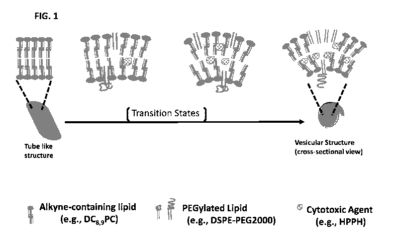
FIG. 1 is a schematic illustration of a proposed model for how a binary lipid
bilayer can be formed
between a PEGylated lipid and a non-bilayer-forming alkyne-containing
phosphocholine lipid.
FIG. 2 is a schematic diagram illustrating one embodiment of a method for
using the disclosed
vesicles to treat a subject having a tumor by injecting the vesicles followed
by targeted delivery of light of a
desired wavelength to the external surface of the subject's skin.
FIGS. 3A and 3B provide chemical structures of 2[1-hexyloxyethy11-2-devinyl
pyropheophorbide-a
(HPPH), (17S,18S)-18-(2-carboxyethyl)-20-(carboxymethyl)-12-ethenyl-7-ethyl-
3,8,13,17-tetramethyl-
17,18,22,23-tetrahydroporphyrin-2-carboxylic acid (or "chlorin e6" or "Ce6"),
1,1'-dioctadecy1-3,3,31,31-
tetramethylindotricarbocyanine iodide (DiR), and Camptothecin (FIG. 3A); 1,2-
bis(tricosa-10,12-diynoy1)-
sn-glycero-3-phosphocholine (DC8,9PC), dipalmitoylphosphatidylcholine (DPPC),
distearoylphosphatidylethanolamine-polyethylene glycol (DSPE-PEG), and 1,2-
bis(10,12-tricosadiynoy1)-
sn-glycero-3-phosphoethanolamine (DC8,9PE) (FIG. 3B).
FIG. 4 is a bar graph showing the effect on UV-triggered photocrosslinking of
DC8,9PC by including
DSPE-PEG2000 in lipid formulations wherein various vesicle embodiments (see
Table 1) were placed in a
96-well plate and exposed to UV light (254 nm) for the indicated time periods
at room temperature and at
the end of each incubation, absorbance was measured at 520 nm to assess
photocrosslinking.
FIG. 5 shows results obtained from using a centrifugation technique disclosed
herein to remove
unincorporated HPPH from HPPH-embedded vesicles, wherein HPPH-loaded vesicles
were placed in
- 2 -
CA 03106008 2021-01-07
WO 2020/014522
PCT/US2019/041464
microcentrifuge tubes and centrifugations were carried out at 6,000 rpm (-3000
RCF) for 30 minutes at 20-
25 C using a fixed-angle rotor centrifuge, supernatants containing the vesicle-
incorporated HPPH were
collected, and any unincorporated HPPH was sedimented in the pellet fraction.
FIG. 6 is a schematic illustration of the centrifugation technique used to
generate the data shown by
FIG. 5.
FIGS. 7A and 7B shows results from analyzing HPPH concentration in exemplary
vesicle
embodiments, wherein HPPH was included at various weight ratios for a known
lipid concentration
(typically 5 or 10 mg lipids were used) and vesicles were prepared by
sonication and unincorporated HPPH
were removed by centrifugation; FIG. 7A shows results of the amount of vesicle-
associated HPPH for the
lipid:HPPH ratios that were examined and FIG. 7B shows the percent of HPPH
encapsulated vesicles for the
different lipid:HPPH ratios examined.
FIG. 8 shows results obtained from analyzing HPPH incorporation in a vesicle
embodiment
disclosed herein and further illustrates that HPPH's PDT efficiency is not
impaired when incorporated in the
vesicle's binary lipid bilayer; to generate these results, vesicle-formulated
HPPH embodiments and
equivalent amounts of HPPH suspended in buffer were treated with a 661 nm
laser for five minutes and the
fluorescence of HPPH remaining after the laser treatments were assessed,
taking the fluorescence of
untreated samples as 100%.
FIGS. 9A-9C show optical imaging results obtained from analyzing the in vivo
tissue distribution of
vesicle-formulated HPPH in the liver, tumor, and skin of tumor-bearing mice;
CT-26 tumor-bearing BALB/c
mice were intravenously injected (groups of five) using either Tween 80-HPPH
(FIG. 9B) or Vesicle2o-
HPPH (FIGS. 9A); a Vesicle20 formulation without HPPH (FIG. 9C) was used to
obtain background signals
and images were collected at various time intervals and average radiant
efficiency of HPPH in tumor, liver
and skin was determined.
FIGS. 10A-10F are graphs of intensity (%) as a function of diameter (nm),
showing the average
diameter and polydispersity index of vesicle embodiments disclosed herein
having different concentrations
of a PEGylated lipid in the binary lipid bilayer; vesicles containing various
DSPE-PEG2000 amounts with
DC8,9PC were prepared by probe sonication, diluted in HBS at either 1:20 or
1:40 ratios (v/v), and dynamic
light scattering measurements were obtained.
FIGS. 11A and 11B are cryo-electron micrograms of a vesicle that does not
comprise an embedded
cytotoxic agent (Vesicle20; FIG. 11A) and a vesicle that does comprise an
embedded cytotoxic agent
(Vesicle20-HPPH; FIG. 11B).
FIGS. 12A and 12B are graphs of intensity (%) as a function of diameter (nm)
which show the
evolution of hydrodynamic size (as monitored as a function of time using
dynamic light scattering) of two
representative vesicle embodiments, Vesicleio-HPPH (FIG. 12A) and Vesicle20-
HPPH (FIG. 12B).
FIG. 13 is a graph showing the relative distribution of vesicles comprising
HPPH upon incubation
with fetal bovine serum and that illustrates that vesicle embodiments
described herein have high serum
stability as most of the detectable HPPH remains embedded within the binary
lipid bilayer and the
- 3 -
CA 03106008 2021-01-07
WO 2020/014522
PCT/US2019/041464
lipid:HPPH ratio remains unaffected in the vesicle fractions; Vesicle20-HPPH
vesicles were prepared at 1:0.1
lipid:HPPH ratios (w/w) and incubated with FBS at 37 C for 2 hours,
fractionated on a Sepharose CL-6B
and fractions were collected after which the lipid Pi, HPPH, and protein in
each fraction were determined;
the inset shows a magnified version for lipid and HPPH (mg/mi) in vesicle
fractions (10-18 ml fractions).
FIGS. 14A and 14B are graphs showing results from evaluating in vitro
cytotoxicity of CT-26 cells
by a vesicle embodiment, Vesicle20-HPPH, after 4 hours (FIG. 14A) and after 24
hours (FIG. 14B) of
incubation; cells on 96-well clusters were incubated with various doses of
Vesicle20-HPPH and laser
treatments were done either at 4 hours or 24 hours post incubations; laser
treatments doses used were either
0, 1, 2, or 4 J/cm2 and cell viability was done using MTT Assays; results are
presented as viability, taking
untreated control cells as 100% viable.
FIG. 15 provides in vivo images of mice (imaged using whole body DiR dorsal 2D-
multispectral
fluorescence imaging) that were injected with vesicle embodiments comprising
DiR and HPPH and which
establish that disclosed vesicle embodiments exhibit enhanced accumulation in
tumors as compared to a
liposome comprising DPPC and DC8,9PC lipids and a low amount of a PEGylated
lipid; to generate the
results, athymic nu/nu mice were injected in the flank with HT29 cells and
upon tumors reaching ¨100 mna3,
0.2 ml of the vesicles (containing 1 mg total lipid) were injected
intravenously and DiR imaging was
performed post 4 hours injections; "L" = liver; "T" = tumor.
FIGS. 16A and 16B are graphs showing results of tumor accumulation of
different vesicle
embodiments disclosed herein (as well as a comparative liposome comprising
DPPC and DC8,9PC lipids and
a low amount of a PEGylated lipid) and relative accumulation of the vesicle
embodiments in tumors versus
the liver; quantitation of the DiR fluorescence in the tumor or liver was done
taking average for the four
mice per group; FIG. 16A shows total radiation efficiency of DiR in tumors (
S.D. 4 animals) and FIG. 16B
shows relative ratios of DiR in tumors versus liver for each group; tumor to
liver ratios obtained for the
vesicles containing 4 mol% of the PEG-lipid were taken as 100 and values are
expressed as an average from
.. four animals ( S.D.).
FIGS. 17A-17C show results obtained from analyzing in vivo PDT response and
antitumor activity
of a vesicle embodiment described herein, Vesicle20-HPPH, in CT-26 bearing
BALB/c mice; to generate
these results, a comparison example, Tween 80-HPPH (FIG. 17A) and an exemplary
vesicle embodiment,
Vesicle20-HPPH (FIGS. 17B and 17C), were intravenously injected in tumor-
bearing mice at 0.47 mot
HPPH/kg body weight; laser treatments were done post 4 hours for Vesicle2o-
HPPH-injected animals and
post 24 hours injections of Tween 80-HPPH and tumor volumes were measured at
indicated days.
FIG. 18 is a Kaplan Meier graph comparing animal survival data obtained for
Vesicle20-HPPH (two
independent experiments) and Tween 80-HPPH.
FIGS. 19A-19H are graphs of intensity (%) as a function of diameter (nm),
showing the average
diameter and polydispersity index of certain formulations comprising DSPE-
PEG2000 and DC8,9PC.
- 4 -
CA 03106008 2021-01-07
WO 2020/014522
PCT/US2019/041464
FIGS. 20A and 20B are graphs of intensity (%) as a function of diameter (nm),
showing the average
diameter and polydispersity index of certain vesicle embodiments comprising
camptothecin embedded in the
bilayer after 1 day (FIG. 20A) and after 7 days (FIG. 20B).
FIGS. 21A and 21B are graphs of fluorescence as a function of camptothecin
concentration (uM)
.. showing results from analyzing camptothecin-induced cytotoxicity in human
breast cancer cells of
camptothecin-formulated vesicles (FIG. 21A) as compared to a free camptothecin
dose (FIG. 21B).
FIGS. 22A and 22B provides in vivo images of two different A549 tumor-bearing
mice that were
implanted with A549 cells subcutaneously and then injected with vesicle
embodiments comprising a binary
lipid bilayer without a cytotoxic agent and trace amounts of DiR; these
figures show that disclosed vesicle
.. embodiments are able to accumulate in tumors and do not exhibit long-term
liver toxicity; to generate the
results, mice were injected in with A549 cells and upon tumors reaching 100
mm3 to 200 mm3, 0.1 ml of the
vesicles (containing 1 mg total lipid) were injected intravenously and DiR
imaging was performed.
FIG. 23 provides images of various tissues of the mice illustrated in FIGS.
22A and 22B one week
after the mice were injected with the vesicle embodiments and wherein there
was a reduction in fluorescent
signal in the liver, indicating clearance of the vesicles; however, the
signals were relatively sustained in the
tumors and there was little to no fluorescence in the heart or lungs,
establishing that the vesicles exhibit
organ-specific accumulation in tumors, but do not accumulate in the heart or
lung and thus do not contribute
to toxicity in these organs.
FIGS. 24A-24C provide results obtained from analyzing vesicle embodiments
comprising DSPE-
PEG2000 and DC8,9PC and Ce6; FIGS. 24A and 24B are graphs of intensity (%) as
a function of diameter
(nm), showing the average diameter and polydispersity index of certain
formulations comprising DSPE-
PEG2000 and DC8,9PC and Ce6 after 1 day (FIG. 24A) and after 7 days (FIG. 24B)
and FIG. 24C shows
Ce6 can be incorporated into the vesicles.
FIGS. 25A and 25B are graphs of intensity (%) as a function of diameter (nm),
showing the average
diameter and polydispersity index of certain formulations comprising DSPE-
PEG350 and DC8,9PC (FIG.
25A) and DSPE-PEG1000 and DC8,9PC (FIG. 25B) after 1 day (top graphs of FIGS.
25A and 25B) and after
4 months (bottom graphs of FIGS. 25A and 25B).
FIGS. 26A-26D are graphs of intensity (%) as a function of diameter (nm),
showing the average
diameter and polydispersity index of certain formulations comprising DSPE-
PEG5000 and DC8,9PC at
different ratios and for different time periods; FIGS. 26A and 26B show
results of DSPE-PEG5000 and
DC8,9PC at a ratio of 90:10 DC8,9PC:DSPE-PEG5000 after 1 day (FIG. 26A) and
after 4 months (FIG. 26B)
and FIGS. 26C and 26D show results of DSPE-PEG5000 and DC8,9PC at a ratio of
99:1 DC8,9PC:DSPE-
PEG5000 after 1 day (FIG. 26C) and after 4 months (FIG. 26D).
FIGS. 27A-27C provide results obtained from analyzing vesicle embodiments
comprising (i) DSPE-
PEG2000, DC8,9PC, and DC8,9PE (FIGS. 27A and 27B) and (ii) DSPE-PEG2000 and
DC8,9PE (FIG. 27C)
and further including HPPH; FIGS. 27A and 27B are graphs of intensity (%) as a
function of diameter (nm),
showing the average diameter and polydispersity index of certain formulations
comprising DSPE-PEG2000,
- 5 -
CA 03106008 2021-01-07
WO 2020/014522
PCT/US2019/041464
DC8,9PC, and DC8,9PE and further including HPPH at different lipid:HPPH
ratios; FIG. 27A shows results
for embodiments comprising a (total) lipid-to-HPPH ratio of 20:1 and FIG. 27B
shows results for
embodiments comprising a (total) lipid-to-HPPH ratio of 100:1.
DETAILED DESCRIPTION
I. Overview of Terms and Abbreviations
The following explanations of terms and abbreviations are provided to better
describe the present
disclosure and to guide those of ordinary skill in the art in the practice of
the present disclosure. As used
herein, "comprising" means "including" and the singular forms "a" or "an" or
"the" include plural references
unless the context clearly dictates otherwise. The term "or" refers to a
single element of stated alternative
elements or a combination of two or more elements, unless the context clearly
indicates otherwise.
Unless explained otherwise, all technical and scientific terms used herein
have the same meaning as
commonly understood to one of ordinary skill in the art to which this
disclosure belongs. Although methods
and materials similar or equivalent to those described herein can be used in
the practice or testing of the
present disclosure, suitable methods and materials are described below. The
materials, methods, and
examples are illustrative only and not intended to be limiting. Other features
of the disclosure are apparent
from the following detailed description and the claims.
Unless otherwise indicated, all numbers expressing quantities of components,
molecular weights,
percentages, temperatures, times, and so forth, as used in the specification
or claims are to be understood as
being modified by the term "about." Accordingly, unless otherwise indicated,
implicitly or explicitly, the
numerical parameters set forth are approximations that may depend on the
desired properties sought and/or
limits of detection under standard test conditions/methods. When directly and
explicitly distinguishing
embodiments from discussed prior art, the embodiment numbers are not
approximates unless the word
"about" is recited.
All chemical compounds include either or both of the (+) and (-)
stereoisomers, as well as any
geometric isomers, such as Z and E isomers and cis and trans isomers. Other
chemistry terms herein are
used according to conventional usage in the art, as exemplified by Hawley's
Condensed Chemical
Dictionary, Richard J. Lewis, Sr. (ed.), published by John Wiley & Sons, Inc.,
1997 (ISBN 0-471-29205-2).
A. Explanation of Terms
The following explanations of terms are provided to better delineate the
subject matter of the present
disclosure and to guide those of ordinary skill in the art in its practice.
Administering: Administration by any route, for example oral, topical,
intravenous, intraperitoneal,
intramuscular, intralesional, intranasal, or subcutaneous administration,
release from a suppository, or the
implantation of a slow-release device (e.g., a mini-osmotic pump) to the
subject. "Parenteral" administration
is by any route other than through the alimentary tract and includes
intravascular administration directly into
a blood vessel, for example by intravenous or intra-arterial administration.
- 6 -
CA 03106008 2021-01-07
WO 2020/014522
PCT/US2019/041464
Alkyne-Containing Phosphocholine Lipid: A lipid comprising a phosphocholine
group (i.e.,
(CH3)3N (CH2)2-0P03--) and at least one alkyne moiety within a carbon chain of
the lipid, with some
embodiments comprising more than one alkyne moiety, which can be in the same
carbon chain or different
carbon chains of the lipid. In some embodiments, the alkyne-containing
phosphocholine lipid can comprise
at least one diyne moiety. An exemplary alkyne-containing phosphocholine lipid
is DC8,9PC.
Alkyne-Containing Phosphoethanolamine Lipid: A lipid comprising a
phosphoethanolamine
group (i.e., H3l\l (CH2)2-0P03--) and at least one alkyne moiety within a
carbon chain of the lipid, with some
embodiments comprising more than one alkyne moiety, which can be in the same
carbon chain or different
carbon chains of the lipid. In some embodiments, the alkyne-containing
phosphoethanolamine lipid can
comprise at least one diyne moiety. An exemplary alkyne-containing
phosphocholine lipid is DC8,9PE.
Alkyne-Containing Phospholipid: A lipid comprising a phosphate group (e.g., a
phosphocholine
or phosphoethanolamine) and at least one alkyne moiety within a carbon chain
of the lipid, with some
embodiments comprising more than one alkyne moiety, which can be in the same
carbon chain or different
carbon chains of the lipid. In some embodiments, the alkyne-containing
phospholipid can comprise at least
one diyne moiety. Exemplary alkyne-containing phospholipids include alkyne-
containing phosphocholine
lipids and alkyne-containing phosphoethanolamine lipids.
Bilayer: A component of a vesicle that defines a core of the vesicle and that
comprises at least two
lipid layers, wherein each layer comprises at least one non-bilayer-forming
lipid (e.g., an alkyne-containing
phospholipid (or a combination of alkyne-containing phospholipids)) and a
PEGylated lipid.
Carrier: An excipient that serves as a component capable of delivering a
compound described
herein. In some embodiments, a carrier can be a suspension aid, solubilizing
aid, or aerosolization aid. In
general, the nature of the carrier will depend on the particular mode of
administration being employed. For
instance, parenteral formulations usually comprise injectable fluids that
include pharmaceutically and
physiologically acceptable fluids such as water, physiological saline,
balanced salt solutions, aqueous
dextrose, glycerol or the like as a vehicle. In some examples, the
pharmaceutically acceptable carrier may
be sterile to be suitable for administration to a subject (for example, by
parenteral, intramuscular, or
subcutaneous injection). In addition to biologically-neutral carriers,
pharmaceutical formulations to be
administered can contain minor amounts of non-toxic auxiliary substances, such
as wetting or emulsifying
agents, preservatives, and pH buffering agents and the like, for example
sodium acetate or sorbitan
monolaurate.
Lipid: A term for fats and fat-derived materials. In some embodiments, lipids
include esters of
fatty acids (simple lipids, such as fats, sterols, waxes, and triglycerides)
or closely related substances
(compound lipids, such as phospholipids). Lipids generally are insoluble in
water but soluble in organic
solvents.
Near-Infrared (NIR): A region of the electromagnetic spectrum between the
visible region and the
infrared region. There is no set definition for the boundaries of the near-
infrared region, but definitions
- 7 -
CA 03106008 2021-01-07
WO 2020/014522
PCT/US2019/041464
include the wavelength ranges from 650-2500 nm, 750-2500 nm, 780-2500 nm, 800-
2500 nm, 700-1400 nm,
or 780-3000 nm. As used herein, NIR refers to the wavelength region of 650-
2500 nm.
Non-Bilayer-Forming Lipid: A lipid that is not, without structural
modification or combination
with a PEGylated lipid, capable of aggregating and forming a bilayer on its
own. Examples of non-bilayer-
forming lipids include alkyne-containing phospholipids, such as alkyne-
containing phosphocholine lipids
(e.g., DC8,9PC), alkyne-containing phosphoethanolamine lipids (e.g., DC8,9PE),
and combinations thereof.
Nucleic Acid Molecule: Includes DNA and RNA. The DNA may be operably linked to
a
promoter and/or contained with an expression vector, such as a plasmid. The
DNA may be genomic (with
introns) or consist only of the intron-less cDNA coding sequence. In some
examples, the DNA sequence
may encode a therapeutic protein, such as an anti-tumor protein. In other
examples, the RNA sequence may
be an inhibitory RNA (iRNA) that inhibits gene expression. Examples include
microRNA (miRNA) and
small interfering RNA (siRNA).
PEGylation: With respect to vesicles, PEGylation refers to incorporating
surface-bound
polyethylene glycol (PEG) to protect vesicles from detection by the
reticuloendothelial system and to
increase blood circulation time of the vesicle. Polyethylene glycols (PEG) are
hydrophilic polymers
composed of repeating ethylene oxide subunits with two terminal hydroxyl
groups that can be chemically
activated. The general structure of PEG is: HO-(CH2CH20).-CH2CH2-0H, wherein n
can be 0 or higher,
such as 0 to 10,000 (or higher), or 1 to 7,500, or 1 to 5,000, or 1 to 3,000,
or 1 to 2,000, or 1 to 1,000. In
some embodiments, n is 350 to 10,000, such as 350 to 5,000, or 350 to 2,000,
or 350 to 1,000. PEG chains
can be linear or branched. PEG conjugation to a pharmaceutically or
biologically useful agent typically
involves activating the PEG by preparing a PEG derivative having functional
groups. The functional group
on PEG is chosen based on the reactive group of the molecule to be conjugated.
The molecular weight of
the PEGs is chosen to avoid rapid clearance by the liver as well as any toxic
effects.
PEGylated Lipid: A lipid comprising a polyethylene glycol (PEG) group
covalently bound to the
lipid, wherein the PEG group is bound directly to a functional group of the
lipid or indirectly to the lipid via
a linker or other functional group.
Pharmaceutical or Bioactive Agent: A molecule that is capable of providing a
therapeutic
(including diagnostic) effect. A bioactive agent has an effect on living
tissue. Examples include anti-cancer
agents, imaging agents, anti-inflammatory agents, and small interfering RNA
(siRNA) molecules.
Pharmaceutically Acceptable: The term "pharmaceutically acceptable" refers to
substance that
can be taken into a subject without significant adverse toxicological effects
on the subject.
Pharmaceutically Acceptable Excipient: A substance, other than an active
compound (e.g., a
compound described herein), that is included in a formulation of the active
compound. As used herein, an
excipient may be incorporated within particles of a pharmaceutical
formulation, or it may be physically
mixed with particles of a pharmaceutical formulation. An excipient also can be
in the form of a solution,
suspension, emulsion, or the like. An excipient can be used, for example, to
dilute an active agent and/or to
modify properties of a pharmaceutical formulation. Excipients can include, but
are not limited to,
- 8 -
CA 03106008 2021-01-07
WO 2020/014522
PCT/US2019/041464
antiadherents, binders, coatings, enteric coatings, disintegrants, flavorings,
sweeteners, colorants, lubricants,
glidants, sorbents, preservatives, adjuvants, carriers or vehicles. Excipients
may be starches and modified
starches, cellulose and cellulose derivatives, saccharides and their
derivatives such as disaccharides,
polysaccharides and sugar alcohols, protein, synthetic polymers, crosslinked
polymers, antioxidants, amino
acids or preservatives. Exemplary excipients include, but are not limited to,
magnesium stearate, stearic
acid, vegetable stearin, sucrose, lactose, starches, hydroxypropyl cellulose,
hydroxypropyl methylcellulose,
xylitol, sorbitol, maltitol, gelatin, polyvinylpyrrolidone (PVP), polyethylene
glycol (PEG), tocopheryl
polyethylene glycol 1000 succinate (also known as vitamin E TPGS, or TPGS),
carboxy methyl cellulose,
dipalmitoyl phosphatidyl choline (DPPC), vitamin A, vitamin E, vitamin C,
retinyl palmitate, selenium,
.. cysteine, methionine, citric acid, sodium citrate, methyl paraben, propyl
paraben, sugar, silica, talc,
magnesium carbonate, sodium starch glycolate, tartrazine, aspartame,
benzalkonium chloride, sesame oil,
propyl gallate, sodium metabisulphite or lanolin.
Phospholipid: A lipid that includes a phosphate group. The phospholipid
comprises a glycerol
bound to the phosphate group and two fatty acid chains.
Photoactivatable / Photo-triggerable: Capable of being activated (e.g.,
converted from an inert
form to an active form) by light energy.
Photoactivation / Photo-triggering: Activating a vesicle using light energy.
As used herein,
activating can comprise promoting reactive oxygen species formation from a
cytotoxic agent disclosed
herein and/or destabilizing a vesicle's binary lipid bilayer wall so that at
least a portion of a cytotoxic agent
embedded within the vesicle's binary lipid bilayer is released. h) some
embodiments, photoactivation occurs
upon exposure of the vesicle to, for example, targeted application of light of
a selected wavelength, intensity,
and/or surface area, to a pre-selected target area.
Photosensitizer: A molecular or atomic species that initiates a photochemical
reaction. The term
"photosensitizer" also refers to a substance that sensitizes an organism,
cell, or tissue to light.
Photosensitizers may be used, for example, in photodynamic therapy for
treatment of cancer. The
photosensitizer absorbs light of a particular wavelength or wavelength range
and becomes excited. The
excited photosensitizer transfers energy to nearby molecules. h) photodynamic
therapy, the photosensitizer
may be taken up by a cancer cell. Upon light absorption, the photosensitizer
transfers energy to oxygen
present within the cell, thereby producing reactive oxygen species (ROS) which
are toxic to cancer cells.
Subject: A mammal and/or other animal, such as humans, companion animals
(e.g., dogs, cats,
rabbits, etc.), utility animals, feed animals and the like; thus, disclosed
methods are applicable to both human
therapy and veterinary applications.
Therapeutically Effective Amount: A quantity or concentration of a specified
compound or
composition sufficient to achieve a desired effect in a subject being treated.
For example, this may be the
.. amount of a vesicle as disclosed herein, or pharmaceutical composition
comprising the vesicle, necessary to
cause tumor cell death or inhibition, thereby eliminating a tumor, reducing
the size of a tumor, and/or
inhibiting tumor growth in a subject. Ideally, a therapeutically effective
amount of a compound or
- 9 -
CA 03106008 2021-01-07
WO 2020/014522
PCT/US2019/041464
composition is an amount sufficient to reduce the desired effect without
substantial cytotoxic effect on
non-tumor cells. However, the therapeutically effective amount of the vesicle
or composition will be
dependent on the subject being treated, the size and characteristics of the
tumor, and the manner of
administration of the therapeutic composition.
Treating/Treatment: Treatment of a disease or condition of interest in a
subject, particularly a
human or mammal having the disease or condition of interest or that may or may
not be prone to developing
the disease or condition, and includes by way of example, and without
limitation:
(i) prophylactic administration to prevent the disease or condition from
occurring in a subject,
or to ameliorate symptoms associated with the condition if required in
particular, when such subject is
predisposed to the condition but has not yet been diagnosed as having it;
(ii) inhibiting the disease or condition, for example, arresting or slowing
its development;
(iii) relieving the disease or condition, for example, causing regression
of the disease or
condition or a symptom thereof; or
(iv) stabilizing the disease or condition.
As used herein, the terms "disease" and "condition" can be used
interchangeably or can be different
in that the particular malady or condition may not have a known causative
agent (so that etiology has not yet
been determined) and it is therefore not yet recognized as a disease but only
as an undesirable condition or
syndrome, where a more or less specific set of symptoms have been identified
by clinicians.
Vesicle: A structural component comprising a lipid bilayer that forms and
encloses a cavity,
wherein the cavity does not comprise a core material such as core materials
found in nanoparticles (e.g., CaP
cores, liquid metal cores, and the like). Instead, the cavity within the
vesicle is a closed internal space.
Vesicles may be characterized by membrane type. Unilamellar vesicles have a
single membrane.
Oligolamellar vesicles and multilamellar vesicles have multiple, usually
concentric, membrane layers and
are typically larger than 0.1 pm. Vesicles with several nonconcentric
membranes, i.e., several small vesicles
contained within a larger vesicle, are termed multivesicular vesicles. In
particular disclosed embodiments,
the vesicles embodiments of the present disclosure are "unilamellar," and thus
have a single binary lipid
bilayer membrane.
Z-average Size: An average size determined by analyzing dynamic light
scattering data using the
technique of cumulants; also referred to as the `cumulants mean' or the
'harmonic intensity averaged particle
diameter' (ISO 22412).
B. Abbreviations
DC8,9PC: 1,2 bis (tricosa-10, 12-diynoy1)-sn-glycero-3-phosphocholine
DC8,9PE: 1,2-bis(10,12-tricosadiynoy1)-sn-glycero-3-phosphoethanolamine
DiR: 1,1'-dioctadecy1-3,3,31,31-tetramethylindotricarbocyanine iodide
DMEM: Dulbecco's Modified Eagle Medium (supplemented with 10% (v/v) heat-
inactivated FBS
(fetal bovine serum), 100 i.u./m1 penicillin and 100 ug/mL streptomycin)
- 10 -
CA 03106008 2021-01-07
WO 2020/014522
PCT/US2019/041464
DPPC: 1,2-dipalmitoyl-sn-glycero-3-phosphocholine
DSPE-PEG2000 (18:0 PEG2 PE): 1,2-Distearoyl-sn-Glycero-3 Phosphoethanolamine-N-
[Methoxy(Polyethylene glycol)-20001 (Ammonium Salt)
HBS: HEPES buffer, 10 mM HEPES, 140 mM NaCl (pH 7.2-7.5)
HPPH: 2- [1-hexyloxyethy11-2-devinyl pyropheophorbide-a
PBS: Phosphate buffered saline (2.66 mM KC1, 1.47 mM KH2PO4, 138 mM NaCl, 8.06
mM
Na2HPO4-7H20 (pH 7.1))
PDT: photodynamic therapy
PI: polydispersity index
Introduction
Clinical utility of anti-cancer drugs is often limited due their poor
solubility, reduced bioavailability,
and non-specific toxicity. These limitations can be alleviated by developing
suitable carriers for transport of
these drugs to desired site(s). Some previously investigated platforms in
cancer nanomedicine include lipid-
based nanocarriers; however, conventional liposomes are limited in their
ability to specifically accumulate in
tumors and avoid being taken up by the mononuclear phagocytic system (MPS).
By introducing PEGylated lipids into a liposome, it is possible to create
"stealth" liposomes that can
partially reduce MPS uptake; however, large fractions of such liposomes are
still taken up by the MPS.
Furthermore, the degree and extent of PEGylated lipid incorporation into
liposomes is often limited due to
their structural constraints. Typically, PEG lipid concentrations that can be
efficiently incorporated into
liposomes are limited by such structural constraints and such liposomes
typically require using bilayer-
forming lipids, such as 1,2-dioleoyl-sn-glycero-3-phosphocholine (or dioleoyl
phosphatidylcholine,
"DOPC").
The present disclosure describes embodiments of a novel vesicle that comprises
a binary lipid
bilayer comprising an alkyne-containing phospholipid and a PEGylated lipid and
that further comprises a
cytotoxic agent embedded in the lipid layer. In some embodiments, the alkyne-
containing phospholipid is
an alkyne-containing phosphocholine lipid, an alkyne-containing
phosphoethanolamine lipid, or a mixture of
the alkyne-containing phosphocholine lipid and the alkyne-containing
phosphoethanolamine lipid (such that
both types of phospholipids are included in the binary lipid bilayer). The
disclosed vesicle embodiments do
not require using conventional phospholipids that typically are used in
liposome delivery systems, such as
phosphatidyl choline lipids, and instead only use two different lipids,
reducing the complexity and cost
associated with making the vesicles. The disclosed vesicle embodiments can
accommodate impressively
high amounts of the PEGylated lipid, while also enabling loading of a
cytotoxic agent at high concentrations
within the binary lipid membrane. The disclosed vesicle embodiments also
maintain their stability upon
storage at ambient temperatures and further accumulate in tumors at high
efficiency, and exhibit remarkably
high tumor care, with no recurrence.
- 11 -
CA 03106008 2021-01-07
WO 2020/014522
PCT/US2019/041464
III. Vesicle and Composition Embodiments
Disclosed herein are embodiments of a vesicle that comprises a binary lipid
bilayer comprising an
alkyne-containing phospholipid and a PEGylated lipid and a cytotoxic agent
embedded in the lipid layer. In
some embodiments, the alkyne-containing phospholipid is an alkyne-containing
phosphocholine lipid, an
alkyne-containing phosphoethanolamine lipid, or a mixture of the alkyne-
containing phosphocholine lipid
and the alkyne-containing phosphoethanolamine lipid. In particular
embodiments, the alkyne-containing
phospholipid is an alkyne-containing phosphocholine lipid or is a mixture of
the alkyne-containing
phosphocholine lipid and an alkyne-containing phosphoethanolamine lipid. In
some embodiments, the
binary lipid bilayer can comprise a plurality (e.g., two or more) of cytotoxic
agents.
In particular disclosed embodiments, the vesicle can comprise (i) a binary
lipid bilayer comprising
an alkyne-containing phospholipid and a PEGylated lipid; and (ii) a cytotoxic
agent embedded in the binary
lipid bilayer, wherein the binary lipid bilayer is free of, or does not
comprise, a lipid other than the alkyne-
containing phospholipid or the PEGylated lipid. In an independent embodiments,
an alkyne-containing
phosphoethanolamine lipid is not what is referred to herein as "a lipid other
than the alkyne-containing
phospholipid or the PEGylated lipid."
In some embodiments, the vesicle can comprise (i) a binary lipid bilayer
comprising an alkyne-
containing phosphocholine lipid and the PEGylated lipid; and (ii) a cytotoxic
agent embedded in the binary
lipid bilayer, wherein the binary lipid bilayer is free of, or does not
comprise, a lipid other than the alkyne-
containing phosphocholine lipid or the PEGylated lipid. In yet additional
embodiments, the vesicle can
comprise (i) a binary lipid bilayer comprising an alkyne-containing
phosphocholine lipid, an alkyne-
containing phosphoethanolamine lipid, and the PEGylated lipid; and (ii) a
cytotoxic agent embedded in the
binary lipid bilayer, wherein the binary lipid bilayer is free of, or does not
comprise, a lipid other than the
alkyne-containing phosphocholine lipid, the alkyne-containing
phosphoethanolamine lipid, or the PEGylated
lipid. Lipids other than the alkyne-containing phosphocholine lipid, the
alkyne-containing
phosphoethanolamine lipid, or the PEGylated lipid can include
phosphatidylcholine lipids (such as
dipalmitoylphosphatidylcholine, or "DPPC"), non-PEGylated DSPE, cholesterol, a
plasmalogen, DPPE-
DVBA, bis-azo PC, bis-sorbyl phosphatidylcholine (or "bis-SorbPC"), and the
like. In some embodiments,
the vesicle can consist essentially of (i) a binary lipid bilayer made of the
alkyne-containing phospholipid
(e.g., an alkyne-containing phosphocholine lipid and/or an alkyne-containing
phosphoethanolamine lipid)
and the PEGylated lipid; and (ii) a cytotoxic agent embedded in the binary
lipid bilayer. In such
embodiments, the vesicle is free of a lipid other than the alkyne-containing
phospholipid or the PEGylated
lipid and any components that would deleteriously affect the ability of the
vesicle to perform its desired
function, such as agents or compounds that would disrupt the vesicle's shape
and/or stability. In yet
additional embodiments, the vesicle can consist of the alkyne-containing
phospholipid (e.g., an alkyne-
containing phosphocholine lipid and/or an alkyne-containing
phosphoethanolamine lipid), the PEGylated
lipid, and the cytotoxic agent. In yet additional embodiments, the vesicle can
comprise (i) the binary lipid
bilayer, which consists of the alkyne-containing phospholipid (e.g., an alkyne-
containing phosphocholine
- 12 -
CA 03106008 2021-01-07
WO 2020/014522
PCT/US2019/041464
lipid and/or an alkyne-containing phosphoethanolamine lipid) and the PEGylated
lipid; and (ii) the cytotoxic
agent. For certain imaging purposes, e.g., bio-distribution studies, trace
amounts of a lipid probe (e.g., 1,1' -
dioctadecyltetramethyl indotricarbocyanine iodide (DiR)) may be included in
the vesicles. This component
does not deleteriously affect the performance of the vesicle and it is not a
necessary component of the
vesicle embodiments
In particular disclosed embodiments, the alkyne-containing phospholipid is a
non-bilayer-forming
lipid that does not, on its own, form a nanostructure in aqueous solution that
is suitable for drug delivery. In
some embodiments, the non-bilayer-forming lipid, alone, forms a tubule-like
morphology. For example, see
FIG. 1, which illustrates the tubule-like structure of an exemplary alkyne-
containing phosphocholine lipid,
DC8,9PC. Other exemplary alkyne-containing phospholipids include, but are not
limited to, alkyne-
containing phosphoethanolamine lipids, such as DC8,9PE and the like.
The binary lipid bilayer can comprise from 80 mol% (or less, such as 75 mol%)
to 97 mol%, such as
85 mol% to 95 mol%, or 85 mol% to 90 mol% of the alkyne-containing
phospholipid (or a combination of
such phospholipids). In particular disclosed embodiments, the alkyne-
containing phospholipid (or
combination of such phospholipids) is present at 80 mol%, 85 mol%, or 90 mol%.
In embodiments
comprising a mixture of an alkyne-containing phosphocholine lipid and an
alkyne-containing
phosphoethanolamine lipid as the alkyne-containing phospholipid, the alkyne-
containing phosphocholine
lipid can be present at 45 mol% to 85 mol%, such as 50 mol% to 80 mol%, or 50
mol% to 75 mol%, or 50
mol% to 70 mol%, or 50 mol% to 65 mol%; and the alkyne-containing
phosphoethanolamine lipid can be
present at 5 mol% to 45 mol%, such as 5 mol% to 40 mol%, or 5 mol% to 35 mol%,
or 5 mol% to 30 mol%,
or 5 mol% to 25 mol%, or 5 mol% to 20 mol%, or 5 mol% to 15 mol%, or 5 mol% to
10 mol%. In
representative embodiments of such mixtures, the alkyne-containing
phosphocholine lipid can be present at
65 mol% and the alkyne-containing phosphoethanolamine lipid can be present at
25 mol%. In yet additional
embodiments, the alkyne-containing phosphocholine lipid can be present at 45
mol% and the alkyne-
containing phosphoethanolamine lipid can be present at 45 mol%.
The non-bilayer-forming lipid is combined with a PEGylated lipid to form a
vesicle structure, as
illustrated schematically in FIG. 1. The PEGylated lipid can be selected from
any PEGylated lipid that is
suitable for therapeutic methods, including administration to a subject, and
also that does not have too high
of a hydrophilicity such that it will not accumulate in cells as desired. In
particular disclosed embodiments,
the PEGylated lipid comprises a phosphoethanolamine lipid modified with a PEG
group. Such lipids can
include, but are not limited to, 1,2-distearoyl-sn-glycero-3-
phosphoethanolamine (DPSE) lipids comprising a
PEG group having a molecular weight ranging from greater than 350 Da to 10,000
Da, such as 350 Da to
5,000 Da (or higher), or 500 Da to 5,000 Da. In some embodiments, the PEG
group can have a molecular
weight ranging from 1,000 Da to 5,000 Da, or 1,000 Da to 4,000 Da, or 1,000 Da
to 3,000 Da, or 1,000 Da
to 2,000 Da. In a representative embodiment, DPSE-PEG2000 is used as the
PEGylated lipid. Other
exemplary PEGylated lipids include, but are not limited to, cholesterol-
PEG600, DPSE-PEG1000, DPSE-
PEG5000, and the like.
- 13 -
CA 03106008 2021-01-07
WO 2020/014522
PCT/US2019/041464
The binary lipid bilayer can comprise 3 mol% to 20 mol% or higher (e.g., 25
mol%), and in
particular embodiments can comprise greater than 6 mol% to 20 mol%, such as 8
mol% to 15 mol%, or 10
mol% to 15 mol% of the PEGylated lipid. In particular disclosed embodiments,
the PEGylated lipid can be
present in an amount of 10 mol%, 15 mol%, or 20 mol%. In an independent
embodiment where the
PEGylated lipid is DSPE-PEG2000, the PEGylated lipid is used in an amount
greater than 6 mol%.
The disclosed alkyne-containing phospholipid and the PEGylated lipid interact
to form a vesicle
structure that defines an inner cavity (FIG. 1). Embodiments of the disclosed
vesicles have a diameter
ranging from 50 nm to 200 nm, such as from 60 nm to 150 nm, or 65 nm to 100
nm. In some embodiments,
the disclosed vesicles can have a PI ranging from 0.2 to 0.3.
One or more cytotoxic agents can be embedded within the bilayer formed by the
alkyne-containing
phospholipid and the PEGylated lipid. In some embodiments, the cytotoxic agent
is a hydrophobic
compound, such as a tetrapyrrollic compound or a camptothecin. Exemplary
tetrapyrrollic compounds
include, but are not limited to, HPPH or tetrapyrrollic analogs thereof, such
as amino diethyl analogs,
aminohexane analogs, and other such analogs as disclosed by WO 2012/006009,
the relevant portion of
which is incorporated herein by reference; chlorin e6 (or "Ce6"); (3S,4S)-9-
Etheny1-14-ethy1-21-
(methoxycarbony1)-4,8,13,18-tetramethy1-20-oxo-3-phorbinepropanoic acid
("Pheophorbide a"); 3,3',3",3"-
(2,3-dihydroporphyrin-5,10,15,20-tetrayl)tetraphenol (or "Temoporfn"), and 3-
[(23S,24R)-14-etheny1-5-(3-
methoxy-3-oxopropy1)-22,23-bis(methoxycarbony1)-4,10,15,24-tetramethyl-
25,26,27,28-
tetraazahexacyclo [16 .6.1.13,6.18,11.113,16.019,241octacosa-
1,3,5,7,9,11(27),12,14,16,18(25),19,21-
dodecaen-9-yl]propanoic acid (or "Verteporfin"). HPPH is a lipophilic compound
with a log P of 5.6 at
physiological pH, a large molar extinction in the near-infrared region, i.e.,
c = 47,500 M-1 cm-1 at 665 nm,
and a singlet oxygen yield of 0.48. HPPH also has anti-cancer properties, and
has been used in PDT, e.g.,
for treatment of esophageal cancer and non-small cell lung cancer. In some
embodiments, activated HPPH
exerts its therapeutic effect through generating reactive oxygen species
(e.g., singlet oxygen) upon
photoactivation. Chlorin e6 is another exemplary PDT compound that can be used
in embodiments
disclosed herein and, like HPPH, can be activated to exert a therapeutic
effect. In some embodiments, the
vesicle can comprise a camptothecin. Camptothecins are hydrophobic lactone
drugs that exhibit
chemotherapeutic activity. Exemplary camptothecins include, but are not
limited to, camptothecin, silatecan
7-t-butyldimethylsily1-10-hydroxycamptothecin (DB-67), 7-ethy1-10-hydroxy-
20(S)-camptothecin (SN-38),
topotecan, irinotecan, 9-nitro-camptothecin, lurtotecan, exatecan, gimatecan,
and karenitecin. In additional
embodiments, the cytotoxic agent can be selected from paclitaxel,
daunorubicin, methotrexate, vincristine,
etoposide, sorafenib, erlotinib, imatinib, or any combination thereof. Any
combination and any number of
cytotoxic agents can be used in the vesicles.
The cytotoxic agent (or combination of cytotoxic agents) can be embedded in
the binary lipid
bilayer at high concentrations, such as amounts ranging from 0.05 to 0.5 mg
cytotoxic agent/mg lipid, such
as 0.075 to 0.5 mg cytotoxic agent/mg lipid, or 0.1 to 0.5 mg cytotoxic
agent/mg lipid, or 0.25 to 0.5 mg
cytotoxic agent/mg lipid. In embodiments comprising a plurality of cytotoxic
agents, the total amount of the
- 14 -
CA 03106008 2021-01-07
WO 2020/014522
PCT/US2019/041464
cytotoxic agents present can range from 0.05 to 0.5 mg cytotoxic agent/mg
lipid, such as 0.075 to 0.5 mg
cytotoxic agent/mg lipid, or 0.1 to 0.5 mg cytotoxic agent/mg lipid, or 0.25
to 0.5 mg cytotoxic agent/mg
lipid. In some embodiments, the cytotoxic agent can be present in an amount
that provides a ratio of total
lipid content to cytotoxic agent ("lipid:cytotoxic agent") ranging from 5:1
lipid:cytotoxic agent to 100:1
lipid:cytotoxic agent, such as 5:1 lipid:cytotoxic agent to 20:1
lipid:cytotoxic agent. In some embodiments,
ratios of 5:1 lipid:cytotoxic agent, 10:1 lipid:cytotoxic agent, or 20:1
lipid:cytotoxic agent are used.
This disclosure includes pharmaceutical compositions comprising at least one
vesicle described
herein. Some embodiments of the disclosed pharmaceutical compositions, when
irradiated with near-
infrared energy, are capable of killing or inhibiting tumor cells, thereby
eliminating a tumor, reducing tumor
size, and/or inhibiting tumor growth. The pharmaceutical compositions may be
applied to tumor cells in
vitro, or the pharmaceutical composition may be formulated for use in human
and/or veterinary medicine
and may be applied to tumor cells in vivo by administering a therapeutically
or diagnostically effective
amount of the pharmaceutical composition to a subject.
Some embodiments of the pharmaceutical compositions include a pharmaceutically
acceptable
carrier and at least one active ingredient. Useful pharmaceutically acceptable
carriers and excipients are
known in the art. Active ingredients may comprise, for example, at least one
vesicle embodiment as
described herein, or any combination of vesicles as described herein (e.g., a
combination of vesicles
comprising one particular type of cytotoxic agent and vesicles comprising a
different type of cytotoxic
agent). In addition, other medicinal or pharmaceutical agents, for example,
with similar, related or
complementary effects on the affliction being treated, may be included as
active ingredients in
pharmaceutical compositions. These agents include, but are not limited to,
pharmaceutical compounds,
chemotherapeutic agents, cytokines, and anti-angiogenic agents.
The pharmaceutical compositions comprising one or more vesicles may be
formulated in a variety of
ways depending, for example, on the mode of administration and/or on the
location and type of disease to be
treated. For example, parenteral formulations may comprise injectable fluids
that are pharmaceutically and
physiologically acceptable fluid vehicles such as water, physiological saline,
other balanced salt solutions,
aqueous dextrose, glycerol or the like. Excipients may include, for example,
nonionic solubilizers, such as
cremophor, or proteins, such as human serum albumin or plasma preparations. If
desired, the
pharmaceutical composition to be administered may also contain non-toxic
auxiliary substances, such as
wetting or emulsifying agents, preservatives, and pH buffering agents and the
like, for example, sodium
acetate or sorbitan monolaurate.
The dosage form of the pharmaceutical composition will be determined by the
mode of
administration chosen. Embodiments of the disclosed pharmaceutical
compositions may take a form
suitable for virtually any mode of administration, including, for example,
topical, ocular, oral, buccal,
systemic, nasal, injection, transdermal, rectal, vaginal, etc., or a form
suitable for administration by
inhalation or insufflation.
- 15 -
CA 03106008 2021-01-07
WO 2020/014522
PCT/US2019/041464
Topical preparations may include eye drops, gels, ointments, creams,
suspensions, sprays and the
like as are well-known in the art.
Useful injectable preparations include sterile suspensions, solutions or
emulsions of the active
compound(s) in aqueous or oily vehicles. The compositions may also contain
formulating agents, such as
suspending, stabilizing and/or dispersing agent. The formulations for
injection may be presented in unit
dosage form, e.g., in ampules or in multidose containers, and may contain
added preservatives. The
composition may take such forms as suspension, solutions or emulsions in oily
or aqueous vehicles, and may
contain formulatory agents such as suspending, stabilizing and/or dispersing
agents. For example, parenteral
administration may be done by bolus injection or continuous infusion.
Alternatively, the active ingredient
may be in powder form for reconstitution with a suitable vehicle, e.g. sterile
water, before use.
Systemic formulations include those designed for administration by injection,
e.g., subcutaneous,
intravenous, intramuscular, intrathecal or intraperitoneal injection, as well
as those designed for transdermal,
transmucosal, oral or pulmonary administration.
Oral formulations may be liquid (e.g., syrups, solutions or suspensions), or
solid (e.g., powder,
tablets, or capsules). Oral formulations may be coupled with targeting ligands
for crossing the endothelial
barrier. Some vesicle formulations may be dried, e.g., by spray-drying with a
disaccharide, to form
liposomal powders. Solid compositions prepared by conventional means with
pharmaceutically acceptable
excipients such as binding agents (e.g., pregelatinised maize starch,
polyvinylpyrrolidone or hydroxypropyl
methylcellulose); fillers (e.g., lactose, mannitol, microcrystalline cellulose
or calcium hydrogen phosphate);
lubricants (e.g., magnesium stearate, talc or silica); disintegrants (e.g.,
potato starch or sodium starch
glycolate); or wetting agents (e.g., sodium lauryl sulfate). The tablets may
be coated by methods well known
in the art with, for example, sugars, films or enteric coatings that mitigate
acid denaturation of the vesicle's
binary lipid bilayer. Actual methods of preparing such dosage forms are known,
or will be apparent, to
those skilled in the art.
Liquid preparations for oral administration may take the form of, for example,
elixirs, solutions,
syrups or suspensions. Such liquid preparations may be prepared by
conventional means with
pharmaceutically acceptable additives such as suspending agents (e.g.,
sorbitol syrup, cellulose derivatives
or hydrogenated edible fats); emulsifying agents (e.g., lecithin or acacia);
non-aqueous vehicles (e.g.,
almond oil, oily esters, ethyl alcohol, cremophoreTM or fractionated vegetable
oils); and preservatives (e.g.,
methyl or propyl-p-hydroxybenzoates or sorbic acid). The preparations may also
contain buffer salts,
preservatives, flavoring, coloring and sweetening agents as appropriate.
Preparations for oral administration
may be suitably formulated to give controlled release of the active compound,
as is well known.
For buccal administration, the compositions may take the form of tablets or
lozenges formulated in
conventional manner.
For rectal and vaginal routes of administration, the active compound(s) may be
formulated as
solutions (for retention enemas) suppositories or ointments containing
conventional suppository bases such
as cocoa butter or other glycerides.
- 16 -
CA 03106008 2021-01-07
WO 2020/014522
PCT/US2019/041464
For nasal administration or administration by inhalation or insufflation, the
active compound(s) can
be conveniently delivered in the form of an aerosol spray or mist from
pressurized packs or a nebulizer with
the use of a suitable propellant, e.g., dichlorodifluoromethane,
trichlorofluoromethane,
dichlorotetrafluoroethane, fluorocarbons, carbon dioxide or other suitable
gas. In the case of a pressurized
aerosol, the dosage unit may be determined by providing a valve to deliver a
metered amount.
For prolonged delivery, the vesicles can be formulated as a depot preparation
for administration by
implantation or intramuscular injection. Alternatively, transdermal delivery
systems manufactured as an
adhesive disc or patch which slowly releases the vesicle for percutaneous
absorption may be used. To this
end, permeation enhancers may be used to facilitate transdermal penetration of
the active compound(s).
Certain embodiments of the pharmaceutical compositions comprising vesicles as
described herein
may be formulated in unit dosage form suitable for individual administration
of precise dosages. The
pharmaceutical compositions may, if desired, be presented in a pack or
dispenser device which may contain
one or more unit dosage forms containing the vesicles. The pack may, for
example, comprise metal or
plastic foil, such as a blister pack. The pack or dispenser device may be
accompanied by instructions for
administration. The amount of vesicles administered will depend on the subject
being treated, the severity of
the affliction (e.g., the size, location, and characteristics of a tumor), and
the manner of administration, and
is known to those skilled in the art. Within these bounds, the formulation to
be administered will contain a
quantity of the vesicles disclosed herein in an amount effective to achieve
the desired effect in the subject
being treated.
Embodiments of the disclosed vesicles will generally be used in an amount
effective to achieve the
intended result, for example in an amount effective to treat or image a tumor.
The vesicles may be
administered therapeutically to achieve therapeutic benefit. By therapeutic
benefit is meant eradication or
amelioration of the underlying disorder being treated and/or eradication or
amelioration of one or more of
the symptoms associated with the underlying disorder such that the patient
reports an improvement in
feeling or condition, notwithstanding that the patient may still be afflicted
with the underlying disorder.
Therapeutic benefit also includes halting or slowing the progression of the
disease, regardless of whether
improvement is realized. In some embodiments, the vesicles are administered to
achieve diagnostic benefit.
Diagnostic benefit includes, for example, the ability to image target tissue
such as tumor tissue.
The amount administered will depend upon a variety of factors, including, for
example, the particular
indication being treated, the mode of administration, the severity of the
indication being treated, the age and
weight of the patient, the bioavailability of the particular bioactive agent
included in the cavity of the
vesicle, etc. Determination of an effective dosage is well within the
capabilities of those skilled in the art.
Effective dosages may be estimated initially from in vitro assays. For
example, an initial dosage may
be formulated to achieve a tumor tissue concentration of reactive oxygen
species produced by the cytotoxic
agent embedded within the vesicle's bilayer that is sufficient to cause tumor
cell necrosis as determined in
an in vitro assay. In additional embodiments, an initial dosage may be
formulated to achieve a tumor tissue
concentration of a released cytotoxic agent following vesicle disruption that
is sufficient to cause tumor cell
- 17 -
CA 03106008 2021-01-07
WO 2020/014522
PCT/US2019/041464
necrosis as determined in an in vitro assay. Calculating dosages to achieve
such concentrations is well
within the capabilities of skilled artisans. For guidance, the reader is
referred to Fingl & Woodbury,
"General Principles," In: Goodman and Gilman's The Pharmaceutical Basis of
Therapeutics, Chapter 1, pp. 1
46, latest edition, Pagamonon Press, and the references cited therein.
Initial dosages can also be estimated from in vivo data, such as animal
models. Animal models useful
for testing the efficacy of compounds to treat tumors are well-known in the
art. A person having ordinary
skill in the art, along with the benefit of the present disclosure, can adapt
such information to determine
dosages suitable for human administration.
Preferably, the vesicles will provide therapeutic or prophylactic benefit
without causing substantial
toxicity. Toxicity of the vesicles may be determined using standard
pharmaceutical procedures. The dose
ratio between toxic and therapeutic effect is the therapeutic index. Vesicles
that exhibit high therapeutic
indices are preferred.
Certain embodiments of the pharmaceutical methods and compositions include co-
administration of
the vesicle as described herein and a therapeutically effective amount of a
second agent other than the
vesicle. The vesicle and the second agent may be administered either
separately or together in a single
composition. The second agent may be, for example, an anti-tumor agent or an
angiogenesis inhibitor.
IV. Methods of Making Vesicle Embodiments
Disclosed herein are embodiments of a method for making the vesicle
embodiments of the present
disclosure. The method can comprise using a probe sonication method to produce
the vesicles. In some
embodiments, the alkyne-containing phospholipid is combined with the PEGylated
lipid in chloroform and
they are mixed. Different ratios of the lipids can be used as described above,
with exemplary ratios being
provided by Tables 1 and 2 in the Examples section of the present disclosure.
A desired amount of the cytotoxic agent is added to the lipid mixture as a
solution (e.g., a DMSO
solution) prior to making a lipid film. Any solvents are removed (e.g., under
nitrogen gas) and the resulting
lipid films can be stored and/or allowed to further dry under an inert
atmosphere. The dried lipid films are
re-suspended using a buffer (e.g., 1 ml HBS, pH=7.4). The lipid mixture is
vortexed and heated and then
sonicated using a probe sonicator in an ice bath. In particular disclosed
embodiments, the lipid mixture is
vortexed and heated at 45-50 C for 15-20 minutes and subjected to at least
five freeze-thaw cycles. In such
embodiments, a probe sonicator can be used, with particular embodiments using
5-10 cycles, with 1 minute
per cycle followed by 1 minute of rest. Specific examples of making exemplary
vesicles disclosed herein
are described in detail in the Examples section of the present disclosure.
V. Methods of Using Vesicle Embodiments
A. Photoactivation
Embodiments of the disclosed vesicles are photoactivated (e.g., the cytotoxic
agent is activated
and/or lipid conformations in the bilayer are modified to facilitate cytotoxic
agent release) by targeted
- 18 -
CA 03106008 2021-01-07
WO 2020/014522
PCT/US2019/041464
application of light having a desired wavelength, intensity, and/or surface
area to a pre-selected target area
for an effective period of time. The wavelength is selected within the near-
infrared range, e.g., from 650 nm
to 2500 nm. When photoactivatable cytotoxic agents, such as HPPH, are used,
the wavelength is selected
from 650-670 nm. Suitable light intensities may range from 1 mW to 500 mW
depending on the target site
.. and method of application. In some examples, a 90 mW, 660 nm laser was
used. Near-infrared light
sources can be obtained from commercial sources, including Thorlabs (Newton,
NJ), Laser Components,
USA (Hudson, NH), ProPhotonix (Salem, NH) and others.
In some embodiments, photoactivation is performed by externally applying light
to a targeted area.
NIR light is capable of penetrating transcutaneously into tissue to a depth of
several centimeters. In other
embodiments, photoactivation may be performed by internally applying light,
such as by using an endoscope
or a fiber optic catheter. Internal application may be used when the target
tissue, such as a tumor, is located
at a depth that is unsuitable for external light application. For example, an
endoscope may be used for light
delivery into the lungs, stomach, or bladder.
The surface area for light application is generally selected to include the
target tissue, e.g., a tumor
.. or portion of a tumor, or an area of skin external to the target tissue.
When targeted, externally applied light
is desired, the surface area can be controlled by using an appropriate light
applicator, such as a micro-lens, a
Fresnel lens, or a diffuser arrangement. For targeted, internally applied
light, a desired endoscope or fiber
optic catheter diameter can be used. In some applications, an indwelling
catheter filled with a light
scattering solution may be internally placed proximate the target tissue, and
an optical fiber light source may
be inserted into the catheter (see, e.g., Madsen et al., Lasers in Surgery and
Medicine 2001, 29, 406-412).
In some embodiments, photoactivation is performed for a period of time
effective to activate at least
a portion of the cytotoxic agent, such as HPPH or other such photoactivable
compounds, within the vesicle's
binary lipid bilayer wall, thereby releasing reactive oxygen species that can
act on the tumor cells. In yet
additional embodiments, photoactivation can be performed for a period of time
effective to activate a
.. conformational change and/or oxidative change in a lipid of the binary
lipid bilayer such that the binary lipid
bilayer wall is destabilized. This destabilization can result in releasing at
least a portion of an embedded
cytotoxic agent, such as a camptothecin. In some embodiments, the effective
period of time ranges from
several seconds to several minutes, e.g., from 30 seconds to 15 minutes. In
certain examples,
photoactivation was performed for 5-10 minutes.
In particular embodiments, HPPH is used as the cytotoxic agent. HPPH is
activated with near-
infrared light energy, such as NIR light having a wavelength of 650 nm to 680
nm. For example, HPPH can
be activated when irradiated for an effective period of time by a laser that
produces light having a
wavelength of 655 nm to 675 nm, e.g., a 660-nm laser. In certain embodiments,
HPPH is activated when
irradiated with a continuous wave (cw)-diode 600 nm laser source (90 mV) for
several seconds to several
minutes.
- 19 -
CA 03106008 2021-01-07
WO 2020/014522
PCT/US2019/041464
B. Applications
Embodiments of the disclosed vesicle are suitable for in vitro uses and/or in
vivo administration to a
subject. As described above, at least a portion of the embedded cytotoxic
agent is activated to promote
reactive oxygen species formation and/or to promote changes in vesicle
morphology so that another
embedded cytotoxic agent can be released upon irradiation with light (e.g.,
near-infrared light energy) for an
effective period of time.
In particular disclosed embodiments, the disclosed vesicle may be administered
to a subject
identified as having a condition that may be treated with a cytotoxic agent,
such as HPPH (or other
tetrapyrrollic compounds, such as chlorin e6) or a camptothecin compound. For
example, with reference to
FIG. 2, a subject 200 with a tumor may be treated with vesicle embodiments
comprising HPPH embedded in
the cavity. Administration of the disclosed vesicle to the subject may impair
growth of the tumor and/or
cause tumor regression. Because the disclosed vesicles have high amounts of a
PEGylated lipid, the vesicles
preferentially are taken up by and accumulate in the tumor 210. A
therapeutically effective amount of the
vesicles, or a pharmaceutical composition comprising the vesicles, is
administered to the subject by any
suitable means including, but not limited to, parenteral, intravenous,
subcutaneous, oral, rectal, vaginal, or
topical administration. In the example shown in FIG. 2, the vesicles 220 are
administered via intravenous
injection. A target portion of the subject subsequently is selectively
irradiated with NIR light energy of a
desired wavelength using an external light applicator 230 for an effective
period of time, such as from 1-15
minutes. The light applicator 230 applies the photoactivation energy to a
target area limited to the region of
the tumor 210, thereby selectively photoactivating the vesicles in and around
the tumor 210 and targeting
delivery of reactive oxygen species generated from the HPPH.
The embedded cytotoxic agent can inhibit tumor cell growth and/or kill tumor
cells, thereby
providing combination chemotherapy to the tumor site. Suitable tumor sites
include, but are not limited to,
the head, neck, skin, bladder, prostate, colon, and lung. Because the
cytotoxic agents and/or reactive oxygen
species generated by the cytotoxic agents are released directly at the tumor
site, the cytotoxic agent's
effectiveness may be increased and/or the cytotoxic agent's side effects may
be reduced compared to other
methods of non-targeted administration.
In a particular disclosed embodiments, colon-26 bearing BALB/c mice,
intravenously injected with
Vesicle20-HPPH showed superior PDT efficacy and animal survival (no tumor
recurrence up to 100 days) as
compared to a formulation currently used in clinical trials, namely Tween 80-
HPPH. Additionally and
advantageously, the vesicles exhibited stability for 60 days upon storage at
room temperature and also were
shown to preferentially accumulate in tumor xenografts in HT29 tumor bearing
athymic mice. Similar
accumulation confirmation was observed in A549 tumor-bearing mice. Additional
details are discussed in
the Examples section of the present disclosures.
Embodiments of the disclosed vesicles also may be useful as nano-imaging
tools, pathogen
diagnostics, oral vaccines, and biomimetics.
- 20 -
CA 03106008 2021-01-07
WO 2020/014522
PCT/US2019/041464
VI. Overview of Several Embodiments
Disclosed herein are embodiments of a vesicle for therapeutic use. In some
embodiments, the
vesicle comprises a binary lipid bilayer comprising an alkyne-containing
phospholipid and a PEGylated
lipid; and a cytotoxic agent embedded in the binary lipid bilayer; wherein the
binary lipid bilayer is free of,
or does not comprise, a lipid other than the alkyne-containing phospholipid or
the PEGylated lipid.
In any or all of the above embodiments, the binary lipid bilayer comprises
greater than 6 mol% to 20
mol% of the PEGylated lipid.
In any or all of the above embodiments, the binary lipid bilayer comprises 10
mol% to 20 mol% of
the PEGylated lipid.
In any or all of the above embodiments, the alkyne-containing phospholipid and
the PEGylated
lipid, taken together, and the cytotoxic agent are present at a ratio of
1:0.05 (total lipid:cytotoxic agent).
In any or all of the above embodiments, the alkyne-containing phospholipid is
an alkyne-containing
phosphocholine lipid or a mixture of the alkyne-containing phosphocholine
lipid and an alkyne-containing
phosphoethanolamine lipid.
In any or all of the above embodiments, the alkyne-containing phosphocholine
lipid is 1,2-
bis(tricosa-10,12-diynoy1)-sn-glycero-3-phosphocholine (DC8,9PC) and wherein
the alkyne-containing
phosphoethanolamine lipid is 1,2-bis(10,12-tricosadiynoy1)-sn-glycero-3-
phosphoethanolamine (DC8,9PE).
In any or all of the above embodiments, the PEGylated lipid is a PEGylated 1,2-
distearoyl-sn-
glycero-3-phosphoethanolamine (DSPE) lipid comprising a PEG group having a
molecular weight ranging
.. from 500 Da to 5000 Da.
In any or all of the above embodiments, the PEGylated lipid is a PEGylated
DSPE lipid comprising
a PEG group having a molecular weight ranging from 1000 Da to 3000 Da.
In any or all of the above embodiments, the PEGylated lipid is 1,2-distearoyl-
sn-glycero-3-
phosphoethanolamine-N-methoxy(polyethylene glycol)-2000 (DSPE-PEG2000).
In any or all of the above embodiments, the cytotoxic agent is a
tetrapyrrollic compound, a
camptothecin compound, paclitaxel, daunorubicin, methotrexate, vincristine,
etoposide, sorafenib, erlotinib,
imatinib, or any combination thereof.
In any or all of the above embodiments, the tetrapyrrollic compound is 241-
hexyloxyethy11-2-
devinyl pyropheophorbide-a (HPPH), (17S,18S)-18-(2-carboxyethyl)-20-
(carboxymethyl)-12-ethenyl-7-
ethyl-3,8,13,17-tetramethy1-17,18,22,23-tetrahydroporphyrin-2-carboxylic acid
(Ce6), (3S,45)-9-Etheny1-14-
ethy1-21-(methoxycarbony1)-4,8,13,18-tetramethyl-20-oxo-3-phorbinepropanoic
acid (Pheophorbide a);
3,3',3",3"-(2,3-dihydroporphyrin-5,10,15,20-tetrayl)tetraphenol (Temoporfn), 3-
[(23S,24R)-14-etheny1-5-(3-
methoxy-3-oxopropy1)-22,23-bis(methoxycarbony1)-4,10,15,24-tetramethyl-
25,26,27,28-
tetraazahexacyclo [16 .6.1.13,6.18,11.113,16.019,241octacosa-
1,3,5,7,9,11(27),12,14,16,18(25),19,21-
dodecaen-9-yl]propanoic acid (Verteporfin), or any combination thereof.
- 21 -
CA 03106008 2021-01-07
WO 2020/014522
PCT/US2019/041464
In any or all of the above embodiments, the camptothecin compound is
camptothecin, silatecan
buty1dirnethy1si1yi- I 0-hydroxycanipto thecin (DB -67), 7-ethyl-I 0-by droxy-
20(S )-caniptothecin (SN -38),
topotecan, irinotecan, 9-nitro-campiotbecin, luriotecan, exatecan, gimatecan,
or karenitecin.
In any or all of the above embodiments, the vesicle comprises a binary lipid
bilayer consisting of an
alkyne-containing lipid, a PEGylated lipid and a cytotoxic agent embedded
within the binary lipid bilayer.
Also disclosed herein are embodiments of a vesicle comprising a binary lipid
bilayer comprising (i)
DSPE-PEG2000 and (ii) DC8,9PC, or a combination of DC8,9PC and DC8,9PE; and
HPPC, Ce6, and/or
camptothecin embedded in the binary lipid bilayer; and wherein the binary
lipid bilayer is free of, or does
not comprise, a lipid other than the DC8,9PC, the DC8,9PE, and the DSPE-
PEG2000.
Also disclosed herein are methods of using any of the vesicle embodiments
disclosed herein. In
some embodiments, the method comprises providing a vesicle according to any
one or all of the above
vesicle embodiments; and irradiating the vesicle with targeted application of
light having a selected
wavelength in the near-infrared range and a selected intensity for an
effective period of time to activate at
least a portion of the cytotoxic agent.
In any or all of the above embodiments, irradiating the vesicle with targeted
application of light
comprises irradiating the vesicle with a laser that produces light having a
wavelength of 650-670 nm.
In any or all of the above embodiments, the selected intensity is from 1 mW to
500 mW.
In any or all of the above embodiments, the effective period of time is at
least 30 seconds.
In any or all of the above embodiments, the method further comprises
identifying a subject as
having a condition that may be treated with the cytotoxic agent; administering
the vesicle to the subject; and
wherein the targeted application of light is directed at a targeted portion of
the subject.
In any or all of the above embodiments, the subject has a tumor and the
targeted portion of the
subject includes an area proximate a location of the tumor.
In any or all of the above embodiments, administering the vesicle to the
subject comprises
administering an amount of the vesicle effective to induce tumor size
regression.
In any or all of the above embodiments, irradiating is performed 4-6 hours
after administering the
vesicle to the subject.
In any or all of the above embodiments, administering the vesicle to the
subject comprises
intravenously injecting the vesicle into the subject.
In any or all of the above embodiments, administering the vesicle to the
subject comprises
administering a pharmaceutical composition comprising the vesicle to the
subject.
In any or all of the above embodiments, the targeted application of light
occurs by externally
applying the light to the targeted portion of the subject for the effective
period of time, thereby
transcutaneously applying the light to the tumor.
In any or all of the above embodiments, the targeted application of light
occurs by internally
applying the light to the targeted portion of the subject for the effective
period of time.
- 22 -
CA 03106008 2021-01-07
WO 2020/014522
PCT/US2019/041464
In any or all of the above embodiments, light is applied internally using an
endoscope or a fiber
optic catheter.
Also disclosed herein are embodiments of a method for impairing growth of a
tumor in a subject. In
some embodiments, the method comprises administering to the subject a
therapeutically effective amount of
a vesicle according to any or all of the above vesicle embodiments; and
irradiating the vesicle by targeted
application of light having a selected wavelength in the near-infrared range
and a selected intensity to a
target area of the subject proximate a location of the tumor for an effective
period of time to activate at least
a portion of the cytotoxic agent to promote reactive oxygen species formation,
thereby impairing growth of
the tumor.
In any or all of the above embodiments, the effective period of time is at
least 30 seconds.
In any or all of the above embodiments, irradiating is performed 4-6 hours
after administering the
vesicle to the subject.
In any or all of the above embodiments, administering the vesicle to the
subject comprises
administering an amount of the vesicle effective to induce tumor size
regression.
In any or all of the above embodiments, administering the vesicle to the
subject comprises
intravenously injecting the vesicle into the subject.
In any or all of the above embodiments, administering the vesicle to the
subject comprises
administering a pharmaceutical composition comprising the vesicle to the
subject.
In any or all of the above embodiments, irradiating the vesicle by targeted
application of light
comprises externally or internally applying the light to the targeted portion
of the subject for the effective
period of time.
In any or all of the above embodiments, the light is applied internally using
an endoscope or a fiber
optic catheter.
VII. Examples
Lipids were from Avanti Polar Lipids, Inc. (Alabaster, AL, USA). PD10 and
Sepharose CL-6B
were from GE Healthcare (Pittsburgh, PA, USA). DiR (DiIC18) (7) was from Life
Technologies (Grand
Island, NY, USA). All other materials were of reagent grade. 2-11-
hexyloxyethy11-2-devinyl
pyropheophorbide-a (HPPH) was synthesized by Dr. Gary Pauly (Chemistry Core,
Chemical Biology
laboratory, CCR).
Cells - HT29 (human colorectal adenocarcinoma) cells were obtained from the
National Cancer Institute 60
cells lines repository. Murine CT-26 colon carcinoma cells were purchased from
American Type Culture
Collection (ATCC, Manassas, VA). The cells were maintained in DMEM
supplemented with 10% FBS,
100 i.u./m1penicillin and 100 g/mL streptomycin in 5% CO2 at 37 C.
- 23 -
CA 03106008 2021-01-07
WO 2020/014522
PCT/US2019/041464
Preparation of Vesicles - Chemical structures of various molecules (see FIGS.
3A and 3B), details of vesicle
preparation (FIG. 4), their purification protocol (FIGS. 5 and 6), and
characterization (e.g., FIGS. 4, 7A, 7B,
8, 24) are described herein. The following set of vesicles were prepared:
(a) PEGylated lipid-DC8,9PC vesicles: Vesicles containing DC8,9PC and DSPE-
PEG2000 at
various mole ratios (0-50 mol% PEGylated lipid) were prepared by probe
sonication. Various formulations
tested in this study are shown in Table 1 (Vesicleo-Vesicle50).
(b) HPPH-Vesicles: Vesicles that contained 10 or 20 mol% of DSPE-PEG2000
(Vesicleio and
Vesicle20) (Table 1) were used to load HPPH (Table 2). HPPH was included
during the formation of lipid
films. Various amounts of HPPH (ranging from 0-0.5 mg HPPH/mg lipid) were
tested to optimize
efficiency of drug incorporation in the vesicles (FIGS. 7A and 7B). A simple
and new method for separating
unincorporated HPPH based on low speed centrifugation (FIG. 6) also was used.
In additional examples,
PEGylated lipid-DC8,9PC-DC8,9PE vesicles that contained 20:1 (w/w) or 100:1
(w/w) lipid:HPPH were
made. In additional examples, vesicles containing HPPH and PEGylated lipids
with different PEG groups
were prepared by probe sonication: (a) DC8,9PC and DSPE-PEG350 (10 mol%
PEGylated lipid, 20:1 w/w
lipid:HPPH); (b) DC8,9PC and DSPE-PEG1000 (10 mol% PEGylated lipid, 20:1 w/w
lipid:HPPH); (c) or
DC8,9PC and DSPE-PEG5000 at various mole ratios (1, 5, and 10 mol% PEGylated
lipid, 20:1 w/w
lipid:HPPH). These various formulations are summarized in Tables 4 and 5.
(c) HPPH-DiR-Vesicles: For mouse-imaging studies, 0.5 mol% of a near IR mouse
imaging lipid,
DiR (Ex/Em 750/780 nm) was included in the Vesicleio and Vesicle20. HPPH was
incorporated at 0.05 mg
HPPH per mg total lipid (Table 2, Vesicleio-HPPH/DiR and Vesicle2o-HPPH/DiR).
A formulation
containing 4 mol% of the PEG lipid (DPPC:DC8,9PC/DiR vesicles, Table 2) was
used for comparison.
(d) PEGylated lipid-DC8,9PC-DC8,9PE vesicles: Vesicles containing DC8,9PC,
DC8,9PE, and
DSPE-PEG2000 at a 65:25:10 mole ratio (DC8,9PC:DC8,9PE:DSPE-PEG2000) were
prepared by probe
sonication. Exemplary embodiments are summarized in Table 3.
(b) Ce6 -Vesicles: Vesicles that contained DC8,9PC:DSPE-PEG2000 at a mole
ratio of
DC8,9PC :DSPE-PEG2000 were used to load Ce6 at amounts ranging from 5:1 (w/w),
10:1 (w/w), and 20:1
(w/w) lipid:Ce6. These embodiments are summarized in Table 6.
Table 1
Formulation DC8,9PC:DSPE- Vesicle UV (254 nm)¨triggered
cross-
Designation PEG2000 Formation linking
Mole Ratio
Vesicleo 100:0 No Yes
Vesicleio 90:10 Yes Yes
Vesicle2o 80:20 Yes Yes
Vesicle3o 70:30 No No
Vesicle4o 60:40 No No
Vesicleso 50:50 No No
- 24 -
CA 03106008 2021-01-07
WO 2020/014522
PCT/US2019/041464
Table 2
Formulation Lipid Mole ratio of DiR Figure Number
Description* Composition lipids (%) (mol%)
Vesicleio-HPPH DC8,9PC: 90:10 none FIGS. 12A, 12B, and
13
DSPE-PEG2000
Vesicle20-HPPH n/a 80:20 none FIGS. 9A-9C, 11A, 11B,
12A,
12B, 14A, and 14B
Vesicleio-HPPH/DiR n/a 90:10 0.5 FIGS. 15, 16A, and
16B
Vesic1e20-HPPH/DiR n/a 80:20 0.5 FIGS. 15, 16A, and
16B
DPPC:DC8,9PC-HPPH DPPC:DC8,9PC: 86:10:04 0.5 FIGS. 15, 16A, and 16B
IDiR DSPE-PEG2000
*Ratio of lipid:HPPH (mg/mg) in all formulations was 1:0.05.
Table 3
Lipid Composition Lipid: HPPH Size/PD! Size/PD!,
Size/PD!,
(mole ratios) ratios) (w/w) Day 1 Day 7 4
months
DC8,9PC:DC8,9PE:DSPE- 20:1 95.16 0.157 93.37 0.592
95.19 0.456
PEG2000 0.192 0.005 0.183 0.008 0.191 0.007
(65:25:10)
DC8,9PC:DC8,9PE:DSPE- 100:1 91.67 0.968 89.15 1.08
93.41 0.838
PEG2000 0.176 0.009 0.158 0.006 0.165 0.011
(65:25:10)
Table 4
Lipid Composition Lipid: HPPH Diameter Diameter
Diameter
(mole ratios) ratios) (nm)/PDI (nm)/PDI
(nm)/PDI
(w/w Day 1 Day 7 4 months
DC8,9PC:DSPE-PEG5000 20:1 99.92 1.00 101.9 0.929
119 2.27
(90:10) 0.218 0.004 0.230 0.020 0.262 0.011
DC8,9PC:DSPE-PEG5000 20:1 91.94 0.395 98.57 1.14
n.d.
(95:05) 0.217 0.004 0.225 0.021
DC8,9PC:DSPE-PEG5000 20:1 97.88 1.24 164.1
16.9 n.d.
(99:01) 0.352 0.013 0.639 0.13
- 25 -
CA 03106008 2021-01-07
WO 2020/014522 PCT/US2019/041464
Table 5
Lipid Composition Lipid: HPPH Diameter (nm) Diameter (nm)
Diameter (nm)
(mole ratios) ratios (w/w) /PDI Day 1 /PDI Day 7 /PDI
4 months
DC8,9PC:DSPE-PEG1000 20:1 90.77 0.308 94.52 0.809 110
1.10
(90:10) 0.214 0.003 0.239 0.003
0.330 0.031
DC8,9PC:DSPE-PEG350 20:1 644.3 28.76 685.7 151.251 0
899.2 39.87
(90:10) 0.682 0.045 1 00
Table 6
Lipid Composition Lipid:Ce6 ratios Diameter
(nm)/PDI Diameter
(mole ratios) (w/w) Day 1 nm)/PDI,
Day 7
DC8,9PC:DSPE-PEG2000 5:1 80.05 3.95
153.8 1.27
(90:10) 0.240 0.058
0.390 0.055
DC8,9PC:DSPE-PEG2000 10:1 145.5 0.814
140.6 1.17
(90:10) 0.386 0.009
0.402 0.007
DC8,9PC:DSPE-PEG2000 20:1 93.49 0.52
93.72 0.782
(90:10) 0.209 0.006
0.218 0.014
Briefly, lipids (in chloroform) were mixed in glass tubes. For vesicles
containing HPPH, desired
amounts of HPPH from a DMSO stock (at 10-100 mg/mi) were added to the lipid
mixtures prior to making
the lipid films. Solvents were removed under nitrogen gas and the lipid films
were kept overnight in a
desiccator at room temperature to remove traces of the solvent. Typically,
vesicles were prepared from 5-10
mg total lipid per sample. Dried films were then resuspended using 1 ml HBS
(pH=7.4). The lipid mixture
was vortexed and heated at 45-50 C for 15-20 minutes and subjected to at least
five freeze-thaw cycles. The
lipid suspensions were sonicated using a Probe sonicator (Branson Sonifier,
Microtip probe, Fisher
Scientific; 5-10 cycles, 1 minute per cycle followed by 1 minute of rest) in
an ice bath.
HPPH-loaded vesicles were placed in microcentrifuge tubes and centrifugations
were carried out at
6,000 rpm (-3000 RCF) for 30 minutes at 20-25 C using a fixed-angle rotor
centrifuge. Supernatants
containing the vesicle-incorporated HPPH were collected, and any
unincorporated HPPH, which aggregates
in aqueous environment, was sedimented in the pellet fraction. A sample of an
equivalent amount of free
HPPH was suspended in HBS (without the lipids), and mixed by vortexing. The
free HPPH aggregated in
the buffer, which could be sedimented in the pelleted fraction upon
centrifugation at low-speed as described
above. This protocol presented a simple procedure to remove unincorporated
HPPH from the vesicle-
associated HPPH.
- 26 -
CA 03106008 2021-01-07
WO 2020/014522
PCT/US2019/041464
Characterization of Vesicles
Total Phospholipid Analysis: Phospholipid recovery in the vesicles was
determined by analysis of
inorganic phosphorus (Pi) according to Ames & Dubin. Typically, >90% lipids
were recovered in various
formulations.
Quantitation of Vesicle-associated HPPH: HPPH incorporation in the vesicles
was determined by
the measurement of absorbance. To quantitate initial input of HPPH in the
samples, measurements were
done in the sonicated samples before and after the centrifugation steps.
Typically, 50 1 of the samples were
placed in a 96-well plate and mixed with equal volumes of methanol and 1%
TX100. The samples were
mixed gently using a pipette and absorbance was measured at 665 nm using a
micro plate reader
(SpectraMax M2, Molecular Devices, Sunnyvale CA, USA).
Size Analysis of Vesicles: Size and population distribution of vesicles was
determined by dynamic
light scattering (DLS) measurements using a Malvern instrument (NANO ZS,
Malvern Instruments, CA,
USA). For a typical sizing experiment, 5-10 1 of vesicles were diluted in HBS
to a final volume of 0.4 ml
and the measurements were done using a microcuvette. Each run consisted of at
least three measurements of
12 to 24 acquisitions per sample. HPPH-loaded vesicles were stored at room
temperature and remained
stable (no change in size analysis by DLS measurements) up to at least 60
days. The morphology of vesicles
was further examined by cryo-electron microscope.
Light Treatments of Vesicles - The light treatments of the vesicles were done
with two independent defined
objectives. First, vesicles prepared without the HPPH were used to assess the
effect of the PEGylated lipid
on intermolecular packing of DC8,9PC. This effect was monitored by 254 nm (UV)-
induced DC8,9PC photo-
crosslinking. Second, vesicles loaded with the HPPH were tested for the
photoactivation of vesicle-
associated HPPH. This effect was monitored using the 661m diode laser to
assess the HPPH photodamage.
HPPH loading in the vesicles was done in the absence of any UV treatments, and
these vesicles were used
for in vitro and in vivo tests in certain examples disclosed herein. Specifics
of light treatment conditions are
described below:
254 nm (UV) Treatment of Vesicles (Without HPPH): Vesicles prior to loading
with HPPH were
used for UV treatments (Table 1A). Vesicles (0.1 ml) were placed in 96-well
clusters and were irradiated
with a UV lamp (UVP, Short Wavelength Assembly 115V, 60Hz, 254 nm) at a
distance of 0.5 to 1 inch for
various time periods (0-40 minutes) at room temperature. Appearance of cross-
linked DC8,9PC was
monitored by measurements at 520 nm (3).
661 nm Laser Treatment of Vesicle20-HPPH: Vesicle20-HPPH (containing 50-100 ng
lipid & 2.5-
5 ng HPPH) were placed in a microcentrifuge tube and irradiated horizontally
in a box fitted with a diode
laser cube at room temperature. Irradiation was done for five minutes using
the 661 nm laser (Coherent
Cube Part Number 1130061) at a power output of 90 mW (125 mW/cm2, as measured
by the Thorlabs
PM200 Energy Meter with the S121C Photodiode Power Sensor). Free HPPH
dispersed in Tween 80
- 27 -
CA 03106008 2021-01-07
WO 2020/014522
PCT/US2019/041464
(Tween 80-HPPH) (4) and DPPC:DC8,9PC:DSPEG2000 vesicles (Table 1B) were used
as controls to
compare the extents of photodamage.
Quantitation of Vesicles and Vesicle-associated HPPH in Animal Tissues
DiR Quantitation in Tissues: For quantitation, a spectral profile of DiR
(Ex/Em 750/780 nm) was
taken to unmix the signal from the auto-fluorescence. The unmixed component
image of the dye was used
for quantitative analysis. A 2D region of interest (ROI) was drawn manually
around the different organs to
measure the total radiant efficiency within them. The background was measured
in an area that did not
evidence any uptake, typically around the neck area. The signal was normalized
by the area of the ROI and
the background was corrected. All the analyses were performed with the Maestro
software version 2.10.0
(Perkin Elmer, Waltham, MA).
Quantitation of HPPH in Tissues: The multi-spectral imaging system IVIS
Spectrum (Perkin-
Elmer) along with Living Image (image acquisition and analysis software) was
used to assay HPPH (Ex/Em
410/ 670 nm; Q-band Absorbancema, =665 nm). IVIS Spectrum has the capability
to use either trans-
illumination (from the bottom) or epi-illumination (from the top) to
illuminate in vivo fluorescent sources.
The instrument is equipped with 10 narrow band excitation filters (30 nm
bandwidth) and 18 narrow band
emission filters (20 nm bandwidth) that assist in significantly reducing
autofluorescence by the spectral
scanning of filters and the use of spectral unmixing algorithms. ROI are
defined for areas of compound
accumulation (tumor, liver, skin), and the total and average signal within the
region are recorded.
Fluorescent intensity is expressed as the total radiant efficiency ([p/s] / [
W/cm21). Results are expressed as
mean total radiant efficiency of three mice SD.
Serum Stability Assay - Vesicle20-HPPH were incubated in 50% FBS for 2 hours
at 37 C. To evaluate
relative partitioning of HPPH between the vesicles and the serum proteins, the
samples were loaded on a 20
ml Sepharose CL-6B column and eluted in HBS. Fractions (0.5 ml) were collected
and analyzed for the
presence of lipid, HPPH, and protein. Protein was determined through the
Bradford assay (Biorad, Hercules
CA) following manufacturers recommendations.
Cytotoxicity assays - Cellular toxicity by vesicle-formulated HPPH (upon light
activation) was determined
using the CT-26 cells. Briefly, cells plated in 96 well plates, were incubated
with the samples at various
concentrations at indicated times prior to light treatments. PDT treatments
were done using the dye lasers
(375; Spectra-Physics, Mt. View, CA) pumped by an argon-ion laser (either 171
or 2080; Spectra-Physics).
Total light doses ranged from 1.0 to 4.0 J/cm2 at a fluence rate of 3.4
mW/cm2. After 48 hours post PDT
treatment, cell viability was determined using the MTT assay (details are
provided herein).
Animal Studies - Initially, athymic nu/nu mice were used for in vivo studies
to establish relative
enhancement of tumor uptake with the disclosed vesicles. Subsequently, immune-
competent BALB/c mice
- 28 -
CA 03106008 2021-01-07
WO 2020/014522
PCT/US2019/041464
were used to quantitate vesicle-associated HPPH and to determine in vivo PDT
efficacy/tumor care of the
formulated HPPH.
To evaluate the tumor uptake of various vesicles, DiR fluorescence in HT29
tumor- bearing athymic
mice was monitored. Mouse imaging studies were performed following the
Frederick National Laboratory
for Cancer Research (Frederick, MD) Animal Care and Use Committee guidelines.
For quantitation of HPPH, PDT efficacy, and tumor regression, BALB/c mice
bearing mouse colon
carcinoma (CT-26) were used. These studies were done following the animal
protocol approved by RPCI
IACUC committee, described below. In additional examples, A549 tumor-bearing
mice were used.
Tumor Implantation
HT29 Tumors: Six-week old athymic nu/nu mice, fed on AIN93G diet (Charles
River Labs Inc.)
were implanted with HT29 tumors for evaluation of DiR-based vesicle
biodistribution. Tumor implantation
of HT29 cells (5x10⁶ cells in 0.1 ml PBS per implant) was done subcutaneously
in the lower flank of the
animals.
CT-26 Tumors: CT-26 cancer cells were suspended at a density of 20 x 106/m1 in
serum-free
media. 50 1 (1 x 10⁶ cells) were injected subcutaneously for tumor
transplantation. Treatments were
initiated 6-7 days later when tumor sizes reached approximately 4 ¨5 mm in
diameter or 32-62.5 mna3 as
measured by length x width x 1/2 width.
A549 Tumors: A549 tumor cells were injected subcutaneously for tumor
transplantation.
.. Treatments were initiated when tumor sizes reached approximately 100 to 200
mna3 as measured by length x
width x 1/2 width.
Tissue Uptake of Vesicles and Vesicle-associated HPPH
Tumor Uptake of Vesicles in HT29 tumor bearing athymic mice: Vesiclem-
HPPH/DiR,
Vesicle20-HPPH/DiR and DPPC:DC8,9PC-HPPH IDiR (Table 2) were intravenously
injected (0.2 ml
containing 1 mg total lipid) in groups of four animals. In addition, two
animals were injected with only 0.2
ml HBS to obtain a background signal. Whole-body, 2D-multispectral
fluorescence imaging (dorsal +
ventral) was performed 4 hours after vesicle injection using the Maestro
fluorescence imager (PerkinElmer,
Waltham, MA) with the 735 25 nm excitation filter and 800 nm longpass LCTF
(liquid crystal tunable
filter) emission filter. Images were captured from 780-950 nm with a step size
of 10 nm and an exposure
time of 5 seconds for each wavelength. Details of quantitation of DiR in the
tissues are provided herein.
Tissue Uptake of Vesicle-Associated HPPH in CT-26 tumor bearing BALB/c mice:
The
Vesicle20-HPPH (Table 2) formulation was used for tissue uptake of vesicle-
associated HPPH by CT-26
tumor bearing BALB/c mice. Tween 80-HPPH formulation (currently in clinical
trials, Identifier:
NCT01140178) was used for comparison. The formulations, diluted with 5%
dextrose solution in water to
achieve a dose of 0.47 mot HPPH/kg body weight, were injected intravenously
(0.2 m1). Near-infrared
- 29 -
CA 03106008 2021-01-07
WO 2020/014522
PCT/US2019/041464
optical imaging was performed at various time periods post-injections.
Detailed procedure for quantitation
of tissue-associated HPPH is described herein.
Tissue Uptake of Vesicle Embodiments in A549 tumor bearing mice: Vesicles
comprising
DC8,9PC: DSPE-PEG2000 (90:10 mole ratio) were used. 0.1 ml of the formulation
containing 1 mg lipid
content & trace amounts of DiR was injected intravenously. Imaging was
performed at various time periods
post-injections.
In Vivo PDT Efficacy and Tumor Regression in CT-26 tumor bearing BALB/c mice:
The Vesicle20-HPPH (Table 2) and Tween 80-HPPH, injected in BALB/c mice
bearing CT-26
tumors (section 2.5.2ii), were investigated for long-term PDT efficacy (tumor
cure). Based on tumor uptake
of HPPH (FIGS. 9A-9C), laser treatments were done at 4 hour and 24 hours post
injections for Vesicle2o-
HPPH and Tween-80-HPPH, respectively. Mice restrained in acrylic holders were
treated with 665 nm laser
(375; Spectra-Physics, Mt. View, CA) pumped by an argon-ion laser (either 171
or 2080; Spectra-Physics),
tuned to 665 nm. The power output from the fiber was 71 mW in a 1.1 cm spot.
Total light dose was 135
J/cm2 for a period of 30 minutes, giving a fluence rate of 75 mW/cm2.
Tumor re-growth was monitored up to 100 days post PDT treatment. Tumors which
re-grew after
treatment were calibrated every other day and tumor volume was recorded as
length x width x V2 width =
volume. When the tumors reached >400mm3, mice were euthanized and the time of
tumor re-growth to 400
mm3 was calculated. The tumor responses that were characterized as partial
response (PR) indicated a tumor
growth inhibition of at least 50% compared with untreated controls. Complete
response (CR) was defined as
the inability to detect tumor by palpation at the initial site of tumor
appearance for up to 100 days post
therapy and were considered cures. Tumor regrowth after CR occurrence was
observed in less than 5% of
mice. Normally, 5 mice per treatment group were included in the experiments.
Example 1
DC8,9PC alone assumes tubule-like morphology in aqueous dispersions, not a
preferred
nanostructure for intended drug delivery applications. It was determined that
a lipid molecule with large
hydrophilic surface (such as a PEGylated lipid) could associate with DC8,9PC
and induce a vesicular
morphology (FIG. 1). The effect of varying concentrations of DSPE-PEG2000 on
the ability of DC8,9PC to
transition to vesicles prior to incorporation of HPPH was evaluated (Table 1).
The vesicle formation was
monitored by hydrodynamic size determination of sonic ated samples (FIGS. 10A-
10F) and visualization by
cryo-electron microscopy (FIGS. 11A and 11B). Additional results of
hydrodynamic size determination are
provided by FIGS. 19A-19H. Also, results of hydrodynamic size determination
for vesicles comprising Ce6
are provided by FIGS. 24A-24C. As can be seen by FIG. 24C vesicle embodiments
are able to load Ce6 and
they exhibit good stability as evidenced by FIGS. 24A and 24B. Details
regarding the evaluated
formulations are provided in Table 6 herein.
- 30 -
CA 03106008 2021-01-07
WO 2020/014522
PCT/US2019/041464
In some embodiments, the data demonstrate that inclusion of the PEG-lipid
ranging at 10 to 20
mol% (Vesicleio and Vesicle20) resulted in the formation of homogeneous
vesicles with 65-100 nm
hydrodynamic diameter (FIGS. 10B and 10C, respectively) with a polydispersity
index (PdI) ranging
between 0.2-0.3. As expected, DC8,9PC in the absence of PEG-lipid was poly-
disperse (see Vesicleo data in
Table 1 and FIG. 10A). PEG-lipid concentrations beyond 20 mol% (Vesicle30,
Vesicle40 and Vesicle50)
resulted in heterogeneous multiple peaks (average diameter 70-370 nm, FIGS.
10D-10F) with a very high
PdI (0.4-0.5). The effect of varying concentrations of DSPE-PEG2000 on
intermolecular packing of
DC8,9PC in the binary lipid bilayer was also examined by quantifying UV-
mediated photo-crosslinking (FIG.
4).
Example 2
After optimizing the desired PEG-lipid to DC8,9PC ratios required for vesicles
formation, the
Vesicleio and Vesicle20 were tested for their ability to incorporate HPPH
(designated as Vesicleio-HPPH or
Vesicle20-HPPH).
Various concentrations of HPPH ranging from the 0.05 to 0.5 mg/mg lipid and
were added to the
lipid mixture and HPPH-loaded vesicles were prepared. A dose-response curve
for efficiency of HPPH
incorporation in Vesicle20 is shown, for example, in FIGS. 7A and 7B. An
impressive dose of HPPH (up to
0.5 mg HPPH/mg lipid) could be included in these vesicles (FIGS. 7A and 7B).
To separate unincorporated
HPPH from vesicle-entrapped HPPH, we used a simple and novel low speed
centrifugation protocol. HPPH
in the absence of lipids, when dispersed in aqueous medium, aggregates and can
be sedimented as pelleted
fraction by low-speed centrifugation. Technical details of the purification
procedure used for HPPH-loaded
vesicles are provided described herein (also see FIG. 6).
Vesicle20-HPPH loaded at 0.05 mg HPPH/mg lipid were further investigated by
cryo-EM, serum
stability, PDT effects on cellular toxicity, tumor uptake, and tumor
regression studies.
Example 3
The Vesicle20-HPPH vesicles (loaded with 0.05 mg HPPH/mg lipid) exhibited
spherical morphology
as visualized by cryo-EM. Inclusion of HPPH in the Vesicle20 had no apparent
effects on the overall
morphology or size of these vesicles (FIGS. 11A and 11B). DLS analysis further
confirmed that the average
size of Vesicle20 and Vesicle20-HPPH were similar (68 nm and 78 nm,
respectively) (FIGS. 12A and 12B).
Without being limited to a single theory of operation, it currently is
believed that the slight increase in the
average size of the Vesicle20-HPPH vesicles is due to the inclusion of HPPH in
the vesicles.
To evaluate the stability of these vesicles upon storage, DLS analysis was
performed periodically up
to at least 60 days. The data, presented in FIGS. 12A and 12B, show results up
to 42 days. These vesicles
retain their original size distribution during the time tested. This property
is likely to be advantageous for
future in vivo applications.
- 31 -
CA 03106008 2021-01-07
WO 2020/014522
PCT/US2019/041464
Example 4
In vivo utility of lipid-based vesicles is often limited due to their
interactions with plasma
components that result in untimely and off-target release of encapsulated
drugs. Since the disclosed vesicles
have significantly higher amounts of PEG-lipids, the vesicles can be stable in
the presence of serum proteins.
The stability of Vesicle20-HPPH (loaded with 0.1 mg HPPH/mg lipid) in the
presence of serum at 37 C for 2
hours was evaluated. The possible exchange of HPPH from the vesicles to serum
proteins was determined
by fractionation on a size-exclusion column with fractionation range of 1x10⁴-
4x10⁶ molecular weight
(Sepharose CL6B, Methods section). This method has been previously
demonstrated to separate vesicles
from the serum as well as plasma proteins of vesicle-injected animals. Column
fractions were analyzed for
lipid, protein, and HPPH (FIG. 13). As expected the vesicles eluted in
fractions 12-15, and major serum
proteins (such as albumin) were eluted in later fractions (after fraction 20)
as determined by the protein
assay. HPPH analysis revealed that most of the detectable HPPH remained
associated with the vesicles and
the lipid/HPPH ratio remained unaffected in the vesicle fractions,
demonstrating that the HPPH did not
exchange at least with the bulk serum proteins.
Example 5
To determine the PDT efficacy of HPPH in these formulations, their
cytotoxicity upon laser
treatment in CT-26 cells was evaluated. The cells were incubated with
Vesicle20-HPPH at various HPPH
doses ranging from 0 to 1 M for 4 or 24 hours at 37 C (FIGS. 14A and 14B,
respectively). Subsequently,
the cells were treated with the laser and cell viability was determined at 48
hours post treatment. As
expected, cytotoxicity was observed only upon laser treatment (FIGS. 14A and
14B). The in vitro
cytotoxicity data at 4 hours and 24 hours incubations indicate that the
Vesicle20-HPPH is slightly more
effective at the longer incubation time. IC50 at 4.0 Joules/cm2 is ¨0.03
micromolar at 4 hours and ¨0.01
micromolar at 24 hours (FIG. 14B).
Example 6
To monitor the potential benefits of high PEG-lipids in these formulations on
tumor accumulation, a
near IR lipid probe, DiR, was incorporated in the vesicles. DiR is a widely
used molecule for mouse
imaging studies due to its absorbance in the near -IR region (Ex/Em 745/845
nm). Vesiclem-HPPH/DiR and
Vesicle20-HPPH/DiR (Table 2) were examined and compared with a formulation
containing 4 mol% DSPE-
PEG2000. Intravenously-injected mice were imaged at 4 hours post injections
and data were analyzed (see
FIG. 15). FIG. 15 shows relative uptake of vesicles containing either 4, 10,
or 20 mol% PEG-lipid. Tumor
accumulation of vesicles is clearly enhanced as a function of increase in PEG-
lipid content, confirming that
higher PEG-lipid in the vesicles favors tumor accumulation, consistent with
our hypothesis. Quantitation of
DiR fluorescence showed a 2.2 and 2.6-fold increase in Vesiclem and Vesicle20,
respectively (see FIG. 16A).
The ratios of an average of DiR fluorescence in tumor/liver (T/L) were
determined and the data are
presented in FIG. 16B. The T/L ratios obtained for DPPC:DC8,9PC:DSPE-PEG2000
(86:10:04 mole ratio)
- 32 -
CA 03106008 2021-01-07
WO 2020/014522
PCT/US2019/041464
were taken as 100 for comparison. An increase in tumor/liver ratios was seen
in Vesicleio and Vesicle20-
injected animals in comparison to vesicles containing 4 mol% PEG-lipid,
consistent with enhanced tumor
accumulation of vesicles as shown in FIG. 16A. Once it was established that
enhanced PEGylation resulted
in increased NPs accumulation in the tumors, Vesicle20 (containing 20 mol%
PEGylated lipid) was used for
therapeutic efficacy studies; in these examples HPPH was measured to monitor
relative accumulation in
various organs including tumors (next section below).
Example 7
After establishing the tumor uptake of Vesicle20-HPPH by DiR imaging, our next
experiments were
designed to quantitate time-dependent tissue distribution in CT-26 tumor
bearing mice. The results were
compared with Tween 80-HPPH formulation. BALB/c mice bearing CT-26 tumors were
intravenously
injected with Vesicle20-HPPH or Tween 80-HPPH at equivalent HPPH doses. The
animals were imaged for
HPPH fluorescence in the tumor, liver and skin at various time intervals post
injections (FIGS. 9A-9C).
Interestingly, Vesicle20-HPPH showed faster kinetics of accumulation with
maximal accumulation at 8 hours
post injection (FIG. 9A). On the other hand, the Tween 80-HPPH formulation
peaked at 24 hours (FIG. 9B).
Faster kinetics of tumor uptake by Vesicle2o-formulation is likely due to its
nanoparticulate assembly/EPR
effect. Relative distribution of HPPH in the liver and skin was found to be
similar (FIGS. 9A-9C). Vesicle20
without HPPH containing equivalent lipid doses were used to obtain background
fluorescence. (FIG. 9C).
No fluorescence above background levels was detected in these animals
confirming that the fluorescence
observed in Vesicle20-HPPH- injected mice indeed was due to the presence of
HPPH in various organs.
Example 8
Accumulation of HPPH in tumors occurred with relatively similar efficiency by
animals injected
with either Tween 80-HPPH or Vesicle20-HPPH (preceding section). However,
interestingly, the maximal
uptake of Vesicle20-HPPH occurred at earlier time points as compared to Tween
80-HPPH. Therefore, the
PDT efficacy for Vesicle20-HPPH at 4 hours post injection and 24 hours post
injection for Tween 80-HPPH
was evaluated.
PDT efficacy was evaluated by exposure of tumors to a laser light (665 nm) at
a dose of 135 J/cm2
and 75 mW/cm2 for 30 minutes. Subsequently, tumor growth was measured daily up
to 100 days post
treatment. The effect of the PDT on the tumor surface was evinced by the lack
of scabbing as compared to
HPPH alone which showed scabbing post PDT treatment (FIGS. 17A-17C). In our
first set of experiments,
tumor regression in an intravenously injected group of five mice with either
Tween 80-HPPH (FIG. 17A) or
Vesicle20-HPPH (FIG. 17B) was tested. The Vesicle2o-HPPH-injected mice
exhibited excellent animal
survival (CR=60%) and tumor response in comparison to Tween 80-HPPH injected
animals (CR=40%).
These results indicated a clear advantage of vesicle-formulated HPPH for
cancer chemotherapy.
To further validate the observed enhanced anti-cancer activity by the
Vesicle20-HPPH, a second set
of animal study was designed (ten animals). Vesicle20-HPPH were injected in
tumor-bearing mice under
- 33 -
CA 03106008 2021-01-07
WO 2020/014522
PCT/US2019/041464
identical conditions, treated with the laser, and tumor cure was monitored up
to 100 days. The combined
results for individual tumor responses by the animals are presented in FIG.
17C. Excellent tumor response
was observed, and 9/15 mice (CR=60%) were found to be tumor-free up to 100
days (FIG. 17C). The
Kaplan Meier graph showed the statistically significant (a P value of <
0.0001) response with tumor-free
survival with Vesicle20-HPPH (FIG. 18). Therefore, vesicle-formulated HPPH
presents a new and efficient
nano-delivery platform for this drug. The vesicle embodiments described herein
can be used with other PDT
drugs, particularly those bearing similar chemical and physical properties to
HPPH.
Example 9
To understand the nature of DC8,9PC/PEG-lipid nano-assemblies, sonicated
samples were treated
with UV (254 nm) at room temperature for various time periods (Methods
section, supplemental). UV
treatments were done in the vesicles that did not include HPPH with the sole
purpose of assessing any
potential interference in DC8,9PC packing properties by the PEGylated lipid.
The extent of photo-
crosslinking was monitored by a shift in chromogenic properties of the samples
(appearance of spectral
__ peaks at 520 nm) as a function of exposure time as indicated. Results are
shown in FIG. 4 and are
representative of at least two independent experiments. Formulations
containing 10 & 20 mol% PEG-lipid
(Vesicleio& Vesicle20, Table 1) had no effect on DC8,9PC packing properties as
demonstrated by a time-
dependent increase in absorbance at 520 nm. Control samples (Vesicleo, without
the PEG-lipid) showed
similar photo-crosslinking. On the other hand, in some embodiments,
concentrations greater than 20 mol%
of the PEG-lipids (Vesicle30, Vesicle4o and Vesicles , Table 1) interfered
with photo-crosslinking indicating
that the DC8,9PC monomer alignment was disrupted at concentrations of the PEG-
lipid that exceed 20
mole% (FIG. 4). It was also determined that the extents of photo-crosslinking
in Vesicleio and Vesiclem
were increased as compared to that of Vesicleo at a given time point (FIG. 4).
This data confirms that PEG-
lipids, at desired mole ratios, assist in enhanced sequestering and alignment
of DC8,9PC in vesicles as
segregated patches in the binary lipid bilayer.
Example 10
The efficiency of photoactivation by HPPH upon laser exposure depends on its
environment and
aggregation state. Laser treatment of HPPH results in photodamage and serves
as an indication of its
__ activity. Therefore, the photodamage of vesicle-formulated HPPH was
compared with Tween 80-HPPH and
a formulation comprising DPPC and a lower concentration of DSPE-PEG2000 (see
Table 2). The extent of
photodamage was tested using Vesicleio-HPPH containing 1:0.05 Lipid:HPPH
ratios (Table 2) upon
treatment with 661 nm laser for five minutes. Data in FIG. 8 shows HPPH
fluorescence remaining in laser-
treated samples, taking 100% as fluorescence of untreated samples. As can be
seen in FIG. 8, the extent of
photodamage for Vesiclem was similar to the DPPC-containing formulation or
Tween 80-HPPH. Laser
exposure of HPPH suspended in the buffer did not result in a decrease in
fluorescence upon laser treatment
- 34 -
CA 03106008 2021-01-07
WO 2020/014522
PCT/US2019/041464
under identical conditions due to its insolubility and presumably its
aggregated state. The results presented
in FIGS. 7A and 7B demonstrate that vesicle-associated HPPH retains its
photoactivation potential.
Example 11
Free, unincorporated HPPH aggregates in aqueous buffer due to its
insolubility, and can be pelleted
upon sedimentation by low-speed centrifugation. Size exclusion chromatography
(PD10 columns, GE
Scientific) was used to separate unincorporated HPPH from the vesicles;
however, in these embodiments, it
was observed that sonication of HPPH dispersion in the HBS (in the absence of
lipids) generated smaller
HPPH aggregates that coeluted (in part) with vesicle-associated HPPH on the
PD10 column. This interfered
with separation of liposomal HPPH from the unincorporated HPPH. Therefore, a
new, simple centrifugation
method was developed to separate unincorporated HPPH from vesicle-associated
HPPH. The data using
Vesicle20-HPPH loaded with the highest concentration of HPPH (0.5 mg HPPH/mg
lipid) are shown in FIG.
5. Centrifugation of HPPH-loaded vesicles at 6,000 rpm for 20-30 minutes
resulted in effective separation
of unincorporated HPPH from the vesicle-associated HPPH (FIG. 6). The HPPH
suspended in HBS (in the
absence of lipids) remained in suspension prior to centrifugation as
determined by absorbance
measurements. The centrifugation step resulted in nearly complete
sedimentation of the HPPH into the
pelleted fraction (FIG. 6). In contrast, when HPPH-loaded vesicles were
centrifuged under identical
conditions, only a very small fraction of HPPH was pelleted (FIG. 5).
Therefore, this centrifugation protocol
provides a simple and efficient method to remove any unincorporated HPPH from
the vesicle-associated
HPPH.
Example 12
In this example, vesicle embodiments comprising camptothecin were made and
evaluated. Vesicle
formation and polydispersity was evaluated using dynamic light scattering
(FIGS. 20A and 20B). The
vesicles were made using a method similar to that disclosed above for the HPPH-
containing embodiments.
Vesicles were made to include a 90:10 ratio of DC8,9PC to DSPE-PEG2000 and
were loaded with
camptothecin at a weight/weight ratio of 20:1 lipid:camptothecin. MDA-MB-231
(human breast cancer)
cells were plated on 96-well clusters at a density of 5x10³ per well for cell
viability assay. The
camptothecin-loaded vesicles or the free camptothecin were diluted to desired
concentrations in the cell
culture medium. 0.1 ml of the diluted samples were added per well in
triplicate, and incubations were
continued for 72 hours at 37 C. Cell viability was determined using the Cell
Titer Blue Assay Kit (Promega
corp. Madison, WI). Results are provided by FIGS. 21A (results for
camptothecin-formulated vesicles) and
21B (results for free camptothecin).
Example 13
In this example, two A549 tumor bearing mice were evaluated after being
injected with a
formulation comprising vesicles comprising DC8,9PC and DSPE-PEG2000 at a 90:10
mole ratio
- 35 -
CA 03106008 2021-01-07
WO 2020/014522
PCT/US2019/041464
(DC8,9PC:DSPE-PEG2000). In particular, 0.1 ml of the vesicle formulation
containing 1 mg lipid content &
trace amounts of DiR was injected in each mouse. Results are shown in FIGS.
22A, 22B, and 23. As can be
seen in FIGS. 22A and 22B, a high intensity signal was observed to be
localized in the tumor of each mouse.
Also, a rapid decrease of the liver signal was observed therefore establishing
that the vesicles should not
exhibit liver toxicity. FIG. 23 also establishes that organ-specific
accumulation can be achieved with the
vesicles after one week post injection.
Example 14
In this example, the effect of PEG chain length variations on vesicle
formation was evaluated. A
vesicle embodiment comprising DSPE-PEG350 was compared to a vesicle embodiment
comprising DSPE-
PEG1000. Details regarding the specific formulations are provided in Table 5.
As can be seen by
comparing the hydrodynamic size and stability results shown in FIGS. 25A and
25B, the vesicles comprising
lipids with shorter PEG chains (e.g., DSPE-PEG350) (see FIG. 25A) exhibited
lower stability after 4 months
than vesicles comprising lipids with longer PEG chains (e.g., DSPE-PEG1000)
(see FIG. 25B).
Example 15
In this example, the hydrodynamic size, stability, and HPPH-loading efficiency
of vesicle
embodiments comprising a DC8,9PC:DSPE-PEG5000 binary lipid bilayer were
evaluated. Details regarding
the specific formulations are provided in Table 4. As can be seen by comparing
FIGS. 26A and 26B with
FIGS. 26C and 26D, vesicles comprising the DC8,9PC:DSPE-PEG5000 binary lipid
bilayer exhibited good
stability over an extended period of time, particularly for embodiments
comprising a 90:10 ratio of
DC8,9PC:DSPE-PEG5000.
Example 16
In this example, the effect of including an alkyne-containing
phosphoethanolamine lipid in a vesicle
(along with the alkyne-containing phospholipid and the PEGylated lipid) was
evaluated. Details about the
specific formulations are provided in Table 3. As can be seen in FIGS. 27A-
27C, vesicle embodiments
comprising the alkyne-containing phosphoethanolamine lipid exhibited good
stability at different ratios of
total lipid content to HPPH content.
In view of the many possible embodiments to which the principles of the
disclosed invention may be
applied, it should be recognized that the illustrated embodiments are only
preferred examples of the
invention and should not be taken as limiting the scope of the invention.
Rather, the scope of the invention
is defined by the following claims. We therefore claim as our invention all
that comes within the scope and
spirit of these claims.
- 36 -