Note : Les descriptions sont présentées dans la langue officielle dans laquelle elles ont été soumises.
ANTI-CD40 ANTIBODIES AND USES THEREOF
Background of the Invention
The invention relates to anti-CD40 antibodies and uses of such antibodies, for
example, to reduce
the likelihood of, or increase the duration prior to, transplant rejection, to
induce immunosuppression, or
to treat an autoimmune disorder.
Suppression of the immune system, particularly the humoral immune system, is
beneficial in
organ transplantation and treatment of autoimmune disorders. Organ
transplantation, for example, has
emerged as a preferred method of treatment for many forms of life-threatening
diseases that involve
organ damage. Transplantation rejection occurs when an organism receiving
transplanted cells or tissue
mounts an undesired immune response to that tissue. Transplant rejection can
be minimized by tissue-
type matching, but even matched tissue is generally rejected by the donor.
Thus, immunosuppressive
therapies are required for virtually all cases of tissue transplantation.
Improved results in clinical transplantation have been achieved primarily
through the
development of increasingly potent non-specific immunosuppressive drugs to
inhibit rejection responses.
While short-term results have improved, long-term outcomes remain inadequate.
Life-long
immunosuppressive agents may be required to combat chronic rejection of the
transplanted organ, and the
use of these agents dramatically increases the risks of cardiovascular
disease, infections, and
malignancies.
One potential target for reducing transplantation rejection is the CD40/CD154
interaction. CD40
is expressed on the surface B lymphocytes and CD154 is expressed on surface of
T cells. The interaction
between these two proteins is associated with B cell activation, which
triggers cytokine expression as
well as expression of cell surface markers including CD23, CD80, and CD86.
Blockade of this
interaction using anti-CD154 antibodies has been shown to reduce or eliminate
rejection of transplanted
tissues in non-human primates.
For any type of immunosuppression (e.g., in a transplantation procedure), a
balance between
efficacy and toxicity is a key factor for its clinical acceptance. Thus, there
is a need for therapies that
specifically target the immunological pathways involved in, for example,
transplant rejection and
autoimmune disorders.
Summary of the Invention
In a first aspect, the invention features an isolated antibody, or antigen-
binding fragment thereof
(e.g., an antibody that lacks an Fc portion or is a F(ab')2, a Fab, an Fv, or
an scFv structure), that
1
Date Recue/Date Received 2021-06-18
specifically binds to an epitope present on CD40 (e.g., rhesus, murine, or
human CD40), where the
epitope is recognized by the 2C10 antibody (e.g., where said epitope is not
recognized by the 3A8 or the
Chi220 antibody, or both). The antibody may be capable of blocking B
lymphocyte (e.g., rhesus or
human B lymphocyte) activation by CD154-expressing Jurkat cells in vitro or
may be capable of
inhibiting rhesus B cells in vitro, e.g., reducing CD23, CD80, or CD86
expression. The antibody may be
the 2C10 antibody. The antibody may have human constant regions. In certain
embodiments, the
antibody is a humanized antibody or a human antibody. In certain embodiments,
the antibody may be
monoclonal antibody or a polyclonal antibody.
In particular embodiments, the antibody includes the heavy chain variable
region defined by
amino acids 20-132 of SEQ ID NO:2, an antibody-binding portion or fragment
thereof, or a humanized
form thereof. In other embodiments, the antibody light chain variable region
of the antibody includes the
sequence of 23-128 of SEQ ID NO:4, an antibody binding portion or fragment
thereof, or a humanized
form thereof. In other embodiments, the heavy chain variable region of the
antibody includes amino
acids 20-132 of SEQ ID NO:2 and the light chain variable sequence of the
antibody includes amino acids
23-128 of SEQ ID NO:4.
The invention also features a polynucleotide encoding the antibody or antibody
fragment of the
first aspect, a vector including the polynucleotide, and a cell including the
vector. The cell may be
eukaryotic (e.g., mammalian such a human, mouse, monkey or rabbit cell) or may
be prokaryotic (e.g., a
bacterial cell such as an E. coli cell).
In another aspect, the invention features a method of suppressing the immune
system in a subject
(e.g., a mammal such as human). The method includes administering to the
subject an effective amount
of an antibody, or antigen-binding fragment thereof, of the first aspect to
the subject.
In yet another aspect, the invention features a method of treating or treating
prophylactically
transplant rejection or increasing the duration of time before transplant
rejection occurs in a subject (e.g.,
a mammal such as a human) in need thereof. The method includes administering
an effective amount of
an antibody, or antigen-binding fragment thereof, of the first aspect to the
subject.
In either of the previous two aspects, the subject may have received,
or may be in need of, an organ transplant (e.g., a heart, kidney, lung, liver,
pancreas, intestine, and
thymus, or a portion thereof) or a tissue transplant (e.g., bone, tendon,
cornea, skin, heart valve, vein, or
bone marrow).
In any of the previous two aspects, administration may be commenced prior to
the transplantation or the
graft. Administration may continue for at least 1, 2, 3, 4, 5, 7 or 10 days;
2, 3, 4, 6, 8, 10, or 12 weeks; 3,
4, 5, 6, 8, 10, 12, 24, or 36 months following the transplantation or the
graft.
In yet another aspect, the invention features a method of treating or treating
prophylactically
graft-versus-host disease in a subject (e.g., a mammal such as a human) in
need thereof. The method
includes administering an effective amount of an antibody, or an antigen-
binding fragment thereof, of the
first aspect to the subject.
2
Date Recue/Date Received 2021-06-18
In another aspect, the invention features a method of treating or treating
prophylactically an
autoimmune disorder in a subject (e.g., a mammal such as a human) in need
thereof. The method
includes administering an effective amount of an antibody, or an antigen-
binding fragment thereof, of the
first aspect to the subject. In certain embodiments, the autoimmune disorder
is associated with or caused
by the presence of an autoantibody (e.g., systemic lupus erythematosus (SLE),
CREST syndrome
(calcinosis, Raynaud's syndrome, esophageal dysmotility, sclerodactyl, and
telangiectasia), opsoclonus,
inflammatory myopathy (e.g., polymyositis, dermatomyositis, and inclusion-body
myositis), systemic
scleroderma, primary biliary cirrhosis, celiac disease (e.g., gluten sensitive
enteropathy), dermatitis
herpetiformis, Miller-Fisher Syndrome, acute motor axonal neuropathy (AMAN),
multifocal motor
neuropathy with conduction block, autoimmune hepatitis, antiphospholipid
syndrome, Wegener's
granulomatosis, microscopic polyangiitis, Churg-Strauss syndrome, rheumatoid
arthritis, chronic
autoimmune hepatitis, scleromyositis, myasthenia gravis, Lambert¨Eaton
myasthenic syndrome,
Hashimoto's thyroiditis, Graves' disease, Paraneoplastic cerebellar
degeneration, Stiff person syndrome,
limbic encephalitis, Isaacs Syndrome, Sydenham's chorea, pediatric autoimmune
neuropsychiatric
disease associated with Streptococcus (PANDAS), encephalitis, diabetes
mellitus type 1, and
Neuromyelitis optica). In other embodiments, the disorder is selected from the
group consisting of
pernicious anemia, Addison's disease, psoriasis, inflammatory bowel disease,
psoriatic arthritis, Sjogren's
syndrome, lupus erythematosus (e.g., discoid lupus erythematosus, drug-induced
lupus erythematosus,
and neonatal lupus erythematosus), multiple sclerosis, and reactive arthritis.
In still other embodiments,
the disorder is selected from the group consisting of polymyositis,
dermatomyositis, multiple endocrine
failure, Schmidt's syndrome, autoimmune uveitis, adrenalitis, thyroiditis,
autoimmune thyroid disease,
gastric atrophy, chronic hepatitis, lupoid hepatitis, atherosclerosis,
presenile dementia, demyelinating
diseases, subacute cutaneous lupus erythematosus, hypoparathyroidism,
Dressler's syndrome,
autoimmune thrombocytopenia, idiopathic thrombocytopenic purpura, hemolytic
anemia, pemphigus
vulgaris, pemphigus, alopecia arcata, pemphigoid, scleroderma, progressive
systemic sclerosis, adult
onset diabetes mellitus (e.g., type II diabetes), male and female autoimmune
infertility, ankylosing
spondolytis, ulcerative colitis, Crohn's disease, mixed connective tissue
disease, polyarteritis nedosa,
systemic necrotizing vasculitis, juvenile onset rheumatoid arthritis,
glomerulonephritis, atopic dermatitis,
atopic rhinitis, Goodpasture's syndrome, Chagas' disease, sarcoidosis,
rheumatic fever, asthma, recurrent
abortion, anti-phospholipid syndrome, farmer's lung, erythema multiforme, post
cardiotomy syndrome,
Cushing's syndrome, autoimmune chronic active hepatitis, bird-fancier's lung,
allergic disease, allergic
encephalomyelitis, toxic epidermal necrolysis, alopecia, Alport's syndrome,
alveolitis, allergic alveolitis,
fibrosing alveolitis, interstitial lung disease, erythema nodosum, pyoderma
gangrenosum, transfusion
reaction, leprosy, malaria, leishmaniasis, trypanosomiasis, Takayasu's
arteritis, polymyalgia rheumatica,
temporal arteritis, schistosomiasis, giant cell arteritis, ascariasis,
aspergillosis, Sampter's syndrome,
eczema, lymphomatoid granulomatosis, Behcet's disease, Caplan's syndrome,
Kawasaki's disease,
dengue, endocarditis, endomyocardial fibrosis, endophthalmitis, erythema
elevatum et diutinum,
3
Date Recue/Date Received 2021-06-18
erythroblastosis fetalis, eosinophilic faciitis, Shulman's syndrome, Felty's
syndrome, filariasis, cyclitis,
chronic cyclitis, heterochronic cyclitis, Fuch's cyclitis, IgA nephropathy,
Henoch-Schonlein purpura,
graft versus host disease, transplantation rejection, human immunodeficiency
virus infection, echovirus
infection, cardiomyopathy, Alzheimer's disease, parvovirus infection, rubella
virus infection, post
vaccination syndromes, congenital rubella infection, Hodgkin's and non-
Hodgkin's lymphoma, renal cell
carcinoma, multiple myeloma, Eaton-Lambert syndrome, relapsing polychondritis,
malignant melanoma,
cryoglobulinemia, Waldenstrom's macroglobulemia, Epstein-Barr virus infection,
mumps, Evan's
syndrome, and autoimmune gonadal failure.
In any of the previous three aspects, administration may be parenteral,
intravenous, subcutaneous,
oral, topical, intrathecal, local, or by any route described herein.
In any of the previous four aspects, the method may further include
administration of second
agent within six months (e.g., within 3, 2, or 1 months; within 4, 3, 2, or 1
weeks; within 6, 5, 4, 3, 2, or 1
days; or within 18, 12, 6, 3, 2, or 1 hours of antibody administration), where
the second agent is an
immunosuppressant. The second agent may be selected from the group consisting
of a calcineurin
inhibitor (e.g., cyclosporin A or cyclosporine G), tacrolimus, an mTor
inhibitor (e.g., sirolimus,
temsirolimus, zotarolimus, or everolimus), fingolimod, myriocin, alemtuzumab,
rituximab, an anti-CD4
monoclonal antibody, an anti-LFA1 monoclonal antibody, an anti-LFA3 monoclonal
antibody, an anti-
CD45 antibody (e.g., an anti-CD45RB antibody), an anti-CD19 antibody,
monabatacept, belatacept,
indolyl-ASC; azathioprine, lymphocyte immune globulin and anti-thymocyte
globulin [equine],
mycophenolate mofetil, mycophenolate sodium, daclizumab, basiliximab,
cyclophosphamide, prednisone,
prednisolone, leflunomide, FK778, FK779, 15-deoxyspergualin, busulfan,
fludarabine, methotrexate, 6-
mercaptopurine, 15-deoxyspergualin, LF15-0195, bredinin, brequinar, and
muromonab-CD3. In certain
embodiments, the second agent is belatacept.
In still another aspect, the invention features a method of making an
antibody. The method
includes: (a) administering to a mammal (e.g., a mouse or a rabbit) a
polypeptide that comprises a
fragment (e.g., less than 50, 40, 30, 20, 10 amino acids in length, but more
than 6, 8, or 10 amino acids in
length) of the CD40 polypeptide that includes the epitope recognized by the
2C10 antibody, but not the
full length CD40 molecule in a manner sufficient to generate an immune
response to said fragment; (b)
isolating spleen cells from the mammal; (c) forming a hybridoma between the
spleen cells and myeloma
cells; and (d) purifying the antibody produced by the hybridoma. The
polypeptide may be a fusion
protein (e.g., between the CD40 fragment and keyhole limpet hemocyanin or
glutathione S-transferase).
The invention also features an antibody produced by such a method.
In another aspect, the invention features a fragment of CD40 fewer than 150
(e.g., fewer than
120, 100, 80, 70, 60, 50, 40, 30, 20, 15, 12, 11, 10, 9, 8, or 7) amino acids
in length that is specifically
bound by the 2C11 antibody. In certain embodiments, the fragment is 8-10, 8-
12, 8-15, 8-20, 8-30, 8-40,
8-50, 8-60, 8-70, 8-80, or 8-100 amino acids in length. In other embodiments,
the fragment is 7-10, 7-12,
7-15, 7-20, 7-30, 7-40, 7-50, 7-60, 7-70, 7-80, or 7-100 in length. The CD40
fragment may be from the
4
Date Recue/Date Received 2021-06-18
extracellular domain of CD40 (e.g., SEQ ID NOS:5 and 6). The invention also
features a fusion protein
including a fragment described herein and a heterologous sequence.
By "specifically binds" is meant a compound or antibody that recognizes and
binds a particular
epitope but does not substantially recognize and bind other molecules present
in a sample (e.g., a
biological sample which naturally includes other polypeptides, nucleic acids,
and/or other biological
molecules). In one example, an antibody that specifically binds the CD40
epitope recognized by the
2C10 antibody does not bind other epitopes present on CD40.
By "antigen-binding fragment" of an antibody is meant any fragment or portion
of an antibody
that has the ability to specifically bind the target antigen of the full
length antibody.
"Humanized" forms of non-human (e.g., murine) antibodies are chimeric
antibodies that contain
minimal sequence derived from non-human immunoglobulin. In one embodiment, a
humanized antibody
is a human immunoglobulin (recipient antibody) in which residues from a
hypervariable region (HVR) of
the recipient are replaced by residues from a HVR of a non-human species
(donor antibody) such as
mouse, rat, rabbit, or nonhuman primate having the desired specificity,
affinity, and/or capacity. In some
instances, framework (FR) residues (i.e., residues in the variable regions
other than the hypervariable
regions) of the human immunoglobulin are replaced by corresponding non-human
residues. Furthermore,
humanized antibodies may comprise residues that are not found in the recipient
antibody or in the donor
antibody. These modifications may be made to further refine antibody
performance. In general, a
humanized antibody can comprise substantially all of at least one, and
typically two, variable domains, in
which all or substantially all of the hypervariable loops correspond to those
of a non-human
immunoglobulin, and all or substantially all of the FRs are those of a human
immunoglobulin sequence.
The humanized antibody optionally will also comprise at least a portion of an
immunoglobulin constant
region (Fc), typically that of a human immunoglobulin. For further details,
see, e.g., Jones et al., Nature
321:522-25, 1986; Riechmann et al., Nature 332:323-29, 1988; and Presta, Curr.
Op. Struct. Biol. 2:593-
6, 1992. See also, e.g., Vaswani et al., Ann. Allergy Asthma & ImmunoL 1:105-
15, 1998; Harris,
Biochem. Soc. Transactions 23:1035-8, 1995; Hurle et al., Curr. Op. Biotech.
5:428-33, 1994; and U.S.
Patent Nos. 6,982,321 and 7,087,409.
A "human antibody" is one that possesses an amino acid sequence corresponding
to that of an
antibody produced by a human and/or has been made using any of the techniques
for making human
antibodies as disclosed herein. This definition of a human antibody
specifically excludes a humanized
antibody comprising non-human antigen-binding residues. Human antibodies can
be produced using
various techniques known in the art, including phage-display libraries
(Hoogenboom et al., J. MoL Biol.
227:381-8, 1992; Marks et al., J. MoL Biol, 222:581-97, 1991). Also available
for the preparation of
human monoclonal antibodies are methods described in Cole et al., Monoclonal
Antibodies and Cancer
Therapy, Alan R. Liss, p. 77 (1985); Boemer et al., J. ImmunoL 147:86-95,
1991. See also van Dijk et
al., Curr. Opin. PharmacoL 5:368-74, 2001. Human antibodies can be prepared by
administering the
antigen to a transgenic animal that has been modified to produce such
antibodies in response to antigenic
Date Recue/Date Received 2021-06-18
challenge, but whose endogenous loci have been disabled, e.g., immunized
xenomice (see, e.g., U.S.
Patent Nos. 6,075,181 and 6,150,584 regarding XenoMouse technology). See
also, for example, Li et
al., Proc. Natl. Acad. Sci. USA 103:3557-62, 2006 regarding human antibodies
generated via a human B-
cell hybridoma technology.
By "treating" a disease, disorder, or condition in a subject is meant reducing
at least one
symptom of the disease, disorder, or condition by administrating a therapeutic
agent to the subject.
By "treating prophylactically" a disease, disorder, or condition in a subject
is meant reducing the
frequency of occurrence or severity of (e.g., preventing) a disease, disorder
or condition by administering
to the subject a therapeutic agent to the subject prior to the appearance of a
disease symptom or
symptoms.
The term "an effective amount" means the dose needed to effectively treat the
physiological
effects of a medical condition (e.g., transplant rejection or graft-versus-
host disease).
By "immunosuppressant" is meant a compound or composition that induces
immunosuppression,
i.e., it reduces (e.g., prevents) or interferes with the development of an
immunologic response (e.g.,
cellular or humoral).
By "subject" is meant a human or non-human animal (e.g., a mammal).
By "fusion protein" is meant a polypeptide that contains (a) a protein or
fragment thereof of
interest; and (b) a heterologous fusion partner.
Other features and advantages of the invention will be apparent from the
following Detailed
Description, the drawings, and the claims.
Brief Description of the Drawings
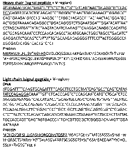
Figure 1 shows the variable regions from the heavy chain and the light chain
of the 2C10
antibody. The nucleotide sequence shown for the heavy chain (SEQ ID NO:1)
includes a signal peptide
(nucleotides 1-57; underlined) and the heavy chain variable sequence
(nucleotides 58-396). The
corresponding amino acid sequence is shown below (SEQ ID NO:2), where amino
acids 1-19
corresponding to the signal sequence (underlined) and amino acids 20-132
correspond to the heavy chain
variable region.
The nucleotide sequence shown for the light chain (SEQ ID NO:3) includes a
signal peptide (nucleotides
1-66; underlined) and the light chain variable sequence (nucleotides 67-384).
The corresponding amino
acid sequence is shown below (SEQ ID NO:4), where amino acids 1-22 correspond
to the signal peptide
(underlined) and amino acids 23-128 correspond to the light chain variable
region.
Figure 2A is a plot showing flow cytometry data confirming the binding of 2C10
to human and
rhesus CD20+ B cells.
Figure 2B is a plot showing CD40 adsorption data from ELISA assays with
varying
concentrations of 2C10 to confirm the binding of 2C10 to human and rhesus CD40
as detected using goat
anti-mouse IgG-HRP.
6
Date Recue/Date Received 2021-06-18
Figure 3 is a graph showing the dose-dependent inhibition of CD154 binding to
B cells by 2C10.
B cells were analyzed for CD154 binding by incubating with histidine-tagged
soluble CD154 and
analyzing for histidine expression. Results are representative of multiple
repetitions of the experiment.
Figure 4 is a schematic diagram and graphs showing the principle of the assay
involving rhesus
or human peripheral blood mononuclear cells (PBMCs) and Jurkat cells.
Figure 5 is a set of graphs showing CD23 expression in CD20+ cells taken from
co-cultures of
rhesus PBMCs and Jurkat cells in the presence of variable concentrations of
3A8, 5C8, or 2C10
antibodies.
Figure 6 is a set of graphs showing CD86 expression in CD20+ cells taken from
co-cultures of
human PBMCs and Jurkat cells in the presence of variable concentrations of
3A8, 5C8, or 2C10
antibodies.
Figure 7 is a set of graphs showing CD23 expression CD20+ cells from either
human or rhesus
PBMCs cultured without Jurkat cells in the presence of either the 3A8 or the
2C10 antibody.
Figure 8 is a graph showing peripheral B cell count of rhesus macaques treated
with mouse-
rhesus chimeric forms of 2C10 engineered to contain either rhesus IgG1
(2C10R1) or IgG4 (2C10R4)
heavy chain constant regions, and chimeric IgG1 forms of anti-CD40 3A8 (3A8R1)
or anti-CD40 Chi220
(Chi220). All animals were immunized with 4-hydroxy-3-nitrophenylacetyl-
conjugated keyhole limpet
hemocyanin (KLH) after the first antibody treatment.
Figure 9 is a graph showing T cell-dependent antibody responses in macaque
monkeys treated
with 2C10R1, 2C10R4, or 3A8R1 antibody. All animals were immunized with KLH
after the first
antibody treatment.
Figure 10 is a diagram showing the standard macaque model of allogeneic islet
transplantation.
Diabetes was induced in macaque monkeys using streptozotocin. Diabetic monkeys
were transplanted
with allogeneic islets and immunosuppresion initiated with basiliximab and
sirolumus. Experimental
animals received 2C10R4 treatment on days 0 and 7 post-transplantation.
Figure 11A is a plot showing free blood glucose levels (FBG) in 4 macaques
following islet
transplantation, background immunosuppresion, and treatment with 2C10R4. The
solid line on the plot
represents the level of 2C10 in the plasma.
Figure 11B is a plot showing FBG in macaques that received only background
immunosuppresion.
Figure 12 is a graph showing results from a competitive blockade assay using
human PBMCs
incubated with increasing concentrations of 2C10, 3A8, or Chi220 antibodies
and stained with an APC-
conjugated 2C10 to assess the ability of each antibody to cross-block 2C10.
Detailed Description
The present invention relates to anti-CD40 antibodies and antibody fragments
having the ability
to bind a particular epitope on the CD40 molecule, as well as methods that
involve the use of such
7
Date Recue/Date Received 2021-06-18
antibodies. This epitopic specificity confers a particular activity profile,
such that the antibodies
generally block the ability of CD40 to interact with its binding partners
(e.g., CD154) and do so without
activating the cell expressing CD40. This activity profile is understood to
make these antibodies
particularly useful for reducing complications associated with organ or tissue
transplantation.
Production and Identification of CD40 antibodies
Mice (strain AJ) were immunized with a fusion protein consisting of the
extracellular domain of
rhesus macaque (M mulatta) CD40 (amino acid sequence:
EPPTACREKQYLINSQCCSLCQPGQKLVSDCTEFTETECLPCSESEFLDTWNRETRCHQHKYCDP
NLGLRVQQKGTSETDTICTCEEGLHCMSESCESCV; SEQ ID NO:5) fused to maltose binding
protein
(CD40-MBP). The amino acid sequence in this region of the rhesus macaque CD40
protein differs from
human CD40 protein at five amino acid positions (human amino acid sequence:
EPPTACREKQYLINSQCCSLCQPGQKLVSDCTEFTETECLPCGESEFLDTWNRETHCHQHKYCDP
NLGLRVQQKGTSETD TICTCEEGWHCTSEACESCV; SEQ ID NO:6). CD40-MBP was
administered to mice multiple times with complete Freund's adjuvant and
incomplete Freund's adjuvant.
Splenocytes from immunized mice were fused with the mouse myeloma cell line
5P2/0 and hybrids
selected using standard hybridoma technology.
Antibodies were selected for reactivity to a second fusion protein consisting
of the same rhesus
CD40 domain fused to glutamine synthetase (CD40-GST). Antibodies reactive to
CD40-GST by ELISA
were further tested for reactivity to native CD40 express on rhesus macaque
blood B cells, human blood
B cells and rhesus macaque B-lymphoblastoid cell lines by flow cytometry. As a
final level of selection,
antibodies were tested in an in vitro assay for their ability to inhibit human
or rhesus macaque B cell
activation after co-culture CD154-expressing Jurkat D1.1 cells. A stable
subclone of anti-CD40 antibody
2C10 was obtained by limiting dilution. The antibody is a mouse IgGl-kappa.
Antibody cloning
Variable regions of monoclonal antibodies can be cloned using any method known
in the art.
PCR-based methods for obtaining antibody variable region sequences for
hybridoma cells are described,
for example, in Larrick et al., Nat. Biotechnol. 7:934-8, 1989 and in Orlandi
et al., Proc. Natl. Acad. Sci.
USA 86:3833-7, 1989. Using these techniques or similar techniques, the
variable regions of monoclonal
antibodies can be cloned and subject to further manipulation.
In the present case, the variable sequences from the heavy and light chains of
the 2C10 antibody
were cloned and were sequenced. The DNA representing the immunoglobulin heavy
and light chain
variable regions from the 2C10 hybridoma were cloned using 5' RACE PCR
employing the following
DNA primers:
Mouse kappa reverse: 5' ¨ CTA ACA CTC ATT CCT GTT GAA GCT CTTGAC (SEQ ID
NO:7);
Mouse kappa forward: 5' ¨ GCT GAT GCT GCA CCA ACT GTA TCC ¨3' (SEQ ID NO:8)
8
Date Recue/Date Received 2021-06-18
Mouse IgG1 reverse: 5' ¨ GGC AAC GTT GCA GGT CTC GC ¨3' (SEQ ID NO:9)
Mouse IgG1 forward: 5' ¨ CTG GAT CTG CTG CCC AAA CTA ACT CC ¨3' (SEQ ID NO:10)
PCR products were cloned into a commercial cloning vector and were sequenced
using standard
sequencing techniques. The resulting sequences are provided in Figure 1.
The immunoglobulin variable region genes were cloned from the hybridomas
secreting anti-
CD40 antibody clone 2C10 and from anti-human CD40 clone 3A8 (Kwekkeboom et
al., Immunology
79:439-44, 1993) (obtained from the American Type Culture Collection, ATCC,
Vienna, VA) using 5'
rapid amplification of cDNA ends-polymerase chain reaction. The immunoglobulin
heavy and light
chain variable regions were subcloned into expression vectors containing
rhesus IgG1 or rhesus IgG4
heavy chain and rhesus kappa light chain constant region sequences.
Recombinant heavy and light chains were subcloned into expression vectors and
packaged in
retroviral vectors used to transduce Chinese hamster ovary cells using the
GPExTM expression technology
(Catalent Pharma Solutions, Middleton, WI). A pool of transduced cells was
grown in serum-free
medium and secreted antibody was purified by protein A affinity
chromatography. The purified chimeric
rhesus IgG1 (2C10R1, 3A8R1) and IgG4 (2C10R4) antibodies were diafiltered into
phosphate buffer;
endotoxin levels were confirmed to be less than 1 endotoxin unit/mg.
Antibody characterization
2C10 binds to CD40 and prevents binding of CD154
To assess the ability of 2C10 to bind to both rhesus and human CD40,
recombinantly expressed
human or rhesus CD40 were adsorbed to ELISA plates and reacted with varying
concentrations of 2C10.
Binding of 2C10 to CD40 was detected using goat anti-mouse IgG-HRP in an
ELISA. The results in
Figure 2B show that 2C10 have similar binding affinities to rhesus and human
CD40, which is important
for clinical translation of 2C10. To confirm the ability of 2C10 to block
binding of its cognate ligand,
CD154, rhesus and human B cells were incubated with escalating concentrations
of 2C10 or an isotype
control and then incubated with histidine-tagged soluble CD154 (R&D Systems,
Minneapolis, MN) and
analyzed for histidine expression. 2C10 blocked the binding of CD154 in a dose-
dependent manner
(Figure 3), indicating that 2C10 can effectively block the interaction of T
cell-bound CD154 with CD40
on B cells and antigen-presenting cells.
2C10 blocks B cell activation in rhesus monkey and human peripheral blood
mononuclear cells
The CD40 antibody 2C10 was characterized with respect to its ability to affect
B cell activation
both using rhesus monkey and human peripheral blood mononuclear cells (PBMCs).
CD20 expression
was chosen as being an indicator of B cells, and expression of CD23, CD80, and
CD86 is associated with
B cell activation. 2C10 was first assessed for its ability to bind to CD20.
Rhesus or human PBMCs were
incubated with fluorochrome-conjugated 2C10 and an anti-CD20 antibody. Flow
cytometric analysis was
used to confirm the binding of 2C10 to human and rhesus CD20+ B cells (Figure
2A). In another set of
experiments, PBMCs from either rhesus monkey or humans were cultured either in
the presence or
9
Date Recue/Date Received 2021-06-18
absence of CD154k Jurkat D1.1 cells, an immortalized T lymophocyte cell line.
Activation of B cells was
determined by measuring expression of three markers (CD23, CD80, and CD86) in
CD20+ cells present
in the PBMCs. The general scheme of this assay is shown in Figure 4. As shown
in Figure 4, culturing
PBMCs in the presence of Jurkat cells resulted in increased expression of all
three markers, indicating
that B cells are activated by the CD154k Jurkat cells.
To test the ability of antibodies to block B cell activation, PBMCs and Jurkat
cells were co-
cultured in the presence or absence of one of three antibodies: 3A8, 5C8, and
2C10. The 3A8 antibody is
a mouse anti-human CD40 antibody (ATCC Deposit No. HB-12024), and 5C8 is an
anti-CD154 antibody
(ATCC Deposit No. CRL-10915). Each was used as a positive control. Co-cultures
were conducted over
a range of five orders of magnitude of antibody concentration (0.001 )1g to 10
)1g). As shown in Figure 5,
3A8 did not block B cell activation in rhesus PBMCs, as measured by CD23
expression, whereas both
2C10 and 5C8 were able to block activation with similar efficiency.
Corresponding changes were also
observed with CD80 and CD86 expression. These results indicate that 2C10 binds
to a different epitope
on CD40 than 3A8. These results also indicate that 2C10 acts primarily as a
CD40 antagonist in contrast
to 3A8 which has previously been shown to act as partial agonists with weak
stimulatory potential
(Adams et al., J. Immunol. 174:542-50, 2005, Badell et al., Am. J. Transplant.
accepted for publication,
2011). When a similar experiment was performed using human, rather than
rhesus, PBMCs, both 2C10
and 5C8 were again observed to block B cell activation, as measured by CD86
expression, with similar
efficiency. Here, the 3A8 antibody, unlike with the rhesus PBMCs, blocked B
cell activation (Figure 6).
The 2C10 and 3A8 antibodies were also tested for their ability to activate B
cells in the absence
of Jurkat cells using either rhesus monkey or human PBMCs. Here, PBMCs were
cultured either in the
presence or absence of either 2C10 or 3A8. Expression of CD23, CD80, and CD86
was then measured in
CD20+ cells. As shown in Figure 7, CD23 expression in rhesus cells was
increased in the presence of the
3A8, but not the 2C10, antibody. By contrast, neither 3A8 nor 2C10 activated
human B cells. The
differences in activity observed between the 3A8 and 2C10 antibody indicate
that the 2C10 antibody
binds to an epitope different from that of the 3A8 antibody.
Date Recue/Date Received 2021-06-18
2C10 prevents a T cell-dependent antibody response
Having established that 2C10 binds to a unique epitope on CD40, inhibits B
cell activation
similarly to an anti-CD154 antibody, and lacks agonistic properties, we then
characterized the effects of
2C10 in vivo. Recombinant mouse-rhesus chimeric forms of 2C10 were generated
using either rhesus
IgG1 (2C10R1) or IgG4 (2C10R4) heavy chain and rhesus kappa light chain
constant region sequences.
A chimeric rhesus IgG1 form of 3A8 (3A8R1) was also generated for use as a
control.
Rhesus macaques were immunized once on day zero with 4-hydroxy-3-
nitrophenylacetyl-
conjugated keyhole limpet hemocyanin (KLH, 10 mg IM) antigen (Biosearch
Technologies, Novato,
CA). Prior to immunization and at one week, cohorts of three animals received
an intravenous dose (50
mg/kg) of 2C10R1, 2C10R4, 3A8R1, or saline. All animals were observed for 70
days, and flow
cytometry was performed weekly. Treatment with either recombinant 2C10
isotypes resulted in modest
change in peripheral B cell counts (Figure 8) compared to the previously
reported significant and
prolonged depletion of peripheral B cells occurring in animals receiving
either 3A8R1 (Badell et al., Am.
J. Transplant. 10:214, 2010) or Chi220 (Adams et al., J. Immunol. 174:542-50,
2005).
T cell-dependent antibody responses to KLH-NP were tested by ELISA. Plates
were coated with
KLH (0.01 mg/ml, Sigma, St. Louis, MO) and blocked with Super Block (Thermo
Scientific, Woodstock,
GA). Pre- and post-treatment plasma samples were serially diluted, plated for
1 hr, and washed with
phosphate-buffered saline/0.05% TweenTm. Anti-KLH antibodies were detected by
incubating for 1 hr
with monoclonal anti-rhesus IgG-horseradish peroxidase (clone 1B3, NHP Reagent
Resource, Boston,
MA). Plates were then incubated with Peroxidase Substrate Solution (KPL). Stop
solution (KPL) was
then added, and optical density was read on an ELISA plate reader at 450 nm. A
sample was considered
positive at a given dilution if the optical density reading of the post-
treatment plasma exceeded the optical
density of the pre-treatment plasma at the same dilution by 2-fold. Following
KLH immunization,
control animals developed high-titer KLH-specific IgG (Figure 9). Animals that
received 3A8R1 also
developed anti-KLH responses, but titers were approximately 10-fold lower than
controls despite
significant depletion of B cells. In contrast, the generation of IgG anti-KLH
antibodies was nearly
completely blocked through day 56 in all animals that received either 2C10R1
or 2C10R4.
2C10 significantly prolongs islet allograft survival in a macaque model of
allogeneic islet transplantation
We further tested 2C10R4, the CD4 purified chimeric rhesis IgG4 antibody, in a
nonhuman
primate allogenic islet transplant model (Figure 10). Rhesus macaques weighing
10-20 kg underwent
donor pancreatectomy one day prior to transplantation via a midline
laparotomy. The pancreas was
isolated and placed on ice after the animals were terminally exsanguinated.
Islet isolation was performed
using Collagenase/Neutral protease (950 Wunsch units and 63 units,
respectively; Serva, Heidelberg,
Germany). The digested pancreas was purified on a four-layer, discontinuous
Euroficoll gradient
(Mediatech, Manassas, VA) and Cobe 2991 blood cell processor (CaridianBCT,
Lakewood, CO).
11
Date Recue/Date Received 2021-06-18
Samples of the final islet preparation were counted and expressed as islet
equivalents (IEQ). Isolated
islets were cultured overnight, counted and suspended in Transplant Media
(Mediatech).
Rhesus macaques weighing 3-5 kg were rendered diabetic using streptozotocin
(1250 mg/m2 IV;
ZanosarTM, Teva Parenteral Medicines, Irvine, CA) four weeks prior to
transplantation. Diabetes was
confirmed by intravenous glucose tolerance test (IVGTT) with a 500 mg/kg bolus
of dextrose and
measurement of primate C-peptide. Glucose levels were monitored and C-peptide
was measured at
baseline and 10, 30, 60 and 90 after injection of dextrose. Diabetes was
confirmed by measurement of
elevated blood glucose levels in the absence of detectable serum C-peptide.
Diabetic recipients
underwent MHC-mismatched islet allotransplantation. A mean of 15,745 ( 4,063)
IEQ were infused via
a small midline laparotomy and cannulation of a mesenteric vein.
Blood glucose levels were measured twice daily by earstick; NPH (Novolin; Novo
Nordisk,
Princeton, NJ) and glargine (Lantus; Sanofi-Aventis, Bridgewater, NJ) insulin
were administered to
maintain fasting blood glucose (FBG) less than 300 mg/dL pre-transplant and
following graft rejection.
IVGTT was performed periodically post-transplant to monitor graft function.
Transplant recipients
underwent weekly flow cytometric analysis to monitor T cell (CD3 V450, CD4
PerCP-Cy5.5, CD8
PerCp; BD Bioscience) and B cell (CD20 PE, BD Bioscience) populations. After
islet engraftment
rejection was defined as FBG greater than 130 mg/dL on two consecutive days.
Primary endpoint was
rejection-free islet graft survival. All animals used in these experiments
were treated in compliance with
the Emory University IACUC and the Guide for the Care and Use of Laboratory
Animals.
Transplant recipients received either 2C10R4, basiliximab (Simulect, Novartis,
Basel,
Switzerland) and sirolimus, or basiliximab and sirolimus alone. 2C10R4 (50
mg/kg) was administered
intravenously on post-operative day (POD) 0 and 7. Basiliximab (0.3 mg/kg) was
administered
intravenously on POD 0 and 3. Sirolimus was administered intramuscularly daily
to achieve trough
levels of 5-15 ng/ml through POD 120. All three animals receiving basiliximab
and sirolimus alone are
historic controls (Bade11 et al., J. Clin. Invest. 120:4520-312, 2010). Two of
these historic controls (RQz6
and RIb7) underwent diabetes induction by pancreatectomy and received oral
sirolimus.
Treatment with the regimens described above resulted in significantly
prolonged islet graft
survival (Figure 11A) compared to controls receiving only basiliximab
induction and sirolimus
maintenance therapy (Figure 11B). Median rejection-free graft survival time
for animals receiving
2C10R4 is 280 days compared to 8 days for control animals (p=0.010, Table 1).
Pharmacokinetic data
predict that plasma 2C10R4 levels would be less than 1 m/m1 by POD 100.
Because sirolimus was
discontinued at POD120, the recipient with the longest survival (304 days)
received no
immunosuppression for approximately 24 weeks prior to rejection. No animals
treated with 2C10R4
developed clinically relevant infectious complications or weight loss. These
results reflect animals that
received the IgG4 isotype of 2C10. Two additional animals that received the
IgG1 isotype of 2C10
(2C10R1) in combination with basiliximab and sirolimus achieved similarly
prolonged graft survival of
12
Date Recue/Date Received 2021-06-18
220 and 162 days (data not shown). Given the positive results with 2C10 used
as induction therapy, the
next step is to assess the effects on graft survival by administering 2C10 as
maintenance therapy.
Table 1
Reciplegit 111L1 viva!
Comment
_________________________________________________ 1..
2C:1 1:31M/Basi I ixi mat)/ Sr Li 21,973 29(3
Rejection
RAD13 A'. 1 OR .11liasil
1J.q13 .1.11:f:: 1 2(6
V.14-11,3 2C10174:4/Bas dixiniA1)./ Sff.u.ti 20,:;1:4::, 1.(i 3
j cctio a
Basil I'M rLu,;: 8 rico:
Basith 10,(p.);,4 8 [14;p1
q.,4:(
.101( Basil! i kl k WS 796 Pri,Ation
Blockade of the CD40/CD154 pathway in conjunction with the CD28/B7 pathway
Blockade of the CD40/CD154 pathway may prove useful in conjunction with other
costimulation
blockade agents. Belatacept, a high affinity version of CTLA4-Ig designed to
block the CD28/B7
costimulatory pathways, has shown efficacy in nonhuman primate models of renal
and islet
transplantation and in phase II and III clinical trials in renal
transplantation (Larsen et al., Transplantation
90:1528-35, 2010, Vincenti et al., Am. J. Transplant. 10:535-46, 2010, Adams
et al., J. Immunol.
174:542-50, 2005, Adams et al., Diabetes 51:265-70, 2002, Larsen et al., Am.
J. Transplant. 5:443-53,
2005, Vincenti et al., N. Engl. J. Med. 358:770-81, 2005). The BENEFIT trial
revealed superior renal
function in patients treated with belatacept; however, these patients had a
higher incidence and more
severe grade of biopsy-proven acute rejection (Larsen et al., Transplantation
90:1528-35, 2010, Vincenti
et al. Am. J. Transplant. 10:535-46, 2010). In light of this increased rate of
acute rejection and the
synergy between CD40 and B7 blockade (Larsen et al., Nature 381:434-8, 1996),
we next want to test the
efficacy of combined 2C10 and belatacept therapy in nonhuman primate kidney
transplantation.
Epitope mapping
Methods for identifying the particular epitope to which an antibody binds are
known to those
skilled in the art. Standard techniques include peptide scanning, in which
overlapping, short peptides (for
example, 10-30 amino acids, e.g., 20, in length) derived from the full length
protein to which the antibody
binds are individually tested for their ability to bind the antibody. From
such experiments, the region of
the protein to which the antibody binds can then be determined.
13
Date Recue/Date Received 2021-06-18
Site-directed mutagenesis can also be used to identify the antigenic region(s)
of a particular
protein. In this approach, point mutations are systematically introduced into
the target polypeptide and
the ability of the antibody to bind the peptide with mutations at various
positions is used to determine
whether a particular region of that protein contains the epitope to which the
antibody binds.
Antibody epitopes can also be identified using high-through mutagenesis
techniques, such as
Shotgun Mutagenesis (Integral Molecular, Inc., Philadelphia, Pa.), which can
be used to generate large
numbers of mutations within the target protein. Such methodologies permit
efficient identification of
eptitopes within the protein.
To determine if various antibodies to CD40 bind similar epitopes, an in vitro
competitive
blockade assay was performed. The antibodies 2C10, 3A8 and Chi220, a chimeric
IgG1 CD40-specific
antibody, were used in the assay. 2C10 was conjugated to allophycocyanin (APC)
using the Lightning
Link antibody labeling kit (Novus Biologics, Littleton, CO). Human PBMCs were
incubated with
escalating concentrations of 2C10, 3A8, or Chi220, and then stained with the
APT-conjugated 2C10 to
assess the ability of each antibody to cross-block 2C10. Binding of APC-
conjugated 2C10 decreased
with increasing concentrations of 2C10 but not Chi220 or 3A8 as shown in
Figure 12. The result
indicates that 2C10 binds a unique epitope distinct from either Chi220 or 3A8.
Generation of additional antibodies
Additional antibodies (e.g., monoclonal, polyclonal, poly-specific, or mono-
specific antibodies)
against the CD40 epitope recognized by 2C10 can be made, e.g., using any of
the numerous methods for
making antibodies known in the art. In one example, a coding sequence for an
eptiope recognized by the
2C10 antibody is expressed as a C-terminal fusion with glutathione S-
transferase (GST) (Smith et al.,
Gene 67:31-40, 1988). The fusion protein is purified on glutathione-Sepharose
TM beads, eluted with
glutathione, cleaved with thrombin (at an engineered cleavage site), and
purified for immunization of
rabbits. Primary immunizations are carried out with Freund's complete adjuvant
and subsequent
immunizations with Freund's incomplete adjuvant. Antibody titers are monitored
by Western blot and
immunoprecipitation analyses using the thrombin-cleaved protein fragment of
the GST fusion protein.
Immune sera are affinity purified using CNBr-SepharoseTm-coupled protein.
Antiserum specificity can
be determined using a panel of unrelated GST proteins.
As an alternate or adjunct immunogen to GST fusion proteins, peptides
corresponding to
relatively unique immunogenic regions of a polypeptide of the invention can be
generated and coupled to
keyhole limpet hemocyanin (KLH) through an introduced C-terminal lysine.
Antiserum to each of these
peptides is similarly affinity purified on peptides conjugated to BSA, and
specificity is tested by ELISA
or Western blot analysis using peptide conjugates, or by Western blot or
immunoprecipitation using the
polypeptide expressed as a GST fusion protein.
14
Date Recue/Date Received 2021-06-18
Alternatively, monoclonal antibodies that specifically bind the CD40 eptiope
recognized the
2C10 antibody can be prepared using standard hybridoma technology (see, e.g.,
Kohler et al., Nature
256:495-7, 1975; Kohler et al., Fur. J. Immunol. 6:511-9, 1976; Kohler et al.,
Fur. J. Immunol. 6:292-5,
1976; Hammerling et al., Monoclonal Antibodies and T Cell Hybridomas,
Elsevier, NY, 1981). Once
produced, monoclonal antibodies can also be tested for specific recognition by
Western blot or
immunoprecipitation analysis. Alternatively, monoclonal antibodies can be
prepared using the
polypeptide of the invention described above and a phage display library
(Vaughan et al., Nat.
Biotechnol. 14:309-14, 1996).
Epitopic fragments can be generated by standard techniques, e.g., using PCR
and cloning the
fragment into a pGEX expression vector. Fusion proteins are expressed in E.
coli and purified using a
glutathione agarose affinity matrix. To minimize potential problems of low
affinity or specificity of
antisera, two or three such fusions are generated for each protein, and each
fusion is injected into at least
two rabbits. Antisera are raised by injections in a series, and can include,
for example, at least three
booster injections.
In order to generate polyclonal antibodies on a large scale and at a low cost
an appropriate animal
species can be chosen. Polyclonal antibodies can be isolated from the milk or
colostrum of, e.g.,
immunized cows. Bovine colostrum contains 28 g of IgG per liter, while bovine
milk contains 1.5 g of
IgG per liter (Ontsouka et al., J. Dairy Sci. 86:2005-11, 2003). Polyclonal
antibodies can also be isolated
from the yolk of eggs from immunized chickens (Sarker et al., J. Pediatr.
Gastroenterol. Nutr. 32:19-25,
2001).
Multiple adjuvants are approved for use in dairy cows. Adjuvants useful in
this invention
include, but are not limited to, Emulsigen , an oil-in-water emulsified
adjuvant, Emulsigee-D, an oil-in-
water emulsified adjuvant with DDA immunostimulant, Emulsigee-P, an oil-in-
water emulsified
adjuvant with co-polymer immunostimulant, Emulsigee-BCL, an oil-in-water
emulsified adjuvant with
block co-polymer immunostimulant, CarbigenTM, a carbomer base, and PolygenTM,
a co-polymer base.
All of the listed adjuvants are commercially available from MVP Laboratories
in Omaha, Nebr.
Useful antibodies can be identified in several different screening assays.
First, antibodies are
assayed by ELISA to determine whether they are specific for the immunizing
antigen (i.e., the CD40
epitope described herein). Using standard techniques, ELISA plates are coated
with immunogen, the
antibody is added to the plate, washed, and the presence of bound antibody
detected by using a second
antibody specific for the Ig of the species in which the antibody was
generated.
A functional in vitro assay can be used to screen antibodies e.g., an
neutralizing assay based on
monocyte-derived dendritic cells.
Direct measurements of bovine immunoglobulin in illeal fluid in human subjects
have shown that
significant amounts of immunoglobulin survive transit through the stomach and
small intestine (Warny et
al., Gut 44:212-7, 1999). Methods have also been described to formulate avian
immunoglobulin (IgY) for
GI delivery (Kovacs-Nolan et al., Immunol. Methods 296:199-209, 2005).
Date Recue/Date Received 2021-06-18
Humanized antibodies
The invention encompasses humanized antibodies. Various methods for humanizing
non-human
antibodies are known in the art. For example, a humanized antibody can have
one or more amino acid
residues introduced into it from a source which is non-human. These non-human
amino acid residues are
often referred to as "import" residues, which are typically taken from an
"import" variable domain.
Humanization can be essentially performed following the method of Winter and
co-workers (Jones et al.,
Nature 321:522-5, 1986; Riechmann et al., Nature 332:323-7, 1988; Verhoeyen et
al., Science 239:1534-
6, 1988), by substituting hypervariable region sequences for the corresponding
sequences of a human
antibody. Accordingly, such "humanized" antibodies are chimeric antibodies
(U.S. Patent No.
4,816,567), where substantially less than an intact human variable domain has
been substituted by the
corresponding sequence from a non-human species. In practice, humanized
antibodies are typically
human antibodies in which at least some hypervariable region residues as well
as other variable region
residues are substituted by residues from analogous sites in rodent
antibodies.
The choice of human variable domains, both light and heavy, to be used in
making the
humanized antibodies can be important to reduce antigenicity. According to the
so-called "best-fit"
method, the sequence of the variable domain of a rodent antibody is screened
against the entire library of
known human variable-domain sequences. The human sequence which is closest to
that of the rodent is
then accepted as the human framework for the humanized antibody. See, e.g.,
Sims et al., J. Immunol.
151:2296-308, 1993; Chothia et al., J. Mol. Biol. 196:901-17, 1987. Another
method uses a particular
framework derived from the consensus sequence of all human antibodies of a
particular subgroup of light
or heavy chains. The same framework may be used for several different
humanized antibodies. See, e.g.,
Carter et al., Proc. Natl. Acad. Sci. USA 89:4285-9, 1992; Presta et al., J.
Immunol. 151:2623-32, 1993.
It is further generally desirable that antibodies be humanized with retention
of high affinity for
the antigen and other favorable biological properties. To achieve this goal,
according to one method,
humanized antibodies are prepared by a process of analysis of the parental
sequences and various
conceptual humanized products using three-dimensional models of the parental
and humanized
sequences. Three-dimensional immunoglobulin models are commonly available and
are familiar to those
skilled in the art. Computer programs are available which illustrate and
display probable three-
dimensional conformational structures of selected candidate immunoglobulin
sequences. Inspection of
these displays permits analysis of the likely role of the residues in the
functioning of the candidate
immunoglobulin sequence, i.e., the analysis of residues that influence the
ability of the candidate
immunoglobulin to bind its antigen. In this way, FR residues can be selected
and combined from the
recipient and import sequences so that the desired antibody characteristic,
such as increased affinity for
the target antigen(s), is achieved. In general, the hypervariable region
residues are directly and most
substantially involved in influencing antigen binding.
16
Date Recue/Date Received 2021-06-18
Human antibodies
Human antibodies of the invention can be constructed by combining Fv clone
variable domain
sequence(s) selected from human-derived phage display libraries with known
human constant domain
sequences(s) (Hoogenboom et al., J. Ma Biol. 227:381-8, 1992; Marks et al., J.
Ma Biol. 222:581-97,
1991). Alternatively, human monoclonal antibodies of the invention can be made
by the hybridoma
method. Human myeloma and mouse-human heteromyeloma cell lines for the
production of human
monoclonal antibodies have been described, for example, by Kozbor, J. Immunol.
133:3001-5, 1984;
Brodeur et al., Monoclonal Antibody Production Techniques and Applications,
pp. 51-63 (Marcel Dekker,
Inc., New York, 1987); and Boerner et al., J. Immunol. 147: 86-95, 1991.
It is now possible to produce transgenic animals (e.g., mice) that are
capable, upon immunization,
of producing a full repertoire of human antibodies in the absence of
endogenous immunoglobulin
production. For example, it has been described that the homozygous deletion of
the antibody heavy-chain
joining region (JH) gene in chimeric and germ-line mutant mice results in
complete inhibition of
endogenous antibody production. Transfer of the human germ-line immunoglobulin
gene array in such
germ-line mutant mice will result in the production of human antibodies upon
antigen challenge. See,
e.g., Jakobovits et al., Proc. Natl. Acad. Sci. USA 90:2551-5, 1993;
Jakobovits et al., Nature 362:255-8,
1993; BrUggemann et al., Year Immunol. 7:33-40, 1993.
Gene shuffling can also be used to derive human antibodies from non-human,
e.g., rodent,
antibodies, where the human antibody has similar affinities and specificities
to the starting non-human
antibody. According to this method, which is also called "epitope imprinting,"
either the heavy or light
chain variable-region of a non-human antibody fragment obtained by phage
display techniques as
described herein is replaced with a repertoire of human V domain genes,
creating a population of non-
human chain/human chain seFv or Fab chimeras. Selection with antigen results
in isolation of a non-
human chain/human chain chimeric seFv or Fab where the human chain restores
the antigen binding site
destroyed upon removal of the corresponding non-human chain in the primary
phage display clone, i.e.,
the epitope governs (imprints) the choice of the human chain partner. When the
process is repeated in
order to replace the remaining non-human chain, a human antibody is obtained
(see PCT Publication WO
93/06213). Unlike traditional humanization of non-human antibodies by CDR
grafting, this technique
provides completely human antibodies, which have no FR or CDR residues of non-
human origin.
Antibody fragments
The invention also features antibody fragments that comprise a portion of an
intact antibody,
preferably comprising the antigen binding region thereof. Examples of antibody
fragments include Fab,
Fab', F(ab')2, and Fv fragments; diabodies; linear antibodies; single-chain
antibody molecules; and
multispecific antibodies formed from antibody fragments.
Papain digestion of antibodies produces two identical antigen-binding
fragments, called "Fab"
fragments, each with a single antigen-binding site, and a residual "Fe"
fragment, whose name reflects its
17
Date Recue/Date Received 2021-06-18
ability to crystallize readily. Pepsin treatment yields an F(ab')2 fragment
that has two antigen-combining
sites and is still capable of cross-linking antigen.
Fv is the minimum antibody fragment which contains a complete antigen-binding
site. In one
embodiment, a two-chain Fv species consists of a dimer of one heavy- and one
light-chain variable
domain in tight, non-covalent association. In a single-chain Fv (scFv)
species, one heavy- and one light-
chain variable domain can be covalently linked by a flexible peptide linker
such that the light and heavy
chains can associate in a "dimeric" structure analogous to that in a two-chain
Fv species. It is in this
configuration that the three hypervariable regions (HVRs) of each variable
domain interact to define an
antigen-binding site on the surface of the VH-VL dimer. Collectively, the six
HVRs confer antigen-
binding specificity to the antibody. However, even a single variable domain
(or half of an Fv comprising
only three HVRs specific for an antigen) has the ability to recognize and bind
antigen, although at a lower
affinity than the entire binding site.
The Fab fragment contains the heavy- and light-chain variable domains and also
contains the
constant domain of the light chain and the first constant domain (CH1) of the
heavy chain. Fab'
fragments differ from Fab fragments by the addition of a few residues at the
carboxy terminus of the
heavy chain CH1 domain including one or more cysteines from the antibody hinge
region. Fab'-SH is
the designation herein for Fab' in which the cysteine residue(s) of the
constant domains bear a free thiol
group. F(ab')2antibody fragments originally were produced as pairs of Fab'
fragments which have hinge
cysteines between them. Other chemical couplings of antibody fragments are
also known.
Single-chain Fv or scFv antibody fragments comprise the VH and VL domains of
antibody, where
these domains are present in a single polypeptide chain. Generally, the scFv
polypeptide further
comprises a polypeptide linker between the VH and VL domains which enables the
scFv to form the
desired structure for antigen binding. For a review of scFv, see, e.g.,
Pluckthiin, in The Pharmacology of
Monoclonal Antibodies, vol. 113, Rosenburg and Moore eds., (Springer-Verlag,
New York, 1994), pp.
269-315.
Diabodies are antibody fragments with two antigen-binding sites, which
fragments comprise a
heavy-chain variable domain (VII) connected to a light-chain variable domain
(VL) in the same
polypeptide chain (VH-VL). By using a linker that is too short to allow
pairing between the two domains
on the same chain, the domains are forced to pair with the complementary
domains of another chain and
create two antigen-binding sites. Diabodies may be bivalent or bispecific.
Diabodies are described more
fully in, for example, European Patent No. 404,097; PCT Publication WO
1993/01161; Hudson et al.,
Nat. Med. 9:129-34, 2003; and Hollinger et al., Proc. Natl. Acad. Sci. USA
90:6444-8, 1993. Triabodies
and tetrabodies are also described in Hudson et al., Nat. Med. 9:129-34, 2003.
Antibody fragments may be generated by traditional means, such as enzymatic
digestion, or by
recombinant techniques. In certain circumstances there are advantages of using
antibody fragments,
rather than whole antibodies. The smaller size of the fragments allows for
rapid clearance, and may lead
18
Date Recue/Date Received 2021-06-18
to improved access to solid tumors. For a review of certain antibody
fragments, see Hudson et al. Nat.
Med. 9:129-134, 2003.
Various techniques have been developed for the production of antibody
fragments. Traditionally,
these fragments were derived via proteolytic digestion of intact antibodies
(see, e.g., Morimoto et al., J.
Biochem. Biophys. Methods 24:107-17, 1992; and Brennan et al., Science 229:81-
3, 1985). However,
these fragments can now be produced directly by recombinant host cells. Fab,
Fv, and ScFv antibody
fragments can all be expressed in and secreted from E. coli, thus allowing the
facile production of large
amounts of these fragments. Antibody fragments can be isolated from the
antibody phage libraries.
Alternatively, Fab'-SH fragments can be directly recovered from E. coli and
chemically coupled to form
F(ab')2fragments (Carter et al., Bio/Technology 10:163-7, 1992). In another
approach, F(ab')2fragments
are isolated directly from recombinant host cell culture. Fab and
F(ab')2fragment with increased in vivo
half-life comprising salvage receptor binding epitope residues are described
in U.S. Patent No. 5,869,046.
Other techniques for the production of antibody fragments will be apparent to
the skilled practitioner.
Pharmaceutical compositions
The present invention provides a composition, e.g., a pharmaceutical
composition, containing an
antibody, or antigen-binding portion(s) thereof, of the present invention,
formulated together with a
pharmaceutically acceptable carrier. Pharmaceutical compositions of the
invention also can be
administered in combination therapy, i.e., combined with other agents (e.g.,
immunosuppressants). As
used herein, "pharmaceutically acceptable carrier" includes any and all
solvents, dispersion media,
coatings, antibacterial and antifungal agents, isotonic and absorption
delaying agents, and the like that are
physiologically compatible. Preferably, the carrier is suitable for
intravenous, intrathecal, intramuscular,
subcutaneous, parenteral, spinal or epidermal administration (e.g., by
injection or infusion). Depending
on the route of administration, the antibodies of the invention may be coated
in a material to protect the
compound from the action of acids and other natural conditions that may
inactivate the compound.
A composition of the present invention can be administered by a variety of
methods known in the
art. As will be appreciated by the skilled artisan, the route and/or mode of
administration will vary
depending upon the desired results. Administration may be parenteral,
intravenous, intrathecal,
subcutaneous, oral, topical, or local, for example, by direct injection into
the cerebrospinal fluid.
Intravenous delivery by continuous infusion is one exemplary method for
administering the therapeutic
antibodies of the present invention. The therapeutic compound may be in the
form of a solution, a
suspension, an emulsion, an infusion device, or a delivery device for
implantation, or it may be presented
as a dry powder to be reconstituted with water or another suitable vehicle
before use. The composition
can be in the form of a pill, tablet, capsule, liquid, or sustained release
tablet for oral administration; or a
liquid for intravenous, intrathecal, subcutaneous or parenteral
administration; or a polymer or other
sustained release vehicle for local administration.
19
Date Recue/Date Received 2021-06-18
Methods well known in the art for making formulations are found, for example,
in "Remington:
The Science and Practice of Pharmacy" (20th ed., ed. A.R. Gennaro AR., 2000,
Lippincott Williams &
Wilkins, Philadelphia, PA). Formulations for parenteral administration may,
for example, contain
excipients, sterile water, saline, polyalkylene glycols such as polyethylene
glycol, oils of vegetable origin,
or hydrogenated napthalenes. Biocompatible, biodegradable lactide polymer,
lactide/glycolide
copolymer, or polyoxyethylene-polyoxypropylene copolymers may be used to
control the release of the
compounds. Nanoparticulate formulations (e.g., biodegradable nanoparticles,
solid lipid nanoparticles,
liposomes) may be used to control the biodistribution of the compounds. Other
potentially useful
delivery systems include ethylene-vinyl acetate copolymer particles, osmotic
pumps, intrathecal pumps,
implantable infusion systems, and liposomes. The concentration of the compound
in the formulation
varies depending upon a number of factors, including the dosage of the drug to
be administered, and the
route of administration.
To administer a compound of the invention by certain routes of administration,
it may be
necessary to coat the compound with, or co-administer the compound with, a
material to prevent its
inactivation. For example, the compound may be administered to a subject in an
appropriate carrier, for
example, liposomes, or a diluent. Pharmaceutically acceptable diluents include
saline and aqueous buffer
solutions. Liposomes include water-in-oil-in-water CGF emulsions as well as
conventional liposomes
(Strejan et al., J. Neuroimmunol. 7:27-41, 1984). Pharmaceutically acceptable
carriers include sterile
aqueous solutions or dispersions and sterile powders for the extemporaneous
preparation of sterile
injectable solutions or dispersion. The use of such media and agents for
pharmaceutically active
substances is known in the art and is included in the invention except where
any conventional media or
agent is incompatible with the active compound. Supplementary active compounds
can also be
incorporated into the compositions.
Therapeutic compositions typically must be sterile and stable under the
conditions of
manufacture and storage. The composition can be formulated as a solution,
microemulsion, liposome, or
other ordered structure suitable to high drug concentration. The carrier can
be a solvent or dispersion
medium containing, for example, water, ethanol, polyol (for example, glycerol,
propylene glycol, and
liquid polyethylene glycol, and the like), and suitable mixtures thereof. The
proper fluidity can be
maintained, for example, by the use of a coating such as lecithin, by the
maintenance of the required
particle size in the case of dispersion and by the use of surfactants. In many
cases, it will be preferable to
include isotonic agents, for example, sugars, polyalcohols such as mannitol,
sorbitol, or sodium chloride
in the composition. Prolonged absorption of the injectable compositions can be
brought about by
including in the composition an agent that delays absorption, for example,
monostearate salts and gelatin.
Sterile injectable solutions can be prepared by incorporating the active
compound in the required
amount in an appropriate solvent with one or a combination of ingredients
enumerated above, as required,
followed by sterilization microfiltration. Generally, dispersions are prepared
by incorporating the active
compound into a sterile vehicle that contains a basic dispersion medium and
the required other
Date Recue/Date Received 2021-06-18
ingredients from those enumerated above. In the case of sterile powders for
the preparation of sterile
injectable solutions, the preferred methods of preparation are vacuum drying
and freeze-drying
(lyophilization) that yield a powder of the active ingredient plus any
additional desired ingredient from a
previously sterile-filtered solution thereof. Dosage regimens are adjusted to
provide the optimum desired
response (e.g., a therapeutic response). For example, a single bolus may be
administered, several divided
doses may be administered over time or the dose may be proportionally reduced
or increased as indicated
by the exigencies of the therapeutic situation. For example, the antibodies of
the invention may be
administered once or twice weekly by subcutaneous injection or once or twice
monthly by subcutaneous
injection.
It is especially advantageous to formulate parenteral compositions in dosage
unit form for ease of
administration and uniformity of dosage. Dosage unit form as used herein
refers to physically discrete
units suited as unitary dosages for the subjects to be treated; each unit
contains a predetermined quantity
of active compound calculated to produce the desired therapeutic effect in
association with the required
pharmaceutical carrier. The specification for the dosage unit forms of the
invention are dictated by and
directly dependent on (a) the unique characteristics of the active compound
and the particular therapeutic
effect to be achieved, and (b) the limitations inherent in the art of
compounding such an active compound
for the treatment of sensitivity in individuals.
The phrases "parenteral administration" and "administered parenterally" as
used herein mean
modes of administration other than enteral and topical administration, usually
by injection, and include,
without limitation, intravenous, intramuscular, intraarterial, intrathecal,
intracapsular, intraorbital,
intracardiac, intradermal, intraperitoneal, transtracheal, subcutaneous,
subcuticular, intraarticular,
subcapsular, subarachnoid, intraspinal, epidural and intrastemal injection and
infusion. Examples of
suitable aqueous and nonaqueous carriers which may be employed in the
pharmaceutical compositions of
the invention include water, ethanol, polyols (such as glycerol, propylene
glycol, polyethylene glycol, and
the like), and suitable mixtures thereof, vegetable oils, such as olive oil,
and injectable organic esters,
such as ethyl oleate. Proper fluidity can be maintained, for example, by the
use of coating materials, such
as lecithin, by the maintenance of the required particle size in the case of
dispersions, and by the use of
surfactants.
Compositions of the invention may also contain adjuvants such as
preservatives, wetting agents,
emulsifying agents and dispersing agents. Prevention of presence of
microorganisms may be ensured
both by sterilization procedures, and by the inclusion of various
antibacterial and antifungal agents, for
example, paraben, chlorobutanol, phenol sorbic acid, and the like. It may also
be desirable to include
isotonic agents, such as sugars, sodium chloride, and the like into the
compositions. In addition,
prolonged absorption of the injectable pharmaceutical form may be brought
about by the inclusion of
agents which delay absorption such as aluminum monostearate and gelatin.
When the compounds of the present invention are administered as
pharmaceuticals, to humans and
animals, they can be given alone or as a pharmaceutical composition
containing, for example, 0.001 to
21
Date Recue/Date Received 2021-06-18
90% (more preferably, 0.005 to 70%, such as 0.01 to 30%) of active ingredient
in combination with a
pharmaceutically acceptable carrier.
For intravenous or intrathecal delivery or direct injection, the composition
must be sterile and
fluid to the extent that the composition is deliverable by syringe. In
addition to water, the carrier can be
an isotonic buffered saline solution, ethanol, polyol (for example, glycerol,
propylene glycol, and liquid
polyetheylene glycol, and the like), and suitable mixtures thereof. Proper
fluidity can be maintained, for
example, by use of coating such as lecithin, by maintenance of required
particle size in the case of
dispersion and by use of surfactants. In many cases, it is preferable to
include isotonic agents, for
example, sugars, polyalcohols such as mannitol or sorbitol, and sodium
chloride in the composition.
Long-term absorption of the injectable compositions can be brought about by
including in the
composition an agent which delays absorption, for example, aluminum
monostearate or gelatin.
Regardless of the route of administration selected, the compounds of the
present invention, which
may be used in a suitable hydrated form, and/or the pharmaceutical
compositions of the present invention,
are formulated into pharmaceutically acceptable dosage forms by conventional
methods known to those
of skill in the art. Actual dosage levels of the active ingredients in the
pharmaceutical compositions of
the present invention may be varied so as to obtain an amount of the active
ingredient which is effective
to achieve the desired therapeutic response for a particular patient,
composition, and mode of
administration, without being toxic to the patient. The selected dosage level
will depend upon a variety
of pharmacokinetic factors including the activity of the particular
compositions of the present invention
employed, or the ester, salt or amide thereof, the route of administration,
the time of administration, the
rate of excretion of the particular compound being employed, the duration of
the treatment, other drugs,
compounds and/or materials used in combination with the particular
compositions employed, the age,
sex, weight, condition, general health and prior medical history of the
patient being treated, and like
factors well known in the medical arts. A physician or veterinarian having
ordinary skill in the art can
readily determine and prescribe the effective amount of the pharmaceutical
composition required. For
example, the physician or veterinarian can start doses of the compounds of the
invention employed in the
pharmaceutical composition at levels lower than that required in order to
achieve the desired therapeutic
effect and gradually increase the dosage until the desired effect is achieved.
In general, a suitable daily
dose of a composition of the invention will be that amount of the compound
which is the lowest dose
effective to produce a therapeutic effect. Such an effective dose will
generally depend upon the factors
described above. If desired, the effective daily dose of a therapeutic
composition may be administered as
two, three, four, five, six or more sub-doses administered separately at
appropriate intervals throughout
the day, optionally, in unit dosage forms. While it is possible for a compound
of the present invention to
be administered alone, it is preferable to administer the compound as a
pharmaceutical formulation
(composition).
22
Date Recue/Date Received 2021-06-18
Therapeutic compositions can be administered with medical devices known in the
art. Examples
of well-known implants, delivery systems, and modules useful in the present
invention are known to
those skilled in the art.
Conditions and disorders
The antibodies and antibody fragments described herein may be used in any
situation in which
immunosuppression is desired (e.g., transplant rejection or autoimmune
disorders). These antibodies are
particularly useful for treating transplant rejection, e.g., reducing the
likelihood that a particular transplant
is rejected by the host or increasing the time before rejection takes place.
The antibodies described herein
can be used in conjunction with transplantation of any organ or any tissue
that is suitable for
transplantation. Exemplary organs include heart, kidney, lung, liver,
pancreas, intestine, and thymus;
exemplary tissues include bone, tendon, cornea, skin, heart valve, vein, and
bone marrow.
The antibodies and antibody fragments can also be used to treat autoimmune
disorders, particular
disorders where autoantibodies are implicated in the pathogenesis of the
disease. Autoimmune diseases
that are or can be associated with autoantibody production include systemic
lupus erythematosus (SLE),
CREST syndrome (calcinosis, Raynaud's syndrome, esophageal dysmotility,
sclerodactyl, and
telangiectasia), opsoclonus, inflammatory myopathy (e.g., polymyositis,
dermatomyositis, and inclusion-
body myositis), systemic scleroderma, primary biliary cirrhosis, celiac
disease (e.g., gluten sensitive
enteropathy), dermatitis herpetiformis, Miller-Fisher Syndrome, acute motor
axonal neuropathy
(AMAN), multifocal motor neuropathy with conduction block, autoimmune
hepatitis, antiphospholipid
syndrome, Wegener '5 granulomatosis, microscopic polyangiitis, Churg-Strauss
syndrome, rheumatoid
arthritis, chronic autoimmune hepatitis, scleromyositis, myasthenia gravis,
Lambert¨Eaton myasthenic
syndrome, Hashimoto's thyroiditis, Graves' disease, Paraneoplastic cerebellar
degeneration, Stiff person
syndrome, limbic encephalitis, Isaacs Syndrome, Sydenham's chorea, pediatric
autoimmune
neuropsychiatric disease associated with Streptococcus (PANDAS), encephalitis,
diabetes mellitus type 1,
and Neuromyelitis optica.
Other autoimmune disorders include pernicious anemia, Addison's disease,
psoriasis, inflammatory
bowel disease, psoriatic arthritis, Sjogren's syndrome, lupus erythematosus
(e.g., discoid lupus
erythematosus, drug-induced lupus erythematosus, and neonatal lupus
erythematosus), multiple sclerosis,
and reactive arthritis.
Additional disorders that may be treated using the methods of the present
invention include, for
example, polymyositis, dermatomyositis, multiple endocrine failure, Schmidt's
syndrome, autoimmune
uveitis, adrenalitis, thyroiditis, autoimmune thyroid disease, gastric
atrophy, chronic hepatitis, lupoid
hepatitis, atherosclerosis, presenile dementia, demyelinating diseases,
subacute cutaneous lupus
23
Date Recue/Date Received 2021-06-18
erythematosus, hypoparathyroidism, Dressler's syndrome, autoimmune
thrombocytopenia, idiopathic
thrombocytopenic purpura, hemolytic anemia, pemphigus vulgaris, pemphigus,
alopecia arcata,
pemphigoid, scleroderma, progressive systemic sclerosis, adult onset diabetes
mellitus (e.g., type II
diabetes), male and female autoimmune infertility, ankylosing spondolytis,
ulcerative colitis, Crohn's
disease, mixed connective tissue disease, polyarteritis nedosa, systemic
necrotizing vasculitis, juvenile
onset rheumatoid arthritis, glomerulonephritis, atopic dermatitis, atopic
rhinitis, Goodpasture's syndrome,
Chagas' disease, sarcoidosis, rheumatic fever, asthma, recurrent abortion,
anti-phospholipid syndrome,
farmer's lung, erythema multiforme, post cardiotomy syndrome, Cushing's
syndrome, autoimmune chronic
active hepatitis, bird-fancier's lung, allergic disease, allergic
encephalomyelitis, toxic epidermal necrolysis,
alopecia, Alport's syndrome, alveolitis, allergic alveolitis, fibrosing
alveolitis, interstitial lung disease,
erythema nodosum, pyoderma gangrenosum, transfusion reaction, leprosy,
malaria, leishmaniasis,
trypanosomiasis, Takayasu's arteritis, polymyalgia rheumatica, temporal
arteritis, schistosomiasis, giant
cell arteritis, ascariasis, aspergillosis, Sampter's syndrome, eczema,
lymphomatoid granulomatosis,
Behcet's disease, Caplan 's syndrome, Kawasaki's disease, dengue,
endocarditis, endomyocardial fibrosis,
endophthalmitis, erythema elevatum et diutinum, erythroblastosis fetalis,
eosinophilic faciitis, Shulman's
syndrome, Felty 's syndrome, filariasis, cyclitis, chronic cyclitis,
heterochronic cyclitis, Fuch's cyclitis, IgA
nephropathy, Henoch-Schonlein purpura, graft versus host disease,
transplantation rejection, human
immunodeficiency virus infection, echovirus infection, cardiomyopathy,
Alzheimer's disease, parvovirus
infection, rubella virus infection, post vaccination syndromes, congenital
rubella infection, Hodgkin's and
non-Hodgkin's lymphoma, renal cell carcinoma, multiple myeloma, Eaton-Lambert
syndrome, relapsing
polychondritis, malignant melanoma, cryoglobulinemia, Waldenstrom's
macroglobulemia, Epstein-Barr
virus infection, mumps, Evan's syndrome, and autoimmune gonadal failure.
Immunosuppressants
The antibodies and antibody fragments described herein can be formulated or
administered in
combination with an immunosuppressant. Examples of immunosuppressants include,
but are not limited
to, calcineurin inhibitors (e.g., cyclosporin A (Sandimmune), cyclosporine G
tacrolimus (Prograr,
Protopie)), mTor inhibitors (e.g., sirolimus (Rapamune , Neorar), temsirolimus
(Toriser), zotarolimus,
and everolimus (Certicae)), fingolimod (GilenyaTm), myriocin, alemtuzumab
(Campath , MabCampath ,
Campath-1H ), rituximab (Rituxan , MabThere), an anti-CD4 monoclonal antibody
(e.g., HuMaxTm-
CD4), an anti-LFA1 monoclonal antibody (e.g., CD1 la), an anti-LFA3 monoclonal
antibody, an anti-CD45
antibody (e.g., an anti-CD45RB antibody), an anti-CD19 antibody (see, e.g.,
U.S. Patent Publication
2006/0280738), monabatacept (Orencie), belatacept, indolyl-ASC (32-indole
ether derivatives of
tacrolimus and ascomycin), azathioprine (Azasan , Imurae), lymphocyte immune
globulin and anti-
thymocyte globulin [equine] (Atgam ), mycophenolate mofetil (Cellcepr),
mycophenolate sodium
(myfortie), daclizumab (Zenapax ), basiliximab (Simulect ), cyclophosphamide
(Endoxan , Cytoxan ,
24
Date Recue/Date Received 2021-06-18
NeosarTM, ProcytoxTM, RevimmuneTm), prednisone, prednisolone, leflunomide
(Arave), FK778, FK779,
15-deoxyspergualin (DSG), busulfan (Myleran , Busulfee), fludarabine
(Fludare), methotrexate
(Rheumatrex , Trexalr), 6-mercaptopurine (Purinethor), 15-deoxyspergualin
(Gusperimus), LF15-
0195, bredinin, brequinar, and muromonab-CD3 (Orthoclone).
Methods for assessing immunosuppressive activity of an agent are known in the
art. For
example, the length of the survival time of the transplanted organ in vivo
with and without
pharmacological intervention serves as a quantitative measure for the
suppression of the immune
response. In vitro assays may also be used, for example, a mixed lymphocyte
reaction (MLR) assay (see,
e.g., Fathman et al., J. Immunol. 118:1232-8, 1977); a CD3 assay (specific
activation of immune cells via
an anti-CD3 antibody (e.g., OKT3)) (see, e.g., Khanna et al., Transplantation
67:882-9, 1999; Khanna et
al. (1999) Transplantation 67:S58); and an IL-2R assay (specific activation of
immune cells with the
exogenously added cytokine IL-2) (see, e.g., Farrar et al., J. Immunol.
126:1120-5, 1981).
Cyclosporine A (CsA; CAS No. 59865-13-3; U.S. Patent No. 3,737,433) and its
analogs may be
used as an immunosuppressant. A number of other cyclosporines and their
derivatives and analogs that
exhibit immunosuppressive activity are known. Cyclosporines and their
formulations are described, for
example, in 2004 Physicians' Desk Reference (2003) Thomson Healthcare, 58th
ed., and U.S. Patent
Nos. 5,766,629; 5,827,822; 4,220,641; 4,639,434; 4,289,851; 4,384,996;
5,047,396; 4,388,307;
4,970,076; 4,990,337; 4,822,618; 4,576,284; 5,120,710; and 4,894,235.
Tacrolimus (FK506) is a macrolide which exerts effects largely similar to CsA,
both with regard
to its molecular mode of action and its clinical efficacy (Liu, Immunol. Today
14:290-5, 1993; Schreiber
et al., Immunol. Today, 13:136-42, 1992); however, these effects are exhibited
at doses that are 20 to 100
times lower than CsA (Peters et al., Drugs 46:746-94, 1993). Tacrolimus and
its formulations are
described, for example, in 2004 Physicians' Desk Reference (2003) Thomson
Healthcare, 58th ed., and
U.S. Patent Nos. 4,894,366; 4,929,611; and 5,164,495.
Sirolimus (rapamycin) is an immunosuppressive lactam macrolide produceable,
for example, by
Streptomyces hygroscopicus. Numerous derivatives of sirolimus and its analogs
and their formulations
are known and described, for example, in 2004 Physicians' Desk Reference
(2003) Thomson
Healthcare, 58th ed., European Patent EP 0467606; PCT Publication Nos. WO
94/02136, WO 94/09010,
WO 92/05179, WO 93/11130, WO 94/02385, WO 95/14023, and WO 94/02136, and U.S.
Patent Nos.
5,023,262; 5,120,725; 5,120,727; 5,177,203; 5,258,389; 5,118,677; 5,118,678;
5,100,883; 5,151,413;
5,120,842; and 5,256,790.
CD40 fragments
The invention also features fragments of CD40 that include the epitope that is
specifically bound
by the 2C10 antibody. The 2C10 antibody was raised against the extracellular
portion of the CD40
polypeptide; it is thus believed that this antibody reacts with a portion of
this sequence (SEQ ID NOS:5
and 6).
Date Recue/Date Received 2021-06-18
The invention therefore features CD40 fragments (e.g., fewer than 150, 120,
100, 80, 70, 60, 50,
40, 30, 20, 18, 15, 12, 11, 10, 9, 8, or 7) amino acids in length that are
specifically bound by the 2C10
antibody. In certain embodiments, the fragment is 8-10, 8-12, 8-15, 8-20, 8-
30, 8-40, 8-50, 8-60, 8-70, 8-
80, or 8-100 amino acids in length. In other embodiments, the fragment is 7-
10, 7-12, 7-15, 7-20, 7-30,
7-40, 7-50, 7-60, 7-70, 7-80, or 7-100 in length.
Fusion proteins
The invention also features fusion protein that includes a fragment described
herein and a
heterologous sequence. In certain embodiments, one of the fusion partners is
the Fc protein (e.g., mouse
Fc or human Fc). The fusion may also be a sequence useful for antibody
production, e.g., a maltose
binding protein or GST. In other embodiments, the fusion protein is a
purification or detection tag, for
example, proteins that may be detected directly or indirectly such as green
fluorescent protein,
hemagglutinin, or alkaline phosphatase), DNA binding domains (for example,
GAL4 or LexA), gene
activation domains (for example, GAL4 or VP16), purification tags, or
secretion signal peptides (e.g.,
preprotyrypsin signal sequence). In other embodiments, the fusion partner may
be a tag, such as c-myc,
poly histidine, or FLAG. Each fusion partner may contain one or more domains,
e.g., a preprotrypsin
signal sequence and FLAG tag.
Production of CD40 fragments and fusion proteins
The CD40 fragments and fusion proteins described herein may be produced by
transformation of
a suitable host cell with a polynucleotide molecule encoding the polypeptide
fragment or fusion protein in
a suitable expression vehicle.
Those skilled in the field of molecular biology will understand that any of a
wide variety of
expression systems may be used and that the precise system or host cell used
is not critical to the
invention. Exemplary expression systems include prokaryotic hosts (e.g., E.
coli) and eukaryotic hosts
(e.g., S. cerevisiae, insect cells, e.g., Sf21 cells, or mammalian cells,
e.g., NIH 3T3, HeLa, or preferably
COS cells). Such cells are available from a wide range of sources (e.g., the
American Type Culture
Collection, Manassas, VA). The method of transformation or transfection and
the choice of expression
vehicle will depend on the host system selected. Transformation and
transfection methods are described,
e.g., in Kucherlapati et al. (CRC Crit. Rev. Biochem. 16:349-379, 1982) and in
DNA Transfer to Cultured
Cells (eds., Ravid and Freshney, Wiley-Liss, 1998); and expression vehicles
may be chosen from those
provided, e.g., in Vectors: Expression Systems: Essential Techniques (ed.,
Jones, Wiley & Sons Ltd.,
1998).
26
Date Recue/Date Received 2021-06-18
Once the recombinant CD40 polypeptide fragment or fusion protein is expressed,
it can be
isolated, e.g., using affinity chromatography. In one example, an antibody
raised against the fragment or
fusion protein (e.g., the 2C10 antibody) may be attached to a column and used
to isolate the polypeptide
fragment or fusion protein. Lysis and fractionation of fragment- or fusion
protein-harboring cells prior to
affinity chromatography may be performed by standard methods (see, e.g.,
Methods in Enzymology,
volume 182, eds., Abelson, Simon, and Deutscher, Elsevier, 1990).
Once isolated, the CD40 polypeptide fragment or fusion protein can, if
desired, be further
purified, e.g., by high performance liquid chromatography (see e.g., Fisher,
Laboratory Techniques in
Biochemistry and Molecular Biology, eds., Work and Burdon, Elsevier, 1980; and
Scopes, Protein
Purification: Principles and Practice, Third Edition, ed., Cantor, Springer,
1994).
The CD40 polypeptide fragments or fusion proteins can also be produced by
chemical synthesis (e.g., by
the methods described in Solid Phase Peptide Synthesis, 2nd ed., 1984, The
Pierce Chemical Co.,
Rockford, IL; and Solid-Phase Synthesis: A Practical Guide, ed., Kates and
Albericio, Marcel Dekker
Inc., 2000).
27
Date Recue/Date Received 2021-06-18