Note : Les descriptions sont présentées dans la langue officielle dans laquelle elles ont été soumises.
CA 03175901 2022-09-16
WO 2021/189061
PCT/US2021/023544
1
IN THE UNITED STATES PATENT AND TRADEMARK OFFICE
APPLICATION FOR PATENT
DENTAL IMPLANT APPARATUS AND METHODS
Applicant: Queventive, LLC, Rochester, New York
Inventor: Dr. Joseph Quevedo, D.D.S., Rochester, New York
PRIORITY
This patent application claims the benefit of priority to United States
provisional
.. patent application serial number 62/992,177, filed March 20, 2020.
This patent application is also a continuation in part of PCT/U521/21258,
which
claims the benefit of priority to US serial numbers 62/985,731, filed March 5,
2020 and
62/992,177, filed March 20, 2020.
All three of the above priority patent applications are incorporated by
reference.
BACKGROUND
Dental implant surgeries and endodontal repair surgeries for, respectively,
replacing
and repairing a decaying tooth, a partially or entirely missing tooth, or an
otherwise painful,
unsightly, or unsuitable tooth are dental surgical options provided by
dentists for resolving
periodontal issues for their patients. Cotton and gauze have been used as
spacer materials to
preserve the way for subsequently coupling abutments and crowns to installed
implants
following osseointegration and/or for facilitating reaccess to a coupling
component or to a
cavity or canal pathway in a follow-on checkup or procedure. However, cotton
and gauze
can become sticky and disheveled over time, especially when soaked with bodily
fluids, and
straggling cotton fibers can provide pathways for microbes. It is desired to
have alternative
spacer articles and materials that exhibit sufficient porosity and
autoclavability and can
maintain their structural integrity, even when soaked in bodily fluids and
subjected to oral
vicissitudes, over extended periods of time.
Gingival retraction involves deflection of marginal gingiva away from a tooth.
There
exist multiple varieties of mechanical, chemo-mechanical, cordless and
surgical retraction
techniques. Retraction cords, chemical reagents, electrosurgery, laser tissue
sculpting and
CA 03175901 2022-09-16
WO 2021/189061
PCT/US2021/023544
2
hemostatic materials are often used when atraumatic displacement of gingival
tissue is
desired. Of these, gingival retraction cords are most commonly used, often in
combination
with chemical solutions, astringent gels, or hemostatic agents such as
aluminum chloride
which can cause gingival recession and can damage epithelial tissue and
underlying
.. connective tissues.
Gingival electrosurgery may be used for crevicular troughing but at a
significant risk of
causing long-term damage.
Retraction pastes have advantages such as comfort reported by patients, faster
techniques, ease of use, no need for anesthesia, and reduced tissue trauma.
Retraction pastes
tend to perform less effectively at the deeper subgingival sites of deeper
implants. Injectable
materials can be used to form an expanding matrix to provide gingival
retraction. As with
retraction pastes, injectable matrices provide less effective retraction
performance in
procedures involving deeper implants.
BRIEF DESCRIPTIONS OF THE DRAWINGS
Figure 1A illustrates a qube having a size, shape and color that has been
selected in
accordance with a specific use and function during an oral surgery in
accordance with an
example embodiment.
Figure 1B illustrates three qubes in accordance with example embodiments
having
different sizes and shapes selected, and that may have been optionally cut
from a larger qube,
such as that illustrated at Figure 1A.
Figures 2A-2G illustrate examples of qubes for use in various roles during
oral
surgical procedures in accordance with example embodiments.
Figure 3 illustrated a wedge that is an example of a tapered shape of a dental
implant
qube or endoqube in accordance with example embodiments.
Figures 4A-4F illustrate seven types of qubes in accordance with example
embodiments.
Figures 5A-5G illustrate schematically examples of retraction qube placement
and
removal tools having smooth, blunt appendages for placement of a qube for
retraction, or for
spacing, cushioning, bandaging, or protecting gum tissue around a bone graft
site, tooth
extraction site or other oral surgical site in accordance with example
embodiments.
CA 03175901 2022-09-16
WO 2021/189061
PCT/US2021/023544
3
Figures 6A-6F illustrate schematically examples of implant qube and endoQube
placement and removal tools in accordance with example embodiments.
Figures 7A-7G illustrate qube placement and removal tools in accordance with
example embodiments.
Figures 8A-8I illustrate photographically certain steps in a process leading
incrementally to completion of the coupling of a dental implant at a site of
tooth extraction,
tooth absence, tooth loss or tooth decay.
Figure 8A includes a photograph that illustrates an incision site reflected in
a mirror in
accordance with an example embodiment.
Figure 8B includes a photograph of a retraction qube inserted into an
underside of a
surgical flap for retraction in accordance with an example embodiment.
Figure 8C includes a photograph of a qube further inserted under a flap in
accordance
with an example embodiment.
Figure 8D includes a photograph including qubes placed on both sides of an
incision
.. to retract a surgical flap and expose underlying bone in accordance with an
example
embodiment.
Figure 8E includes a photograph of a visible qube retracting a flap in
accordance with
an example embodiment.
Figure 8F includes a photograph that illustrates a retraction qube allowing
access to
osteotomy for implant site preparation and better visibility in accordance
with an example
embodiment.
Figure 8G includes a photograph wherein the retraction qube of Figure 8F has
been
removed in accordance with an example embodiment.
Figure 8H includes a photograph of a qube removed from an opposite side of an
implant in place below a gingival surface in accordance with an example
embodiment.
Figure 81 includes a photograph of a surgical flap closed and sutured around a
dental
implant site in accordance with an example embodiment.
Figures 9A-9U illustrate photographically examples of endocubes and/or dental
implant cubes in an example endodontal oral surgical repair procedure in
accordance with
example embodiments.
Figures 10A-10I include surgical photographs that include example embodiments
of
retraction qubes as demonstrably suitable retraction media.
CA 03175901 2022-09-16
WO 2021/189061
PCT/US2021/023544
4
Figures 11A-11K illustrate example embodiments of example retraction steps and
example retraction qubes useful in multiple roles during a dental implant
procedure involving
a screw-coupled dental implant in accordance with example embodiments.
Figures 12A-12J illustrate example embodiments of example retraction steps and
example retraction qubes useful in multiple roles during a dental implant
procedure involving
a cement-coupled dental implant in accordance with example embodiments.
Figures 13A-13G photographically illustrate further example qube uses and
applications and example qubes.
Figure 14A-14T illustrate photographically an example procedure using
retraction
qubes in accordance with another embodiment.
Figures 15A-15K schematically illustrate example embodiments of grafting
processes
for preparing a bone socket site for coupling a dental implant thereto.
DETAILED DESCRIPTIONS OF EXAMPLE EMBODIMENTS
A dental surgical retraction article is provided that may include a polymeric
foam
sponge that may be autoclavable at 250 F and may have a porosity that is not
less than a
porosity of polyurethane. The dental article may be configured in size and
shape for
retracting a gingival flap during an oral surgery.
The polymeric foam sponge may exhibit an elongated shape. The polymeric foam
sponge may include a cylindrical, ellipsoidal, tubular, wedge, prism, ovoid,
triovoid, egg or
pear shape, or combinations thereof.
The polymeric foam sponge may include polyurethane, polytetrafluoroethylene
(PTFE), polyolefin, polyamide-imide, polymethylpentene (PMP), polyoxymethylene
(POM),
polyaryletherketone (PAEK), polyetheretherketone (PEEK), partially reticulated
polyether
type polyurethane, polyethyl polyurethane, thermoplastic foam, reactive resin
foam,
polyurethane foam, reaction injection molding plastic foam, flexible foam,
thermoplastic
polyurethane, mica-particulated polyurethane, resin-particulated polyurethane,
resin-blended
polyurethane, porous polyurethane, or polyurethane blend, or combinations
thereof.
The polymeric foam sponge may include polyurethane blended with one or more
additives for enhancing one or more characteristic material attributes. The
one or more
additives may include silicon oil, silicone surfactant, polyether, polyethyl,
or molybdenum.
CA 03175901 2022-09-16
WO 2021/189061
PCT/US2021/023544
The one or more additives may include ethylene glycol, 1,4-butanediol (1,4-BDO
or
BDO), 1,6-hexanediol, cyclohexane dimethanol or hydroquinone bis(2-
hydroxyethyl) ether
(HQEE), or combinations thereof
The one or more additives may include one or more difunctional, trifunctional
or
5 tetrafunctional Hydroxyl compounds or one or more difunctional amine
compounds, or
combinations thereof.
The one or more additives may include one or more difunctional hydroxyl
compounds
including Ethylene glycol, Diethylene glycol, Triethylene glycol,
Tetraethylene glycol,
Propylene glycol, Dipropylene glycol, Tripropylene glycol, 1,3-Propanediol,
1,3-Butanediol,
1,4-Butanediol, Neopentyl glycol, 1,6-Hexanediol, 1,4-Cyclohexanedimethanol,
HQEE,
Ethanolamine, Diethanolamine, Methyldiethanolamine, or Phenyldiethanolamine,
or
combinations thereof.
The one or more additives may include one or more trifunctional hydroxyl
compounds including Glycerol, Trimethylolpropane, 1,2,6-Hexanetriol, or
Triethanolamine,
or combinations thereof.
The one or more additives may include one or more tetrafunctional hydroxyl
compounds including Pentaerythritol, N,N,N,N1-Tetralcis, (2-hydroxypropyl), or
ethylenediamine, or combinations thereof.
The one or more additives may include one or more difunctional amine compounds
including Diethyltoluenediamine or Dimethylthiotoluenediamine, or both.
A dental surgical retraction article is also provided that includes a
sustainable green
polyhydroxurethane foam sponge formed by combining polyamines and cyclic
carbonates
with polyols prepared from vegetable oils, dimer fatty acids, or fatty acids,
or combinations
thereof.
A method of manufacturing a dental surgical retraction article is also
provided. The
method may involve combining one or more aliphatic or cycloaliphatic
isocyanates with one
or more polyols including at least one polyether polyol that has a molecular
weight of at least
2000.
The one or more polyols may include polycarbonate, polycaprolactone,
polybutadiene, polysulfide, castor oil, soybean oil, cotton seed oil, neem
seed oil, vegetable
oil, dipropylene glycol, glycerine, or a sorbitol/water solution, or
combinations thereof.
CA 03175901 2022-09-16
WO 2021/189061
PCT/US2021/023544
6
The method may also include chemically grafting dispersed styrene-
acrylonitrile,
acrylonitrile, or polyurea (PHD) polymer solids to a polyether backbone.
The one or more isocyanates may include 1,6-hexamethylene diisocyanate (HDI),
1-
isocyanato-3-isocyanatomethy1-3,5,5-trimethyl-cyclohexane, isophorone
diisocyanate (IPDI),
or 4,4-diisocyanato dicyclohexylmethane (H12MDI or hydrogenated MDI), or
combinations
thereof.
A dental implant surgical spacer article is also provided. This dental article
may
include a polymeric foam sponge that is autoclavable at 250 F and has a
porosity not less
than a porosity of polyurethane. The dental article may be configured in size
and shape to
preserve a volume above a dental implant for coupling an abutment to the
dental implant
during an osseointegration period.
The polymeric sponge may include a base end opposite a tapered end.
The polymeric sponge may include a tapered end to base end weight density
ratio of
at least 2:1.
The polymeric sponge may exhibit a conic or truncated conic shape.
The polymeric sponge may include a pyramid or truncated pyramid shape.
An endodontic spacer article may include a polymeric foam sponge that may be
configured to temporarily preserve a prepared tooth cavity volume until
filling material is
ready for filling the cavity volume with permanent filling material.
The polymeric sponge may include an absorbed, adhered or trapped medicinal
dosage, or combinations thereof.
The polymeric sponge may include a base end opposite a tapered end.
The polymeric sponge may include a tapered end to base end weight density
ratio of
at least 2:1.
The polymeric sponge may include a conic or truncated conic shape.
The polymeric sponge may exhibit a pyramid or truncated pyramid shape.
A polymeric foam sponge may be configured to protect sensitive or vulnerable
mouth
tissue from surgical equipment and ambient exposure during an oral surgery.
A dental surgical retraction method is also provided that may include placing
a
polymeric foam sponge at a gingival incision location to retract the gingival
flap during an
oral surgery.
CA 03175901 2022-09-16
WO 2021/189061
PCT/US2021/023544
7
A dental surgical spacer method is also provided. The method may include
placing a
polymeric foam sponge in a space next to an embedded dental implant to
preserve a spacing
for coupling an abutment to the dental implant after an osseointegration
period.
A dental surgical protection method is also provided. The method may include
placing a polymeric foam sponge against sensitive or vulnerable mouth tissue
as protection
from surgical equipment impacts and ambient exposure during an oral surgery.
A dental article is also provided. The dental article may include a polymeric
foam
sponge that is autoclavable at 250 F and has a porosity not less than a
porosity of
polyurethane and is configured for insertion into a bone socket recess to
nonadhesively
contact and compress loose graft material contained therein.
The polymeric foam sponge may include polyurethane or polyurethane blend.
The polymeric foam sponge may include a porosity not less than a porosity of
polyurethane.
The polymeric foam sponge may be further configured for maintaining a volume
density integrity of compressed graft material when removing bodily fluids
from the bone
socket recess by suctioning said fluids through the polymeric foam sponge.
A guided tissue regeneration membrane may be configured to be disposed between
graft material and a polymeric foam sponge during compression of the graft
material by
applying contact pressure nonadhesively to the sponge.
The membrane may be configured to remain over the graft material within the
socket
graft recess during an osseointegration period.
A dental bone socket grafting method is also provided. A bone socket recess
defined
within a patient's jawbone is prepared. After the preparing of the bone socket
recess, the
bone socket recess is filled with loose graft material. The loose graft
material may be
compressed within the bone socket recess by inserting a nonadhesive polymeric
foam sponge
into contact with the loose graft material therein and applying pressure to
the sponge. The
nonadhesive polymeric foam sponge may exhibit autoclavability at 250 F and may
have a
porosity which is not less than a porosity of polyurethane.
The preparing of a bone socket recess may involve shape cutting or drilling
into a
tooth, or through gum tissue, or into some bone tissue, or combinations
thereof.
CA 03175901 2022-09-16
WO 2021/189061
PCT/US2021/023544
8
The preparing of a bone socket recess may involve removing one or more of a
decayed tooth, decayed tissue, excess tissue, microbial organic material, or
inorganic debris,
or combinations thereof.
The method may include suctioning fluid from the bone socket recess through
the
sponge.
DETAILED DESCRIPTIONS OF THE EMBODIMENTS
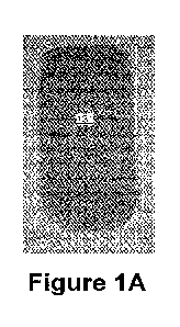
Figure 1A illustrates a qube having a size, shape and color that has been
selected in
accordance with a specific use and function during an oral surgery in
accordance with an
example embodiment.
Figure 1B illustrates three qubes having different sizes and shapes selected,
and
optionally cut from, a larger qube, such as that illustrated at Figure 1A,
each for a specific
intended use during an oral surgery in accordance with example embodiments.
Several qubes
of each of several types, shapes, sizes, and compositions are illustrated and
described in
.. example embodiments herein.
In some example embodiments, a qube may relate to an article for application
to
human and animal teeth and human and animal dental implants as a medicated and
non-
medicated space maintainer and/or retraction medium (referred to herein as a
QUBE, a Qube,
or a qube). A Qube may include, in an example embodiment, a synthetic sponge-
like material
with a 1) specific porosity size 2) which is autoclavable 3) which can be
colored 4) which
can be used a vehicle to carry a medicament 1.2% Chlorohexidine, 5) which can
be used a
vehicle to carry a medicament Calcium hydroxide Ca(OH),6)which can be used a
vehicle to
carry a medicament Povodine ¨Iodine solution, 7) which can be used a vehicle
to carry a
medicament 2% Iodine Potassium Iodide, 8) which can be used a vehicle to carry
a Sterile
saline. The Qube is to be applied as an interappointment dressing for
endodontically treated
teeth in the access cavity to serve as a barrier from microbial invasion of
the canal space as
well as a mechanism to prevent damage to surrounding tooth structure when a
dentist re-
accesses the tooth for permanent restoration. The Qube can also be used as a
barrier from
microbial invasion within the internal aspect of the coronal access of screw
retained dental
implants.
CA 03175901 2022-09-16
WO 2021/189061
PCT/US2021/023544
9
The Qube can also be used as a retraction medium for gingival flaps during
dental
surgery. The Qube can be contoured in specific shapes. The Qube can be
impregnated with
barium sulfate so it can be visible radiographically. The Qube can be inserted
and compacted
against gingival soft tissue to allow for atraumatic retraction.
Figures 2A-2G illustrate examples of qubes for use in various roles during
oral
surgical procedures, including as dental implant spacer qubes at Figure 2A,
and as grafting
qubes in Figure 2B, and as exo socket medicated qubes as in Figure 2C, and as
tapered
endoQubes in Figures 2D-2G.
Figures 2A-2C illustrate multiple examples of each of three different qube
types,
including implant cubes, grafting qubes and endo-socket medicated qubes in
accordance with
example embodiments.
Figure 2D-2G illustrates multiple examples of a fourth qube type, including
tapered
endo qubes in accordance with example embodiments.
The tapered shape of the wedge shaped qube of Figure 3 renders it advantageous
for
insertion into a dental implant space reserved for coupling with an abutment
component or an
abutment space reserved for coupling with a crown component for a duration of
an
osseointegration of the implant. The wedge of Figure 3 has four long sides and
a square or
rectangular base. Two long sides are parallel, tapered and/or triangularly-
shaped and the
other two sides are rectangular and form an acute angle at the tapered end.
One or both of the rectangular long sides may also be tapered or be
triangularly-
shaped, and a pyramidal qube or tetrahedral qube or truncated pyramid or
truncated
tetrahedron or cone-shaped, four or five sided pyramid, or pentagonal cone,
pentagonal
pyramid, truncated cone, half ellipsoid or partial ellipsoid or truncated
ellipsoid. One or more
of the long sides of a regular rectangular box, cube or polyhedron may be
tapered or
compressed spatially at one end.
An implant qube may be more densely-weighted at a tapered end, which may taper
to
a point or may round off or may be truncated such that a plane at a tapered
end may be
parallel to a base plane of greater area of a truncated implant qube, which
may have small and
large diameter circular end planes, or an elongated end plane quadrilateral
having at least one
short dimension which may taper to a point in one or both cross-plane
dimensions.
Figures 4A-4F illustrate seven types of qubes in accordance with example
embodiments. Different qubes may have different physical, chemical or
biological
CA 03175901 2022-09-16
WO 2021/189061
PCT/US2021/023544
properties, different functions, different uses, different roles to play
within oral surgical
applications, different compositions (polymer units or polymer side chains,
molecular
component monomers or side chains, monomer units or monomer side groups,
different sizes
(millimeters to centimeters) and shapes (spheres, ellipsoids, cubes,
polyhedrons with four to
5 twenty-four sides, wedges, pyramids, tetrahedrons, tapered polyhedrons,
truncated
polyhedrons, ovoid) and being grouped according to anticipated, intended or
scheduled uses,
functions, or specific applications among multiple example oral surgical
applications in
accordance with example embodiments.
Other qube types may include qubes having different colors or color
distributions or
10 different weights, or different overall weight densities (10kg/m3,
15kg/m3, 20kg/m3,
25kg/m3, 30kg/m3, 35kg/m3, 40kg/m3), or different weight or weight density
distributions
(20&30kg/m3õ or different porosities, autoclavabilities (thermal: 250F-300F,
225F-325F,
200F-275F, 200F-280F, 270F-280F, 275F-300F, 275F-325F; pressure: 20p5i-30p5i,
25-35p5i,
25-30p5i, 23-28p5i, 24=27p5i, 28-36p5i, 24-38psi)-indentation force deflection
(IFD)
capabilities, cell openness, cell densities (15-16 cells/cm, 10-20 cells/cmõ
medicament
chemistry (calcium hydroxide CaOH, barium sulfide BaS, titanium dioxide TiO2,
silver
nitride Ag3N, silver nitrate AgNO3, silver ion Ag+, silver ion Ag-, 1.2%
chlorohexidine,
povodine-iodine 2% iodine potassium iodide, sterile saline), or medicament
biology
(bacteriocidal, medicament combinational process types (e.g., soaking,
coating,
encapsulating, embedding, integrating, release rate).
Figures 5A-5G illustrates a qube placement tool having smooth, blunt
appendages for
placement of a qube for retraction, or for spacing, cushioning, bandaging, or
protecting gum
tissue around a bone graft site, tooth extraction site or other oral surgical
site in accordance
with an embodiment. The blunt appendages of the qube placement tool may be
fixed or may
be movable towards or away from each other at one end. Examples include plyers
without
sharp edges or forks with two or more prongs having rounded ends.
Figures 6A-6F illustrates a qube removal tool having sharp, jagged and/or
barbed
appendages for removing a qube from an oral surgical retraction site, or from
a dental
implant, or following use during oral surgery cushioning, bandaging, and/or
protecting gum
tissue at a bone graft site, a tooth extraction site, a dental implant site,
or another oral surgical
site in accordance with embodiments. An example qube removal tool may
articulate such
that the barbed ends of two appendages may be safely enclosed or sheathed or
enfolded or
CA 03175901 2022-09-16
WO 2021/189061
PCT/US2021/023544
11
interlocked in a "safety-on" position and may be actuated or articulated into
a "safety-off'
position such as to emerge to grab a qube for removal from a retraction site,
or a space
maintaining site or a tissue protection side or from a dental implant site or
other oral surgical
qube use site.
Figures 7A-7G illustrate a qube placement and/or removal tool that may have
articulated arms or articulated ends or both for, respectively, pushing,
maneuvering,
reorienting or bluntly pinching or holding a qube for placement at an oral
surgical site as a
qube placement tool and/or for grabbing, entangling or adhering to a qube to
remove it from
an oral surgical site as a qube removal tool.
The photographs of Figures 8A-8I illustrate certain steps in a process leading
incrementally to completion of the coupling of a dental implant at a site of
tooth extraction,
tooth absence, tooth loss or tooth decay. Advantageously, only minimal tissue
trauma was
caused by use of the qube.
Moreover, in certain example embodiments, use of a qube during an oral surgery
or
during a step or subset of steps of an oral surgery, e.g., a dental implant
surgery, a tooth or
jawbone grafting surgery, or another oral surgery involving one or more
retraction uses of
one or more cubes. In example embodiments, a dental impression may be made,
formed,
generated or located such as to make a dental impression for molding a
synthetic tooth, a
grown organic tooth or a tooth graft or set of teeth to replace a tooth or
teeth that may have
.. become decayed or that may be colliding with another tooth or gum, cheek,
tongue or lip area
causing pain or that may be rooted unevenly within an upper or lower jaw in
the front or back
of the mouth or may have fallen out such that a synthetic replacement tooth or
a grown
organic replacement dental implant or similar oral constituent may be desired
to take its
place.
Example embodiments are provided herein that may involve one or more oral
surgical
steps, sequences of two or more steps, subsets of multiple steps or several
steps, or complete
oral surgical processes that involve use of a qube for retraction, maintaining
space above or
within a dental implant, abutment or crown, or providing temporary structural
integrity
support for a tooth, gum, dentin, pulp, root, enamel, bone-cementum, crown or
combinations
or component parts thereof, or for catching, filtering, redirecting,
accumulating, or stabilizing
or controlling flow rate, area coverage or contained volume density of bodily
fluids, saliva,
CA 03175901 2022-09-16
WO 2021/189061
PCT/US2021/023544
12
blood, mucous, water, partly digested food or dislodged food fragments or
combinations or
evolving quantities or components thereof during an oral surgery.
Example embodiments may advantageously further involve reduced pain, reduced
swelling, and reduced tearing, scratching, slicing, stabbing or poking by
sharp edges or
jagged components of dental instruments, and reduced time to heal and enhanced
effectiveness by placement and use of one or more qubes for protecting,
cushioning,
deflecting, bandaging, or covering one or more exposed, wounded, inflamed or
otherwise
sensitive areas within a patient's mouth during an oral surgery.
Example embodiments of dental processes, both surgical and non-surgical,
advantageously include sequences of steps involving use of one or more qubes
for retraction,
maintaining space, cushioning, absorbing, softening, providing flexibility,
strength without
rigidity, and cohesiveness. After any of a wide variety of oral surgical
steps, and in various
orders and sequences of oral surgical steps, use of qubes throughout the
surgical processes
characteristically maintains an availability of choices of next steps, when to
stop, how to
provide a first dental care process and transition to a different oral state
prepared to provide a
second dental care process, while continuously, discretely, periodically
and/or increasingly
having an ability to return, and/or returning, suturing or positioning or
orienting tissue to an
original position or orientation due to no distortion or damage being caused
by this retraction
method involving use of a qube rather than a conventional retraction cord or
other
conventional retraction device or component.
Figure 8A includes a photograph that illustrates an incision site reflected in
a mirror in
accordance with an example embodiment.
Figure 8B includes a photograph of a retraction qube inserted into an
underside of a
surgical flap for retraction in accordance with an example embodiment.
Figure 8C includes a photograph of a qube further inserted under a flap in
accordance
with an example embodiment.
Figure 8D includes a photograph including qubes placed on both sides of an
incision
to retract a surgical flap and expose underlying bone in accordance with an
example
embodiment.
Figure 8E includes a photograph of a visible qube retracting a flap in
accordance with
an example embodiment.
CA 03175901 2022-09-16
WO 2021/189061
PCT/US2021/023544
13
Figure 8F includes a photograph that illustrates retraction qube allowing
access to
osteotomy for implant site preparation and better visibility in accordance
with an example
embodiment. A floor of a maxillary sinus is visible in the photograph of
Figure 8F as the
grey circular area in the image. Post-operatively, however, the patient had
minimal pain,
swelling and inflammation due to use of a qube retraction medium in an
advantageous form
of retraction during an oral surgery. There was also a strong unobstructed
healing response
due to lack of trauma during the surgery.
Figure 8G includes a photograph wherein the retraction qube of Figure 8F has
been
removed in accordance with an example embodiment.
Figure 8H includes a photograph of a qube removed from an opposite side of an
implant in place below a gingival surface in accordance with an example
embodiment.
Figure 81 includes a photograph of a surgical flap closed and sutured around a
dental
implant site in accordance with an example embodiment. In fact, a pair of
surgical flaps are
shown sutured in place on opposite sides of a dental implant site that
includes an abutment
component coupled to a dental implant that is secured to the jawbone of a
dental customer or
orthodontal patient. A crown may be next coupled to the abutment component of
the
example embodiment that is illustrated photographically at Figure 81.
Figures 9A-9U illustrate photographically examples of endocubes and/or dental
implant cubes in accordance with example embodiments.
Figures 10A-10I include surgical photographs that include qubes in place and
in use
during performance of various oral surgical steps. In these example
embodiments, the Qube
material exhibits advantageous usefulness and functionality as a demonstrably
suitable
retraction medium.
Figures 10A-10I are photographs illustrating multiple uses of qubes of
different sizes
and shapes specifically configured for a planned use during one or more
scheduled oral
surgeries in accordance with example embodiments. Figures 10A-10H include
multiple
photographs that include one or more qubes each in place performing a
retraction function.
Other uses of qubes include performing a spacer or space-maintaining function
during an oral
surgical procedure that includes two or more subsets of an overall surgery or
of a complete
procedure, such as between coupling a dental implant at a grafted or ungrafted
jawbone
socket site which has become decoupled from a tooth suddenly or gradually over
time, or a
jawbone site that is at risk of becoming decayed unless a rotting tooth is
extracted or repaired.
CA 03175901 2022-09-16
WO 2021/189061
PCT/US2021/023544
14
The two or more subsets of sequential oral surgical steps, processes, actions
or
modifications may, in one example embodiment, be spaced apart in time. In an
example
embodiment, a time delay advantageously allows for sufficient osseointegration
of a bone
graft within a jawbone socket, or socket graft, for example, prior to a dental
implant
procedure. Such a dental implant procedure may itself follow a sudden,
unexpected tooth
loss collision event or a long and steady incremental tooth decay process, or
an ordinary tooth
extraction, or a drawn-out, crumbling tooth disintegration lasting perhaps
years or another
tooth and/or jawbone volume reducing event.
The two or more surgical process subsets may, in another example embodiment,
be
spaced apart in time in order to allow sufficient osseointegration of a dental
implant inserted
within a jawbone socket at a depth below a gingival margin anywhere in a range
between a
shallow implant coupling location through an average implant depth location to
a deep
implant coupling location that may be significantly below a gingival margin.
In this example
embodiment, a second surgical process subset may involve coupling within a
dental implant
component for maintaining a space for attaching an abutment after sufficient
osseointegration
of the implant has occurred over the passage of time.
Figures 10A-10H include surgical photographs that include qubes in place and
in use
during performance of various oral surgical steps. In these example
embodiments, the Qube
material exhibits advantageous usefulness and functionality as a demonstrably
suitable
retraction medium.
Figures 11A-11K illustrate example embodiments of example retraction steps and
example retraction qubes useful in multiple roles during a dental implant
procedure involving
a screw-coupled dental implant in accordance with example embodiments.
Figures 12A-12J illustrate example embodiments of example retraction steps and
example retraction qubes useful in multiple roles during a dental implant
procedure involving
a cement-coupled dental implant in accordance with example embodiments.
Figures 13A-13G photographically illustrate further example qube uses and
applications and example qubes. One or more qubes may be used to retract a
rubber dam. A
qube may be used to retract soft tissue as well. A qube may be used to protect
cheek tissue,
tongue tissue, lip tissue, gum tissue, tonsil tissue, and/or tissue at the
roof of the mouth or
under the tongue from a surgical drill or other surgical instruments. A qube
may protect
CA 03175901 2022-09-16
WO 2021/189061
PCT/US2021/023544
tissues of the mouth from encountering tooth or implant fragments which may
have sharp or
jagged edges by adhering, blocking or deflecting such items.
Figure 14A-14T illustrate photographically an example procedure using
retraction
qubes in accordance with another embodiment.
5 Figure 15A schematically illustrates a decayed tooth 1512, which may
also be a
deformed tooth, a misplaced tooth, a misoriented tooth, a pain-producing
tooth, an outsized
molar or an otherwise unwanted tooth 1512, which is located between a pair of
healthy teeth
1511, 1513, and which is prior to extraction of the decayed tooth 1512, or
prior to a
collisional tooth loss, a disintegrational or naturally decaying tooth loss,
or another
10 unintended tooth loss, in accordance with example embodiments.
Figure 15B schematically illustrates the pair of healthy teeth of Figure 15A
following
extraction or other loss of decayed tooth 1512 leaving a gap both between the
healthy teeth
1511, 1513 above the gumline 1521 and extending beneath the gumline 1521 into
a socket
recess defined in a jawbone region from which a root region of the extracted
decayed tooth
15 1512 of Figure 15A has also been removed following incision and
retraction of gingival flaps
1551, 1552 around the decayed tooth 1512 in accordance with an embodiment.
Figure 15C schematically illustrates teeth with a gap above the gumline and a
socket
recess defined through the gumline and into the jawbone beneath after a
decayed tooth
extraction with the socket filled or partially filled with graft material in
accordance with an
embodiment.
Figure 15D1 schematically illustrates teeth with a gap above the gumline and a
socket
recess defined as extending into the gum tissue and into bone tissue beneath
with a qube 1533
draped over a graft-filled socket as in Figure 15C to protect and promote
osseointegration at
the socket graft site and to cushion and bandage the gums around the socket
for healing in
accordance with an example embodiment.
Figure 15D2 schematically illustrates teeth with a gap above the gumline and a
socket
recess defined as extending into the gum tissue and into the bone tissue
beneath with a qube
1534 inserted or partially inserted into a partially graft-filled socket as in
Figure 15C to
protect and promote osseointegration at the socket graft site and to cushion
and bandage the
gums around the socket for healing in accordance with an embodiment.
CA 03175901 2022-09-16
WO 2021/189061
PCT/US2021/023544
16
Figure 15E schematically illustrates sutured gingival flaps 1571, 1572
following
removal of retraction qubes 1531, 1532 to close a socket graft site draped
with qube 1533 for
osseointegration in accordance with an embodiment.
Figure 15F schematically illustrates teeth with a gap following
osseointegration,
removal of sutures, incision and retraction again of gingival flaps 1551, 1552
at a socket graft
site that is still protected by a blood-soaked qube 1543 in preparation for a
dental implant
procedure in accordance with an embodiment.
Figure 15G schematically illustrates an osseointegrated socket graft site
following
removal of a blood-soaked qube 1541 in accordance with an embodiment just
prior to
coupling a dental implant into the socket graft site in accordance with an
embodiment.
While the invention has been described in terms of several embodiments, those
skilled
in the art will recognize that the invention can be practiced with
modification and alteration.
The description is thus to be regarded as illustrative.