Note : Les descriptions sont présentées dans la langue officielle dans laquelle elles ont été soumises.
CA 03188767 2023-01-03
WO 2022/011018 PCT/US2021/040697
OPTIMIZED MULTIFOCAL WAVEFRONTS FOR PRESBYOPIA
CORRECTION
CROSS-REFERENCES TO RELATED APPLICATIONS
100011 This application claims ptiority to U.S, Provisional Patent Application
Ser. No.
63/049,277, filed July 8, 2020, which is herein incorporated by reference in
its entirety and
for all purposes.
BACKGROUND
100021 Optical aberrations that degrade visual acuity are common. Optical
aberrations are
imperfections of the eye that degrade focusing of light onto the retina.
Common optical
aberrations include lower-order aberrations (e.g., astigmatism, positive
defocus (myopia) and
negative defocus (hyperopia)) and higher-order aberrations (e.g., spherical
aberrations, coma
and trefoil).
100031 Existing treatment options for correcting optical aberrations include
glasses, contact
lenses, and reshaping of the cornea via laser eye surgery. Additionally,
intraocular lenses are
often implanted to replace native lenses removed during cataract surgery.
100041 Presbyopia may be defined as a gradual loss of near vision, or the
ability to focus on
nearby objects, that may occur naturally with age. Presbyopia may become
noticeable for
patients in their early to mid-40s and may continue to worsen over time as
they age until
around age 65. As patients age, the crystalline lens gradually stiffens and
grows in size,
generally making it difficult for the lens to accommodate (or change shape)
adequately to
focus on nearby objects.
BRIEF SUMMARY
100051 The following presents a simplified summary of some embodiments of the
invention in order to provide a basic understanding of the invention. This
summary is not an
extensive overview of the invention. It is not intended to identify
key/critical elements of the
invention or to delineate the scope of the invention. Its sole purpose is to
present some
embodiments of the invention in a simplified form as a prelude to the more
detailed
description that is presented later.
CA 03188767 2023-01-03
WO 2022/011018 PCT/US2021/040697
100061 Embodiments described herein are directed to ophthalmic lenses having
at least one
subsurface optical structure (e.g., diffractive optical structures and/or non-
diffractive optical
structures) with enhanced distribution of refractive index values. In many
embodiments, the
subsurface refractive index variations are formed via focusing ferntosecond
duration laser
pulses onto a targeted sequence of subsurface volumes of an ophthalmic lens.
The refractive
indexes of the annular optical structure vary radially relative to the optical
axis up to an upper
limit refractive index (e.g., providing any suitable phase change less than
1.0 wave). The
refractive indexes of the annular optical structure are equal to the upper
limit refractive index
over a range of radii (e.g., at least 0.15 mm length) from the optical axis.
In many
embodiments, the refractive indexes of the annular optical structure are equal
to a lower limit
refractive index (e.g., providing a phase change of 0.0 waves) over a range of
radii (e.g., at
least 0.15 mm length) from the optical axis. The enhanced distribution of
refractive index
values can be formed using fewer laser pulses in comparison with a
corresponding
distribution of refractive index values determined via a ratio approach.
Additionally, limiting
the refractive index values to equal to or less than the upper limit
refractive index helps to
reduce damage induced by- the sequence of laser pulses at a given pulse energy
level as
compared to forming a corresponding subsurface optical structure(s) using
refractive index
values that are greater than the upper limit refractive index. The approaches
described herein
may be useful in forming a subsurface optical structure(s) in any suitable
ophthalmic lenses
(e.g., intraocular lenses, contact lenses, corneas, glasses, and/or native
lenses).
100071 In some embodiments, methods, systems, and devices are described for
determining
parameters for forming an optical structure (e.g., a subsurface optical
structure) in an
ophthalmic lens for improving vision in a patient. These parameters may be
used to control
an energy source to appropriately form the desired optical structure.
100081 In many cases, presbyopia patients less than 45 years old may be
classified as early
presbyopes requiring relatively minor correction; presbyopia patients between
45 and 55
years old may be classified as mid presbyopes requiring a moderate level of
correction; and
presbyopia patients over the age of 55 years old (or patients who have
received a non-
accommodating manner focal intraocular lens (IGL)) may be classified as
advanced
presbyopes requiring a relatively large level of correction.
100091 Disclosed herein are methods for forming subsurface optical structures
in an
ophthalmic lens for improving patient vision (e.g., for correcting
presbyopia). In some
embodiments, the method includes defining a first phase-wrapped wavefront
corresponding
2
CA 03188767 2023-01-03
WO 2022/011018 PCT/US2021/040697
to a first optical structure configured to cause the ophthalmic lens to
diffract light to multiple
focal points, wherein the first phase-wrapped wavefront is a wavefront having
a first
predetermined phase height (e.g., not equal to 1 wave); defining a first
spherical wavefront
configured for inducing a first spherical aberration in the ophthalmic lens;
and generating,
based on the first phase-wrapped wavefront and the first spherical wavefront,
energy output
parameters for forming a first subsurface optical structure in the ophthalmic
lens using an
energy source, wherein the first subsurface optical structure is configured to
correct
presbyopia by providing extended depth of focus that produces increased
intermediate vision
quality.
100101 in some embodiments, the method may include accessing an optical
prescription for
the patient, wherein the optical prescription comprises one or more
prescription parameters
for refracting light directed at a retina of the patient so as to improve
vision; and generating a
first variable wavefront based on the optical prescription, wherein the first
variable wavefront
comprises at least one portion that has a phase height greater than 1 wave;
wherein generating
the first phase-wrapped wavefront comprises collapsing the first variable
wavefront to the
first predetermined phase height.
100111 In sonic embodiments, the energy output parameters specify a plurality
of power
levels corresponding to a plurality of optical zones on the ophthalmic lens.
The method may
include directing a first energy beam from the energy source at a first
subsurface optical zone
of the ophthalmic lens for a first duration, wherein a power level of the
first energy beam is
based on a corresponding power level as specified by the energy output
parameters; and
directing a second energy beam from the energy source at a second subsurface
optical zone
on the ophthalmic lens for a second duration, wherein a power level of the
second energy
beam is based on a corresponding power level as specified by the energy output
parameters.
The first energy beam and the second energy beam may alter refractive indexes
of the first
subsurface optical zone and the second subsurface optical zone, respectively,
and wherein the
first subsurface optical structure comprises the first subsurface optical zone
and the second
subsurface optical zone.
100121 in some embodiments, the first optical structure is configured to cause
the
ophthalmic lens to be a bifocal lens having a 2 diopter add power. In some
embodiments, the
first optical structure is configured to cause the ophthalmic lens to be a
bifocal lens having a
1.5 diopter add power. In some embodiments, the first predetermined phase
height is between
3
CA 03188767 2023-01-03
WO 2022/011018 PCT/US2021/040697
about 0.5 to 0.6 waves. In some embodiments, the first spherical aberration is
around
Inn. In some embodiments, the first spherical aberration is around 0.2 Rm.
[00131 In some embodiments, forming the subsurface optical structure comprises
directing
an energy beam toward a volume of the ophthalmic lens so as to change a
refractive index of
the volume.
[0014] in some embodiments, the method may include defining a second phase-
wrapped
wavefront corresponds to a second optical structure configured to cause the
ophthalmic lens
to diffract light to multiple focal points, wherein the second phase-wrapped
wavefront is a
wavefront having a second predetermined phase height (e.g., not equal to 1
wave); defining a
second spherical wavefront configured to cause a second spherical aberration
in the
ophthalmic lens; and generating, based on the second phase-wrapped wavefront
and the
second spherical wavefront, energy output parameters for forming a second
subsurface
optical structure in the ophthalmic lens using an energy source. In some
embodiments, the
first subsurface optical structure is configured to correct a first stage of
presbyopia in the
patient, the second subsurface optical structure is configured to correct a
second stage of
presbyopia in the patient, and the second stage of presbyopia in the patient
is later than the
first stage of presbyopia in the patient.
[0015] Also disclosed are of ophthalmic lenses for improving vision (e.g., for
correcting
presbyopia in a patient), which may in some embodiments be performed using the
described
methods. In some embodiments an ophthalmic lens may include a first subsurface
optical
structure comprising concentric Fresnel rings within an interior of the
ophthalmic lens. Each
of the concentric Fresnel rings can define a volume having a desired
refractive index. 'The
first subsurface optical structure may be configured to: induce a first
spherical aberration in
the ophthalmic lens; and diffract light to multiple focal points based on a
phase-wrapped
wavefront having a first predetermined phase height (e.g., not equal to 1
wave).
[0016] In some embodiments, the ophthalmic lens is an intraocular lens, a
contact lens, or a
cornea of the patient. In some embodiments, wherein the first subsurface
optical structure is
configured to cause the ophthalmic lens to be a bifocal lens having a 2
diopter add power. In
some embodiments, the first subsurface optical structure is configured to
cause the
ophthalmic lens to be a bifocal lens having a 1.5 diopter add power. In some
embodiments,
the first predetermined phase height is between about 0.5 to 0.6 waves, In
some
4
CA 03188767 2023-01-03
WO 2022/011018 PCT/US2021/040697
embodiments, the first spherical aberration is around ¨0.2 gm. In some
embodiments, the first
spherical aberration is around 0.2 gm
100171 In some embodiments, the ophthalmic lens includes a second subsurface
optical
structure. The first subsurface optical structure can be embedded in a first
layer of the
ophthalmic lens. The second subsurface optical structure can be embedded in a
second layer
of the ophthalmic lens. In some embodiments, the first subsurface optical
structure is
configured to correct a first stage of presbyopia in the patient and the
second subsurface
optical structure is configured to correct a second stage of presbyopia in the
patient. The
second stage of presbyopia in the patient can be later than the first stage of
presbyopia in the
patient.
BRIEF DESCRIPTION OF THE DRAWINGS
100181 FIG. 1 is a plan view illustration of an ophthalmic lens that includes
subsurface
optical structures with enhanced distribution of refractive index variations,
in accordance
with embodiments.
100191 FIG. 2 is a plan view illustration of a layer of the subsurface optical
structures of
the ophthalmic lens of FIG. 1.
100201 FIGS. 3A-3B illustrate example wavefronts through a medium for parallel
and
converging rays of light.
100211 FIGS. 3C-3D illustrate example wavefronts that may simulate aberrations
of the
eye.
100221 FIG. 3E illustrates a two-dimensional wavefront map and a corresponding
first
variable wavefront.
100231 FIG. 3F illustrates a first phase-wrapped wavefront corresponding to
the first
variable wavefront.
100241 FIG. 4 illustrates a second phase-wrapped wavefront having a phase
height less
than l wave.
100251 FIG. 5 illustrates a two-dimensional map representation of a phase-
wrapped
wavefront phase-wrapped at an optical phase height less than I wave, such as
the wavefront
in FIG. 4.
100261 FIG. 6 illustrates an example of an optical structure having
diffractive properties.
CA 03188767 2023-01-03
WO 2022/011018 PCT/US2021/040697
[00271 FIG. 7 is a graph illustrating the relative distribution of light
between a near-vision
focal point and a far-vision focal point as phase height of a wavefront is
adjusted between 0
wave and I wave.
100281 FIG. 8 illustrates a cross section of an ophthalmic lens including a
subsurface
optical structure having multiple substructures.
100291 FIGS. 9A-9B illustrate example conceptualizations of an ophthalmic lens
having a
plurality of optical zones.
[0030] FIG. 10 illustrates an example method for determining parameters for
forming a
subsurface optical structure for improving vision in a patient.
10031] FIG. 11 illustrates an example of presbyopia progression in a patient.
100321 FIG. 12 illustrates an example chart of presbyopia progression.
100331 FIG. 13 illustrates example image quality metrics across a diopter
range using a
number of bifocal wavefronts.
[0034] FIG. 14 illustrates the concept of spherical aberrations in lenses.
[0035] FIG. 15 illustrates example image quality metrics for a patient with
presbyopia with
lenses having positive and negative spherical aberrations as compared to a
control with zero
spherical aberration.
[0036] FIG. 16 illustrates a graph overlaying the line for a 2-di opter
bifocal of FIG. 13
with the spherical aberration lines, and the control line of FIG. 15.
[0037] FIG. 17 illustrates the graph of FIG. 16 further overlaying lines
corresponding to
image quality metrics of phase-wrapped trifocals.
[0038] FIG. 18 illustrates a graph including a number of the previously
described lines as
well as lines corresponding to bifocals with spherical aberrations.
100391 FIGS, 19A-19B illustrate cross-sections of the wavefronts corresponding
to
particular lines of FIG. 18.
[0040] FIG. 20 is a table showing example wavefronts that may be implemented
for
different stages of presbyopia.
6
CA 03188767 2023-01-03
WO 2022/011018 PCT/US2021/040697
[0041] FIG. 21 illustrates an example method 2000 for forming a subsurface
optical
structure in an ophthalmic lens for correcting presbyopia in a patient.
[0042] FIG. 22 illustrates simulated resulting phase-wrapped wavefronts for a
design
0.4 wave height phase-wrapped wavefront due to practical limitations
associated with
inducing the design 0.4 wave height phase-wrapped wavefront in an artificial
or biological
optical material.
[0043] FIG. 23 illustrates simulated resulting through-focus retinal image
quality for the
simulated resulting phase-wrapped wavefronts of FIG. 22.
[0044] FIG. 24 illustrates simulated resulting phase-wrapped wavefronts for a
scaled-up
version of the 0.4 wave height phase-wrapped wavefront of FIG. 22.
10045] FIG. 25 illustrates simulated resulting through-focus retinal image
quality for the
simulated resulting phase-wrapped wavefronts of FIG. 24,
[0046] FIG. 26 shows simulated resulting through-focus retinal image qualities
illustrating
that near visual benefit can be recovered by scaling a design wavefront
height.
DETAILED DESCRIPTION
[0047[ In the following description, various embodiments of the present
invention will be
described. For purposes of explanation, specific configurations and details
are set forth in
order to provide a thorough understanding of the embodiments. However, it will
also be
apparent to one skilled in the art that the present invention may be practiced
without the
specific details. Furthermore, well-known features may be omitted or
simplified in order not
to obscure the embodiment being described.
[0048] FIG. 1 is a plan view illustration of an ophthalmic lens 10 that
includes one or more
subsurface optical structures 12 with annular distribution of refractive index
variations, in
accordance with embodiments. The one or more subsurface structures 12
described herein
can be formed in any suitable type of ophthalmic lens including, but not
limited to, intra-
ocular lenses, contact lenses, corneas, spectacle lenses, and native lenses
(e.g., a human
native lens). The one or more subsurface optical structures 12 with annular
distribution of
refractive index variations can be configured to provide a suitable refractive
correction for
each of many optical aberrations such as astigmatism, myopia, hyperopia,
spherical
aberrations, coma and trefoil, as well as any suitable combination thereof.
7
CA 03188767 2023-01-03
WO 2022/011018 PCT/US2021/040697
[0049] FIG. 2 is a plan view illustration of one of the subsurface optical
structures 12 of
the ophthalmic lens 10, The illustrated subsurface optical structure 12
includes concentric
circular sub-structures 14 separated by intervening line spaces or gaps 16, In
FIG. 2, the size
of the intervening line spaces 16 is shown much larger than in many actual
embodiments. For
example, example embodiments described herein have an outer diameter of the
concentric
circular sub-structures 14 of 3.75 ram and intervening line spaces 16 of 0.25
urn, thereby
having 1,875 of the concentric circular sub-structures 14 in embodiments where
the
concentric circular substructures 14 extend to the center of the subsurface
optical
structure 12. Each of the concentric circular sub-structures 14 can be formed
by focusing
suitable laser pulses onto contiguous sub-volumes of the ophthalmic lens 10 so
as to induce
changes in refractive index of the sub-volumes so that each of the sub-volumes
has a
respective refractive index different from an adjacent portion of the
ophthalmic lens 10 that
surrounds the sub-structure 14 and is not part of any of the subsurface
optical structures 12.
[0050] In many embodiments, a refractive index change is defined for each sub-
volume of
the ophthalmic lens 10 that form the subsurface optical structures 12 so that
the resulting
subsurface optical structures 12 would provide a desired optical correction
when formed
within the ophthalmic lens 10. The defined refractive index changes are then
used to
determine parameters (e.g., laser pulse power (mW), laser pulse width (fs)) of
laser pulses
that are focused onto the respective sub-volumes to induce the desired
refractive index
changes in the sub-volumes of the ophthalmic lens 10.
100511 While the sub-structures 14 of the subsurface optical structures 12
have a circular
shape in the illustrated embodiment, the sub-structures 14 can have any
suitable shape and
distribution of refractive index variations. For example, a single sub-
structure 14 having an
overlapping spiral shape can be employed. In general, one or more
substructures 14 having
any suitable shapes can be distributed with intervening spaces so as to
provide a desired
diffraction of light incident on the subsurface optical structure 12ss. More
information about
subsurface optical structures and forming such structures may be found in U.S.-
Provisional
Application No. 63/001,993, which is incorporated herein by reference in its
entirety for all
purposes.
100521 In some embodiments, a system including one or more processors may be
configured to determine parameters for forming one or more optical structures
(e.g.,
subsurface optical structures) for improving or correcting vision. In some
embodiments, the
8
CA 03188767 2023-01-03
WO 2022/011018 PCT/US2021/040697
one or more processors of the system may be configured to access a first
optical prescription
for the patient. The first optical prescription may be prescribed by, for
example, an
optometrist. The first optical prescription may include one or more
prescription parameters
for refracting light directed at a retina of the patient so as to improve
vision. The prescription
parameters may be determined based on any suitable means of measurement. The
prescription parameters may specify any suitable parameters for correcting or
improving
vision. For example, the prescription parameters may include diopter values of
sphere,
cylinder, or axis. The prescription parameters may include parameters for
correcting one or
more of a variety of low-order aberrations (e.g., myopia, hyperopia,
astigmatism) and high-
order aberrations (e.g., spherical aberration, coma, trefoil).
100531 FIGS. 3A--3B illustrate example wavefronts 305, 306 through a medium
for parallel
and converging rays of light. Prescriptions for correcting or improving vision
of a patient can
essentially be described as a prescription for creating an optical structure
that effects a
wavefront configured to modify incoming rays of light before they reach the
retina of the
patient. A wavefront is an imaginary surface of constant phase. A wavefront
can also be
thought of as a surface that is normal or perpendicular to rays of light
passing through the
wavefront. FIG. 3A illustrates a planar wavefront 305 from parallel rays of
light. A.s is
evident, the wavefront 305 is perpendicular to the parallel rays of light at
each point of
intersection. FIG. 3B illustrates a spherical wavefront 306 from converging
rays of light.
FIG. 3B simulates an ideal configuration of an eye, where the rays of light
converge at a
single point (on the retina 302). Each of the rays is perpendicular to the
wavefront 307 at its
respective point of intersection with the wavefront 307. The illustrated rays
converge at a
single point.
100541 FIGS. 3C-31) illustrate example wavefronts 308, 309 that may simulate
aberrations
of the eye. Unlike the rays in FIG. 3B, the rays in FIG. 3C do not converge at
a single point
on the retina 302 (e.g., at or near the macula). Such non-convergence may
cause issues with
vision by not allowing for a focused image (e.g., causing myopia). FIG. 31)
illustrates an
aberrated wavefront 309 simulating another aberration of the eye (e.g. higher
order
aberrations). Again, each of the rays is perpendicular to the wavefront 309 at
its respective
point of intersection with the wavefront 309. And again, as illustrated, the
rays in FIG. 31) do
not converge at a single point on the retina 302 (and in fact do not converge
at all), causing
issues with vision. An appropriate optical structure with a corrective
wavefront may be used
to correct issues produced by aberrations by, for example, refracting light
such that the light
9
CA 03188767 2023-01-03
WO 2022/011018 PCT/US2021/040697
rays are made to converge at a single appropriate point on the retina 302.
Disclosed herein are
methods, devices, and system .s for use in forming such optical structures.
Although the
disclosure focuses on methods, devices, and systems for correcting aberrations
of the eye, the
disclosure also contemplates enhancing what may be considered normal vision by
similar
methods, devices, and systems.
100551 FIG. 3E illustrates a two-dimensional wavefront map 310 and a
corresponding first
variable wavefront 320. In some embodiments, the one or more processors may
use the first
optical prescription to determine a wavefront for an optical structure for
correcting or
improving vision of the patient. In some embodiments, the one or more
processors may
generate a wavefront map, which may be visualized, for example, by the two-
dimensional
wavefront map 310. The contours of the two-dimensional wavefront map 310 may
specify
different optical phases of the corresponding wavefront. For example, the
different shades in
the two-dimensional wavefront map 310 specifies different optical phases of
the
corresponding wavefront. In some embodiments, the one or more processors may
do so by
first computing the Zernike coefficient for defocus (C2,o) using the following
equation:
(1) C2,o = P*rmax2/(4*sqrt(3)), where P is an add power specified in the first
prescription,
and Tmax is the maximum radius of an optical zone.
The Zernike coefficient is a scalar that may be expressed in units of
micrometers, In some
embodiments, the two-dimensional wavefront map may then be calculated using
the
following equation:
(2) Wum = C2,0 * sqrt(3) * (2 * p2 - 1), where p is a normalized radial pupil
coordinate
(radial coordinate / rmax)
Wum provides a value (e.g., in units of micrometers) for each point of a two-
dimensional
wavefront map. Referencing FIG. 3D, the two-dimensional wavefront map 310 for
a
particular optical prescription may be generated using this equation.
100561 In some embodiments, the one or more processors may be configured to
generate a
first variable wavefront based on the first optical prescription. Referencing
FIG. 3D, for
example, the first variable wavefront 320 may be generated based on
specifications provided
by the first optical prescription. The first variable wavefront describes a
wavefront in units of
waves with respect to a specified wavelength. In some embodiments, the first
variable
wavefront comprises at least one portion that has a phase height greater than
1 wave, In some
CA 03188767 2023-01-03
WO 2022/011018 PCT/US2021/040697
embodiments, the first variable wavefront may be generated based on the two-
dimensional
wavefront map. The first variable wavefront may be determined with respect to
any desired
wavelength by dividing Wum for each point by the desired wavelength. For
example, the first
variable wavefront may be determined with respect to a center of the visible
spectrum (e.g.,
0.555 p.m in daylight). In this example, the equation below may be used to
generate a first
variable wavefront at 0.555 um).
(3) Wwv= 'Num / 0.555 urn
[0057] FIG. 3F illustrates a first phase-wrapped wavefront 325 corresponding
to the first
variable wavefront 320. In some embodiments, the one or more processors may be
configured to phase wrap the first variable wavefront, which may include
collapsing the first
variable wavefront to generate a first phase-wrapped wavefront. Phase wrapping
the first
variable wavefront may involve collapsing the first variable wavefront into a
wavefront
having a predetermined phase height (i.e., the height from peak to valley of
the wavefront).
For example, referencing FIG, 3B, the first phase-wrapped wavefront 325 may
have a phase
height of I wave. Phase-wrapping a variable wavefront to l wave causes no
appreciable
change in diffraction or refraction of light rays, and may thus be suitable,
for example, for a
patient having only myopia. An example Matlab algorithm for phase-wrapping to
a phase
height of I wave is shown below, where W555 = Wwv and Wrap =
while cnt == 0
W555( W555 < -Wrap ) = W555( W555 < -Wrap ) + Wrap;
if sum( W555(:) < -Wrap ) == 0
cot = I;
end
end
cnt = 0;
while cnt == 0
W555( W555 > Wrap) = W555( W555 > Wrap ) - Wrap;
if sum( W555(:) > Wrap ) == 0
cnt =
end
end
[0058] In some embodiments, collapsing the first variable wavefront may
include
identifying a plurality of discrete segments of the first variable wavefront,
in some
embodiments, as is the case in FIG. 3F, each of these discrete segments (e.g.,
320-1 to 320-n)
may be circumferential discrete segments that extend radially around the two-
dimensional
II
CA 03188767 2023-01-03
WO 2022/011018 PCT/US2021/040697
wavefront map 310 of the ophthalmic lens. For example, the discrete segment
320-1 in the
first variable wavefront 320 may correspond to the portion 310-1 in the two-
dimensional
wavefront 310, the discrete segment 320-2 may correspond to the segment 310-2,
the discrete
segment 320-3 may correspond to the segment 310-3, and so on. In other
embodiments, the
discrete segments may not be circumferential, and the first variable wavefront
may be
segmented based on, for example, phase height. In the example illustrated in
FIG. 3F, each
of the discrete segments (325-Ito 325-n) is circumferential, and each discrete
segment is
adjacent to and concentric with another discrete segment. For example, the
discrete segment
325-2 is adjacent to and concentric with the discrete segment 325-1
(similarly, the discrete
segment 325-3 is adjacent to and concentric with the discrete segment 325-2,
and so on). In
some embodiments, the one or more processors of the system may reduce a phase
height of
each discrete segment by a respective scalar such that a peak of the first
discrete segment is at
a desired phase height. For example, in FIG. 3F, the phase height of each
discrete segment is
reduced to a predetermined phase height of 1 wave, yielding the first phase-
wrapped
wavefront 325. As mentioned above, collapsing the first variable wavefront 320
to the phase-
wrapped wavefront 325 (which is collapsed to I wave) causes no appreciable
change in
diffraction or refraction, and light rays passing an optical structure based
on the collapsed
phase-wrapped wavefront 325 essentially behave in the same manner as light
rays passing an
optical structure formed based on the first variable wavefront 320. The
resulting phase-
wrapped wavefront may include a central discrete segment (e.g., the discrete
segment 325-1)
and a number of surrounding circumferential, adjacent echelettes (e.g., the
discrete segments
325-2 to 325-n) as illustrated in FIG. 3E.
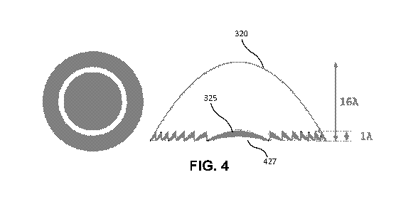
100591 FIG. 4 illustrates a second phase-wrapped wavefront 427 having a phase
height less
than 1 wave. In some embodiments, the system may be configured to phase wrap
the first
variable wavefront at a predetermined phase height that is not at 1 wave to
generate a second
phase-wrapped wavefront. For example, referencing FIG. 5, the predetermined
phase height
of the illustrated phase-wrapped wavefront 427 is less than I wave. As
discussed further
below, collapsing a wavefront to a phase height other than 1 wave causes
diffraction, which
may be useful for creating a multifocal optical structure. Thus, such a
wavefront may be
referred to herein as a "diffractive phase-wrapped wavefront." in some
embodiments, the
phase-wrapped wavefront may be collapsed at a phase height greater than 1
wave. The
decision as to whether a wavefront is collapsed to a phase height greater than
1 wave or to a
phase height less than 1 wave may have some practical effects. For example,
phase wrapping
12
CA 03188767 2023-01-03
WO 2022/011018 PCT/US2021/040697
at greater than 1 wave may reduce diffractive chromatic effects. However,
phase wrapping to
greater than 1 wave requires more available refractive index change as
compared to phase
wrapping to less than 1 wave, and any material used is subject to a given
range of possible
refractive index changes, which may be a limiting factor (e.g., limited by the
properties of the
material). This may be ultimately overcome in many cases by writing multiple
layers or
volume filling, however, but there are still limits. So there is a tradeoff
between phase
wrapping at greater than 1 wave or less than I wave. Whether a wave front is
phase-wrapped
to less than 1 wave or greater than 1 wave may also have implications for
energy distribution
of far/near vision (e.g., for patients with presbyopia), and the practitioner
can control this as
necessary to achieve a desired effect.
100601 FIG. 5 illustrates a two-dimensional map representation of a phase-
wrapped
wavefront 500 phase-wrapped at an optical phase height less than 1 wave, such
as the
wavefront 427 in FIG. 4. The illustrated phase-wrapped wavefront has a 3.0 mm
diameter
optical zone and a diffractive bifocal with 2.5 Diopters (1)) of add-power.
The diffractive
bifocal wavefront is designed to have an optical phase height of 0.35 waves at
555 nm
wavelength. As illustrated, the phase-wrapped wavefront 500 includes five
discrete
circumferential segments, each segment gradually decreasing in phase height
(from 0.35
waves to 0 waves) from an inner boundary of the segment to an outer boundary
of the
segment.
[00611 FIG. 6 illustrates an example of an optical structure 610 having
diffractive
properties. In some embodiments, an optical structure having a phase-wrapped
wavefront
collapsed at a phase height other than 1 wave (e.g., less than I wave) has
diffractive effects
that create multiple focal points, which may be useful, for example, in
correcting vision in
patients having presbyopia. As illustrated in -FIG 6, light rays passing
through the optical
structure 610, which is an optical structure with diffractive properties, an
incident beam can
be focused simultaneously at several positions along the propagation axis.
Diffraction in this
manner can be used to create multiple focal points, for example, to improve
the vision of
patients with presbyopia. For example, an optical structure having diffractive
properties may
have a first focal point for near-vision and a second focal point for far-
vision.
100621 FIG. 7 is a graph 700 illustrating the relative distribution of light
between a near-
vision focal point and a far-vision focal point as phase height of a wavefront
is adjusted
between 0 wave and 1 wave. In some embodiments, the system may generate
diffractive
13
CA 03188767 2023-01-03
WO 2022/011018 PCT/US2021/040697
phase-wrapped wavefronts (e.g., phase-wrapped wavefronts at less than 1 wave
or greater
than I wave), for conditions such as presbyopi a that are designed to provide
both high optical
quality for far-vision and intermediate- and near-vision (e.g., good through-
focus image
quality), but with the understanding that there may be a trade-off. An example
representation
of this trade-off is illustrated in FIG. 7. As illustrated by the far-vision
curve 710, as the
phase height increases to I wave, the percentage of light distributed to the
far-vision focal
point by the diffraction of incoming light decreases (and therefore image
quality for far-
vision generally decreases). By contrast, referencing the near-vision curve
720, as the phase
height increases to I wave, the percentage of light distributed to the near-
vision focal point
increases (and therefore image quality for near-vision generally increases).
In some
embodiments, a desired distribution for this tradeoff may be specified in an
optical
prescription (e.g., as add power), and may be determined based on any suitable
of patient-
dependent factors. For example, the patient who often engages in high-detail
work (e.g., a
watchmaker) may require a relatively high add power (e.g., 4.0 diopters). A
relatively low
add power (e.g., 1.0 diopters) may be suitable for a patient who does not
engage in such high-
detail work. A diffractive phase-wrapped wavefront may be generated with a
prescription
having such considerations in mind to come to a desired trade-off.
100631 In some embodiments, the one or more processors may be configured to
generate
multiple wavefronts, for example, to correct multiple aberrations of the eye.
In some
embodiments, the one or more processors may generate a second variable
wavefront based on
a second optical prescription, wherein the second optical prescription
comprises an add
power for multifocal vision correction. The term second optical prescription
does not
necessarily reference a separate prescription, and may instead refer to
separate one or more
parameters for correcting a different aberration than the first optical
prescription. For
example, a patient may receive a single prescription from an optometrist for
correcting near-
vision based on parameters of a first optical prescription and for correcting
far-vision based
on parameters of a second optical prescription (e.g., including an add power).
In some
embodiments, the one or processors may phase-wrap the second variable
wavefront, wherein
phase wrapping the second variable wavefront comprises collapsing the second
variable
wavefront to a second phase-wrapped wavefront having a second predetermined
phase
height. The second predetermined phase height may be less than 1 wave, so as
to allow for
diffractive effects as discussed above. In some embodiments, a first phase-
wrapped
wavefront may have a phase height of I wave, and the second phase-wrapped
wavefront may
14
CA 03188767 2023-01-03
WO 2022/011018 PCT/US2021/040697
have phase height less than I wave. In these embodiments, the first phase-
wrapped wavefront
may be useful for correcting myopia and the second phase-wrapped wavefront may
be useful
for correcting presbyopia, for example.
100641 FIG. 8 illustrates a cross section of an ophthalmic lens including a
subsurface
optical structure having multiple substructures 810. In some embodiments, the
one or more
processors may be configured to generate, based on the first phase-wrapped
wavefront,
energy output parameters for forming a first optical structure using an energy
source. In some
embodiments, the first optical structure may be configured to refract light
directed at the
retina of the patient so as to improve vision. In some embodiments, the
optical structure may
be a subsurface optical structure. :For example, referencing the cross-section
illustrated in
FIG. 8, the optical structure may be a subsurface optical structure having
multiple
substructures 810 that may be concentric. As discussed in further detail
above, subsurface
optical structures may be achieved by focusing laser pulses appropriately to
depths within the
ophthalmic lens such that changes in refractive property occur to sub-volumes
in the interior
of the ophthalmic lens.
100651 The conventional approach for forming a diffractive ophthalmic lens
involves
creating Fresnel rings that project outward from an exterior surface of the
ophthalmic lens.
Such a configuration not only increases the thickness profile of the lens, but
it may also cause
issues with the optical properties of the ophthalmic lens. For example, in the
case of a contact
lens, disposing Fresnel rings on the outward-facing exterior surface of the
contact lens may
cause errors in light diffraction or refraction because the level of tear film
may vary across
the peaks and valleys of the Fresnel rings. And disposing the Fresnel rings on
the inward-
facing exterior surface of the contact lens may cause patient discomfort.
Additionally, rings
disposed on an exterior surface of the ophthalmic lens may become sites for
the accumulation
of debris which causes light scatter and loss of contrast.
100661 Moreover, conventional approaches rely on changes in the thickness of
ophthalmic
lenses to supply the base power of the ophthalmic lenses. In these approaches,
the refractive
index of the material throughout an ophthalmic lens may remain constant. This
reliance on
thickness necessarily means that lenses with relatively high base powers are
relatively thick.
For contact lenses, this may mean patient discomfort. For 10Ls, this may mean
an increase in
patient risk during surgery, and a higher potential for complications (e.g.,
because it may be
more difficult to get the JOL seated in the capsular bag). By contrast, the
disclosed methods
CA 03188767 2023-01-03
WO 2022/011018 PCT/US2021/040697
of creating subsurface optical structures using an energy system (e.g., a
laser) does not rely
on changing the thickness of an ophthalmic lens for the base power. Rather, as
explained
above, refractive indices of subyolumes within the ophthalmic lens are
modified to supply the
base power of the ophthalmic lens and thereby refract and/or diffract light as
desired. Finally
the use of an energy system as described below with respect to optical zones
provides
increased resolution as compared to more conventional techniques such as
cryolathes or
molded injection.
[0067] FIGS. 9A-9B illustrate example conceptualizations of an ophthalmic lens
900
having a plurality of optical zones. In some embodiments, an ophthalmic lens
may be divided
up into a plurality of pixels, each pixel corresponding to an optical zone. An
optical zone may
be a sub-region or a sub-volume of an ophthalmic lens. This is illustrated in
FIG. 9A, which
shows the ophthalmic lens 900 divided up into a plurality of pixels (e.g., the
pixels 910 and
920) in a grid fashion. Although FIG. 9A illustrates uniform pixels that are
square shaped,
this disclosure contemplates that pixels may be of any suitable shape (e.g.,
hexagonal,
pentagonal, circular) and that they may not be uniform (e.g., they may of
different shapes and
sizes). A pixel area may correspond to the resolution of an energy delivery
system (e.g., a
laser system) configured to form an optical structure corresponding to a phase-
wrapped
wavefront. That is, a pixel area may correspond to a minimum area of a sub-
region of the
ophthalmic lens at which the energy delivery system may focus an energy beam
(e.g., a laser
pulse) to change a refractive index of the sub-volume associated with the sub-
region. FIG.
9B illustrates another conceptualization of optical zones, where the
ophthalmic lens is not
divided up into discrete pixels. Instead, the ophthalmic lens is mapped out
using a coordinate
system (e.g., a two-dimensional x-y coordinate system, a three-dimensional x-y-
z coordinate
system, or a polar coordinate system (radius and angle)). For example, the
points 912 and 922
may each have a respective coordinate in the coordinate system.
[0068] In some embodiments, the generated energy output parameters may specify
an
amount of power that is to be delivered by the energy delivery system at one
or more optical
zones. For example, referencing FIG. 9A, the energy output parameters may
specify power
levels (e.g., in Watts) for one or more laser pulses that are to be delivered
by a laser system at
the pixel 910 and the pixel 920. Similarly, referencing FIG. 9B, the energy
output parameters
may specify power levels for a plurality of coordinates associated with the
ophthalmic lens
(e.g., the points 912 and 922). In some embodiments, the generated energy
output parameters
may specify a duration during which energy beam may be directed at one or more
optical
16
CA 03188767 2023-01-03
WO 2022/011018 PCT/US2021/040697
zones. For example, the energy output parameters may specify pulse durations
for directing a
laser beam at one or more of the optical zones. In some embodiments, the
energy output
parameters may specify a depth at which energy beam is to be delivered in
forming an optical
structure. For example, the energy output parameters may specify that a first
set of pulses is
to be delivered to a set of optical zones at a first depth along a first layer
of the ophthalmic
lens, and may further specify that a second set of pulses is to be delivered
to a second set of
optical zones at a second depth along a second layer of the ophthalmic lens.
In this example,
the first layer may be based on a phase-wrapped wavefront collapsed at 1 wave
(e.g., for
correcting myopia), and the second layer may be based on a phase-wrapped
wavefront
collapsed at less than l wave (e.g., for correcting presbyopia). The first set
of pulses in this
example may be associated with a first set of energy output parameters (e.g.,
power levels,
pulse durations, depths) for a plurality of optical zones, and the second set
of pulses in this
example may be associated with a second set of energy output parameters.
[0069] In some embodiments, the one or more processors, and generating the
energy output
parameters, may apply a calibration function so as to create a tailored set of
parameters for
real-world conditions. The calibration function may depend on any suitable
factors. For
example, the one or more processors may apply a calibration function based on
one or more
of a material property of the ophthalmic lens, a gender of the patient, an age
of the patient, a
depth at which an optical structure (e.g., a subsurface optical structure) is
to be formed in the
ophthalmic lens, a number of layers, the distance by which different layers
are separated,
and/or properties of an energy source for which the energy output parameters
are generated
(e.g., scan speed, numerical aperture, wavelength, pulse width, repetition
rate, writing depth,
line-spacing, scan architecture).
[0070] In some embodiments, the one or more processors may be configured to
generate
energy output parameters for forming multiple optical structures. For example,
the one or
more processors may generate energy output parameters for forming a first
subsurface optical
structure based on a first phase-wrapped wavefront having a phase height of I
wave (e.g., for
correcting myopia) and a second subsurface optical structure based on a second
phase-
wrapped wavefront having a phase height less than I wave so as to diffract
light (e.g., for
correcting presbyopia). In these embodiments, what results may be a multifocal
ophthalmic
lens configured to create multiple focal points within the eye. In some
embodiments, these
optical structures may be formed as distinct layers (e.g., in a cornea, a
contact lens, an
intraocular lens). In other embodiments, the one or more processors may
generate parameters
CA 03188767 2023-01-03
WO 2022/011018 PCT/US2021/040697
for forming a single optical structure as a single layer that combines the
first phase-wrapped
wavefront and the second phase-wrapped wavefront such that the single layer
has the effects
specified by the two wavefronts.
100711 In some embodiments, the system may further include an energy source
configured
to direct one or more energy beams toward the optical structure so as to form
the first optical
structure based on the energy output parameters. In other embodiments, the
system may not
include such an energy source, and may simply send the energy output
parameters to a
different system that includes an energy source for forming optical
structures. In some
embodiments, the energy source may be a laser source configured to deliver
targeted pulsed
or continuous-wave laser beams.
100721 Although the examples in the disclosure focus on correction of standard
sphere/cylinder error and/or presbyopia, the disclosure contemplates the
generation of
wavefronts that may be used to form optical structures for correcting any
suitable aberration
(e.g., customized higher order aberrations, myopia progression peripheral
error). For
example, wavefronts described by any combination of Zernike polynomials may be
generated. Although the disclosure focus is on subsurface optical structures,
disclosure
contemplates any suitable optical structures, for example, optical structures
that are not
subsurface.
100731 FIG. 10 illustrates an example method 1000 for determining parameters
for forming
a subsurface optical structure for improving vision in a patient. The method
may include, at
step 1010, accessing a first optical prescription for the patient, wherein the
first optical
prescription comprises one or more prescription parameters for refracting
light directed at a
retina of the patient so as to improve vision. At step 1020, the method may
include generating
a first variable wavefront based on the first optical prescription, wherein
the first variable
wavefront comprises at least one portion that has a phase height greater than
I wave, At step
1030, the method may include phase wrapping the first variable wavefront,
wherein phase
wrapping the first variable wavefront comprises collapsing the first variable
wavefront to a
first phase-wrapped wavefront having a first predetermined phase height. At
step 1040, the
method may include generating, based on the first phase-wrapped wavefront,
energy output
parameters for forming a first subsurface optical structure in an ophthalmic
lens using an
energy source, wherein the first subsurface optical structure is configured to
refract light
directed at the retina of the patient so as to improve vision.
18
CA 03188767 2023-01-03
WO 2022/011018 PCT/US2021/040697
100741 Particular embodiments may repeat one or more steps of the method of
FIG. 10,
where appropriate. Although this disclosure describes and illustrates
particular steps of the
method of FIG. 10 as occurring in a particular order, this disclosure
contemplates any
suitable steps of the method of FIG. 10 occurring in any suitable order.
Moreover, although
this disclosure describes and illustrates an example method for determining
parameters for
forming a subsurface optical structure for improving vision in a patient,
including the
particular steps of the method of FIG. 10, this disclosure contemplates any
suitable method
for determining parameters for forming a subsurface optical structure for
improving vision in
a patient, including any suitable steps, which may include all, some, or none
of the steps of
the method of FIG. 10, where appropriate. Furthermore, although this
disclosure describes
and illustrates particular components, devices, or systems carrying out
particular steps of the
method of FIG. 10, this disclosure contemplates any suitable combination of
any suitable
components, devices, or systems carrying out any suitable steps of the method
of FIG-. 10.
[0075] FIG. 11 illustrates an example of presbyopia progression in a patient.
In order to
focus on objects near to the eye, the natural lens of the eye (e.g., the human
crystalline lens)
needs to be able to accommodate, or change its shape to appropriately focus
the convergence
of light rays on the retina from the object. This is accomplished by
contraction of the ciliary
muscles coupled to the lens. As a patient ages, the natural lens tends to
stiffen (a reduction in
elasticity) and/or grow in size (axial and/or equatorial growth) with age,
making it
increasingly difficult for the ciliary muscle to cause the lens to accommodate
appropriately.
As a result, the patient may experience a reduction in the ability to focus on
near or
intermediate objects. This condition may be termed presbyopia, and an example
progression
is illustrated in FIG. 11, which shows the amplitude of accommodation possible
with a
patient's natural lens as a function of age. A diopter may be defined as ltd,
where d is an
distance between the eye and an object in meters. As illustrated, the patient
may have a
relatively high amplitude of accommodation at age 10, being able to
appropriately
accommodate for objects as near as around 1/13 or 1/14 meters away from the
eye (i.e., 13 or
14 di opters). As the patient ages, this amplitude of accommodation gradually
begins to
decrease. At around the age of 40, presbyopia typically begins to be
noticeable. In the
example of FIG. 11, at around the age of 40, the patient may be unable to
appropriately
accommodate for objects farther than 1/4 meters away. Generally, patients less
than 45 years
old may be classified as early presbvopes requiring relatively minor
correction, Presbyopia.
patients between 45 and 55 years old may be classified as mid presbyopes
requiring a
19
CA 03188767 2023-01-03
WO 2022/011018 PCT/US2021/040697
moderate level of correction. Referencing FIG. 11, the presbyopia in the
patient during this
age range may have progressed such that the patient is unable to appropriately
accommodate
for objects farther than 1/2 meters away. Presbyopia patients over the age of
55 years old (or
patients who have received a non-accommodating intraocular lens) may be
classified as
advanced presbyopes requiring a relatively large level of correction.
Referencing FIG. 11,
the presbyopia in the patient after 55 years may have progressed such that the
patient can no
longer accommodate for objects closer than 1 meter away.
100761 FIG. 12 illustrates an example chart of presbyopia progression. FIG. 12
shows
typical accommodating ability for early presbyopes, mid presbyopes, and
advanced
presbyopes (or those with a monofocal non-accommodating TOL). FIG. 12 also
shows
appropriate add powers that may be needed to improve near and/or intermediate
vision for
each respective stage of presbyopia progression. For example, an early
presbyope may need I
diopter of add power, a mid presbyope may need 2 diopters of add power, and an
advanced
presbyope may need 3 diopters of add power. These add powers may be provided
by, for
example, providing optical structures (e.g., subsurface optical structures
within an ophthalmic
lens) that implement an appropriate wavefront capable of diffracting light so
as to refocus
light rays coming from an object.
[0077] FIG. 13 illustrates example image quality metrics across a diopter
range using a
number of bifocal wavefmnts. Referencing FIG. 13, the line 1310 illustrates an
example of
image quality as a function of defocus (in units of diopters) for a patient
with presbyopia. The
patient has relatively high image quality at low diopters corresponding to far
vision (e.g., an
image-quality value of around 0.9 at 0 diopters where an object is infinitely
far away) and
relatively low image quality at high diopters corresponding to near vision
(e.g., an image-
quality value of around 0.2 at 2 diopters where an object is 0.5 meters away).
The image
quality metrics shown in FIG. 13 (and similarly in FIGS. 15, 16, 17, and 18)
are known as
the "image convolution metric," which numerous studies have shown to be an
excellent
proxy for high contrast visual acuity. More information about such metrics may
be found in
the following references, which are incorporated herein in their entirety for
all purposes:
Watson, Andrew B. et at, "Predicting visual acuity from wavefront
aberrations." Journal of
Vision 8.4 (2008): 17-17; Zheleznyak, Len et al., "Modified monovision with
spherical
aberration to improve presbyopia through-focus visual performance,"
Investigative
Ophthalmology & Visual Science 54.5 (2013): 3157-3165; Z heleznyak, Len et
al., "Impact of
pupil transmission apodization on presbyopia through-focus visual performance
with
spherical aberration," Investigative Ophthalmology & Visual Science 55.1
(2014): 70-77; and
CA 03188767 2023-01-03
WO 2022/011018
PCT/US2021/040697
Kim, Myoung Joon, et al., "improving Through-Focus Visual Performance Using
Primary
And Secondary Spherical Aberrations," investigative Ophthalmology & Visual
Science 53.14
(2012): 6332-6332.
10078] The typical way of improving near vision in patients with presbyopia is
causing
light to diffract to multiple focal points using an optical element. For
example, a bifocal
contact lens, a bifocal IOL, or a cornea modification may be used to focus
light rays from
objects at two focal points e.g., a first focal point for nearby objects
and a second focal
point for far objects. Referencing FIG. 13, the line 1320 corresponds to a
conventional
bifocal lens with a 2-diopter add power. As illustrated, the bifocal lens
diffract slate so as to
create two peaks of high image quality -------------------------------- the
first peak at 0 diopters and the second peak at
around 2 diopters corresponding to the two focal points of the bifocal.
This typically results
in an overall improvement of vision by allowing the patient to see relatively
well around the
two peaks, but it is nonetheless suboptimal because there is a large range in
between the
peaks (intermediate vision) where image quality drops off significantly.
10791 In some embodiments, the range in between the peaks can be shortened
using an
ophthalmic lens with a lower diopter value. For example, a 1.5-diopter bifocal
may be used
instead of a 2-diopter bifocal. Doing so shifts the image-quality peak toward
better
intermediate vision as compared to an ophthalmic lens with a higher diopter
value, but
reduces image quality for a range of near vision. In some embodiments, the
ophthalmic lens
may be made to correspond to a wavefront generated using the phase-wrapping
process
described previously. That is, the wavefront of a typical bifocal may be
collapsed to a
predetermined phase height that is less than I wave. For example, referencing
FIG. 13, the
lines 1330 and 1340 correspond to 1.5-diopter bifocal with a wavefront that
has been
collapsed to 0.4 waves and 0.5 waves respectively. As illustrated, phase-
wrapping the
wavefront adjusts the curvature of the image-quality line. An optimal phase
height and an
optimal add power of the lens may be determined based on the "visual diet" of
the patient,
e.g., which may correspond to the relative percentages of time the patient
focuses at each
distance on an average day. As is evident from these lines, implementing
diffractive
wavefronts generally involves significant trade-offs among near, intermediate,
and far vision.
That is, these diffractive wavefronts on their own are typically unable to
create optimal vision
across the entire range of a patient's vision from near vision to far vision.
For example, while
the lines 1330 and 1340 corresponding to the phase-wrapped wavefronts may be
an
improvement over the line 1320 corresponding to the conventional bifocal, they
still have an
21
CA 03188767 2023-01-03
WO 2022/011018 PCT/US2021/040697
intermediate-vision range in between their respective peaks that provides
suboptimal image
quality.
100801 FIG. 14 illustrates the concept of spherical aberrations in lenses.
Typically, all
spherical lenses have some degree of spherical aberration. As illustrated in
FIG. 14, a lens
1410 with zero spherical aberration focuses all incoming rays of light at a
single focal point.
In some embodiments, an ophthalmic lens may be made to deliberately introduce
a spherical
aberration in order to refocus light to help correct presbyopia. There may be
two general
types of spherical aberrations: negative spherical aberrations and positive
spherical
aberrations. Negative spherical aberrations cause peripheral rays (rays closer
to the periphery
of the lens 1420) passing through the lens 1420 to be refracted by a smaller
amount than
central rays (rays closer to the center, or optical axis, of the lens 1420),
Thus, as illustrated in
FIG. 14, the more central rays passing through the lens 1420 come to a focal
point prior to
the more peripheral rays. Positive spherical aberrations cause the peripheral
rays passing
through the lens 1430 to be refracted by a larger amount than the central
rays. Thus, as
illustrated in FIG. 14, the more peripheral rays passing through the lens 1430
come to a focal
point prior to the more central rays.
100811 FIG. 15 illustrates example image quality metrics for a patient with
presbyopia with
lenses having positive and negative spherical aberrations as compared to a
control with zero
spherical aberration. Introducing spherical aberrations (both positive and
negative) may
generally decrease image quality for far vision as compared to the control,
but may increase
image quality for near and intermediate vision. For example, referencing FIG.
15, the lines
1520 and 1530 corresponding to positive and negative spherical aberrations,
respectively,
produce a decrease in image quality at the extreme of far vision (e.g., at 0
diopters) as
compared to the control 1510, and an increase in image quality at more
intermediate and near
ranges (e.g., after about 0.4 diopters) as compared to the control 1510..As is
evident from
FIG. 15, positive and negative spherical aberrations have their own trade-offs
(e.g., with
positive spherical aberrations as illustrated by the line 1520 producing
better intermediate
vision but worse near vision than negative spherical aberrations as
illustrated by the line
1530). As FIG. 15 illustrates, although spherical aberrations can be used to
provide an
improvement over a control with zero aberrations, they are overall limited in
their capability
for providing an extended range of high image quality from near to far vision.
That is, while
they provide some gains in far vision, there is a drop-off when it comes to
near and/or
intermediate vision.
22
CA 03188767 2023-01-03
WO 2022/011018 PCT/US2021/040697
[00821 FIG. 16 illustrates a graph overlaying the line 1320 for a 2-diopter
bifocal of FIG.
13 with the spherical aberration lines 1520, 1530 and the control line 1510 of
FIG. 15, As
can be seen in the example of FIG. 16, the image quality metrics of the
bifocal line 1320
(e.g., at and around the 2-diopter peak) provide an improvement to the drop-
off in near and/or
intermediate vision that occurs on the spherical aberration lines 1520, 1530.
And the image
quality metrics of the spherical aberration lines 1520, 1530 provide an
improvement to the
valley between the peaks of the bifocal -line 1320 (e.g., between about zero
and 2 diopters).
Thus, there are qualities for both spherical aberrations and multifocals
(e.g., bifocals) that
may be complementary to each other. Embodiments of the disclosure attempt to
create a lens
corresponding to a unified wavefront that merges both qualities together, as
will be explained
below.
[0083] FIG. 17 illustrates the graph of FIG. 16 further overlaying lines
corresponding to
image quality metrics of phase-wrapped trifocals. The lines 1710 and 1720 both
correspond
to trifocals centered at 1 diopter and 2 diopters, but the line 1710
corresponds to a trifocal
phase wrapped at 0.6 waves and the line 1720 corresponds to a trifocal phase
wrapped at 0.5
waves. As illustrated, the trifocals provide an improvement over the bifocal
corresponding to
the line 1610 over the range between 0 and 2 diopters. For example, the
trifocals provide an
additional peak at 1 diopter and generally reduce drop-offs in image quality
between their
peaks (i.e., in the illustrated example, between the peaks at 0 and 1 diopter
and between the
peaks at 1 and 2 diopters) due to their respective phase wrapping. However,
the drop-offs
between peaks may not allow for consistent image quality, which may be
perceptible to the
patient, and as such may still not be ideal in providing a seamless extended
range of vision.
[0084] FIG. 18A illustrates a graph including a number of the previously
described lines as
well as lines 1810, 1820 corresponding phase-wrapped wavefronts (at 0.5 waves)
including
both defocus (of 1.5 diopters and 2.0 diopters, respectively) and spherical
aberration. The line
1810 corresponds to a 1.5-diopter bifocal with a ¨0.2 pm spherical aberration.
The tine 1820
corresponds to a 2-diopter bifocal with a ¨0.2 tan spherical aberration. As
shown in FIG 18,
the lines 1810 and 1820 provide image quality that is generally high and
consistent across a
large range of vision. For example, the line 1810 provides relatively high
image quality up to
around 2 diopters, with image quality for a large portion of this range being
relatively
constant. Similarly, the line 1820 provides high image quality up to about 2.5
diopters, again
remaining relatively constant for much of this range (hut with a slight dip).
By contrast, the
other lines all exhibit sharp drop-offs in image quality at one or more points
along this range.
23
CA 03188767 2023-01-03
WO 2022/011018 PCT/US2021/040697
100851 FIGS. 19A-19B illustrate cross-sections of the wavefronts corresponding
to the
lines 1810 and 1820 of FIG. 18. FIG. 19A corresponds to the line 1810 (1.5-
diopter bifocal
with a -0.2 um spherical aberration) and FIG. 19B corresponds to the line 1820
(2-diopter
bifocal with a ¨0.2 um spherical aberration). As shown, these wavefronts have
been phase-
wrapped to have a phase height of 0.5 waves.
100861 In some embodiments, the wavefronts may be phase wrapped as described
previously. Any suitable phase height may be predetermined for the phase
wrapping. In some
embodiments, the phase height may be less than 1 wave. For example, a
wavefront may be
phase wrapped to 0.5 waves or 0.6 waves. As previously discussed, the phase
height chosen
for phase wrapping affects how light energy is distributed between near,
intermediate, and far
vision. For example, referencing the example graph in FIG. 7, at a phase
height of 0.5 waves,
light is equally distributed between near and far vision. As phase height is
increased toward 1
wave, more of the light is distributed toward near vision than toward far
vision. By contrast,
as phase height is decreased toward 0 waves, more of the light is distributed
toward far vision
than toward near vision. A suitable phase height may be determined for the
patient based on,
for example, the "visual diet" of the patient as explained previously.
100871 FIG. 20 is a table showing example wavefronts that may be implemented
for
different stages of presbyopia. As previously explained, presbyopia typically
progresses with
age, and patients can be characterized broadly as early presbyopes, mid
presbyopes, and
advanced presbyopes. As previously expressed, any suitable wavefront may be
implemented
by the described system to form a necessary ophthalmic lens. Some example
wavefront
characteristics for each stage are noted in FIG. 20. Using an energy source
(e.g., a laser),
optical structures may be formed in an ophthalmic lens (e.g., subsurface
optical structures
within the ophthalmic lens) to implement any suitable wavefront so as to
correct a patient's
vision as desired.
100881 In some embodiments, these implementations may be phased in as
presbyopia
progresses. For example, an early presbyope patient may be treated with an
ophthalmic lens
implementing a wavefront suitable for early presbyopes. The same patient may
later get a
further treatment suitable for a mid presbyope once the patient's presbyopia
has progressed to
that stage. Similarly, the same patient may later get a further treatment
suitably for an
advanced presbyope once the patient's presbyopia has progressed to that stage.
The systems
and methods described herein are advantageous in that they allow this phasing
in approach
even in corneal or IOL ophthalmic lenses. For example, a patient with an IOL
for early
presbyopia can get a further treatment for mid or advanced presbyopia without
needing a new
24
CA 03188767 2023-01-03
WO 2022/011018 PCT/US2021/040697
KA, implant surgery. Instead, an energy system (e.g., a laser system) can
simply modify the
refractive index of the 10I., as needed to implement a suitable wavefront.
[0089] FIG. 21 illustrates an example method 2100 for generating parameters
for forming
a subsurface optical structure in an ophthalmic lens for correcting presbyopia
in a patient.
The method may include, at step 2110, generating a first phase-wrapped
wavefront
corresponding to a first optical structure configured to cause the ophthalmic
lens to diffract
light to multiple focal points, wherein the first phase-wrapped wavefront is a
wavefront
having a first predetermined phase height less than 1 wave. The first phase-
wrapped
wavefront may be generated based on an optical prescription for the patient,
where the optical
prescription includes one or more prescription parameters for refracting light
directed at a
retina of the patient so as to improve vision. From this optical prescription,
a first variable
wavefront may be generated, wherein the first variable wavefront comprises at
least one
portion that has a phase height greater than 1 wave. This variable wavefront
may then be
collapsed to the first predetermined phase height to generate the first phase-
wrapped
wavefront. At step 2120, the method may include generating a first spherical
wavefront
configured to cause a first spherical aberration in the ophthalmic lens, The
first spherical
wavefront may also be based on the first optical prescription, and may be
generated based on
simulations of image quality metrics that would result from combining the
first spherical
wavefront with the first phase-wrapped wavefront. Optimal spherical and phase-
wrapped
wavefronts may be determined based on the simulations, in light of the
patient's lifestyle or
"visual diet" as explained above. At step 2130, the method may include
generating, based on
the first phase-wrapped wavefront and the first spherical wavefront, energy
output parameters
for forming a first subsurface optical structure in the ophthalmic lens using
an energy source,
wherein the first subsurface optical structure is configured to correct
presbyopia with an
extended depth of focus that allows for increased intermediate vision quality.
[0090] Particular embodiments may repeat one or more steps of the method of
FIG. 21,
where appropriate. Although this disclosure describes and illustrates
particular steps of the
method of FIG. 21 as occurring in a particular order, this disclosure
contemplates any
suitable steps of the method of FIG. 21 occurring in any suitable order,
Moreover, although
this disclosure describes and illustrates an example method for generating
parameters for
forming a subsurface optical structure in an ophthalmic lens for correcting
presbyopia in a.
patient, including the particular steps of the method of FIG. 21, this
disclosure contemplates
any suitable method for generating parameters for forming a subsurface optical
structure in
an ophthalmic lens for correcting presbyopia in a patient, including any
suitable steps, which
CA 03188767 2023-01-03
WO 2022/011018 PCT/US2021/040697
may include all, some, or none of the steps of the method of FIG. 21, where
appropriate.
Furthermore, although this disclosure describes and illustrates particular
components,
devices, or systems carrying out particular steps of the method of FIG. 21,
this disclosure
contemplates any suitable combination of any suitable components, devices, or
systems
carrying out any suitable steps of the method of FIG. 21.
100911 Adjusting for Implementation Limitations
100921 A design phase-wrapped wavefront, as an abstract construct, can have
vertical steps
with infinitely abrupt changes in wavefront slope as described herein.
Implementation of the
design phase-wrapped wavefront in an artificial or biological optical
material, however, can
result in differences between the resulting optical correction and the optical
correction
corresponding to the design phase-wrapped wavefront. The resulting optical
differences can
result from what is referred to herein as a low-pass filtering of the design
phase-wrapped
wavefront. The low-pass filtering of the design phase-wrapped wavefront can
have many
causes including, but not limited to, the size of the laser point spread
function, the volumes of
the laser induced refractive index changes (L1RIC) in the artificial or
biological optical
material, and/or post-L1RIC changes in the artificial or biological optical
material (e.g.,
biological remodeling, swelling, etc.).
100931 To assess the impact of the low-pass filtering, simulation of through-
focus retinal
image quality (RIQ) (monochromatic at 550 nm, 3 mm pupil diameter, 3 diopter
add-power
diffractive multifocal wavefront, Image Convolution Metric) was simulated
using Matlab for
different amounts of low-pass filtering with a Gaussian function (full width
at half maximum
(FWI-IM) from 2 um to 188 um). FIG. 22 illustrates simulated resulting phase-
wrapped
wavefronts 2210, 2220, 2230, 2240, 2250, 2260, 2270 for an intended 0.4 wave
height phase-
wrapped wavefront due to the impact of the different amounts of the low-pass
filtering. As
illustrated, the peak wave heights of the resulting effective phase-wrapped
wavefront are
increasingly reduced in magnitude from the design 0.4 wave height for
increasing magnitudes
of the low-pass filtering. FIG. 23 illustrates simulated resulting through-
focus (RIC)) 2310,
2320, 2330, 2340, 2350, 2360, 2370 for the simulated resulting phase-wrapped
wavefronts of
FIG. 22. The simulated resulting through-focus RIQs show increasing reduction
in near
visual benefit and near vision RIQ, and increases distance RIQ for increasing
magnitudes of
the low-pass filtering, ultimately reverting to the pre-treatment RIQ at the
highest magnitude
of the low-pass filtering.
100941 To compensate for the impact of the low-pass filtering on the through-
focus (RIQ),
the design peak wave height can be increased or scaled by a suitable amount.
For example,
26
CA 03188767 2023-01-03
WO 2022/011018 PCT/US2021/040697
FIG. 24 illustrates simulated resulting phase-wrapped wavefronts 2410, 2420,
2430, 2440,
2450, 2460, 2470 for a scaled-up version of the design 0.4 wave height phase-
wrapped
wavefront of FIG. 22. The scaled-up version of the design 0.4 wave height
phase-wrapped
wavefront has been scaled up by (1.0/0.4) to increase the wavefront peaks from
0.4 wave to
1.0 wave. FIG. 24 illustrates simulated resulting phase-wrapped wavefronts for
the scaled-
up version of the design 0.4 wave height phase-wrapped wavefront due to the
impact of the
different amounts of the low-pass filtering. A.s illustrated, the peak wave
heights of the
resulting effective phase-wrapped wavefront are increasingly reduced in
magnitude from the
resulting 1.0 wave height for increasing magnitudes of the low-pass filtering.
FIG. 25
illustrates simulated resulting through-focus (RIQ) 2510, 2520, 2530, 2540,
2550, 2560, 2570
for the simulated resulting phase-wrapped wavefronts of FIG. 24. The simulated
resulting
through-focus RIQs for the scaled-up version show increased near visual
benefit and near
R1Q relative to the design 0.4 wave height phase-wrapped wavefront for the
different
amounts of the low-pass filtering. FIG 26 shows simulated resulting through-
focus retinal
image qualities illustrating that near visual benefit can be recovered by
scaling the design
wavefront height. As illustrated, the scaled-up version of the design 0.4 wave
height phase-
wrapped wavefront has a comparable through focus RIQ 262.0 at a higher level
of low-pass
filtering (94 urn FMITM) relative to the design 0.4 wave height phase-wrapped
wavefront
2610 at a lower level of low-pass filtering (2 um FWHM). Accordingly, scaling
up of a
design phase-wrapped wavefront can be used to compensate for the impact of
resulting low-
pass filtering associated with physically inducing the phase-wrapped wavefront
in an
artificial or biological optical material.
[0095] Example 1 is a method of generating parameters for forming a subsurface
optical
structure in an ophthalmic lens for correcting presbyopia in a patient. The
example 1 method
includes: defining a first phase-wrapped wavefront corresponding to a first
optical structure
configured to cause an ophthalmic lens to diffract light to multiple focal
points, wherein the
first phase-wrapped wavefront is a wavefront having a first predetermined
phase height;
defining a first spherical wavefront configured for inducing a first spherical
aberration in the
ophthalmic lens; and generating, based on the first phase-wrapped wavefront
and the first
spherical wavefront, energy output parameters for forming a first subsurface
optical structure
in the ophthalmic lens using an energy source, wherein the first subsurface
optical structure is
configured to correct presbyopia by providing an extended depth of focus that
produces
increased intermediate vision quality.
27
CA 03188767 2023-01-03
WO 2022/011018 PCT/US2021/040697
[0096] Example 2 is the method of example 1 (or of any other preceding or
subsequent
examples individually or in combination), wherein the first predetermined
phase height is not
equal to 1. wave.
[0097] Example 3 is the method of example 1 (or of any other preceding or
subsequent
examples individually or in combination), further including: accessing an
optical prescription
for the patient, wherein the optical prescription includes one or more
prescription parameters
for refracting light directed at a retina of the patient so as to improve
vision; and defining a
first variable wavefront based on the optical prescription. In the example 3,
the first variable
wavefront includes at least one portion that has a phase height greater than 1
wave. In the
example 3, defining the first phase-wrapped wavefront includes collapsing the
first variable
wavefront to the first predetermined phase height.
[0098] Example 4 is the method of example 1 (or of any other preceding or
subsequent
examples individually or in combination), wherein the energy output parameters
specify a
plurality of power levels corresponding to a plurality of optical zones on the
ophthalmic lens.
Example 4 further includes: directing a first energy beam from the energy
source at a first
subsurface optical zone of the ophthalmic lens for a first duration, wherein a
power level of
the first energy beam is based on a corresponding power level as specified by
the energy
output parameters; and directing a second energy beam from the energy source
at a second
subsurface optical zone of the ophthalmic lens for a second duration. In
example 4, a power
level of the second energy beam is based on a corresponding power level as
specified by the
energy output parameters. In example 4, the first energy beam and the second
energy beam
alter refractive indexes of the first subsurface optical zone and the second
subsurface optical
zone, respectively. In example 4, the first subsurface optical structure
includes the first
subsurface optical zone and the second subsurface optical zone.
[0099] Example 5 is the method of any one of example 1 through example 4 (or
of any
other preceding or subsequent examples individually or in combination),
wherein the first
optical structure is configured to cause the ophthalmic lens to be a bifocal
lens having a 2
diopter add power.
[0100] Example 6 is the method of any one of example 1 through example 4 (or
of any
other preceding or subsequent examples individually or in combination),
wherein the first
optical structure is configured to cause the ophthalmic lens to be a bifocal
lens having a 1.5
diopter add power.
28
CA 03188767 2023-01-03
WO 2022/011018 PCT/US2021/040697
[0101] Example 7 is the method of any one of example 1 through example 4 (or
of any
other preceding or subsequent examples individually or in combination.),
wherein the first
predetermined phase height is between about 0.5 to 0.6 waves.
[0102] Example 8 is the method of example 7 (or of any other preceding or
subsequent
examples individually or in combination), wherein the first spherical
aberration is
around -0.2 urn.
[0103] Example 9 is the method of example 7 (or of any other preceding or
subsequent
examples individually or in combination), wherein the first spherical
aberration is around
0.2 p.m.
[0104] Example 10 is the method of any one of example 1 through example 4 (or
of any
other preceding or subsequent examples individually or in combination),
wherein forming the
subsurface optical structure includes directing an energy beam toward a volume
of the
ophthalmic lens so as to change a refractive index of the volume.
[0105] Example 11 is the method of any one of example 1 through example 4 (or
of any
other preceding or subsequent examples individually or in combination);
further including:
defining a second phase-wrapped wavefront corresponding to a second optical
structure
configured to cause the ophthalmic lens to diffract light to multiple focal
points, wherein the
second phase-wrapped wavefront is a wavefront having a second predetermined
phase height;
defining a second spherical wavefront configured for inducing a second
spherical aberration
in the ophthalmic lens; and generating, based on the second phase-wrapped
wavefront and the
second spherical wavefront, energy output parameters for forming a second
subsurface
optical structure in the ophthalmic lens using an energy source.
[0106] Example 12 is the method of example 11 (or of any other preceding or
subsequent
examples individually or in combination), wherein the second predetermined
phase height is
not equal to 1 wave.
[0107] Example 13 is the method of example 11 (or of any other preceding or
subsequent
examples individually or in combination), wherein the first subsurface optical
structure is
configured to correct a first stage of presbyopia in the patient, and wherein
the second.
subsurface optical structure is configured to correct a second stage of
presbyopia in the
patient, wherein the second stage of presbyopia in the patient occurs after
the first stage of
presbyopia in the patient.
29
CA 03188767 2023-01-03
WO 2022/011018 PCT/US2021/040697
[0108] Example 14 is an ophthalmic lens for correcting presbyopia in a
patient. The
example 14 ophthalmic lens includes a first subsurface optical structure
including concentric
Fresnel rings within an interior of the ophthalmic lens. Each of the
concentric Fresnel rings
defines a volume having a desired refractive index. The first subsurface
optical structure is
configured to: cause a first spherical aberration in the ophthalmic lens; and
diffract light to
multiple focal points based on a phase-wrapped wavefront having a first
predetermined phase
height not equal to 1 wave.
[0109] Example 15 is the ophthalmic lens of example 14 (or of any other
preceding or
subsequent examples individually or in combination), wherein the ophthalmic
lens is an
intraocular lens, a contact lens, or a cornea of the patient.
[0110] Example 16 is the ophthalmic lens of example 14 (or of any other
preceding or
subsequent examples individually or in combination), wherein the first
subsurface optical
structure is configured to cause the ophthalmic lens to be a bifocal lens
having a 2 diopter add
power.
[0111] Example 17 is the ophthalmic lens of example 14 (or of any other
preceding or
subsequent examples individually or in combination), wherein the first
subsurface optical
structure is configured to cause the ophthalmic lens to be a bifocal lens
having a 1.5 diopter
add power.
[0112] Example 18 is the ophthalmic lens of example 14 (or of any other
preceding or
subsequent examples individually or in combination), wherein the first
predetermined phase
height is between about 0.5 to 0.6 waves.
[0113] Example 19 is the ophthalmic lens of example 14 (or of any other
preceding or
subsequent examples individually or in combination), wherein the first
spherical aberration is
around ¨0.2 pm.
[0114] Example 20 is the ophthalmic lens of example 14 (or of any other
preceding or
subsequent examples individually or in combination), wherein the first
spherical aberration is
around 0.2 pm.
[0115] Example 21 is the ophthalmic lens of any one of example 14 through
example 20
(or of any other preceding or subsequent examples individually or in
combination), wherein
the ophthalmic lens further includes a second subsurface optical structure,
wherein the first
CA 03188767 2023-01-03
WO 2022/011018 PCT/US2021/040697
subsurface optical structure is embedded in a first layer of the ophthalmic
lens and the second
subsurface optical structure is embedded in a second layer of the ophthalmic
lens.
[0116] Example 22 is the ophthalmic lens of example 21 (or of any other
preceding or
subsequent examples individually or in combination), wherein the first
subsurface optical
structure is configured to correct a first stage of presbyopia in the patient,
and wherein the
second subsurface optical structure is configured to correct a second stage of
presbyopia in
the patient, wherein the second stage of presbyopia in the patient occurs
after the first stage of
presbyopia in the patient.
101.1.71 Other variations are within the spirit of the present invention.
Thus, while the
invention is susceptible to various modifications and alternative
constructions, certain
illustrated embodiments thereof are shown in the drawings and have been
described above in
detail. It should be understood, however, that there is no intention to limit
the invention to the
specific form or forms disclosed, but on the contrary, the intention is to
cover all
modifications, alternative constructions, and equivalents falling within the
spirit and scope of
the invention, as defined in the appended claims.
101181 The use of the terms "a" and "an" and "the" and similar referents in
the context of
describing the invention (especially in the context of the following claims)
are to be
construed to cover both the singular and the plural, unless otherwise
indicated herein or
clearly contradicted by context. The terms "comprising," "having,"
"including," and
"containing" are to be construed as open-ended terms (i.e., meaning
"including, but not
limited to,") unless otherwise noted. The term "connected" is to be construed
as partly or
wholly contained within, attached to, or joined together, even if there is
something
intervening. Recitation of ranges of values herein are merely intended to
serve as a shorthand
method of referring individually to each separate value falling within the
range, unless
otherwise indicated herein, and each separate value is incorporated into the
specification as if
it were individually recited herein. All methods described herein can be
performed in any
suitable order unless otherwise indicated herein or otherwise clearly
contradicted by context.
The use of any and all examples, or exemplary language (e.g., "such as")
provided herein, is
intended merely to better illuminate embodiments of the invention and does not
pose a
limitation on the scope of the invention unless otherwise claimed. No language
in the
specification should be construed as indicating any n.on-claimed element as
essential to the
practice of the invention,
31
CA 03188767 2023-01-03
WO 2022/011018 PCT/US2021/040697
10119-1 Preferred embodiments of this invention are described herein,
including the best
mode known to the inventors for carrying out the invention. Variations of
those preferred
embodiments may become apparent to those of ordinary skill in the art upon
reading the
foregoing description. The inventors expect skilled artisans to employ such
variations as
appropriate, and the inventors intend for the invention to be practiced
otherwise than as
specifically described herein. Accordingly, this invention includes all
modifications and
equivalents of the subject matter recited in the claims appended hereto as
permitted by
applicable law. Moreover, any combination of the above-described elements in
all possible
variations thereof is encompassed by the invention unless otherwise indicated
herein or
otherwise clearly contradicted by context.
101201 All references, including publications, patent applications, and
patents, cited herein
are hereby incorporated by reference to the same extent as if each reference
were individually
and specifically indicated to be incorporated by reference and were set forth
in its entirety
herein.
32