Note : Les descriptions sont présentées dans la langue officielle dans laquelle elles ont été soumises.
WO 2022/130138
PCT/IB2021/061510
1
COMPENSATING FOR DISTORTION OF IMAGES OF AN EYE
FOR A SURGICAL PROCEDURE
TECHNICAL FIELD
[0001] The present disclosure relates generally to ophthalmic surgical
systems, and
more particularly to compensating for distortion of images of an eye for a
surgical procedure.
BACKGROUND
[0002] Ophthalmic laser surgical systems generate a pulsed laser beam to
perform a
surgical procedure on an eye. In some procedures, the laser beam creates
photodisruptions at
specific points in the eye according to a laser focal spot pattern. The eye
should be stabilized
throughout the procedure so the laser beam can create photodisruptions that
precisely match
the pattern.
[0003] A patient interface (PI) is usually used to hold the eye in position
during the
procedure. The patient interface is typically affixed to the eye by a vacuum
to secure the eye
in place to allow the laser beam to operate on the surgical site during the
procedure. Certain
patient interfaces change the shape of the cornea. For example, a patient
interface may apply
pressure to the cornea that may even substantially flatten the cornea.
Changing the shape of the
cornea typically changes the refractive properties of the cornea.
BRIEF SUMMARY
[0004] In certain embodiments, an ophthalmic surgical system for adjusting a
dimension of an eye includes a camera and a computer. The camera generates a
surgical image
of the eye in contact with a patient interface. The eye has a cornea and an
iris defining a pupil
with a real pupil diameter. The cornea is distorted by the patient interface.
The surgical image
includes the pupil with the real pupil diameter. The computer: accesses the
surgical image of
the eye with the distorted cornea; accesses a diagnostic image of the eye with
the cornea having
a natural curvature, the natural curvature affecting the real pupil diameter
to yield a diagnostic
pupil diameter of the diagnostic image that is different from the real pupil
diameter of the
surgical image; adjusts the real pupil diameter of the surgical image using an
eye model to yield
a refracted pupil diameter that takes into account the curvature of the
cornea; and uses the
CA 03198540 2023- 5- 11
WO 2022/130138
PCT/1B2021/061510
2
refracted pupil diameter to compensate for a difference between the diagnostic
pupil diameter
of the diagnostic image and the real pupil diameter of the surgical image.
[0005] Embodiments may have none, one, two or more, or all of the following
features:
The ophthalmic surgical system further comprises a laser device that directs a
laser beam
towards the eye. The computer further uses the refracted pupil diameter to
perform a surgical
procedure on the eye to compensate for difference between the diagnostic pupil
diameter of the
diagnostic image and the real pupil diameter of the surgical image. Adjusting
the real pupil
diameter of the surgical image using the eye model comprises: accessing
information
describing one or more of the following of the eye: a distance between
structures of the eye, a
refractive power of a structure of the eye, a thickness of a structure of the
eye, and a curvature
of a structure of the eye; and including the information in the eye model.
Using the refracted
pupil diameter to compensate for the difference comprises. aligning the
surgical image with
the diagnostic image according to the refracted pupil diameter. Using the
refracted pupil
diameter to compensate for the difference comprises: determining a pupil
centroid shift
according to the refracted pupil diameter; and determining a pupil center
according to the pupil
centroid shift. The computer further: adjusts a dimension of the iris of the
surgical image using
the eye model; and corrects for torsion according to the adjusted iris
dimension. The computer
may adjust the dimension of the iris of the surgical image by: determining an
imaging ratio of
the real pupil diameter to the refracted pupil diameter; and adjusting the
dimension of the iris
according to the imaging ratio. The computer may correct for torsion according
to the adjusted
iris structure by: identifying a pseudo-rotation of the iris according to the
dimension of the iris;
and taking the pseudo-rotation into account to correct for torsion. The cornea
may have a
decreased curvature or may be substantially flattened.
[0006] In certain embodiments, an ophthalmic surgical system for adjusting a
dimension of an eye includes a camera and a computer. The camera generates a
surgical image
of the eye in contact with a patient interface. The eye has a cornea and an
iris defining a pupil
with a real pupil diameter. The cornea is distorted by the patient interface.
The surgical image
includes the pupil with an interface pupil diameter. The computer: accesses
the surgical image
of the eye with the distorted cornea; accesses a diagnostic image of the eye
with the cornea
having a natural curvature, the natural curvature affecting the real pupil
diameter to yield a
diagnostic pupil diameter of the diagnostic image that is different from the
interface pupil
diameter of the surgical image; adjusts the interface pupil diameter of the
surgical image using
an eye model to yield a refracted pupil diameter that takes into account the
curvature of the
cornea; and uses the refracted pupil diameter to compensate for a difference
between the
CA 03198540 2023- 5- 11
WO 2022/130138
PCT/1B2021/061510
3
diagnostic pupil diameter of the diagnostic image and the interface pupil
diameter of the
surgical image.
[0007] Embodiments may have none, one, two or more, or all of the following
features:
The ophthalmic surgical system further comprises a laser device configured to
direct a laser
beam towards the eye. The computer further uses the refracted pupil diameter
to perform a
surgical procedure on the eye to compensate for difference between the
diagnostic pupil
diameter of the diagnostic image and the interface pupil diameter of the
surgical image.
Adjusting the interface pupil diameter of the surgical image using the eye
model comprises:
accessing information describing one or more of the following of the eye: a
distance between
structures of the eye, a refractive power of a structure of the eye, a
thickness of a structure of
the eye, and a curvature of a structure of the eye; and including the
information in the eye
model. Using the refracted pupil diameter to compensate for the difference
comprises. aligning
the surgical image with the diagnostic image according to the refracted pupil
diameter. Using
the refracted pupil diameter to compensate for the difference comprises:
determining a pupil
centroid shift according to the refracted pupil diameter; and determining a
pupil center
according to the pupil centroid shift. The computer further: adjusts a
dimension of the iris of
the surgical image using the eye model; and corrects for torsion according to
the adjusted iris
dimension. The computer may adjust the dimension of the iris of the surgical
image by:
determining an imaging ratio of the interface pupil diameter to the refracted
pupil diameter;
and adjusting the dimension of the iris according to the imaging ratio. The
computer may
correct for torsion according to the adjusted iris structure by: identifying a
pseudo-rotation of
the iris according to the dimension of the iris; and taking the pseudo-
rotation into account to
correct for torsion.
[0008] In certain embodiments, an ophthalmic surgical system for adjusting a
dimension of an eye includes a camera, a laser device, and a computer. The
camera generates
a surgical image of the eye in contact with a patient interface. The eye has a
cornea and an iris
defining a pupil with a real pupil diameter. The cornea is distorted by the
patient interface. The
surgical image includes the pupil with the real pupil diameter. The laser
device directs a laser
beam towards the eye. The computer accesses the surgical image of the eye with
the distorted
cornea, and accesses a diagnostic image of the eye with the cornea having a
natural curvature.
The natural curvature affects the real pupil diameter to yield a diagnostic
pupil diameter of the
diagnostic image that is different from the real pupil diameter of the
surgical image. The
computer adjusts the real pupil diameter of the surgical image using an eye
model to yield a
refracted pupil diameter that takes into account the curvature of the cornea,
where adjusting the
CA 03198540 2023- 5- 11
WO 2022/130138
PCT/1B2021/061510
4
real pupil diameter of the surgical image using the eye model comprises:
accessing information
describing one or more of the following of the eye: a distance between
structures of the eye, a
refractive power of a structure of the eye, a thickness of a structure of the
eye, and a curvature
of a structure of the eye; and including the information in the eye model. The
computer adjusts
a dimension of the iris of the surgical image using the eye model and corrects
for torsion
according to the adjusted iris dimension, where adjusting the iris structure
of the surgical image
comprises: determining an imaging ratio of the real pupil diameter to the
refracted pupil
diameter; and adjusting the dimension of the iris according to the imaging
ratio, and where
correcting for torsion according to the adjusted iris structure comprises:
identifying a pseudo-
rotation of the iris according to the dimension of the iris; and taking the
pseudo-rotation into
account to correct for torsion. The computer uses the refracted pupil diameter
to compensate
for a difference between the diagnostic pupil diameter of the diagnostic image
and the real
pupil diameter of the surgical image, where using the refracted pupil diameter
to compensate
for the difference comprises: determining a pupil centroid shift according to
the refracted pupil
diameter; and determining a pupil center according to the pupil centroid
shift; and aligning the
surgical image with the diagnostic image according to the refracted pupil
diameter. The
computer uses the refracted pupil diameter to perform a surgical procedure on
the eye to
compensate for difference between the diagnostic pupil diameter of the
diagnostic image and
the real pupil diameter of the surgical image.
[0009] Embodiments may or may not have the following feature: The cornea may
have
a decreased curvature or be substantially flattened.
BRIEF DESCRIPTION OF THE DRAWINGS
[0010] FIGURE 1 illustrates an example of an ophthalmic surgical system
configured
to compensate for distortion of images of an eye, according to certain
embodiments;
[0011] FIGURES 2A and 2B illustrate how the curvature of the cornea affects
the pupil
diameter in a diagnostic image;
[0012] FIGURES 3A and 3B illustrate an example of eye models describing
diagnostic
and surgical imaging of an eye;
[0013] FIGURES 4A, 4B, and 4C illustrate the linear relationship between real
pupil
diameter PDreal and refracted pupil diameter PDrerract,a at different anterior
chamber depths
(ACDs);
CA 03198540 2023- 5- 11
WO 2022/130138
PCT/1B2021/061510
[0014] FIGURE 5 illustrates a substantial linear relationship between the
anterior
chamber depth (ACD) and the imaging ratio PDreal/PDrerracted, as described in
FIGURES 4A to
4C; and
[0015] FIGURE 6 illustrates an example of a method for compensating for
distortion
of images of an eye for a surgical procedure, which may be performed by system
10 of FIGURE
1.
DESCRIPTION OF EXAMPLE EMBODIMENTS
[0016] Referring now to the description and drawings, example embodiments of
the
disclosed apparatuses, systems, and methods are shown in detail. The
description and drawings
are not intended to be exhaustive or otherwise limit the claims to the
specific embodiments
shown in the drawings and disclosed in the description. Although the drawings
represent
possible embodiments, the drawings are not necessarily to scale and certain
features may be
simplified, exaggerated, removed, or partially sectioned to better illustrate
the embodiments.
[0017] Diagnostic measurements of the eye may be performed when the cornea is
in its
natural curved state. This curvature refracts light reflected from eye
structures (e.g., the pupil
and iris), such that the structures may appear larger. During surgery, certain
patient interfaces
flatten the cornea, such that the corneal surface does not affect the size of
the structures.
Accordingly, there may be differences between diagnostic and surgical images.
To compensate
for the differences, an eye model is used to adjust dimensions of the eye
structures of the
surgical image to correspond with the eye structures of the diagnostic image.
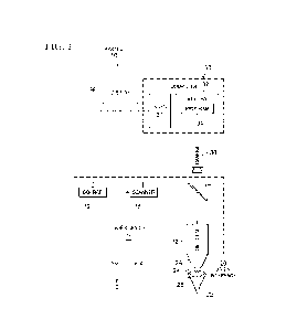
[0018] FIGURE 1 illustrates an example of an ophthalmic surgical system 10
configured to compensate for distortion of images of an eye, according to
certain embodiments.
In the embodiments, a computer uses an eye model to compensate for differences
between a
diagnostic pupil diameter of a diagnostic image and a real pupil diameter of a
surgical image.
A surgical image is taken when the cornea is substantially flattened by a
patient interface. The
flattened cornea generally does not affect the imaging of the real pupil
diameter. A diagnostic
image is taken when the cornea has its natural curvature. The curvature
affects the real pupil
diameter to yield a diagnostic pupil diameter that is different from the real
pupil diameter. The
computer uses an eye model to adjust the real pupil diameter of the surgical
image to yield a
refracted pupil diameter that takes into account the curvature of the cornea
The computer then
uses the refracted pupil diameter of the surgical image to perform the
surgical procedure on the
eye to compensate for the difference between the diagnostic pupil diameter and
the real pupil
diameter.
CA 03198540 2023- 5- 11
WO 2022/130138
PCT/1B2021/061510
6
[0019] In the illustrated example, ophthalmic surgical system 10 includes a
laser device
15, a patient interface 20, a camera 38, and a control computer 30, coupled as
shown. Laser
device 15 includes controllable components, such as a laser source 12, a
scanner 16, one or
more optical elements 17, and/or a focusing objective 18, controllable by a
computer such as
computer 30, coupled as shown. Patient interface 20 includes a contact portion
24 (with an
abutment face 26) and a sleeve 28 coupled as shown. Computer 30 includes logic
31, a memory
32 (which stores a computer program 34), and a display 36, coupled as shown.
[0020] Ophthalmic surgical system 10 may perform any suitable surgical
procedure,
such as corneal refractive or laser coagulation surgery. The surgical
procedure may have an
associated laser focal spot pattern that describes the target locations of the
laser pulses in the
cornea. Certain types of procedures, e.g., lenticule extraction, require
precise placement of the
laser pulses according to the laser focal spot pattern, which in turn requires
precise alignment
of surgical and diagnostic images.
[0021] Turning to the parts of system 10, as an example overview of laser
device 15,
laser source 12 generates a laser beam having ultrashort pulses, where a
propagation direction
of the laser beam defines a z-axis and/or z-direction. Scanner 16 directs a
focal point of the
laser beam in an xy-plane that is orthogonal to the z-axis. Objective 18
focuses the focal point
towards the cornea of eye 22.
[0022] In certain embodiments, laser source 12 generates a laser beam with
ultrashort
pulses. An ultrashort pulse refers to a light pulse that has a duration that
is less than a
nanosecond, such as on the order of picoseconds, femtoseconds, or attoseconds.
The laser beam
may have any suitable wavelength, such as a wavelength in the range of 300 to
1500
nanometers (nm), e.g., a wavelength in the range of 300 to 650, 650 to 1050,
1050 to 1250,
and/or 1250 to 1500 nm, such as 340 to 350 nm, e.g., 347 nm + 1 nm. The focal
point of the
laser beam may create a laser-induced optical breakdown (LIOB) in tissue
(e.g., the cornea) to
yield a photodisruption in the tissue. The laser beam may be precisely focused
to yield precise
photodisruptions, which may reduce or avoid unnecessary destruction of other
tissue
[0023] Scanner 16 longitudinally and transversely directs the focal point of
the laser
beam. The longitudinal direction refers to the direction of the laser beam
propagation, i.e., the
z-direction. Scanner 16 may longitudinally direct the laser beam in any
suitable manner. For
example, scanner 16 may include a longitudinally adjustable lens, a lens of
variable refractive
power, or a deformable mirror that can control the z-position of the focal
point. The transverse
direction refers to directions orthogonal to the direction of beam
propagation, i.e., the x- and
y-directions. Scanner 16 may transversely direct the laser beam in any
suitable manner. For
CA 03198540 2023- 5- 11
WO 2022/130138
PCT/1B2021/061510
7
example, scanner 16 may include a pair of galvanometrically-actuated scanner
mirrors that can
be tilted about mutually perpendicular axes. As another example, scanner 16
may include an
electro-optical crystal that can electro-optically steer the laser beam
[0024] One (or more) optical elements 17 direct the laser beam towards
focusing
objective 18. An optical element 17 can act on (e.g., transmit, reflect,
refract, diffract, collimate,
condition, shape, focus, modulate, and/or otherwise act on) a laser beam
Examples of optical
elements include a lens, prism, mirror, diffractive optical element (DOE),
holographic optical
element (HOE), and spatial light modulator (SLM). In the example, optical
element 17 is a
mirror. Focusing objective 18 focuses the focal point of laser beam through
the patient interface
20 towards a point of eye 22. In the example, focusing objective 18 is an
objective lens, e.g.,
an f-theta objective.
[0025] Patient interface 20 interfaces with the cornea of eye 22 to couple eye
22 to laser
device 15. In the example, patient interface 20 has sleeve 28 coupled to
contact portion 24.
Sleeve 28 detachably couples to focusing objective 18. Contact portion 24 may
be translucent
or transparent to the laser beam and has an abutment face 26 that interfaces
with the cornea.
Abutment face 26 may have any suitable shape, e.g., planar, convex, or
concave.
[0026] Camera 38 records surgical images of eye 22 in real time during a
surgical
procedure. Examples of camera 38 include a video, optical coherence tomography
(OCT), or
eye-tracking camera. Camera 38 delivers image data, which represent recorded
surgical images
of the eye 22, to computer 30.
[0027] Computer 30 controls controllable components (e.g., laser source 12,
scanner
16, optical elements 17, and/or focusing objective 18) in accordance with
instructions (which
may be stored in computer program 34) to photodisrupt corneal tissue. Memory
32 stores
information that can be accessed by computer 30. Examples of information
include: images
(e.g., surgical and/or diagnostic images), an eye model, information
describing a particular eye,
information describing pupil centroid shifts, and other suitable information.
[0028] In certain embodiments, computer 30 uses an eye model to compensate for
differences between a diagnostic pupil diameter of a diagnostic image and a
real pupil diameter
of a surgical image. In the embodiments, computer 30 accesses the surgical
image and
diagnostic image of the eye and uses an eye model to adjust the real pupil
diameter of the
surgical image to yield a refracted pupil diameter that takes into account the
curvature of the
cornea. For example, computer 30 determines how the eye model predicts the
natural curvature
of the cornea affects the real pupil diameter PDreai to determine the
refracted pupil diameter
PDrefracted. Computer 30 then uses the refracted pupil diameter to perform the
surgical procedure
CA 03198540 2023- 5- 11
WO 2022/130138
PCT/1B2021/061510
8
on the eye to compensate for difference between the diagnostic pupil diameter
and the real
pupil diameter.
[0029] Computer 30 may use the refracted pupil diameter to perform the
surgical
procedure in any suitable manner, such as aligning surgical and diagnostic
images according
to the refracted pupil diameter. In certain embodiments, computer 30 uses the
refracted pupil
diameter to compensate for pupil centroid shift. Pupil centroid shift occurs
when the pupil
center moves as the pupil diameter changes. In the embodiments, computer 30
determines the
pupil centroid shift according to the refracted pupil diameter of the surgical
image, and
determines the pupil center according to the pupil centroid shift and the
refracted pupil diameter
of the surgical image. For example, computer 30 accesses a table of pupil
diameters and
associated pupil centroid shifts to determine the centroid shift associated
with the refractive
pupil diameter. Computer 30 then applies the centroid shift to determine the
pupil center.
[0030] In certain embodiments, computer 30 uses the eye model to correct for
torsion.
Torsion refers to twisting of the eye, which may occur when the patient moves
from a seated
to a lying position. An asymmetric eye structure, such as the iris, can be
used to correct torsion.
In the embodiments, computer 30 uses the eye model to adjust a dimension of
the iris of the
surgical image and then corrects for torsion according to the adjusted iris
dimension. Computer
30 may use the eye model to adjust the iris dimension of the surgical image
by: determining an
imaging ratio of the real pupil diameter to the refracted pupil diameter; and
adjusting the iris
dimension according to the imaging ratio. This is described in more detail
with reference to
FIGURES 4A through 4C.
[0031] Computer 30 may take into account pseudo-rotation, which is an apparent
rotation which arises from the change in pupil size and hence a shift in the
structures of the iris,
but which does not imply a real torsion of the eye. In the embodiments,
computer 30 corrects
for torsion according to the adjusted iris structure by: identifying a pseudo-
rotation of the iris
structure according to the adjusted iris structure; and taking the pseudo-
rotation into account to
correct for torsion For example, computer 30 accesses a table of pupil
diameters and associated
pseudo-rotations to determine the pseudo-rotation associated with the
refractive pupil diameter.
This is described in more detail with reference to FIGURES 4A through 4C.
[0032] FIGURES 2A and 2B illustrate how the curvature of the cornea affects
the pupil
diameter in a diagnostic image_ FIGURE 2A shows a diagnostic device 40
measuring eye 22,
which includes a cornea 50 and an iris 52 that defines a pupil 54 having a
pupil diameter PD.
Diagnostic device 40 generates an image of eye 22, typically without contact
with cornea 50
or without changing the shape of cornea 50. Ophthalmic surgical system 10 may
use the
CA 03198540 2023- 5- 11
WO 2022/130138
PCT/1B2021/061510
9
diagnostic image to treat eye 22. For example, the diagnostic image or a
treatment pattern based
on the diagnostic image may be aligned with a surgical image of eye 22.
[0033] In the illustrated example, pupil 54 has a real pupil diameter PDreat
The
curvature of cornea 50 refracts light reflected from eye structures, such as
iris 52 and pupil 54,
which changes the imaging ratios. As a result, a diagnostic image of an eye
structure has
refracted dimensions that are larger than the real dimensions For example,
pupil 54 has a
refracted pupil diameter PDrefracted that is larger than real pupil diameter
PDreat. Similarly, iris
52 has a refracted diameter that is larger than the real diameter.
[0034] FIGURE 2B shows patient interface 20 of ophthalmic surgical system 10
applanating eye 22. A patient interface 20 may distort the shape of the cornea
such that the
distortion affects the dimensions of' structures of eye 22. In the illustrated
example, patent
interface 20 flattens cornea 50 such that cornea 50 does not refract light
reflected from eye
structures. As a result, a surgical image of an eye structure has dimensions
that are substantially
the same as the real dimensions. For example, pupil 54 has a pupil diameter
that is substantially
the same size as real pupil diameter PDreai. In other examples, patient
interface 20 may decrease
the curvature of the surface of the cornea, but not flatten the surface, such
that the pupil has an
interface pupil diameter that is closer to, but not the same size as, real
pupil diameter PDicat.
[0035] FIGURES 3A and 3B illustrate an example of eye models describing
diagnostic
and surgical imaging of an eye. An eye model uses geometric optics to describe
the paths of
light rays through an eye. Any suitable eye model may be used, e.g., a
standardized eye model,
such as the Navarro model.
[0036] Computer 30 may use an eye model to adjust the real pupil diameter in
any
suitable manner. For example, computer 30 may use an eye model to determine
the refracted
pupil diameter PDrefracted that corresponds to a given real pupil diameter
PDrcal and/or to
determine the real pupil diameter PDreal that corresponds to a given refracted
pupil diameter
PDrefracted. In certain embodiments, computer 30 determines how the eye model
predicts the
natural curvature of the cornea affects the real pupil diameter PDteat to
determine the refracted
pupil diameter PDrefracted. In certain embodiments, computer 30 determines how
the eye model
predicts a decreased curvature of the cornea (resulting from a patient
interface 20) affects the
real pupil diameter PDreat to determine an interface pupil diameter
PDinterface. In the
embodiments, computer 30 may use this information to determine a relationship
between the
interface pupil diameter PDintereace and the refracted pupil diameter
PDrefracted.
[0037] In certain embodiments, computer 30 may customize an eye model with
information (e.g., measurements) describing a specific eye 22. For example,
the information
CA 03198540 2023- 5- 11
WO 2022/130138
PCT/1B2021/061510
may describe one or more of the following: the distance between structures of
an eye (e.g., the
anterior chamber depth and/or eye length); the refractive power of a structure
(e.g., the
refractive power of the cornea and/or lens), the thickness of a structure
(e.g., the lens thickness);
and/or the curvature of a structure (e.g., the curvature of the cornea, lens,
and/or retina). The
information may describe the eye with the cornea having its natural curvature,
a distorted
curvature, or substantially flattened.
[0038] In the example, model shows an eye with cornea 50, lens 56, and retina
58. A
pupil plane 60 is the plane at which pupil 54 is located. FIGURE 3A shows an
eye model with
a cornea 50 having a curvature. The curvature of cornea 50 generally focuses
incoming light
rays through lens 56 and onto retina 58, i.e., the light rays substantially
converge to meet at
retina 58. In doing so, the light rays converge slightly at pupil plane 60.
Accordingly, light
reflected from eye structures at pupil plane 60 is refracted by cornea 50,
yielding a refracted
pupil diameter PDrefracted that is larger than real pupil diameter PDreal.
[0039] FIGURE 3B shows an eye model with a flattened cornea 50. The flattened
cornea 50 does not refractively affect the light rays. Lens 56 refracts the
light rays slightly, but
this occurs between the pupil plane 60 and retina 58. Accordingly, the pupil
diameter is
substantially the same as real pupil diameter PDiem. In other examples,
patient interface 20 may
decrease the curvature of the surface of the cornea, but not flatten the
surface, such that the
pupil has an interface pupil diameter that is closer to, but not the same size
as, real pupil
diameter PDreai.
[0040] FIGURES 4A, 4B, and 4C illustrate the linear relationship between real
pupil
diameter PDreat and refracted pupil diameter Paefr acted at different anterior
chamber depths
(ACDs). The ratio PDreal/PDrefracted is an imaging ratio that provides an
estimate of a refracted
dimension, given a real dimension, or vice-versa. For example, given real
dimension Dreai,
refracted dimension Drerractea may be calculated as Drerractea = PDrerractea
/PDreal x Dreai. Given
refracted dimension Drefracted, real dimension Dreai may be calculated as
Dreai = PDreal/PDrefracted
X Drefiacted.
[0041] In certain embodiments, computer 30 uses imaging ratio Paeal/PDLefr
acted to
correct for torsion. In the embodiments, computer 30 adjusts a real dimension
of an iris in a
surgical image according to imaging ratio PDreal/PDrefracted to yield a
refracted iris dimension.
Computer 30 then uses the refracted iris dimension to correct for torsion to
align the surgical
image with a diagnostic image. In certain embodiments, computer 30 may take
into account
pseudo-rotation. In the embodiments, computer 30 identifies a pseudo-rotation
of the iris
CA 03198540 2023- 5- 11
WO 2022/130138
PCT/1B2021/061510
11
structure using the refracted iris dimension. Computer 30 then does not treat
the pseudo-
rotation as a real rotation in correcting for torsion.
[0042] As patient interface 20 applanates eye 22, interface 20 presses on
cornea 50,
decreasing the anterior chamber depth. The imaging ratio PDreal/PDrefracted
varies with the
anterior chamber depth. FIGURE 4A shows the linear relationship at an ACD of
3.50
millimeters (mm), where PDrcal/PDrcfracted is 0.8833. FIGURE 4B shows the
linear relationship
at an ACD of 3.05 mm, where PDreal/PDrefracted is 0.8983. FIGURE 4C shows the
linear
relationship at an ACD of 2.50 mm, where PDreal/PDrefracted is 0.9166.
[0043] FIGURE 5 illustrates a substantial linear relationship between the
anterior
chamber depth (ACD) and the imaging ratio PDreal/PDrefracted, as described in
FIGURES 4A to
4C. FIGURE 5 presents a graph 63 that plots the imaging ratios
PDreal/PDrefracted along the y-
axis relative to the anterior chamber depths along the x-axis. The
relationship may be described
by y = 2- 5x2 ¨ 0.0334x +1 ¨ 0.0334x +1. Accordingly, given the anterior
chamber depth (of
the applanated eye) and real pupil diameter Pazai, refracted pupil diameter
Paetiacted can be
determined.
[0044] FIGURE 6 illustrates an example of a method for compensating for
distortion
of images of an eye for a surgical procedure, which may be performed by system
10 of FIGURE
1. Certain steps of the method may be performed by computer 30 sending
instructions to other
components of system 10.
[0045] The method starts at step 110, where computer 30 accesses diagnostic
and
surgical images of the eye. Computer 30 adjusts the pupil diameter of the
surgical image using
an eye model to yield a refracted pupil diameter at step 112. For example,
computer 30
determines how the eye model shows the natural curvature of the cornea affects
the real pupil
diameter PDreal to determine the refracted pupil diameter PDrefracted.
[0046] Computer 30 deteimines a pupil centroid shift using the refracted pupil
diameter
at step 114. For example, computer 30 accesses a table of pupil diameters and
associated pupil
centroid shifts to determine the centroid shift associated with the refractive
pupil diameter.
Computer 30 determines the pupil center according to the pupil centroid shift
at step 116. For
example, computer 30 applies the centroid shift to determine the pupil center,
i.e., the position
of the diagnostic pupil center.
[0047] Computer 30 adjusts the iris stnicture of the surgical image using the
refracted
pupil diameter at step 120. For example, computer 30 determines imaging ratio
PDreal/PDrefracted
and adjusts a real dimension of the iris according to an eye model in a
surgical image according
to imaging ratio PDreal/PDrefracted to yield a refracted iris dimension.
Computer 30 may also
CA 03198540 2023- 5- 11
WO 2022/130138
PCT/1B2021/061510
12
identify a pseudo-rotation of the iris structure using the refracted iris
dimension. Computer 30
compensates for torsion using the adjusted iris structure at step 122. For
example, computer 30
uses the refracted iris dimension to correct for torsion to align the surgical
image with a
diagnostic image, but does not treat the pseudo-rotation as a real rotation in
correcting for
torsion. The method then ends.
[0048] A component (such as the control computer) of the systems and
apparatuses
disclosed herein may include an interface, logic, and/or memory, any of which
may include
computer hardware and/or software. An interface can receive input to the
component and/or
send output from the component, and is typically used to exchange information
between, e.g.,
software, hardware, peripheral devices, users, and combinations of these. A
user interface (e.g.,
a Graphical User Interface (GUI)) is a type of interface that a user can
utilize to interact with a
computer. Examples of user interfaces include a display, touchscreen,
keyboard, mouse,
gesture sensor, microphone, and speakers.
[0049] Logic can perform operations of the component. Logic may include one or
more
electronic devices that process data, e.g., execute instructions to generate
output from input.
Examples of such an electronic device include a computer, processor,
microprocessor (e.g., a
Central Processing Unit (CPU)), and computer chip. Logic may include computer
software that
encodes instructions capable of being executed by the electronic device to
perform operations.
Examples of computer software include a computer program, application, and
operating
system.
[0050] A memory can store information and may comprise tangible, computer-
readable, and/or computer-executable storage medium. Examples of memory
include computer
memory (e.g., Random Access Memory (RAM) or Read Only Memory (ROM)), mass
storage
media (e.g., a hard disk), removable storage media (e.g., a Compact Disk (CD)
or Digital Video
or Versatile Disk (DVD)), database, network storage (e.g., a server), and/or
other computer-
readable media. Particular embodiments may be directed to memory encoded with
computer
software
[0051] Although this disclosure has been described in terms of certain
embodiments,
modifications (such as changes, substitutions, additions, omissions, and/or
other modifications)
of the embodiments will be apparent to those skilled in the art. Accordingly,
modifications may
be made to the embodiments without departing from the scope of the invention.
For example,
modifications may be made to the systems and apparatuses disclosed herein. The
components
of the systems and apparatuses may be integrated or separated, or the
operations of the systems
and apparatuses may be performed by more, fewer, or other components, as
apparent to those
CA 03198540 2023- 5- 11
WO 2022/130138
PCT/1B2021/061510
13
skilled in the art. As another example, modifications may be made to the
methods disclosed
herein. The methods may include more, fewer, or other steps, and the steps may
be performed
in any suitable order, as apparent to those skilled in the art
[0052] To aid the Patent Office and readers in interpreting the claims,
Applicants note
that they do not intend any of the claims or claim elements to invoke 35
U.S.C. 112(f), unless
the words "means for" or "step for" are explicitly used in the particular
claim. Use of any other
term (e.g., "mechanism," "module," "device," "unit," "component," "element,"
"member,"
"apparatus," "machine," "system," "processor," or "controller") within a claim
is understood
by the applicants to refer to structures known to those skilled in the
relevant art and is not
intended to invoke 35 U.S.C. 112(f).
CA 03198540 2023- 5- 11