Note : Les descriptions sont présentées dans la langue officielle dans laquelle elles ont été soumises.
WO 2022/112651
PCT/F12021/050796
1
An apparatus and a method for measuring jugular vein pressure waveform
Field of the disclosure
The disclosure relates to an apparatus and a method for measuring jugular vein
pressure "JVP" waveform. The measured jugular vein pressure waveform can be
used for example for detecting pulmonary hypertension "PAH".
Background
Abnormalities that may occur in a cardiovascular system, if not diagnosed and
appropriately treated and/or remedied, may progressively decrease the health
of an
individual. For example, pulmonary hypertension "PAH" represents in many cases
an early indication for an oncoming worsening phase of a heart failure that
will take
place in average after from three to four weeks from the occurrence of the
pulmonary
hypertension. In many cases, the pulmonary hypertension can predict the
worsening
phase of a heart failure in a so early stage that traditional indications of
the heart
failure such as e.g. increase in weight and increase in blood pressure are
typically
not present. A heart failure diagnosed at an early phase can often be treated
and/or
remedied and thereby mortality and a need for hospitalization can decrease
significantly.
An examination that concerns behaviour of a jugular vein (lat. vena jugularis)
represents a useful tool for diagnosing for example a heart failure. Variation
in the
jugular vein pressure is produced by changes in a blood flow and pressure in
central
veins caused by fillings and contractions of the right atrium and the right
ventricle of
a heart. The jugular vein is directly connected to the right atrium, and this
opens a
door for a non-invasive examination directed to the right side of the heart
i.e. the
right ventricle and the right atrium. Publication US2019254542 describes a
venous
pressure monitoring system which is configured to determine central venous
pressure "CVP" based on the jugular vein pressure "JVP". The venous pressure
monitoring system described in US2019254542 comprises a signal processor, at
least one accelerometer, at least one display, and at least one patch adapted
to be
CA 03199959 2023- 5- 24
WO 2022/112651
PCT/F12021/050796
2
held in place or otherwise secured to the neck of an individual. The signal
processor
is in communication with the at least one accelerometer to compute an estimate
for
the central venous pressure. An inherent challenge related to the above-
described
system based on one or more accelerometers is that an output signal of each
accelerometer comprises not only a signal component caused by the variation in
the
jugular vein pressure but also a signal component caused by movements not
related
to the variation of the jugular vein pressure. The last-mentioned signal
component
impairs the accuracy of the estimate of the central venous pressure.
Publication Bagyaraj, S., Ragumathulla, M., Vaithiyanathan, D.: Acquisition of
Jugular Venous Pulse Waveform by a Non-invasive Technique, Recent advances
in mechanical engineering, Lecture notes in mechanical engineering. Springer,
Singapore, 25.01.2020 describes a method for measuring a jugular vein pressure
"JVP" with the aid of an accelerometer. Publication Baumann, U., Marquis, C.,
Stoupis, C., Willenberg, T., Takala, J. & Jakob, S.: Estimation on central
venous
pressure by ultrasound, Resuscitation 64(2), 193-199, 01.02.2005 describes a
method for estimating central venous pressure "CVP" with the aid of ultrasound
signals.
Publication US2018184977 describes a method for measuring a jugular vein
property. The method comprises: coupling a device including an imager to the
neck
of a patient, imaging the jugular vein at an imaged location using the imager,
and
analysing at least one image provided by the imager in order to estimate at
least
one property of the Jugular vein. Publication US2012136240 describes a system
for
detecting and measuring increased global or local intracranial pressure. The
system
comprises: devices for performing controlled occlusion of jugular cranial
blood
outflow and generating occlusion data related to the controlled occlusion, a
cranial
blood outflow pressure measurement device, and a processor for processing
jugular
cranial blood outflow occlusion data and cranial blood outflow data to
identify and/or
measure a functional relationship between the jugular controlled occlusion and
the
jugular cranial blood outflow pressure. Publication W02008098353 describes a
device for non-invasively measuring at least one parameter of a cardiac blood
vessel. The device comprises at least one light source that emits light in the
400 nm
to 1000 nm wavelength range, at least one photodetector adapted to receive
light
CA 03199959 2023- 5- 24
WO 2022/112651
PCT/F12021/050796
3
from a tissue of a patient in the proximity of the cardiac blood vessel and
generate
an output based on the received light, and at least one probe for delivering
the light
from the light source to the tissue of the patient. Publication US2012197118
describes an ultrasonic monitoring device for measuring physiological
parameters
of a mammal. The ultrasonic monitoring device comprises a substrate, a
plurality of
ultrasonic transducer elements, a computer readable memory, a microprocessor,
and a power source. The ultrasonic transducer elements are coupled to the
substrate. Each ultrasonic transducer element is separately configured to
transmit
a signal to a target area of a mammal and to receive an echo return signal
from the
target area. Publication W02018161159 describes a device for measuring the
jugular venous pressure of a patient. The device comprises a body defining a
longitudinal enclosure and having a window along a length of the longitudinal
enclosure to allow light to exit the longitudinal enclosure and a beam
generator
comprising a moveable portion mounted within the longitudinal enclosure. The
beam
generator is configured to generate a sheet of light along a plane
perpendicular to
the longitudinal direction and at an adjustable position along the
longitudinal
direction and to direct the sheet of light out of the window. The device
further
comprises an adjustment mechanism for adjusting the position of the moveable
portion of the beam generator relative to the body along the longitudinal
direction
and a readout device indicating the position of the sheet of light along the
longitudinal direction. Publication US2010094141 describes a jugular venous
pressure "JVP" ruler and a method for its use in measuring jugular venous
pressure
of a patient. The JVP ruler comprises a transducer configured to detect
displacements of a skin of the patient.
Summary
The following presents a simplified summary in order to provide a basic
understanding of some aspects of various invention embodiments. The summary is
not an extensive overview of the invention. It is neither intended to identify
key or
critical elements of the invention nor to delineate the scope of the
invention. The
following summary merely presents some concepts of the invention in a
simplified
form as a prelude to a more detailed description of exemplifying embodiments
of the
invention.
CA 03199959 2023- 5- 24
WO 2022/112651
PCT/F12021/050796
4
In this document, the word "geometric" when used as a prefix means a geometric
concept that is not necessarily a part of any physical object. The geometric
concept
can be for example a geometric point, a straight or curved geometric line, a
geometric plane, a non-planar geometric surface, a geometric space, or any
other
geometric entity that is zero, one, two, or three dimensional.
In accordance with the invention, there is provided a new apparatus for
measuring
a jugular vein pressure "JVP" waveform. The measured jugular vein pressure
waveform can be used for example for detecting pulmonary hypertension "PAH".
An apparatus according to the invention comprises:
- a rotation sensor, e.g. a gyroscope, configured to produce a measurement
signal indicative of rotation of the rotation sensor when being against a skin
of an individual and in a movement sensing relation with a jugular vein of the
individual, and
- a processing system configured to receive the measurement signal and
produce a waveform of a motion of the skin in a direction perpendicular to the
skin based on the measurement signal, the waveform of the motion of the
skin being indicative of the jugular vein pressure waveform.
The rotation sensor is advantageously positioned so that one end of the
rotation
sensor is nearer to the jugular vein than another end of the rotation sensor.
Thus,
variation in the jugular vein pressure causes more movement at the first-
mentioned
end of the rotation sensor than at the last-mentioned end of the rotation
sensor, and
this difference appears as rotational movement of the rotation sensor. A
movement
which is not related to the jugular vein pressure and which has a
substantially same
amplitude and direction over a whole skin area covered by the rotation sensor
does
not cause a significant rotational movement of the rotation sensor but a
translational
movement only, and thereby this movement does not cause a significant signal
component in the output signal of the rotation sensor. Therefore, the rotation
sensor
that measures rotation is more insensitive to many movements not related to
the
variation of the jugular vein pressure than for example an acceleration sensor
that
measures translational movements.
CA 03199959 2023- 5- 24
WO 2022/112651
PCT/F12021/050796
The pulsation caused by a jugular vein differs from the pulsation caused by a
carotid
artery due to the difference between the structures of the thin-walled and
flexible
jugular vein and the thick-walled and muscular carotid artery and also due to
different pressures in the jugular vein and in the carotid artery, about 10
mmHg in
5 the jugular vein and about 100 mmHg in the carotid artery. According to
experiments, a signal produced by a rotation sensor is more purely a signal
produced by a jugular vein whereas a signal produced by an acceleration sensor
is
a mixture of signals produced by a jugular vein and by a carotid artery. This
can be
explained based on differences between types of movements measured with a
rotation sensor and an acceleration sensor and on differences between types of
movements caused by a jugular vein and a carotid artery. A jugular vein causes
a
local movement on tissue covering the jugular vein whereas a carotid artery
causes
a sharper pulse that causes a translational movement on a larger area. As
mentioned earlier in this document, a movement which is not related to the
jugular
vein pressure and which has a substantially same amplitude and direction over
a
larger skin area does not cause a significant rotational movement on a
rotation
sensor but a translational movement only, and thereby this movement does not
cause a significant signal component in the output signal of the rotation
sensor.
Therefore, the rotation sensor is more insensitive to movements caused by a
carotid
artery than for example an acceleration sensor that measures translational
movements.
An advantage of a rotation sensor with respect to an optical sensor is that an
optical
sensor can measure only pulsation caused by an outer jugular vein, vena
jugularis
externa, and thus the optical sensor must be placed accurately to cover the
outer
jugular vein, which complicates the use of an optical sensor. A rotation
sensor
measures the pulsation caused mainly by an inner jugular vein, vena jugularis
interna, and thus there are no so hard requirements related to positioning
than when
using an optical sensor.
In accordance with the invention, there is provided a new method for measuring
a
jugular vein pressure "JVP" waveform. A method according to the invention
comprises:
CA 03199959 2023- 5- 24
WO 2022/112651
PCT/F12021/050796
6
- producing a measurement signal with a rotation sensor that is against a
skin
of an individual and in a movement sensing relation with a jugular vein of the
individual, and
- producing a waveform of a motion of the skin in a direction perpendicular
to
the skin based on the measurement signal indicative of rotation of the
rotation
sensor, the waveform of the motion of the skin being indicative of the jugular
vein pressure waveform.
Exemplifying and non-limiting embodiments are described in accompanied
dependent claims.
Various exemplifying and non-limiting embodiments both as to constructions and
to
methods of operation, together with additional objects and advantages thereof,
will
be best understood from the following description of specific exemplifying
embodiments when read in conjunction with the accompanying drawings.
The verbs "to comprise" and "to include" are used in this document as open
limitations that neither exclude nor require the existence of also un-recited
features.
The features recited in the accompanied dependent claims are mutually freely
combinable unless otherwise explicitly stated. Furthermore, it is to be
understood
that the use of "a" or "an", i.e. a singular form, throughout this document
does not
exclude a plurality.
Brief description of figures
Exemplifying and non-limiting embodiments and their advantages are explained
in
greater detail below with reference to the accompanying drawings, in which:
figure 1 illustrates an apparatus according to an exemplifying and non-
limiting
embodiment for measuring a jugular vein pressure "JVP" waveform,
figure 2a shows exemplifying waveforms produced with an apparatus according to
an exemplifying and non-limiting embodiment,
CA 03199959 2023- 5- 24
WO 2022/112651
PCT/F12021/050796
7
figure 2b shows an exemplifying waveform of angular displacement produced with
an apparatus according to an exemplifying and non-limiting embodiment, and
figure
2c shows a corresponding waveform of displacement produced with an
acceleration
sensor,
figure 3 illustrates an apparatus according to an exemplifying and non-
limiting
embodiment for measuring a jugular vein pressure "JVP" waveform, and
figure 4 shows a flowchart of a method according to an exemplifying and non-
limiting
embodiment for measuring a jugular vein pressure "JVP" waveform.
Description of exemplifying and non-limiting embodiments
The specific examples provided in the description below should not be
construed as
limiting the scope and/or the applicability of the appended claims. Lists and
groups
of examples provided in the description are not exhaustive unless otherwise
explicitly stated.
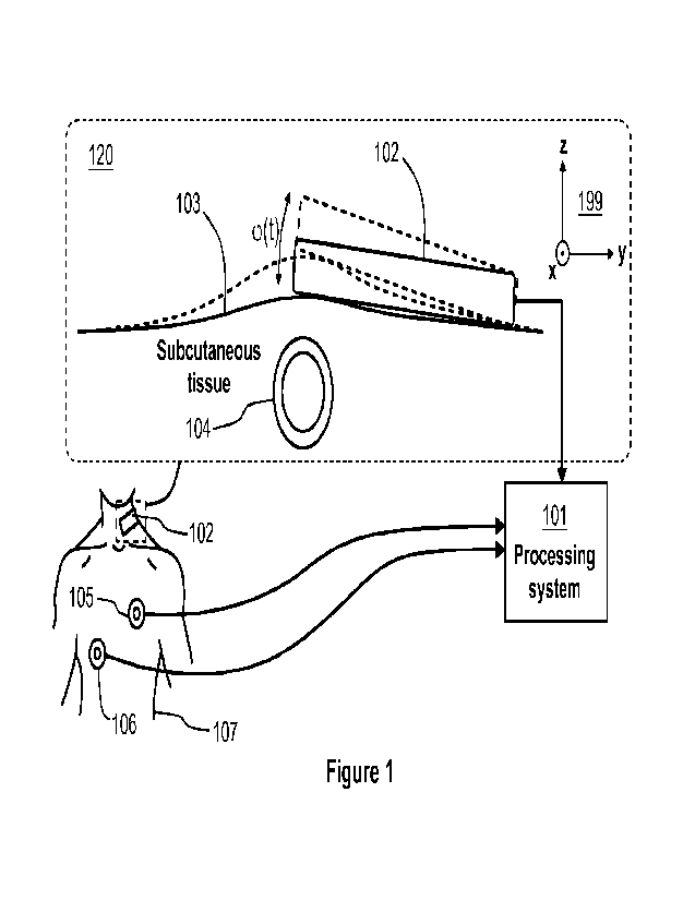
Figure 1 illustrates an apparatus according to an exemplifying and non-
limiting
embodiment for measuring a jugular vein pressure "JVP" waveform. The apparatus
comprises a rotation sensor 102, e.g. a gyroscope, configured to produce a
measurement signal indicative of rotation of the rotation sensor 102 when the
rotation sensor 102 is against a skin 103 of an individual 107 and in a
movement
sensing relation with a jugular vein 104 of the individual. The rotation
sensor 102
can be for example a part of a mobile phone that is placed against the skin of
the
individual 107. The apparatus comprises a processing system 101 configured to
receive the measurement signal and to produce a waveform of a motion of the
skin
103 in a direction perpendicular to the skin based on the measurement signal.
The
waveform of the motion of the skin is indicative of the jugular vein pressure
"JVP"
waveform. In a part 120 of figure 1, the direction perpendicular to the skin
103 is
substantially parallel with the z-axis of a coordinate system 199. The
processing
system 101 can be for example a part of a mobile phone. Furthermore, both the
processing system 101 and rotation sensor 102 can be for example parts of a
same
mobile phone. In this exemplifying case, the mobile phone constitutes the
apparatus
for measuring a jugular vein pressure "JVP" waveform. In the exemplifying case
CA 03199959 2023- 5- 24
WO 2022/112651
PCT/F12021/050796
8
illustrated in figure 1, the waveform of the motion of the skin 103 and
thereby the
jugular vein pressure "JVP" waveform are expressed with temporal variation of
a
rotation angle cp of the rotation sensor 102. The apparatus may comprise for
example a display for presenting the measured jugular vein pressure "JVP"
waveform. The display is not shown in figure 1. It is also possible that the
apparatus
comprises a data transfer interface for supplying data expressing the jugular
vein
pressure "JVP" waveform to an external device. The data transfer interface is
not
shown in figure 1.
In an apparatus according to an exemplifying and non-limiting embodiment, the
rotation sensor 102 is configured to measure angular velocity co of the
rotation
sensor 102 and the processing system 101 is configured to compute a time
integral
of the angular velocity:
q(t) = fot co(T)dy,
(1)
where the angular velocity 0) of the rotation sensor 102 represents the
measurement
signal and the time integral of the angular velocity, i.e. the angular
displacement cp,
is indicative of the jugular vein pressure "JVP" waveform.
In an apparatus according to an exemplifying and non-limiting embodiment, the
rotation sensor 102 is configured to measure angular acceleration a of the
rotation
sensor 102 and the processing system 101 is configured to compute a first time
integral 11 that is a time integral of the angular a acceleration and a second
time
integral 12 that is a time integral of the first time integral:
/1: (0(0 = fot aer)dr,
(2)
12: (p(t) = fot
(3)
where the angular acceleration a of the rotation sensor 102 represents the
measurement signal and the second time integral 12, i.e. the angular
displacement
cp, is indicative of the jugular vein pressure "JVP" waveform.
CA 03199959 2023- 5- 24
WO 2022/112651
PCT/F12021/050796
9
The above-mentioned embodiment in which the rotation sensor 102, e.g. a
gyroscope, is configured to measure the angular velocity co is advantageous in
the
sense that it requires only one integration with respect to time to obtain the
waveform
of the angular displacement cp of the skin. An integration with respect to
time has a
low-pass filtering effect and thus it is advantageous if only one integration
with
respect to time is needed. For example, in a case in which an acceleration
sensor
is used, it is possible to obtain the waveform of the angular displacement y
with the
aid of trigonometric functions but the need for two integrations with respect
to time
weakens the quality of the measurement and furthermore the need for
trigonometric
functions complicate the data processing.
In an apparatus according to an exemplifying and non-limiting embodiment, the
processing system 101 is configured to receive electric signals from
electrodes 105
and 106 on the skin of the individual 107 and the processing system 101 is
configured to produce an electrocardiogram "ECG" for a time interval of the
jugular
vein pressure waveform, i.e. the jugular vein pressure waveform and the
electrocardiogram are synchronized with each other.
It is also possible that an apparatus according to an exemplifying and non-
limiting
embodiment comprises two or more rotation sensors for measuring a same jugular
vein in order to improve accuracy. Furthermore, one or more rotation sensors
of an
apparatus according to an exemplifying and non-limiting embodiment can be one
or
more implants to be placed under a skin. An implant may utilize the radio
frequency
identifier "RFID" technology for transferring a measurement signal from the
implant
to a processing system of the apparatus.
Figure 2a shows an exemplifying jugular vein pressure waveform 210 and an
exemplifying electrocardiogram 211 produced with an apparatus according to an
exemplifying and non-limiting embodiment. The jugular vein pressure waveform
210
and the electrocardiogram 211 are measured simultaneously.
In an apparatus according to an exemplifying and non-limiting embodiment, the
processing system 101 is configured to produce an indicator signal expressing
pulmonary hypertension "PAH" in response to a situation in which an a-wave of
the
CA 03199959 2023- 5- 24
WO 2022/112651
PCT/F12021/050796
jugular vein pressure waveform exceeds a predetermined threshold. The a-wave
of
the jugular vein pressure waveform 210 is illustrated in figure 2a. An
increase in the
a-wave is characteristics to the pulmonary hypertension "PAH" which is caused
by
increased flow resistance via the pulmonary valve to the pulmonary artery.
This is
5 reflected via the right atrium to the jugular vein.
Figure 2b shows an exemplifying waveform of angular displacement produced with
an apparatus according to an exemplifying and non-limiting embodiment. In this
exemplifying case, the apparatus comprises a gyroscope that is configured to
measure the angular velocity and therefore only one integration with respect
to time
10 is needed to obtain the waveform of the angular displacement. As shown
in figure
2b, the waveform of the angular displacement obtained with the aid of the
gyroscope
is able to express the a-, c-, h-, and v-waves. Figure 2c shows a waveform of
displacement that has been obtained with two integrations with respect to time
based on a signal measured with an acceleration sensor in a direction
perpendicular
to the skin. As shown in figure 2c, it is not possible to identify the a-, c-,
h-, and v-
waves from the waveform obtained with the acceleration sensor.
Figure 3 illustrates an apparatus according to an exemplifying and non-
limiting
embodiment for measuring a jugular vein pressure "JVP" waveform. The apparatus
comprises a rotation sensor 302, e.g. a gyroscope, configured to produce a
measurement signal indicative of rotation of the rotation sensor 302 when the
rotation sensor 302 is against a skin 303 of an individual and in a movement
sensing
relation with a jugular vein 304 of the individual. The apparatus comprises a
processing system 301 configured to receive the measurement signal and to
produce a waveform of a motion of the skin 303 in a direction perpendicular to
the
skin based on the measurement signal. In figure 3, the direction perpendicular
to
the skin 303 is substantially parallel with the z-axis of a coordinate system
399.The
waveform of the motion of the skin is indicative of the jugular vein pressure
"JVP"
waveform. In the exemplifying case illustrated in figure 3, the waveform of
the motion
of the skin 303 and thereby the jugular vein pressure "JVP" waveform are
expressed
with temporal variation of a rotation angle cp of the rotation sensor 302.
CA 03199959 2023- 5- 24
WO 2022/112651
PCT/F12021/050796
11
The exemplifying apparatus illustrated in figure 3 comprises a sheet of
flexible
material 308 that is provided with glue to attach the rotation sensor 302 to
the skin
303 of the individual. Thus, the apparatus can be used in different positions
of the
individual, e.g. when the individual is standing.
In the exemplifying apparatus illustrated in figure 3, the processing system
301 and
the rotation sensor 302 are configured to maintain a wireless link to transfer
the
measurement signal from the rotation sensor 302 to the processing system 301.
The wireless link can be for example a radio link such as e.g. a Bluetoothe
link or a
Near Field Communication "NFC" link. It is also possible that the wireless
link is an
optical or infrared link.
Each of the processing systems 101 and 301 shown in figures 1 and 3 can be
implemented for example with one or more processor circuits, each of which can
be
a programmable processor circuit provided with appropriate software, a
dedicated
hardware processor such as for example an application specific integrated
circuit
"ASIC", or a configurable hardware processor such as for example a field
programmable gate array "FPGA". Each of the processing systems 101 and 301
may further comprise memory implemented for example with one or more memory
circuits each of which can be e.g. a random-access memory "RAM" device.
Figure 4 shows a flowchart of a method according to an exemplifying and non-
limiting embodiment for measuring a jugular vein pressure "JVP" waveform. The
method comprises the following actions:
- action 401: producing a measurement signal with a rotation sensor that is
against a skin of an individual and in a movement sensing relation with a
jugular vein of the individual, and
- action 402: producing a waveform of a motion of the skin in a direction
perpendicular to the skin based on the measurement signal indicative of
rotation of the rotation sensor, the waveform of the motion of the skin being
indicative of the jugular vein pressure waveform.
CA 03199959 2023- 5- 24
WO 2022/112651
PCT/F12021/050796
12
In a method according to an exemplifying and non-limiting embodiment, the
rotation
sensor measures angular velocity of the rotation sensor and the method
comprises
computing a time integral of the measured angular velocity. The measured
angular
velocity of the rotation sensor represents the above-mentioned measurement
signal
and the time integral of the measured angular velocity is indicative of the
jugular
vein pressure waveform.
In a method according to an exemplifying and non-limiting embodiment, the
rotation
sensor measures angular acceleration of the rotation sensor and the method
comprises computing a first time integral that is a time integral of the
measured
angular acceleration and a second time integral that is a time integral of the
first time
integral. The measured angular acceleration of the rotation sensor represents
the
above-mentioned measurement signal and the second time integral is indicative
of
the jugular vein pressure waveform.
A method according to an exemplifying and non-limiting embodiment comprises
receiving one or more electric signals from electrodes on the skin of the
individual
and producing an electrocardiogram for a time interval of the jugular vein
pressure
waveform.
In a method according to an exemplifying and non-limiting embodiment, the
measurement signal is received from the rotation sensor via a wireless link.
A method according to an exemplifying and non-limiting embodiment comprises
producing an indicator signal expressing pulmonary hypertension "PAH" in
response to a situation in which an a-wave of the jugular vein pressure
waveform
exceeds a predetermined threshold.
The specific examples provided in the description given above should not be
construed as limiting the scope and/or the applicability of the appended
claims. Lists
and groups of examples provided in the description given above are not
exhaustive
unless otherwise explicitly stated. Correspondingly, exemplifying waveforms
and
other exemplifying results presented above and/or in figures should not be
construed as limiting the scope and/or the applicability of the appended
claims.
CA 03199959 2023- 5- 24