Note : Les descriptions sont présentées dans la langue officielle dans laquelle elles ont été soumises.
P1 241 8CA00 1
DEVICE FOR PROSTATE PALPATION
DESCRIPTION
OBJECT OF THE INVENTION
The present invention discloses a device for palpation of the prostate as a
screening
method for prostate cancer. The object of the invention is to provide a device
that
allows palpation of the rectal face of the prostate, identifying areas of high
rigidity and
the limits of the gland, with the aim of detecting prostate cancer.
To characterize prostate stiffness (e.g. stiffness determined by the
deformation of the
fat layer and stiffness determined by the overall soft tissue layer), the
device is
configured to measure force and deformation at points on the rectal face of
the
prostate, by the rectum.
The invention discloses a device and method for performing palpation of the
rectal
face of the prostate providing an objective and reproducible index for the
assessment
of tissue stiffness.
Currently, the most common diagnostic maneuver for prostate cancer is the
digital
rectal examination, which is a manual procedure, making the test subjective
and
unreliable. In addition, the finger often barely reaches the prostatic apex,
resulting in
a partial assessment of tissue stiffness.
The methods and devices disclosed in this invention overcome these drawbacks
by
allowing the identification of areas of high rigidity in a completely
objective, less
aggressive way, reaching the entire peripheral area, and with a test time
similar to
that of a digital rectal examination. The device of the invention also has an
ultrasonic
imaging system that makes it possible to visualize the anatomy of the prostate
during
the test.
TECHNICAL FIELD
CA 03208435 2023-8- 14
P1 241 8CA00 2
The invention falls within the sector of medical diagnostic device
manufacturers, as
well as the industry dedicated to the treatment of prostate cancer.
BACKGROUND OF THE INVENTION
The prostate is a gland that lies below the bladder in men and produces the
fluid for
semen. Cancer screening is a test that is done before you have any symptoms,
as
cancer that is found early is easier to treat.
The absolute number of cancers diagnosed in Spain and worldwide has continued
to
rise for decades in probable relation to the increase in life expectancy
of the
population and is currently the most frequent cancer in men.
In this regard, one of the tests used as a screening method for prostate
cancer is the
digital rectal examination. In 18 % of prostate cancers there is only an
abnormal digital
rectal examination without an alteration in the blood marker called prostate
specific
antigen (PSA).
The doctor inserts a gloved and lubricated finger into the rectum to palpate
the
prostate and, depending on the stiffness of the palpated area, determine a
suspected
diagnosis.
However, there are a number of problems with this technique:
- The first and biggest problem is the lack of objectivity in the analysis
of the stiffness
of the palpated area, as it is based on the subjective perception of the
doctor with
respect to the pressure he/she can exert with his/her finger, which can lead
to
diagnoses that are poorly reliable.
- The length of the finger, the dimensions of the patient and the
experience of the
examiner limit the quality of the examination.
- This is a psychologically unpleasant technique for the examiner and the
patient. It
is therefore desirable to reduce this aspect, which is very uncomfortable and
yields
little.
CA 03208435 2023-8- 14
P1 241 8CA00 3
In an attempt to obviate this problem, devices for prostate palpation that
include a
force/pressure sensor are known, as is the case of US 2007293792, which
discloses
a rectal probe comprising a force/pressure sensor configured to measure
prostate
rigidity through the rectal wall adjacent to the prostate.
Ideally, the force/pressure sensor is large enough to detect prostate pressure
or
stiffness and/or prostate tumors. The forces imposed on the sensing device by
tumors
will typically be greater than those applied by adjacent rectal wall tissues
supported
by the benign prostate.
In this device the force/pressure sensors are rigidly mounted on the probe to
ensure
that the force measured by the force sensor corresponds to the applied force.
Due to the physiognomy of the prostate tissues, the application of a
pressure/force
sensor can give erroneous readings, as it can coincide with small folds so
that
obtaining different measurements relative to each other is decisive in making
a good
diagnosis.
On the other hand, these devices do not include means to visualize the anatomy
of
the prostate, so it is not possible to know precisely the specific area being
scanned,
and even more so if it is the prostate that is being scanned.
While there are devices that can objectively detect prostate hardening, they
are
sophisticated ultrasound machines equipped with special probes for shear wave
elastography and specific software.
The use of such equipment could be feasible and useful in specialized hospital
consultations, but their high cost, large size and limited portability make
them
unsuitable for initial screening of patients in primary care, where patients
first come.
In this context, doctors with little training in digital rectal examination
have to decide
whether a given patient has the conditions to justify a urological
consultation, which
can lead to false negatives, resulting in complications of the pathology due
to late
identification or false positives that collapse specialty consultations and
generate long
CA 03208435 2023-8- 14
P1 241 8CA00 4
waiting lists.
A similar situation is found in other devices of this type such as those
disclosed in
US6511427, US 8016777 or US 2002143275.
In short, it can be concluded that to date there is no system for palpation of
the
rectal face of the prostate that transforms the information collected with the
device
itself into objective, reproducible and useful information, all in a device
that is easy to
use and low cost.
DESCRIPTION OF THE INVENTION
The device for prostate palpation disclosed solves the above-mentioned problem
in a
fully satisfactory manner, allowing palpation of the rectal face of the
prostate,
identifying areas of high rigidity in the peripheral zone of the gland, with
the aim of
detecting indurations suggestive of prostate cancer.
For this purpose, the device of the invention takes the form of a hand-held
electronic
instrument, provided with a handle for its handling joined to a transrectal
scanning
rod.
The control electronics are integrated in the handle, via the corresponding
push
button, as well as a series of indicators that show the results of the
measurements
obtained.
The scanning rod has two concentric indenters near its distal end with
different
heights, the central one being higher than the external or perimeter one, by
means of
which pressure is applied to the scanning point, and which are linked to
respective
internal pressure sensors by means of a lever (external indenter) and a
cylinder
(internal indenter).
These elements make it possible to measure in vivo synchronized force and
deformation at defined points of the prostate through the rectum.
CA 03208435 2023-8- 14
P1 241 8CA00 5
In accordance with another feature of the invention, the device incorporates
an
ultrasonic imaging system that allows to locate and visualize the anatomy of
the
prostate, as well as the correct positioning of the pressure sensors.
The ultrasonic imaging system is based on the use of a phased array of at
least 8
piezoelectric emitter/receiver elements and a center frequency greater than 2
MHz,
with at least 50% bandwidth, with transverse focusing so that a sectoral slice
of the
prostate can be observed.
The arrangement of the transrectal ultrasound imaging system's emitting
elements,
preferably curved to fit the cylindrical shape of the stem and to come into
contact with
the tissue, as well as the size and number of them and the separation distance
between them, is optimized so as not to interfere with the correct arrangement
of the
pressure sensor line, to obtain the best image resolution and the greatest
aperture
and depth of field.
The transrectal ultrasound imaging system has electronics associated with a
wireless
communications module or a wired communications interface that allows it to be
used
with a computer, tablet, smartphone, or other computing device that allows the
visualization of ultrasound information. This transrectal ultrasound imaging
system
provides a visual reference of the prostate to the medical staff performing
the
examination in real time.
The simultaneous performance of transrectal ultrasound makes it possible to
rule out
the false positives that can be caused by frequent prostatic calcifications,
which would
otherwise be associated with areas of elevated rigidity.
Alternatively, the ultrasonic imaging system may be composed of a two-
dimensional
arrangement of the emitting elements for electronic anatomical image
addressing or
a one-dimensional arrangement that allows mechanical rotation from the handle
for
mechanical and low-cost anatomical sectorial image addressing. In this way,
the
stiffness sensor and probe may not be aligned with the transrectal ultrasound
image,
in order to obtain images of different prostate slices from the same position
of the
stiffness sensor and to improve scanning and rule out false positives.
CA 03208435 2023-8- 14
P1 241 8CA00 6
Alternatively, the ultrasonic imaging system may be located at a position
inside the
stem such that the ultrasonic image includes the area of the pressure sensor
and
the tissue in contact with it. In that case the inside of the stem will
include a medium
with an elasticity and density that allows for proper ultrasound propagation
using an
acoustic impedance matching layer. This provision provides additional
information
about the quality of the contact between the pressure sensor and the tissue,
which is
used to ensure correct physical contact between the sensor and the tissue.
In addition, this configuration allows correlation of echogenic information
with
information from pressure sensors.
Alternatively, there may be a dedicated ultrasonic imaging system for quality
control
of the coupling between the pressure sensor and the tissue.
In this way, the device will be introduced through the anus of the patient to
be explored
by means of its exploration stem so that the rectal side of the prostate will
be mapped
through it in order to detect indurated areas that suggest prostate cancer.
Optionally, the number of sensors could be increased, in order to simplify the
use of
the device by simultaneously palpation a larger surface area.
Based on this structure, a system of palpation of the rectal face of the
prostate is
achieved that transforms the information collected with the device into
objective,
reproducible and useful information, making it possible to identify areas of
high rigidity
by reaching the entire peripheral area when, currently, on many occasions the
finger
barely reaches the prostatic apex during the manual procedure, and
transforming a
diagnostic maneuver for prostate cancer that until now has been subjective and
unreliable into an objective maneuver. All this in a short space of time,
performed in
a less aggressive and more objective manner than digital rectal examination.
In addition, the health care operator performing the examination must avoid
contact
between the bowel wall and the indenters during the insertion and removal of
the
device in the rectum.
CA 03208435 2023-8- 14
P1 241 8CA00 7
For this purpose, the device can incorporate two alternative systems to
facilitate the
maneuver:
1. A sheath that covers the indenters and that is removed once the area to be
assessed has been reached; or
2. A retraction system, internal to the stem, which retracts the indenters
into the body
of the stem during insertion and allows the indenters to be extracted when the
area to
be assessed has been reached, by means of a control system in the handle.
DESCRIPTION OF THE DRAWINGS
In order to complement the description to be given below and in order to help
a better
understanding of the features of the invention, in accordance with a
preferential
example of practical implementation of the same, a set of drawings is attached
as an
integral part of this description, in which the following is illustrated for
illustrative
purposes and without limitation:
Figure 1 shows a perspective view of a device for prostate palpation made
in
accordance with the object of the present invention.
Figure 2 shows an enlarged detail of the control interface located on the
handle of the
device.
Figure 3 shows the position of the pressure sensors, located in the mechanical
assembly formed by the support base and the hinged lever at the end of the
base
(raised in the figure). The left sensor (10 in the figure) receives the force
from the
inner indenter directly; the inner sensor (11 in the figure) receives the
force from the
outer indenter indirectly, through the central support of the lever where this
second
indenter is located.
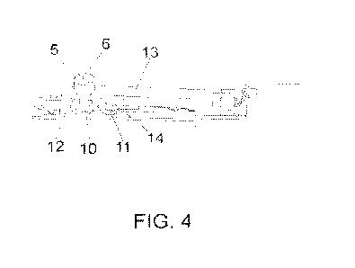
Figure 4 shows the indenter and sensor base assembly, mounted in its working
position, which is inserted into the scanning rod (3) at its distal end and
closed by a
CA 03208435 2023-8- 14
P1 241 8CA00 8
rounded cap (4). It has a support base for the sensors (12). The outer
indenter (5) is
integrated in a lever (13), hinged at its end on the base, and transmits the
force to the
corresponding sensor (11), by means of a central support (14). The inner
indenter (6)
passes through the inside of the outer indenter (5), being of greater overall
height,
and transmits the force to the corresponding sensor (10).
PREFERRED EMBODIMENT OF THE INVENTION
In view of the figures shown above, it can be seen that the device for
prostate
palpation disclosed consists of a hand-held electronic instrument, equipped
with a
handle (1) for its operation, and a probe (3).
More specifically, the control electronics are integrated in the handle, with
a cover (2)
for access to it, so that it has a pushbutton (7) for managing the device (on
or off;
activation or deletion of readings), as well as a scale (8) on which the
different
pressure levels (9) are displayed, and may include indicator lights (10) to
show the
measurement results, the power on of the equipment or the state of charge of
the
battery, all as can be seen in figure 2.
Returning again to figure 1, in proximity to its distal end (4) of the
scanning rod (3)
there are two concentric indenters (5-6) of different heights, the central one
being
higher than the external or perimeter one, the latter having a diameter of
about 5
millimeters, The latter having a diameter of around 5 millimeters, elements by
means
of which pressure is applied to the exploration point, and which are linked to
the
respective internal pressure/force sensors by means of a lever (external
indenter) and
a cylinder (internal indenter).
In order to be able to visualize the anatomy of the prostate, as well as the
exact point
on which the device is positioned when taking measurements, the device is
intended
to include an ultrasonic imaging system, based on the use of a "phased array"
or
phase emitters, i.e. a set of emitters in which the relative phases of the
signals fed to
each emitter are intentionally varied in order to alter the radiation pattern
of the set,
involving at least 8 elements with a central frequency of at least 2 MHz,
allowing the
visualization of a sectorial section of the prostate, electronics associated
with a
CA 03208435 2023-8- 14
P1 241 8CA00 9
wireless communications module or cable connection that allows it to be used
with a
computer, tablet, smartphone or other computer device that allows the
visualization
of the ultrasound information.
As shown in figures 3 and 4, the device makes it possible to simultaneously
transmit
the force exerted by two indenters (5 and 6) to two sensors (10 and 11), by
means of
a lever mechanism (13) with central support (11) and external indenter (5),
and with
concentric internal cylindrical indenter (6).
In addition, as indicated above, the health care operator performing the
examination
must avoid contact between the bowel wall and the indenters during the
insertion and
removal of the device in the rectum.
For this purpose, the device can incorporate two alternative systems to
facilitate the
maneuver:
1. A sheath that covers the indenter and that is removed once the area to be
assessed
has been reached; or
2. A retraction system, internal to the stem, which retracts the indenter into
the stem
body during insertion and allows it to be extracted when the area to be
assessed has
been reached, by means of a control system in the handle.
Finally, it only remains to note that, as mentioned above, the number of
pressure
sensors could optionally be increased in order to simplify the use of the
device by
simultaneously palpation a larger surface area.
CA 03208435 2023-8- 14
