Note: Descriptions are shown in the official language in which they were submitted.
13(~ 4 1 9
DIAGNOSTIC CATHETER FOR MONITORING CARDIAC OUTPUT
~ACKGROUND OF THE lNVeNTlON
Pleld o~ the Inventlons Thls Inventlon relates gonorally to
medical apparatus for measuring characteristics of an anlmal heart, and
5 more partlcularly to a diagnostic catheter to be used wlth approprlate
equipment whereby cardiac outputs can be monltored on B beat-by-beat
basis over a prolonged period of time.
Discussion of the Prior Art: In assessing cardlac performance
and in diagnosing heart abnormalltles, an Important parameter to be
10 observed is cardiac output, which is generaJly measured In terms of llters-
por-mlnute and whlch corresponds to the heart s stroko volume multlpllod
by heart rate. Por example, followlng the occurronco of a cardlac
Infarct, the attendlng cardiologist may want to assess the amount of
dama8e In terms of the heart s ability to pump blood. Also, when certaln
15 drugs are administered, the attending physician will want to monitor the
effects of such drugs on cardiac performance.
Varlous methods are known In the art for measuring cardlac
output . A common approach has been the use of a thorm al dllutlon
technlque In which a catheter is wed to inject cold sallne solution Into the
20 heart and further means are provided on the catheter for senslng
temperature at a point exterlor to the heart, usually In the pulmonary
outflow tract. By Its very nature, the procedure can only be used on an
Intermittent bads at relatively wldely spaced lntervals. The thermal
dilution technlque Is not capable of provlding real-time data on a beat-by
25 beat basis.
More recently, researchers have found a way to measure stroko
volume through the use of a technlque calJed impedance pleythsmography.
Here, a catheter havlng a plurallty of surface electrodes Is Inserted Into
the rlght ventrlcb and an AC voltage is appllcd across one palr of spaced-
30 apart surface electrodes, whlch may be referred to as tho drlve palr. At
,~ .
13(~4 ~9
the sa~e t~ne. ~-o~a~e s~gr.als are sensed at l~.er~er.~e ~)al--s
o~ sensing electrodes ar.d i-t is found that these s gnals are
proportional to t~e impedance between the sensing e:lectrodes,
which impedance is a runction of the quantit~ of ~'ood con~aln.ed
ln the heart chamber between the sensing elect~odes in question.
The beating action of the heart thus modulates the appli~d AC
carrie~ signal and, using available signal processing techniques,
the modulating signal can be removed from the carrier and it ls
found to be proportional to stroke volume.
Those readers desiring more inrormation on t~.e
impedance ple~hysmography technique are referred to the United
States patent of Rodney Salo et al, United States patent No.
4,686,g8~ which is assigned to the assignee of the instant
application, as well as to the published references cited
therein.
The present invention is concerned with the design of a
special-purpose catheter which has been developed to facilitate
the real-time monitoring of stroke volume and, therefore, cardiac
outputs using the impedance plethysmography technique.
Specifically, the catheter has been designed to facilitate the
positioning of the driving and sensing electrode pairs within the
ventricular chamber of the heart in such an orientation that
accurate readings can be insured. The catheter is deslgned so
that it will be disposed in the right ventricle with a drive
electrode located in the apex and with another drive electrode
being located near the pulmonic valve and with the intermediate
sensing electrodes space away from the endocardial tissue
improving the quality of the intracardiac impedance signals and
minimizing cardiac induced PVCs. The construction thus reduces
the risk of catheter-induced arrhythmias and allows thee catheter
to remain in place for prolonged periods while providir.g the
physical placement of electrodes necessary for accurate cardiac
~utput determinations by impedance plethysmography.
--- 13~?04~
SUMMARY OF THE INVENTION
The catheter of the present invention comprises an elongated
plastic tubular member having a plurality of lumens runnlng the length
thereof, the catheter body beJng sufficiently flexible that It may be
5 routed through the vascular system and into the rlght ventrlcle of tho
heart. To assist in the catheter placement, a floatatlon devlce in the
form of a balJoon is located near the distal end of the catheter and may
be inflated by a suitable fluid via one of the plural lumens and an
appropriately positioned port extending through the wall of the catheter
10 in the zone occupied by the balloon. Located a predetermlned distance or
length proximal from the distal cnd of the catheter are a serles of surface
eîectrodes in the form of conductive rings mounted on the exterlor
surface of the catheter body and extending in an axlally spaced manner
over a second predetermined length of the cathetcr. Each of the surface
15 electrodes is connected to an appropriate electrical connector located at
the proxlmal end of the catheter by way of wires whlch pass through a
second lumen.
To provide an independent measure of forward flow, both to
permit the verification of cardiac outputs determlned by Impedance
20 plethysmography and for the quantitation of valvular regurgitatlon,
thermal dilutlon capability may be included In the catheter. In order to
accompllsh this, a port Is formed throu8h the side wall of the cathetcr
body proxlmally of the balloon, this port contalnlng a thermistor-type
sensor whose electrical leads extend the length of the catheter body vla a
25 further lumen. The thermistor can also be used for dcterminlng blood
temperature upon demand. A still further port is located proxlmally of
the most proximal one of the rin8 electrodes and communlcates wlth yet
another lumen, the proximal lumen. This lumen Is used to measure ri8ht
atriaJ pressure and for drug delivery. Also, cold saline may be injected at
30 the proximal end of the catheter through this proximal lumen and out the
004 ~9
port so as to be ejected into the right atrial chamber. Then, on the next
contraction, a ten~perature change may be detected by the thermistor and
by noting the temperature change, the cardiac output can be inferred, all
as is weJI known in the art.
5Located in the same lumen through which the cold saJlno 15
introduced, but dlstally to the proximal ejectlon port In tho catheter slde
wall, are first and second stlffener members whlch are longltudlnally
spaced from one another by a short predetermined distance, with one such
stiffener member bein8 in the zone of the catheter spanned by the surface
10electrodes and the other stiffener member being proximal thereto. The
spacing between the stiffener members allows the catheter to bend at an
acute angle with the most proxlmal surface electrode belng located In the
apex of the right ventricle and the segment of the catheter bearlng the
more dlstal surface electrodes projectlng upward through the right
1~ventricle whcn the flow-dlrectlng balloon portlon o~ the catheter Is
located wlthln the pulmonary outflow tract of the heart. Furthermorc,
ehe spaclng b~twecn the stlffener members contained wlthln tho samo
lumen In the cathcter permlts the catheter to bend at an acute angle
wlthout kinklng and occluding the catheter's plural lumens. Moreover, the
20routlng of the catheter into and out from the right ventricle is such that
there is minimal contact between the catheter body and excitable tlssue.
Hence, the occurrence of catheter-lnduced PVCs may be reduced.
OBJeCTS
It is accordlngly a principal object of the present Invention to
;~5provlde a new and Improved catheter for use In monltorlng stroke volume.
Another object of the invention is to provlde a catheter for use
with stroke volume monltorlng equipment that facllltates the
measurements of cardiac output on a beat-by-beat basis.
Stiil another object of the invention Is to provlde, In a single
3Ucatheter structure, means for conductlng stroke volume measurements
~` 13~
using two different techniques so that comparison and/or callbration can
be performed.
Yet another object of the invention is to provlde a rlght
ventrlcular, flow-directed cathcter having a serles o~ axlally allgned
5 surface electrodes extending over a predetermlned Icngth proxlmally of
the balloon such that when the balloon is guided into the pulmonary
outflow tract, the portion of the catheter bearing the surface eJectrodes
extends substantially the entire length of the right ventricle and remains
substantially straight.
A yet further object of the invention Is to provlde a flow-
directed catheter havlng spaced-apart stiffening members contained In
the lumen thcreof for causing the catheter to bend in a predetermlned
fashion proximate the apex of the right ventricle.
These and othcr objects and advantages of thc Inventlon wlll
15 becomc apparent to those skilled In the art from the iollowlng detalled
descrlption of a prcferred embodiment, especially when consldered In
conjunctJon with the accompanying drawings in which llke numerals in the
sevcral vlews refer to corresponding parts.
DESCRIPTION OF THE DRAWINGS
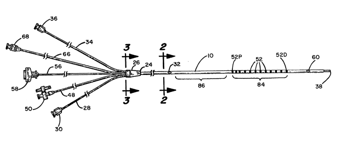
Pigure 1 is 8 drawing showing the preferred embodiment of the
present invention;
Figure 2 is a cross-sectional view taken along the llne 2-2 In
Flgure 1~
Flgure 3 Is a cross-sectional view taken along the line 3-3 In
25 Flgure l;
Figure 4 is a longitudinal cross-section view of the distal end
portion of the catheter of Figure l;
Figure S is a further longitudinal sectlonal vlew showing the
manner In whlch a typical surface electrode is conflguredt
S
.
!
--- 13UO~
Figure 6 Is a cross-sectional view showlng the manner In whlch
the thermistor-type temperature sensor Is dlsposed In the lumen of
Figure 1~
Flgure 7 Is a drawlng showlng the constructlon of ~tlffenor
S members used in the embodlment of Plgure l;
Fi~ure 8 is an alternative stiffener member used In the
embodiment of Figure l;
Figure 9 shows the manner in which the stiffener of Figure 7 is
installed in the proximal lumen of the catheter of Figure l; and
Figure 10 is a sectioned view of the heart showlng the catheter
of this invention installed in the right ventricle.
DESCRIPTION OF THe PREFERReD eMBODIMENT
Referrlng to Flgure 1, the dlagnostic catheter of the present I-
lnventlon 18 seen to comprl# an elongated tubular member 10 whlch Is
lS extruded so as to have a predetermined outer diametor whlch, for
purposes of example only, may be about 2.28 mm and whlch is preferably
formed from sllicone rubber, polyurethane or some other suitable plastlc
whlch tends to be non-thrombogenic. With reference to the cros~
sectlonal vlew of Flgure 2, there are a plurallty o~ separate lumens 14, 16,
18, 20 and 22 extendlng the length thereof. For reasons whlch wlll
become apparent from a continued reading of the speciflcation, the
lumen 14 i5 referred to as the inflatJon lumen, 16 the dlstal lumen, 18 the
sensing electrode lumen, 20 the thermistor lumen, and 22 the proximal
lumen. For a catheter 10 of the typical size set forth above, the Inflatlon
lumen 1~ may ~e about 0.37 mm ~n diameter. The lumens 16 and 22 may
each be approximately 0.81 mm in diameter. The thermlstor lumen 20
may also be 0.Sl mm, while lumen 18 is about 0.71 mm in diametcr.
l~onded to the proximal end 24 of the extruded catheter 101s a
yoke member 26 whlch provides a means whereby varlow devlcos may be
connected to the several lumens runnlng through the c theter body 10.
1 3
The yoke 26 Is preferably molded from a thermosetting, medical 8rade .
plastic. Wlth the ald of the cross-sectlonal vlew of Flgure 3 and the vlew
of Figure 1, there is shown a length of PVC tubing 28 whlch Is adheslvoly
bonded withln the yoke 26 and a Luer lock 30 is afflxed to the othor end
5 of the tube 28. Thus, fluid communication Is establlshcd between the
Luer lock 30 and the proximal lumen 22 (Figure 2) of the catheter 10. As
shown In Pigure 1 and as better seen in the enlarged view of Flgure 9,
extending through the side wall of the tubular catheter 10 and
communicating with the proximal lumen is a port 32. Thus, fluid
10 communication is established from the Luer lock 30, through the tube 28
and the yoke 26, and through the proximal lumen 22 out through the
port 32.
In a ~ornewhat slmilar fashlon, a tube 34, preterably formed from
PVC, is termlnated at its proxlmal end wlth a Luer lock 36. The other end
lS i5 adheslvcly bondcd into a borc in the yoke 26 leadlng to the distal
lumcn 16 (Flgure 2) which extends the entire length of the catheter
body 10 and termlnates In a distal port 38. Thus, fluld such as radlopaque
dyes, drugs, ctc. may be Introduced through the Luer connector 36 and
will flow through the tubing 34, the yoke 26 and the dlttal 16 to exit the
20 distal port 38.
As can best be seen In the enlarged vlew of Plgure 4, the distal
end portlon of the catheter 10 Is formed to a reduced dlamcter. Pltted
over that end portlon ls a plece of expandable balloon tu~lng 40. It Is
bonded to the catheter body 10 at locations 42 and 44 wlth a sultable
25 adhesive. Formed through the side wall of the cathetcr 10 In the zone
spanned by the balloon member 40 Is a port 46 whlch communlcates wlth
the inflation lumen 14. The inflation lumen runs the entlre length of the
catheter body and extends through the yoke 26 where a PVC tube 48 jolns
it to a Luer valve S0. Thus, when a fluld, under pressure, is Introduced
30 through the opened Luer valve S0, it flows through the tube 48, the
1~()` (:) 4 L~ 9
yoke 26, the inflation lumen 14 and out the port 46 to inflate the
balloon 40 By then closing the valve S0, the balloon can be retalned In lts
Inflated state.
Ncxt, wlth rcference to Flguros 1 and 5, It can bo soon that
there are afflxed to the outer surface of the tubular catheter 10, a
plurality of rlng-type surfacc electrodes S2, the most proxlmal rlng belng
Identlfied by numeral S2P and the most distal rlng bclng identlflcd by
numeral S2D. For a catheter to be used wlth an adult heart, the rin8
electrode 52D may typically be disposed approximately 80 mm from the
10 distal end of the catheter 10. The spacing between adjacent surface
electrodes may typlcally be 10 mm, but It to be understood that an
alternatlvc spaclng may be used, especially In pedlatrlc operatlon~.
Wlth reference to Figure 5, independently connected to each of
the ~urface clectrodes 52, ~2D and S2P are Insulated conductors, as at 54,
15 which extend proxlmally through the senslng electrode lumen 18 and
through a length of PVC tubing S6 to the indivldual connector plns (not
shown) contalned wlthin the connector houslng S8. Thls connector is
adapted to be jolned to the electronic circuitry used In the measurement
of stroke volume using impedance pleysthmography.
Next, wlth reference to Flgures I and 6, formed through the slde
wall of the tubular catheter 10 is an openlng 60, and just below the
openlng 60 Is a thermistor element 62 whlch Is disposed wlthln the
thermi~tor lumen 20. Its electrical leads 64 extend down thls lumen and
through the yoke 26 and the PVC tubing 66 to a further electrlcal
25 connector 68. A plug formed from silicone rubber adhesive is identlfied
by numeral 70. lhen, a plastic, such as polyurethane, havlng good heat
conducting properties, is made to cover the opening 60 to prevent the
ingress of blood and other body fluids.
Referring now to Figure 9, it can be #en that a polyurethane
30 potting adhesive plu~ 72 is injected into the proximal lumen at a locatlon
,
' ' - .''
. .
13004 ~9
just distal o~ the proxlrnal port 32 so as to block that lumen agalnst any
fluid flow therebeyond. The proximal lumen 22 continues distally of the
plug 72, however, and disposed in this lumen are first and second
stiffening members of the type shown in Figure 7 of the drawlngs. The
5 stiffenJn6 members are indlcated generally by numeral 74 and comprl e a
stainless steel coil 76 surrounding a s~ainle~s steel cor~ wlro 78. The core
wire 78 Is welded at each end (80 and 82) to the surroundlng coll wlre 76.
In the case of a catheter made in accordance wlth thc preferrcd
embodiment being described herein, the coil may be made f rom a
10 O.lSO mm wire wound as a unifiler coil and preferably i5 fabricated from
Type 304 stainless steel. The core wire may typically have a diameter of
0.3SS mm and aJso may be Type 304 stainless steel. ay welding the core
wire to the coll on each end thereof, unravellng of thc coll Is precluded
when the stiffener Is subjected to tensile forces. The weldod core wlre
15 also prccludes penetration of the lumen walls.
As shown in Flgure 8, It Is also contemplated that one end of the
corc wlre 78 may be tapered as shown at end 80 thereof to thereby
Increase the relative flcxibility of the stiffener member at that end. Thc
purpose ol thls will become more apparent as the descrlption proceeds.
20 lrrespective of the type of stiffener mcmber used, they may be
approximately 0.81S mm in diameter and may have an overall Icngth of
approximately lO cm.
Referring a~ain to Figures 1 and 9, a llrst stlffener member 74
may be fed down the proxlmal lumen untll positloned In the zone occupled
25 by the spaced-apart surface electrodes S2 and Identlfled by the
bracket 84. Spaced proxlmally from the above-mentloned dlstal stlffener
member ls a second stiffener member which extends distally from the end
of the potting adhesive plug 72 near the proximal port 32 (Flgure 9) In the
zone identified by bracket 86. These two stlffener members, beJng0 located in the proximal lumen of the catheter 10 and spaced apart from
13~04.~9
,~
one another ~y a snort dis~ance glves the catheter a ~endencl ~;o
berd ~n the ~one ~etween the two, but in such a manner that tre
catheter does not ~lnk so as tc occlude the lumen.
~ he surface electrodes 5~ are crimped in place only
after the stiffener member 74 has been fitted into zone 84, and
the crimping opera~ion not only secures the right electrodes to
the outer sur~ace of the catheter, but also tends to hold the
stiffener member 7~ in place.
OPE~ATION
Referring to E'igure 10, there is shown a se~tioned view
of a heart with a catheter of the present invention nstalled so
as to facilitate the monitoring of the patient's stroke volume
using right ventricular impedance pleysthmosgraphy. The Catheter
is installed by entering the patient's subclavean vein or
brachial vein and routing it through the superior vena cava into
the right atrium and from there through the tricuspid valve into
the right ven~ricle. At this point, and inflating fluid is
applied under pressure to the inflation lumen, via the valve 50,
and the fluid exits the port 46 (Figure 4) to inflate the
expander (balloon) 40. As blood is pumped from the right
ventrlcle, the balloon 40 tends to be carried by the flow into
the pulmonary outflow tract. Because of the first and second
stiffener members, which are disposed in the proximal lumen
downstream distally of the proximal port 32 and the relat~ve
dimensions of those stiffener members and the spacing
therebetween, the catheter tends to bend at a point proximate
the apex of the right ventricle, as illustrated in Figure lo,
with the segment 84 on which the surface electrodes are arrayed
; ~ extending upwardly through the right ventricle. The proximal
-~ right electrode 52P is located in the apex of the heart while
the distal surface electrode 52~ is at the entrance to the
~` pulmonary outflow tract.
-. Once the catheter is so installed, stroke volume
measurements can be taken using the technique set out in the Salo
~, 10
: ~,
"
l3no4~
J~lted States patent ~o. ~ Li enti'led "~ J ~J~ A~'UA'~ALU5
FOR MEASURING VENTRlC,ULAR VOLUME." ~ecause the present invention
~s concerned With physlcaï constructlon of the catheter, it ~s
deemed unnecessary to explain in detail how the strohe ~701ume
measurements are obtained. Those desiring an ex31an.ation ol
the lmpedance pleysthmography technique for measurin~ stroke
volume can refer to the aforementioned application and to the
publications referenced therein.
To be able to calibrate the stroke volume measurements~
the catheter of the present invention also permits a measurement
of cardiac output using the thermal dilution tecnnique. As is
well known to practitioners in the field, a cold saline solution
may be injected through the proximal lumen via Luer lock 30
whereupon it exits the proximal port 32 which, as seen in Figure
10, will be located in the right atrium. The temperature chanse
occasioned by the flow of the cold saline diluted blood will be
picked up by the thermistor element 60 exposed through the port
60 in the pulmonary outflow tract, and suitable instrumentation
coupled to the electrlcal termi~al 68 is used to convert that
temperature changed information to a stroke volume value for
comparison with the stroke volume obtained using the impedance
pleysthmography technique.
In using a catheter of the type described herein to
measure relative stroke volume, a fewer number of sensing
electrodes, e.g., four, positioned along the catheter body
from the apex of the right ventrical to the pulmonic valve
would be sufficient. Where absolute stroke volume is being
assessed, however, an increased number of sensing electrodes,
e.g. ten, is more appropriate. Furthermore, with the catheter
designed for measuring absolute stroke volume, it is not
re~uired that provision be made for conducting thermal dilution
measurements such that the thermistor sensor can be eliminated.
It is important, however, that it be
.~
1 1
13Q~
Included where relatlve stroke volume measurements aro to be obtalncd
so that periodic correlatlons can be made.
~ ecause of the inclusion of the stiffener members 74, the
catheter does not tend to lay along excitable heart tlssue and, hence,
5 catheter-lnduced PVCs are minlmlzed.
By using a stiffener mernber of the type shown In Figure 8 wlth a
tapered core wire 78 and by orlenting that stiffener in the prox~mal lumen
so that the tapered end of the stlffener member Is pointlng toward the
balloon 40 In the zone 84 of the catheter, the abillty of the catheter to
10 snake around turns Is enhanced. This is partlcularly advantageous In
pedlatric UJO of the catheter.
Thus, therc has been Jhown and deacrlbed the doslgn o~ a ~1
diagnostic coronary catheter whlch can be left In place over a perlod of ~-
hours and even days so that a varlety of medical procedures and
15 measurements may be carried out. One or more drugs may be injected
into the heart cavity via the distal end port 38 and the effect of those
drugs on cardiac performance can be monitored as previously descrlbed.
Thia inventlon has been descrlbed hereln In conslderable detall In
order to comply wlth the Patent Statutes and to provlde those skllled in
2û the art with the Information needed to apply the novel prlnclplca and to
con~truct and use such spoclallzed components aa are requlred. Howover,
lt 1~ to be understood that the Inventlon can be carrled out by spoclflcally
d}~ferent egulpment and devlces, and that varlous modl~lcstlons, both as
to egulpment detalls and operatlng procedures, can bo accompllshod
25 wlthout departing from the scope of the invent.on Itself.
What is clalmed is: