Note: Descriptions are shown in the official language in which they were submitted.
~3~66~;~
3A~:KGROUND OF THE INVENTION
The present invention relates to a spectroscope
apparatus, and more particularly to a two-dimensional
imaging monochrometer apparatus which can continuously
form a plurality of two-dimensional images of a to-be-
measured body due to different light components each
having a desired spectral width.
Furt'ner, the present invention relates to a
method of and an apparatus for controlling reaction which
is accompanied by light emission due to reaction,
discharge or others, and more particularly to a method of
and an apparatus for controlling a reactor or instrument
containing a light emitting body, on the basis of those
images of the light emitting body which are formed hy a
; 15 two-dimensional spectroscope apparatus and are formed of
different wavelength component emitted from the light
emitting body.
In a conventional apparatus for monitoring or
controlling a reactor or instrument containing a substance
20~ which emits light on the basis of reaction, discharge and
others, a monitor window is provided at the wall of the
~:~ reactor or instrument, and the inside of the reactor or
: instrument is observed through the window to control
variable quantities contribut1ng to the chemlcal change of
the substance. :
.~ 1
.: .
,
~3~CP~i6~2
For example, in a thermal power station or the
like~ the state of combustion flames is observed by an
industrial television camera through the monitor window,
and it is judged on the basis o~ the above observation
and analytical values of exhaust gas whether the state of
combustion flames is appropriate or not, to control the
quantities of air and fuel so as to obtain optimum
flames. Further, an image indicating the brightness
distribution in combustion flames is formed on the basis
of the observation on combustion flames by the industrial
television camera, to be used for monitoring and
controlling the combustion flames. A method of
monitoring and controlling combustion flames on the basis
of the output of a photodetector which receives light
from the combustion flames is known in the prior art. A
control method using a video signal from a television
camera is also known in the prior art. However, in these
known methods the output of an industrial television
camera due to all the wavelength components emitted from
a light emi~ting substance (namely, light emitting body)
is used for monitoring and controlling the light emitting
body. That is, the methods fail to use only a desired
wavelength component emitted from the light emitting
bod~, of the purpose of monitoring and controlling the
above body. In general, the emission spectrum of the
light emitting body is based upon active atoms, molecules
and r~dicals which are contained in this body. The
- 2 -
" ~ ' ' . , :
,
,
~31~63~:
1 inforrnation due to each of wavelength components from the
light emitting body makes it possible to estimate the
state of the body on the level of atom, molecule and
radical, and is indispensable for accurate monitoring and
control operations.
A method of monitoring or controlling a flame on
the basis of the information due to each of wavelength
components emitted from the flame, is disclosed in, for
; example, a Japanese Patent Application JP-A-53-107,890.
In this method, the state of a flame is monitored and
controlled on the basis of the correlation between the
intensities of OH-radical line, C2-radical line and
CH-radical line appearing on the emission spectrum of the
flame and analytical values of exhaust gas. In the
method, however, the intensity of each wavelength com-
ponent emitted from a point in the flame or the sum of
intensities of all wavelength components emitted from the
whole region of the flame is used, and thus it is
impossible to obtain an image which indicates the
distribution of each wavelength component in the flame.
Generally speaking, in the reaction generatlng a light
emitting body which always moves, such as a flame,
detailed information on the distribution of each wave-
length component, that is, the distribution of each
chemical species in the flame, teaches the progress of the
reaction and the fine structure of the flame, and suggests
a position where nitrogen oxlde and soot are generated.
Accordingly, a method is required which has not
::
; - 3 -
32
only an advantage of spectrochemical analysis (that is,
an advantage that information on each of chemical species
in the flame is obtained), but also are advantage of an
industrial television camera (that is, an advantage that
an image o~ the flame is formed). There has been known a
method, in which an interference filter capable of
transmitting only a desired wavelength is provided in
front of an industrial television camera. According to
this method, an image of a light emitting body can be
formed of a desired one of wavelength components emitted
from the body. In this method, however, it is necessary
to prepare a plurality of interference filters, and it is
impossible to change the measuring wavelength
continuously, since replacement of interference filter is
required for changing the measuring wavelength. Further,
an interference filter attenuates light in a great
degree, and thus makes it impossible to obtain a clear
image by the industrial television camera.
Further, reaction accompanied by light emission
occurs in the following apparatuses and methods, that is,
a photochemical vapor deposition apparatus, a vapor phase
epitaxial growth apparatus, a semiconductor fabricating
method, a method of forming a nitride film and a chemical
vapor deposition apparatus. In any one of these patent
applications, the distribution of each of chemical
' ~
13~6~32
components of a light emitting body in the body is not
measured, and it is not disclosed to monitor and control
the light emitting bod~ on the basis of information on
the above distribution.
In the simplest conventional method for forming
a plurality of images of an object to be measured, of
different light components each having a spectral width,
optical filters are used, each of which transmits only a
light component having a desired spectral width and
absorbs or reflects other light components. For example,
in a case where a photograph is taken by an ordinary
camera in a state that strong ultraviolet rays are
present, when an ordinary film is used, blurs in color
tone are produced. Accordingly, an ultraviolet cut
filter is used, to form an image only of visible light.
Further, in order to thoroughly investigate
discharge and combustion phenomena, it is necessary
to observe the spatial intensity distribution of a
wavelength component peculiar to each of unstable
chemical species e~isting in a discharge. plasma or
flame such as radicals and active molecules. In this
case, only specified wavelength components are measured,
and thus optical filters each capable of transmitting
one of the specified wavelength components are used.
For example, the measurement of unstable chemical
species contained in a flame is described in an
article (Applied Physics B. Vol. 29, 19~2,
-- 5 --
~,' , ~,
13~663Z
1 pages 93 to 97). It is shown in Fig. 1 of this article to
use a filter for light from the OH-radical and another
filter for light from the C2-radical. However, in a
method of forrning an image due to light within a specified
wavelength range by using a filter, it is required to
change the specified wavelength range by the replacement
of filter, and hence it is impossible to change a measur-
ing wavelength continuously. Further, a filter attenuates
light in a great degree. In the above article, no regard
is paid to such problems.
An optical apparatus for forming a plurality of
images of an object due to different wavelength components
continuously by using a spectroscope, is described on page
20 of the abstracts of the spring meeting of the Spectro-
scopical Society of Japan held in May, 1985. In thisoptical apparatus, the measuring wavelength can be
continuously varied by rotating a grating included in the
spectroscope. Further, in the spectroscope, light
reflection is repeated, and no optical filter is used.
Thus, light is scarcely attenuated in the spectroscope.
In this optical apparatus, however, as is apparent from
the description that, since a background having a
continuous spectrum is present, a wavelength component
which exists in the vicinity of a band head and is not
affected by a band spectrum, is used, and an image formed
of the wavelength component is corrected by software,
there arises the following problem. That is, in a case
where light emitted from an object to be measured has a
-- 6
~3C~663Z
l continuous spectrum, the measurement is restricted as
above. Further, it is required to correct an image by
software, and thus the optical apparatus is complicated in
structure.
As mentioned above, in a method of taking
desired wavelength components out of light emitted from an
object to be measured, by using filters to form a
plurality of images of the to-be-measured object, there
arise problems that it is impossible to change the
taken-out wavelength component continuously, since the
wavelength component is changed by the replacement of
filter, and that each filter absorbs light, and thus the
intensity of the taken-out wavelength component is greatly
reduced. ~urther, in a method of taking out desired
wavelength components by using a spectroscope, the taken-
out wavelength can be continuously varied, but there arise
optical problems that when light incident on the spectro-
scope has a continuous spectrum within a wavelength range,
measurement is restricted as mentioned above or a desired
image cannot be formed.
SUMMARY OF THE INVENTION
An object of the present invention is to provide
a two-dimensional imaging monochrometer apparatus
(spectroscope apparatus) which can form an image of an
object to be measured, of a~desired wavelength component
emitted from the to-be-measured body and moreover can
change the desired wavelength component continuously, and
-- 7
13~663~
1 which can form the image of the to-be-measured body with-
out being subject to any restriction, even when the light
incident on (that is, received by) the spectroscope
apparatus has a continuous spectrum in a wavelength range.
Another object oE the present invention is to
provide a method of and an apparatus for monitoring or
controlling reaction accompanied by light emission, by
using a two-dimensional imaging monochrometer apparatus
which can form an image of an object to be measured, of a
desired wavelength component of light emitted from the
to-be-measured body and moreover can change the desired
wavelength component continuously, and which can form the
image of the to-be-measured body without being subject to
any restriction, even when the light received by the
lS spectroscope apparatus has a continuous spectrum in a
wavelength range.
According to an aspect of the present invention,
there is provided a spectroscope apparatus which comprises
means Eor separating light emitted from an object to be
measured, into spectral components, means for mixing that
part of the spectral components which exists in a desired
wavelength range, and means for forming an image of the
to-be-measured body, of mixed light. Further, the
spectroscope apparatus may be provided with means for
maklng light rays which are formed of the desired spectral
part,~diverge, and Eor focusing the divergent light rays
to a point before the desired spectral part is mixed.
In more detall, a spectroscope apparatus
8 -
,
~3~6~i3;~
1 according to the present invention includes a first
spectroscope, a second spectroscope which is coupled with
the first spectroscope through an intermediate slit, and
drive means for driving the first and second spectro-
scopes. The first spectroscope includes a collimatorsystem for forming an image of an object to be measured on
a light dispersing grating, and includes a light dispers-
ing optical system which is made up of the light
dispersing grating and a first optical system for guiding
the diffracted light from the light dispersing grating to
the intermediate slit. The second spectroscope includes a
light mixing optical system which is made up of a light
mixing grating and a second optical system for focusing
light having passed through the intermediate slit on the
light mixing grating, and includes an image formation
optical system for forming an i.mage of mixed light from
the light mixing optical system. The drive means drives
the light dispersing optical system and the light mixing
optical system so that these systems are optically
symmetrical with respect to the intermediate slit.
A spectroscope apparatus according to the
present invention is applicable to light emitted from a
flame due to combustion, light due to photochemical
reaction which is generated by irradiating a photochemi-
cally reactive gas with stimulating light, fluorescencewhich is emitted from a pigment for staining a desired
tissue in a cell, when the pigment is irradiated with
predetermined light, and light emitted from a flame at a
~30~ 3~
1 time a solution containing a metal ion is introduced into
the flame. That is, the spectroscope apparatus can
separate the above light into spectral components, mix
that part of the spectral components which exists in a
desired wavelength range, and form an image of mixed
light. Thus, a spectroscope apparatus according to the
present invention is applicable to a method of monitoring
the combustion state of a flame, a method of monitoring
photochemical reaction which proceeds in a photochemical
reaction apparatus, a method of monitoring biochemical
reaction which occurs at a predetermined tissue of a cell,
and a method of determining a metal ion by flame spectro-
chemical analysis.
Thus, according to another aspect to the present
invention, there are provided a boiler provided with a
spectroscope apparatus which receives light from a flame
generated in the ~urnace of the boiler; a gas turbine made
up of a compressor for compressing air, a combustor for
burning fuel with the aid of compressed air, a turbine
driven by a combustion gas, and a spectroscope apparatus
which receives light from a flame generated in the
combustor; a photochemical reaction apparatus for proceed-
ing photochemical reaction by irradiating photochemically
reactive gas with stimulating light which reaction
apparatus is provided with a spectroscope apparatus
recelving light due to the photochemical reaction; a
biochemical reaction apparatus for irradiating a pigment
having stained a desired tissue in a cell, with light to
-- 10 --
~3C~6~
1 generate fluorescence from the pigment which reaction
apparatus is provided with a spectroscope apparatus
receiving the ~luorescence; and an analytical apparatus
for determining a metal ion in a solution by flame
spectrophotometry which analytical apparatus is provided
with a spectroscope apparatus receiving light from a
flame. Each of the above spectroscope apparatus includes
means for separating incident light into spectral
components, means for mixing that part of the spectral
components which exists in a wavelength range, and means
for forming an image of mixed light.
Incidentally, the above-mentioned photochemical
reaction apparatus includes a photochemical vapor deposi-
tion apparatus, a vapor phase epitaxial growth apparatus,
and a chemical deposition apparatus. Further, the
stimulating light is selected from visible light, infrared
rays, ultraviolet rays and a laser beam~
Further, according to a further aspect of the
present invention, there is provided a method of
controlling the reaction accompanied by light emission, in
which light from a light emitting body is separated into
spectral components, that part of the spectral components
which exists in a desired wavelength range is mixed to
form an image of the light emltting body of mixed light, a
plurality of images of the llght emitting body are formed
in accordance with a plurality of desired wavelength
ranges, the images thus obtained are compared with
previously-prepared reference images, and variable
;632
l quantities concerning the state of the light emitting body
are controlled so that the images agree with the reference
images.
In the above control method, it is preferable
that a region where one of chemical species contained in
the light emitting body is present, is increased in area
and a region where another chemical species is present, is
reduced, when tne supply quantity of one of raw materials
of the light emitting body is changed. For example, it is
preferable that the light emitting body is a combustion
gas, and one and another chemical species are C2-radical
and ~O-radical, respectively.
The desired wavelength range is selected from
the whole spectral range of light emitted from the light
emitting body, and it is preferable that the desired
wavelength range is a wavelength range frorn a wavelength
longer than a specified wavelength by 2.5 nm to a
wavelength shorter than the specified wavelength by 2.5 nm.
Further, according to still another aspect of
the present invention, there is provided an apparatus for
controlling the reaction accompanied by light emission
which apparatus includes a reaction apparatus for forming
a light emitting body therein, an optical guide for
forming an optical path for light emltted from the light
emitting body, two-dimensional imaging monochrometer
apparatus for separating light from the optical guide into
spectral components, a monitor for displaying a plurality
of images which are formed of light components having
- 12 -
131~63~
1 different wavelength ranges, a memory for storing a
plurality of reference images, and a controller for
displaying each of the images and a corresponding one of
the reference images at the same time to control variable
quantities concerning the state of the light emitting body
so that the images agrees with the reference image. The
two~dimensional imaging monochrometer apparatus includes a
flrst spectroscope, a second spectroscope which is coupled
with the first spectroscope through an intermediate slit,
and drive means for driving the first and second spectro-
scopes. The first spectroscope includes a collimator
system for focusing the light to be measured on a light
dispersing grating, and includes a light dispersing
optical system which is made up of the light dispersing
grating and a first optical system for leading the
diffracted light from the light di.spersing grating to the
intermediate slit. The second spectroscope includes a
light mixing optical system which is made up of a light
mixing yrating and a second optical system for focusing
light having passed through the intermediate slit, on the
light mixing grating, and includes an image formation
optical system for forming an image of the mixed light
from the light mixing optical system. The drive means
drives the light dispersing optical system and the light
mixing optical system so that these systems are optically
symmetrical with respect to the intermediate slit.
The above apparatus for controlling the reaction
accompanied by light emission can control the state of the
- ~3 ~
~3~
1 light emitting body accurately, provided that the image
formation optical system of the second spectroscope is
provided with a light amplifying element, the width of the
intermediate slit is variable, the rotational angle of
each of the light dispersing grating and the light mixing
grating can be varied continuously, while maintaining a
state that these gratings are optically symmetrical with
respect to the intermediate slit, the optical guide
includes a lens capable of transmitting light within a
wavelength range from an ultraviolet region to an infrared
region, and the light intensity distribution at an image
formed of mixed light can be expressed in colors.
According to the above apparatus for controlling
the reaction accompanied by light emission, light is drawn
from a reactor or instrument in which the reaction or
phenomenon accompanied by ligllt emission proceeds, an
image due to part of the spectral components of the drawn
light is continuously formed by the two-dimensional
imaging monochrometer apparatus, and the chemical species
distribution in the light emitting body is monitored with
the aid of the images, or the reaction or phenomenon is
controlled so that the chemical species distribution is
optimum. That is, the state of the light emitting body is
estimated on the level of chemical species such as an
atom, a molecule and a radical, and thus can be accurately
monitored or controlled. Further, information on the
distri`oution of each of chemical species such as an atom,
: a molecule and a radical, can be obtained, and thus
- 14 -
~3~ 32
1 generation and extinction processes in reaction can be
observed That is, the progress of the reaction can be
estimated, and the reaction can be monitored or controlled
more accurately.
As mentioned above, in an apparatus for
controlling the reaction accompanied by light emission
according to the present invention, light from an object
to be measured is separated into spectral components, that
part of the spectral components which exists in a desired
wavelength range is mixed, and a very clear image of the
to-be-measured object is formed of mixed light. Such an
image provides information useul for controlling the
combustion state of a fuel generated in a furnace, the
progress of photochemical reaction and the progress of
biochemical reaction, and useful for the observation on a
cellular texture and the flame spectrophotometric analysis
of a metal i.on contained in a solution. That is, the
above image makes possible a precise control operation,
and provides accurate information.
In a two-dimensional imaging monochrometer
apparatus according to the present invention, incident
light passes through the collimator system and the light
dispersing optical system of the first spectroscope, and
then only part of the spectral components of the incident
light reaches the intermediate slit. ThUS/ the first
spectroscope functions as a light dispersing element. The
intermediate slit passes a desired range of the wavelength
of the light. The spectral part from the intermediate
- 15 -
~3~6~2
l slit passes through the light mixing optical system and
the image formation optical system of the second
spectroscope, to form an image of mixed light. Thus, the
second spectroscope functions as a light mixing element.
By driving the light dispersing optical syste~ of the
first spectroscope and the light mixing optical system of
the second spectroscope so that these systems are
optically symmetrical with respect to the intermediate
slit, the wavelength of mixed light used for forming the
image can be continuously varied.
As mentioned above, a two-dimensional imaging
monochrometer apparatus according to the present invention
can continuously ~orm a plurality of images due to part of
the spectral components of incident light. Thus, the
spectroscope apparatus can clearly shows the chemical
species distribution in a light emitting body which is
generated in a reactor or instrument, and makes it
possible to monitor the light emitting body or control
variable quantities concerning the yeneration of the light
emitting body so that the optimum distribution of a
chemical species in the light emitting body is achieved.
BRIEF DESCRIPTION OF THE DRAWINGS
Fig. 1 is a schematic diagram showing an optical
path in an embodiment of a spectroscope apparatus accord-
ing to the present invention.
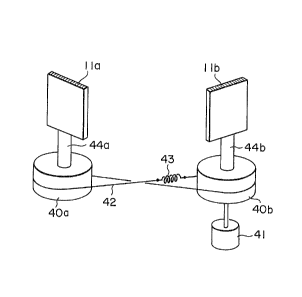
Fig. 2 is a schematic diagram showing anembodiment of the arrangement for driving gratings.
- 16 -
:,
1 Fig. 3 iS a schematic diagram showing a test
plate which is used for measuring the wavelength resolving
power and spatial resolving power of the embodiment of
Fig. 1.
Figs. ~A to 4~ are schematic diagrams showing
the results of measurements of the wavelength resolving
power and spatial resolving power of the embodiment of
Fig. 1.
Fig. 5 is a schematic diagram showing an
embodiment of a monitor/control apparatus according to the
present invention.
Fig. 6 is a schematic diagram for explaining the
optical principle of a pinhole camera.
Fig. 7 is a schematic diagram for explaining the
optical principle of image formation which is carried out
by a spectroscope apparatus according to the peesent
invention.
Fig. 8 is a schematic diagram showing a cross
section of an echelette plane grating which is usable in a
spectroscope apparatus according to the present invention.
Fig. 9 is a schematic diagram for explaining
light reflection from an echelette plane grating.
Fig. 10 shows an example of the spectrum of
incident light.
Fig. 11 is a schematic dlagram for explaining
the optical path formed in a spectroscope.
Fig. 12 shows an example of the spectrum of
light emerging from the spectroscope of Fig. 11.
- 17 -
~3~6~32
1Fig. 13 shows an example of the spectrum of
light incident on the spectroscope of Fig. 11.
Fig. 14 is a schematic diagram ~Ihich shows an
optical path in the spectroscope of Fig. 11 corresponding
to the incident light of Fig. 13.
Fig. 15 shows the spectrum of outgoing light
corresponding to the incident light of Fig. 13.
Fig. 16 is a perspective view showing a main
part of another embodiment of a monitor/control apparatus
according to the present invention.
Fig. 17 is a schematic diagram for explaining
the optical principle of the light amplifying element of
Fig. 16.
Fig. 18 shows an example of the emission
spectrum of flame.
Fig. 19 is a schematic diagram showing radical
distribution in a flame on the basis of those images of
the flame which are obtained by the present invention.
Fig. 20 shows how light emitted from flames is
led to a monitor/control apparatus according to the
present invention.
Fig. 21 shows that light from a photochemical
reaction apparatus is received by a monitor/control
apparatus according to the present invention.
25Fig, 22 shows an example of the spectrum of
light emission due to photochemical reaction.
Fig. 23 is a schematic diagram showing that the
present invention is applicable to the determination of a
- 18 ~
'
~3~
1 sampling position for emission spectrochemical analysis.
Fig. 24 is a schematic diagram showing an
example of the observation on a desired tissue of a cell,
according to the present invention.
DESCRIPTION OF THE PREFE~RED EMBODIMENTS
Example I
Fig. 5 shows an embodiment of a monitor/control
apparatus according to the present invention. Referring
to Fig. 5, the present embodiment includes an optical
guide 36 receiving light from a light emitting body which
is formed in a reaction apparatus 35 as the result of
reaction (or a phenomenon) accompanied by light emission,
a two-dimensional imaging monochrometer apparatus for
successively outputting a plurality of images formed of
desired ones of wavelength components of incident light, a
camera 8 Eor forming an image which shows the reaction
product (namely, chemical species) distribution in the
light emitting body, on the basis of the output images
from the spectroscope apparatus, a monitor 37, a memory 38
for previously storing reference images, and a controller
33 for controlling variable quantities concerning the
generation of the light emitting body on the basis of the
comparison of the image from the camera 8 with a reference
image from the memory 38 so that optimum reaction-product
distribution in the light emitting body is obtained. The
reference image may be displayed on the display screen of
-- 19 --
3&~6~32
1 the monitor 37 together with the image from the camera 8,
or may he displayed by another monitor ~not shown). The
reference image is used as the standard of the image
obtained from the camera 8, and the reaction product
distribution and a light and shade pattern in the image
are compared with those in the reference image. Accord-
ingly, a plurality of reference images corresponding to a
plurality of images which are formed of different wave-
length components, are stored in the memory 38. In a case
where only a monitoring operation is performed, the memory
38 and the controller 39 may be omitted from the present
embodiment.
The two-dimensional imaging monochrometer
apparatus includes a first spectroscope 2 which is
provided with a condenser lens 1 for collecting light rays
from the optical guide 36, an intermediate slit 3, and a
second spectroscope 3. A relay lens group 6 and a
focusing lens group 7 act as a focusing optical system for
the second spectroscope ~. In other words, the lens
groups 6 and 7 forms an image due to that part of the
spectral components of light incident on the condenser
lens 1 which exists in a desired wavelength range, on the
light receiving surface of the camera, without producing
astigmatism chromatic aberration.
First, the operation principle of the two-
dimensional imaging monocilrometer apparatus capable of
forming an image of a light emitting body to be measured,
of a desired wavelength component, will be explained while
- 20 -
~3~6G~2
1 being compared with that of a pinhole camera, with
reference to Figs. 6 and 7. Referring to Fig. 6 which
shows the operation principle of a pinhole camera, a
pinhole 9 acts as the point source of light rays for
forming an image, and the upper and lower parts of light
rays passing through the pinhole 9 are replaced with each
other at the pinhole 9. Further, the left and right parts
of the light rays are replaced with each other at the
pinhole 9. Then, the image is formed on a film 10. That
isj all wavelength components ~T contains in incident
light contribute to the formation of the image. In the
two-dimensional imaging monochrometer apparatus, a grating
11 which diffracts different wavelength components in
different directions, is disposed in place of the film
10. Accordingly, as shown in Fig. 7, images due to wave-
length components ~ and ~k are formed on screens 12,
13 and 14, respectively. In Fig. 7, a slit 15 performs a
~unction corresponding to that of the pinhole 9. That is,
at the slit 15, the upper and lower parts of light rays
are replaced with each other, and the left and right parts
of the light rays are replaced with each other. As shown
in Fig. 7, light rays having passed through the slit 15
form a divergent light beam, which is converted by a
concave mirror 16 into parallel light rays. The parallel
light rays thus obtained are difracted by the grating 11,
that is, di~ferent wavelength components are reflected
from the grating 11 in different directions.
Now, an echelette plane grating used in a
- 21 -
Z
1 spectroscope apparatus according to the present invention
will be ex~lained, with reference to Fig. 8. As shown in
FigO 8, the cross section of one main surface of an
echellette plane grating 11 has the form of saw-teeth
parallel to two planes. An angle a between a groove
surface 17 and a grating plane 18 is called blaze angle.
Let us express the distance between adjacent grooves
(namely, grating constant), an incident angle between
incident light OA and a normal ON to the grating plane 18,
and an angle between reflected light OB and tne normal ON,
by d, ~ and ~, respectively. When an optical path
difference between light beams diffracted from adjacent
grooves is equal to an integer multiple of a wavelength ~,
the light beams are in phase. That is, constructive
interference takes place between the light beams, when the
factors d, ~, ~ and ~ satisfy the following equation:
m~ = d (sin ~ + sin ~), when m = 0, +1, +2, and so on
.......... (lj
Incidentally, m indicates the spectral orders.
For example, let us consider the diffracted
light of the first order indicated by m = 1. When the
incident light AO having a wavelength ~ makes an angle ~
;~ with the normal ON to the gratlng plane 18, the light OB
diffracted from the groove surface 17 having a grating
constant d makes an angle ~ with the normal ON.
Referring to Fig. 9, when light ~T incident on
- 22 -
, . , ~,
66~
1 the echelette plane grating has two wave components ~1
and ~2~ an angle x between the diffracted wavelength
component ~1 and a normal to the grating plane 18 and an
angle y between the diffracted wavelength component ~2
and the normal r can be calculated from the equation (1).
It is to be noted that when the wavelength ~1 is longer
than the wavelength ~2' the angle x is greater than the
angle y.
The above fact holds for a case where incident
light is formed o~ three or more spectral lines, that is,
for a case where the inci.dent light is formed of a
plurality of spectral lines each having a very small
spectral width, or a difference in wavelength between
adjacent spectral lines of incident light is greater than
the resolving power of the spectroscope 2. Accordingly,
when incident light has three wavelength components ~i'
~j and ~k as shown in Fig. 10, three images due to the
wavelength components ~ and ~k are formed on the
screens 12, 13 and 14, respectively. In an ordinary case
where the imaging surface of the camera 8 is used as the
screen, that is, only a single screen is used, three
images are successively projected on the screen by
rotating the grating 11. In a spectroscope or the like,
~ as shown in Fig. 11, a detector 20 for detecting light
from an exit slit 19 corresponds to the screen, and only a
- spectral component incident on the exit slit 20 at right
angles is detected by the detector 20. That is, when
light ~T incident on the spectroscope contains three
- 23 -
~3~ i32
1 spectral lines ~ and ~k as shown in Fig. 10, the
light ~T is separated by the grating 11 into three wave-
length components ~ and ~k~ which are focused on the
exit slit 19 by a concave mirror 16. In Fig. 11, the
wavelength component ~j is incident on the exit slit 20
at right angles, and thus an image formed of only the
wavelength component ~j is detected by the detector 20.
Fig. 12 shows the spectrum of the detected image. In
order to detect the wavelength component ~j, the grating
11 is rotated in a direction A so that the wavelength
component Ai is incident on the exit slit 20 at right
angles. Similarly, in order to detect the wavelength
component ~k' the grating 11 is rotated in a direction B.
In the above, explanation has been made of a
case where the incident light ~T is composed of discrete
spectral lines. In a case where the incident light ~T
has a band spectrum shown in Fig. 13, a light component
detected by the detector 20 has a spectral width as shown
in Fig. 14. That is, the detected light component has a
spectrum shown in Fig. 15. In other words, a correct
image is not formed on each of the screens 12, 13 and ]4
of Fig. 7, but a beltlike image spread or blurred in a
longitudinal direction is formed on each screen (it is to
be noted that the length of the image in a transverse
direction is determined by the length of the slit and
hence the image is not blurred in the transverse direc-
tion). That is, in a case where the incident light has a
band spectrum as shown in Fig. 13, it is impossible to
- 24 -
, .
663~
1 form a plurality of correct images by using a single
spectroscope.
Fig. 1 shows an optical system according to the
present invention capable of forming an image which is not
blurred, even in a case where incident light has a band
spectrum as shown in Fig. 13 and outgoing light from the
exit slit 19 has a continuous spectrum in a wavelength
range. Needless to say, the above optical system is used
in the embodiment of Fig. 5. Referring to Fig. 1, the
first spectroscope 2 has the same function as the
spectroscope shown in Figs. 11 and 14. Accordingly, a
grating lla in the spectroscope 2 functions as a light
dispersing element. That is, the light dispersing grating
lla diffracts wavelength components of incident light in
different directions. If the incident light on and the
diffracted light from the grating lla are propagated in
reverse directions, the grating will function as a light
mixing element. In more detail, referring back to Fig. 9,
when the wavelength component ~1 impinges on the grating
11 in a direction which makes an angle x with a normal to
the grating plane, and the w~velength component ~2
impinges on the grating 11 in a direction which makes an
angle y with the normal, the light AT composed of the
wavelength components ~1 and ~2 is reflected from the
grating 11 in a direction which makes an angle z with the
normal. Thus, the grating 11 can act as a light mixing
element. The second spectroscope 4 of Fig. 1 is disposed
so as to perform a light mixing function. That is, the
- 25 -
~6632,
1 optical elements oE the first spectroscope 2 and those of
the second spectroscope 4 are made optically symmetrical
with respect to the intermediate slit 3. In more detail,
a light dispersing optical system of the first spectro-
scope 2 is made up of the light dispersing grating lla anda first optical system including a concave mirror 16 and a
plane mirror 21 for guiding the dispersed light from the
grating lla to the intermediate slit 3. A light mixing
optical system of the second spectroscope 4 is made up of
a light mixing grating llb and a second optical system
including a plane mirror 22 and a concave mirror 16 for
focusing the dispersed light on the grating llb. Drive
means (not shown) drives the light dispersing optical
system and the light mixing optical system so that these
optical systems are optically symmetrical with respect to
the intermediate slit 3. Thus, the outgoing light from
the exit slit 19 of the second spectroscope 4 is not
dispersed. When a screen is disposed in front of the exit
slit 19, the outgoing light can form an image which is not
blurred, on the screen. The condenser lens 1 of Fig. 5
and a concave mirror 16 confronting an entrance slit 15
make up a collimator. A concave mirror 16 confronting the
exit slit 19, the relay lens group 6 and the ~ocusing lens
group 7 make up an image formation optical system. It is
determined by the rotational angle of the gratings lla and
llb what part of the spectrum of incident light passes
through the exit slit 19, and the wavelength range used
for forming the outgolng light from the exit slit 19 is
- 26 -
. . ,
~6632
1 determined by the width of the intermediate slit 3.
Hence, it is desirable to make variable the width of the
intermediate slit 3.
The optical system is so arranged that the
incident light is focused on the light dispersing grating
lla is the first spectroscope 2. Fig. 2 shows the
schematic diagram of one embodiment in which the light
dispersing grating lla in the first spectroscope 2 and the
light mixing grating llb in the second spectroscope 4 are
driven in optically symmetrical with a center of symmetry
about the intermediate slit 3. In this embodiment, pulley
40a is connected to grating lla by sha~t 44a and light
mixing grating llb is connected to pulley 40b by shaft
44b. The pulley 40b is driven by motor 41. The pulley
40a is communicated with the pulley 40b by wire 42 which
is crossed and includes tension spring 43 connected
between the ends of wire 42. The wire 42 and the tension
spring 43 may be made of high strength steel like a piano
wire. In abov~ system, the gratings lla and llb are
arranged in optically symmetry each other about the
intermediate slit 3 and the light dispersing grating lla
and the light mixing grating are driven by the motor 41 in
optically symmetrical about the intermediate slit 3.
Now, explanatlon; will be made of experiments on
the wavelength resolving power and the spatial resolving
power (namely, the resolution of lmage) oE the two-dimen-
sion~al imaging monochrometer apparatus according to the
present invention. ~eferring to Fig. 3, square, circular
- 27 -
13(~ 32
l and trianglar through holes are formed in a black board
23, and filled with color filters. That is, a color
filter 24 capable of transmitting wavelengths more than
390 nm is inserted in the square through hole, a color
filter 25 capable of transmitting wavelengths more than
460 nm is inserted in the circular through hole, and a
color filter 26 capable of transmitting wavelengths more
than 620 nm is inserted in the triangular through hole.
The black board 23 is illuminated wlth white light
(namely, sunlight) 27 as shown in Fig. 3, and light having
passed through the color filters 24, 25 and 26 is led to
the spectroscopes 2 and 4 through the condensor lens l to
form images of the color filters. The gratings lla and
llb are rotated so as to be optically symmetrical with
respect to the intermediate slit 3, to project a plurality
of images on the light receiving surface of the camera 8,
and the images are observed. The results of the experl-
ments are shown in Figs. 4A to 4E. In a case where the
gratings lla and llb were rotated so that light having a
wavelength of 300 nm passed through the exit slit 19, no
image was formed as shown in Fig. 4A, since the filters
24, 2S and 26 were unable to transmit the above light. In
a case where the gratings were set so that light having a
wavelength of 400 nm passed through the exit slit 19, only
an image of the square through hole was formed as shown in
Fig. 4~, since the filter 24 was able to transmit the
light. In a case where the gratings were set so as to
send out light having a wavelength of 500 nm, images of
- 28 -
3~6~2
1 the square and circular through holes were obtained as
shown in Fig. 4C, since the filters 24 and 25 were able to
transmit the light. In a case where the gratings were set
so as to send out light having a wavelength of 600 nm, the
same images as shown in Fig. 4C were obtained as shown in
Fig. ~D, since the filter 26 was unable to transmit the
light. Further, in a case where the gratings were set so
as to send out light having a wavelength of 700 nm, images
of the square, circular and triangular through holes were
formed as shown in Fig. 4E, since all of the filters 24,
25 and 26 were able to transmit the light. Figs. 4A to 4E
show that the two-dimensional imaging monochrometer
apparatus has favorable wavelength resolving power, and
images formed by the spectroscope apparatus in excellent
in resolution.
In the above experiments, the black board 23
having dimensions of 150 mm x 100 mm was used. However,
the size of an ob]ect to be measured can be varied by
changing the condenser lens 1. In the above experiments,
a wavelength range from 300 nm to 700 nm was used.
However, the measuring wavelength range is dependent upon
the~characteristics of the gr~atings lla and llb. The
present embodiment can use ultravolet rays, visible rays
and~infrared rays. Further, it was confirmed by exper-
ments that the wavelength range of that spectral portionof lncldent light which contributed to the formation of
one image could be increased to about 70 A by setting the
width of the intermediate slit 3 appropriately.
: :
- 29 -
'' ,
3~
l As mentioned above, in the present embodiment,
light rays from an object to be measured, are collected by
the condenser lens, and then separated by the first
spectroscope into spectral components. A desired part of
the spectral components is mixed by the second spectro-
scope which is disposed so that the first and second
spectroscopes are optically symmetric with respect to the
intermediate slit, and an image due to mixed light is
formed on the light receiving surface of the camera with-
out having astigmatism and chromatic aberration. Accord-
ingly, even in a case where light from the to-be-measured
body has a continuous spectrum, a desired spectral part
can be continuously taken out of the continuous spectrum
by the first spectroscope, and the taken-out spectral part
is converted by the second spectroscope into mixed light.
Thus, images can be continuously detected without being
subjected to any restriction.
Example II
The outgoing light from the exit slit 19 is a
spectral part of incident light. Accordingly, in some
cases, the outgoing light has a very weak intensity, and
cannot form a clear image. Another embodlment of a
;monitor/control apparatus according to the present inven-
tion can solve the above problem. The present embodiment
ls~different from the embodiment of Fig. 5 only in that,
as shown in Fig. 16, a two-dimensional amplifying element
28 for amplifying a faint image is interposed between the
- 30 -
63~
1 relay lens group 6 and the focusing lens group 7.
The operation principle of the amplifying
element 28 will be explained below, ~ith reference to Fig.
17. It is impossible to multiply a photon 29 itself.
Hence, the photon 29 is converted into electron, which is
converted into a multiplicity of secondary electrons.
Then, the secondary electrons are converted into photon.
In more detail, the photon 29 is converted by a photo-
cathode surface 30 into a primary electron, which is
multiplied to one thousand or more secondary electrons by
a secondary electron multiplier 31. The multiplier 31
utilizes a phenomenon that when a metal wall is bombarded
with an electron, a plurality of secondary electrons are
emitted from the metal wall, and such electron mutlipli-
cation is repeated a plurality o times in the multiplier31, as shown in Fig. 17. The secondary electron
mutliplier 31 has a length of about 300 ~m, and a voltage
of about 1,000 V is applied between both ends of the
multiplier 31 so that electrons are accelerated in a
direction from the photocathode toward an anode.
Secondary electrons 32 emerging from the multiplier 31 are
accelerated by an acceleration voltage of 4,500 V, and
then bombard a fluorescent screen 33, to be converted into
~ photons 34. Thus, very weak light is converted into
strong light whose intensity is more than one thousand
times greater than the intensity of the very weak light.
63~
1 Example III
A burner made up of a fuel supply nozzle and an
air supply n~zzle disposed out.side of the fuel supply
nozzle coaxially t~erewith was used for making a diffused
flame from propane and air, and an emission spectrochemi-
cal analysis ~as made for the flame. Fig. 18 shows the
emission spectrum of the flame. It was known from ~ig. 18
that OH-, CH-, C2- and NO-radicals were present in the
flame. Thus, the radical distribution in the flame was
monitored by the embodiment of Fig. 5. That is, the
burner was used as the reaction apparatus 35, and an image
of the flame was displayed on the display screen of the
monitor 37. Thus, the distribution of each radical in the
flame was displayed as shown in Fig. 19. It was known
that when the supply of air was reduced, a C2-radical
existing region was enlarged and soot was generated.
Further, it was known that when the supply of air was
increased, an NO-radical existing region was enlarged and
the amount of resulting nitrogen oxide was increased.
However, OH- and CH-radicals were scarcely affected by a
change in air supply. By utilizing the above facts, it is
possible to maintain an optimum combustion state, in which
soot is not generated and a very small amount of nitrogen
oxide is produced. The control operation will be
explained below in more detail, with reference to Fig.
20. Referring to Fig. 20, burners 91 provided in a
furnace 90 generate flames 92, and light emitted from the
flames 32 is introduced into image fibers 93. The output
- 32 -
6~32
1 light from the fibers 93 is recieved by the condenser lens
1 of Fig. 5. Then the gratings lla and llb are set so as
to select a wavelength component due to a desired radial
from the spectrum of the flames, the distribution of the
desired radical in the flames can be monitored. By
process variables concerning the state of flames such as
the pressure and flow rate of each of supplied fuel and
supplied air on the basis of the comparison of an image
indicating the distribution of the desired radical with a
corresponding reference image, a favorable flame can be
maintained. Further, when the controller 39 is operated,
the air supply and fuel supply can be controlled
accurately and instantaneously on the basis of information
from the camera 8. According to the present invention,
the state of a flame is controlled on the basis of the
reaction product (namely, chemical species) distribution
in the flame, and thus the flame can be controlled
reliably.
Example IV
In a photochemical vapor deposition apparatus, a
plasma chemical vapor deposition apparatus and others, as
shown in Fig. 21, a raw material is introduced from a
nozzle 101 into a vacuum reactor 100, and light 104 having
a wavelength necessary for photochemical reaction illumi-
nates the raw material through a light transmitting window
103, to deposit a solid substance on a substrate 102. By
using Fe(CO)3 and NH3 as the raw material, a thin iron
- 33 -
~3C~66~
1 nitride film was deposited on the substrate 102. This
reaction was accompanied by light emission. An emission
spectral analysis was made for the emitted light, to
obtain a spectrum shown in Fig. 22. Thus, it was confirm-
ed that chemical species such as Fe, CO and NH + H2 werepresent. Needless to say, it is desirable that each of
the chemical species is distributed in the reactor 100 in
an optimum state. Accordingly, the embodiment of Eig. 5
was applied to the light generated by the photochemical
reaction. That is, the supply of raw material, the
intensity of the illumination light 104, an exposure time
and others were controlled by the controller 39 on the
basis of the comparison of an image indicating the present
distribution of a desired chemical species with a refer-
ence image indicating the optimum distribution of thechemical species. The optimum distribution of the
chemical species was determined on the basis of the
properties of the thin iron nitride film deposited, and
the properties of the iron nitride ilm were measured by
appropriate methods.
Example V
In flame spectrochemical analysis, as shown in
Flg. 23, a flame 110 is strongly activated by a magnetic
field due to an induction coil 114, and a solution
containing a metal ion and other is ejected from a nozzle
into the flame 110. At this time, light from the metal
ion and others is led to a spectroscope 113 through a
- 34 -
~3~;ti3~
1 condenser lens 11~, to obtain an emission spectrum, there-
by determining the metal ion and others quantitatively.
In the above analytical method, the condenser lens 112 is
disposed so that light from that portion of the flame 110
where the light emission from the metal ion is strongest,
is incident on the entrance slit of the spectroscope 113.
The light emission from a metal ion is based upon the
following process. That is, a metal ion in t~e solution
is vaporized in the flame 110, and then excited to emit
light. Accordingly, the position where the light emission
from the metal ion is strongest, varies with the kind of
metal ion. In the prior art, it takes a lot of time to
find the above position. When the embodiment of Fig. 5 is
used, an image due to a wavelength component emitted from
the metal ion can be formed and monitored. Accordingly,
the position where the light emission from the metal ion
is strongest, can be instantaneously found, and the
condenser lens 112 and the spectroscope 113 are set so
that the entrance slit receives light from the above
position.
Example VI
In order to observe a desired tissue in a cell,
a pigment capable of staining the tissue efficiently is
added to the cell, and the tissue is observed with the aid
of fluorescence emitted from the pigment. Accordingly, in
a case where it is desired to observe a plurality of
tissues in a cell, it is necessary to prepare samples, the
- 35 -
6~Z
1 number of ~hich is equal to the number of tissues.
However, according to the present invention, as
shown in Fig. 24, two tissues 200 and 201 in one sample
can be observed. That is, a pigment capable of staining
the tissue 200 efficiently and another pigment capable of
staining the tissue 201 efflciently are added to the
sample, and ligilt emitted from the sample is led to the
optical guide 36 through an objective lens group 202. The
output wavelength of the two-dimensional imaging monochro-
meter apparatus is first set to the fluorescence from thepigment used for the tissue 200, and then set to the
fluorescence from the pigment used for the tissue 201.
Thus, respective images of the tissues 200 and 201 due to
fluorescence are successively obtained. That is, a
plurality of tissues in one sample can be observed.
However, lt is necessary to appropriately choose the
pigments so that the wavelength of fluorescence emitted
from a pigment which is used to stain the tissue 200, is
different from the wavelength of fluorescence emitted from
another pigment which is used to stain the tissue 201.
::
:~: : :
- 36 -