Note: Descriptions are shown in the official language in which they were submitted.
20 6 5 6 3 4
This invention relates to an endovascular graft having bifurcation and
an apparatus and a method for deploying the same.
In U.S. Patent No. 4,617,932 there is disclosed a bifurcated graft
which has two legs with one leg being longer than the other leg. There is also
disclosed a device and a method for inserting the graft into an artery. However,
there is a need for an improved endovascular bifurcated graft and an apparatus
and a method for deploying the same.
The present invention provides a graft device having a bifurcation for
repairing an aortic aneurysm close to or involving the aortic bifurcation having an
10 arterial wall and comprising the aorta and the first and second iliac arteries
extending therefrom and in fluid communication therewith in a patient, the graft
device comprising: a main tubular body; first and second tubular legs joined to
said main tubular body in a bifurcation, said main tubular body and said first and
second tubular legs being formed of a flexible surgically implantable material, said
main tubular body and said first and second tubular legs having respectively first,
second and third openings therein in communication with each other; first
expandable attachment means for anchoring said main body, said first attachment
means being secured to said main tubular body adjacent the first opening; and
second expandable attachment means for anchoring said first tubular leg said
20 second attachment means being secured to said first tubular leg adjacent the
second opening, wherein said graft device is capable of intraluminal implantation
by a catheter into the aortic bifurcation through said first iliac artery such that said
main body can be anchored by said first attachment means in said aorta, said first
tubular leg can be anchored by said second attachment means in said first iliac
62948-1 71
~'
20`65634
artery, and said second tubular leg can be deployed in said second iliac artery.
The invention also provides an endovascular kit for repairing an
aortic aneurysm close to or involving the aortic bifurcation, said kit comprising: a
graft having a main tubular body and having first and second tubular legs joined to
the main body in a bifurcation; wherein the main body and the first and second
legs are formed of a flexible surgically implantable material; wherein the main
body and the first and second legs have respectively first, second and third
openings therein in communication with each other; an expandable anchor means
secured to the main body adjacent the first opening and additional expandable
10 anchor means secured to the first leg adjacent the second opening; and a pull line
removably connected to the second leg.
The invention also provides an endovascular graft suitable for
permanent implantation in the vasculature of a patient for repairing an aortic
aneurysm proximate the aortic bifurcation, the endovascular graft comprising: a)
support means for reinforcing the aortic bifurcation proximate the aneurysm to
prevent rupture, said support means having a main tubular member and first and
second tubular leg members in fluid communication with the vasculature; b) a
plurality of anchoring means for attaching the main tubular member and at least
one of the first and second tubular leg member to the vasculature and for forming
20 a substantially fluid-tight seal with the vasculature; and c) marker means for
positioning said support means in the aortic bifurcation relative to the aneurysm so
that said anchoring means may be amxed to healthy vasculature tissue on either
side of said aneurysm, wherein the endovascular graft is intraluminally deployed
into the vasculature using a catheter.
- 1a-
-
C 62948-1 71
20fi563~
The invention also provides an endovascular graft for attachment in a
patient's vasculature for repairing an aortic aneurysm proximate to the aortic
bifurcation, the endovascular graft comprising: a) a main tubular body having a
proximal end; b) first and second tubular members each having distal ends and
each joined to and in fluid communication with said main tubular body; c) first
means for anchoring said main tubular body at its proximal end to the vasculature;
d) second means for anchoring each of said first and second tubular members at
their respective distal ends to the vasculature, said first means and second means
configured to provide a substantially fluid-tight seal between the vasculature and
10 the endovascular graft so that there is fluid communication between the
vasculature and said main tubular body and said first and second tubular
members, wherein the endovascular graft is intraluminally deployed into the
vasculature using a catheter.
Additional features of the invention will appear from the following
description in which the preferred embodiments are set forth in detail in
conjunction with the accompanying drawings.
- 1b-
62948-1 71
aofis~34
BRIEF DESCRIPTION OF THE DRAWINGS
FIGURE 1 is a plan view of the apparatus for
deploying an endovascular graft having a bifurcation of the
present invention in which the graft is disposed within the
capsule ready for deployment.
FIGURE 2 is a cross-sectional view taken along the
line 2-2 of Figure 1.
FIGURE 3 is an enlarged cross-sectional view showing
the sliding seal assembly utilized in the device shown in
Figure l.
FIGURE 4 is an enlarged perspective view of a graft
having a bifurcation incorporating the present invention.
FIGURE 5 is an enlarged schematic view of the capsule
showing the manner in which the graft having bifurcation is
stored therein for deployment.
FIGURE 6 is an elevational view partially in cross
section of a minor deployment device utilized as a part of the
apparatus for deploying the graft of the present invention.
FIGURE 7 is an elevational view partially in cross
section of the balloon dilatation catheter utilized in the
minor deployment device shown in Figure 6.
FIGURE 8 is a perspective view of the hook assembly
forming a part of the minor deployment device shown in Figure
6 to be utilized with the graft shown in Figure 4.
FIGURES 9-19 are diagrams showing the method and
apparatus utilized in deploying the graft of the present
invention.
DETAILED DESCRIPTION
In general, the present invention provides a graft
having a bifurcation for repairing an aneurysm in the aorta
2~ ~ 6 3 ~
extending to or beyond the aortic bifurcation in a patient
comprising a main tubular body and first and second tubular
legs joined to said main body in a bifurcation. The main body
and the legs are formed of a flexible surgically implantable
material. The main body and the first and second legs each
have an opening therein in communication with the other
openings. Expandable spring attachment means is secured to
the main body adjacent the opening in the main body.
Additional spring attachment means is secured to the first leg
adjacent the opening in that leg. The major deployment device
comprises a capsule catheter and a balloon catheter. The
capsule catheter comprises a flexible elongate tubular member
having proximal and distal extremities. A capsule is mounted
on the distal extremity of the flexible elongate tubular member
and has an open end. A graft is disposed within the capsule.
The balloon catheter comprises a flexible elongate tubular
member having proximal and distal extremities. A balloon is
secured to the distal extremity of the flexible elongate
tubular member of the balloon catheter. The flexible elongate
tubular member of the balloon catheter extends through the
graft and through the capsule in which the graft is disposed
and through the flexible elongated tubular member of the
capsule catheter. Retention means is carried by the flexible
elongate tubular member of the balloon catheter and engages
the graft. A control m~ch~n;sm is provided and has a handle
portion adapted to be grasped by a human hand and has first
and second parts movable relative to each other. Means is
provided for securing the flexible elongate tubular member of
the capsule catheter to the first part. The flexible elongate
tubular member of the balloon catheter extends through the
first part and through the control mech~n;sm. Means is carried
by the control mechanism for causing movement of the first part
with respect to the second part to thereby cause the capsule
to be withdrawn from over the graft and permitting the
retention means to retain the graft in position so that it is
ejected from the capsule as the first part is moved relative
to the second part.
The method for deploying a graft having bifurcation
with a main body and first and second legs for deployment
20~5634 ~
across the aortlc blfurcatlon and into the flrst and second
illac arteries of a patlent to repalr an aneurysm thereln
comprlslng foldlng one of the legs of the graft so lt lles
substantlally parallel to the maln body of the graft,
lntroduclng the graft through the femoral artery untll the
dlstal portlon of the graft ls dlsposed proxlmal of the aortlc
aneurysm, securlng the proxlmal extremlty of the graft wlth
the other leg of the graft belng dlsposed ln the flrst lllac
artery, pulllng down the folded over leg lnto the second lllac
artery securlng the dlstal extremlty of the flrst leg of the
graft ln the flrst lllac artery and thereafter securlng the
second leg of the graft ln the second lllac artery.
The apparatus for deploylng a graft 20 havlng a
blfurcatlon of the present lnventlon conslsts of a ma~or
deployment devlce 21 whlch ls shown partlcularly ln Flgure 1
and a mlnor deployment device 22 whlch ls shown ln Flgure 6.
The ma~or deployment devlce 21 lncorporates a capsule catheter
26 whlch ls very slmllar to a capsule catheter dlsclosed ln
Canadlan Patent Appllcatlon No. 2,046,974 flled July 12, 1991
whlch lssued to patent on January 30, 1996. As dlsclosed
thereln, the capsule catheter 26 ls provlded wlth a flexlble
elongated tubular member 27 formed of a plastlc whlch ls
loaded wlth a radlopaque materlal so that lt wlll be vlslble
under X-rays. An lnner llner 28 of lubrlcous materlal ls
dlsposed wlthin the tubular member 27. A flexlble capsule 31
ls secured to the dlstal extremlty of the tubular member 27.
The capsule can have a length ranglng from 10-40 centlmeters
and a diameter ranglng from 6-9 mllllmeters.
A control mechanlsm 36 ls secured to the proxlmal
-- 4
62948-171
20~63~
extremity of the tubular member 27. The control mechanlsm 36
is provlded wlth a multlpart houslng 37, a portlon of whlch
serves as a handle adapted to be engaged by the adult human
hand. The houslng 37 ls formed ln two parts 37a and 37b of a
sultable material such as plastlc. The part 37a serves as a
cyllndrlcal plnlon houslng whlch has a longltudlnally
extendlng bore 39 formed thereln openlng through one end
thereof. A smaller bore 41 ls provided in the pinlon houslng
37a and extends axlally thereof and opens lnto the bore 39.
The part 37b ls secured to the part 37a by sultable means such
- 4a -
62948-171
B
2 ~
as ultrasonic bonding. The part 37b serves as a rack housing.
A generally cylindrical rack member 42 is slideably mounted
in the bore 39. Means is provided for causing relative
movement between the rack member 42 and the pinion housing 37a
and consists of a rack and pinion assembly 43. The rack and
pinion assembly 43 consists of a rack 44 which is mounted in
a flat 46 provided on the rack member 42. The rack 44 is
engaged by a pinion 47 mounted on a shaft 48. The shaft 48
extends through the pinion housing 37a and is provided with
an enlargement 48a on one end. A knob 49 is mounted on the
other end of the shaft 48 and is provided for rotating the
shaft 48 by fingers of one hand of the operator. The other
hand of the operator holds the control mechanism 36.
A detent assembly 51 is provided for permitting step-
by-step rotation of the knob 49 in one direction but preventing
rotation in an opposite direction. The detent assembly 51
consists of a plastic cylindrical housing 52 mounted in the
wall of part 37a and has a plunger 53 slideably mounted therein
which is yieldably urged in a direction towards the knob 49
by a coil spring 54. The plunger 53 serves as a detent which
is adapted to engage the circumferentially spaced notches 56
provided in the knob 49. The notches 56 are shaped so that
the knob 49 can only be rotated in one direction and not in
the other direction.
The distal extremity of the rack housing 37b is
provided with a bore 61 (see Figure 3) which opens through the
distal extremity of the same. A smaller bore 62 is provided
within the rack member 42 and extends axially of the bore 61
and opens into the bore 61 and also opens through the proximal
extremity of the rack member 42. A sliding seal housing 63
is provided within the bore 61 and is secured therein by
suitable means such as an adhesive. The housing 63 is provided
with a bore 64 which opens through the proximal extremity of
the housing 63 and a smaller bore 66 extending axially of the
bore 64 and opening into the bore 64 and opening through the
distal extremity of the housing 63. The sliding seal housing
63 is provided with an annular recess 67 on its distal
extremity which is adapted to receive the proximal extremity
~ fi5 ~ 3 ~
of the flexible elongate member 27 and is bonded thereto by
suitable means such as an adhesive.
The major deployment device 21 also includes a
balloon catheter assembly 71 of the type disclosed in EP-A-
0,466,518 and which includes a flexible elongate tubular member
in the form of a balloon catheter shaft 72 having a single
lumen therein and formed of a suitable material such as
irradiated polyethylene tubing. A separate balloon 74 is
secured to the distal extremity of the balloon catheter shaft
72 and is formed of a suitable material such as polyethylene.
The balloon catheter shaft 72 can have a suitable outside
diameter such as .050" (1.27 mm) and extend into a metal hypo
tube 76 formed of a suitable material such as stainless steel
having a suitable outside diameter, for example .062" (1.57
mm). The metal tube 76 extends into the inner liner 28 and
extends into the bore 66 of the sliding seal housing 63 and
into the bore 64 where it engages a pair of the spaced-apart
cylindrical numbers 77 and 78 formed of a suitable material
such as polycarbonate and a pair of spaced-apart silicone O-
rings 79 and 81, all of which are disposed within the bore 64
to form sliding seals. These sliding seals formed by the
cylindrical members 77 and 78 in conjunction with the O-rings
79 and 81 serve to prevent body fluids from coming into contact
with operating parts of the control mechanism 36 as for
example, the rack pinion assembly 43. The stainless steel hypo
tube 76 extends rearwardly towards the proximal extremity
through the passage 62 of the rack member 42 and into the bore
41 of the pinion housing 37a. A collet 82 is provided on the
proximal extremity of the pinion housing 37a. Means is
provided for permitting free rotational movement of the hypo
tube 76 in a fixed longitudinal position and consists of a
collet housing 83 having a threaded split cylindrical
protrusion 83a with a collet cover 84 threaded thereon. The
collet cover 84 has a hole 85 therein through which the hypo
tube 76 passes. The collet housing 83 is rotatably mounted
by an isolation ball bearing assembly 86 on a base 87 provided
on the housing part 37a. When the collet cover 84 is rotated
in one direction, the collet housing protrusion 83a is
permitted to move to its normally open position to permit the
~n ~ 5 ~ 3 ~
collet 82 to open allowing the tube 76 to pass therethrough.
When the collet cover 84 is rotated in an opposite direction
it will close the housing protrusion 83a and lock the collet
82 onto the tube 76. A Luer-type fitting 88 is mounted on the
proximal extremity of the hypo tube 76.
A pusher wire 89 of a suitable material such as
stainless steel and of a suitable diameter as, for example,
.018" (0.46mm) is disposed within the balloon catheter shaft
72 and extends the length thereof. The proximal extremity 89a
of the pusher wire 89 is secured in a fixed position to the
luer fitting 88 in a suitable manner such as by embedding in
the wall of the fitting 88 as shown in Figure 1. The pusher
wire 89 extends through the lumen of the balloon catheter shaft
72 into the balloon 74 where it is fastened in a fixed position
in the distal extremity of the balloon 74. A flexible, pre-
shaped spring-like guide wire 91 is secured to the distal
extremity of the balloon 74 by use of a plug 92 which also
receives the distal extremity of the pusher wire 89.
Means is provided as a part of the control mec-h~n;cm
36 for supplying liquids for injection into the capsule 31 and
consists of a fitting 96 (see Figure 3) which is mounted in
the rack member 42 and which is provided with a bore 97 in
communication with the bore 66. A flexible tube 99 is
connected to the fitting 96 and is provided with a Luer-type
fitting 101 having a stop cock 102 therein. The rack housing
or cover 37b is provided with a slot 103 through which the tube
99 extends and can move longitudinally during rectilinear
movement of the rack member 42.
A stabilization button 106 is mounted on the balloon
catheter shaft 72 in a fixed position spaced a predetermined
distance from the proximal extremity of the balloon 74 as for
example, a distance of 5-10 centimeters. A pair of spaced-
apart radiopaque markers 107 in the form of platinum bands are
provided on the balloon catheter shaft 72 within the balloon
74.
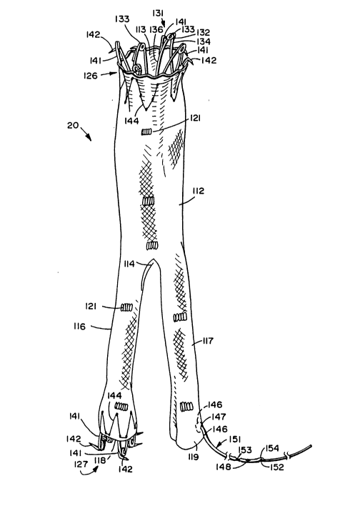
The endovascular graft 20 having a bifurcation is
shown in Figure 4. The graft 20 has many characteristics which
are similar to the expandable intraluminal vascular graft
disclosed in EP-A-0,466,518. However, graft 20 differs
2 û ~ ~ ~ 3 ~ ~
significantly from the grant disclosed therein in that it is
provided with a bifurcation as hereinafter described. The
graft 20 is an expandable intraluminal vascular graft which
is provided with a main deformable cylindrical body 112 having
an open end 113. The body 112 is provided with a bifurcation
or crotch 114 at the other end which opens into first and
second legs 116 and 117, having open ends 118 and 119 generally
opposite the open end 113. Continuous walls form the body 112
and the legs 116 and 117 and are woven of surgically
implantable material such as Dacron-type fiber. One material
found to be particularly satisfactory is a USCI DeBakey soft
woven Dacron vascular prosthesis. The main body 112 can have
a length ranging from 5 to 30 centimeters with each of the legs
having a length ranging from 2 to 15 centimeters. The body
112 can have a maximum expandable diameter ranging from 12 to
30 millimeters whereas the legs 116 and 117 can have maximum
diameters ranging from 6 to 12 millimeters.
Radiopaque markers 121 are provided on the main body
112 and also on the legs 116 and 117 and can be formed of a
suitable material such as lengths of platinum wire secured to
the fabric of the graft by suitable means such as Dacron
sutures.
Expandable spring attachment means 126 is secured
to the expandable main body adjacent the opening 113. Also
expandable spring attachment means 127 is secured to the first
leg 116 adjacent the opening 118. These expandable spring
attachment means 126 and 127 serve as anchoring means for
securing the graft 20 to vessel wall in which the graft 20 is
disposed. The expandable spring attachment means 126 is
constructed in a manner similar to that described in
EP-A-0,466,518 and serves to yieldably urge the opening 113
in the main body 112 from an initial compressed or collapsed
position to a subsequent expanded position. Similarly, the
expandable spring attachment means 127 serves to yieldably urge
the open end 118 from an initial compressed or collapsed
position to a subsequent expanded position. As explained in
EP-A-0,466,518, the expandable spring attachment means 126 and
127 are formed of a plurality of vees 131 with the apices 132
being formed with helical coil springs 133 to yieldably urge
3 4 ~
the legs 134 and 136 of each of the vees 131 outwardly in
directions in the planes in which the vees lie. As disclosed
in EP-A-0,466,518, the apices 133 lie in three longitudinally
spaced-apart parallelplanesextending transversely oftheaxis
of the expandable spring attachment means in which the first
plane is disposed internally of the open end and the second
plane lies in a position which is external of but in close
proximity to the open end and the third plane is spaced a
substantial distance beyond the open end.
Hook-like elements 141 are provided at the apices
132 which are disposed beyond the open end 113 for the
attachment means 126 and the open end 118 for the attachment
means 127. The hook-like elements 141 are bonded to the legs
136 of the vees 131 by suitable means such as welding. The
hook-like elements 141 have hooks 142 which are of a length
which is sufficient to penetrate into the vessel wall and
slightly beyond the vessel wall in which the graft is to be
placed. The expandable spring attachment means 126 and 127
are secured to the graft by Dacron polyester suture material
144 as shown particularly in Figure 4.
A pull line 146 is secured to the leg 117 in a region
which is closely approximate the end of the leg 117 at the
opening 119. The pull line can be formed of a suitable
material such as Nylon having a diameter from .005"-0.010"
(0.13-0.25 mm). The pull line 146 is continuous and extends
through small holes 147 provided in the material forming the
graft 20. The pull line 146 which is doubled over onto itself
and has a doubled-over length of approximately 40-60
centimeters with the ends of the pull line 146 being tied
together in a knot 148. A lead tube 151 with a lumen 152 is
positioned over the pull line 146 so it is adjacent the leg
117. The lead tube 151 is necked down at 153 by suitable means
such as by heat in a region distal of the knot 148 (see Figure
4) so that the lead tube 151 is retained on the pull line 146.
A cutout 154 is provided in the lead tube 151 proximal of the
knot 148.
The balloon catheter assembly 71 is disposed within
the capsule 31 in a manner also shown in Figure 5 in which the
balloon tube or shaft 72 extends coaxially of the main body
206a634
of the graft 112 coaxially of the first leg 116. The
stabilization button 106 is preferably disposed within the
graft in a position which is just proximal of the bifurcation
or crotch 114. By positioning the pusher button 106 where
shown in Figure 5, it is near to the major portion of the
material forming the graft 20 which is folded up within the
capsule 31. This is desirable because the mass of material
provided in that region of the capsule facilitates pushing the
graft 20 out of the capsule as hereinafter described.
The minor deployment device 22 as particularly shown
in Figure 6 consists of a capsule catheter 161, a balloon
catheter 162 and a separate expandable spring attachment means
163. The separate balloon catheter 162 is shown in greater
detail in Figure 7 and the separate spring attachment means
163 is shown in Figure 8. The capsule catheter 161 consists
of a flexible tubular member 166 formed of a suitable material
such as polyethylene having an inside diameter ranging from
.050 to .080" (1.27 mm to 2.03 mm) and an outside diameter
ranging from .075 to 0.100" (1.91 to 2.54 mm). The tubular
member 166 can have a suitable length as for example, 15-25
centimeters. The tubular member 166 has a lumen 167 extending
the length thereof and has proximal and distal extremities 168
and 169. A conventional Tuohy Borst adapter 171 is mounted
on the proximal extremity 168. A small capsule 172 formed of
suitable material such as stainless steel is mounted on the
distal extremity 169 of the tubular member 166. It can be of
a suitable size, as for example a length of 10 to 30
millimeters and an inside diameter of 4 to 6 millimeters with
a wall thickness ranging from .006 to .015" (0.150 to 0.381
mm). The capsule 172 is provided with an open end 173 through
which the separate expandable spring attachment means 163 is
adapted to be inserted.
The balloon catheter 162 as shown in Figure 7
consists of a flexible elongated tubular member 176 formed of
a suitable material such as polyethylene and which serves as
the balloon shaft and is provided with an outside diameter
ranging from 0.04 to 0.060" (1.02 to 0.15 mm) and an inside
diameter ranging from 0.015 to 0.030" (0.38 to 0.76 mm). An
expandable balloon 177 is formed integral with the flexible
~0~6 3~
11
elongate tubular member 176 near the distal extremity thereof.
The balloon 177 is formed of the same polyethylene material
as the tubular member 176 and can have a diameter ranging from
6 to 12 millimeters and a length ranging from 1 to 2
centimeters. A wye adapter 179 is mounted on the proximal
extremity 181 of the flexible elongated tubular member 176.
A Tuohy Borst adapter 182 is mounted on the main arm 183 of
the wye adapter 179. A stop cock 184 is mounted on the side
arm 186 of the wye adapter 179.
An additional elongate flexible tubular member 188
of a suitable material such as polyethylene is provided and
extends from the Tuohy Borst adapter 182 through the lumen 189
provided in the tubular member 176 and through the balloon 177
where the distal extremity of the elongate flexible tubular
of the member 188 is bonded to the distal extremity of the
tubular member 176 to provide an airtight seal for the balloon
177. The tubular member 188 is provided with a lumen 191
extending the length thereof as adapted to receive a guide wire
196 of a suitable size as for example, one having a diameter
of .018" so that the guide wire 196 can extend through the
tubular member 176 and through the balloon 177 and extend
beyond the distal extremity of the tubular member 176. The
guide wire 196 is of a conventional type and is utilized for
guiding the balloon catheter as hereinafter described. A pair
of spaced-apart radiopaque markers of a suitable material such
as gold bands 198 are provided on the tubular member 188 within
the balloon 177.
The coaxial annular space between the exterior of
the tubular member 188 and the interior of the tubular member
176 serves as an annular balloon inflation passage and is in
communication with the side arm 186 so that the inflation and
deflation of the balloon can be controlled by the stop cock
184.
The expandable spring attachment means 163 shown in
Figure 8 has a construction very similar to the expandable
spring attachment means 126 and 127 hereinbefore described.
The expandable spring attachment means 163 is provided with
a plurality of vees 201 having apices 202 formed by coil
springs 203 which have legs 204 and 206 expandable and
2 0 6 ~
- 12
contractible within the plane of the vee. The vees 201 are
configured in such a manner so that the apices 202 lie in only
two spaced-apart parallel planes perpendicular to the
longitudinal axis of the expandable spring attachment means,
rather than the three planes disclosed for expandable spring
attachment means 126 and 127. Hook-like elements 207 are
bonded to the legs or struts 204 or 206. The hook-like
elements 207 are provided with hooks 208 which face outwardly
of the exp~n~hle spring attachment means and in a direction
towards the other end of the spring attachment means.
Additional hook-like elements 209 are provided on the other
end of the spring attachment means 163 by bonding the same by
suitable means such as welding to the struts 204 and are
provided with hooks 211 which face outwardly and extend in an
opposite direction to the hooks 208, toward the other end of
the spring attachment means. In this way it can be seen that
the hooks 208 and 211 face in opposite directions, hooks 208
being angled slightly distally and hooks 211 being angled
slightly proximally, and serve to prevent distal and proximal
migration of the graft leg 117 to which the expandable
attachment means 163 is attached as hereinafter described.
Theexpandable springattachment means 163 is adapted
to be compressed and mounted within the capsule 172 as shown
particularly in Figure 6. Means is provided for pushing the
expandable spring attachment means 163 out of the open end 173
of the capsule 172 and consists of a stabilization button 216
which is formed on the balloon shaft or flexible elongate
tubular member 176. ~he pusher member 216 can be formed in
a suitable manner such as by forming a ring of longitudinally
compressed polyethylene on the shaft 176.
operation and use of the apparatus hereinbefore
described for performing the method of the present invention
for deploying an endovascular graft having bifurcation may now
be briefly described as follows.
In conjunction with the diagrams which are set forth
in Figures 9-19, let it be assumed that it is desired to repair
an aneurysm in the abdominal aorta 222 close to or involving
the aortic bifurcation 221 and possibly involving the left and
right iliac arteries 223 and 224 in a human patient. In this
8n ~ ~ ~ 3 ~
13
example, the left iliac artery 223 is referred to as the first
iliac artery, and the right iliac artery 224 is referred to
as the second iliac artery. Graft legs 116 and 117 are
identified similarly. Initially the patient is prepared with
either general, regional, or local anesthesia. A cut-down is
performed in the left femoral aEtery as indicated by the
opening 226 in the first leg 223. Similarly, a cut-down or
percutaneous access is performed in the right femoral artery
as indicated by the opening 227 in the second leg 224. A guide
wire 231 of a conventional type, as for example a guide wire
having a diameter of .038" (0.97 mm), is introduced through
the opening 226 in the left femoral artery 223 and then is
passed over the aortic bifurcation 221 and down through the
right artery 224 and out through the opening 227 in the right
femoral artery. This procedure is accomplished in a
conventional manner under fluoroscopy as shown in Figure 9.
Thereafter as shown in Figure 10 the lead tube 151
which is extending out of the distal extremity of the capsule
31 is threaded over the guide wire 231 extending out of the
hole 226 in the first artery 223 and thence into the left cut-
down or hole 226 and over the guide wire 231 in the left
artery, over the guide wire 231 in the aortic bifurcation 221
and then down the second artery 224 through the right cut-down
227 so that the distal extremity of the lead tube 151 extends
for a substantial distance out of the cut-down 227. During
the time that the lead tube 151 is being advanced, the distal
extremity of the guide wire 231 is caused to pass through the
cut-out 154 so that the distal extremity of the guide wire 231
is accessible and can be held steady while the lead tube 151
is advanced over it.
Thereafter, the guide wire 231 can be pulled out by
grasping the proximal extremity of the guide wire 231 adjacent
the cut-out 154 in the lead tube 151 and pulling out the guide
wire 231 while holding the distal extremity of the lead tube
151 so as to prevent the lead tube 151 from being pulled back
into the cut-down 227. The distal extremity of the lead tube
151 is then clamped with a hemostat 236 as shown in Figure 11
to be sure that the lead tube 151 is not pulled back into the
cut-down 227 during future steps in the method of the present
~ O ~ ~ ~ 3 4 -
14
invention. The major deployment device 21 is then introduced
into the left cut-down 226 by first passing the balloon guide
wire 91 and then the balloon 74 through the left cut-down 226
followed by the capsule 31, which is advanced to the position
shown in Figure 11 by pushing on the tubular member 27. During
the advancement, the operator may need to place gentle traction
on the lead tube 151 to facilitate advancement of the capsule
31 toward the aortic bifurcation 221. When the capsule 31
reaches the aortic bifurcation 221, it is necessary for the
operator holding the lead tube 151 to permit more of the lead
tube 151 to enter the cut-down 227 to permit further
advancement of the capsule 31 up the aorta so that the distal
spring attachment means 126 of the graft 20 within the capsule
31 can be positioned in a region which is 1-2 centimeters
proximal of the proximal extremity of the aneurysm to be
corrected by the graft 20 being deployed. As shown in Figure
12 the distal extremity of the capsule 31 is deployed well
beyond the aortic bifurcation 221. As soon as the physician
has determined that the capsule 31 is in the proper position,
the physician uses one hand to hold the control mechanism 36
while at the same time using the fingers of the other hand to
rotate the knob 49 and the pinion 47 to retract the rack member
42. This causes retraction of the tubular member 27 and the
capsule 31 mounted thereon while the hypo tube 76 is retained
in a stationary position by the collet 82 that is retained by
the collet housing 83. As the capsule 31 is withdrawn, the
stabilization button 106 carried by the tubular member 72 in
engagement with the graft 20 as shown particularly in Figure
5 causes the graft 20 to be gradually ejected from the capsule
31 as the capsule 31 is withdrawn. Upon continued retraction
of the capsule 31, the proximal expandable spring attachment
means 126 will clear the capsule 31 and will spring outwardly
to cause the hooks 142 carried thereby to come into engagement
with the aortic vessel wall proximal to the aneurysm to be
repaired as shown in Figure 12.
The physician, using one hand to hold the control
mechanism 36, uses his other hand to release the collet 82 in
order to unlock the tube 76 by rotating the collet cover 84
relative to the control mechanism 36. The physician
~n fiS ~ P~ ~
repositions the hand not holding the control mechanism 36 so
as to grasp the portion of the metal hypo tube 76 extending
proximally of the control mechanism 36. The hypo tube 76 is
then pulled rearwardly or proximally. The balloon 74 is
thereby drawn into the proximal extremity of the main body
portion 112 of the graft 20 as shown in Figure 13 so that the
intermediate portion of the balloon 74 is in general
registration with the expandable spring attachment means 126.
The balloon 74 is then inflated by supplying gas to the balloon
inflation lumen by attachment of a syringe or other suitable
inflation means to the Luer fitting 88. Upon inflation of the
balloon 74 the hooks 142 carried by the proximal expandable
spring attachmentmeans126are firmly seatedcircumferentially
in the normal aortic wall proximal to the aortic aneurysm.
With the balloon 74 still inflated and firmly holding the
proximal attachment means 126 against the aortic wall, the
capsule 31 is then further retracted by holding tube 76 in
fixed position relative to the patient with one hand and
retracting the handle 36 with the other hand in order to expose
the entire length of the second leg 117 as shown in Figure 13.
The capsule 31 is still further retracted to clear most of the
first leg 116 as shown in Figure 14. As this is being
accomplished, the second leg 117 of the graft 20 is pulled down
into the artery 224 by pulling on the lead tube 151 so that
the entire length of the leg 117 of the graft 20 is disposed
in the arterial vessel 224 and extends substantially below the
bifurcation 221 and below the aneurysm which is to be repaired.
Further retraction of the capsule 31 is accomplished by holding
tube 76 fixed with one hand and retracting the handle 36 with
the other hand until the distal expandable spring attachment
means 127 carried by the first leg 116 clears the capsule 31
and springs into engagement with the wall of the arterial
vessel 223. It should be appreciated that during the foregoing
procedures, the balloon 74 remains inflated in the attachment
means 126 to prevent any accidental dislodgement of the
attachment means 126 during the removal of the capsule 31 and
during the placement of the second leg 117 of the graft 20 into
the artery 224 by pulling on the lead line 151.
~n ~s ~ 3 ~
16
The balloon 74 is then deflated so that it is in a
collapsed position and the balloon is withdrawn from the
attachment means 126 into the first leg 117 until its
intermediate portion is in registration with the attachment
means 127. The balloon 74 is then reinflated to expand the
hooks 142 of the attachment means 127 into firm engagement with
the arterial wall of the vessel 223 as shown in Figure 15.
After this has been accomplished, the balloon 74 is
again deflated and is advanced up through the main body of the
graft 112 and again into the attachment means 126. The balloon
74 is then reinflated as shown in Figure 16 and serves to hold
the graft 20 in place while the procedures for securing the
distal extremity of the second leg 117 are accomplished. It
is likely in many instances that this step of again securing
the proximal extremity of the graft by inflating the balloon
in the attachment means 126 may be unnecessary. However to
ensure that the graft 20 will not move after it has been
deployed, as additional insurance, the balloon 74 can be
positioned in the attachment means 126 and reinflated.
The minor deployment device 22 is next utilized.
The guide wire 196 forming a part thereof is introduced through
the cutdown 227 into the second artery 224 so that it extends
into the second leg 117 of the graft 20 and beyond the
bifurcation. The balloon catheter 162 is threaded onto or
advanced over the guide wire 196. The balloon catheter 162
is disposed within the capsule catheter 161. The minor
deployment device 22, with its balloon catheter 162 and capsule
catheter 161, is advanced into the cutdown 227 while applying
gentle traction to the lead tube 151 to keep the second leg
117 of the graft 20 taut. The balloon 177 and the capsule 172
are thus introduced into the second leg 117. The capsule 172
is positioned so that when the expandable spring attachment
means 163 contained therein is deployed therefrom, the spring
attachment means 163 will be at the distal extremity of the
second leg 117 of the graft 20 as shown in Figure 16. The
expandable spring attachment means 163 is then forced out of
the capsule 172 by the physician using one hand to grasp the
wye adapter 179 and hold it in a fixed position relative to
the patient and using the other hand to grasp the Tuohy Borst
~ ~ fi ~
17
adapter 171 and gradually withdraw the same to retract the
capsule 172 from over the expandable spring attachment means
163 which is held in the desired position by the stabilization
button 216 carried by the tubular member 176. As soon as the
expandable spring attachment means 163 clears the capsule 172
it will spring out with one row of hooks 208 moving into
engagement with the distal extremity of the second leg 117 and
with the other row of hooks 211 moving into engagement with
the wall of the arterial vessel 224. Alternatively the capsule
172 is positioned so that when the expandable spring attachment
means 163 contained therein is displaced therefrom, the
expandable spring attachment means 163 is disposed within the
second leg 117 so that both rows of hooks 208 and 211 move into
engagement with the distal extremity of the leg 117 and engage
the wall of the vessel 224.
In order to firmly implant the hooks 208 and 211 of
the expandable spring attachment means 163, the balloon 177
in its deflated condition is brought down into the attachment
means 163 so that its intermediate portion is disposed within
the attachment means 163. This is accomplished by pulling on
the wye adapter 179 which applies a pulling force to the
tubular member 176 to pull the balloon 177 towards the distal
extremity of the leg 117 of the graft 20 while at the same time
withdrawing, if so desired, the capsule catheter 161 by pulling
on the adapter 171 which applies a pulling force to the tubular
member 166. As soon as the balloon 177 is in the proper
position, the balloon 177 is inflated by suitable inflation
means as, for example, a syringe attached to the stop cock
fitting 184 and inflating the balloon 177 to the desired
pressure to force the hooks 208 and 211 firmly into the distal
extremity of the leg 117 of the graft 20 and the arterial
vessel 224.
After the inflation of the balloon 177 has been
accomplished, the balloon 177 can be deflated by removing the
syringe and opening the stop cock 184. The balloon catheter
162 and the capsule catheter 161 then can be removed through
the cutdown 227 so that all that remains is the lead tube 151
extending through the cutdown 227. The lead tube 151 is cut
distal to the knot 148 in the vicinity of the necked down
3 ~
18
section 153 and the lead tube 151 is pulled off of the pull
line 146. One end of the Nylon pull line 146 is then grasped
to pull out the Nylon pull line 146 by having the free end
travel up into the cutdown 227 and pass through the distal
extremity of the leg 117 of the graft 20. It is then removed
in its entirety through the cutdown 227. The right cutdown
227 is then repaired. Following that, the balloon 74 is
deflated. The hypo tube 76 is retracted relative to the
control mechanism 36 to move the balloon into engagement with
the capsule 31. The collet 82 is then locked onto the hypo
tube 76 by turning the knob 84 relative to the control
mechanism 36. The control mechanism 36 is then withdrawn to
remove the capsule catheter 27, the balloon catheter shaft 72,
and the balloon 74 through the cutdown 226. The left cutdown
226 is then repaired. This completes the steps for deployment
of the graft 20 across an aortic bifurcation to repair an
aneurysm. The patient can then be brought out of general
anesthesia if employed.
It should be appreciated that the graft having
bifurcation can have legs of various lengths depending upon
the type of aneurysm which is to be repaired. For example,
one leg can be longer than the other. The legs can both be
short in cases in which the aneurysm has a short distal aortic
neck and does not include the iliac arteries. They would be
longer in aneurysms which involve the iliac arteries as well.
It is generally desirable that the graft extend at least one
centimeter beyond the most distal portion of the most distal
aneurysm in the vessels.
From the foregoing it can be seen that there has been
provided a graft having a bifurcation in which the main body
of the graft as well as the legs are firmly attached in the
arterial vessels so that they accidentally cannot become
dislodged from the location in which they are fixed in the
arterial walls. The method which is utilized for deploying
the graft with legs is relatively simple and can be
accomplished within a relatively short period of time. The
major and minor deployment devices which are utilized in the
procedure are constructed in such a manner that they are easy
to utilize with a minimum of training. The use of a folded-
2 ~ 3 4 -
19
over second leg of the graft in the capsule makes it
unnecessary to move the main body of the graft as high in the
aorta as would be otherwise necessary in order to permit the
second leg of the graft to clear the aortic bifurcation to
thereby permit the second leg to be placed in the second iliac
artery. Thus, the risk incurred by moving the graft and its
capsule and any associated debris past the renal arteries
located well above the aortic bifurcation is greatly reduced
thereby reducing the chance of occluding the renal arteries
and causing embolization to the renal arteries.