Note: Descriptions are shown in the official language in which they were submitted.
2~~~~
P-2393 PATENT
METHODS AND APPARATUS FOR DETECTING BACTERIAL GROWTH
BY SPECTROPHOTOMETRIC SAMPLING OF A
FIBER-OPTIC ARRAY
The present invention relates to non-invasive
methods and apparatus for detecting biological activity in a
specimen sample such as blood, that may include a culture
medium and which is introduced into a sealable container or
vial and exposed to conditions enabling metabolic processes
to take place in the presence of any microorganisms in the
sample.
BACKGROUND OF THE INVENTION
Usually, the presence of bacteria in a patient's
body fluid, especially blood, is determined using blood
culture vials. A small quantity of blood is injected through
an enclosing rubber septum into a sterile sample vial
containing a culture medium. The vial is incubated at normal
human body temperature and monitored for bacterial growth.
Common visual inspection involves monitoring the turbidity of
the liquidrsuspension. Known instrumental methods detect
changes in the COZ content of the culture vials, which is a
metabolic by-product of the bacterial growth. Monitoring the
GOZ content can be accomplished by methods well established
in the art, such as radiochemical, infrared absorption at a
spectral line characteristic of CO2, or pressure/vacuum
measurement techniques such as those disclosed in U.S. Patent
No. 4,152,213--Ahnell.
P-2393 PATENT
Recently, non-invasive methods have been developed
involving the optical interrogation of chemical sensors
disposed inside a sample vial that utilize colorimetric or
fluorometric spectroscopic techniques. Some of these methods
have also implemented remote sensing of multiple sample sites
via optical fibers and switches. Additional non-invasive
optical methods have been devised which rely on properties
inherent in the liquid suspension and do not require the use
of a chemical sensort these include automated techniques for
scattered photon migration measurements.
Typically, when using these non-invasive
techniques, the sample vial must be agitated. Since it is
both cost effective and time efficient to process samples in
a batch, equipment must therefore be provided that agitates a
large number of vials. Agitation, however, requires that the
structure holding the vials moves relative to a stationary
reference frame, and it is usually preferable to mount
electronics and other equipment within the stationary portion
of the system, nat on the moving portion. This results in
systems where both electrical cables and/or optical fibers
must be designed to permit this relative motion by allowing
sufficient excess at an appropriate point in the system.
There remains a need, however, to permit a plurality of
sample vials to be agitated while also permitting the
interrogation of each vial by an optical fiber. It is
accordingly an object of the present invention to provide an
agitating rack and an optical excitation/detection system for
transmitting electromagnetic energy to each sample vial in
the rack and receiving electromagnetic energy from each
sample. '
S~(JMMARY OF THE INVENTION
To fulfill this and other objects of the present
invention, an instrument is provided for incubating and
detecting bacterial growth in multiple sample vials. A drive
mechanism is connected to a rack that combines agitation of
the vials with the sequential coupling of one or more optical
P-2393 - ~ PATENT
fibers to a spectrophotometric detector or similar sensor.
Selection of each vial provides an optical signal useful in
the detection of bacterial growth by fluorescence or other
spectrophotometric measurements. The rack is preferably the
only moving assembly, and requires no mechanical or
electrical connection to an optical excitation/detection
system for its operation. The present invention therefore
eliminates the above-described flexing of electrical cables
or optical fibers.
Thus, in a most preferred embodiment, the present
invention provides apparatus for transmitting electromagnetic
radiation to a plurality of sample vials and receiving
electromagnetic radiation from the sample vials. The
apparatus includes a rack for retaining the plurality of
sample vials and one or more optical guides coupled to each
of the plurality of sample vials and to the rack at one or
more optical coupling locations. An excitation/detection
system is provided for transmitting and receiving
electromagnetic radiation between the excitation/detection
system and the sample vials using the optical guides that are
aligned with the optical coupling locations.
The optical guide can include either a single
optical fiber that terminates at a single optical coupling
location, or multiple fibers that are grouped together to
define an optical coupling location. Alternatively, the
optical guide.can include separate fibers for excitation and
emission that transmit electromagnetic radiation to the
sample vial and collect it from the sample vials,
respectively. The fibers or groups of fibers can either be
aligned with the central axis of the rack or arranged in one
or more groups along a portion of the rack. In the
embodiments using groups of either excitation fibers,
emission fibers, or both, different types of systems are
useful to enable electromagnetic energy to be transmitted to
and collected from each individual sample vial.
Methods of transmitting electromagnetic radiation
to sample vials and receiving electromagnetic radiation from
P-2393 - 4 - PATENT
the sample vials are also disclosed. In general, the present
invention discloses the optically indexed presentation of the
optical guides, e.g., the emission and excitation fibers, to
many remote sample sites in the form of a geometric array at
the interface between the moving culture vial rack assembly
and the spectrophotometric excitation/detection system fixed
to the body of the instrument. This interface is defined as
an optical coupling location.
The methods of the present invention include the
steps of coupling the sample vials to an optical~fiber and
terminating the optical fiber at an optical coupling
location. The rack is then moved while aligning at least one
of the optical coupling locations with the
excitation/detection system.
BRIEF DESCRIPTION OF THE DRAWINGS
The various features, objects, benefits, and
advantages of the present invention will become more apparent
upon reading the following detailed description of the
preferred embodiments, along with the appended claims in
2.0 conjunction with the drawings, wherein like reference
numerals identify corresponding components, and:
FIGS. 1A-1C respectively depict front elevation,
plan, and side elevation views of a first embodiment of the
rack apparatus of the present invention.
FIG. 2 is an enlarged partially schematic,
partially broken away side elevation view depicting further
details of the optical excitation/detection system used with
the rack apparatus illustrated in FIGS. 1A-1C.
FIGS. 3A-3B show another embodiment of the
apparatus of the present invention adapted to process 120
samples.
FIG. 4 is a partially schematic illustration of two
of the apparatus depicted in FIGS. 3A-3B partially connected
to share certain components.
3
2~0002~
P-2393 - 5 - PATENT
FIGS. 5A is a side elevation view illustrating
details of an alternate detection systems used in the present
invention.
FIGS. 5B-5C are side elevation views similar to
FIG. 1C illustrating alternate arrangements of optical
coupling locations.
FIG. 6 illustrates details of another alternate
embodiment an optical system for use in the present
invention.
FIGS. 7A-7B illustrate details of still another
alternate embodiment of the rack apparatus and optical system
used in the present invention.
FIG. 8 is a partial, front elevation cross-
sectional view of a light source and photodetector used in
certain embodiments of the apparatus of the present
invention.
FIGS. 9A-9B depict another alternate embodiment of
the rack apparatus of the present invention.
FIGS. l0A-lOB illustrate further details of the
rack apparatus depicted in FIGS. 9A-9B.
DETAILED DESCRIPTION OF THE PREFERRED EMBODIMENTS
The various embodiments of the apparatus of the
present invention are illustrated in Figures lA-lOB, with the
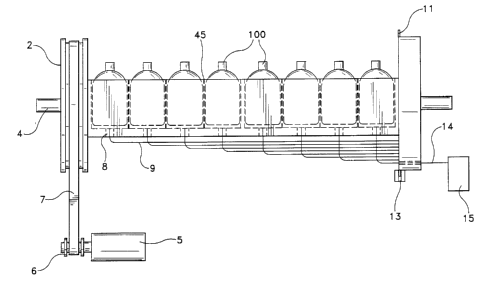
apparatus generally designated as 1. A first embodiment of
the apparatus of the present invention is depicted in FIGS.
lA-1C. A plurality of samples, each contained in an
individual vial or container 100 are disposed in a rack 1.
In this embodiment, the rack 1 is a block in which many
cylindrical cavities have been bored, each sized to hold a
single vial 100. A first end of the rack 1 is attached to a
large drive pulley 2; a second end is attached to a circular
sampling disk 3. The rack assembly 45 is supported by and
rotates about an axis defined by a pivot shaft 4. The
culture vials 100 held in the cavities of the rack 1 are
preferably rocked or rotated by a stepper motor 5 that is
connected to a small. pulley 6. A belt 7 interconnecting the
"°° ~.
2.00020
P-2393 - 6 - PATENT
pulleys 2,6 to the stepper motor 5 controls the angular
position of the rack 1.
Those of skill in the art will understand that
structural elements such as the pivot shaft bearings, mounts
and supports required to locate the pivot shaft within the
body of an instrument, and other associated structures, have
been omitted from FIGS. lA-1C and the other figures herein
for clarity of description, as have the mechanical and
electrical arrangements required to maintain the apparatus of
the present invention at a constant temperature suitable to
support the growth of any bacteria present within the culture
vials in certain applications. Additionally, it should be
noted at the outset numerous other drive systems such as, for
one example, a gear train would be equivalent to and easily
substituted for the belt 7 and pulleys 2,6 shown in FIGS. lA-
1C.
Referring still to FIGS. lA-1C, in the bottom of
each cavity is a through-hole 8, in which an optical fiber 9
is inserted and retained. The optical fibers 9 from each of
the plurality of cavities terminate in an array of fiber ends
10 that extend through the sampling disk 3, with their
centers preferably equally spaced along an arc centered on
the axis of the pivot shaft 4, as shown in FIG. 1C. The
angular increment between the centers of the fiber ends 10
most preferably corresponds to an integral number of steps of
the stepper motor 5. A preferred manner for indexing the
disclosed apparatus comprises providing a thin extended rim
11 on the perimeter of the sampling disk 3 in which a series
of rectangular notches 12 has been cut. The leading edge of
each notch 'is angularly aligned with the centerline of the
nearest optical fiber end 10. As seen in FIG. lA and
illustrated in greater detail in FIG. 2, a transmissive
optical index fiducial 13 is angularly aligned with the
optical axis 14 of an excitation/detection system 15. In
this way, the edge transition of a notch 12 through the
fiducial 13 produces an electrical signal indicating the
precise alignment of the exeitation/detection system 15 with
Y:
~~00~20
P-2393 - 7 - PATENT
one of the optical fibers 9. When aligned, fluorescence from
the vial 100 in the cavity which is also associated with that
particular the optical fiber 9 is measured. In this
embodiment the optical fiber 9 thus acts as both an
excitation fiber by transmitting the energy received from the
excitation/detection system to the vial 100, and as an
emission fiber by receiving energy that escapes the vial 100
and transmitting it to the detection system.
Additional details of the apparatus shown in FIGS.
lA-1C are illustrated in FIG. 2. A preferred embodiment of a
fluorescence excitd'tion/detection system 15 for use in the
present invention is shown schematically. As explained
above, the metabolic production of COZ by bacteria growing in
the vial 100 can be determined by the fluorescence intensity
of a chemical sensor 102 in the vial 100. Those of ordinary
skill will understand that the sensor 102 may be a membrane
or thin film disposed within the vial 100. As seen in FIG.
2, this fluorescence is preferably excited by a beam of
electromagnetic radiation from a green Helium-Neon (He-Ne)
laser 16 which is deflected by an alignment mirror 17 and
passes through a small center hole in an emission mirror 18
and the center of the objective lens 19. The excitation beam
travels along the optical axis 14 shown and is directed
through the optical fiber 9, in its excitation mode, to
interrogate the sensor 102 that is disposed in the bottom of
the vial 100. Fluorescent emission is collected from the
sensor 102 by the same optical fiber 9, now in its emission
mode, and is acquired by the objective lens 19, but is
deflected by the emission mirror 18 to an emission filter 20
and focused by a detector lens 21 on the photocathode of a
photomultiplier tube 22. In other embodiments, the
photomultiplier 22 can be a photodiode or other suitable
detector. As is well known, the photomultiplier anode
current is a measurement of the fluorescent emission
intensity. As understood by those of ordinary skill, after
correctian for excitation intensity, as measured by an
optical source monitor or laser current monitor (riot shown),
P-2393 - 8 - PATENT
this intensity value serves as an indicator of bacterial
growth in the culture medium.
The pierced mirror beam splitter and fiber-optic
sampling of culture vial fluorescence shown in FIG. 2A uses
conventional laser excitation technology as is the common
practice in fluorescence measurement. The present invention
is differentiated from other bacterial growth-based detection
instruments, as discussed above, by the non-mechanical link
from the rack 1 to the excitation/detection system 15, and by
use of the agitation drive system to multiplex the
measurement of a plurality of culture vials 100 via the array
of optical fibers 9 optically connected to each of the vials
and to the rack at a plurality of "optical coupling
locations" that are in this case defined by the optical fiber
ends 10. The present invention therefore provides an
optically indexed presentation of the fiber-optic links 9 to
many remote sample sites, i.e., each vial 100, in the form of
a geometric array of optical coupling locations defined at
the interface between the moving rack assembly 45 and the
excitation/detection system 15, the latter being fixed
relative to the body of the instrument. In operation, the
rack 1 may either be "stepped" to stop at each optical
coupling location or it may "sweep" smoothly through an arc,
successively passing the optical coupling locations through
the optical axis 14 of the beam.
Another embodiment of the present invention on a
scale suitable for a commercial instrument is shown in FIGS.
3A-38. This embodiment of the present invention holds up to
120 culture vials 100, most preferably arranged in the rack
1' in an array of six rows and twenty columns, as shown in
FTG. 3A. All components serve the same function as those
described above with reference to FIGS lA-1C and 2, and are
thus labeled with the same reference numerals. As will be
understood by those of ordinary skill, apparatus made in
accordance with the present invention can be constructed to
accommodate any number of vials 100 by varying the number of
rows and columns in the rack.
. . ,.... ''
P-2393 - 9 - PATENT
The productivity of the present invention can be
enhanced by having two rack assemblies share a single light
source 16 as illustrated in FIG. 4 which depicts a 240-vial
instrument based on joining two of the 120-vial assemblies
that are shown in FIGS. 3A-3B. Each of the 120-vial racks 1'
has its own emission detector components 22 and
excitation/emission beam splitter 18. However, the two racks
1' obtain excitation energy from a single source 16 by use of
a semi-transparent alignment mirror 117 as shown. One-half
the energy of the source 16 is deflected to one of the two
racks 1' and one-half is transmitted to the other. The use
of a common source 16 and parallel detectors 22 for the two
racks 1' allows the incorporation of supporting electronics
which simultaneously sample the fluorescence from the samples
contained within both racks 1'. The embodiment illustrated
in FIG. 4 thus obtains doubled vial sampling throughput
without the expense of multiple laser sources, thereby
reducing the cost per sample analyzed.
Referring now to FIGS. 5A-5B, an alternate
embodiment of the present invention is illustrated in which
separate optical fibers 28,9 are used for excitation 28 and
emission 29. FIG. 5A depicts a partially broken away
elevation view of an instrument similar to those described
above with reference to FIGS. lA, 2 and 3B, except that the
excitation/detection system 15 differs, as explained herein.
In this embodiment, three lenses 25,26,27 form a Galilean
laser beam expander along the excitation beam axis 29: this
beam simultaneously illuminates multiple vials 100 via a
circular bundle of excitation fibers 28, the ends 10' of
which may 13e arranged as seen in FIGS. 5B-5C or in any
suitable pattern, in any reasonable subdivision, based upon
the number of samples to be tested. The ends 10 of the
emission fibers 9 from each of the simultaneously excited
groups of vials 100 are preferably arranged into linear
arrays by terminating each emission fiber 9 the sampling disk
3, as explained above. The arrays of fiber ends 10,10' are
most preferably centered on equi-angularly spaced radii of
P°2393 - 10 - PATENT
the sampling disk 3. As explained above, the apparatus is
preferably indexed between optical coupling locations,
defined by the array of fiber ends 10,10' in this embodiment,
using a series of notches 12 that cooperate with the fiducial
13 to produce an electrical signal indicating precise
alignment.
Once aligned, a selected array of emission fiber
ends 10 is optically coupled to a photodetector 24, seen in
FIG. 5A, by an objective lens 19 centered on a central
emission axis 23 of the array of the emission fiber ends 10.
The photodetector 24 is either a linear photodiode array,
avalanche photodiode array, or charge-coupled device (CCD).
Alternatively, a fiber-optic face plate is used in some
embodiments in place of the objective lens 19. In these
embodiments, electronic readout of the elements of the linear
photodetector array 20 then provides emission intensity
measurements for the selected vials. As shown in FIG. 5C,
the linear format of the arrays of emission fiber ends 10
that define the optical coupling locations is replaced in
some embodiments with a square 4 x 4. In these embodiments,
the linear photodetector array 24 is replaced with a sixteen
channel photomultiplier tube.
FIG. 6 shows another alternative embodiment of the
apparatus of the present invention, in a view similar to FIG.
5A, in which the laser source 16 has been replaced by an arc
lamp or filament lamp 216. The beam expander lenses 25,26,27
shown in FIG. 5A are eliminated, but due to the non-coherent
nature of the arc lamp 216, a collimating lens 31, excitation
filter 30 and focusing lens 27' are required. In this
embodiment, the mirror 217 is most preferably a so-called
"cold mirror" which directs only short-wavelength radiation
toward the excitation fiber bundles 28. A cold mirror is a
heat transmitting filter that permits the transmission of
infrared wavelengths while reflecting visible light, such as
those available from Driel Corporation, 250 Long Beach Blvd.,
P.O. Box 872, Stratford, Connecticut, USA 06497. In certain
embodiments, the collimating lens 31 and the cold mirror 217
,"°
210002
P-2393 - 11 - PATENT
can also be combined and replaced with a single collimating
cold mirror. The emission side of this detection system is
similar to that described with reference to FIG. 5A.
FIGS. 7A-7B illustrate an embodiment of the present
invention that allows for the highest sampling throughput,
although it also requires the greatest optical source power.
In this embodiment, there is one registered rack position, a
single bundle of excitation fiber ends 10', and one array of
emission fiber ends 10. The photodetector 33 is most
preferably a two-dimensional charge-coupled device (CCD),
silicon-intensified target (SIT) detector or other high-
sensitivity imaging detector. Also in this embodiment, the
objective lens 19 can be replaced with a fiber-optic face
plate, as described above with reference to FIG. 5A.
A further modification of an apparatus according to
the present invention is illustrated by the partial, cross-
sectional side view of FIG. 8. In this embodiment, the
emission fibers 34 are grouped together in an array located
along the center axis of the circular sampling disk 3 of the
rack 1. The rack 1 and disk 3 are connected to a supporting
structure 38 via a rotatable coupling 35 that rides on a
bearing 36 disposed within a housing 37, as shown. Emission
light reemerging from the fiber bundle 34 is focussed on a
photodetector 41 by an optical lens 40. In this embodiment,
the excitation fibers 42 are preferably illuminated by a
light source 43 using a second optical lens system 44. In a
most preferred embodiment, a laser is used as the excitation
light source 43. It should also be noted that the lens
systems 40,44 are preferably constructed and adapted to act
as insulating walls in order not to impair the thermal
insulation 39 that insulates the structure 38 to retain heat
in the area of the rack 1.
In this embodiment, the emission fiber bundle 34
can have an irregular statistical distribution of individual
fibers and is still capable of monitoring individual sample
vials 100. The photodetector 41 is most preferably a
photomultiplier, and thus electromagnetic energy reemerging
t
P-2393 - 12 - PATENT
from each individual fiber in the emission bundle 34 always
reaches the photodetector 41, independent of the particular
sampling disk's angular position. However, the emission
fiber bundle 34 may also be arranged in a regular 2-
dimensional array comprising rows and columns, as shown for
example in FIG. 7B. In this case, the photodetector 41 is
most preferably an electronic camera. It is advantageous,
however, to concentrate all excitation fibers into one
irregular bundle 34. Therefore, as in the embodiment
illustrated in FIGS. 7A-7B, the rack 1 most preferably has
only one position for reading all sample vials. The
embodiment shown in FIG. 8 provides an excellent optical
shielding between the bundle of excitation fibers 42 and the
emission fibers 34. This is due to the cylinder 35
surrounding the emission path that also forms part of the
structure that acts as the pivot shaft for the rack 1. With
regard to modifications, it is advantageous if the optical
lens system 44 comprises a cylindrical lens to generate a
slit-shaped fiber illumination beam with the slit axis
oriented radially on the circular sampling disk 3. Using a
slit-shaped fiber illumination beam results in a significant
reduction of the mechanical precision requirements. This
also results in an excellent long-time stability of the
instrument.
One modification applicable to any of the
embodiments of the apparatus of the present invention
discussed above is the replacement of the stepper motor 5
with an ordinary electrical motor. In this case, the rack 1
and the circular sampling disk 3 rotate back and forth
continuously. Whenever an excitation fiber crosses the
focussed laser beam, light reemerges from the corresponding
emission fiber. Consequently, the output signal generated by
the phatodetector consists of a series of pulses, and the
pulse amplitudes contain information indicative of
bacteriological activity. This multi-pulse signal is stored
and analyzed by a computer.
~~~fl~~~
P-2393 - 13 - PATENT
An additional feature of preferred embodiments of
apparatus made according to the present invention is
described in FIGS. 9A-9B and l0A-lOB. In this embodiment, a
mechanism is provided that allows interchangeable rack
subassemblies 101 holding a plurality of vials 100 to be
easily mounted and dismounted. These embodiments of the
present invention therefore can use a relatively small number
of vials 100 per interchangeable rack subassembly 101 and
make use of multiple racks 101 maintained in an external
incubator (not illustrated). Each rack subassembly 101 is
placed in the apparatus by an operator at appropriate times,
and each of the vials 100 is spectrophotometrically tested
for bacteria growth or otherwise exposed to electromagnetic
radiation, as described above. The rack subassembly 101 is
then replaced in the external incubator.
Further details of this embodiment are illustrated
in FIGS. 9A-9B. The rack subassembly 101 has a pivot shaft 4
and contains a set of emission/excitation fibers 9 optically
linked to an array of mounting holes 51 on the sampling disk
3. The anterior end 46 of the pivot shaft 4 is preferably
formed into a square cross-section, as shown in FIG. 9B, and
inserted into a matching square female socket 47 centered in
hub 48 of the drive pulley 2. The drive pulley hub 48
rotates within a bearing 49, which is fixed in the incubator
wall 50. For purposes of illustration, the remainder of the
drive system is not shown. Spectrophotometric sampling of an
individual optical fiber 9 aligned with the optical axis 14
is accomplished through an aperture 52 in the anterior
incubator wall 50.
The posterior incubator wall 53 serves as fixed
mounting for a bolt-action receiver 54 that supports the
posterior end 55 of the pivot shaft 4, In a preferred
embodiment, the receiver housing 56 is a hollow cylinder
having a section removed from its anterior end in order to
allow placement of the pivot shaft 4 within the open end. As
seen in the side elevation view of FIG. lOB, the pivot shaft
4 rests on a semi-cylindrical supporting bearing 57 fixed
P-2393 - 19 - PATENT
inside the open end of the receiver 56. A hollow cylindrical
bolt 58 nests inside the housing 56 and slides freely on the
extended axis of the pivot shaft 4. When the bolt 58 is
located in the retracted position as shown in FIGS 9A and
10A, the pivot shaft 4 may be withdrawn and lifted out of the
bolt-action receiver 54. This enables the entire rack
subassembly 101 to be removed and placed in an external
incubator or otherwise removed to a remote location.
When a rack subassembly 101 containing samples to
be tested is put in place, the bolt 58 is placed in an
extended position and captures the posterior end 55 of the
pivot shaft 4, thereby preventing its removal. The
compression spring 59, illustrated in FIG. 10A, is centered
in the hollow bolt 58 and forces the pivot shaft 4 to the
limit of its anterior travel in the socket 47 of the drive
pulley hub 48. This mechanism controls the distance from the
end of the optical fiber 9 to the aperture 52. Projecting
from the solid rear wall of the bolt 58 is the actuator shaft
60, on which the actuator ring 61 rotates freely, and is
retained by snap ring 62. The actuator handle 63 projects
from the actuator ring 61 through the inclined slot 64 in the
housing 56. By moving the actuator handle 63 in the slot 64,
the bolt 58 may be extended or retracted; the short vertical
detent slot 65 allows the bolt 58 to be locked in the
extended position.
In addition to the various embodiments of the
apparatus of the present invention described above, the
present invention also provides methods for transmitting
electromagnetic radiation to sample vials and receiving
electromagnetic radiation from the sample vials. In order to
practice the methods of the present invention, a plurality of
sample vials are retained in a rack wherein one or more
optical guides are coupled to each one of the plurality of
sample vials and to the rack at one or more optical coupling
locations. Next, an excitation/detection system, as
described above, is provided for transmitting and receiving
electromagnetic radiation between the excitation/detection
P-2393 - 15 - PATENT
system and the optical guides. The optical locations are
aligned with the axis of the excitation/detection system.
Finally, agitation of the sample vials is accomplished by a
mechanical drive system, which also indexes the optical
guides and aligns them with the axis of the
excitation/detection system.
Those skilled in the art will realize that the
various embodiments of the apparatus and methods disclosed
herein are not limited to bacterial detection systems
emplaying fluorescent sensors. In particular, the principles
and concepts of the invention can be readily applied to
chromophoric and absorptive sensors, to detection systems
based on scattered photon migration, and to other optical
sensor types that detect radiation at ultraviolet (UV),
visible (VIS) or infrared (IR) wavelengths. In this regard,
the present invention can be utilized in any situation where
the size and the large number of agitated samples make direct
sample imaging impractical. In a most preferred embodiment,
however, the present invention provides an optical method and
apparatus for promoting and detecting bacterial growth in
blood culture vials which is simple, non-invasive, and that
can be economically scaled for commercially-sized
instruments. This design is potentially more reliable than
designs having mechanical vial or detector transport systems
because no electrical cables or fiber-optic cables cross the
mechanical interface between the movable rack and fixed
excitation/detection system. This design allows a single
high-performance spectrophotometric detector to be
economically shared among many culture vials; this enables
the required measurements to be made with much better
sensitivity, stability and dynamic range than can be achieved
economically with detector components of low enough cost to
be replicated at every vial position in a large instrument.
Thus, while the preferred embodiments of the
present invention have been described so as to enable one
skilled in the art to practice the apparatus and methods of
the present invention, it is to be understood that variations
'.
P-2393 - 16 - PATENT
and modifications may be employed without departing from the
concept and intent of the present invention as defined in the
following claims. The preceding description is intended to
be exemplary and should not be used to limit the scope of the
invention. The scope of the invention should therefore be
determined only by reference to the following claims.