Note: Descriptions are shown in the official language in which they were submitted.
CA 02227756 2008-12-11
-1-
APPARATUS FOR PERFORMING CORNEAL SURGERY
The invention relates to the surgical correction
of astigmatism taking into account refractive and
topographical measurements of the eye of the patient.
BACKGROUND
Current methods of analyzing astigmatism are
confined to calculation of the vector of change
surgically induced in attaining the post-operative
result from the pre-operative state.
This ably allows determination of total
induced astigmatism and the direction of the vector
force acting in the eye. It also enables calculation of
the mean total surgical astigmatism induced when a
series of operations are compared and analyzed.
25
CA 02227756 1998-01-23
WO 96/40027 PCT/AU96/00257
-2-
However, the axes of surgical induced astigmatism (SIA)
generally varies considerably within the 180 arc of
range. This makes it extremely difficult to make
meaningful comparisons of astigmatic change for a
series, as one cannot obtain an average directional
change of vectors, as vectors in opposing or partly
opposing directions cancel each other out in varying
amounts.
One practice carried out by some surgeons is
to resort to the sole option of tabulating each
patient's results individually, leaving it to the
reader to estimate any trend. Some surgeons attempt to
provide an overview of results, but lack the means to
deduce a trend in induced astigmatism vectors as a
group, because they have variable orientation.
Taking a mean of the angles has no validity
in determining the trend for axes, nor does it address
the change in axes from their pre-operative to post-
operative astigmatic status. It does not assess the
success or desirability of the achieved result;
furthermore, it does not indicate the extent to which
the surgical aim was achieved. An attempt has been
made to address the complexities of correcting the
magnitude for the degrees of axis change by introducing
the approximation that this component varies as the
CA 02227756 1998-01-23
WO 96/40027 PCT/AU96/00257
-3-
cosine of the difference between the attempted and the
observed (achieved) axes. This corrected value of
magnitude was substituted as the amount of surgically
induced astigmatism measured on a cylinder 90 to the
axis of the incisions, the so-called "proper" axis. It
has been proposed to program so called Naylor's
equations into a computer program that requires slight
modifications to resolve the ambiguity and essentially
reproduce the Naylor table.
The formula for calculation of SIA is derived
from the resultant of two plano-cylindrical lenses with
axes at different angles; this was subsequently
employed by some surgeons using graphical methods
confirming the magnitude and axis of the astigmatic
change. Jaffe and Clayman employ rectangular and polar
co-ordinates to determine, by vector analysis, the
formula for calculating SIA and its axis with the known
values for pre- and post-operative corneal astigmatism.
Analogous formulae were derived by Hall based on Martin
and Welford's derivation of Euler's theorem of curved
surfaces (investigated by Airy in 1827).
Euler's theorem, which states "that the sum
of the curvatures of any two perpendicular sections of
a cylindrical or toric surface has a constant value",
provides the link between Jaffe's and Naeser's methods
of vector analysis. Naeser's method calculates the
CA 02227756 1998-01-23
WO 96/40027 PCT/AU96/00257
-4 -
polar values of astigmatism, arising when the axis of
astigmatism does not lie on 90" or 180 meridia; its =
use lies primarily in interpreting results of surgery =
which induces polar (with-the-rule and against-the-
rule) changes, such as cataract and implant surgery
(with or without transverse astigmatic keratotomy).
Astigmatism is a unique refractive error that
causes reduced visual acuity and produces symptoms such
as glare, monocular diplopia, asthenopia and
distortion. For some years now, astigmatism control
and correction has been of great concern to refractive,
cataract and corneal surgeons. Reduction or
elimination of astigmatism, as a single or combined
procedure, is only possible if one possesses an
understanding of astigmatic change, in its component
parts of magnitude and axis. Current analytical
techniques do not allow us to compare magnitudes and
axes separately for a series of paired groups of
procedures or for a single procedure, yet it is only in
this way that we are able to perfect techniques of
astigmatism surgery. We need to be able to determine
the preferable technique to employ; we also need to be
able to determine whether any failure to achieve
surgical goals is attributable to an individual patient
factor or to machine or technique error. Modern laser
technologies have empowered us with the ability to
. . ._ . . . .. . . _ ... . ........ :_._ . ..... . . .:.. ....-... _.. . . ~
... .. ._,..... . ........ _._........ ..,...: . . . . . ....
CA 02227756 2008-12-11
-5-
modify our procedures with degrees of sophistication not
previously possible; this in turn requires analysis systems
which will allow us to accurately quantify and
scientifically assess the results.
SUMMARY OF THE INVENTION
The present invention provides an apparatus for
performing corneal surgery comprising:
means for performing surgery on a patient's
cornea;
control means for controlling the means for
performing surgery; and
processing means having an input and providing an
output which is connected to said control means for
controlling surgery on the patient's cornea based on said
output; wherein
said input of the processing means receives:
both a magnitude and an axis of astigmatism
(T) of an eye of a patient based on topography of the
cornea of the eye of the patient;
both a magnitude and an axis of astigmatism
(R) of the eye of the patient based on refractive
correction of said eye,
said processing means determines surgical
parameters (TIA) based on the measurements of
astigmatism both refractively (R) and topographically
(T) by
a) determining in a double angle vector
diagram target induced astigmatism vectors
topographically (TIA T) and refractively
(TIA R) to achieve zero astigmatism
. . .... ... .. . .. .. ....... . ..._..... .e...... .......... . I . . .
....... ,..... . ... . . . . . . . .
CA 02227756 2008-12-11
-6-
topographically and refractively
respectively,
b) determining a maximal correction
line connecting the end points of vectors
(TIA T) and (TIA R), and
c) establishing said surgical
parameters (TIA) as a vector intermediate
vectors (TIA T and TIA R) with an end point
on the maximal correction line such that
total residual astigmatism in the eye
following surgery, will be a minimum when
measured topographically and refractively.
Also disclosed herein is a method of correcting
astigmatism of an eye of a patient taking into account
refractive and topographical measurements of the
astigmatism by a method comprising:
measuring magnitude and axis of astigmatism of an
eye of a patient based on topography of the cornea of the
eye of the patient,
measuring magnitude and axis of astigmatism of
the eye of the patient based on refractive correction of
said eye,
determining surgical parameters based on the
measurements of astigmatism both refractively and
topographically, and
surgically treating the eye according to said
surgical parameters,
said surgical parameters being determined by
a) summating the values of astigmatism measured
topographically on the values of astigmatism measured
refractively, on the one hand, and the values of
astigmatism measured refractively on the values of
CA 02227756 2008-12-11
- 7-
astigmatism measured topographically, on the other hand, to
obtain respective non-zero target astigmatism values for
refraction and topography, and
b) establishing said surgical parameters based
on both said target astigmatism values such that the sum of
the target astigmatism values for refraction and topography
is a minimum,
whereby astigmatism in the eye following surgery
will be a minimum when measured topographically and
refractively.
The step of summating the astigmatism values
comprises vectorially subtracting the respective
astigmatism values from one another.
Also disclosed herein is a method of
CA 02227756 1998-01-23
WO 96/40027 PCT/AU96/00257
- 8 -
surgically correcting astigmatism of an eye of a patient
taking into account refractive and topographical =
measurements of the astigmatism comprising:
measuring magnitude and axis of astigmatism of an
eye of a patient based on topography of the cornea of the
eye of the patient,
measuring magnitude and axis of astigmatism of
the eye of the patient based on refractive correction of
said eye,
determining surgical parameters based on the
measurements of astigmatism both refractively and
topographically, and
surgically treating the eye according to said
CA 02227756 1998-01-23
WO 96/40027 PCT/AU96/00257
- 9 -
surgical parameters,
said surgical parameters being determined by
a) s mmating the values of astigmatism measured
topographically on the values of astigmatism measured
refractively, on the one hand, and the values of
astigmatism measured refractively on the values of
astigmatism measured topographically, on the other hand, to
obtain respective non-zero target astigmatism values for
refraction and topography,
b) establishing said surgical parameters based
on both said target astigmatism values, and
c) considering the cornea as divided into two
hemi-divisions and determining said surgical parameters for
CA 02227756 2008-12-11
'= - 10 -
each hemi-division independently of one another.
The present disclosure still further includes a
method of surgically correcting astigmatism of an eye of a
patient taking into account refractive and topographical
measurements of the astigmatism comprising:
measuring magnitude and axis of astigmatism of an
eye of a patient based on topography of the cornea of the
eye of the patient,
measuring magnitude and axis of astigmatism of
the eye of the patient based on refractive correction of
said eye,
determining surgical parameters based on the
CA 02227756 1998-01-23
WO 96/40027 PCT/AU96/00257
- 11 -
measurements of astigmatism both refractively and
topographically,
combining the measured values of astigmatism
based on topography and refraction to obtain surgical
parameters which will produce a minimum resultant
astigmatism measured topographically and refractively, or a
determined shift of the axis of the astigmatism or
orthogonal symmetry of the eye, and
surgically treating the eye according to said
surgical parameters.
BRIEF DESCRIPTION OF THE FIGURES OF THE DRAWING
Preferred embodiments of the invention will be
described, by way of example, with reference to the
CA 02227756 1998-01-23
WO 96/40027 PCT/AU96/00257
- 12 -
accompanying drawings in which:
Figure 1 is a graph showing typical pre-
operative,
aimed and achieved astigmatism values for a
patient;
Figures 2, 3 and 4 are double angle vector
diagrams for the astigmatism values shown in Figure 1;
Figure 5 is a diagram showing double angle vector
diagrams and in particular showing a target
CA 02227756 1998-01-23
WO 96/40027 PCT/AU96/00257
-13-
astigmatism value, a target induced astigmatism vector
and a difference vector;
Figure 6 is a view showing half angle
surgical vectors;
Figure 7 shows a diagram in which the
surgica,l vectors of Figure 6 are analyzed;
Figure 8 is a schematic diagram of an
apparatus for performing corneal surgery;
Figure 9 is a graphical illustration showing
magnitude of astigmatism measured refractively and
topographically for 100 random study patients;
Figure 10 graphically illustrates axis
variance relative to topography meridian for the
patients;
Figure 11 graphically illustrates magnitude
variance relative to topography meridian for the
patients;
Figure 12 graphically illustrates residual
astigmatism for the patients;
Figure 13 graphically illustrates magnitude
of residual astigmatism vs. magnitude variance for the
patients;
Figure 14 graphically illustrates the
magnitude of residual astigmatism vs. axis variance for
the patients;
Figure 15a is an astigmatism and surgical
CA 02227756 1998-01-23
WO 96/40027 PCT/AU96/00257
-14-
vector diagram;
Figure 15b shows the vectors in Figure 15a
plotted on a double angle vector diagram with the
parameters of the vectors set forth in the box adjacent
to the vector diagram;
Figure 16a is a double angle vector diagram
showing treatment by topography with the parameters of
the vectors set forth in the box adjacent to the vector
diagram;
Figure 16b is a double angle vector diagram
showing treatment by refraction, with the parameters of
the vectors set forth in the box adjacent the vector
diagram;
Figure 17a is a double angle vector diagram
showing treatment with a target induced astigmatism
vector to produce minimum target astigmatism with the
parameters of the vectors set forth in the box adjacent
to the vector diagram;
Figure 17b is a double angle vector diagram
showing treatment with an optimal target induced
astigmatism vector to achieve optimal minimum target
astigmatism with the parameters of the vectors set
forth in the box adjacent to the vector diagram;
Figure 18a is a double angle vector diagram
showing treatment without regard to minimal target
astigmatism to illustrate overcorrection, with the
CA 02227756 1998-01-23
WO 96/40027 PCT/AU96/00257
-15-
parameters of the vectors set forth in the box adjacent
to the vector diagram;
Figure 18b is a double angle vector diagram
showing treatment without regard to minimal target
astigmatism to illustrate undercorrection, with the
parameters of the vectors set forth in the box adjacent
to the vector diagram;
Figure 19 graphically illustrates variation
of surgical treatment emphasis for different paradigms;
Figure 20a graphically illustrates in
superimposition on an eye vector diagrams for superior
and inferior hemi-divisions of the eye, with the
parameters of the vectors set forth in the box adjacent
to the vector diagram;
Figure 20b graphically illustrates a double
angle vector diagram showing resolution of the
treatment vectors of Figure 20a;
Figure 20c graphically illustrates a double
angle vector diagram showing optimal treatment for
hemi-divisions of the eye of Figure 20a;
Figure 21a graphically illustrates
astigmatism and surgical vectors to produce astigmatic
torque on the eye, with the parameters of the vectors
set forth in the box adjacent to the vector diagram;
Figure 21b graphically illustrates the
parameters of Figure 21a on a double angle vector
CA 02227756 1998-01-23
WO 96/40027 PCT/AU96/00257
-16-
diagram, with the parameters of the vectors set forth
in the box adjacent to the vector diagram;
Figure 22a graphically illustrates
astigmatism and surgical vectors similar to Figure 21a,
but with corneal flattening, with the parameters of the
vectorp set forth in the box adjacent to the vector
diagram;
Figure 22b illustrates the vectors of Figure
22a on a double angle vector diagram, with the
parameters of the vectors set forth in the box adjacent
to the vector diagram;
Figure 23a is a vector diagram illustrating
the effect of flattening or steepening on astigmatism
following surgery, with the parameters of the vectors
set forth in the box adjacent to the vector diagram;
Figure 23b illustrates the vectors of Figure
23a on a double angle vector diagram, with the
parameters of the vectors set forth in the box adjacent
to the vector diagram;
Figure 24a graphically illustrates in
superimposition on an eye, vector diagrams for superior
and inferior hemi-divisions of the eye for achieving
astigmatic torque, with the parameters of the vectors
set forth in the box adjacent to the vector diagram;
Figure 24b shows the vectors of Figure 24a on
a double angle vector diagram;
CA 02227756 1998-01-23
WO 96/40027 PCT/AU96/00257
-17-
Figure 25a is similar to Figure 24a for
treatment to achieve orthogonal symmetrical astigmatism
without change in refractive astigmatism, with the
parameters of the vectors set forth in the box adjacent
to the vector diagram;
Figure 25b shows the vectors of Figure 25a on
a double angle vector diagram;
Figure 25c shows the resolution of treatment
vectors from Figure 25b;
Figure 26a is similar to Figure 25a for
treatment to achieve orthogonal symmetrical astigmatism
with orientation shifted towards favorable "with the
rule" orientation, with the parameters of the vectors
set forth in the box adjacent to the vector diagram;
Figure 26b shows the vectors of Figure 26a on
a double angle vector diagram;
Figure 26c shows the resolution of treatment
vectors from Figure 26b;
Figure 27a is similar to Figure 26a for
treatment to achieve orthogonal symmetrical astigmatism
with minimum residual astigmatism, with the parameters
of the vectors set forth in the box adjacent to the
vector diagram;
Figure 27b shows the vectors of Figure 27a on
a double angle vector diagram;
Figure 27c shows the resolution of treatment
CA 02227756 1998-01-23
WO 96/40027 PCT/AU96/00257
-18-
vectors from Figure 27b;
Figure 28a is similar to Figure 27a for
treatment to achieve orthogonal symmetrical astigznatism
in a preferred orientation, with the parameters of the
vectors set forth in the box adjacent to the vector
diagram;
Figure 28b shows the vectors of Figure 28a on
a double angle vector diagram;
Figure 28c shows the resolution of treatment
vectors from Figure 28b;
Figure 29a is similar to Figure 28a for
treatment to achieve any nominal desired corneal
astigmatism;
Figure 29b shows the vectors of Figure 29a on
a double angle vector diagram; and
Figure 29c shows the resolution of treatment
vectors from Figure 29b.
DETAILED DESCRIPTION
The astigmatism values used to assess results
are shown in Figure 1 for a typical patient and are:
(1) Pre-operative astigmatism, magnitude Ki
diopters at steepest axis 6l
(2) Targeted or aimed astigmatism, magnitude
K2
diopters at steepest axis 6Z
CA 02227756 1998-01-23
WO 96/40027 PCT/AU96/00257
-19-
(3) Achieved astigmatism, magnitude K3
diopters at steepest axis e3
where K,, K2 and K3 are the dioptric
differences between the steepest and
flattest curvatures of the cornea, at
the steepest axes el, eZ and e3
For example the pre-operative astigmatism is
4.00 diopters at 20 , the targeted or aimed astigmatism
is 0.75 diopters at 70 and the achieved astigmatism is
1.25 diopters at 125 .
Astigmatism is normally represented in a 0
to 180 sense. This representation complicates
interpretation of results in that a change in
astigmatism from, say, a pre-operative value of 5 to a
post-operative value of 175 appears both visually, on
a graph, and numerically to be a 170" change whereas it
is in fact only a 10' change.
Doubling the angles ensures that results are
examined in a 360 sense, so that rectangular
coordinates may be used. Doubling the angles simplifies
interpretation of differences between pre-operative,
targeted or aimed and achieved astigmatic values, and
is necessary in order to determine the magnitude and
direction of the surgical vectors. Figures 2 to 4 show
a double angle vector diagram in which the angles shown
CA 02227756 1998-01-23
WO 96/40027 PCT/AU96/00257
-20-
in Figure 1 have been doubled.
In order to calculate angles and magnitudes, polar coordinates are first
converted to rectangular
coordinates as follows:
X,=Kl cos.ine (261)
Y,=K1 sine ( 26j)
X2=K2 cosine (262)
Y2=K2 sine (262)
X3=K3 cosine (203)
Y3=K3 sine (203)
where: Xl , X. and X3 are the X axis co-
ordinates on a 360 vector diagram and Yl, Y2 and Y3 are
the Y axis coordinates.
Fig. 5 shows the Aimed or Target Induced
Astigmatism (TIA) vector, the surgical Induced
Astigmatism (SIA) vector and the Difference Vector.
The differences between the X and Y axis co-
ordinates of the pre-operative (1), target (2) and
achieved (3) astigmatisms are therefore:
X12=X2-X1
Y12=Y2-Yl
X13=X3-X1
Y13=Y3-Yl
X32=X2-X3
Y32=Y2-Y3
CA 02227756 1998-01-23
WO 96/40027 PCT/AU96/00257
-21-
Double-angle values of the astigmatism
vectors are calculated using the X and Y axis
differences:
(Y12)
612d arctan --------
(X1z)
(Y13)
e1,=arctan --------
(X13)
(Y32)
632d arctan --------
(X32)
The subscript d refers to double angle.
The arctangent calculation returns a value
within the first and fourth quadrants. That is, it
does not distinguish whether the angle is in a "to-
from" or "from-to" sense. A 180 correction is
required when the magnitude (see below) is calculated
to be a negative value, as the required angle actually
lies in the second and third quadrants.
The magnitude of the astigmatism vectors K 12
(TIA Target Induced Astigmatism), K13 (SIA Surgically
Induced Astigmatism) and K32 (Difference Vector) can
now be calculated:
CA 02227756 1998-01-23
WO 96/40027 PCT/AU96/00257
-22-
Y12
K12= ----------
sine (612d)
Y13
K13= ----------
sine (e13d)
Y32
K32= ----------
sine (632d)
Both positive and negative values for K12, K13
and K32 are possible. Negative values indicate, that
the values of 612d and 013d need to be adjusted by 180 .
Once such corrections to the angles are made, the
absolute values of the magnitudes are used.
The above method of calculation differs from
the method adopted by Jaffe and Clayman who used the
Law of Cosines to determine the magnitude of the SIA as
below (conformed for Fig. 5):
K13=(Ki2+K32-2K,K3 cosine 2 (A1-63) ) 1/2
The problem with using the Law of Cosines is
that the sign of the value calculated is not
determinable and by convention is taken as being
positive (i.e. the square root of the square of -4 is
evaluated as +4).
The alternative method of calculation used
CA 02227756 1998-01-23
WO 96/40027 PCT/AU96/00257
-23-
here to determine K12, K13 and K32 returns the same
absolute value as that obtained via the Law of Cosines,
but with either a positive or negative sign. A
positive value indicates that the value calculated for
612d, 613cl or 632d does not require adj ustment . A
negative value means that the required angle is 180'
different from that calculated, i.e. it lies in the
second and third quadrants.
If the Law of Cosines is used, additional
calculations and tests are required to determine when a
180' correction must be made to the double-angle value
of 612d' 613d or 632d=
The calculated values for the vector angles
e12d, e13cl or e32d are derived via the double-angle vector
diagram. The actual vector angles are of half the
size:
e 12d
612= -----
2
e13d
e13= - - - - -
2
e32d
e32= -----
2
The angle of error is expressed as being
CA 02227756 1998-01-23
WO 96/40027 PCT/AU96/00257
-24-
positive when the SIA vector lies further anti-
clockwise than the TIA vector, and as negative if the
change is further clockwise (see Fig. 7). The
magnitude of error is expressed as a positive value if
the SIA vector is larger than the TIA vector and as
negative if smaller than the TIA vector.
The angle of error is most readily calculated
from the double-angle values of the TIA vector and the
SIA vector (Fig. 5). On the 0' to 180 single-angle
vector diagram (Fig. 7), the angle appears as the angle
between the vectors. However, if the absolute value of
the eerror is greater than 90 degrees, the angle is
adjusted to bring it into the 0 to 90 degree range, by
adding the smaller angle to 180' minus the larger
angle.
The angle of error is calculated as:
(e13d-e12d)
--------------
error
2
The magnitude of the error is calculated as:
Kerror-K13-K12
The difference vector represents the amount of
astigmatic correction still to be induced to reach the
targeted or aimed result from the achieved result; its
corresponding orientation of action is from point 3 to
point 2 (Fig. 5).
CA 02227756 1998-01-23
WO 96/40027 PCT/AU96/00257
-25-
The angle of the difference vector is:
e32
edi f f ------
2
The magnitude of the difference vector is:
Kdtff-K32
Whereas the angle of error relates to the TIA
vector and SIA vector, the angle of correction deals
with the targeted or aimed and achieved astigmatism.
The difference between the targeted or aimed and
achieved astigmatism angles is defined as the angle of
correction.
The angle of correction is:
e3-62
A positive value indicates that the result is
counter-clockwise of the aim and a negative value means
that it is further clockwise. The value is independent
of the preoperative astigmatism.
Whilst the angle of correction is a measure of
the final astigmatic result, it is not as useful as the
angle and magnitude of error values in determining and
comparing the success of astigmatic surgery.
The Coefficient of Adjustment adapts future
astigmatism values to take account of a past trend of
variance between the targeted or aimed and achieved
astigmatism vectors. The coefficient of adjustment is:
CA 02227756 1998-01-23
WO 96/40027 PCT/AU96/00257
-26-
K12 K13
The index of success relates to the magnitude
of the differences vector and to the magnitude of the
TIA vector.
Index of success:
K32
K12
The index of success can only be used if an
attempt has been made to induce an astigmatic change in
the eye.
Unlike astigmatism, vectors cannot be measured;
they can only be calculated. Vectors are like surgical
navigation aids. They indicate both the direction of
future surgery and the success of past surgeries.
The difference vector is specific to the one
eye in which it is calculated; however, utilizing the
magnitude of this vector alone does provide a measure
of the success of surgery, and can provide a useful
basis for statistical analysis between multiple
operations when axis direction is ignored. (This is
similar to the current method of averaging SIA (Jaffe
method) to determine mean total induced astigmatism for
CA 02227756 1998-01-23
WO 96/40027 PCT/AU96/00257
-27-
a series of eyes). It specifically represents the
magnitude*and axis of the difference in achieved. The
angle is half that subtended on the double angle vector
diagram; by placing its magnitude on a 180 chart, it
would describe in a practical sense, the dioptric
correction (the amount of steepening and its axis)
required for a "top-up" operation to achieve the aimed
result for that eye.
The magnitude in diopters gives a measure of
the total vector distance between the aimed and the
achieved results on the vector diagram.
Magnitude and angle of error are both
standardized parameters that are measurable for, and
directly comparable between, a series of multiple
refractive surgery procedures and can determine the
trend of a particular procedure. Mean and standard
deviation values can be derived, providing statistical
analysis. This method separates the components of the
operative error, namely magnitude and axis, and
indicates modifications to the original surgical plan
required to achieve the aimed result, thereby enabling
improved technique for subsequent surgery.
The success of a series of operations can be
assessed by determining how close the mean magnitude
and axis of error are to zero.
Methods of surgical technique currently
CA 02227756 1998-01-23
WO 96/40027 PCT/AU96/00257
-28-
employed to make separate alterations to magnitude and
axis include:-
For magnitude: * changing the number of T-(tangential) cuts;
* increasing or decreasing the optical zone size;
* changing the length or depth of T-cuts;
* altering the dimensions of the major or minor
axes thickness of the ablatable mask in the
excimer lasers employing these respective
techniques.
For axis:
changing the steepest axis by 90 by correcting
astigmatism in excess of the preoperative
magnitude;
* offsetting T-cuts from the steepest axis.
The potential exists for future excimer laser
techniques, utilizing the TIA vector, to rotate the
ellipse or the ablatable mask by a calculated amount
from the steepest meridian of the corneal to achieve a
nominated refractive and astigmatic aim.
a) Magnitude of error:
This is the difference in length or magnitude
between the SIA (surgically induced astigmatism) vector
and the TIA (aimed or target induced astigmatism)
vector (Fig. 7). An over-correction has occurred if
the SIA vector is longer than the TIA vector; an under-
CA 02227756 1998-01-23
WO 96/40027 PCT/AU96/00257
-29-
correction if it is shorter.
b) Angle of error:
This is half the angle substended on the vector
diagram (Fig. 5) by the TIA and SIA vectors at the
point (1) of the pre-operative astigmatism value. it
can determine, in a series of eyes, for example if
there is an error bias occurring towards a consistent
axis, which is indicative of technique or machine
error. Randomly spread error both positive and
negative signs would suggest patient factors are more
likely to be at play.
The sign of the angle indicates the direction
in which the angle is in error; future corrective
surgical action can then be adjusted accordingly.
The TIA vector and the SIA vector can be
represented on a 180' diagram (Figs. 6 & 7) by halving
their respective angles; this determines the angle of
error and its orientation. Here, the separation
between the two vectors is the angle of error, and the
correction of surgical axis direction required is from
the induced towards the aimed.
The angle of correction is simply the angle
between aimed and achieved astigmatism.
The angle of correction is zero if the aimed
and achieved astigmatism axes coincide; the same can be
said on the vector diagrams if the axes coincide on the
CA 02227756 1998-01-23
WO 96/40027 PCT/AU96/00257
-30-
same side of the zero co-ordinates. If the achieved
and the aimed astigmatism differ in magnitude but
coincide in axis there is a residual difference vectoK,
angle of error and magnitude of error.
However, distinguishing between under and over
correct.ion according to the relative proximity of aimed
or achieved astigmatism to pre-operative astigmatism
would not appear to provide information of practical
value.
A coefficient measuring the adjustment required
to improve future surgeries can be derived from past
surgical data, by dividing the TIA vector by the SIA
vector. This coefficient can be averaged for a series
of eyes. If it varies significantly from unity, a
trend is apparent. If warranted, the magnitude of
astigmatism to be corrected in future surgeries can be
adjusted accordingly, to take account of the
discernible trend. By multiplying the magnitude of the
pre-operative astigmatism by the coefficient of
adjustment, a magnitude parameter can be obtained,
indicating treatment required to obtain the optimal
surgical result.
A coefficient value of one indicates that there
is no magnitude of error, and that there is no need to
make this adjustment to future treatment. A value
greater than one indicates that magnitude has been
CA 02227756 1998-01-23
WO 96/40027 PCT/AU96/00257
-31-
under-corrected; if the value is less than one, over-
correction has occurred.
The index of success is a useful measure of the
success of the surgery. It is proportional directly to
the difference vector and inversely to the TIA vector.
The ratio is independent of the size of pre-operative
astigmatism. A value of zero on the index of success
indicates complete success in achieving the surgical
aim; and axiomatically the difference vector magnitude
would also be zero. If only one of the angle of error
or magnitude or error is zero, the index of success
figure will be a number greater than zero. If the
index might lie between 0 and one; for example, a value
of 0.2 would indicate 80% success has been achieved in
attaining the surgical goal. If the index of success
is one, then surgery has resulted in achieved
astigmatism being equally far away from the aimed as
pre-operative astiginatism was. There may or may not
have been an astigmatic change; either way the
situation has been made worse because the eye has
undergone surgery without improvement in its astigmatic
state. The index of success can exceed one, indicating
a result worse than the pre-operative state.
The index can only be used if the surgeon has
attempted to change the astigmatic state of the eye.
For example, in an eye that has a small amount of
CA 02227756 1998-01-23
WO 96/40027 PCT/AU96/00257
-32-
astigmatism associated with myopia, the surgeon may
choose only to induce a spherical correction to correct
the refractive error. In such as case, the index of
success cannot be used.
With reference to Figure 8 an apparatus for
performing corneal surgery is shown in schematic form.
Such apparatus are generally well known and therefore
the apparatus is not shown in full detail. The
apparatus comprises a source of ultraviolet radiation
12 for producing a beam of ultraviolet radiation (193
nm) which will perform one or more cuts or contouring
of a patient's cornea to change the astigmatic state of
the patient's eye. A shutter 14 is provided for
selectively allowing or shutting off the beam of
radiation from the source 12. A control mechanism 16
is provided to control the intensity of the source 12
and also the opening time duration and the speed of
opening or closing of the shutter 14 so that a beam of
particular intensity for a particular time period can
be provided. A microprocessor 18 is coupled to the
control mechanism 16 and includes an input keyboard 20
for inputting data into the microprocessor. Data
relating to the pre-operative astigmatism of the
patient and the aimed astigmatism of the patient is fed
into the keyboard 20 and the microprocessor 18
calculates a target induced astigmatism vector which is
CA 02227756 1998-01-23
WO 96/40027 PCT/AU96/00257
-33-
the difference between the targeted or aimed
astigmatism and the pre-operative astigmatism and used
as that vector to produce output commands 4t4o_-,-~the
control mechanism 16 for controlling the source 12 and
shutter 14.
The microprocessor may also be programmed in
accordance with the method hereinbefore disclosed to
calculate the other parameters in accordance with the
method of this invention for use in analysis and/or
further surgery.
The astigmatic module for elliptical treatment
patterns recently introduced for the Excimer laser has
enabled the corneal shape to be changed in a precise
and graduated manner to match the astigmatic refractive
error. Current accepted practice is to treat the
spectacle refraction adjusted for effectivity at the
corneal plane, with secondary regard to the corneal
shape. There is frequently a significant variance
between spectacle and corneal astigmatism, and this
becomes perplexing when one considers that differing
readings are obtained with various types of
keratometers according to the optical zone measured.
The recent introduction of corneal topography
technology has made this inconsistency more prevalent.
Clearly, to obtain meaningful data, the same type of
instrument should be used for all sequential readings:
CA 02227756 1998-01-23
WO 96/40027 PCT/AU96/00257
-34-
corneal topography, where available, is likely to
become the preferred mode.
If the eye is treated using refractd~on as the
treatment parameter, and there is a variance between
corneal and refractive astigmatism, it is axiomatic
that uhavoidable non-zero corneal astigmatism will
result. With astigmatic keratotomy, it is accepted
practice to apply the tangential incisions at the
steepest axis, with secondary regard to refraction: the
same unavoidable consequence of non-zero astigmatism is
conversely destined for refraction. After astigmatic
keratotomy, it is not an infrequent occurrence to be
satisfied with our surgical endeavors, using the
keratometry reading as a criteria of success, and yet
be disappointed that the patient may complain of
symptoms such as monocular diplopia and oblique
contours, or may still require astigmatic correction in
their spectacles.
The cornea is a convex surface and is steeper
in its vertical meridian when with-the-rule astigmatism
is present - the axis of the convex cylinder lying at
180'. The clearest retinal image to this eye lies in
the vertical meridian. Eggers has shown that this
provides an advantage to visual acuity as measured by
Snellen's type, as vertical strokes predominate in the
English alphabet characters. Testing by a mathematical
CA 02227756 1998-01-23
WO 96/40027 PCT/AU96/00257
-35-
model confirmed that, for cases of mild myopia, viewing
test objects from 0.5 - 6.0 meters, 0.50D-0.75D of
with-the-rule astigmatism is optimal;- resuk9bIng in the
least amount of summated blur. The nasotemporal
overlap of ganglion cells which supply both optic
tracts'are bilaterally cortically represented. They
lie on the vertical midline raphe of retinal receptors
and neuronal fibres, centered on the fovea, with a
width extending greater than one degree of arc. This
provides the mechanism to explain a much lower
stereoscopic threshold for vertical objects than those
orientated in any other meridian. Monocular clues for
determining distance are obtained by utilizing parallax
error between two objects, and this is achieved most
frequently with vertical contour clues, such as light
poles. In addition, the cyclodisparity range for
fusion is greater for vertical than horizontal line
segments.
We should state and write down our goals for
astigmatism surgery, just as we do for many other
tasks, to enable us to assess our success or
shortcomings in achieving our initial aims. By stating
our astigmatic goal, we are able to determine how the
SIA Vector differs from the TIA Vector. Comparative
analyses of surgery, utilizing this concept of vector
analysis is then made possible, because we can
CA 02227756 1998-01-23
WO 96/40027 PCT/AU96/00257
-36-
determine differences and errors, and thereby ascertain
the correction required for future surgeries. The more
accurate and predictable the surgery,- the srarrower will
be the spread of the results.
The concept of the TIA Vector is the key to
future. astigmatism surgery, utilizing techniques such
as the Excimer laser. As mentioned earlier, the
tendency of past and present techniques of astigmatism
surgery is to aim to achieve zero astigmatism, by
effectively utilizing a TIA Vector force equal in
magnitude to the pre-operative astigmatism and at 90
to the axis of the astigmatism. The cornea is
flattened in the meridian of the astigmatism, with a
net steepening in the direction of the TIA Vector.
It is likely that zero astigmatism will
continue to be the astigmatic goal, but aiming for zero
astigmatism is a self-imposed limitation that may no
longer be necessary or reasonable because of the
subtleties afforded us by new technology. Any desired
post-operative astigmatism may be sought, such as, for
example, 0.5D-0.75D with-the-rule for the reasons given
above. By utilizing the TIA Vector calculated, the
required surgery can be keyed in to the appropriate
software program of the Excimer laser to achieve the
intended corneal toroidal shape.
Non-zero astigmatism is an ineluctable
CA 02227756 1998-01-23
WO 96/40027 PCT/AU96/00257
-37-
consequence of the conflict between a variance of
refractive and corneal astigmatism. A dilemma exists
as to whether the corneal shape or the refv=tion
should be the primary determinative factor addressed in
any mode of astigmatism surgery. The method addresses
how this dilemma can best be resolved by pre-
operatively assessing the least unfavorable result for
the secondary surface, to which unavoidable astigmatism
will be directed. This can be done by analyzing what
the astigmatism consequence would be for each surface
if a TIA Vector were applied to achieve zero
astigmatism at the other surface. The surgeon can then
select the preferable TIA Vector to be applied (or a
suitable compromise between the two calculated), so
that the refractive surface(s) destined to receive non-
zero astigmatism is (or are) altered in the most
optically and physiologically favorable orientation.
The surgeon may choose to preoperatively select the
primary treatment that directs the secondary result
closest to with-the-rule astigmatism, with the steepest
refracting axis closest to the 90' meridian. Without
calculating and specifying a non-zero goal(s), we are
unable to determine how successful our astigmatism
surgery has been.
The ability to calculate the angle of error
accurately now exposes the weakest link in our
CA 02227756 1998-01-23
WO 96/40027 PCT/AU96/00257
-38-
refractive surgery armamentarium - our inability to
identify the steepest corneal meridian precisely by
real-time topography through the operating~md;croscope
during surgery. Achieving this would enable accuracy
in applying treatment to approach the accuracy we
possess in measuring and calculating the treatment
parameters.
The method described herein provides the
astigmatism surgeon with additional information not
previously available, enabling a mathematically precise
evaluation of surgery, using parameters which will
allow comparison both between different eyes and
different techniques. These parameters also enable the
surgeon to ascertain the means of attaining any desired
level of post-operative astigmatism. It is only by
meaningfully and critically analyzing our astigmatism
surgery that we will be able to improve it. Now that
we can determine specific errors, we are provided with
the means of correcting each component of our error
separately. By being able to make better use of
current technologies we will achieve better control and
ultimately, more accurate surgery.
Hereafter, the resolution of the problems in
correcting astigmatism taking into account measurements
made topographically and refractively will be explained in detail.
CA 02227756 1998-01-23
WO 96/40027 PCT/AU96/00257
-39-
In a randomly chosen population of 100 patients
who underwent PARK surgery, the patients were screened
prior to surgery and their pre-operative parameters
were determined.
The refractive astigmatism (R) at the corneal
plane is determined by manifest refraction with Jackson
cross-cylinder confirmation performed in a standard
refracting lane, with the appropriate correction for
back vertex distance and the associated myopia. This
was found to be a mean 1.69D, SD 1.03D, range 0.39D to
5.15D. The topographic astigmatism (T), as determined
by the Simulated Keratometry value utilizing the TMS
Topographic Modelling System (Computed Anatomy, Inc.,
New York, NY), showed a mean 1.83D, SD 0.96D, range
0.2D to 5.5D. The mean absolute difference between T
and R values of pre-operative magnitude was mean 0.58D,
SD 0.46D, range O.OOD to 2.30D and the axis was 11.93',
12.03 SD, range 0 to 78'. The magnitude of astigmatism
measured topographically exceeded the magnitude of
astigmatism measured refractively in 59 patients, and
the magnitude of refractive astigmatism exceeded the
magnitude of astigmatism measured topographically in 41
patients.
Scatterplots of the refractive versus
topographic astigmatism magnitude values are shown in
Fig. 9 and indicate the trend for topography to exceed
CA 02227756 1998-01-23
WO 96/40027 PCT/AU96/00257
-40-
refraction (corneal plane) values. The axis variance
between topography and refraction values shows no
clockwise or counter-clockwise trend (mean -+.-_U.57 ; SD
16.97'; range -78'to +52'. A scatterplot shown in Fig.
10 displays this variance in relation to topographic
axis; positive values indicate refraction to be
clockwise to topography and negative values counter-
clockwise. The magnitude variance between topography
or refraction is displayed on the scatterplot in Fig.
11 compared to topographic axis.
The residual astigmatism is a combined measure
of axis and magnitude variance between the refractive
and corneal astigmatism. The magnitude and orientation
values are illustrated in Figure 12, with a trend to
greater frequency and magnitude in the 60=-120' range.
A scatterplot of the residual astigmatism magnitudes
versus the magnitude variances as shown Fig. 13 and the
axis variances as shown in Fig. 14 of each patient
shows a trend of linearity in their relationships. The
residual astigmatism R for the group as determined by
vector analysis has a mean 0.81D, SD 0.49D, range 0.O1D
2.32D. The residual astigmatism exceeded 1.OOD in 34
patients, and 7 of these exceeded the pre-operative
magnitude of topographic astigmatism. When surgically
treating by refractive astigmatism parameters alone,
this astigmatism would be targeted onto the cornea,
CA 02227756 1998-01-23
WO 96/40027 PCT/AU96/00257
-41-
which in the latter group would be targeting an
increase in the existing corneal astigmatism. Hence,
if zero residual astigmatism is targeted bas.~d on
topography or refractive measurements alone, the
achieved astigmatism following surgery on the basis of
the selected class of measurement may approach zero,
but the astigmatism measured by the other class can
remain high and even exceed the originally measured
astigmatism. The invention seeks to provide a surgical
treatment method which takes into account both
refractive and topographic astigmatism measurements.
An example is given hereafter to assist in
gaining the required understanding of the method of the
invention in which vectors are analyzed for the
planning of astigmatism surgery. An apportionment of
total target astigmatism into its topographic and
refractive components, and the methodology for choosing
the targeted induced astigmatism (TIA) in order to
achieve a minimum target astigmatism measured
refractively and topographically, is described and
illustrated. Various modes of optimal and asymmetrical
treatment are proposed.
Figure 15a illustrates an example of a cornea
with differing values of magnitude and orientation of
refractive (corneal plane) and topographic astigmatism
R and T respectively. The orientation of the
CA 02227756 1998-01-23
WO 96/40027 PCT/AU96/00257
-42-
refractive astigmatism R is graphically illustrated at
the power meridian of the negative cylinder (or the
cylinder axis of the positive cylinder), tor facilitate
refraction and shape comparisons. All examples
containing refractive astigmatism values are calculated
using;both "plus" and "minus" cylinder notation. Each
of the steepening forces required to correct
astigmatisms T and R respectively, are the target
induced astigmatism (TIA) which are equal in magnitude
and orientated at 90'to each respective astigmatism T
or R. Thus, as shown in the box in Fig. 15b, the
astigmatism T measured topographically is 1.70D at axis
120 and the TIA is 1.70D at axis 30 to produce a target
residual value of zero (considering topography alone)
whereas the astigmatism R measured refractively is
1.40D at axis 107 (for plus cylinder refraction) and
the TIA is 1.40D at axis 17 to produce a target
residual value of zero (considering refraction alone).
Each of the two astigmatisms T and R are
displayed as vectors on the double-angle vector diagram
of Fig. 15b and each TIA is now opposite, i.e.
orientated at 180', to the respective astigmatism, T
and R. The vectors TIA for measurements by topography
(TIA T) and for orientation (TIA R) show the force and
its orientation necessary to sphericize the cornea
topographically or refractively. The residual
CA 02227756 1998-01-23
WO 96/40027 PCT/AU96/00257
-43-
astigmatism (KTR) is the vectorial difference between
the total astigmatism as measured by refraction at the
corneal plane, and the corneal astigmatism,~-measured
by topography values:
KTR=KR. KT
The value of KR-RT KTR in the example is 0.76D
Ax 147'.
The refractive effect of choosing a TIA to
sphericize the cornea (TIA T) can be ascertained by
vectorially adding that TIA to the pre-operative
refraction to determine the target refraction R as
shown in Fig. 16a. The target refraction R has the
same magnitude as the residual astigmatism and lies
parallel to the line displaying it. Similarly, the
.15 topographical target astigmatism can be determined by
vectorially adding the vector TIA R to the
topographically measured astigmatism T to achieve a
summating or combining of the spherical refraction and
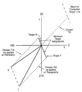
the pre-operative topography. The arrows in Figures
15b, 16a, 16b indicate the direction of the vectors.
The residual astigmatism is equivalent in
length and orientation to the maximal correction target
line in Fig. 17a. The magnitudes of the residual
astigmatism in Fig. 15, the target refraction, Fig. 16a
and the target topography Fig. 16b, minimum target
astigmatism Fig. 17a and maximal correction target line
CA 02227756 1998-01-23
WO 96/40027 PCT/AU96/00257
-44-
in Fig. 17a are all equal, and all lines representing
these values are parallel to each other, as evidenced
by the parallelograms formed by joining the,nazpactorial
combinations.
In other words, when the target induced
astigmatism determined topographically (TIA T) is
vectorially added to the vector R of astigmatism values
measured refractively to obtain the residual
astigmatism shown in Fig. 16a, this residual
astigmatism is equal to the residual astigmatism in
Fig. 16b which is the vector sum of the astigmatism
values T measured topographically and the target
induced astigmatism determined refractively (TIA R).
Moreover, the residual astigmatism values determined in
Figs. 16a and 16b, which are equal to one another, are
also equal to the maximum correction target line in
Fig. 17a where the chosen TIA is intermediate TIA T and
TIA R. In accordance with the invention, by observing
the above conditions, the total residual astiginatisms T
and R measured topographically and refractively
following surgery will be a minimum. Essentially, when
the TIA is between TIA R and TIA T its magnitude is
established by the vector having one end at the origin
and whose other end is on the maximal correction target
line.
The intermediate TIA in Fig. 17a can be chosen
CA 02227756 1998-01-23
PCT/AU96/00257
WO 96/40027
-45-
between the boundaries of the TIA T and TIA R and its
vector length terminates on the maximal correction
target line. The relative proximity of the-%-w-;
intermediate TIA to topography astigmatism values T and
refraction astigmatism values R determines the emphasis
of treatment shown in Fig. 17b. Any TIA utilized which
achieves the minimum target astigmatism for the
prevailing topographic and refractive parameters must
terminate on this line. Any chosen TIA can then be
applied to both refraction and topography (as in Figure
17a) to leave the minimum target astigmatism, which is
apportioned between topography and refraction according
to the chosen emphasis. The target refraction and
topography are orientated at 180 to each other on the
double angle vector diagram; that is, they form a
straight line, and hence their total magnitudes are a
minimum for the optical system of that eye. In Figs.
17a and 17b, the total astigmatism values of T and R
are 0.76 (0.50+0.26 in Fig. 17a and 0.28 and 0.48 in
Fig. 17b).
The parameter that best determines the optimal
point of termination of the TIA with the emphasis line
(where it intersects the maximal correction target
line) is the orientation of the target corneal
astigmatism. In this example, the meridian or target
topography is 147'. As this lies 57' from a with-the-
CA 02227756 1998-01-23
WO 96/40027 PCT/AU96/00257
-46-
rule orientation of 90', the surgeon may decide to use
a linear relationship as illustrated in Figure 19 and
apportion 57/90 or 63.3% emphasis to a-topogv-aphy-based
treatment goal. Consequently, the TIA (Fig. 17b) will
be positioned in relative proximity to the TIA T vector
(Fig..16a) compared to the TIA R vector (Fig. 16b). In
the box associated with Figure 17b, the emphasis is 63%
topographically and 37' refractively. This determines
the parameters for TIA and the target astigmatism
values for R and T.
If a TIA is chosen without regard to the
maximal correction target line this will result in a
total target astigmatism (T plus R) greater than the
minimum. When the TIA is longer than if it terminated
at the maximal correction target line 11overcorrection"
will be obtained as illustrated in Figure 18a. The two
values of target astigmatism T and R are determined by
applying the TIA to the pre-operative topography and
refraction values and when the two target values T and
R are added together, the result is 0.54+0.58=1.12
which is greater than the minimum target astigmatism of
0.76, and when compared to each other a linear (1801)
relationship is not present. Thus, as seen in Fig.
18a, the angle between T and R is not 180' (86'),
whereas in Figs. 17a and 17b the angle is 180'and T and
R lie on a straight line.
CA 02227756 1998-01-23
WO 96/40027 PCT/AU96/00257
-47-
Similarly, if a TIA is chosen that is shorter
than that required to reach the maximal correction
target line (Fig. 18b) the sum of T and R w-1 be
0.30+0.63=0.93 which greater than the minimum value of
0.76. This may, for example, be the case where the
refractive magnitude is chosen with the topographic
meridian, to "under-correct" the astigmatism. The
resultant target astigmatism (T & R) is again greater
than the minimum achievable as the angle between T and
R is not 180' (51'). A full correction of astigmatism
is only achieved when the TIA employed targets the
minimum astigmatism that is equal to the residual
astigmatism (as in Figures 17a and 17b). The target
astigmatism in excess of the minimum increases
hyperbolically as a function of increase of the
distance of the end of the line representing the chosen
TIA from the maximal correction target line.
Note that in this context as illustrated in
Figures 18a and 18b "over" and "under" corrections do
not refer to the relative relationship between SIA and
TIA, but a comparison between the targeted change and
what might be a preferable choice. It is also to be
noted that when the astigmatism values T and R measured
topographically and refractively are different in
magnitude and/or axis, the refractive and topographic
target astigmatism values T and R are non-zero and the
CA 02227756 1998-01-23
WO 96/40027 PCT/AU96/00257
-48-
sum of their vectors is equal and parallel to the
vectorial difference between astigmatism values T and R
and respective TIA R and TIA T values as sho;tn in
Figures. 17a and 17b. This is referred to as
"summating" the astigmatism values T and R measured
topographically and refractively.
The emphasis of treatment is the relative
position between any targeted topographic and
refractive goals, expressed as a percentage. When
these goals are both zero, the maximal correction of
astigmatism is possible as the emphasis line coincides
with the maximal correction target line. The treatment
emphasis can only be apportioned when the TIA
terminates at its point of intersection with the
emphasis line.
The emphasis paradigm chosen in Figures 17a and
17b follows linearity as represented graphically in
Figure 19. For the refractive surgeon, a decision is
to be made in the choice of emphasis of treatment. The
majority of current astigmatism surgery using
incisional or non-incisionally techniques is performed
with the chosen TIA at either end of the emphasis line.
Preferably, a choice should be made as to how much
emphasis is given to topography or refraction in the
surgical plan, according to the orientation of the
target astigmatism. The surgeon may choose an emphasis
CA 02227756 1998-01-23
WO 96/40027 PCT/AU96/00257
-49-
that adheres to linearity, to the square of the cosine
of the target astigmatism, or to another function of
cosine. The lower the line on the graph in-&ig. 19,
the more the emphasis given to refraction.
Alternatively, the surgeon may choose to vary the
treatment emphasis according to prevailing knowledge or
understanding of how much degradation is imposed upon
the visual image according to the orientation of the
existing corneal astigmatism. The effect on the
quality of this perceived image is also likely to vary
according to the associated spherical equivalent. In
the example given for Fig. 17b, the selected paradigm
is a "with the rule" orientation for the residual
astigmatism. The axis of 147' for target astigmatism
(topographically) is taken on the abscissa in Fig. 19
and this intersects the linear emphasis line at an
emphasis of 63%. This value of 63% is selected for
topography and 37% for refraction. The resultant TIA
is plotted in Fig. 17b and its parameters are given in
the appended box.
Hereafter the invention will be described with
reference to eyes having a non-symmetrical topography
wherein analysis and treatment will be made on
respective superior and inferior hemi-divisions of the
eye.
Referring to Fig. 20a, therein it is seen that
CA 02227756 1998-01-23
WO 96/40027 PCT/AU96/00257
-50-
the eye is divided into superior and inferior hemi-
divisions, each having respective topographical
astigmatism values which differ from one ane~6her. The
refractive astigmatism values are measured for the
entire eye and accordingly are the same for the two
hemi-d,ivisions.
The optimal treatment described with reference
to Figs. 15a, 15b and 17b is shown on the superior
hemi-division of the cornea in Fig. 20a. Note that
this is a single angle astigmatism and surgical vector
diagram, illustrating the parameters as they would
appear schematically on an eye, as in Figure 15a.
Employing polar co-ordinates does not allow for
vectorial comparisons of astigmatism provided by the
double angle vector diagrams as in Figures 15b, 18a,
18b. In the two examples in Figure 20a, the emphasis
in the surgical plan given to topography follows the
linear relationship in Figure 19. The closer the
target astigmatism approaches against-the-rule, the
more the emphasis is given to topography in the
surgical plan to achieve a spherical cornea, thus
targeting less unfavorable corneal astigmatism.
Where treatment differs between the two hemi-
divisions of the cornea, a separate evaluation is
required to determine the effect on refractive
astigmatism of the two differing TIAs applied to the
CA 02227756 1998-01-23
WO 96/40027 PCT/AU96/00257
-51-
corneal shape. Resolution of the treatment vectors
(Fig. 20b) is required when performing hemi-meridian
treatment of astigmatism, so that the change-4n
refractive astigmatism is the vector sum of the two
treatment components of the superior and inferior hemi-
divisions. One half of the vector sum of the TIA
topography parameters of the superior and inferior
halves is vectorially applied to the refractive
astigmatism value in both hemi-divisions as shown in
Fig. 20c.
The values in the parentheses in the boxes in
Fig. 20a for target refractive astigmatism in the
superior and inferior divisions are each determined for
the case where a single topography value exists, either
superior or inferior values for both divisions of the
cornea, as if the cornea were symmetrical. In this
example, it can be seen that the orientation of these
two refractive values, being separated by close to 90 ,
results in the single target value calculated from the
resolved treatment vector being smaller than each
individual target value.
Hence, according to the invention a TIA net
value is obtained by vectorially averaging the TIA
values obtained from the superior and inferior hemi-
divisions, said TIA values being based on the pre-
operative astigmatisms T and R respectively, and the
CA 02227756 1998-01-23
WO 96/40027 PCT/AU96/00257
-52-
emphasis in each hemi-division. The TIA net is then
taken globally with the pre-operative astigmatism R
measured refractively to obtain the non-zero-.~,arget
astigmatism measured refractively.
The invention will next be described with
reference to treatment in which the axis of astigmatism
is shifted without changing the magnitude of the
astigmatism. This treatment is in the nature of
application of an "astigmatic torque" to the eye.
A force applied to the eye, having existing
corneal astigmatism, at an oblique angle of 45' to the
astigmatism axis will exert a purely torque effect and
have no steepening or flattening effect on the original
astigmatism axis. The effect of this force on
astigmatism can readily be appreciated on the double
angle vector diagram as shown in Fig. 21b, where the
target astigmatism increases in magnitude as the
tangential force applied increases. As torque force
exerted increases, so does the pre-operative axis
shift, up to a limit of 45', which is in the direction
of the torque force. Referring to Figs. 21b the
following relationships are seen.
Xl = Ki cosine (2ej)
Yl = Ki sine (261)
X2 = K2 cosine (262)
Y2 = K2 sine (26Z)
CA 02227756 1998-01-23
WO 96/40027 PCT/AU96/00257
-53-
The axis and magnitude of the astigmatism
vector on the double angle vector diagram are
determined from the relation:
YZ-Y1
TIA axis = arctan
X2-X1
YZ-Y1
TIA magnitude = arctan
sine (TIA) axis
The magnitude of the astigmatism torque force
(TIA torque):
KTORQUE = K2 sine (2A2 - 26j)
If the result is positive the force is in a
counter-clockwise (CCW) direction and if negative it is
clockwise (CW).
The amount of flattening/steepening that has
occurred with respect to the pre-operative astigmatism
axis is expressed as follows:
KFLATTENNING/STEEPENING = K2 cosine (262-26j)-K,
In this example the value of
KFLATTENNING/STEEPENING is taken as zero if there is no
flattening/steepening effect to be obtained. If the
value was positive then steepening has occurred, and if
negative flattening has occurred.
Where only a change in meridian is desired and
CA 02227756 1998-01-23
WO 96/40027 PCT/AU96/00257
-54-
no change in magnitude of the astigmatism is targeted,
any change in the orientation of astigmatism requires
an amount of flattening in combination with.the TIA
torque. The greater the change in orientation, the
greater the proportion of flattening force and the less
the torque. At the limit of change of axis, which is
180=, the force required is wholly a flattening one,
and the torque component becomes zero.
It is useful to divide the change into its two
component parts one being either flattening or
steepening, and the other torque. This is an essential
consideration when differences exist between topography
and refraction, as for example, a refractive cataract
surgery incision placed "on axis" for flattening of one
modality will be "off axis" for the other, and will
have a torque as well as a flattening effect on its
magnitude. When performing non-incisional refractive
surgery, a treatment will have a flattening/steepening
and torque effect on one or both modalities, as it may
not be aligned with either T or R axis.
The effect of flattening and steepening on
torque and the compensating effect to achieve
astigmatic torque without flattening and steepening
will be described with reference to Figs. 22a, 22b, 23a
and 23b.
Referring to Figs. 22a and 22b the amount
CA 02227756 1998-01-23
WO 96/40027 PCT/AU96/00257
-55-
the TIA is "off-axis" from the steepening force
required to reduce the existing astigmatism:
KTORQUE = TIA sine 2
In Figures 21a, 21b, 22a, 22b the effect of
steepening or flattening produced by applying a torque
force at 45'to the astigmatism axis (90' in the double
angle vector diagram) is compensated by angularly
shifting the TIA by an angle 90-2 (Fig. 22b), to
obtain torsional rotation without change in magnitude.
In the numerical example, the astigmatism has a
magnitude of 1.40D and the axis is shifted from 8 to
25' and to achieve this the TIA has a magnitude of 0.83
and an axis of 62=. The effect of steepening,
flattening, CW torque and CCW torque applied to the
pre-operative astigmatism by the TIA force at the
respective orientations is illustrated in the Figures.
When examining the change that has occurred at
the intended axis of the astigmatism surgery, the
flattening/steepening effects of surgery can be
determined by the relationship illustrated in Figures
23a and 23b. The angle 0 is subtended between the SIA
(surgically induced astigmatism) axis, and the TIA axis
90' to the axis of the incision.
KFLATTENING/STEEPENING = SIA cosine 20
= 25 The method can be applied to determine the
astigmatic effect of a cataract surgery incision (SIA)
.
CA 02227756 1998-01-23
WO 96/40027 PCT/AU96/00257
-56-
at its meridian of placement. The flattening/
steepening component of the SIA determined by this
means utilizing surgical vectors, provides arx-:
alternative formula to achieve the same magnitude value
as that calculated by the formulas for the meridional
power of cylindrical lenses and surfaces employed by
Holladay and Naeser. The above formula is directly
linked to the SIA and the angular deviation from its
intended meridian of placement, the angle of error.
This eliminates the requirement when determining
meridional power, of calculating the contribution of
the pre- and post-operative astigmatism on the surgical
meridian and at 90' to it, followed by subtracting one
net value from the other to determine the change at
that incision's axis.
The description of this change by either of the
terms flattening or steepening, according to the
corneal change at the surgical meridian, may provide
some advantages of simplicity to the descriptive terms
"with and against-the-wound", and "with and against-
the-power". However, the terms "with-the-rule 1 and
"against-the-rule" refer to polarity at the specific
meridia at 90' and 180', and are in common accepted
usage.
In the example in Figures 24a and 24b, a torque
force is applied to the entire cornea with orthogonal
CA 02227756 1998-01-23
WO 96/40027 PCT/AU96/00257
-57-
symmetrical astigmatism, to target the refractive
cylinder axis. Any induced change of the corneal shape
would target an equivalent vectorial change-on the
refractive modality, and one would expect the
refractive cylinder to have rotated by the appropriate
amount=of the corneal change induced by that force.
When treatment is symmetrical no resolution of
treatment vectors is necessary.
Next will be explained the treatment of
irregular astigmatism referring to Figs. 25a and 25b.
Irregular astigmatism is present where
topographic values of the two hemi-divisions of the
cornea do not coincide either in magnitude (asymmetry)
or meridian (non-orthogonal) or both. Existing
differences of some order between the two halves of the
cornea are widely prevalent. The topographical
disparity is a measure of astigmatism irregularity in
diopters, by vectorially comparing the topographic
values of magnitude and axis between the two corneal
hemi-divisions. For this example the topographic
disparity is 1.29D.
In the presence of either non-orthogonal or
asymmetrical astigmatism or both, there may be a desire
to regularize the corneal shape to provide the
opportunity for improved unaided or best-corrected
vision. It may be advantageous to have the ability to
CA 02227756 1998-01-23
WO 96/40027 PCT/AU96/00257
-58-
perform this task without any net change in refractive
astigmatism or spectacle correction. By treating the
cornea in two independent halves, the appropr.a.ate
amount of astigmatic change can be applied at the
appropriate meridia for the desired change in
topographic astigmatism, in opposite cyclical
directions. The existing topographical astigmatism can
be targeted to coincide in both magnitude and meridian
(actually 360' apart) on the double angle vector
diagram, to create the orthogonal state (180" apart) on
an astigmatism diagram of the eye. Resolution of the
two treatment vectors shows that the two TIAs, when
applied in equal magnitudes and at 90' to each other,
negate each other's effect and cancel any net effect on
refractive astigmatism.
Reduced vision due to apparent amblyopia or
other causes of sub-optimal vision associated with
irregular astigmatism may benefit from improved unaided
and best corrected vision by regularizing the cornea.
It is possible that lower grades of keratoconus may
also benefit from differential flattening and
steepening on the opposite hemi-meridian to reduce or
eliminate the existing asymmetry of the condition.
This method of changing the topography of the
eye without any net-effect on the refractive
astigmatism can also be employed with purely flattening
CA 02227756 1998-01-23
WO 96/40027 PCT/AU96/00257
-59-
and steepening or torque effects.
The correction of irregular astigmatism to an
orthogonal symmetrical state may be achieved ip other
ways. This may be achieved by the application of
treatment to a single hemi-division of the cornea, as
in Figures 26a and 26b (The TIA for the inferior hemi-
division is zero). In this example, by moving the less
favorable astigmatism to coipcide with the more
favorably placed hemi-meridian closer to a with-the-
rule orientation (180'/540= on the double angle vector
diagram, the result is an improvement in the
orientation of both the shifted superior hemi-meridian
of the corneal astigmatism and the refractive
astigmatism with the least change to achieve
regularity. One half of the net TIA effect is applied
to each half of the refractive astigmatism. No change
of the inferior topographic hemi-meridian is targeted.
The topographic astigmatism of the eye can be
improved by rendering it orthogonal and symmetrical in
another manner as shown in Figs. 27a, 27b and 27c. By
targeting the refractive magnitude and axis for the
topography in both hemi-divisions of the cornea, a net
reduction in the amount of residual astigmatism will
result from a decrease in the amount of disparity
between topography and refraction in each corneal half.
There may be a shift of the refractive astigmatism that
CA 02227756 1998-01-23
WO 96/40027 PCT/AU96/00257
-60-
is induced by the net TIA determined by resolving the
superior and inferior treatment vectors. The resultant
residual astigmatism of both hemi-divisions will be
equal and at minimal levels.
Referring to Figs. 28a, 28b and 28c, these show
that the TIA can be determined to change the prevailing
refractive or topographic astigmatism to any desired
target. The topographic change can be symmetrical or
asymmetrical. In this example, both hemi-meridia of
the cornea are targeted at 0.75 in a with-the-rule
orientation (90' & 270') to render the cornea
symmetrical. After resolution of the treatment
vectors, it can be seen that there is a net favorable
shift in the refractive astigmatism, with a reduction
in its magnitude and the amount of residual
astigmatism.
Figs. 29a, 29b, 29c show a treatment to achieve
any desired nominal corneal astigmatism where there is
no apparent relation between the corneal shape in the
hemi-divisions. Thus, the target astigmatism of both
hemi-divisions represent two randomly chosen targets.
The TIA vectors are separately calculated for each of
the chosen targets. The net TIA is obtained by
vectorially adding the TIA values in the hemi-
divisions. This could have benefits not immediately
predictable at the present but which could have future
CA 02227756 1998-01-23
WO 96/40027 PCT/AU96/00257
-61-
benefits in different surgical techniques and to
achieve different optical effects based on retinal and
optic nerve investigations.
The separation of the differing forces of
flattening/steepening and torque, and the treatment of
the cofnea as the two significantly differing halves
(as it frequently is), provides the ability to change
the relative shape of the cornea in various ways. The
ability to achieve orthogonal and symmetrical
topographic astigmatism, either affecting the
refractive astigmatism in a favorable way, or not
necessarily changing it at all, enables a potential and
significant expansion in the means to produce
astigmatic enhancement and improved visual performance.
Hereafter, the invention will be discussed
further with reference to a series of topical headings
as follows:
1. A concept for undertaking astigmatism
analysis.
2. The optical correction of astigmatism.
3. Objective and Subjective analyses.
4. Adjustments and Nomograms.
5. A computer Assisted Video Keratotomy (CAVK)
6. Keratoconus.
7. Vector Analysis Vector.
8. Common principle - Target T and R.
CA 02227756 1998-01-23
WO 96/40027 PCT/AU96/00257
-62-
9. Optical treatment of astigmatism.
10. Emphasis.
11. Complexities and ConsideratiorYs:-bf Corneal
Shape.
12. Astigmatic torque.
13. Quantitative Surgical Planning.
14. Vector Change Maps.
15. Conclusion.
1. A CONCEPT FOR UNDERSTANDING ASTIGMATISM ANALYSIS
Any new concept that expands existing
understanding of a subject requires many qualities to
gain general acceptance. First and most importantly,
the underlying concept should be simple to understand.
Any inadequacy in past understanding of a subject
should readily become apparent. New information
provided by the introduction of a concept should be
useful and easy to apply. The acceptance of a new
analysis technique will be enhanced if its application
assists in the decision-making process for future
endeavors as well as discerning success of past
efforts.
Stroking a golf ball into the hole on a flat
putting green is a simple process to understand, but
not always easy to accomplish. Golf putting shares
common concepts with the treatment and analysis of
CA 02227756 1998-01-23
WO 96/40027 PCT/AU96/00257
-63-
astigmatism. When one is unsuccessful in hitting a
ball along a chosen path into a hole, one of two events
has happened: the force with which it was struck was
either firm or too soft, or the direction in which it
was propelled was either clockwise or counter-clockwise
from that desired. A combination of these two is most
common. 'The single most reliable parameter of the
overall success of a putt is the length required for a
second putt to place the ball in the hole.
The principle underlying the planning and
analysis of astigmatism surgery is no more complicated
than this. The intended path of the astigmatism
surgery - that is, the required force and its
direction, is the TIA (targeted induced astigmatism
vector). The actual path taken by the astigmatism
surgery is the SIA (surgically induced astigmatism
vector) and the Difference Vector (DV) measures the
force and its orientation still required to achieve the
initial goal - effectively the second putt, using the
above analogy. It is the various relationships between
the SIA and TIA that tell us whether the treatment was
on axis or off axis, or whether too much or too little
force has been applied, and how to adjust for it. It
is the magnitude of the DV, and its relationship with
the TIA, that gauges the absolute and relative success
of our surgery.
CA 02227756 1998-01-23
WO 96/40027 PCT/AU96/00257
-64-
The Correction Index determined by the ratio of
the SIA to the TIA is a measure of the amount of
correction, and is optimally unity. It is-Vxeater than
one if an over-correction has occurred and less than
one if there has been an under-correction. The
Coefficient of Adjustment is simply the inverse of the
Correction Index, and quantifies the modification
required to the initial surgery treatment plan to have
achieved a Correction Index of one -the desired
correction.
The measures of success obtained utilizing this
method of astigmatism analysis are both absolute and
relative. The magnitude of the DV provides an absolute
measure of the success achieved in astigmatism surgery,
and is a measure of the second surgery; that is, the
amount of steepening and its orientation, required to
achieve the goal of the initial surgery. Using the
golf analogy, the DV is represented by the second putt.
This vectorial comparison can be determined for
all measurement modalities (R, K and T), and the DV's
mean for a series of surgeries usefully compares
astigmatism surgery techniques. The orientation of its
DVs does not seem to provide useful information for the
determination of success. The planning required for
re-operation is best performed by freshly addressing
the optimal refractive and topographic goals.
,
CA 02227756 1998-01-23
WO 96/40027 PCT/AU96/00257
-65-
The relationship of the DV to the TIA is the
Index of Success (IOS), which provides a relative
measure of surgical success. The greater the targeted
change in astigmatism, the smaller the IOS value would
be for any constant value of the DV, hence the more
successful was the surgery. Employing the golfing
analogy, to determine which of two putts that finished
equidistant from the hole was more effective, the
answer is clearly the one that resulted from a longer
first putt.
Where the achieved astigmatism coincides with
the target, no astigmatism induction is required to
achieve the initial goal. Just as one may one-putt a
green, so in this case the DV is zero and so the Index
of Success value is also zero.
Where smaller values of pre-operative
astigmatism exist with consequently smaller targeted
changes in astigmatism, less reliance can be placed on
the measures that involve a ratio as the sole
determinant in judging the success of surgery (IOS,
Coefficient of Adjustment and Correction Index),
because larger swings in values can be induced by
smaller variations in the components of a fraction.
Reference to means of absolute values such as the DV is
necessary for comprehensive interpretation of results.
The TIA is the key enabling an analysis to be
CA 02227756 1998-01-23
WO 96/40027 PCT/AU96/00257
-66-
performed both at the corneal and spectacle plane. It has in effect removed
the barrier that has until now
prevented astigmatism surgeons who operate'W" refraction from appreciating the
effect of surgery on
the corneal shape and vice versa, by converting an
interVening mirror barrier into a pane of glass; now
each can see the effect of their surgery at the other's
reference plane.
While current astigmatism analysis is adequate
for determining what has happened (SIA), whether or not
a change in the astigmatism was intended, until now
analysis systems have not compared how what has
happened differs from what was intended to happen.
Comparing pre-operative and post-operative astigmatism
values ignores any change in the astigmatism's axis and
consequently is misleading, because it inevitably
renders all imperfect corrections to be
"undercorrections". It does not identify the separate
errors of magnitude and axis.
For example, when analyzing the Angle of Error,
a consistent error of 5' in a clockwise direction in
all eyes may indicate a system error, such as beam
rotation when performing excimer laser surgery.
However, this 5' may be consistently clockwise in right
eyes and counter-clockwise in left eyes, indicating the
likely cause to be extortion of the eyes when lying
CA 02227756 1998-01-23
WO 96/40027 PCT/AU96/00257
-67-
supine.
2,.- THE OPTICAL CORRECTION OF ASTIGMATISM
REFRACTIVE, TOPOGRAPHIC AND RESIDUAL ASTIGMATISM
The optical correction of astigmatism measured
refractive,ly can be performed with spectacles, contact
lenses or by surgery. Where the correction of
astigmatism is performed by spectacles, then the
subjective astigmatism is determined by manifest
testing at the spectacle plane. This refractive
astigmatism value provides a measure of the total net
astigmatism of all the refracting surfaces of the eye.
That is, the net astigmatic effect encountered by light
during its passage through the optical system of the
eye via the front and back surfaces of the cornea and
lens, and the vitreal body. The subjective test will
also be influenced by any tilt of the retina, and the
conscious perception of this retinal image by the
visual cortex of the occipital lobe.
The anterior surface of the cornea is the major
refracting surface of the optical system of the eye.
The variations between the values of the corneal
astigmatism and the refractive astigmatism at the
corneal plane is responsible for the residual
astigmatism that cannot be eliminated from the optical
system of the eye and its refractive correction. Until
CA 02227756 1998-01-23
WO 96/40027 PCT/AU96/00257
-68-
recently, these differences have been disregarded in
the treatment of astigmatism; the consequence of this
has significant implications for resultant oor-aneal
astigmatism.
The variance that occurs between topographic
and refractive astigmatism cannot be dismissed, nor can
residual astigmatism be loosely termed as "Ienticular
astigmatism". If this term were an adequate
description for these differences, then one would
expect corneal and refractive astigmatism to coincide
both in magnitude and axis after cataract extraction
and spheric lens implantation; however, this is not the
case. The existence of two modalities for measuring
astigmatism, subjective and objective, together with
multiple measuring devices within each group, will
ensure the perpetuation of these differences and the
need to address them in the treatment.
Where refractive error is corrected by a soft
contact lens, which provides only a spherical
correction, then, in a theoretical model, a
topographical image obtained from the front surface of
the contact lens in place on the cornea would be
equivalent to that image obtained of the cornea without
the contact lens, and would provide a measure of
topographical astigmatism. A manifest refraction,
performed with or without this lens in place, provides
CA 02227756 1998-01-23
WO 96/40027 PCT/AU96/00257
-69-
, a measure of refractive astigmatism at the spectacle
plane.
Toric soft contact lenses are presc=~ibed with
the refractive astigmatism corrected for back vertex
distance, at the appropriate axis. Where this contact
lens iS of the same refractive index as the cornea, and
accurately situated on the cornea at the prescribed
meridian, the CAVK (Computer Assisted Video
Keratography) image obtained from the front surface of
that lens as it lies on the cornea would, in a
theoretical model, provide an image of the magnitude
and axis of residual astigmatism of the eye. A
manifest over-refraction should not detect any
refractive astigmatic error.
A hard contact lens with spherical surfaces
when in place on the cornea effectively sphericises the
anterior refractive surface of the eye, so that the
manifest over-refraction provides a measure of the
amount of residual (not refractive) astigmatism. This
over-refraction magnitude corrected to the cornea plane
theoretically should be equal to the value obtained by
CAVK on the surface of the in-situ toric soft contact
lens, but the orientation of its refractive axis would
be 90' to the orientation of the residual astigmatism.
3. OBJECTIVE & SUBJECTIVE ANALYSIS
CA 02227756 1998-01-23
WO 96/40027 PCT/AU96/00257
-70-
Corneal topography displayed by CAVK provides
the treatment and analysis of astigmatism with
objective and reproducible images. Analyzing-an
astigmatism result by topography as well as refraction
will become an essential step in monitoring the changes
at the surface where they are effected - on the cornea.
CAVK provides us with an objective measure; however,
some limitations are caused by the reduced definition
of dioptric contour separation, present after non-
incisional refractive surgery.
Manifest refraction is a subjective test that
depends on observer and patient responses, which may
vary according to testing conditions in a refracting
lane such as lighting, chart distance and illumination
inconsistencies. The inadequacy of refraction as the
sole measurement parameter for astigmatism is
accentuated by the large changes in spherical
equivalents induced by refractive surgery, which may
cause less attention than warranted to be paid to
residual astigmatic refractive errors. The trends
discernible by the objective measurements determined by
corneal topography, or conventional keratometry,
provide a balance for the subjective trends in the
analyses_ This additional information gives the
required assurance necessary to determine the success
of astigmatism surgery, the errors occurring and what
CA 02227756 1998-01-23
WO 96/40027 PCT/AU96/00257
-71-
adjustments are required to improve results.
Utilizing the TIA, the topographical and
refractive targets are now determinable. The, TIA
provides the linkage to enable an analysis by
refraction, topography or other device that measures
astigmatism. The trend now emerging when performing
laser astigmatism surgery is that significant
differences exist in the results when comparing those
achieved by refraction with those occurring on the
cornea. These differing trends in analyses may be
attributable in part to undue emphasis on refraction as
the sole treatment parameter.
4. ADJUSTMENTS AND NOMOGRAMS
The targeted change in corneal astigmatism
(TIA) attempts to alter the pre-operative state to
achieve the desired astigmatism goal as determined by
all modalities; refraction, topography and keratometry.
Nomograms are utilized for incisional astigmatism
surgery (astigmatic keratotomy) to allow for the
biological variance caused by such factors as age,
ocular tension and corneal diameter. To achieve the
targeted correction more consistently, adjustment
factors can be determined to modify these incisional
nomograms further or to fine-tune the performance of
laser machines performing non-incisional surgery.
Analysis of PARK surgeries performed with the
CA 02227756 1998-01-23
WO 96/40027 PCT/AU96/00257
-72-
VisX twenty/twenty laser prior to February 1993,
revealed a general trend of under-correction of
astigmatism magnitude. Insufficient "force'4::-was being
applied, the average amounts of surgically induced
astigmatism (SIA) being less than the average TIA
intended, in a proportion of around 10 to 12.
Subsequent results for magnitude achieved after April
1993, after applying an adjustment factor of 1.2 to the
astigmatism requiring treatment, have shown that the
SIA by refraction and topography subsequently more
closely approximates the targeted change, achieving a
correction index closer to one.
5. COMPUTER-ASSISTED VIDEO KERATOGRAPHY (CAVK)
The devices currently available for examining
and measuring corneal contour fall broadly into two
groups. The dioptric plots derive their data from
reflective placido disc technology. The illuminated
placido disc may be composed of either alternating
black and white rings or a colored sequence of rings.
The true elevation maps are derived from images
generated by non-reflective images, either from
scanning slits or the viewing of a projected grid by
sterophotogrammetry. The latter technology enables
non-axial viewing of non-reflective surfaces and the
potential for providing a real-time image to be viewed
CA 02227756 1998-01-23
WO 96/40027 PCT/AU96/00257
-73-
on the operating table for intra-operative use.
The point of steepest curvature depicted at the
highest peak of the bowtie on a dioptric plot,requires
the least amount of tissue ablation to sphericize the
cornea. The point of highest altitude depicted on a
true elevation topography map requires the greatest
amount of tissue ablation utilizing the excimer laser
to sphericize the cornea. This corresponds to the
meridian of flattest curvature and is aligned with the
orientation of the steepening vector (the TIA). Tissue
addition procedures such as astigmatic keratotomy act
maximally, perpendicular to the axis of the TIA.
KERATOCONUS
Corneal astigmatism varies in the amount of
asymmetry and non-orthogonal orientation. The spectrum
of these physiological aberrations of corneal shape
stretches from high amounts of astigmatism to the
grosser amounts of asymmetry that appears as "forme
fruste" and "early" or "subclinical" levels of
keratoconus now more frequently identified with the aid
of CAVK technology. The fine line that divides what
lies within and outside the physiological spectrum may
well be determined by clinical signs such as excessive
corneal steepening, apical thinning or scarring. The
differing patterns found in keratoconus may represent a
CA 02227756 1998-01-23
WO 96/40027 PCT/AU96/00257
-74-
continuum of different stages in the progression of the
topographical alterations that occur over time in
keratoconus. However, on careful examination- of the
topographical maps in eyes with the cornea showing
keratoconus change, there commonly exists a vestigial
remnant of one half of the bowtie, indicating the
presence of both hemi-meridians of the astigmatism,
even when in a grossly asymmetrical state.
7. VECTOR ANALYSIS VALUE
Current methods of measurement of visual
perception, such as visual acuity or contrast
sensitivity, do not have the sensitivity required to
demonstrate differing performance for alternative
methods of astigmatism treatment. For example,
heretofore, one was unable to demonstrate any
discernible difference between the elliptical shaped
ablation created by the opening of parallel blades
limiting the beam aperture on two sides and the
concurrent closing of a round diaphragm aperture, and
sequential technique using the plano-cylindrical mode
methods of treatment of astigmatism, when utilizing the
VisX twenty/twenty excimer laser. This difference in
performance was detectable when astigmatism change is
separated from co-existing spherical changes by
utilizing vector analysis.
CA 02227756 1998-01-23
WO 96/40027 PCT/AU96/00257
-75-
rt
_~l COMMON PRINCIPLE-TARGET T & R
= There is an existing conflict in the two
contrasting treatment paradigms that employ one of the
two pre-operative astigmatism values, topography or
refra(-',tion, as the determinant for their differing
surgical treatment. This variance can be resolved by
utilizing the orientation of the target corneal
astigmatism as the guiding surgical paradigm, rather
than either of the two pre-operative astigmatisms. In
this way, all astigmatism surgeons, whether they use
blade or laser technology, will be operating under the
same guiding principle.
All forms of refractive surgery would then come
under the one common principle of treatment; that is,
to assess the effect of the proposed change in
astigmatism on both refraction and corneal shape, in
order to determine the optimal procedure for each
individual eye. Current practice follows no common or
consistent theme, totally emphasizing pre-operative
corneal shape for cataract and penetrating
keratoplasty, either shape or refraction for astigmatic
keratotomy and refraction for excimer laser PARK
surgery; each adopts only one or other extreme of the
treatment spectrum.
It is only by determining the refractive and
CA 02227756 1998-01-23
WO 96/40027 PCT/AU96/00257
-76-
topographic targets prior to surgery that one can
perform two essential tasks in the astigmatism surgery;
firstly, to optimize the treatment according to prevailing parameters, and
secondly, to enable a valid
analysis by knowing where the targets lie. The process
of setting precise goals enables measurement of
success, determination of errors and making the various
adjustments necessary to improve future procedures.
When performing excimer laser surgery for
myopia and astigmatism, one may ask - if the goal is to
eliminate the need for glasses, why would we not use
the refraction as the only treatment parameter for both
cylinder as well as the sphere? The answer lies in the
targeting of a zero spherical equivalent for the
correction of myopia, which takes care of the goal of
eliminating spectacles. However, the penalty for
sculpting the spectacle astigmatism onto the cornea is
the maximizing of the resultant corneal astigmatism,
and this is contrary to established and conventional
principles of corneal surgery requiring that corneal
shape be considered in surgical planning. The optical
system of the eye that is independent of spectacle
correction will continue to depend upon optimal
regularity of the shape of the anterior cornea surface.
9. OPTIMAL TREATMENT OF ASTIGMATISM
CA 02227756 1998-01-23
WO 96/40027 PCT/AU96/00257
-77-
When differences prevail between topography and
rt
refraction, residual astigmatism unavoidably remains in
the optical system of the eye, either in topography or
refraction, or shared between the two leaving non-zero
targets at both surfaces. The consequence of excluding
topography from the surgical plan, as is widely
practiced with Excimer laser surgery, is the inevitable
destination of the residual astigmatism onto the
cornea, the maximizing of the resultant corneal
astigmatism and the potential for increased spherical
aberration and resultant degradation of the perceived
image.
The optimal treatment of astigmatism according
to the invention is achieved when the sum of the
targeted non-zero values of topographic and refractive
astigmatism equals the minimum target value achievable
for that optical system, and is appropriately
apportioned to topography and refraction according to
the orientation of the target astigmatism.
The orientation of the targeted corneal
astigmatism optimally determines the amount of emphasis
apportioned to topography and refraction in the
surgical treatment plan. However, this apportionment
may be directed according to how the cortical
perception of the image degrades according to its
orientation. If the image suffers maximal degradation,
CA 02227756 1998-01-23
WO 96/40027 PCT/AU96/00257
-78-
then in this case the goal of a spherical cornea would
be preferable and the residual astigmatism would be
directed to the refraction. Should the targeted =
astigmatism fall in the orientation where the image is
minimally degraded, then any residual astigmatism
optimally remains on the cornea, and no astigmatic
correction remains in the refraction.
It may be demonstrable in a laboratory or
clinical setting that oblique astigmatism may in fact
be the least favorable option; if so, the treatment
emphasis could be apportioned accordingly. The
spherical equivalent of the eye that co-exists with the
corneal astigmatism influences the clarity of the image
according to the meridian at which it lies.
10. EMPHASIS
The emphasis is a measure, expressed as a
percentage, of the relative position between the
topographic and refractive goals when the treatment
vector i.e. the TIA, terminates on a line in the double
angle vector diagram connecting these two goals. Where
the treatment emphasis line connects the two goals of
zero topographic and refractive astigmatism, then the
emphasis line coincides with the maximal correction
target line, making possible the achievement of minimum
target astigmatism by the maximal correction of
CA 02227756 1998-01-23
WO 96/40027 PCT/AU96/00257
-79-
existing astigmatism.
The greater the targeted corneal astigmatism is
closer to 90 (WTR), the less the emphasis will be
towards a zero topography goal as the sole treatment
parameter.
; However, the relative emphasis given to
topography and refraction in the surgical plan may not
bear a direct linear relationship according to the
orientation of the target astigmatism (Fig. 19).
Instead the surgeon must decide on the relative
emphasis to be given to topography and refraction in
the surgical plan, based on knowledge of the relative
degradation of the visual image according to the
orientation of the existing corneal astigmatism when
the targeted spherical equivalent is zero. Current
knowledge would suggest a WTR orientation to be the
most favorable.
11. COMPLEXITIES AND CONSIDERATIONS OF CORNEAL SHAPE
Traditionally, the primary concern of the
cornea surgeon is the cornea shape; that is, the amount
of astigmatism and its orientation. The optimal
treatment of astigmatism would seek to achieve less
corneal astigmatism, with an attempt to influence its
orientation in a favorable orientation. Zero
astigmatism by all measurement modalities is likely to
CA 02227756 1998-01-23
WO 96/40027 PCT/AU96/00257
-80-
continue to be the ultimate goal of the refractive
surgeon.
Until now, conventional methods of astigmatism
treatment have maintained zero astigmatism, when
achievable, to be the surgical goal, whether the
surgical method employed was based on shape or
refractive astigmatism. The invention recognizes the
inability to avoid non-zero goals imposed by prevailing
differences that exist between refraction and
topography. The introduction of the TIA enables any
refractive or topographical goal to be reached in one
surgical procedure, without requiring the intermediate
step of attaining zero astigmatism. This is analogous
to a flight to a destination not requiring a stop at
the hub airport.
The expansion of the treatment parameters now
possible, according to the invention, opens the way for
determining new forms of astigmatism treatment not
previously considered.
While in the past refraction has been the
primary determinant of treatment, the introduction of
topography into consideration has advantages; however,
it also introduces complexities. The advantages gained
are the reduction of targeted corneal astigmatism by
the inclusion of shape parameters into the surgical
plan, and the ability to use objective measurements for
CA 02227756 1998-01-23
WO 96/40027 PCT/AU96/00257
-81-
vectorial analysis of surgical results.
However, significant complexities are
introduced by the inclusion of topography, which must
be addressed. One complexity already discussed is the
differences existing between the refraction, and the
topography represented by the simulated K. The
simulated K, better termed the "T", provided by CAVK is
only a best-fit compromise, and is derived in different
ways by different devices. In general, it is a mean
value achieved over a number of measured constant
reference points on both hemi-divisions of the cornea.
The existing differences in the dioptric
magnitude of the astigmatism on the two hemi-meridians
of the cornea are as prevalent as differences that
exist between R and T values. The asymmetry of these
hemi-meridian T values are further complicated by the
non-orthogonal relationship of the two hemi-meridians
of the astigmatism; that is, very frequently their axes
are not aligned in a 180= orientation to each other.
Current topography technologies occasionally vary in
their determination of the orientation of the simulated
K readings in the case of non-orthogonal astigmatism,
by selecting at various readings on the same cornea one
of three axes - either one of the two non-orthogonal
values, or an orientation intersecting the two.
The differences between refraction and
CA 02227756 1998-01-23
WO 96/40027 PCT/AU96/00257
-82-
topography values cannot be dismissed as
inconsequential. Nor should it be suggested that an
accurate and methodical refraction can resolve these
differences, as there is no mechanism to reconcile
which hemi-meridian of the cornea topography has been
resolved with the manifest refractive astigmatism
value.
As stated above, examination of the corneal
topography usually shows some amount of asymmetry in
the two hemi-meridia of the cornea. This asymmetry may
lie either in the magnitude or the non-orthogonal
orientation of the two halves of the bow-tie. To
accomplish the optimal treatment of the cornea, two
resolutions of the common refractive and differing
topographic magnitude and axis are required. One can
determine a differing target corneal astigmatism from
each half of the cornea, and from each of these can be
determined an optimal TIA. This hemi-meridian solution
for treatment is relevant to whatever incisional or
non-incisional techniques are employed.
The refractive astigmatism frequently
intersects the meridia of opposite non orthogonal
topographic axes. Employing the optimal treatment for
each hemi-division of the cornea can result in an
enhanced synergistic effect in reducing targeted
refractive astigmatism, yet attaining an overall more
CA 02227756 1998-01-23
WO 96/40027 PCT/AU96/00257
-83-
regular and closer to spherical cornea. Hence,
treating each hemi-division more closely aligned to
each topographic steep axis by recognizing the
inevitable differences between the two topographic
corneal halves, as well as each of their differences
from the refractive astigmatic astigmatism, the optimal
treatment according to the individual conditions
prevailing for that patient can then be determined.
The regularizing of non-orthogonal and
0 asymmetrical elements of corneal astigmatism, with or
without a change in refractive astigmatism, could
potentially improve the best-corrected as well as
unaided visual acuity of an eye. This treatment could
be applied by ablative or intrastromal lasers,
incisional keratotomy or combinations of these
technologies to improve the visual function of an eye
without any change in its refractive status. It may be
useful for the treatment of amblyopia ex anopsia in
children, or in adult eyes that are functioning
suboptimally with irregular astigmatism.
The treatment of irregular astigmatism to
achieve an orthogonal symmetrical state can be achieved
in a number of differing ways, depending on the goal of
surgery. The achievement of regular orthogonal
astigmatism can be performed without any change in
refraction (Figs. 25a-25c). The rotation of the less
CA 02227756 1998-01-23
WO 96/40027 PCT/AU96/00257
-84-
favorable cornea hemi-meridian towards the other more
favorably placed half will achieve a concurrent
favorable rotation of the refractive astigmatism (Figs.
26a-26c). The disparity between topography and
refraction can be reduced on one or both hemi-meridia
by targeting in each the pre-operative refractive
astigmatism. In this way, the net change in refractive
astigmatism will equal the residual astigmatism of both
topographical hemi-meridia (Figs. 27a-27c).
The treatment of astigmatism may be directed at
correcting existing aberrations of symmetry of corneal
shape. In this manner, the clarity of the retinal
image that has been degraded by the prevailing corneal
irregularities could potentially be improved. One
might anticipate that a significant proportion of eyes
suffering visual obscuration may be likely to coexist
with either non-orthogonal or asymmetrical astigmatism,
or both. The correction of this aberrant state at any
age by treatment of the cornea as two dissimilar halves
could potentially improve the quality and quantity of
vision perceived by that eye.
12. ASTIGMATIC TOROUE
Where astigmatic torque is exerted on the pre-
operative existing astigmatic state of the cornea, the
force applied is at 45' to the existing astigmatism. A
rI
~ __ _ - _
CA 02227756 1998-01-23
WO 96/40027 PCT/AU96/00257
-85-
purely tangential (in double angle terms) uni-
directional force applied to the steepest corneal axis
will result in a target astigmatism that is greater in
magnitude than the pre-operative value, with an
orientation that has moved in the direction of the
rotati,onal force (Figs. 21a, 21b).
The effect of applying this TIA force to change
the corneal astigmatism will affect the refractive
astigmatism. One would anticipate any change in the
axis of the pre-existing corneal astigmatism would
change the refractive astigmatism in the same clockwise
or counter-clockwise orientation by the same TIA vector
amount. This change in the amount or orientation of
the refractive astigmatism (Figs. 24a, 24b) may be
favorable or unfavorable.
The division of treatment of the astigmatism of
the eye into hemi-divisions would enable a change on
the orientation of the two hemi-meridian corneal
astigmatism, while reducing or eliminating any effect
on refractive astigmatism. Two opposing steepening
forces can be applied in opposite cyclical directions,
negating each other's effect on refractive astigmatism.
In this way, the use of opposing torsional forces can
realign non-orthogonal asymmetrical hemi-meridians to
achieve a coincident and therefore regular relationship
(Figs. 25a-25c). By changing the orientation of each
CA 02227756 1998-01-23
WO 96/40027 PCT/AU96/00257
-86-
in opposite directions, that is, clockwise and counter-
clockwise, the net effect on refractive astigmatism can
be reduced or eliminated. A closer relationship
between the realigned topography and the refractive
astigmatism would be anticipated.
The separation of flattening/steepening effects
and clockwise or counter-clockwise torsional effects
give control of the corneal shape by separating the
changes into their component parts. If necessary, the
linkage between refractive and topographic changes can
in this way be simplified for analysis and therapy
purposes.
However, applying the TIA to exert some
proportion of astigmatic torque on the cornea without
necessarily reducing the magnitude of the targeted
corneal astigmatism achieves neither the maximal nor
the optimal treatment of astigmatism of the eye. The
maximal treatment of astigmatism occurs where the
target astigmatism (topographic plus refractive) at its
minimum value, equals the residual astigmatism of the
eye. The treatment is optimal where this target
astigmatism is distributed proportionately between
topography and refraction, according to its
orientation.
13. OUALITATIVE SURGICAL PLANNING
CA 02227756 1998-01-23
WO 96/40027 PCT/AU96/00257
-87-
The invention has stressed the importance of
ti
the quantitative information derived from CAVK, but
necessarily recognizes the value of further qualitative
information that can be derived by this quantitative
planning and analysis technique. The method can be
applied in a predictive manner, by applying the
targeted change in astigmatism - either symmetrical or
hemi-divisional, to the pre-operative astigmatism
state, as displayed on the topographical map. The
multiple individual measurements points, either singly
or in groups, can have that treatment applied during
the planning process to enable the viewing of the
expected topographical picture that would be generated
from that proposed treatment. A value for the target
refractive astigmatism can also be determined in
accordance with the resolution of the proposed
treatment vectors.
The surgical planning can be fine-tuned
according to the appearance of the topography as
estimated by the calculated targeted change. Small
changes in emphasis on either of the upper or lower
treatment vectors (TIA) can modify the target
appearance to that which is deemed most desirable
according to the surgeon's qualitative criteria of a
favorable result. These changes in emphasis of one or
both TIAs will concurrently alter the target refractive
CA 02227756 1998-01-23
WO 96/40027 PCT/AU96/00257
-88-
astigmatism. It is worth noting that zero or close-to-
zero refractive astigmatism can be achieved with
asymmetric treatment vectors without necessarily either
of these individually targeting zero refraction, while
enabling some emphasis be given to both topography
hemi-meridia to target less overall topographical
astigmatism.
Further alternatives to fine-tuning the
treatment by change in emphasis can be achieved by
modifying the flattening/steepening effect, the torque
or a combination of these for either of the two
treatment vectors. This can be performed under a real-
time display of the intended topography which would
result from any alteration of the treatment plan.
Movement of the TIA in these modes away from the
maximal correction target line would be performed only
if maximal correction of astigmatism was not a
priority.
14. VECTOR CHANGE MAPS
After surgery has been performed, the post-
operative and the pre-operative topography maps can
then be compared by examining the vectored change map
in addition to performing simple arithmetic change or
difference analysis at each point as is currently
CA 02227756 1998-01-23
WO 96/40027 PCT/AU96/00257
-89-
performed. This could he done for all relevant
modalities of the vector analysis. For example, the
correction index map would provide the relative areas
of under and over-correction by the surgical laser or
blade, and this could be expressed in terms of positive
and negative amounts of magnitude of error. Absolute
and relative areas of success can be mapped by the DV
and Index of Success, and any variations in alignment
or misalignment can be mapped by an Angle of Error map.
The information generated by point-by-point vectored
change can be used for individual or groups of patients
to improve the performance of the refractive tool,
whether it be laser or incisional.
In time, the current separate disciplines of
CAVK and laser modulation of the corneal shape can
merge as an integrated entity for the control and the
evaluation of the surgical procedures where relative
meridional changes in corneal shape are performed. The
eventual coupling of these complementary and
interdependent technologies can only synergistically
enhance each of their individual values in the
treatment and analysis of refractive errors.
15. CONCLUSION
The cornea is a transparent tissue whose
function is affected by its shape. It consists of
CA 02227756 1998-01-23
WO 96/40027 PCT/AU96/00257
-90-
collagen and elastic tissue, which enables the
induction of changes in shape by tissue ablation,
incision and contraction. These relative steepening
and flattening changes of the cornea occur as part of
the concurrent general steepening or flattening
spherical changes that occur in refractive surgery.
The planning of desired changes in toroidal
shape to gain the maximum function requires the
incorporation of measurement parameters for both
modalities of topography (shape) and refraction
(function). Recognizing and addressing the differences
between not only the shape and the function, but also
the two hemi-divisions of the shape, is an essential
step on the path to realizing the maximum potential
vision for an astigmatic eye.
The utilization of both subjective and objective
parameters in the retrospective analysis further
enhances the ability to fine-tune the devices employed
to perform astigmatism surgery, by adjusting for any
system inaccuracies. The laser machines of the future
will need to address the requirement for asymmetrical
as well as symmetrical treatments, to enable the
modification of astigmatism on both sides of the cornea
to either their chosen configuration or the optimal
levels. Establishing the orthogonal symmetrical state
will create regularity of topographic astigmatism,
CA 02227756 1998-01-23
WO 96/40027 PCT/AU96/00257
-91-
potentially improving the quality of the perceived
image on the retina.
This invention has sought to extend the
methodology of astigmatism analysis to gain predictive
value in surgical planning and treatment. The
incorporation of topography into the surgical plan
provides the immediate benefit of reduced corneal
astigmatism. However, complexity is introduced into
the surgical decision-making process by the need to
address the differences of symmetry in the two halves
of the cornea, in addition to the differences between
topography and refraction. The method detailed in this
paper for determining the optimal treatment of
astigmatism is guided by the targeted, not the
preoperative astigmatism and can be applied separately
to both halves of the cornea. This methodology
provides a blueprint for the control of any desired
change in the corneal shape. Furthermore, it presents
a means for ultimately achieving the integration of
diagnostic, analytic and therapeutic refractive surgery
tools.
WHAT IS CLAIMED IS: