Note: Descriptions are shown in the official language in which they were submitted.
CA 02272647 1999-OS-20
WO 98/23325 PCT/US97/22166
- 1 -
COMPOUND DELIVERY USING IMPULSE TRANSIENTS
Background of the Invention
This invention relates to the delivery of
compounds through epithelial cell layers using impulse
transients, i.e., stress waves.
Various methods have been employed for
facilitating the delivery of pharmaceutical agents
through the skin. One layer of the skin is the stratum
1o corneum, which forms the outermost layer of the epidermis
and is thought to act as the skin's primary barrier to
molecular transport. It has a thickness of 10 to 15 ~m
and is composed of layers of corneocytes, with the layers
varying in thickness from 10 to 50 cells. Corneocytes
1s are keratin-filled cells that lack nuclei and cytoplasmic
organelles. Intercellular regions of the stratum corneum
are composed mostly of neutral lipids and comprise 5 to
21% of the stratum corneum volume.
One method of delivering drugs through the skin is
2o iontophoresis, in which electric current applied to the
surface of the skin increases the penetration of charged
drugs (Singh et al., Med. Re. Rev., 13:569, 1993).
However, the efficiency of drug delivery using this
method depends on the ionization state of the drug. In
2s addition, because iontophoresis uses high current
densities, it can burn the skin (Singh et al., supra).
In another method, phonophoresis, a drug is
delivered through intact skin using ultrasound (Skauen et
al., Intern. J. Pharm., 20:235, 1984; Mitragotri et al.,
3o J. Pharmaceut. Sci., 84:697, 1995). However, the tensile
component of ultrasound waves (negative pressure), which
is always present in ultrasound waves, can cause tissue
injury (Ter Haar, Biological Effects of Ultrasound in
Clinical Applications, In Ultrasound: Its Chemical,
35 Physical, and Biological effects, Suslick, ed., VCH
Publishers, pp. 305-20; 1988). In addition, the method
CA 02272647 1999-OS-20
WO 98/23325 PCT/US97/22166
- 2 -
requires long exposure to deliver a therapeutic dose of
the drug.
Summary of the Invention
The invention is based on the discovery that high
s pressure impulse transients, e.g., stress waves (e. g.,
laser stress waves (LSW) when generated by a laser), with
specific rise times and peak stresses (or pressures), can
safely and efficiently effect the transport of compounds,
such as pharmaceutical agents, through layers of
1o epithelial tissues, such as the stratum corneum and
mucosal membranes. The new methods can be used to
deliver compounds of a wide range of sizes regardless of
their net charge. In addition, impulse transients used
in the methods avoid tissue injury.
1s The compounds that can be transported through
epithelial tissue layers by the new methods include
pharmaceutical compounds such as photosensitizers,
anesthetic agents, polypeptides, nucleic acids, and
antineoplastic agents such as cisplatin, and mixtures of
2o compounds.
In general, the invention features a method of
delivering a compound, e.g., an anesthetic, such as
lidocaine, a hormone, such as insulin, an anti-neoplastic
agent, or a nucleic acid, through an epithelial tissue
2s layer by (a) mixing the compound with a coupling medium
to form a compound-coupling medium mixture; (b)
contacting a surface of the epithelial tissue layer with
the compound-coupling medium mixture; and (c) propagating
one or more impulse transients through the compound-
3o coupling medium mixture to contact and enter the
epithelial tissue layer, whereby the compound passes
through the epithelial tissue layer.
Each impulse transient can be a broad-band
compressive wave having a rise time of at least 1 ns and
CA 02272647 1999-OS-20
WO 98/23325 PCTIUS97/22166
- 3 -
a peak pressure of at least 300 bar less than that which
will damage tissues, e.g., about 2000 bar. In certain
embodiments, the impulse transient can have a duration of
about 100 ns to 1 microsecond. The impulse transient can
s be generated by exposing a target material to a pulsed
laser beam. The method can be enhanced by adding a step
of applying hydrostatic pressure.
In certain embodiments, a transparent material can
be bonded to a surface of the target material to enable
to confined ablation. In other embodiments, the target
material can be a metallic foil, e.g., of aluminum or
copper, or a plastic sheet, e.g., of a polymer like
polystyrene, and the impulse transient is generated by a
laser-induced plasma formed by ablation of the target
is material. In another embodiment, the target material can
be an absorbing material, and the impulse transient is
generated by laser-induced rapid heating of the absorbing
material.
In another aspect, the invention features an
20 apparatus for delivering a compound through an epithelial
tissue layer. The apparatus includes a reservoir for
containing a coupling medium suitable for mixing with the
compound, wherein the reservoir is arranged to enable the
coupling medium to directly contact a surface of the
2s epithelial tissue layer; and an energy source, e.g., a
laser or lithotripter, arranged and controlled to
propagate an impulse transient within the reservoir when
filled with the coupling medium.
In another embodiment, the apparatus further
3o includes a target material, e.g., a metal foil or plastic
sheet, arranged between the laser and the reservoir, and
the reservoir is configured to enable the target material
to directly contact the coupling material in the
reservoir. The apparatus can further include a
3s transparent material bonded to a surface of the target
CA 02272647 1999-OS-20
WO 98/23325 PCT/US97/22166
- 4 -
material and interposed between the surface and the
laser, and arranged to confine pressure forces resulting
from ablation of the target material within the
reservoir. The invention also features a system for
s delivering a compound through an epithelial cell layer in
an animal. This system includes the apparatus and a
coupling medium suitable for mixing with the compound.
The laser pulse can have a duration of about 10 to
70 nanoseconds (ns), or in certain embodiments, a
1o duration of about 20 to 40 ns. About 1 to 10 laser
pulses, and consequently 1 to 10 impulse transients, are
applied to an epithelial cell layer during any one
exposure period. In certain embodiments, about 1 to 3
laser pulses are applied.
is The impulse transients can have a rise time of
about 1 to 200 ns. Typically, the impulse transients can
have a rise time of about 5 to 15 ns.
The impulse transients can have a peak stress or
pressure of about 300 to 2000 bars, depending on the
2o nature of the epithelial cell layer. In particular
embodiments, the impulse transients can have a peak
stress or pressure of about 500 to 1500 bars, e.g., about
550 to 650 bars.
The impulse transients can have a duration of
25 about 100 ns to 1.1 microseconds (us). In specific
embodiments, the laser pulse can have a duration of about
150 to about 750 ns, or about 200 to about 300 ns.
An impulse transient is a broad-band, compressive
wave having a peak pressure of up to about-2000 bar, and
3o a fast, but not discontinuous, rise time (on the order of
200 ns or less). Accordingly, impulse transients are not
shock waves, which are characterized by a discontinuous
rise time. Further, an impulse transient is preferably a
unipolar compressive wave, but in addition to the major
3s compressive component, can include a minor tensile
CA 02272647 1999-OS-20
WO 98/23325 PCT/US97/22166
- 5 -
component that is less than 5 to 10% of the compressive
peak pressure.
A coupling medium is a non-linear liquid or gel
medium in which the impulse transients are generated and
propagated. The coupling medium enables a direct contact
of the impulse transients to the surface of the
epithelial cell layer and minimizes acoustic reflections.
The coupling medium may optionally contain a
io surfactant to enhance delivery of the compound across the
epithelial tissue, e.g., by increasing the time required
for the epithelial tissue to become impermeable following
generation of an impulse transient. The surfactant can
be a detergent and thus can include, e.g., sodium lauryl
sulfate, cetyl trimethyl ammonium bromide, and lauryl
dimethyl amine oxide.
The invention has many advantages. In particular,
the specific rise time and magnitude of the impulse
transients used in the new methods induce a temporary
2o permeability in epithelial tissue layers. This increases
the diffusion of compounds through these layers for a
short period of time, and allows effective delivery of
the compounds such as drugs without causing destruction
or killing of cells. Thus, the method can be used to
deliver drugs to desired locations underlying epithelial
cell layers. For example, impulse transients can be used
to deliver chemotherapeutic agents to the site of a skin
cancer lesion. In this manner, a host of maladies can be
treated.
3o Moreover, drugs that have been previously
dismissed because they could not be transported through
epithelial tissue layers, e.g., the stratum corneum
layer, can be delivered using the new methods.
Similarly, the new methods can also be used to deliver
- CA 02272647 1999-OS-20
WO 98/23325 PCT/US97/22166
- 6 -
drugs whose toxicity or high cost precludes or
discourages systemic administration.
Unless otherwise defined, all technical and
scientific terms used herein have the same meaning as
s commonly understood by one of ordinary skill in the art
to which this invention pertains. Although methods and
materials similar or equivalent to those described herein
can be used in the practice or testing of the present
invention, suitable methods and materials are described
io below. All publications, patent applications, patents,
and other references mentioned herein are incorporated by
reference in their entirety. In case of conflict, the
present document, including definitions, will control.
Unless otherwise indicated, materials, methods, and
15 examples described herein are illustrative only and not
intended to be limiting.
Various features and advantages of the invention
will be apparent from the following detailed description
and from the claims.
2o Brief Description of the Drawings
Fig. 1 is a graph illustrating the change in
fluorescence of skin over time after the addition of 5-
aminolevulenic acid (ALA) and a single impulse transient
to the skin.
2s Fig. 2 is a graph illustrating the change in
fluorescence of skin over time after the addition of ALA
to the skin without an impulse transient.
Fig. 3 is a graph illustrating the comparative
changes in fluorescence of skin following the addition of
3o ALA and a single impulse transient under the indicated
peak stresses.
Fig. 4 is a graph illustrating the waveform of an
impulse transient generated by ablation of a black
CA 02272647 1999-OS-20
WO 98/23325 PCT/US97/221b6
_ 7 _
polystyrene target with a single 23 nsec Q-switched ruby
laser pulse.
Figs. 5A and 5B are graphs illustrating the
fluorescence spectra (excitation: 486 nm) before
generation of an impulse transient (baseline),
immediately after exposure to the impulse transient
(laser stress wave "LSW") in the presence of 40 kDa
dextran (after LSW) and after the stratum corneum was
removed by tape stripping ("SC removed") (Fig. 5A; and of
io the emission spectra of the exposed (+LSW) and control (-
LSW) sites after subtraction of baseline fluorescence
(Fig. 58).
Fig. 6 is a graph illustrating the fluorescence
spectra (excitation: 568 nm) of a site exposed to an
impulse transient wave using 20 nm latex particles as the
probe material (+LSW) and the control site (-LSW).
Fig. 7 is a graph illustrating the fluorescence
spectra (excitation: 568 nm) of two sites exposed to an
impulse transient using 40 kDA dextran as the probe in
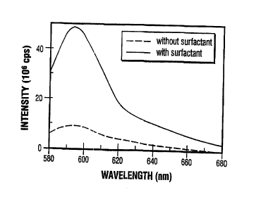
2o the presence (solid line) and absence (dashed line) of a
surfactant.
Fig. 8 is a schematic drawing of a device using
hydrostatic pressure to enhance delivery of a compound
through the stratum corneum following the application of
2s an impulse transient.
Detailed Description
The invention provides new methods for delivering
compounds, e.g., pharmaceutical compounds, through
multiple cellular layers of epithelial tissue of a person
30 or animal using impulse transients. Impulse transients
induce a transient increase in the permeability of the
epithelial tissue layer, thereby increasing diffusion of
a compound from an exterior region of the epithelial
tissue layer, through the epithelial tissue.
CA 02272647 1999-OS-20
WO 98/23325 PCT/US97/22166
_ g _
Prior to exposure to an impulse transient, an
epithelial tissue layer, e.g., the stratum corneum or a
mucosal layer, is likely impermeable to a foreign
compound; this prevents diffusion of the compound into
cells underlying the epithelial layer. Exposure of the
epithelial layer to the impulse transients enables the
compound to diffuse through the epithelial layer. The
rate of diffusion, in general, is dictated by the nature
of the impulse transients and the size of the compound to
1o be delivered.
The rate of penetration through specific
epithelial tissue layers such as the stratum corneum of
the skin also depends on several other factors including
pH, the metabolism of the cutaneous substrate tissue,
pressure differences between the region external to the
stratum corneum, and the region internal to the stratum
corneum, as well as the anatomical site and physical
condition of the skin. In turn, the physical condition
of the skin depends on health, age, sex, race, skin care,
2o and history, for example, prior contacts with organic
solvents or surfactants.
The amount of compound delivered through the
epithelial tissue layer will also depend on the length of
time the epithelial layer remains permeable, and the size
of the surface area of the epithelial layer which is made
permeable.
Properties of Impulse Transients
The properties and characteristics-of impulse
transients are controlled by the energy source used to
3o create them. However, their characteristics are modified
by the linear and non-linear properties of the coupling
medium through which they propagate. The linear
attenuation caused by the coupling medium attenuates
predominantly the high frequency components of the
CA 02272647 1999-OS-20
WO 98/23325 PCT/US97/22166
_ g -
impulse transients. This causes the bandwidth to
decrease with a corresponding increase of the rise time
of the impulse transient. The non-linear properties of
the coupling medium, on the other hand, cause the rise
s time to decrease. The decrease of the rise time is the
result of the dependence of the sound and particle
velocity on stress (pressure). As the stress increases,
the sound and the particle velocity increase as well.
This causes the leading edge of the impulse transient to
io steepen.
The relative strengths of the linear attenuation,
non-linear coefficient, and the peak stress determine how
long the wave has to travel for the rise time steepening
to become substantial. This distance can be calculated
is from the theory of non-linear acoustics (Lyamshev Sov.
Phys. Usp., 24:977, 1981).
For a planar impulse transient, the distance (L)
travelled through the coupling medium that leads to non-
linear distortions is given by equation (1) (Lyamshev
20 Sov. Phys. Usp. 24:977; 1981):
lpc2
L = --- (1)
sP
where 1 is the spatial width of the rise time (temporal
2s rise time multiplied by the sound velocity), p the
density of the medium, c the sound velocity, s the non-
linear coefficient and P the peak stress or pressure. If
the coupling medium is water, for example, an impulse
transient with a temporal rise time of 20 ns and peak
3o pressure of 500 bar will show significant steepening
within a propagation distance of about 1.5 mm.
CA 02272647 1999-OS-20
WO 98/23325 PCT/US97/22166
- 10 -
The steepening can be calculated from equation (2):
pcv
a = --- (a)
eP
where d is the width of the rise time and v the
dissipative coefficient (defined by equation 3):
f 2c31
v=I-- I (3)
t ~PJ
1o where o is the frequency of the peak stress or pressure,
and a is the absorption coefficient at frequency W.
The rise time, magnitude, and duration of the
impulse transient are chosen to create a non-destructive
(i.e., non-shock wave) impulse transient that temporarily
1s increases the permeability of the epithelial tissue
layer. Equations 1, 2, and 3, described above, can be
used for calculating the parameters from published values
for different coupling media. Generally, the rise time
is at least 1 ns, and is more preferably about 10 ns.
2o The peak stress or pressure of the impulse
transients varies for different epithelial tissue or cell
layers. For example, to transport compounds through the
stratum corneum, the peak stress or pressure of the
impulse transient should be set to at least 400 bar; more
25 preferably at least 1,000 bar, but no more than about
2,000 bar.
For epithelial mucosal layers, the peak pressure
should be set to between 300 bar and 800 bar, and is
preferably between 300 bar and 600 bar.
3o The impulse transients preferably have durations
on the order of a few tens of ns, and thus interact with
the epithelial tissue for only a short period of time.
CA 02272647 1999-OS-20
WO 98/23325 PCT/US97/22166
- il -
Following interaction with the impulse transient, the
epithelial tissue is not permanently damaged, but remains
permeable for up to about three minutes.
In addition, the new methods involve the
s application of only a few discrete high amplitude pulses
to the patient. The number of impulse transients
administered to the patient is typically less than 100,
more preferably less than 50, and most preferably less
than 10. If multiple optical pulses are used to generate
1o the impulse transient, the time duration between
sequential pulses is 10 to 120 seconds, which is long
enough to prevent permanent damage to the epithelial
tissue.
Properties of impulse transients can be measured
is using methods standard in the art. For example, peak
stress or pressure, and rise time can be measured using a
polyvinylidene fluoride (PVDF) transducer method as
described in Doukas et al., Ultrasound Med. Biol., 21:961
(1995).
2o Generation of Impulse Transients
Impulse transients can be generated by various
energy sources. For example, impulse transients can be
generated by ablation or thermoelastic expansion of an
appropriate target material by a high energy optical
2s source such as a laser {Doukas et al., Physical
Characteristics and Biological Effects of Laser-Induced
Stress Waves, Ultrasound in Med. & Biol., 22:151-164,
1996). When impulse transients are generated by laser,
they can be referred to as laser stress waves.
3o The efficiency of conversion of laser energy to
mechanical energy of the impulse transient is given by
the coupling coefficient of the target material. The
coupling coefficient (Cm) is defined as the total
momentum transfer to the target material during ablation
CA 02272647 1999-OS-20
WO 98/23325 PCT/US97/22166
- 12 -
divided by the pulse energy. The physical phenomenon
responsible for launching the impulse transient is, in
general, chosen from three different mechanisms: (1)
thermoelastic generation; (2) optical breakdown; or (3)
ablation.
For example, the impulse transients can be
initiated by applying a high energy laser source to
ablate a target material, and the impulse transient is
then coupled to an epithelial tissue or cell layer by a
io coupling medium. The coupling medium can be, for
example, a liquid or a gel, as long as it is non-linear.
Thus, water, oil such as castor oil, an isotonic medium
such as phosphate buffered saline (PBS), or a gel such as
a collagenous gel, can be used as the coupling medium.
1s The coupling medium can in addition include a
surfactant that enhances transport, e.g., by prolonging
the period of time in which the stratum corneum remains
permeable to the compound following the generation of an
impulse transient. The surfactant can be, e.g., ionic
2o detergents or nonionic detergents and thus can include,
e.g., sodium lauryl sulfate, cetyl trimethyl ammonium
bromide, and lauryl dimethyl amine oxide.
The absorbing target material acts as an optically
triggered transducer. Following absorption of light, the
2s target material undergoes rapid thermal expansion, or is
ablated, to launch an impulse transient. Typically,
metal and polymer films have high absorption coefficients
in the visible and ultraviolet spectral regions.
Many types of materials can be used as the target
3o material in conjunction with a laser beam, provided they
fully absorb light at the wavelength of the laser used.
The target material can be composed of a metal such as
aluminum or copper; a plastic, such as polystyrene, e.g.,
black polystyrene; a ceramic; or a highly concentrated
3s dye solution. The target material must have dimensions
CA 02272647 1999-OS-20
WO 98/23325 PCT/US97/22166
- 13 -
larger than the cross-sectional area of the applied laser
energy. In addition, the target material must be thicker
than the optical penetration depth so that no light
strikes the surface of the skin. The target material
must also be sufficiently thick to provide mechanical
support. When the target material is made of a metal,
the typical thickness will be 1/32 to 1/16 inch. For
plastic target materials, the thickness will be 1/16 to
1 / 8 inch .
1o Impulse transients can be also enhanced using
confined ablation. In confined ablation, a laser beam-
transparent material, such as a quartz optical window, is
placed in close contact with the target material.
Confinement of the plasma created by ablating the target
material by using the transparent material increases the
coupling coefficient by an order of magnitude (Fabro et
al., J. Appl. Phys., 68:775, 1990). The transparent
material can be quartz, glass, or transparent plastic.
Since voids between the target material and the
2o confining transparent material allow the plasma to
expand, and thus decrease the momentum imparted to the
target, the transparent material is preferably bonded to
the target material using an initially liquid adhesive,
such as carbon-containing epoxies, to prevent such voids.
The laser beam can be generated by standard
optical modulation techniques known in the art, such as
by employing Q-switched or mode-locked lasers using, for
example, electro or acousto-optic devices. Standard
commercially available lasers that can operate in a
3o pulsed mode in the infrared, visible, and/or infrared
spectrum include Nd:YAG, Nd:YLF, C02, excimer, dye,
Ti: sapphire, diode, holmium (and other rare-earth
materials), and metal-vapor lasers. The pulse widths of
these light sources are adjustable, and can vary from
several tens of picoseconds (ps) to several hundred
CA 02272647 1999-OS-20
WO 98/23325 PCT/US97/22166
- 14 -
microseconds. For use in the new methods, the optical
pulse width can vary from 100 ps to about 200 ns and is
preferably between about 500 ps and 40 ns.
Impulse transients can also be generated by
s extracorporeal lithotripters (one example is described in
Coleman et al., Ultrasound Med. Biol., 15:213-227, 1989-)-.
These impulse transients have rise times of 30 to 450 ns,
which is longer than laser-generated impulse transients.
To form an impulse transient of the appropriate rise time
1o for the new methods using an extracorporeal lithotripter,
the impulse transient is propagated in a non-linear
coupling medium (e. g., water) for a distance determined
by equation (1), above. For example, when using a
lithotripter creating an impulse transient having a rise
15 time of 100 ns and a peak pressure of 500 barr, the
distance that the impulse transient should travel through
the coupling medium before contacting an epithelial cell
layer is approximately 5 millimeters (mm).
An additional advantage of this approach for
2o shaping impulse transients generated by lithotripters is
that the tensile component of the wave will be broadened
and attenuated as a result of propagating through the
non-linear coupling medium. This propagation distance
should be adjusted to produce an impulse transient having
25 a tensile component that has a pressure of only about 5
to 10% of the peak pressure of the compressive component
of the wave. Thus, the shaped impulse transient will not
damage tissue.
The type of lithotripter used is not critical.
3o Either a electrohydraulic, electromagnetic, or
piezoelectric lithotripter can be used.
The impulse transients can also be generated using
transducers, such as piezoelectric transducers.
Preferably, the transducer is in direct contact with the
35 coupling medium, and undergoes rapid displacement
CA 02272647 1999-OS-20
WO 98/23325 PCT/US97/22166
- 15 -
following application of an optical, thermal, or electric
field to generate the impulse transient. For example,
dielectric breakdown can be used, and is typically
induced by a high-voltage spark or piezoelectric
transducer (similar to those used in certain
extracorporeal lithotripters, Coleman et al., Ultrasound
Med. Biol., 15:213-227, 1989). In the case of a
piezoelectric transducer, the transducer undergoes rapid
expansion following application of an electrical field to
to cause a rapid displacement in the coupling medium.
In addition, impulse transients can be generated
with the aid of fiber optics. Fiber optic delivery
systems are particularly maneuverable and can be used to
irradiate target materials located adjacent epithelial
tissue layers to generate impulse transients in hard-to-
reach places. These types of delivery systems, when
optically coupled to lasers, are preferred as they can be
integrated into catheters and related flexible devices,
and used to irradiate most organs in the human body. In
2o addition, to launch an impulse transient having the
desired rise times and peak stress, the wavelength of the
optical source can be easily tailored to generate the
appropriate absorption in a particular target material.
Delivery of Compounds Usinq Impulse Transients
Because impulse transients exert physical forces
to increase the permeability of the epithelial tissue,
they can be used to transport many different types of
compounds. Thus, chemotherapeutic agents such as
cisplatin, polypeptides, such as antibodies, nucleic
3o acids, such as oligonucleotides, DNA, RNA, and plasmids,
local anesthetics, such as lidocaine and benzocaine, and
photosensitizers, such as benzoporpherene derivative
monoacid ring A (BPD-MA), all can be delivered through
epithelial tissue layers, e.g., transdermally, using
CA 02272647 1999-OS-20
WO 98/23325 PCT/US97/22166
- 16 -
impulse transients.' The compounds may optionally be
heated prior to generation of the impulse transient to
facilitate their transport through the skin.
Localization of the compound using the methods of
the invention is advantageous, as it allows impulse
transients to be administered with highly localized
effects to areas of diseased cells, thus sparing the
other tissues of the body. In this way, healthy tissues
and organs are spared from adverse effects of a
to systemically administered drug.
Compounds which have a toxic effect at higher
dosages can be administered to a patient using guidelines
for administration that will produce greater
concentrations of the drugs in the treated tissues or
cells. compared to the surrounding tissues, while
maintaining adequate levels of the drug in these treated
tissues or cells. In general, this differential drug
localization can be achieved using guidelines for
administration determined using standard techniques known
2o in the field of pharmacology. Preferably, the compound
dosage and time course are such that a 2:1 or greater
concentration ratio is achieved in the treated tissues or
cells compared to the surrounding, untreated tissues.
Determining the appropriate dosage for a specific
2s compound, and for a particular subject or patient (human
or animal) is a routine matter to one skilled in the art
of pharmaceutical administration. Two approaches are
commonly used to assay directly the quantity of drug in
the diseased (treated) and surrounding tissues. First,
3o tissue samples are obtained from animals (e.g., pigs) or
patients who have been treated with different dosage and
timing protocols. Cadaver skin samples can also be used
in this assay. The quantity of drug in each tissue is
then measured either chemically, or if there is a unique
3s optical signal such as fluorescence, then by quantitative
CA 02272647 1999-OS-20
WO 98/23325 PCT/US97/22166
- 17 -
microscopy or laser-induced fluorescence. The results
are tabulated to determine a scale of optimum drug
dosages and types of impulse transients for a given
epithelial tissue layer, body region, and compound.
The compound or compounds to be delivered through
an epithelial cell layer are administered by mixing the
compound with the coupling medium, and applying the
coupling medium-compound mixture to the surface of the
epithelial cell layer, e.g., the stratum corneum, in the
1o region in which transport is desired. The compound must
be thoroughly dispersed in, and is preferably dissolved
in, the coupling medium. Thus, hydrophilic compounds can
be mixed with an aqueous coupling medium, and hydrophobic
compounds can be mixed with an oil-based coupling medium.
1s Once the target material and coupling medium in a
container are set in position on a particular region of
the surface of an epithelial tissue layer, impulse
transients are used to permeabilize the epithelial tissue
layer in the region in which the coupling medium directly
2o contacts the cell layer, using the methods described
herein. The methods result in the delivery of the
compounds to the cells underlying the epithelial tissue
layer in the region of interest that normally would not
cross the epithelial tissue layer barrier.
2s Hydrostatic pressure can be used in conjunction
with impulse transients to enhance the transport of a
compound through the epithelial tissue layer. Since the
effects induced by the impulse transients last for
several minutes, the transport rate of a drug diffusing
3o passively through the epithelial cell layer along its
concentration gradient can be increased by applying
hydrostatic pressure on the surface of the epithelial
tissue layer, e.g., the stratum corneum of the skin,
following application of the impulse transient. This
CA 02272647 1999-OS-20
WO 98/23325 PCT/iiS97/22166
- 18 -
method is described in further detail in the examples
below. The hydrostatic medium can be any liquid, such as
water or phosphate buffered saline.
Topical application and delivery of compounds by
the new methods allow the compounds to be localized to a
site of interest. Thus, the compound, e.g., a drug, is-
more concentrated at the site of action aad has a
minimal, if any, systemic concentration. This enhances
the therapeutic effect of the drug and simultaneously
1o minimizes systemic side-effects. Another advantage
compared to systemic administration is that compounds
transported through epithelial tissue bypass systemic
deactivation or degradation (e. g., hepatic "first-pass"
effects). Gastrointestinal incompatibility and potential
toxicological risks are also minimized relative to
systemic administration. In addition, drugs developed
for topical application can be designed so that they are
deactivated systematically (i.e., the "soft drug"
concept), using standard techniques. Topical
2o administration may also be desired when the compound is
rare or expensive.
Examples
The following examples are used to describe the
delivery of compounds using impulse transients.
Example 1. Transdermal Delivery of ALA
5-aminolevulenic acid (ALA) was used as a compound
to demonstrate the permeation effect of impulse
transients on the stratum corneum. ALA is converted in
cells to protoporphyrin IX, which fluoresces at 634 nm
(405 nm excitation), while ALA does not fluoresce. Thus,
the transport of ALA can be followed, non-invasively, by
monitoring the fluorescence of the skin. In addition,
since the conversion of ALA to protophyrin IX requires
CA 02272647 1999-OS-20
WO 98/23325 PCTIUS97/22166
- 19 -
that cells be viable, the measurement of protophyrin IX
fluorescence also assays cell viability in vivo.
For these experiments, a Q-switched solid state
ruby laser (20 ns pulse duration, capable of generating
up to 2 joules per pulse) was used to generate the laser
beam, which hit the target material (black polystyrene
sheet about 1 mm thick) and generated a single impulse
transient. Impulse transients of up to 1000 bar peak
stress and 50 ns duration and with a 1/2 inch beam
1o diameter can be generated with this laser-target system.
The large target ensures that the impulse transients
generated are plane waves, because the thickness of the
coupling medium is much shorter than the diameter of the
impulse transient. An articulating arm was used and the
laser path was totally covered for safety.
A plastic (flexible) washer approximately 1 inch
in diameter and 1/16 inches thick was used as a reservoir
for the sample solution (5~ concentration of the ALA in
PBS coupling medium) to be delivered through the stratum
2o corneum. The washer was attached onto the skin with
grease. The sample filled the central opening of the
washer, which was approximately 1/4 inch in diameter.
The target material was positioned on top of the washer
and irradiated with 1 laser pulse.
The black polystyrene target completely absorbed
the laser radiation so that the skin was exposed only to
impulse transients, and not laser radiation. The impulse
transients, even at the highest peak stress of 1,000 bar,
did not produce any pain in the subject. After exposure
3o to the impulse transients, the excess ALA solution was
removed. The skin was monitored for fluorescence of
protophyrin IX thirty minutes after exposure to the
impulse transients. The fluorescence intensity increased
for approximately 4 hours, at which point it reached the
s5 maximum intensity and subsequently decreased.
CA 02272647 1999-OS-20
WO 98123325 PCT/ITS97/22166
- 20 -
Fig. 1 shows the change of fluorescence intensity
at different wavelengths over time after the application
of a single impulse transient. As shown in the graph,
the peak in intensity occurs at about 640 nm and is
s highest after 210 minutes (dashed line) post-treatment.
Fig. 2 shows the fluorescence from an adjacent
site (control) where ALA. was applied without any impulse
transients. As shown in this graph, there is little
change in the intensity at different time points.
1o Fig. 3 shows the effects of varying the applied
peak stress of the impulse transient on ALA transport.
As shown in the graph, the degree of permeabilization of
the stratum corneum depends on the peak stress. In three
separate experiments, a single impulse transient was
is applied at 500 mJ, 600 mJ, or 1 J to generate applied
peak stresses of 300 bar, 400 bar, and 600 bar,
respectively. Fig. 3 shows that protophyrin IX
fluorescence increased with increasing peak pressure,
demonstrating that transdermal transport of ALA increases
2o with increasing peak stress. The onset of the
permeabilization of the stratum corneum was observed
above 300 bar.
The permeabilization of the stratum corneum is
transient. When sites on the stratum corneum were
2s exposed first to impulse transients and ALA was then
applied on the same sites after 5 minutes, no
fluorescence emission from protophyrin IX was observed.
Therefore, the permeabilization of the stratum corneum
lasted less than 5 minutes.
3o Penetration of ALA through the skin (without the
action of impulse transients) depends on many conditions,
such as skin hydration, skin temperature, anatomical
site, condition of skin and contact time. All
fluorescence measurements were compared to the
CA 02272647 1999-OS-20
WO 98/23325 PCT/CTS97122166
- 21 -
fluorescence emission of the target site on the stratum
corneum before the experiments.
Example 2. Transdermal Delivery of 40 kDa Dextran and 20
nm Latex Particles
s The ability of impulse transients to deliver large
macromolecules across the stratum corneum was determined
using probes of rhodamine B dextran having a molecular
weight of 40 kDa and a diameter of about 8.8 nm, and
fluorescent latex particles 20 nm in diameter.
io Ten-week old male fuzzy rats, each having a mass
of 300-400 g, were obtained from Iiarlan-Sprague-Dawley
(Indianapolis, IN) and acclimated for a minimum of 48
hours prior to use. Animals were anesthetized by
intramuscular injection of ketamine (120 mg/kg), xylazine
is (20 mg/kg), and atropine (0.04 mg/kg).
A single laser pulse was delivered to the target
material, which generated a single impulse transient.
Aqueous probe solutions of 500 ~M rhodamine B dextran of
40 kDA molecular weight (Molecular Probes, Eugene, OR) or
20 2% (weight/volume) fluorescent latex particles, 20 nm in
diameter (Molecular Probes, Eugene, OR) were allowed to
remain in contact with the skin for five minutes after
the application of the impulse transient. Subsequently,
the solution was removed and the surface of the skin was
2s cleaned with water. Control sites adjacent to the sites
exposed to impulse transients were treated with the donor
solution in an identical manner except that they were not
exposed to impulse transients. In addition, some control
sites were exposed to a impulse transient using sterile
3o water only as the coupling medium.
A flexible washer approximately 19 mm in diameter
was used as a reservoir for the donor solution to be
delivered through the stratum corneum. The washer was
attached on the skin on the dorsal side of each rat with
3s grease, and a black polystyrene target material was
CA 02272647 1999-OS-20
WO 98/23325 PCT/US97/22166
- 22 -
placed on top of the washer in contact with the solution.
The solution acted as the acoustic coupling medium.
Impulse transients were generated by ablation of
the target material (Perri, Phys. Fluids 16:1435-1440,
s 1973) with a 23 nsec pulse from a Q-switched ruby laser
and launched into the reservoir containing the molecular
probe solution. An articulated arm was used to deliver
the beam to the target. The beam size at the target was
about 6 mm in diameter to achieve a fluence of about 7
to J/cm2. The laser pulse was completely absorbed by the
target so that only the impulse transient propagated
through the probe solution and impinged onto the skin of
the rat. The impulse transients were measured in
separate experiments under identical conditions of laser
15 parameters, target material, and propagation distance
through the coupling medium with a calibrated
polyvinylidene fluoride transducer (Doukas et al.
Ultrasound Med. Biol. 21:961-967, 1995).
The temporal profile of the impulse transients
2o used in these experiments is shown in Fig. 4. The peak
stress in the skin (PS) was calculated from the peak
pressure in water (PW) and the acoustic impedance of
water (ZW = 1.48x10⁶ kgiri 2s-1) and skin (ZS = 1.54x10⁶ kcpri
2s-1) (Payne et al., Sound Skin Models-Acoustic Properties
2s of Epidermis and Dermis. In Skin Models To Study
Function and Disease of Skin. Parks et al., ed., Springer
Verlag, Berlin, pp. 402-411; 1986) using the equation
PS/PW =2Zw/(ZSZW). The peak stress on the skin in all
experiments was calculated to be 589 ~ 23 bar.
3o The delivery of the dextran and latex beads across
the stratum corneum following the generation of impulse
transients was observed using transmission
photomicrographs and fluorescence emission spectra of
biopsy samples. For these studies, skin samples were
CA 02272647 1999-OS-20
WO 98/23325 PCT/US97/22166
- 23 -
obtained one hour post-treatment using a 6 mm biopsy
punch. Biopsies were embedded in OCT 4583' (Sakura
Finetek USA, Torrence, California) and frozen. The skin
samples were then sectioned in a cryostat microtome, and
s microphotographs were obtained with a Zeiss inverted
microscope using a Rhodamine B filter set (XF39, Omega
Optical, Brattleboro, Vermont). Fluorescence emission
spectra of the exposed and control sites were collected
from another group of animals while they were alive and
io under full anesthesia using a fiber-based fluorimeter
(FLUORMAX'", Spex Industries, Edison, NJ).
Transmission photomicrography revealed that
rhodamine B dextran penetrated to a depth of
approximately 50 ~m into the skin. Fluorescence spectra
is also demonstrated that the rhodamine dextran penetrated
the stratum corneum following induction of an impulse
transient. The fluorescence emission spectra of skin
exposed to a single impulse transient in the presence of
40 kDa dextran is shown in Fig. 5A. Emission spectra
2o were taken at three different times: (1) before
application of the dextran probe and generation of the
impulse transient, in order to establish the baseline
fluorescence (shown as the dashed line marked
"baseline"), (2) immediately after generation of the
2s impulse transient (shown as the broken line labeled
"after LSW," for laser stress wave), and (3) after the
stratum corneum of the exposed site was removed by tape
stripping (shown as the solid line marked "SC removed").
Tape stripping was performed to eliminate_the
3o fluorescence from the probe molecules located in the
stratum corneum. Thus, the fluorescence signal in the
tape stripping experiment represented only the probe
molecules located in the viable epidermis and dermis.
Twenty tape strippings were sufficient to remove the
35 stratum corneum (Wells, Br. J. Dermatol. 108:87-91,
CA 02272647 1999-OS-20
WO 98/23325 PCT/CJS97122166
- 24 -
1957). The data shown in Fig. 5A represent raw
fluorescence.
The spectra shown in Fig. 5A indicate that
rhodamine-associated fluorescence increased following
s delivery of an impulse transient to skin exposed to the
40 kDa rhodamine dextran probe (spectra labeled "after
LSW"). Most of this fluorescence remained after removal
of the stratum corneum (compare the spectra labeled "SC
removed" with "after LSW"). Both of these spectra show
io significantly higher intensity than that shown by the
baseline spectra. In addition, exposure of skin to
impulse transients only did not induce any change in the
fluorescence emission of the skin (data not shown).
These data suggest that application of an impulse
is transient (in the form of a laser stress wave) caused the
40 kDa rhodamine dextran probe to be transported into the
dermis, i.e., to be localized in tissues that are not
sensitive to procedures that remove the stratum corneum.
Fig. 5B shows the comparative fluorescence of
2o sites exposed to the LSW (+LSW) and control sites (-LSW)
after tape stripping and after the baseline fluorescence
has been subtracted. The site subjected to an impulse
transient showed over two-fold higher rhodamine
associated fluorescence than the control site, which also
25 demonstrates that impulse transients promote transport of
the dextran probe across the stratum corneum.
Latex fluorescent particles of 10 nm diameter were
also delivered through the stratum corneum using impulse
transients. Fig. 6 shows the fluorescence emission
3o spectra after tape stripping of a site exposed to a
impulse transient using latex particles as the
fluorescent probe. The fluorescence emission of the
control site under identical conditions is shown for
comparison.
CA 02272647 1999-OS-20
WO 98/23325 PCT/US97I22166
- 25 -
To measure the amount of time required for the
stratum corneum to regain its barrier function for a
probe molecule having the size of 40 kDa dextran, a
single impulse transient was applied to skin using
sterile water as the acoustic coupling medium. The
coupling medium was immediately removed, and the probe
solution added to the reservoir 2 minutes after the
application of the impulse transient. The fluorescence
emission spectra were then measured as described above.
io No fluorescence was detected. This indicates that the
stratum corneum becomes impermeable to 40 kDa dextran
within 2 minutes after the generation of the impulse
transient.
Example 3. Transdermal Delivery Using Surfactants in the
Coupling Medium
The effect of a surfactant was examined by using a
solution of 2% sodium lauryl sulfate (SLS) as the
coupling medium. Fig. 7 shows the fluorescence spectra
(from which baseline fluorescence had been subtracted) of
2o the 40 kDa dextran probe molecule in skin at two sites at
which an impulse transient had been generated. At one
site (solid line), 2~ SLS was used as the coupling
medium; at the other, no SLS was added (dashed line).
The two skin sites were tape stripped prior to generating
the two spectra shown in Fig. 7
A comparison of the spectra shown in Fig. 7
reveals that the fluorescence intensity of 40 kDa dextran
was approximately 8-9 fold greater when 2~ SLS was used
in the coupling medium. Thus, a surfactant can
3o significantly increase the amount of an agent delivered
across the stratum corneum using an impulse transient.
To determine if surfactants in the coupling medium
affect transport by increasing the time of recovery of
the barrier function of the stratum corneum, the length
of time to restore the barrier function of the stratum
. _. u.~4_ _. _ _ .~.~ .~._.~~... ._.._._...~ _...~_ __.._..,_
CA 02272647 1999-OS-20
WO 98/23325 PCT/IJS97/22166
- 26 -
corneum was compared using water or an aqueous solution
of 2% sodium lauryl sulfate (SLS) as the coupling medium.
As discussed above in Example 2, the stratum
corneum becomes impermeable to the 40 kDa dextran probe
within two minutes after generation of an impulse
transient. To determine the length of time the stratum
corneum remains permeable when impulse transients are
generated in the presence of a surfactant, a single pulse
was applied in which the initial coupling medium was an
1o aqueous solution of 2% SLS. The surfactant was removed,
and the aqueous solution of the 40 kDa dextran probe was
added 15, 30, 45, and 60 minutes after generation of the
impulse transient. The presence of the probe was then
measured.
1s Probe molecules added as long as 45 to 60 minutes
after generation of the impulse transient emitted
fluorescence that was resistant to procedures that remove
the stratum corneum. These observations indicate that
when the surfactant was used in the coupling medium, the
2o recovery of the barrier function of the stratum increased
to 45-60minutes. This compares to recovery of the
barrier function within 2 minutes without the surfactant.
Surfactants therefore can act to increase the time
required for the stratum corneum to regain its barrier
25 function.
Example 4. Transdermal Delivery of Anti-neoplastia
Agents
5-fluorouracil (5-FU) is dissolved in an aqueous
solution of PBS, which serves as the coupling medium, and
so applied to a skin cancer lesion in a suitable container
as described in Example 1. A black polystyrene sheet is
used as the target material and is placed on the
container in direct contact with the coupling medium,
which in turn transmits the impulse transients to the
CA 02272647 1999-OS-20
WO 98/23325 PCT/US97/22166
- 27 -
surface of the lesion. Five pulses from a Q-switched
solid state ruby laser (1 J, 20 ns pulse duration) are
applied to generate 5 separate impulse transients. The
amount of 5-FU dissolved in the PBS is determined using
standard techniques described herein and based on the
nature of the lesion, the desired drug concentration in
the lesion, and the patient s skin type.
Example 5. Transdermal Delivery of ALA Using Impulse
Transients and Confined Ablation
io In confined ablation, a transparent material is
placed in close contact with the target material.
Confinement of the plasma increases the coupling
coefficient by an order of magnitude.
A quartz optical window with a thickness of 3/8
inch is used as the transparent material, and a black
polystyrene sheet is used as the target material. To
eliminate microscopic voids, a solvent is used to
dissolve the surface of the polystyrene to allow it to
bond to the quartz transparent material. This combined
2o transparent material and target material is used in the
same way as the target material in Example 1.
Plasma confinement causes an increase in the rise
time of the impulse transient, which may decrease the
effectiveness of the impulse transient. To counteract
this effect, the distance the impulse transient
propagates through the coupling medium is increased to
shape the impulse transient to have an appropriate rise
time when it contacts the surface of the epithelial
tissue layer. The non-linear properties of the coupling
3o medium cause the rise time to decrease. An impulse
transient of 500 bar propagating through 1 mm of water
undergoes a decrease in the rise time from 30 ns to 15
ns. Therefore, the initial increase of the rise time due
to confinement can be compensated by appropriately
CA 02272647 1999-OS-20
WO 98/23325 PCT/LTS97/22166
- 28 -
adjusting the propagation length of the impulse transient
through the reservoir.
The impulse transient is measured in separate
experiments under identical conditions using a calibrated
s polyvinylidene fluoride transducer as described in Doukas
et al., Ultrasound Med. Biol., 21:961 (1995). The
temporal resolution of the combination of the transducer
and oscilloscope is 5 ns. The pressure in the skin PS is
calculated from the pressure in Water Pw and the acoustic
1o impedances of water (ZW) and skin (ZS) using the equation
PS/PW = 2 ZW/ ( ZS+ZW)
Impulse transients are generated by using one
pulse of a Q-switched ruby laser (4J, 30 ns). An ArF
excimer laser can also be used (650 mJ, 25 ns).
1s Transcutaneous delivery of ALA is measured by measuring
protophyrin IX fluorescence as described above.
Example 6. Transdermal Delivery of Benzoporpherene
Derivative Monoaaid Ring A (BPD-MA) Using
Impulse Transients and Hydrostatic Pressure
2o Fig. 8 shows a schematic diagram of a device 10
that is used to control drug delivery by varying the
hydrostatic pressure. A reservoir 12 is made from a
plastic washer 14 about 3 mm in height and 1 cm in
diameter and is attached to rabbit skin 16 with silicon
2s grease. An outlet 20 that connects to a groove 18 in the
bottom of the washer is connected to a suction pump (not
shown). This allows the washer to remain firmly vacuum
sealed on the skin during the application of hydrostatic
pressure. A black polystyrene target material 22 is
3o attached to the surface of the washer 14, and a quartz
overlay 23 is placed on the target material. Laser
radiation 24 from Q-switched ruby laser (4J, 30 ns) 26 is
directed onto the target material 22 using an
articulating arm (not shown). A tube 28 is connected to
CA 02272647 1999-OS-20
WO 98/23325 PCT/HS97/22166
- 29 -
an opening 21 in the side of the washer 14. The tube 28
is connected to a pressure regulator 30, and the
reservoir 12 is filled with a solution of benzoporpherene
derivative monoacid ring A (BPD-MA).
The fur on the back of a New Zealand albino rabbit
is removed. The animal is anesthetized and the device-10
for applying hydrostatic pressure is attached to the back
of the animal. Subsequently, hydrostatic pressure is
applied and a single impulse transient is generated. The
1o hydrostatic pressure is applied for 5 minutes. The
device is then removed, the skin cleaned, and the
fluorescence is measured using a fiber-based
spectrofluorimeter (450 nm excitation, 650-750 nm
emission).
These measurements are compared to three control
sites in which BPD-MA is applied. One site is exposed to
a single impulse transient with no hydrostatic pressure
applied. The second is exposed only to hydrostatic
pressure and no impulse transient, and the third is
2o exposed to neither an impulse transient nor hydrostatic
pressure.
The fluorescence measurements indicate the amount
of BPD-MA present in the skin. To confirm these
measurements, BPD-MA is extracted from the tissue and
2s measured using standard techniques. Briefly, skin
biopsies are weighed, mixed with 1 ml dimethyl sulfoxide
(DMSO) and homogenized. The homogenized samples are kept
at room temperature overnight and then centrifuged. The
integrated fluorescence of the supernatant is measured in
3o a spectrofluorimeter and the amount of drug is estimated
from a calibration curve.
CA 02272647 1999-OS-20
WO 98/23325 PCT/LTS97/22166
- 30 -
Euample 7. Transdermal Delivery of a Compound
Using a Lithotripter
A subject places a limb having the desired target
area of skin into a water bath. The target area is
covered by a small reservoir containing a 5o ALA solution
in PBS. This reservoir differs from others described
above in that it is covered on the side opposite the skin
with a thin plastic film or membrane that has an
impedance near that of water, i.e., it is designed not to
1o reflect impulse transients generated by the lithotripter
that propagate first through the water and then through
the reservoir to reach the target epithelial layer. The
ALA-PBS solution in the reservoir serves as both a source
of the compound (ALA) and the coupling medium.
Ten pulses from a electrohydraulic lithotripter
are applied in the water bath to generate ten impulse
transients. The rise times and peak stress are adjusted
to be about 5 to 15 ns and 500 bar, respectively, at the
point of contact with the skin, following propagation
2o through the water bath and the coupling medium.
Transdermal delivery of ALA is determined by
measuring protophyrin IX fluorescence as described above.
CA 02272647 1999-OS-20
WO 98/23325 PCT/US97/22166
- 31 -
Other Embodiments
It is to be understood that while the invention
has been described in conjunction with the detailed
description thereof, that the foregoing description is
s intended to illustrate and not limit the scope of the
invention, which is defined by the scope of the appended
claims. Other aspects, advantages, and modifications are
within the scope of the following claims.