Note: Descriptions are shown in the official language in which they were submitted.
CA 02294981 2007-04-04
WO 99/00113 PCT/US90/13272
1
NOVEL FORMULATIONS OF PHARMACOLOGICAL AGENTS.
METHODS FOR THE PREPARATION THEREOF AND
METHODS FOR THE USE THEREOF
FIELD OF THE INVENTION
The present invention relates to methods for the
production of particulate vehicles for the intravenous
administration of pharmacologically active agents, as well as
novel compositions produced thereby. In a particular aspect,
the invention relates to methods for the in vivo delivery of
substantially water insoluble pharmacologically active agents
(e.g., the anticancer drug Taxol ). In another aspect,
dispersible colloidal systems containing water insoluble
pharmacologically active agents are provided. The suspended
particles may be formed of 100% active agent, or may be encased
in a polymeric shell formulated from a biocompatible polymer,
and have a diameter of less than about 1 micron. Invention
colloidal systems may be prepared without the use of
conventional surfactant or any polymeric core matrix. In a
presently preferred aspect of the invention, there is provided
a method for preparation of extremely small particles which.can
be sterile-filtered. The polymeric shell contains particles of
pharmacologically active agent, and optionally a biocompatible
dispersing agent in which pharmacologically active agent can be
either dissolved or suspended. Thus, the invention provides a
drug delivery system in either liquid form or in the form of a
redispersible powder. Either form provides both immediately
bioavailable drug molecules (i.e., drug molecules which are
molecularly bound to a protein), and pure drug particles coated
with a protein.
CA 02294981 2007-04-04
WO 99/00113 PCf/U398/13272
2
The invention also relates to the method of use and
preparation of compositions (formulations) of drugs such as the
anticancer agent paclitaxel.. In one aspect, the formulation of
paclitaxel, known as Capxol!~ is significantly less toxic and
more efficacious than Taxole, a commercially available
formulation of paclitaxel. In another aspect, the novel
formulation Capxofm, localizes in certain tissues after
parenteral administration thereby increasing the efficacy of
treatment of cancers associated with such tissues.
HACKGROUND OF THE INVENTION
Intravenous drug delivery permits rapid and direct
equilibration with the blood stream which carries the
medication to the rest of the body. To avoid the peak serum
levels which are achieved within a short time after
intravascular injection, administration of drugs carried within
stable carriers would allow gradual release of the drugs inside
the intravascular compartment following a bolus intravenous
injection of the therapeutic nanoparticles.
Injectable controlled-release nanoparticles can
provide a pre-programmed duration of action, ranging from days
to weeks to months from a single injection. They also can
offer several profound advantages over conventionally
administered medicaments, including automatic assured patient
compliance with the dose regimen, as well as drug targeting to
specific tissues or organs (Tice and Gilley, Journal of
Controlled Release 2:343-352 (1985)).
CA 02294981 2007-04-04
WO 99/00113 PCT/US98/13272
3
Microparticles and foreign bodies present in the
blood are generally cleared from the circulation by the "blood
filtering organs", namely the spleen, lungs and liver. The
particulate matter contained in normal whole blood comprises
red blood cells (typically 8 microns in diameter), white blood
cells (typically 6-8 microns in diameter), and platelets
(typically 1-3 microns in diameter). The microcirculation in
most organs and tissues allows the free passage of these blood
cells. When microthrombii (blood clots) of size greater than
10-15 microns are present in circulation, a risk of infarction
or blockage of the capillaries results, leading to ischemia or
oxygen deprivation and possible tissue death. Injection into
the circulation of particles greater than 10-15 microns in
diameter, therefore, must be avoided. A suspension of
particles less than 7-8 microns, is however, relatively safe
and has been used for the delivery of pharmacologically active
agents in the form of liposomes and emulsions, nutritional
agents, and contrast media for imaging applications.
The size of particles and their mode of delivery
determines their biological behavior. Strand et al. (in
Microspheres-Biomedical Applications, ed. A. Rembaum, pp 193-
227, CRC Press (1988)) have described the fate of particles to
be dependent on their size. Particles in the size range of a
few nanometers (nm) to 100 nm enter the lymphatic capillaries
following interstitial injection, and phagocytosis may occur
within the lymph nodes. After intravenous/intraarterial
injection, particles less than about 2 microns will be rapidly
cleared from the blood stream by the reticuloendothelial system
(RES), also known as the mononuclear phagocyte system (MPS).
CA 02294981 2007-04-04
WO 99/00113 PCT/US98/13272
4
Particles larger than about 7 microns will, after intravenous
injection, be trapped in the lung capillaries. After
intraarterial injection, particles are trapped in the first
capillary bed reached. Inhaled particles are trapped by the
alveolar macrophages.
Pharmaceuticals that are water-insoluble or poorly
water-soluble and sensitive to acid environments in the stomach
cannot be conventionally administered (e.g., by intravenous
injection or oral administration). The parenteral
administration of such pharmaceuticals has been achieved by
emulsification of the oil solubilized drug with an aqueous
liquid (such as normal saline) in the presence of surfactants
or emulsion stabilizers to produce stable microemulsions.
These emulsions may be injected intravenously, provided the
components of the emulsion are pharmacologically inert. US
Patent No. 4,073,943 describes the administration of water-
insoluble pharmacologically active agents dissolved in oils and
emulsified with water in the presence of surfactants such as
egg phosphatides, pluronics (copolymers of polypropylene glycol
and polyethylene glycol), polyglycerol oleate, etc. PCT
International Publication No. W085/00011 describes
pharmaceutical microdroplets of an anaesthetic coated with a
phospholipid such as dimyristoyl phosphatidylcholine having
suitable dimensions for intradermal or intravenous injection.
An example of a water-insoluble drug is Taxol , a
natural product first isolated from the Pacific Yew tree, Taxus
brevifolia, by Wani et al. (J. Am. Chem. Soc. 2;2325 (1971)).
Among the antimitotic agents, Taxol, which contains a diterpene
carbon skeleton, exhibits a unique mode of action on
CA 02294981 2007-04-04
WO 99/00113 PCT/US98/13272
microtubule proteins responsible for the formation
of the mitotic spindle. In contrast with other antimitotic
agents such as vinblastine or coichicine, which prevent the
assembly of tubulin, Taxol is the only plant product known to
5 inhibit the depolymerization process of tubulin, thus
preventing the cell replication process.
Taxol, a naturally occurring diterpenoid, has been
shown to have significant antineoplastic and anticancer effects
in drug-refractory ovarian cancer. Taxol has shown excellent
antitumor activity in a wide variety of tumor models such as
the B16 melanoma, L1210 leukemias, NIX-1 mammary tumors, and CS-
1 colon tumor xenografts. Several recent press releases have
termed Taxol as the new anticancer wonder-drug. Indeed, Taxol
has recently been approved by the Federal Drug Administration
for treatment of ovarian cancer. The poor aqueous solubility
of Taxol, however, presents a problem for human administration.
Indeed, the delivery of drugs that are inherently insoluble or
poorly soluble in an aqueous medium can be seriously impaired
if oral delivery is not effective. Accordingly, currently used
Taxol formulations require a cremaphor to solubilize the drug.
The human clinical dose range is 200-500 mg. This dose is
dissolved in a 1:1 solution of ethanol:cremaphor and diluted
with saline of about 300-1000 ml of fluid given intravenously.
The cremaphor currently used is polyethoxylated castor oil. The
presence of cremaphor in this formulation has been linked to
severe hypersensitivity reactions in animals (Lorenz et al.,
Agents Actions 1987, 7, 63-67) and humans (Weiss at al., J.
Clin. Oncol. 1990, 8, 1263-68) and consequently requires
premedication of patients with corticosteroids (dexamethasone)
and antihistamines. The large dilution results in large
CA 02294981 2007-04-04
WO 99/00113 PCT/US98/13272
6
volumes of infusion (typical dose 175 mg/M2) up to 1 liter
and infusion times ranging from 3 hours to 24 hours. Thus,
there is a need for an alternative less toxic formulation for
paclitaxel.
In phase I clinical trials, Taxol itself did not
show excessive toxic effects, but severe allergic reactions
were caused by the emulsifiers employed to solubilize the drug.
The current regimen of administration involves treatment of the
patient with antihistamines and steroids prior to injection of
the drug to reduce the allergic side effects of the cremaphor.
In an effort to improve the water solubility of
Taxol, several investigators have modified its chemical
structure with functional groups that impart enhanced water-
solubility. Among them are the sulfonated derivatives
(Kingston et al., U.S. Patent 5,059,699 (1991)), and amino acid
esters (Mathew et al., J. Med. Chem. U:145-151 (1992)) which
show significant biological activity. Modifications to produce
a water-soluble derivative facilitate the intravenous delivery
of Taxol dissolved in an innocuous carrier such as normal
saline. Such modifications, however, add to the cost of drug
preparation, may induce undesired side-reactions and/or
allergic reactions, and/or may decrease the efficiency of the
drug.
Protein microspheres have been reported in the
literature as carriers of pharmacological or diagnostic agents.
Microspheres of albumin have been prepared by either heat
denaturation or chemical crosslinking. Heat denatured
microspheres are produced from an emulsified mixture (e.g.,
CA 02294981 2007-04-04
WO 99/00113 PCT/US98/13272
7
albumin, the agent to be incorporated, and a suitable
oil) at temperatures between 100 C and 150 C. The microspheres
are then washed with a suitable solvent and stored. Leucuta et
al. (International Journal of Pharmaceutics 41:213-217 (1988))
describe the method of preparation of heat denatured
microspheres.
The procedure for preparing chemically crosslinked
microspheres involves treating the emulsion with glutaraldehyde
to crosslink the protein, followed by washing and storage. Lee
et al. (Science 2_11:233-235 (1981)) and U.S. Patent No.
4,671,954 teach this method of preparation.
The above techniques for the preparation of protein
microspheres as carriers of pharmacologically active agents,
although suitable for the delivery of water-soluble agents, are
incapable of entrapping water-insoluble ones. This limitation
is inherent in the technique of preparation which relies on
crosslinking or heat denaturation of the protein component in
the aqueous phase of a water-in-oil emulsion. Any aqueous-
soluble agent dissolved in the protein-containing aqueous phase
may be entrapped within the resultant crosslinked or
heat-denatured protein matrix, but a poorly aqueous-soluble or
oil-soluble agent cannot be incorporated into a protein matrix
formed by these techniques.
One conventional method for manufacturing
drug-containing nanoparticles comprises dissolving polylactic
acid (or other biocompatible, water insoluble polymers) in a
water-immiscible solvent (such as methylene chloride or other
chlorinated, aliphatic, or aromatic solvent), dissolving the
CA 02294981 2007-04-04
WO 99/00113 PCTIUS99113272
8
pharmaceutically active agent in the polymer solution, adding
a surfactant to the oil phase or the aqueous phase, forming an
oil-in-water emulsion by suitable means, and evaporating the
emulsion slowly under vacuum. If the oil droplets are
sufficiently small and stable during evaporation, a suspension
of the polymer in water is obtained. Since the drug is
initially present in the polymer solution, it is possible to
obtain by this method, a composition in which the drug
molecules are entrapped within particles composed of a
polymeric matrix. The formation of microspheres and
nanoparticles by using the solvent evaporation method has been
reported by several researchers (see, for example, Tice and
Gilley, in Journal of Controlled Release 2.:343-352 (1985);
Bodmeier and McGinity, in Int. J. Pharmaceutics Aa:179 (1988);
Cavalier et al., in J. Pharm. Pharmacol. x$:249 (1985); and
D'Souza et al., WO 94/10980) while using various drugs.
Bazile et. al., in Biomaterials la:1093 (1992), and
Spenlehauer et al., in Fr Patent 2 660 556, have reported the
formation of nanoparticles by using two biocompatible polymers,
one (e.g., polylactide) is dissolved in the organic phase,
together with an active component such as a drug, and the other
polymer, such as albumin, is used as the surface active agent.
After emulsification and removal of the solvent, nanoparticles
are formed, in which the drug is present inside the polymeric
matrix of the polylactide particles.
The properties of the polymer solution from which the
polymeric matrix is formed are very important to obtain the
proper emulsion in the first stage. For example, polylactide
(the polymer commonly used in the preparation of injectable
CA 02294981 2007-04-04
WO 99/00113 PCT/US98/13272
9
nanoparticles), has a surface activity which causes the rapid
adsorption thereof at the dichloromethane-water interface,
causing reduced interfacial tension (see, for example, Boury et
al., in Langmuir 11:1636 (1995)), which in turn improves the
emulsification process. In addition, the same researchers
found that Bovine Serum Albumin (BSA) interacts with the
polylactide, and penetrates into the polylactide monolayer
present at the oil-water interface. Therefore, it is expected,
based on the above reference, that emulsification during the
conventional solvent evaporation method is greatly favored by
the presence of the surface active polymer (polylactide) in the
nonaqueous organic phase. In fact, the presence of polylactide
is not only a sufficient condition, but it is actually
necessary for the formation of nanoparticles of suitable size.
Another process which is based on the solvent
evaporation method comprises dissolving the drug in a
hydrophobic solvent (e.g., toluene or cyclohexane), without any
polymer dissolved in the organic solvent, adding a conventional
surfactant to the mixture as an emulsifier, forming an oil-in-
water emulsion by use of sonication on high-shear equipment,
and then evaporating the solvent to obtain dry particles of the
drug (see, for example, Sjostrom et al., in J. Dispersion
Science and Technology la:89-117 (1994)). Upon removal of the
nonpolar solvent, precipitation of the drug inside the solvent
droplets occurs, and submicron particles are obtained.
It has been found that the size of the particles is
mainly controlled by the initial size of the emulsion droplets.
In addition, it is interesting to note that the final particle
size is reported to decrease with a decrease in the drug
CA 02294981 2007-04-04
wo 99/00113 PCT/US98/13272
concentration in the organic phase. This finding is
contrary to the results reported herein, wherein no
conventional surfactant is used for the preparation of
nanoparticles (in same embodiments of the invention). In
5 addition, it is noted by the authors of the Sjostrom paper that
the drug used, cholesteryl acetate, is surface active in
toluene, and hence may be oriented at the oil-water interface;
therefore the concentration of drug at the interface is higher,
thus increasing the potential for precipitation.
Formation of submicron particles has also been
achieved by a precipitation process, as described by Calvo et
al. in J. Pharm. Sci. 11:530 (1996). The process is based on
dissolving the drug (e.g., indomethacin) and the polymer (poly-
caprolactone) in methylene chloride and acetone, and then
pouring the solution into an aqueous phase containing a
surfactant (Poloxamern'" 188), to yield submicron size particles
(216 nm). However, the process is performed at solvent
concentrations at which no emulsion is formed.
Taxol is a naturally occurring compound which has
shown great promise as an anti-cancer drug. For example, Taxol
has been found to be an active agent against drug-refractory
ovarian cancer by McGuire et al. See "Taxol: A Unique Anti-
Neoplastic Agent With Significant Activity Against Advanced
Ovarian Epithelial Neoplasms." Ann. Int. Med., 111, 273-279
(1989).
CA 02294981 2007-04-04
WO 99/00113 PCT/US98/13272
11
Unfortunately, Taxol has extremely low solubility in
water, which makes it difficult to provide a suitable dosage
form. In fact, in Phase I clinical trials, severe allergic
reactions were caused by the emulsifiers administered in
conjunction with Taxol to compensate for Taxol's low water
solubility; at least one patient's death was caused by an
allergic reaction induced by the emulsifiers. Dose limiting
toxicities include neutropenia, peripheral neuropathy, and
hypersensitivity reactions.
Brown et al., in "A Phase I Trial of Taxol Given by
A 6-Hour Intravenous Infusion" J of Clin Oncol, Vol. 9 No. 7,
pp. 1261-1267 (July 1991) report on a Phase I Trial in which
Taxol was provided as a 6-hour IV infusion every 21 days
without premedication. 31 patients received 64 assessable
courses of Taxol. One patient had a severe (or acute)
hypersensitivity reaction, which required discontinuation of
the infusion and immediate treatment to save the patient's
life. Another patient experienced a hypersensitivity reaction,
but it was not so severe as to require discontinuing the
infusion. Myelosuppression was dose-limiting, with 2
fatalities due to sepsis. Non-hematologic toxicity was of
Grade 1 and 2, except for one patient with Grade 3 mucositis
and 2 patients with Grade 3 neuropathy. The neuropathy
consisted of reversible painful paresthesias, requiring
discontinuation of Taxol in two patients. Four partial
responses were seen (3 in patients with non-small-cell lung
cancer, and one in a patient with adenocarcinoma of unknown
primary). The maximum tolerated dose reported was 275 mg/m2,
and the recommended Phase II starting dose was 225 mg/m2. The
CA 02294981 2007-04-04
WO 99/00113 PCT/US9V13272
12
incidence of hypersensitivity reaction was reported to be
schedule-dependent, with 6 to 24-hour infusions of drug having
a O% to at incidence of hypersensitivity reactions. It was
also reported that hypersensitivity reactions persist with or
without premedication despite prolongation of infusion times.
Since these Phase I studies were conducted on terminally ill
patients suffering from a variety of cancers, the efficacy of
the Taxol treatments could not be determined.
In a study by Kris et al., Taxol formulated with
CremaphoTM EL in dehydrated alcohol was given as a 3-hour IV
infusion every 21 days, with the administered dosage ranging
from 15 to 230 mg/m2 in nine escalation steps. Kris et al.
concluded that "with the severity and unpredictability of the
hypersensitivity reactions, further usage of Taxol is not
indicated with this drug formulation on this administration
schedule." See Cancer Treat. Rep., Vol. 70, No. 5, May 1986.
Since early trials using a bolus injection or short
(1-3 hour) infusions induced anaphylactic reactions or other
hypersensitivity responses, further studies were carried out
in which Taxol was administered only after premedication with
steroids (such as dexamethasone), antihistamines (such as
diphenhydramine), and H2-antagonists (such as cimetidine or
ranitidine), and the infusion time was extended to 24 hours in
an attempt to eliminate the most serious allergic reactions.
Various Phase I and Phase II study results have been published
utilizing 24-hour infusions of Taxol with maximum total
dosages of 250 mg/m2, generally with the course being repeated
every 3 weeks. Patients were pre-treated with dexamethasone,
diphenhydramine, and cimetidine to offset allergic reactions.
CA 02294981 2007-04-04
WO 99/00113 PCTIUS98/13272
13
See Einzig, et al., "Phase II Trial of Taxol in Patients
with Metastatic Renal Cell Carcinoma," Cancer Investigation,
9(2) 133-136 (1991), and A. B. Miller et al., "Reporting
Results of Cancer Treatment," Cancer, Vol 47, 207-214 (1981).
Koeller et al., in "A Phase I Pharmacokinetic Study
of Taxol Given By a Prolonged Infusion Without Premedication,"
Proceedings of ASCO, Vol. 8 (March, 1989), recommends routine
premedication in order to avoid the significant number of
allergic reactions believed to be caused by the cremophor
(polyethoxylated castor oil) vehicle used for Taxol infusions.
Patients received dosages ranging from 175 mg/m2 to 275 mg/m2.
Wiernik et al. in "Phase I Clinical and
Pharmacokinetic Study of Taxol," Cancer Research, 47, 2486-
2493 (May 1, 1987), also report the administration of Taxol in
a cremophor vehicle by IV infusion over a 6-hour period in a
Phase I study. Grade 3-4 hypersensitivity reactions incurred
in 4 of 13 courses. The starting dose for the study was 15
mg/m2 (one-third of the lowest toxic dose in dogs). Doses were
escalated, and a minimum of 3 patients were treated at each
dose level until toxicity was identified, and then 4-6
patients were treated at each subsequent level. The study
concluded that neurotoxicity and leukopenia were
dose-limiting, and the recommended Phase II trial dose was 250
mg/m2 with premedication.
Other exemplary studies on Taxol include: Legha et
al., "Phase II Trial of Taxol in Metastatic Melanoma," Vol. 65
(June 1990) pp. 2478-2481; Rowinsky et al., "Phase I and
Pharmacodynamic Study of Taxol in Refractory Acute Leukemias,"
CA 02294981 2007-04-04
WO 99/00113 PCT/US98/13272
14
Cancer Research, 49, 4640-4647 (Aug. 15, 1989); Grem et al.,
"Phase I Study of Taxol Administered as a Short IV Infusion
Daily For 5 Days," Cancer Treatment Reports, Vol. 71 No. 12,
(December, 1987); Donehower et al., "Phase I Trial of Taxol in
Patients With Advanced Cancer," Cancer Treatment Reports, Vol.
71, No. 12, (December, 1987); Holmes et al., "Phase II Study
of Taxol in Patients (PT) with Metastatic Breast Cancer
(MBC)," Proceedings of the American Society of Clinical
Oncology, Vol. 10, (March, 1991), pp. 60. See also Suffness.
"Development of Antitumor Natural Products at the National
Cancer Institute," Gann Monograph or Cancer Research, 31
(1989) pp. 21-44 (which recommends that Taxol only be given as
a 24-hour infusion).
Weiss et al., in "Hypersensitivity Reactions from
Taxol," Journal of Clinical Oncology, Vol. 8, No. 7 (July
1990) pp. 1263-1268, reported that it was difficult to
determine a reliable overall incidence of hypersensitivity
reactions, HSRs, because of the wide variations in Taxol doses
and schedules used, and the unknown degree of influence that
changing the infusion schedule and using premedication has on
HSR incidents. For example, of five patients who received
Taxol in a 3-
hour infusion at greater than 190 mg/m2 with no premedication,
three had reactions, while only one out of 30 patients
administered even higher doses over a 6-hour infusion with no
premedication had a reaction. Therefore, this suggests that
prolonging the infusion to beyond 6 hours is sufficient to
reduce HSR incidents. Nevertheless, Weiss et al. found that
patients receiving 250 mg/m2 of Taxol administered via a 24-
hour infusion still had definite HSRs. Thus, while prolonging
CA 02294981 2007-04-04
WO 99/00113 PCT/US98/13272
drug infusion to 6 or 24-hours may reduce the risk for an
acute reaction, this conclusion can not be confirmed, since
78% of the HSR reactions occurred within ten minutes of
initiating the Taxol infusion, which indicates that the length
5 of time planned for the total infusion would have no bearing.
Further, concentration of Taxol in the infusion may also not
make a difference since substantial numbers of patients had
reactions to various small Taxol dosages. Finally, not only is
the mechanism of Taxol HSR unknown, it is also not clear
10 whether Taxol itself is inducing HSRs, or if the HSRs are due
to the excipient (Cremaphor EL; Badische Anilin and Soda
Fabrik AG (BASF), Ludwigshafen, Federal Republic of Germany).
Despite the uncertainty as to whether or not premedication had
any influence on reducing the severity or number of HSRs,
15 prophylactic therapy was recommended, since there is no known
danger from its use.
The conflicting recommendations in the prior art
concerning whether premedication should be used to avoid
hypersensitivity reactions when using prolonged infusion
durations, and the lack of efficacy data for infusions done
over a six-hour period has led to the use of a 24-hour
infusion of high doses (above 170 mg/m2) of Taxol in a
Cremaphor EL emulsion as an accepted cancer treatment
protocol.
Although it appears possible to minimize the side
effects of administering Taxol in an emulsion by use of a long
infusion duration, the long infusion duration is inconvenient
for patients, and is expensive due to the need to monitor the
patients for the entire 6 to 24-hour infusion duration.
CA 02294981 2007-04-04
WO 99/00113 PCT/US98/13272
16
Further, the long infusion duration requires that
patients spend at least one night in a hospital or treatment
clinic.
Higher doses of paclitaxel have also been described
in the literature. To determine the maximal-tolerated dose
(MTD) of paclitaxel in combination with high-dose
cyclophosphamide and cisplatin followed by autologous
hematopoietic progenitor-cell support (AHPCS), Stemmer et al
(Stemmer SM, Cagnoni PJ, Shpall EJ, et al: High-dose
paclitaxel, cyclophosphamide, and cisplatin with autologous
hematopoietic progenitor-cell support: A phase I trial. J
Clin Oncol 14:1463-1472, 1996) have conducted a phase I trial
in forty-nine patients with poor-prognosis breast cancer, non-
Hodgkin's lymphoma (NHL) or ovarian cancer with escalating
doses of paclitaxel infused over 24 hours, followed by
cyclophosphamide (5,625 mg/m2) and cisplatin (165 mg/m2) and
AHPCS. Dose-limiting toxicity was encountered in two patients
at 825 mg/m2 of paclitaxel; one patient died of multi-organ
failure and the other developed grade 3 respiratory, CNS, and
renal toxicity, which resolved. Grade 3 polyneuropathy and
grade 4 CNS toxicity were also observed. The MTD of this
combination was determined to be paclitaxel (775 mg/m2),
cyclophosphamide (5,625 mg/m2), and cisplatin (165
mg/m2).followed by AHPCS. Sensory polyneuropathy and mucositis
were prominent toxicities, but both were reversible and
tolerable. Eighteen of 33 patients (54%) with breast cancer
achieved a partial response. Responses were also observed in
patients with NHL (four of five patients) and ovarian cancer
(two of two patients).
CA 02294981 2007-04-04
WO 99/00113 PCT/US98/13272
17
US Patent 5,641,803 reports the use of Taxol at
doses 175 and 135 mg/m2 administered in a 3 hour infusion.
The infusion protocols require the use premedication and
reports the incidences of hypersensitivity reactions in 35% of
the patients. Neurotoxicity was reported in 51% of patients
with 66% of patients experiencing neurotoxicity in the high
dose group and 37% in the low dose group. Furthermore, it was
noted that 48% of patients experienced neurotoxicity for
longer infusion times of 24 hours while 54% of patients
experienced neurotoxicity for the shorter 3 hour infusion.
There is evidence in the literature that higher
doses of paclitaxel result in a higher response rate.
The optimal doses and schedules for paclitaxel are still under
investigation. To assess the possibility that paclitaxel dose
intensity may be important in the induction of disease
response, Reed et al of NCI (Reed E, Bitton R, Sarosy G, Kohn
E: Paclitaxel dose intensity. Journal of Infusional
Chemotherapy 6:59-63, 1996) analyzed the available phase II
trial data in the treatment of ovarian cancer and breast
cancer. Their results suggest that the relationship between
objective disease response and paclitaxel dose intensity in
recurrent ovarian cancer is highly statistically significant
with two-side p value of 0.022. The relationship in breast
cancer is even stronger, with a two-sided p value of 0.004.
At 135 mg/m2/21 days, the objective response rate was 13.2%;
and at 250 mg/m2/21 days, the objective response rate was
35.9%. The response rate seen at the intermediate dose of 175
mg/m2 was linear with the 135 mg/m2 and 250 mg/m2 results and
the linear regression analysis shows a correlation coefficient
for these data of 0.946 (Reed et al, 1996).
CA 02294981 2007-04-04
WO 99/00113 PCT/US98/13272
18
In a study by Holmes (Holmes FA, Walters RS,
Theriault RL, et al: Phase II trial of Taxol, an active drug
in the treatment of metastatic breast cancer. J Natl Cancer
Inst 83:1797-1805, 1991), and at MSKCC (Reichman BS, Seidman
AD, Crown JPA, et al: Paclitaxel and recombinant human
granulocyte colony-stimulating factor as initial chemotherapy
for metastatic breast cancer. J Clin Oncol 11:1943-1951,
1993), it was shown that higher doses of TAXOL up to 250 mg/m2
produced greater responses (60%) than the 175 mg/m2 dose (26%)
currently approved for TAXOL. These results however, have not
been reproduced due to higher toxicity at these higher doses.
These studies, however, bear proof to the potential increase
in response rate at increased doses of paclitaxel.
Since premedication is required for Taxol, that
often necessitates overnight stays of the patient at the
hospital, it is highly desirable to develop a formulation of
paclitaxel that obviates the need for premedication.
Since premedication is required for Taxol, due to
HSR's associated with administration of the drug,, it is
highly desirable to develop a formulation of paclitaxel that
does not cause hypersensitivity reactions. It is also -
desirable to develop a formulation of paclitaxel that does not
cause neurotoxicity.
Since Taxol infusions are generally preceded by
premedication, and require post-infusion monitoring and record
keeping,, that often necessitates overnight stays of the
patient at the hospital, it is highly desirable to develop a
CA 02294981 2007-04-04
WO 99/00113 PCT/US98/13272
19
formulation of paclitaxel which would allow for
recipients to be treated on an out-patient basis.
Since it has been demonstrated that higher doses of
Taxol achieve improved clinical responses albeit with higher
toxicity, it is desirable to develop a formulation of
paclitaxel which can achieve these doses without this toxicity.
Since it has been demonstrated that the dose
limiting toxicity of Taxol is cerebral and neurotoxicity, it
is desirable to develop a formulation of paclitaxel that
decreases such toxicity.
It is also desirable to eliminate premedication
since this increases patient discomfort and increases the
expense and duration of treatment.
It is also desirable to shorten the duration of
infusion of Taxol, currently administered in 3 hours - 24
hours to minimize patient stay at the hospital or clinic.
Since Taxol is currently approved for administration
at concentrations between 0.6 - 1.2 mg/ml and a typical dose
in humans is about 250 - 350 mg, this results in infusion
volumes typically greater than 300 ml. It is desirable to
reduce these infusion volumes, by developing formulations of
paclitaxel that are stable at higher concentrations so as to
reduce the time of administration.
Since infusion of Taxol is limited to the use of
special I.V. tubing and bags or bottles due to the leaching of
CA 02294981 2011-07-21
54449-12
plasticizers by the cremaphor in the Taxol formulation, it is desirable to
develop a
formulation of paclitaxel that does not have cremaphor and does not leach
potentially
toxic materials from the conventionally used plastic tubings or bags used for
intravenous infusion.
5 BRIEF DESCRIPTION OF THE INVENTION
Thus it is an aspect of this invention to deliver pharmacologically active
agents (e.g., Taxol, taxane, Taxotere, and the like) in unmodified form in a
composition that does not cause allergic reactions due to the presence of
added
emulsifiers and solubilizing agents, as are currently employed in drug
delivery.
10 It is a further aspect of the present invention to deliver
pharmacologically active agents in a composition of microparticles or
nanoparticles,
optionally suspended in a suitable biocompatible liquid.
It is yet another aspect of the present invention to provide methods for
the formation of submicron particles (nanoparticles) of pharmacologically
active
15 agents by a solvent evaporation technique from an oil-in-water emulsion.
Some
methods use proteins as stabilizing agents. Some methods are performed in the
absence of any conventional surfactants, and in the absence of any polymeric
core
material.
These and other aspects of the invention will become apparent upon
20 review of the specification and claims.
In one embodiment of the invention, there is provided use for reducing
the toxicity of paclitaxel in a subject undergoing treatment with paclitaxel,
of a
pharmaceutically acceptable cremophor-free formulation comprising
nanoparticles
comprising paclitaxel having a protein coating, wherein the amount of
paclitaxel is
at a dose of at least 175 mg/m2 over an administration period of no
greater than 2 hours, wherein said use is systemic.
CA 02294981 2011-07-21
54449-12
20a
In another embodiment of the invention, there is provided use for the
administration of paclitaxel to a subject in need thereof, without the need
for pre-
medication prior to administration of said paclitaxel, of a pharmaceutically
acceptable
cremophor-free formulation comprising nanoparticles comprising paclitaxel
having a
protein coating, wherein the amount of paclitaxel is at a dose of at least 135
mg/m2
over an administration period of no greater than 2 hours.
In another embodiment of the invention, there is provided use for the
administration of paclitaxel to a subject in need thereof, of a complete dose
of said
paclitaxel in a cremophor-free formulation comprising nanoparticles comprising
paclitaxel having a protein coating to said subject in a volume of less than
250 ml.
In another embodiment of the invention, there is provided use for the
administration of paclitaxel to a subject in need thereof, of a
pharmaceutically
acceptable cremophor-free formulation comprising nanoparticles comprising said
paclitaxel having a protein coating, wherein the amount of paclitaxel is at a
dose of at
least 250 mg/m2.
In another embodiment of the invention, there is provided use for the
administration of paclitaxel to a subject in need thereof, of a cremophor-free
formulation comprising nanoparticles comprising said paclitaxel having a
protein
coating, wherein the amount of paclitaxel is at a rate of at least 50
mg/m2/hour,
wherein said use is systemic.
In another embodiment of the invention, there is provided a formulation
of paclitaxel having reduced hematologic toxicity to a subject undergoing
treatment
with paclitaxel, said formulation comprising nanoparticles comprising
paclitaxel
having a protein coating in a pharmaceutically acceptable sterile-filterable,
cremophor-free formulation, wherein the formulation is adapted for the
administration
of paclitaxel at a dose of at least 175 mg/m2 over an administration period of
no
greater than 2 hours.
CA 02294981 2011-07-21
54449-12
20b
In another embodiment of the invention, there is provided a formulation
of paclitaxel for administration of paclitaxel to a subject in need thereof,
without the
need for pre-medication prior to administration of said paclitaxel, said
formulation
comprising nanoparticles comprising paclitaxel having a protein coating in a
pharmaceutically acceptable sterile-filterable, cremophor-free formulation,
wherein
the formulation is adapted for the administration of paclitaxel at a dose of
at least
135 mg/m2 over an administration period of no greater than 2 hours.
In another embodiment of the invention, there is provided a sterile-
filterable, cremophor-free liquid formulation of paclitaxel comprising water
and
paclitaxel at a concentration of at least 2.0 mg/ml, wherein the formulation
comprises
nanoparticles comprising paclitaxel having a protein coating, wherein the
pharmaceutical formulation is an aqueous suspension of nanoparticles that is
stable
for at least 3 days under at least one of room temperature or refrigeration
conditions.
In another embodiment of the invention, there is provided a unit dose of
paclitaxel comprising the formulation as described above.
In another embodiment of the invention, there is provided a sealed vial
containing the formulation as described above.
In another embodiment of the invention, there is provided use of
paclitaxel in a formulation as described above, for the treatment of
metastatic breast
cancer.
CA 02294981 2007-04-04
WO 99/00113 PCT/US98/13272
21
In accordance with the present invention, we have
discovered that substantially water insoluble pharmacologically
active agents can be delivered in the form of microparticles or
nanoparticles that are suitable for parenteral administration
in aqueous suspension. This mode of delivery obviates the
necessity for administration of substantially water insoluble
pharmacologically active agents (e.g., Taxol) in an emulsion
containing, for example, ethanol and polyethoxylated castor
oil, diluted in normal saline (see, for example, Norton et al.,
in Abstracts of the 2nd National Cancer Institute Workshop on
Taxol & Taxus, September 23-24, 1992). A disadvantage of such
known compositions is their propensity to produce allergic side
effects.
Thus, in accordance with the present invention, there
are provided methods for the formation of nanoparticles of
pharmacologically active agents by a solvent evaporation
technique from an oil-in-water emulsion prepared under a
variety of conditions. For example, high shear forces (e.g.,
sonication, high pressure homogenization, or the like) may be
used in the absence of any conventional surfactants, and
without the use of any polymeric core material to form the
matrix of the nanoparticle. Instead, proteins (e.g., human
serum albumin) are employed as a stabilizing agent. In an
alternative method, nanoparticles may be formed without the
need for any high shear forces, simply by selecting materials
that spontaneously form microemulsions.
The invention further provides a method for the
reproducible formation of unusually small nanoparticles (less
than 200 nm diameter), which can be sterile-filtered through a
CA 02294981 2007-04-04
WO 99/00113 PCT1US98113272
22
0.22 micron filter. This is achieved by addition of a
water soluble solvent (e.g. ethanol) to the organic phase and
by carefully selecting the type of organic phase, the phase
fraction and the drug concentration in the organic phase. The
ability to form nanoparticles of a size that is filterable by
0.22 micron filters is of great importance and significance,
since formulations which contain a significant amount of any
protein (e.g., albumin), cannot be sterilized by conventional
methods such as autoclaving, due to the heat coagulation of the
protein.
In accordance with another embodiment of the present
invention, we have developed compositions useful for in vivo
delivery of substantially water insoluble pharmacologically
active agents. Invention compositions comprise substantially
water insoluble pharmacologically active agents (as a solid or
liquid) contained within a polymeric shell. The polymeric
shell is a crosslinked biocompatible polymer. The polymeric
shell, containing substantially water insoluble
pharmacologically active agents therein, can then be suspended
in a biocompatible aqueous liquid for administration.
The invention further provides a drug delivery system
in which part of the molecules of pharmacologically active
agent are bound to the protein (e.g., human serum albumin), and
are therefore immediately bioavailable upon administration to a
mammal. The other portion of the pharmacologically active
agent is contained within nanoparticles coated by protein. The
nanoparticles containing the pharmacologically active agent are
present as a pure active component, without dilution by any
polymeric matrix.
CA 02294981 2007-04-04
WO 99/00113 PCP/US98/13272
23
A large number of conventional pharmacologically
active agents circulate in the blood stream bound to carrier
proteins (through hydrophobic or ionic interactions) of which
the most common example is serum albumin. Invention methods
and compositions produced thereby provide for a
pharmacologically active agent that is "pre-bound" to a protein
(through hydrophobic or ionic interactions) prior to
administration.
The present disclosure demonstrates both of the
above-described modes of bioavailability for Taxol
(Paclitaxel), an anticancer drug capable of binding to human
serum albumin (see, for example, Kumar et al., in Research
Communications in Chemical Pathology and Pharmacology .Q.:337
(1993)). The high concentration of albumin in invention
particles, compared to Taxol, provides a significant amount of
the drug in the form of molecules bound to albumin, which is
also the natural carrier of the drug in the blood stream.
In addition, advantage is taken of the capability of
human serum albumin to bind Taxol, as well as other drugs,
which enhances the capability of Taxol to absorb on the surface
of the particles. Since albumin is present on the colloidal
drug particles (formed upon removal of the organic solvent),
formation of a colloidal dispersion which is stable for
prolonged periods is facilitated, due to a combination of
electrical repulsion and steric stabilization.
In accordance with the present invention, there are
also provided submicron particles in powder form, which can
CA 02294981 2007-04-04
WO 99/00113 PCT/US98/13272
24
easily be reconstituted in water or saline. The powder
is obtained after removal of water by lyophilization. Human
serum albumin serves as the structural component of some
invention nanoparticles, and also as a cryoprotectant and
reconstitution aid. The preparation of particles filterable
through a 0.22 micron filter according to the invention method
as described herein, followed by drying or lyophilization,
produces a sterile solid formulation useful for intravenous
injection.
The invention provides, in a particular aspect, a
composition of anti-cancer drugs, e.g., Taxol, in the form of
nanoparticles in a liquid dispersion or as a solid which can be
easily reconstituted for administration. Due to specific
properties of certain drugs, e.g., Taxol, such compositions can
not be obtained by conventional solvent evaporation methods
that rely on the use of surfactants. In the presence of
various surfactants, very large drug crystals (e.g., size of
about 5 microns to several hundred microns) are formed within a
few minutes of storage, after the preparation process. The
size of such crystals is typically much greater than the
allowed size for intravenous injection.
While it is recognized that particles produced
according to the invention can be either crystalline,
amorphous, or a mixture thereof, it is generally preferred that
the drug be present in the formulation in an amorphous form.
This would lead to greater ease of dissolution and absorption,
resulting in better bioavailability.
CA 02294981 2007-04-04
WO 99/80113 PCTNS98/13272
The anticancer agent paclitaxel (TAXOL, Bristol
Myers Squibb, BMS,) has remarkable clinical activity in a
5 number of human cancers including cancers of the ovary,
breast, lung, esophagus, head and neck region, bladder and
lymphomas. It is currently approved for the treatment of
ovarian carcinoma where it is used in combination with
cisplatin and for metastatic breast cancer that has failed
10 prior treatment with one combination chemotherapy regimen.
The major limitation of Taxol is its poor solubility and
consequently the BMS formulation contains 50% Cremaphor EL and
50% ethanol as the solubilizing vehicle. Each vial of this
= - formulation contains 30 mg of paclitaxel dissolved at a
15 concentration of 6 mg/ml. Prior to intravenous
administration, this formulation must be diluted 1:10 in
saline for a final dosing solution containing 0.6 mg/ml of
paclitaxel. This formulation has been linked to severe
hypersensitivity reactions in animals (Lorenz et al., Agents
20 Actions 1987, 7, 63-67) and humans (Weiss et al., J. Cliri.
Oncol. 1990, 8, 1263-68) and consequently requires
premedication of patients with corticosteroids (dexamethasone)
and antihistamines. The large dilution results in large
volumes of infusion (typical dose 175 mg/m2) upto 1 liter and
25 infusion times ranging from 3 hours to 24 hours. Thus, there
is a need for an alternative less toxic formulation for
paclitaxel.
CapxolT" is a novel, cremophor-free formulation of
the anticancer drug paclitaxel. The inventors, based on
animal studies, believe that a cremophor-free formulation will
CA 02294981 2007-04-04
WO 99/00113 PCT/US98/13272
26
be significantly less toxic and will not require
premedication of patients. Premedication is necessary to
reduce the hypersensitivity and anaphylaxis that occurs as a
result of cremophor in the currently approved and marketed BMS
(Bristol Myers Squibb) formulation of paclitaxel. CapxolT" is
a lyophilized powder for reconstitution and intravenous
administration. When reconstituted with a suitable aqueous
medium such as 0.99. sodium chloride injection or 5% dextrose
injection, CapxolT" forms a stable colloidal solution of
paclitaxel. The size of the colloidal suspension may range
from 20nm to 8 microns with a preferred range of about 20-400
nm. The two major components of CapxolT" are unmodified
paclitaxel and human serum albumin (HSA). Since HSA is freely
soluble in water, CapxolTM can be reconstituted to any desired
concentration of paclitaxel limited only by the solubility
limits for HSA. Thus CapxolTM can be reconstituted in a wide
range of concentrations ranging from dilute (0.1 mg/ml
paclitaxel) to concentrated (20 mg/ml paclitaxel). This can
result in fairly small volumes of administration.
CA 02294981 2007-04-04
WO 99/00113 PCT/US98/13272
27
In accordance with the present invention, there
are provided compositions and methods useful for in vivo
delivery of biologics, in the form of nanoparticles that are
suitable for parenteral administration in aqueous suspension.
Invention compositions comprise stabilized by a polymer. The
polymer is a biocompatible material, such as the protein
albumin. Use of invention compositions for the delivery of
biologics obviates the necessity for administration of
biologics in toxic diluents of vehicles, for example, ethanol
and polyethoxylated castor oil, diluted in normal saline (see,
for example, Norton et al., in Abstracts of the 2nd National
Cancer Institute Workshop on Taxol & Taxus, September 23-24,
1992). A disadvantage of such known compositions is their
propensity to produce severe allergic and other side effects.
It is known that the delivery of biologics in the
form of a particulate suspension allows targeting to organs
such as the liver, lungs, spleen, lymphatic circulation, and
the like, due to the uptake in these organs, of the particles
by the reticuloendothelial (RES) system of cells. Targeting to
the RES containing organs may be controlled through the use of
particles of varying size, and through administration by
different routes. But when administered to rats, Capxol was
unexpectedly and surprisingly found to accumulate in tissues
other than those containing the RES such as the prostate,
pancreas, testes, seminiferous tubules, bone, etc. to a
significantly greater level than Taxol at similar doses.
Thus, it is very surprising that the invention
formulation of paclitaxel, Capxol, a nanoparticle formulation,
concentrates in tissues such as the prostate, pancreas, testes,
CA 02294981 2007-04-04
WO 99/00113 PCT/US98/13272
28
seminiferous tubules, bone, etc., i.e., in organs not
containing the RES, at a significantly higher level than a non-
particulate formulation of paclitaxel such as Taxol. Thus,
Capxol may be utilized to treat cancers of these tissues with a
higher efficacy than Taxol. However, the distribution to many
other tissues is similar for Capxol and Taxol, therefore Capxol
is expected to maintain anticancer activity at least equal to
that of TAXOL in other tissues.
The basis for the localization within the prostate
could be a result of the particle size of the formulation (20-
400 nm), or the presence the protein albumin in the
formulation which may cause localization into the prostatic
tissue through specific membrane receptors (gp 60, gp 18, gp
13 and the like). It is also likely that other biocompatible,
biodegradable polymers other than albumin may show specificity
to certain tissues such as the prostate resulting in high
local concentration of paclitaxel in these tissues as a result
of the properties described above. Such biocompatible
materials are contemplated within the scope of this invention.
A preferred embodiment of a composition to achieve high local
concentrations of paclitaxel in the prostate is a formulation
containing paclitaxel and albumin with a particle size in the
range of 20-400 nm, and free of cremophor. This embodiment
has also been demonstrated to result in higher level
concentrations of paclitaxel in the, pancreas, kidney, lung,
heart, bone, and spleen when compared to Taxol at equivalent
doses. These properties provide novel applications of this
formulation of paclitaxel including methods of lowering
testosterone levels, achieving medical orchiectomy, providing
CA 02294981 2007-04-04
WO 99/00113 PCT/US98/13272
29
high local concentrations to coronary vasculature for the
treatment of restenosis.
It is also very surprising that paclitaxel is
metabolized into its metabolites at a much slower rate than
Taxol when administered as Capxol. This represents increased
anticancer activity for longer periods with similar doses of
paclitaxel.
It is also very surprising that when Capxol and Taxol
are administered to rats at equivalent doses of paclitaxel, a
much higher degree of myelosuppression results for the Taxol
group compared to the Capxol group. This can result in lower
incidences of infections and fever episodes (e.g., febrile
neutropenia). It can also reduce the cycle time in between
treatment s which is currently 21 days. Thus the use of Capxol
may provide substantial advantage over Taxol.
It was surprisingly found that the Taxol vehicle,
Cremophor/Ethanol diluted in saline, alone caused strong
myelosuppression and caused severe hypersensitivity reactions
and death in several dose groups of mice. No such reactions
were observed for the Capxol groups at equivalent and higher
doses. Thus Capxol, a formulation of paclitaxel that is free
of the Taxol vehicle is of substantial advantage.
It is also very surprising that when Capxol and Taxol
are administered to rats at equivalent doses of paclitaxel, a
much lower toxicity is seen for the Capxol compared to Taxol as
evidenced by significantly higher LD50 values. This may allow
for higher more therapeutically effective doses of paclitaxel
CA 02294981 2007-04-04
WO 99/00113 PCT/US98/13272
to be administered to patients. There is evidence in
the literature showing increases response rates to higher doses
of paclitaxel. The Capxol formulation may allow the
administration of these higher doses due to lower toxicity and
5 thereby exploit the full potential of this drug.
It is also surprising that Capxol, a formulation of
the substantially water-insoluble drug, paclitaxel, is stable
when reconstituted in an aqueous medium at several different
10 concentrations ranging from, but not limited to 0.1 - 20 mg/ml.
This offers substantial advantage over Taxol during
administration of the drug as it results in smaller infusion
volumes, overcomes instability issues known for Taxol, such as
precipitation, and avoids the use of an in-line filter in the
15 infusion line. Thus Capxol greatly simplifies and improves the
administration of paclitaxel to patients.
It is also surprising that Capxol when administered
to rats at equivalent doses of paclitaxel as Taxol, shows no
20 sign of neurotoxicity while Taxol even at low doses shows
neurotoxic effects.
The invention formulation further allows the
administration of paclitaxel, and other substantially water
25 insoluble pharmacologically active agents, employing a much
smaller volume of liquid and requiring greatly reduced
administration time relative to administration volumes and
times required by prior art delivery systems.
30 In combination with a biocompatible polymer matrix,
the invention formulation (Capxol) allows for local sustained
CA 02294981 2007-04-04
WO 99/00113 PCT/US98/13272
31
delivery of paclitaxel with lower toxicity and prolonged
activity.
The above surprising findings for Capxol offer the
potential to substantially improve the quality of life of
patients receiving paclitaxel.
Potential Advantages of the Capxol2K formulation for
Paclitaxel:
= Capxol1" is a lyophilized powder containing only paclitaxel and
human serum albumin. Due to the nature of the colloidal
solution formed upon reconstitution of the lyophilized powder
toxic emulsifiers such as cremophor (in the BMS formulation of
paclitaxel) or polysorbate 80 (as in the Rhone Poulenc
formulation of docetaxel) and solvents such as ethanol to
solubilize the drug are not required. Removing toxic
emulsifers will reduce the incidences of severe
hypersensitivity and anaphylactic reactions that are known to
occur in products TAXOL.
= In addition, no premedication with steroids and antihistamines
are anticipated prior to administration of the drug.
= Due to reduced toxicities, as evidenced by the LD10 / LDsO
studies, higher doses may be employed for greater efficacy.
#5 The reduction in myelosuppression (as compared with the BMS
formulation) is expected to reduce the period of the treatment
cycle (currently 3 weeks) and improve the therapeutic
outcomes.
= Capxol'" can be administered at much higher concentrations
(upto 20 mg/ml) compared with the BMS formulation (0.6 mg/ml),
CA 02294981 2007-04-04
WO 99/00113 PCT/US98/13272
32
allowing much lower volume infusions, and administration
as an intravenous bolus.
= TAXOL may be infused only with nitroglycerin polyolefin
infusion sets due to leaching of plasticizers from standard
infusion tubing into the formulation. Capxol shows no
leaching and may be utilized with any standard infusion
tubing. In addition, only glass or polyolefin containers are
to be used for storing all cremophor containing solutions.
The Capxol formulation has no such limitations.
40 A recognized problem with TAXOL formulation is the
precipitation of paclitaxel in indwelling catheters. This
results in erratic and poorly controlled dosing. Due to the
inherent stability of the colloidal solution of the new
formulation, CapxolTM, the problem of precipitation is
alleviated.
= The administration of Taxol requires the use of in line
filters to remove precipitates and other particulate matter.
Capxol has no such requirement due to inherent stability.
= The literature suggests that particles in the low hundred
nanometer size range preferentially partition into tumors
through leaky blood vessels at the tumor site. The colloidal
particles of paclitaxel in the CapxolT" formulation may
therefore show a preferential targeting effect, greatly
reducing the side effects of paclitaxel administered in the
BMS formulation.
Therefore, it is a primary object of the present
invention to provide a new formulation of paclitaxel that
provides the above desirable characteristics.
CA 02294981 2007-04-04
WO 99/00113 PCT/US98/13272
33
It is another object of the present invention to
provide a new formulation of paclitaxel that localizes
paclitaxel in certain tissues, thereby providing higher
anticancer activity at these sites.
It is another object of the invention to administer
paclitaxel at concentrations greater than about 2 mg/ml in
order to reduce infusion volumes.
It is also an object of the invention to provide a
formulation of paclitaxel that is free of the Taxol vehicle.
It is yet another object of the invention to provide
a formulation of paclitaxel that improves the quality of life
of patients receiving Taxol for the treatment of cancer.
BRIEF DESCRIPTION OF THE FIGURES
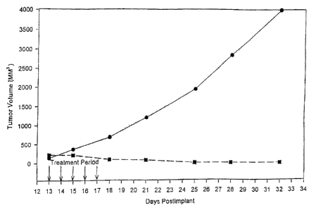
Figure 1 presents the results of intravenous
administration of paclitaxel nanoparticles to tumor bearing
mice (n=5 in each group), showing a complete regression of
tumor in the treatment group (U) compared with a control group
receiving saline W. Virtually uncontrolled tumor growth is
seen in the control group. Dose for the treatment group is 20
mg/kg of paclitaxel administered as an intravenous bolus for
five consecutive days.
Figure 2 presents the results of intraperitoneal
administration of paclitaxel nanoparticles in rats that have
developed arthritis in their paws following intradermal
injection of collagen. Paw volumes are measured and indicate
CA 02294981 2007-04-04
WO 99100113 PCT/US98/13272
34
the severity of the disease. The paw volumes are normalized
to 100% at the beginning of treatment. Day 0 represents the
initiation of treatment. There are 3 groups - control group
receiving saline (n-2, shown as a thin line and labelled in the
figure a "non-treatment"); a first treatment group receiving
paclitaxel nanoparticles at a dose of 1 mg/kg (n-4, shown as a
heavy line and labelled in the figure as "paclitaxel
nanoparticles 1.0 mg/kg"), and a second treatment group
receiving combination therapy of paclitaxel nanoparticles at a
dose of 0.5 mg/kg and prednisone at a dose of 0.2 mg/kg (n-4,
shown as a heavy line and labelled in the figure as "prednisone
0.2 mg/kg + paclitaxel nanoparticles 0.5 mg/kg"). The two
treatment groups show a dramatic reduction in paw volume with
time, indicating a regression of arthritis, while the control
group showed an increase in paw volume over the same period.
DETAILED DESCRIPTION OF TIE INVENTION
In accordance with the present invention, there are
provided methods for reducing the hematologic toxicity of
paclitaxel in a subject undergoing treatment with paclitaxel,
said method comprising systemically administering said
paclitaxel to said subject in a pharmaceutically acceptable
formulation at a does of at least 175 mg/m2 over an
administration period of no greater than two hours.
In accordance with the present invention, there are
also provided methods for the preparation of substantially
CA 02294981 2007-04-04
WO 99/00113 PCTIUS98/13272
water insoluble pharmacologically active
agents for in vivo delivery, said method comprising:
a) combining
i) an organic solvent having said active
5 agent dissolved therein;
ii) water or an aqueous solution;
iii) a surfactant; and
iv) a cosurfactant
that spontaneously form a microemulsion; and
10 b) removing said organic solvent to yield a
suspension of nanoparticles of said active agent in said water.
In accordance with a still further embodiment of the
present invention, there is provided a drug delivery system
15 comprising particles of a solid or liquid, substantially water
insoluble pharmacologically active agent, coated with a
protein,
wherein said protein coating has free protein
associated therewith,
20 wherein a portion of said pharmacologically active
agent is contained within said protein coating and a
portion of said pharmacologically active agent is
associated with said free protein, and
wherein the average diameter of said particles is
25 no greater than about 1 micron.
Compositions produced by the above-described methods
are particularly advantageous as they have been observed to
provide a very low toxicity form of a variety of
30 pharmacologically active agents. Also described herein are
CA 02294981 2007-04-04
WO 99/00113 PCT/US98/13272
36
other methods of making low toxicity forms of
pharmacologically active agents, e.g., paclitaxel.
In a preferred embodiment, the average diameter of
the above-described particles is no greater than about 200 nm.
Such particles are particularly advantageous as they can be
subjected to sterile filtration, thereby obviating the need for
more vigorous treatment to achieve sterilization of solutions
containing the desired pharmacologically active agent.
As used herein, unless specified to the contrary, the
term "paclitaxel" encompasses all forms, modifications and
derivatives of paclitaxel, e.g., taxotere, and the like.
CapxolTM is the trademark for the paclitaxel
formulation to be marketed by Applicants' assignees. As used
herein, CapxolTM is merely a shorthand means of reference to
protein-coated paclitaxel nanoparticles produced by the method
of Example 1. CapxolTM is a proprietary new, cremaphor-free
formulation of the anticancer drug paclitaxel. Inventors,
based on animal studies, believe that a cremaphor-free
formulation will be significantly less toxic and will not
require premedication of patients. Premedication is necessary
to reduce the hypersensitivity and anaphylaxis that occurs as
a result of cremaphor in the currently approved and marketed
BMS (Bristol Myers Squibb) formulation of paclitaxel. CapxolTM
is a lyophilized powder for reconstitution and intravenous
administration. Each vial of CapxolTM contains 30 mg of
paclitaxel and approximately 400 mg of human serum albumin.
When reconstituted with a suitable aqueous medium such as 0.9%
CA 02294981 2007-04-04
WO 99/00113 PCT/US98/13272
37
sodium chloride injection or 5% dextrose injection,
CapxolTM forms a stable colloidal solution of paclitaxel. The
size of the colloidal nanoparticles is typically less than 400
nm. The nanoparticles are prepared by high pressure
homogenization of a solution of USP human serum albumin and a
solution of paclitaxel in an organic solvent. The solvent is
then removed to generate the colloidal suspension or solution
of paclitaxel in human albumin. This suspension is sterile
filtered and lyophilized to obtain CapxolTM. The formulation
contains no other added excipients or stabilizers. The
sterility of the product is assured by an aseptic
manufacturing process and/or by sterile filtration. The two
major components of CapxolTM are unmodified paclitaxel and
human serum albumin (HSA). Since HSA is freely soluble in
water, CapxolTM can be reconstituted to any desired
concentration of paclitaxel limited only by the solubility
limits for HSA. Thus CapxolTM can be reconstituted in a wide
range of concentrations ranging from dilute (0.1 mg/ml
paclitaxel) to concentrated (20 mg/ml paclitaxel). This can
result in fairly small volumes of administration.
As used herein, the term "in vivo delivery" refers to
delivery of a pharmacologically active agent by such routes of
administration as oral, intravenous, subcutaneous,
intraperitoneal, intrathecal, intramuscular, inhalational,
topical, transdermal, suppository (rectal), pessary (vaginal),
intra urethral, intraportal, intrahepatic, intra-arterial,
intraumoral,and the like.
CA 02294981 2007-04-04
WO 99/00113 PCT/US98/13272
38
As used herein, the term "micron" refers to a unit
of measure of one one-thousandth of a millimeter.
As used herein, the term "biocompatible" describes a
substance that does not appreciably alter or affect in any
adverse way, the biological system into which it is introduced.
Substantially water insoluble pharmacologically
active agents contemplated for use in the practice of the
present invention include pharmaceutically active agents,
diagnostic agents, agents of nutritional value, and the like.
Examples of pharmaceutically active agents include:
analgesics/antipyretics (e.g., aspirin,
acetaminophen, ibuprofen, naproxen sodium, buprenorphine
hydrochloride, propoxyphene hydrochloride, propoxyphene
napsylate, meperidine hydrochloride, hydromorphone
hydrochloride, morphine sulfate, oxycodone hydrochloride,
codeine phosphate, dihydrocodeine bitartrate, pentazocine
hydrochloride, hydrocodone bitartrate, levorphanol
tartrate, diflunisal, trolamine salicylate, nalbuphine
hydrochloride, mefenamic acid, butorphanol tartrate,
choline salicylate, butalbital, phenyltoloxamine citrate,
diphenhydramine citrate, methotrimeprazine, cinnamedrine
hydrochloride, meprobamate, and the like);
anesthetics (e.g., cyclopropane, enflurane,
halothane, isoflurane, methoxyflurane, nitrous oxide,
propofol, and the like);
antiasthmatics (e.g., Azelastine, Ketotifen,
Traxanox, Amlexanox, Cromolyn, Ibudilast, Montelukast,
Nedocromil, Oxatomide, Pranlukast, Seratrodast, Suplatast
CA 02294981 2007-04-04
WO 99/00113 PCT/US98/13272
39
Tosylate, Tiaramide, Zafirlukast, Zileuton,
Beclomethasone, Budesonide, Dexamethasone, Flunisolide,
Trimcinolone Acetonide, and the like);
antibiotics (e.g., neomycin, streptomycin,
chloramphenicol, cephalosporin, ampicillin, penicillin,
tetracycline, and the like);
antidepressants (e.g., nefopam, oxypertine, doxepin
hydrochloride, amoxapine, trazodone hydrochloride,
amitriptyline hydrochloride, maprotiline hydrochloride,
phenelzine sulfate, desipramine hydrochloride,
nortriptyline hydrochloride, tranylcypromine sulfate,
fluoxetine hydrochloride, doxepin hydrochloride,
imipramine hydrochloride, imipramine pamoate,
nortriptyline, amitriptyline hydrochloride, isocarboxazid,
desipramine hydrochloride, trimipramine maleate,
protriptyline hydrochloride, and the like);
antidiabetics (e.g., biguanides, hormones,
sulfonylurea derivatives, and the like);
antifungal agents (e.g., griseofulvin, keloconazole,
amphotericin B, Nystatin, candicidin, and the like);
antihypertensive agents (e.g., propanolol,
propafenone, oxyprenolol, Nifedipine, reserpine,
trimethaphan camsylate, phenoxybenzamine hydrochloride,
pargyline hydrochloride, deserpidine, diazoxide,
guanethidine monosulfate, minoxidil, rescinnamine, sodium
nitroprusside, rauwolfia serpentina, alseroxylon,
phentolamine mesylate, reserpine, and the like);
anti-inflammatories (e.g., (non-steroidal)
indomethacin, naproxen, ibuprofen, ramifenazone,
piroxicam, (steroidal) cortisone, dexamethasone,
CA 02294981 2007-04-04
WO 99/00113 PCT/US98/13272
fluazacort, hydrocortisone, prednisolone,
prednisone, and the like);
antineoplastics (e.g., adriamycin, cyclophosphamide,
actinomycin, bleomycin, duanorubicin, doxorubicin,
5 epirubicin, mitomycin, methotrexate, fluorouracil,
carboplatin, carmustine (BCNU), methyl-CCNU, cisplatin,
etoposide, interferons, camptothecin and derivatives
thereof, phenesterine, Taxol and derivatives thereof,
taxotere and derivatives thereof, vinblastine,
10 vincristine, tamoxifen, etoposide, piposulfan, and the
like);
antianxiety agents (e.g., lorazepam, buspirone
hydrochloride, prazepam, chlordiazepoxide hydrochloride,
oxazepam, clorazepate dipotassium, diazepam, hydroxyzine
15 pamoate, hydroxyzine hydrochloride, alprazolam,
droperidol, halazepam, chlormezanone, dantrolene, and the
like);
immunosuppressive agents (e.g., cyclosporine,
azathioprine, mizoribine, FK506 (tacrolimus), and the
20 like) ;
antimigraine agents (e.g., ergotamine tartrate,
propanolol hydrochloride, isometheptene mucate,
dichloralphenazone, and the like);
sedatives/hypnotics (e.g., barbiturates (e.g.,
25 pentobarbital, pentobarbital sodium, secobarbital sodium),
benzodiazapines (e.g., flurazepam hydrochloride,
triazolam, tomazeparm, midazolam hydrochloride, and the
like);
antianginal agents (e.g., beta-adrenergic blockers,
30 calcium channel blockers (e.g., nifedipine, diltiazem
hydrochloride, and the like), nitrates (e.g.,
CA 02294981 2007-04-04
WO 99/00113 PCT/US98/13272
41
nitroglycerin, isosorbide dinitrate, pentaerythritol
tetranitrate, erythrityl tetranitrate, and the like));
antipsychotic agents (e.g., haloperidol, loxapine
succinate, loxapine hydrochloride, thioridazine,
thioridazine hydrochloride, thiothixene, fluphenazine
hydrochloride, fluphenazine decanoate, fluphenazine
enanthate, trifluoperazine hydrochloride, chlorpromazine
hydrochloride, perphenazine, lithium citrate,
prochlorperazine, and the like);
antimanic agents (e.g., lithium carbonate);
antiarrhythmics (e.g., bretylium tosylate, esmolol
hydrochloride, verapamil hydrochloride, amiodarone,
encainide hydrochloride, digoxin, digitoxin, mexiletine
hydrochloride, disopyramide phosphate, procainamide
hydrochloride, quinidine sulfate, quinidine gluconate,
quinidine polygalacturonate, flecainide acetate, tocainide
hydrochloride, lidocaine hydrochloride, and the like);
antiarthritic agents (e.g., phenylbutazone, sulindac,
penicillamine, salsalate, piroxicam, azathioprine,
indomethacin, meclofenamate sodium, gold sodium
thiomalate, ketoprofen, auranofin, aurothioglucose,
tolmetin sodium, and the like);
antigout agents (e.g., colchicine, allopurinol, and
the like);
anticoagulants (e.g., heparin, heparin sodium,
warfarin sodium, and the like);
thrombolytic agents (e.g., urokinase, streptokinase,
altoplase, and the like);
antifibrinolytic agents (e.g., aminocaproic acid);
hemorheologic agents (e.g., pentoxifylline);
CA 02294981 2007-04-04
WO 99/00113 PCT/US98/13272
42
antiplatelet agents (e.g., aspirin, empirin,
ascriptin, and the like);
anticonvulsants (e.g., valproic acid, divaiproate
sodium, phenytoin, phenytoin sodium, clonazepam,
primidone, phenobarbitol, phenobarbitol sodium,
carbamazepine, amobarbital sodium, methsuximide,
metharbital, mephobarbital, mephenytoin, phensuximide,
paramethadione, ethotoin, phenacemide, secobarbitol
sodium, clorazepate dipotassium, trimethadione, and the
like) ;
antiparkinson agents (e.g., ethosuximide, and the
like);
antihistamines/antipruritics (e.g., hydroxyzine
hydrochloride, diphenhydramine hydrochloride,
chlorpheniramine maleate, brompheniramine maleate,
cyproheptadine hydrochloride, terfenadine, clemastine
fumarate, triprolidine hydrochloride, carbinoxamine
maleate, diphenylpyraline hydrochloride, phenindamine
tartrate, azatadine maleate, tripelennamine hydrochloride,
dexchlorpheniramine maleate, methdilazine hydrochloride,
trimprazine tartrate and the like);
agents useful for calcium regulation (e.g.,
calcitonin, parathyroid hormone, and the like);
antibacterial agents (e.g., amikacin sulfate,
aztreonam, chloramphenicol, chloramphenicol palmitate,
chloramphenicol sodium succinate, ciprofloxacin
hydrochloride, clindamycin hydrochloride, clindamycin
palmitate, clindamycin phosphate, metronidazole,
metronidazole hydrochloride, gentamicin sulfate,
lincomycin hydrochloride, tobramycin sulfate, vancomycin
CA 02294981 2007-04-04
WO 99/00113 PCT/US98/13272
43
hydrochloride, polymyxin B sulfate, colistimethate
sodium, colistin sulfate, and the like);
antiviral agents (e.g., interferon gamma, zidovudine,
amantadine hydrochloride, ribavirin, acyclovir, and the
like);
antimicrobials (e.g., cephalosporins (e.g., cefazolin
sodium, cephradine, cefaclor, cephapirin sodium,
ceftizoxime sodium, cefoperazone sodium, cefotetan
disodium, cefutoxime azotil, cefotaxime sodium, cefadroxil
monohydrate, ceftazidime, cephalexin, cephalothin sodium,
cephalexin hydrochloride monohydrate, cefamandole nafate,
cefoxitin sodium, cefonicid sodium, ceforanide,
ceftriaxone sodium, ceftazidime, cefadroxil, cephradine,
cefuroxime sodium, and the like), penicillins (e.g.,
ampicillin, amoxicillin, penicillin G benzathine,
cyclacillin, ampicillin sodium, penicillin G potassium,
penicillin V potassium, piperacillin sodium, oxacillin
sodium, bacampicillin hydrochloride, cloxacillin sodium,
ticarcillin disodium, azlocillin sodium, carbenicillin
indanyl sodium, penicillin G potassium, penicillin G
procaine, methicillin sodium, nafcillin sodium, and the
like), erythromycins (e.g., erythromycin ethylsuccinate,
erythromycin, erythromycin estolate, erythromycin
lactobionate, erythromycin siearate, erythromycin
ethylsuccinate, and the like), tetracyclines (e.g.,
tetracycline hydrochloride, doxycycline hyclate,
minocycline hydrochloride, and the like), and the like);
anti-infectives (e.g., GM-CSF);
bronchodialators (e.g., sympathomimetics (e.g.,
epinephrine hydrochloride, metaproterenol sulfate,
terbutaline sulfate, isoetharine, isoetharine mesylate,
CA 02294981 2007-04-04
WO 99/00113 PCT/US98/13272
44
isoetharine hydrochloride, albuterol
sulfate, albuterol, bitolterol, mesylate isoproterenol
hydrochloride, terbutaline sulfate, epinephrine
bitartrate, metaproterenol sulfate, epinephrine,
epinephrine bitartrate), anticholinergic agents (e.g.,
ipratropium bromide), xanthines (e.g., aminophylline,
dyphylline, metaproterenol sulfate, aminophylline), mast
cell stabilizers (e.g., cromolyn sodium), inhalant
corticosteroids (e.g., flurisolidebeclomethasone
dipropionate, beclomethasone dipropionate monohydrate),
salbutamol, beclomethasone dipropionate (BDP), ipratropium
bromide, budesonide, ketotifen, salmeterol, xinafoate,
terbutaline sulfate, triamcinolone, theophylline,
nedocromil sodium, metaproterenol sulfate, albuterol,
flunisolide, and the like);
hormones (e.g., androgens (e.g., danazol,
testosterone cypionate, fluoxymesterone,
ethyltostosterone, testosterone enanihate,
methyltestosterone, fluoxymesterone, testosterone
cypionate), estrogens (e.g., estradiol, estropipate,
conjugated estrogens), progestins (e.g.,
methoxyprogesterone acetate, norethindrone acetate),
corticosteroids (e.g., triamcinolone, betamethasone,
betamethasone sodium phosphate, dexamethasone,
dexamethasone sodium phosphate, dexamethasone acetate,
prednisone, methylprednisolone acetate suspension,
triamcinolone acetonide, methylprednisolone, prednisolone
sodium phosphate methylprednisolone sodium succinate,
hydrocortisone sodium succinate, methylprednisolone sodium
succinate, triamcinolone hexacatonide, hydrocortisone,
hydrocortisone cypionate, prednisolone, fluorocortisone
CA 02294981 2007-04-04
WO 99/00113 PCT/US98/13272
acetate, paramethasone acetate, prednisolone
tebulate, prednisolone acetate, prednisolone sodium
phosphate, hydrocortisone sodium succinate, and the like),
thyroid hormones (e.g., levothyroxine sodium) and the
5 like), and the like;
hypoglycemic agents (e.g., human insulin, purified
beef insulin, purified pork insulin, glyburide,
chlorpropamide, glipizide, tolbutamide, tolazamide, and
the like);
10 hypolipidemic agents (e.g., clofibrate,
dextrothyroxine sodium, probucol, lovastatin, niacin, and
the like);
proteins (e.g., DNase, alginase, superoxide
dismutase, lipase, and the like);
15 nucleic acids (e.g., sense or anti-sense nucleic
acids encoding any therapeutically useful protein,
including any of the proteins described herein, and the
like);
agents useful for erythropoiesis stimulation (e.g.,
20 erythropoietin);
antiulcer/antireflux agents (e.g., famotidine,
cimetidine, ranitidine hydrochloride, and the like);
antinauseants/antiemetics (e.g., meclizine
hydrochloride, nabilone, prochlorperazine, dimenhydrinate,
25 promethazine hydrochloride, thiethylperazine, scopolamine,
and the like);
oil-soluble vitamins (e.g., vitamins A, D, E, K, and
the like); and
as well as other drugs such as mitotane, visadine,
30 halonitrosoureas, anthrocyclines, ellipticine, and the
like.
CA 02294981 2007-04-04
WO 99/00113 PCT/US98/13272
46
Examples of diagnostic agents contemplated for use in
the practice of the present invention include ultrasound
contrast agents, radiocontrast agents (e.g., iodo-octanes,
halocarbons, renografin, and the like), magnetic contrast
agents (e.g., fluorocarbons, lipid soluble paramagnetic
compounds, and the like), as well as other diagnostic agents
which cannot readily be delivered without some physical and/or
chemical modification to accommodate the substantially water
insoluble nature thereof.
Examples of agents of nutritional value contemplated
for use in the practice of the present invention include amino
acids, sugars, proteins, carbohydrates, fat-soluble vitamins
(e.g., vitamins A, D, E, K, and the like) or fat, or
combinations of any two or more thereof.
A. FORMATION OF NANOPARTICLES USING
HIGH SHEAR HOMOGENIZATION
Key differences between the pharmacologically active
agents contained in a polymeric shell according to the
invention and protein microspheres of the prior art are in the
nature of formation and the final state of the protein after
formation of the particle, and its ability to carry poorly
aqueous-soluble or substantially aqueous-insoluble agents. In
accordance with the present invention, the polymer (e.g., a
protein) may be crosslinked as a result of exposure to high
shear conditions in a high pressure homogenizer. High shear is
used to disperse a dispersing agent containing dissolved or
suspended pharmacologically active agent into an aqueous
CA 02294981 2007-04-04
WO 99/00113 PCT/US98/13272
47
solution of a biocompatible polymer, optionally bearing
sulfhydryl or disulfide groups (e.g., albumin) whereby a shell
of crosslinked polymer is formed around fine droplets of non-
aqueous medium. The high shear conditions produce cavitation
in the liquid that causes tremendous local heating and results
in the formation of superoxide ions that are capable of
crosslinking the polymer, for example, by oxidizing the
sulfhydryl residues (and/or disrupting existing disulfide
bonds) to form new, crosslinking disulfide bonds.
In contrast to the invention process, the prior art
method of glutaraldehyde crosslinking is nonspecific and
essentially reactive with any nucleophilic group present in the
protein structure (e.g., amines and hydroxyls). Heat
denaturation as taught by the prior art significantly and
irreversibly alters protein structure. In contrast, disulfide
formation contemplated by the present invention does not
substantially denature the protein. In addition, particles of
substantially water insoluble pharmacologically active agents
contained within a shell differ from crosslinked or heat
denatured protein microspheres of the prior art because the
polymeric shell produced by the invention process is relatively
thin compared to the diameter of the coated particle. It has
been determined (by transmission electron microscopy) that the
"shell thickness" of the polymeric coat is approximately 25
nanometers for a coated particle having a diameter of 1 micron
(1000 nanometers). In contrast, microspheres of the prior art
do not have protein shells, but rather, have protein dispersed
throughout the volume of the microsphere.
CA 02294981 2007-04-04
WO 99/00113 PCT/US98/13272
48
Thus, in accordance with the present invention, a
pharmacologically active agent is dissolved in a suitable
solvent (e.g., chloroform, methylene chloride, ethyl acetate,
ethanol, tetrahydrofuran, dioxane, butanol, butyl acetate,
acetonitrile, acetone, dimethyl sulfoxide, dimethyl formamide,
methyl pyrrolidinone, or the like, as well as mixtures of any
two or more thereof). Additional solvents contemplated for use
in the practice of the present invention include soybean oil,
coconut oil, olive oil, safflower oil, cotton seed oil, sesame
oil, orange oil, limonene oil, Cl-C20 alcohols, C2-C20 esters,
C3-C20 ketones, polyethylene glycols, aliphatic hydrocarbons,
aromatic hydrocarbons, halogenated hydrocarbons and
combinations thereof.
Unlike conventional methods for nanoparticle
formation, a polymer (e.g. polylactic acid) is not dissolved in
the solvent. The oil phase employed in the preparation of
invention compositions typically contains only the
pharmacologically active agent dissolved in solvent.
Next, a protein (e.g., human serum albumin) is added
(into the aqueous phase) to act as a stabilizing agent for the
formation of stable nanodroplets. Protein is added at a
concentration in the range of about 0.05 to 25 % (w/v), more
preferably in the range of about 0.5% - 5% (w/v). Unlike
conventional methods for nanoparticle formation, no surfactant
(e.g. sodium lauryl sulfate, lecithin, tween 80, pluronic F-68
and the like) is added to the mixture.
Next, an emulsion is formed by homogenization under
high pressure and high shear forces. Such homogenization is
CA 02294981 2007-04-04
WO 99/00113 PCT/US98/13272
49
conveniently carried out in a high pressure homogenizer,
typically operated at pressures in the range of about 3,000 up
to 60,000 psi. Preferably, such processes are carried out at
pressures in the range of about 6,000 up to 40,000 psi. The
resulting emulsion comprises very small nanodroplets of the
nonaqueous solvent (containing the dissolved pharmacologically
active agent) and very small nanodroplets of the protein
stabilizing agent. Acceptable methods of homogenization
include processes imparting high shear and cavitation such as
high pressure homogenization, high shear mixers, sonication,
high shear impellers, and the like.
Finally, the solvent is evaporated under reduced
pressure to yield a colloidal system composed of protein coated
nanoparticles of pharmacologically active agent and protein.
Acceptable methods of evaporation include the use of rotary
evaporators, falling film evaporators, spray driers, freeze
driers, and the like. Ultrafiltration may also be used for
solvent removal.
Following evaporation of solvent, the liquid
suspension may be dried to obtain a powder containing the
pharmacologically active agent and protein. The resulting
powder can be redispersed at any convenient time into a
suitable aqueous medium such as saline, buffered saline, water,
buffered aqueous media, solutions of amino acids, solutions of
vitamins, solutions of carbohydrates, or the like, as well as
combinations of any two or more thereof, to obtain a suspension
that can be administered to mammals. Methods contemplated for
obtaining this powder include freeze-drying, spray drying, and
the like.
CA 02294981 2007-04-04
WO 99/00113 PCT/US98/13272
In accordance with another embodiment of the present
invention, there is provided an alternative method for the
formation of unusually small submicron particles
5 (nanoparticles), i.e., particles which are less than
200 nanometers in diameter. Such particles are capable of
being sterile-filtered before use in the form of a liquid
suspension. The ability to sterile-filter the end product of
the invention formulation process (i.e., the drug particles) is
10 of great importance since it is impossible to sterilize
dispersions which contain high concentrations of protein (e.g.,
serum albumin) by conventional means such as autoclaving.
In order to obtain sterile-filterable particles
15 (i.e., particles <200 nm), the pharmacologically active agent
is initially dissolved in a substantially water immiscible
organic solvent (e.g., a solvent having less than about 5%
solubility in water, such as, for example, chloroform) at high
concentration, thereby forming an oil phase containing the
20 pharmacologically active agent. Suitable solvents are set
forth above. Unlike conventional methods for nanoparticle
formation, a polymer (e.g. polylactic acid) is not dissolved in
the solvent. The oil phase employed in the process of the
present invention contains only the pharmacologically active
25 agent dissolved in solvent.
Next, a water miscible organic solvent (e.g., a
solvent having greater than about 10% solubility in water, such
as, for example, ethanol) is added to the oil phase at a final
30 concentration in the range of about it - 99% v/v, more
preferably in the range of about 5% - 25% v/v of the total
CA 02294981 2007-04-04
WO 99/00113 PCTIUS98/13272
51
organic phase. The water miscible organic solvent can
be selected from such solvents as ethyl acetate, ethanol,
tetrahydrofuran, dioxane, acetonitrile, gutanol, acetone,
propylene glycol, glycerol, dimethyl sulfoxide, dimethyl
formamide, methyl pyrrolidinone, and the like. Alternatively,
the mixture of water immiscible solvent with the water miscible
solvent is prepared first, followed by dissolution of the
pharmaceutically active agent in the mixture.
Next, human serum albumin or any other suitable
stabilizing agent as described above is dissolved in aqueous
media. This component acts as a stabilizing agent for the
formation of stable nanodroplets. Optionally, a sufficient
amount of the first organic solvent (e.g. chloroform) is
dissolved in the aqueous phase to bring it close to the
saturation concentration. A separate, measured amount of the
organic phase (which now contains the pharmacologically active
agent, the first organic solvent and the second organic
solvent) is added to the saturated aqueous phase, so that the
phase fraction of the organic phase is between about 0.5% - 1516
v/v, and more preferably between 1% and 8% v/v.
Next, a mixture composed of micro and nanodroplets is
formed by homogenization at low shear forces. This can be
accomplished in a variety of ways, as can readily be identified
by those of skill in the art, employing, for example, a
conventional laboratory homogenizer operated in the range of
about 2,000 up to about 15,000 rpm. This is followed by
homogenization under high pressure (i.e., in the range of about
3,000 up to 60,000 psi). The resulting mixture comprises an
aqueous protein solution (e.g., human serum albumin), the water
CA 02294981 2007-04-04
WO 99/00113 PCf/US98/13272
52
insoluble pharmacologically active agent, the first
solvent and the second solvent. Finally, solvent is rapidly
evaporated under vacuum to yield a colloidal dispersion system
(pharmacologically active agent and protein) in the form of
extremely small nanoparticles (i.e., particles in the range of
about 10nm - 200 nm diameter) that can be sterile-filtered.
The preferred size range of the particles is between about 50
nm - 170 nm, depending on the formulation and operational
parameters.
Colloidal systems prepared in accordance with the
present invention may be further converted into powder form by
removal of the water therefrom, e.g., by lyophilization or
spray drying at a suitable temperature-time profile. The
protein (e.g., human serum albumin) itself acts as a
cryoprotectant or lyoprotectant, and the powder is easily
reconstituted by addition of water, saline or buffer, without
the need to use such conventional cryoprotectants as mannitol,
sucrose, glycine, and the like. While not required, it is of
course understood that conventional cryoprotectants may be
added to invention formulations if so desired.
The colloidal system of pharmacologically active
agent allows for the delivery of high doses of the
pharmacologically active agent in relatively small volumes.
This minimizes patient discomfort at receiving large volumes of
fluid and minimizes hospital stay. In addition, the walls of
the polymeric shell or coating are generally completely
degradable in vivo by proteolytic enzymes (e.g., when the
polymer is a protein), resulting in substantially no side
CA 02294981 2007-04-04
WO 99/00113 PCTIUS98/13272
53
effects from the delivery system, which is in sharp
contrast to the significant side effects caused by current
formulations.
A number of biocompatible polymers may be employed in
the practice of the present invention for the formation of the
polymeric shell which surrounds the substantially water
insoluble pharmacologically active agents. Essentially any
polymer, natural or synthetic, optionally bearing sulfhydryl
groups or disulfide bonds within its structure may be utilized
for the preparation of a disulfide crosslinked shell about
particles of substantially water insoluble pharmacologically
active agents. The sulfhydryl groups or disulfide linkages may
be preexisting within the polymer structure or they may be
introduced by a suitable chemical modification. For example,
natural polymers such as proteins, peptides, polynucleic acids,
polysaccharides (e.g., starch, cellulose, dextrans, alginates,
chitosan, pectin, hyaluronic acid, and the like),
proteoglycans, lipoproteins, and so on, are candidates for such
modification.
Proteins contemplated for use as stabilizing agents
in accordance with the present invention include albumins
(which contain 35 cysteine residues), immunoglobulins, caseins,
insulins (which contain 6 cysteines), hemoglobins (which
contain 6 cysteine residues per a292 unit), lysozymes (which
contain 8 cysteine residues), immunoglobulins, alpah-2-
macroglobulin, fibronectins, vitronectins, fibrinogens,
lipases, and the like. Proteins, peptides, enzymes, antibodies
and combinations thereof, are general classes of stabilizers
contemplated for use in the present invention.
CA 02294981 2007-04-04
WO 99/00113 PCf/US98/13272
54
A presently preferred protein for use as a
stabilizing agent is albumin. Optionally, proteins such as
alpha-2-macroglobulin, a known opsonin, could be used to
enhance uptake of the shell encased particles of substantially
water insoluble pharmacologically active agents by macrophage-
like cells, or to enhance the uptake of the shell encased
particles into the liver and spleen. Specific antibodies may
also be utilized to target the nanoparticles to specific
locations.
Other functional proteins, such as antibodies or
enzymes, which could facilitate targeting of biologic to a
desired site, can also be used as components of the stabilizing
protein.
Similarly, synthetic polymers are also good
candidates for formation of particles having a polymeric shell.
In addition, polyalkylene glycols (e.g., linear or branched
chain), polyvinyl alcohol, polyacrylates, polyhydroxyethyl
methacrylate, polyacrylic acid, polyethyloxazoline,
polyacrylamides, polyisopropyl acrylamides, polyvinyl
pyrrolidinone, polylactide/glycolide and the like, and
combinations thereof, are good candidates for the biocompatible
polymer in the invention formulation.
Similarly, synthetic polypeptides are also good
candidates for stabilizing agents for the substantially water
insoluble pharmacologically active agents. In addition,
contemplated for use in the practice of the present invention
are such materials as synthetic polyamino acids containing
CA 02294981 2007-04-04
WO 99/00113 PCT/US99/13272
cysteine residues and/or disulfide groups; polyvinyl
alcohol modified to contain free sulfhydryl groups and/or
disulfide groups; polyhydroxyethyl methacrylate modified to
contain free sulfhydryl groups and/or disulfide groups;
5 polyacrylic acid modified to contain free sulfhydryl groups
and/or disulfide groups; polyethyloxazoline modified to contain
free sulfhydryl groups and/or disulfide groups; polyacrylamide
modified to contain free sulfhydryl groups and/or disulfide
groups; polyvinyl pyrrolidinone modified to contain free
10 sulfhydryl groups and/or disulfide groups; polyalkylene glycols
modified to contain free sulfhydryl groups and/or disulfide
groups; polylactides, polyglycolides, polycaprolactones, or
copolymers thereof, modified to contain free sulfhydryl groups
and/or disulfide groups; as well as mixtures of any two or more
15 thereof.
In the preparation of invention compositions, a wide
variety of organic media can be employed to suspend or dissolve
the substantially water insoluble pharmacologically active
20 agent. Organic media contemplated for use in the practice of
the present invention include any nonaqueous liquid that is
capable of suspending or dissolving the pharmacologically
active agent, but does not chemically react with either the
polymer employed to produce the shell, or the pharmacologically
25 active agent itself. Examples include vegetable oils (e.g.,
soybean oil, olive oil, and the like), coconut oil, safflower
oil, cotton seed oil, sesame oil, orange oil, limonene oil,
aliphatic, cycloaliphatic, or aromatic hydrocarbons having 4-30
carbon atoms (e.g., n-dodecane, n-decane, n-hexane,
30 cyclohexane, toluene, benzene, and the like), aliphatic or
aromatic alcohols having 2-30 carbon atoms (e.g., octanol, and
CA 02294981 2007-04-04
WO 99/00113 PCT/US98/13272
56
the like), aliphatic or aromatic esters having 2-30
carbon atoms (e.g., ethyl caprylate (octanoate), and the like),
alkyl, aryl, or cyclic ethers having 2-30 carbon atoms (e.g.,
diethyl ether, tetrahydrofuran, and the like), alkyl or aryl
halides having 1-30 carbon atoms (and optionally more than one
halogen substituent, e.g., CH3C1, CH2C121 CH2C1-CH2C1, and the
like), ketones having 3-30 carbon atoms (e.g., acetone, methyl
ethyl ketone, and the like), polyalkylene glycols (e.g.,
polyethylene glycol, and the like), or combinations of any two
or more thereof.
Especially preferred combinations of organic media
contemplated for use in the practice of the present invention
typically have a boiling point of no greater than about 200 C,
and include volatile liquids such as dichloromethane,
chloroform, ethyl acetate, benzene, ethanol, butanol, butyl
acetate, and the like (i.e., solvents that have a high degree
of solubility for the pharmacologically active agent, and are
soluble in the other organic medium employed), along with a
higher molecular weight (less volatile) organic medium. When
added to the other organic medium, these volatile additives
help to drive the solubility of the pharmacologically active
agent into the organic medium. This is desirable since this
step is usually time consuming. Following dissolution, the
volatile component may be removed by evaporation (optionally
under vacuum).
Particles of pharmacologically active agent
associated with a polymeric shell, prepared as described above,
are delivered as a suspension in a biocompatible aqueous
liquid. This liquid may be selected from water, saline, a
CA 02294981 2007-04-04
WO 99/00113 PCT/US98/13272
57
solution containing appropriate buffers, a
solution containing nutritional agents such as amino acids,
sugars, proteins, carbohydrates, vitamins or fat, and the like.
These biocompatible materials may also be employed in
several physical forms such as gels, crosslinked or
uncrosslinked to provide matrices from which the
pharmacologically active ingredient, for example paclitaxel,
may be released by diffusion and/or degradation of the matrix.
Temperature sensitive materials may also be utilized as the
dispersing matrix for the invention formulation. Thus for
example, the Capxol may be injected in a liquid formulation of
the temperature sensitive material (e.g., copolymers of
polyacrylamides or copolymers of polyalkylene glycols and
polylactide/glycolides) which gel at the tumor site and provide
slow release of Capxol. The Capxol formulation may be
dispersed into a matrix of the above mentioned biocompatible
polymers to provide a controlled release formulation of
paclitaxel, which through the properties of the Capxol
formulation (albumin associated with paclitaxel) results in
lower toxicity to brain tissue as well as lower systemic
toxicity as discussed below. This combination of Capxol or
other chemotherapeutic agents formulated similar to Capxol
together with a biocompatible polymer matrix may be useful for
the controlled local delivery of chemotherapeutic agents for
treating solid tumors in the brain and peritoneum (ovarian
cancer) and in local applications to other solid tumors. These
combination formulations are not limited to the use of
paclitaxel and may be utilized with a wide variety of
pharmacologically active ingredients including antiinfectives,
immunosuppressives and other chemotherapeutics and the like.
CA 02294981 2007-04-04
WO 99/00113 PCT/US98/13272
58
Particles colloidal substantially completely
contained within a polymeric stabilizing layerl, or associated
therewith, prepared as described herein, are delivered neat, or
optionally as a suspension in a biocompatible medium. This
medium may be selected from water, buffered aqueous media,
saline, buffered saline, optionally buffered solutions of amino
acids, optionally buffered solutions of proteins, optionally
buffered solutions of sugars, optionally buffered solutions of
carbohydrates, optionally buffered solutions of vitamins,
optionally buffered solutions of synthetic polymers, lipid-
containing emulsions, and the like.
In addition, the colloidal particles can optionally
be modified by a suitable agent, wherein the agent is
associated with the polymeric layer through an optional
covalent bond. Covalent bonds contemplated for such linkages
include ester, ether, urethane, diester, amide, secondary or
tertiary amine, phosphate ester, sulfate ester, and the like
bonds. Suitable agents contemplated for this optional
modification of the polymeric shell include synthetic polymers
(polyalkylene glycols (e.g., linear or branched chain
polyethylene glycol), polyvinyl alcohol, polyhydroxyethyl
methacrylate, polyacrylic acid, polyethyloxazoline,
polyacrylamide, polyvinyl pyrrolidinone, and the like),
phospholipids (such as phosphatidyl choline (PC), phosphatidyl
ethanolamine (PE), phosphatidyl inositol (PI), sphingomyelin,
and the like), proteins (such as enzymes, antibodies, and the
like), polysaccharides (such as starch, cellulose, dextrans,
alginates, chitosan, pectin, hyaluronic acid, and the like),
chemical modifying agents (such as pyridoxal 5'-phosphate,
CA 02294981 2007-04-04
WO 99/00113 PCT/US98/13272
59
derivatives of pyridoxal, dialdehydes, diaspirin esters,
and the like), or combinations of any two or more thereof.
Variations on the general theme of stabilized
colloidal particles are possible. A suspension of fine
particles of pharmacological agent in a biocompatible
dispersing agent could be used (in place of a biocompatible
cispersing agent containing dissolved biologic) to produce a
polymeric shell containing dispersing agent-suspended particles
of biologic. In other words, the polymeric shell could contain
a saturated solution of biologic in dispersing agent. Another
variation is a polymeric shell containing a solid core of
biologic produced by initially dissolving the biologic in a
volatile organic solvent (e.g. benzene), forming the polymeric
shell and evaporating the volatile solvent under vacuum, e.g.,
in an evaporator, spray drier or freeze-drying the entire
suspension. This results in a structure having a solid core of
biologic surrounded by a polymer coat. This latter method is
particularly advantageous for delivering high doses of biologic
in a relatively small volume. In some cases, the biocompatible
material forming the shell about the core could itself be a
therapeutic or diagnostic agent, e.g., in the case of insulin,
which may be delivered as part of a polymeric shell formed in
the process described above. In other cases, the polymer -
forming the shell could participate in the delivery of a
biologic, e.g., in the case of antibodies used for targeting,
or in the case of hemoglobin, which may be delivered as part of
a polymeric shell formed in the ultrasonic irradiation process
described above, thereby providing a blood substitute having a
high binding capacity for oxygen.
CA 02294981 2007-04-04
WO 99/00113 PCT/US98/13272
Those skilled in the art will recognize that
several variations are possible within the scope and spirit of
this aspect of the invention. The organic medium within the
polymeric shell may be varied, a large variety of
5 pharmacologically active agents may be utilized, and a wide
range of proteins as well as other natural and synthetic
polymers may be used in the formation of the walls of the
polymeric shell. Applications are also fairly wide ranging.
Other than biomedical applications such as the delivery of
10 drugs, diagnostic agents (in imaging applications), artificial
blood and parenteral nutritional agents, the polymeric shell
structures of the invention may be incorporated into cosmetic
applications such as skin creams or hair care products, in
perfumery applications, in pressure sensitive inks, and the
15 like.
This aspect of the invention will now be described in
greater detail by reference to the following non-limiting
examples.
Exa e 1
Preparation of Nano2articles by High Pressure
Homogenization
30 mg paclitaxel is dissolved in 3.0 ml methylene
chloride. The solution was added to 27.0 ml of human serum
abumin solution (1% w/v). The mixture was homogenized for 5
minutes at low RPM (Vitris homogenizer, model: Tempest I.Q.) in
order to form a crude emulsion, and then transferred into a
high pressure homogenizer (Avestin). The emulsification was
performed at 9000-40,000 psi while recycling the emulsion for
CA 02294981 2007-04-04
WO 99/00113 PCT/US98/13272
61
at least 5 cycles. The resulting system was
transferred into a Rotary evaporator, and methylene chloride
was rapidly removed at 40 C, at reduced pressure (30 mm Hg),
for 20-30 minutes. The resulting dispersion was translucent,
and the typical diameter of the resulting paclitaxel particles
was 160-220 (Z-average, Malvern Zetasizer).
The dispersion was further lyophilized for 48 hrs
without adding any cryoprotectant. The resulting cake could be
easily reconstituted to the original dispersion by addition of
sterile water or saline. The particle size after
reconstitution was the same as before lyophilization.
Example 2
Use of Conventional Surfactants and Proteins Results
in Formation of Large Crystals
The following example demonstrates the effect of
adding surfactants which are used in the conventional solvent
evaporation method. A series of experiments was conducted
employing a similar procedure to that described in Example 1,
but a surfactant such as Tween 80 (1% to 10%) is added to the
organic solvent. It was found that after removal of the
methylene chloride, a large number of paclitaxel crystals is
obtained having an average size of 1-2 micron, as viewed by
light microscopy and under polarized light. The crystals grow
within a few hours to form very large needle-like crystals,
with a size in the range of about 5-15 micron. A similar
phenomenon is observed with other commonly used surfactants,
such as Pluronic F-68, Pluronic F-127, Cremophor EL and Brij
58.
CA 02294981 2007-04-04
WO 99/00113 PCT/US98/13272
62
From these results it can be concluded that the
conventional solvent evaporation method utilizing conventional
surfactants in combination with a protein such as albumin is
not suitable for the formation of submicron drug particles
(e.g. Paclitaxel) without a polymeric core, while using a polar
solvent (e.g., methylene chloride).
Example 3
Use of Conventional Surfactants Alone
Results in Formation of Large Crystals
This example demonstrates that it is not possible to
form nanoparticles while using conventional surfactants,
without a polymeric core material, with pharmacologically
active agents which are soluble in polar, water immiscible
solvents (e.g. chloroform).
30 mg Taxol is dissolved in 0.55 ml chloroform and
0.05 ml ethanol. The solution is added to 29.4 ml of Tween 80
solution (1t w/v), which is presaturated with it chloroform.
The mixture is homogenized for 5 minutes at low RPM (Vitris
homogenizer, model: Tempest I.Q.) in order to form a crude
emulsion, and then transferred into a high pressure homogenizer
(Avestin). The emulsification is performed at 9000-40,000 psi
while recycling the emulsion for at least 6 cycles. The
resulting system was transferred into a Rotary evaporator, and
the chloroform was rapidly removed at 40 C, at reduced pressure
(30 mm Hg), for 15-30 minutes. The resulting dispersion was
opaque, and contained large needle-like crystals of the drug.
The initial size of the crystals (observed also by polarized
CA 02294981 2007-04-04
WO 99/00113 PCT/US98/13272
63
light), was 0.7-5 micron. Storage of the dispersion for
several hours at room temperature led to further increase in
crystal size, and ultimately to precipitation.
Example 4
Preparation of Less than 200 nm
Sterile-Filterable Nanoparticles
This example describes a process by which sterile-
filterable drug particles can be obtained. Thus, 30 mg Taxol
is dissolved in 0.55 ml chloroform and 0.05 ml ethanol. The
solution is added to 29.4 ml of human serum abumin solution (1%
w/v), which is presaturated with 1% chloroform. The mixture is
homogenized for 5 minutes at low RPM (Vitris homogenizer,
model: Tempest I.Q.) in order to form a crude emulsion, and
then transferred into a high pressure homogenizer (Avestin).
The emulsification is performed at 9000-40,000 psi while
recycling the emulsion for at least 6 cycles. The resulting
system is transferred into a Rotary evaporator, and the
chloroform is rapidly removed at 40 C, at reduced pressure (30
mm Hg), for 15-30 minutes. The resulting dispersion is
translucent, and the typical diameter of the resulting Taxol
particles is 140-160 nm (Z-average, Malvern Zeta Sizer). The
dispersion is filtered through a 0.22 micron filter
(Millipore), without any significant change in turbidity, or
particle size. HPLC analysis of the Taxol content revealed
that more than 97% of the Taxol was recovered after filtration,
thus providing a sterile Taxol dispersion.
The sterile dispersion was further lyophilized for 48
hrs without adding any cryoprotectant. The resulting cake
CA 02294981 2007-04-04
WO 99/00113 PCT/US98/13272
64
could be easily reconstituted to the original dispersion by
addition of sterile water or saline. The particle size after
reconstitution was the same as before lyophilization.
Example 5
Preparation of Less than 200 nm
Sterile-Filterable Nan=articles
This example describes a process by which sterile-
filterable drug particles can be obtained. Thus, 225 mg Taxol
is dissolved in 2.7 ml chloroform and 0.3 ml ethanol. The
solution is added to 97 ml of human serum abumin solution (3%
w/v). The mixture is homogenized for 5 minutes at low RPM
(Vitris homogenizer, model: Tempest I.Q.) in order to form a
crude emulsion, and then transferred into a high pressure
homogenizer (Avestin). The emulsification is performed at
9000-40,000 psi while recycling the emulsion for at least 6
cycles. The resulting system is transferred into a Rotary
evaporator, and the chloroform is rapidly removed at 40 C, at
reduced pressure (30 mm Hg), for 15-30 minutes. The resulting
dispersion is translucent, and the typical diameter of the
resulting Taxol particles is 140-160 nm (Z-average, Malvern
Zeta Sizer). The dispersion is filtered through a 0.22 micron
filter (Sartorius, sartobran 300), without any significant.
change in turbidity, or particle size. HPLC analysis of the
Taxol content typically revealed that 70-100% of the Taxol
could be recovered after filtration, depending on the
conditions employed. Thus, a sterile Taxol dispersion was
obtained.
CA 02294981 2007-04-04
WO 99/00113 PCT/US98/13272
The sterile dispersion was aseptically
filled into sterile glass vials and lyophilized without adding
any cryoprotectant. The resulting cake could be easily
reconstituted to the original dispersion by addition of sterile
5 water or saline. The particle size after reconstitution was
the same as before lyophilization.
Example 8
Nanoparticle Formation of a Model Drug
30 mg Isoreserpine (a model drug) is dissolved in 3.0
ml methylene chloride. The solution is added to 27.0 ml of
human serum abumin solution (1% w/v). The mixture is
homogenized for 5 minutes at low RPM (Vitris homogenizer,
model: Tempest I.Q.) in order to form a crude emulsion, and
then transferred into a high pressure homogenizer (Avestin).
The emulsification is performed at 9000-18,000 psi while
recycling the emulsion for at least 5 cycles. The resulting
system is transferred into a Rotary evaporator, and methylene
chloride is rapidly removed at 40 C, at reduced pressure (30 mm
Hg), for 20-30 minutes. The resulting dispersion is
translucent, and the typical diameter of the resulting
paclitaxel particles was 120-140 nm (Z-average, Malvern
Zetasizer). The dispersion was filtered through a 0.22 micron
filter (Millipore).
The sterile dispersion was further lyophilized for 48
hrs without adding any cryoprotectant. The resulting cake
could be easily reconstituted to the original dispersion by
addition of sterile water or saline. The particle size after
reconstitution was the same as before lyophilization.
CA 02294981 2007-04-04
WO 99/00113 PCT/US98/13272
66
Example 9
Extremely Small Particle Formation with a Model Drua
The effect of ethanol addition on reducing particle
size is demonstrated for Isoreserpine. Thus, 30 mg
Isoreserpine is dissolved in 2.7 ml methylene chloride and 0.3
ml ethanol. The solution is added to 27.0 ml of human serum
abumin solution (lo w/v). The mixture is homogenized for 5
minutes at low RPM (Vitris homogenizer, model: Tempest I.Q.) in
order to form a crude emulsion, and then transferred into a
high pressure homogenizer (Avestin). The emulsification was
performed at 9000-40,000 psi while recycling the emulsion for
at least 5 cycles. The resulting system was transferred into a
Rotary evaporator, and methylene chloride was rapidly removed
at 40 C, at reduced pressure (30 mm Hg), for 20-30 minutes.
The resulting dispersion was translucent, and the typical
diameter of the resulting paclitaxel particles was 90-110 nm
(Z-average, Malvern Zetasizer). The dispersion was filtered
through a 0.22 micron filter (Millipore).
The sterile dispersion was further lyophilized for 48
hrs without adding any cryoprotectant. The resulting cake
could be easily reconstituted to the original dispersion by
addition of sterile water or saline. The particle size after
reconstitution was the same as before lyophilization.
Example 10
Use of a Water miscible Solvent Alone. Supersaturated with Drug
- Not Suitable for Invention Process
CA 02294981 2007-04-04
WO 99/00113 PCT/NS9S/13272
67
30 mg Taxol is dispersed in 0.6 ml ethanol.
At this concentration (50 mg/ml), the Taxol is not completely
soluble and forms a supersaturated dispersion. The dispersion
is added to 29.4 ml of human serum abumin solution (1% w/v).
The mixture is homogenized for 5 minutes at low RPM (Vitris
homogenizer, model: Tempest I.Q.) in order to form a crude
dispersion, and then transferred into a high pressure
homogenizer (Avestin). The emulsification is performed at
9000-40,000 psi while recycling the emulsion for at least 6
cycles. The resulting system is transferred into a Rotary
evaporator, and the ethanol is rapidly removed at 40 C, at
reduced pressure (30 mm Hg), for 15-30 minutes. The resulting
dispersion particle size is extremely broad, ranging from about
250 nm to several microns.
Observation under the microscope revealed the
presence of large particles and typical needle shaped crystals
of Taxol. These particles were too large for intravenous
injection. This experiment demonstrates that the use of
solvents such as ethanol that are freely miscible in water in
the invention process results in the formation of large
particles with very broad particle size distribution and as
such cannot be used alone for the invention process. Thus the
invention process specifically excludes the use of water
miscible solvents when used alone for the dissolution or
dispersion of the drug component. The invention process
requires that such solvents, when used, must be mixed with
essentially water immiscible solvents to allow production of
the invention nanoparticles.
Example 12
CA 02294981 2007-04-04
WO 99/00113 PGT/US98/13272
68
Determination of Physical State of Paclitaxxel in
Nanouarticle Form by X-Ray Powder Diffraction
Paclitaxel raw material is usually present as needle
shaped crystals of varying sizes typically between 5-500
microns. The presence of crystals in a drug formulation for
intravenous injection is obviously detrimental if crystals are
present in size above a few microns due to potential blockage
of capillaries. In addition, the solubility of drug crystals
in general would be lower than for amorphous drug, thereby
lowering the bioavailability of the drug following intravenous
administration. It is also known that as the loading of the
drug in a formulation is increased, the tendency for
crystallization also increases. Thus it is advantageous that
the formulation contain the drug in essentially amorphous form.
X-Ray powder diffraction was used to determine the
crystalline or non-crystalline nature of paclitaxel in the
lyophilized powder formulation. The following samples were
analyzed: Sample 1 - Paclitaxel powder; Sample 2 - Lyophilized
serum albumin; Sample 3 - a physical mixture of paclitaxel and
albumin; and Sample 4 - formulated paclitaxel. Each sample was
x-rayed from 2 to 70 2-theta angles using CuKa radiation, an
accelerating voltage of 40KeV/3OmA, a step size of 0.05 2-
theta and a data acquisition time of 2.0 seconds per step.
Sample 1 showed strong peaks typical of a crystalline sample.
The most intense paclitaxel peak was located at 5.1 2-theta.
Sample 2 showed broad humps typical of amorphous material.
Sample 3 showed largely the broad humps of Sample 2, but in
addition, the peak at 5.1 2-theta of paclitaxel was visible.
Sample 4, the formulated paclitaxel showed no evidence of
CA 02294981 2007-04-04
WO 99/00113 PCT/US98/13272
69
crystallinity characteristic of paclitaxel and appeared
identical to Sample 2, indicating the presence of substantially
amorphous pharmacologically active agent in the formulated
sample.
The amorphous nature of the nanoparticles produced
according to the invention stands in direct contrast to the
products produced by other methods described in the art for
producing nanoparticles. For example, the use of grinding
techniques, as described in U.S. Patent 5,145,684 (Liversidge
et al.), and as described by Liversidge-Merisko et al.,
Pharmaceutical Research 13(2):272-278 (1996), produces a
substantially crystalline product.
Example 13
Preparation of Nanoparticl_es of Cvcl_osporine (Capsorine 7.V.)
by High Pressure Homogenization
30 mg cyclosporine is dissolved in 3.0 ml methylene
chloride. The solution is then added into 27.0 ml of human
serum albumin solution (1% w/v). The mixture is homogenized
for 5 minutes at low RPM (Vitris homogenizer model: Tempest
I.Q.) in order to form a crude emulsion, and then transferred
into a high pressure homogenizer (Avestin). The emulsification
was performed at 9000-40,000 psi while recycling the emulsion
for at least 5 cycles. The resulting system was transferred
into a Rotavap and methylene chloride was rapidly removed at
40 C, at reduced pressure (30 mm Hg), for 20-30 minutes. The
resulting dispersion was translucent and the typical diameter
of the resulting cyclosporine particles was 160-220 (Z-average,
Malvern Zetasizer).
CA 02294981 2007-04-04
WO 99/00113 PCT/US98/13272
The dispersion was further lyophilized for 48 hours,
without adding any cryoprotectant. The resulting cake could be
easily reconstituted to the original dispersion by addition of
5 sterile water or saline. The particle size after
reconstitution was the same as before lyophilization.
Example 14
Preparation of Nanodroplets of Cyclosporine (Capsorine Oral) by
10 High Pressure Homogenization
30 mg cyclosporine is dissolved in 3.0 ml of a
suitable oil (sesame oil containing 10% orange oil). The
solution is then added into 27.0 ml of human serum albumin
15 solution (1`6 v/w). The mixture is homogenized for 5 minutes at
low RPM (Vitris homogenizer, model: Tempest I.Q.) in order to
form a crude emulsion, and then transferred into a high
pressure homogenizer (Avestin). The emulsification is
performed at 9000-40,000 psi while recycling the emulsion for
20 at least 5 cycles. The resulting dispersion had a typical
diameter of 160-220 (Z-average, Malvern Zetasizer).
The dispersion could be used directly or lyophilized
for 48 hours by optionally adding a suitable cryoprotectant.
25 The resulting cake could be easily reconstituted to the
original dispersion by addition of sterile water or saline.
B. Formation of Nanoparticles Using Sonication
CA 02294981 2007-04-04
WO 99/00113 PCT/US"/13272
71
Similar to the use of high shear homogenization,
the use of sonication to form protein-coated nanoparticles of
water insoluble pharmacologically active agents is believed to
operate by crosslinking proteins through the formation of
inter-molecular disulfide bonds. Many of the advantages over
the prior art enjoyed by the high shear homogenization
techniques described above apply equally to the sonication
methods described below.
With respect to the organic solvents, proteins, and
non-proteinaceous polymers that may be used in the sonication
method, reference is made to those components described above
with respect to the high shear homogenization method. All of
the same components are expected to work equally well in both
methods.
This aspect of the invention will now be described in
greater detail by reference to the following non-limiting
examples.
Example 15
Formulation for Inhalation of Anti-Asthmatic Drug
Anti-asthmatic pharmaceuticals have been prepared
using microparticle techniques to yield effective formulations
for dry powder inhalers (DPI). Starting with a steroidal drug
(e.g., beclomethasone, beclomethasone dipropionate,
budesonide, dexamethasone, flunisolide, triamcinolone
acetonide, and the like), a dry formulation is prepared of
appropriate particle size and release characteristics to
ensure efficacious delivery in the respiratory system.
CA 02294981 2007-04-04
WO 99/00113 PCf/US98/13272
72
The formulation is prepared using sonication
techniques, or homogenization in which the active drug,
dissolved in solvent, is dispersed into an aqueous protein
solution to form an emulsion of nanoparticles. This emulsion
is then evaporated to remove solvents, leaving the active drug
coated with protein in solution. This liquid sample
containing the colloidal drug particles is measured by Malvern
Zetasizer and gives a Z-average size of 260 nm. In a
preferred embodiment, the range of sizes of these colloidal
particles is about 50-1,000 nm, and more preferably about 70-
400 nm.
In this liquid form, other excipients may be
dissolved. Such excipients include (but are not limited to)
mannitol 0.5-150 lactose 0.1-5%, and maltodextrin. At this
stage, the resulting solution of active drug, protein, and
excipient can be either spray-dried or lyophilized and milled
to yield a dry powder. After spray-drying, the dry particle
size is determined by Malvern Mastersizer as D(vØ5) of about
1-10 m. The preferred size range for these particles is 0.5-
15 m, with a more preferred range of 0.7-8 m.
This spray dried powder is then mixed with an
excipient carrier powder. Again, several carriers are
available, including lactose, trehalose, Pharmatose 325 M,
sucrose, mannitol, and the like. The size of the carrier
powder is significantly larger than that of the formulated
drug particles (-63-90 m for lactose, 40-100 m for
Pharmatose).
CA 02294981 2007-04-04
WO 99/00113 PCT/US98113272
73
The efficacy of the dry powder formulation is
demonstrated by testing with an Andersen eight-stage cascade
impactor. Results of impactor trials show a fine particle
fraction (FPF) of -60%. This indicates a highly effective
release of particles, appropriately sized for respiratory
deposition. This FPF is surprisingly high and is a result of
the formulation composition that contains colloidal
nanoparticles of the drug within larger formulation particles.
This formulation shows the applicability of
microparticle and spray-dry techniques in the processing and
composing of dry powder formulations for aerosol delivery via
DPI. The high FPF results shown indicate an efficacious and
promising approach to DPI formulations.
Example 16
Summary of the Presently Preferred Manufacturing Process:
Starting with 1 Gram Paclitaxel as the SOS
Prepare a 3% HSA solution. To 51.7 ml of 25%
Albutein add 379.3 ml water for injection. Mix thoroughly and
filter the solution through a sterile 0.22 m Nalgene
disposable filterware. Keep at 4 C until used.
Weigh out 1.0 g of paclitaxel in a glass bottle.
Combine CHC13 and ethyl alcohol in appropriate proportions in a
vial. Mix well. To the paclitaxel, add 13.33 ml of the
chloroform/ethyl alcohol mixture. Agitate to ensure all
paclitaxel dissolves into solution. Filter the solution
through a 0.22 micron sterile Teflon filter and collect in a
sterile glass bottle.
CA 02294981 2007-04-04
WO 99/00113 PCT/U598/13272
74
To the dissolved paclitaxel solution in the glass
bottle, add the HSA solution. Use the Sentry Microprocessor
mixer to mix the paclitaxel/HSA solution.
When the solution is mixed, pour the contents into the chamber
of the Homogenizer. Cycle the mixture through the homogenizer
at a pressure until the desired particle size is obtained.
Collect the homogenized sample in a sterile Kontes round
bottom flask.
Attach the flask with the final sample to the Rotary
evaporator. Turn on the vacuum and the rotation to maximum in
the rotavapor and evaporate the organic solvent. This results
in the colloidal solution of paclitaxel in human albumin.
Save -- 3ml of this rotavaped sample for analysis of particle
size.
Under a sterile hood, filter the colloidal solution
using sterile 0.45/0.2 m filter and collect in a sterile
receiving vessel. Save - 3m1 of filtered sample for analysis
by HPLC for paclitaxel concentration.
Determine the fill volume to obtain 30 mg (or other
derived amount) of paclitaxel per vial- Fill the sterile
filtered sample into autoclaved Wheaton 30 ml vials at
approximately 17 ml each (based on assay). Close the vials
with autoclaved Wheaton serum vial stoppers. Each vial should
contain approximately 30 mg of paclitaxel.
Lyophilize the samples in the FTS System Stoppering
tray lyophilizer using a predetermined lyophilization cycle.
CA 02294981 2007-04-04
WO 99/00113 PCTIUS98/13272
After the samples have been lyophilized, stopper the vials
and seal the vials by crimping them with the 20mm Wheaton
aluminum tear-off caps. Label the samples appropriately. The
entire process is carried out in a clean room environment
5 under aseptic conditions.
The lyophilized samples contain residual solvent at
levels <1000ppm, and more preferably <500 ppm, or even <100
ppm.
Final Product Sterile filtration: Following removal
of solvent by evaporation, the colloidal solution of
paclitaxel in the flask is sterile filtered through a
combination 0.45/0.2 micron sterilizing filter. The filtered
solution is collected in a sterile beaker and sterile filled
into 30 ml vials. Vials are then placed in the lyophilizer.
Following completion of the lyophilization cycle the vials are
blanketed with dry sterile nitrogen gas and stoppered under
the nitrogen blanket.
It is of note that high pressure homogenization
processes are utilized to rupture and kill bacterial and other
cells to extract their contents.
Example 17
Preparation of Protein Shell Containing oil
Three ml of a USP (United States Pharmacopia) 5%
human serum albumin solution (Alpha Therapeutic Corporation)
were taken in a cylindrical vessel that could be attached to a
CA 02294981 2007-04-04
WO 99/00113 PCT/US98/13272
76
sonicating probe (Heat Systems, Model XL2020). The
albumin solution was overlayered with 6.5 ml of USP grade
soybean oil (soya oil). The tip of the sonicator probe was
brought to the interface between the two solutions and the
assembly was maintained in a cooling bath at 200C. The system
was allowed to equilibriate and the sonicator turned on for 30
seconds. Vigorous mixing occurred and a white milky suspension
was obtained. The suspension was diluted 1:5 with normal
saline. A particle counter (Particle Data Systems, Elzone,
Model 280 PC) was utilized to determine size distribution and
concentration of oil-containing protein shells. The resulting
protein shells were determined to have a maximum cross-
sectional dimension of about 1.35 0.73 microns, and the total
concentration determined to be --109 shells/ml in the original
suspension.
As a control, the above components, absent the
protein, did not form a stable miocroemulsion when subjected to
ultrasonic irradiation. This result suggests that the protein
is essential for formation of microspheres. This is confirmed
by scanning electron micrograph and transmission electron
micrograph studies as described below.
Example 18
Preparation of Polymeric Shells
Containing Dissolved Paclitaxel
Taxol was dissolved in USP grade soybean oil at a
concentration of 2 mg/ml. 3 ml of a USP 5% human serum albumin
solution was taken in a cylindrical vessel that could be
attached to a sonicating probe. The albumin solution was
CA 02294981 2007-04-04
WO 99/00113 PCT/US98/13272
77
overlayered with 6.5 ml of soybean oil/Taxol solution.
The tip of the sonicator probe was brought to the interface
between the two solutions and the assembly was maintained in
equilibrium and the sonicator turned on for 30 seconds.
Vigorous mixing occurred and a stable white milky suspension
was obtained that contained protein-walled polymeric shells
enclosing the oil/Taxol solution.
In order to obtain a higher loading of drug into the
crosslinked protein shell, a mutual solvent for the oil and the
drug (in which the drug has a considerably higher solubility)
can be mixed with the oil. Provided this solvent is relatively
non-toxic (e.g., ethyl acetate), it may be injected along with
the original carrier. In other cases,, it may be removed by
evaporation of the liquid under vacuum following preparation of
the polymeric shells.
It is recognized that several different methods may
be employed to achieve the physical characteristics of the
invention formulation. The biological properties associated
with this formulation of higher local concentrations at
specific organ sites (prostate, lung, pancreas, bone, kidney,
heart) as well as lower toxicities (increased LD50, decreased
myelosuppression, decreased cerebral toxicity) associated with
higher efficacies is independent of the method of manufacture.
Example 19
Preparation of Nanoparticles by Sonication
20 mg paclitaxel is dissolved in 1.0 ml methylene
chloride. The solution is added to 4.0 ml of human serum
CA 02294981 2007-04-04
WO 99/00113 PCT/US98/13272
78
abumin solution (5t w/v). The mixture is homogenized for 5
minutes at low RPM (Vitris homogenizer, model: Tempest I.Q.) in
order to form a crude emulsion, and then transferred into a 40
kHz sonicator cell. The sonicator is performed at 60-90% power
at 0 degree for 1 min (550 Sonic Dismembrator;. The mixture is
transferred into a Rotary evaporator, and methylene chloride is
rapidly removed at 40 C, at reduced pressure (30 mm Hg), for
20-30 minutes. The typical diameter of the resulting
paclitaxel particles was 350-420 nm (Z-average, Malvern
Zetasizer).
The dispersion was further lyophilized for 48 hrs
without adding any cryoprotectant. The resulting cake could be
easily reconstituted to the original dispersion by addition of
sterile water or saline. The particle size after
reconstitution was the same as before lyophilization.
Example 20
In Vivo Biodi stributi on of ro ink d Protein Shells
Containing a Fluorophore
To determine the uptake and biodistribution of liquid
entrapped within protein polymeric shells after intravenous
injection, a fluorescent dye (rubrene, available from Aldrich)
was entrapped within a human serum albumin (HSA) protein
polymeric shell and used as a marker. Thus, rubrene was
dissolved in toluene, and albumin shells containing
toluene/rubrene were prepared as described above by ultrasonic
irradiation. The resulting milky suspension was diluted five
times in normal saline. Two ml of the diluted suspension was
then injected into the tail vein of a rat over 10 minutes. One
CA 02294981 2007-04-04
WO 99/00113 PCT/US98/13272
79
animal was sacrificed an hour after injection and another 24
hours after injection.
100 micron frozen sections of lung, liver, kidney,
spleen, and bone marrow were examined under a fluorescent
microscope for the presence of polymeric shell-entrapped
fluorescent dye or released dye. At one hour, the majority of
the polymeric shells appeared to be intact (i.e., appearing as
brightly fluorescing particles of about 1 micron diameter), and
located in the lungs and liver. At 24 hours, the dye was
observed in the liver, lungs, spleen, and bone marrow. A
general staining of the tissue was also observed, indicating
that the shell wall of the polymeric shells had been digested,
and the dye liberated from within. This result was consistent
with expectations and demonstrates the potential use of
invention compositions for delayed or controlled release of an
entrapped pharmaceutical agent such as Taxol.
Example 21
Toxicity of Polymeric Shells Containing Soybean Oil (SBO)
Polymeric shells containing soybean oil were prepared
as described in Example 15. The resulting suspension was
diluted in normal saline to produce two different solutions,
one containing 20% SBO and the other containing 30% SBO.
Intralipid, a commercially available TPN agent,
contains 20% SBO. The LD50 for Intralipid in mice is 120 ml/kg,
or about 4 ml for a 30 g mouse, when injected at 1 cc/min.
CA 02294981 2007-04-04
WO 99/00113 PCT/US98/13272
Two groups of mice (three mice in each group;
each mouse weighing about 30 g) were treated with invention
composition containing SBO as follows. Each mouse was injected
with 4 ml of the prepared suspension of SBO-containing
5 polymeric shells. Each member of one group received the
suspension containing 20% SBO, while each member of the other
group received the suspension containing 30% SBO.
All three mice in the group receiving the suspension
10 containing 20% SBO survived such treatment, and showed no gross
toxicity in any tissues or organs when observed one week after
SBO treatment. Only one of the three mice in the group
receiving suspension containing 30% SBO died after injection.
These results clearly demonstrate that oil contained within
15 polymeric shells according to the present invention is not
toxic at its LDso dose, as compared to a commercially available
SBO formulation (Intralipid). This effect can be attributed to
the slow release (i.e., controlled rate of becoming
bioavailable) of the oil from within the polymeric shell. Such
20 slow release prevents the attainment of a lethal dose of oil,
in contrast to the high oil dosages attained with commercially
available emulsions.
Example 22
25 In Vivo Bioavailability of Soybean Oil Released from Polymeric
Shells
A test was performed to determine the slow or
sustained release of polymeric shell-enclosed material
30 following the injection of a suspension of polymeric shells
into the blood stream of rats. Crosslinked protein (albumin)
CA 02294981 2007-04-04
WO 99/00113 PCT/US98/13272
81
walled polymeric shells containing soybean oil (SBO)
were prepared by sonication as described above. The resulting
suspension of oil-containing polymeric shells was diluted in
saline to a final suspension containing 20% oil. Five ml of
this suspension was injected into the cannulated external
jugular vein of rats over a 10 minute period. Blood was
collected from these rats at several time points following the
injection and the level of triglycerides (soybean oil is
predominantly triglyceride) in the blood determined by routine
analysis.
Five ml of a commercially available fat emulsion
(Intralipid, an aqueous parenteral nutrition agent---containing
20% soybean oil, 1.2% egg yolk phospholipids, and 2.25%
glycerin) was used as a control. The control utilizes egg
phosphatide as an emulsifier to stabilize the emulsion. A
comparison of serum levels of the triglycerides in the two
cases would give a direct comparison of the bioavailability of
the oil as a function of time. In addition to the suspension
of polymeric shells containing 200 oil, 5 ml of a sample of
oil-containing polymeric shells in saline at a final
concentration of 30% oil was also injected. Two rats were used
in each of the three groups. The blood levels of triglycerides
in each case are tabulated in Table 1, given in units of mg/dl.
CA 02294981 2007-04-04
WO 99/00113 PCT/US98/13272
82
Table 1
GROUP SERUM TRIGLYCERIDES (mg/dl)
Pre 1 hr 4 hr 24 hr 48 hr 72 hr
Intralipid Control 11.4 941.9 382.9 15.0 8.8 23.8
(20% SBO)
Polymeric Shells 24.8 46.7 43.8 29.3 24.2 43.4
(20% SBO)
Polymeric Shells 33.4 56.1 134.5 83.2 34.3 33.9
(30% SBO)
Blood levels before injection are shown in the column
marked 'Pre'. Clearly, for the Intralipid control, very high
triglyceride levels are seen following injection. Triglyceride
levels are then seen to take about 24 hours to come down to
preinjection levels. Thus the oil is seen to be immediately
available for metabolism following injection.
The suspension of oil-containing polymeric shells
containing the same amount of total oil as Intralipid (20%)
show a dramatically different availability of detectible
triglyceride in the serum. The level rises to about twice its
normal value and is maintained at this level for many hours,
indicating a slow or sustained release of triglyceride into the
blood at levels fairly close to normal. The group receiving
oil-containing polymeric shells having 30% oil shows a higher
level of triglycerides (concomitant with the higher
administered dose) that falls to normal within 48 hours. Once
again, the blood levels of triglyceride do not rise
astronomically in this group, compared to the control group
receiving Intralipid. This again, indicates the slow and
sustained availability of the oil from invention composition,
which has the advantages of avoiding dangerously high blood
CA 02294981 2007-04-04
WO 99/00113 PCT/US98/13272
83
levels of material contained within the polymeric shells
and availability over an extended period at acceptable levels.
Clearly, drugs delivered within polymeric shells of the present
invention would achieve these same advantages.
Such a system of soybean oil-containing polymeric
shells could be suspended in an aqueous solution of amino
acids, essential electrolytes, vitamins, and sugars to form a
total parenteral nutrition (TPN) agent. Such a TPN cannot be
formulated from currently available fat emulsions (e.g.,
Intralipid) due to the instability of the emulsion in the
presence of electrolytes.
ExamD 23
Preparation of Protein-Walled Polymeric Shells Containing -a
Solid Core of Pharmaceutically Active Agent
Another method of delivering a poorly water-soluble
drug such as Taxol within a polymeric shell is to prepare a
shell of polymeric material around a solid drug core. Such a
'protein coated' drug particle may be obtained as follows. The
procedure described in Example 16 is repeated using an organic
solvent to dissolve Taxol at a relatively high concentration.
Solvents generally used are organics such as benzene, toluene,
hexane, ethyl ether, chloroform, alcohol and the like.
Polymeric shells are produced as described in Example 15. Five
ml of the milky suspension of polymeric shells containing
dissolved Taxol are diluted to 10 ml in normal saline. This
suspension is placed in a rotary evaporator and the volatile
organic removed by vacuum. The resultant suspension is
examined under a microscope to reveal opaque cores, indicating
CA 02294981 2007-04-04
WO 99/00113 PCf/US98/13272
84
removal of substantially all organic solvent, and the
presence of solid Taxol. The suspension can be frozen and
stored indefinitely and used directly or lyophilized at a later
time.
Alternatively, the polymeric shells with cores of
organic solvent-containing dissolved drug are freeze-dried to
obtain a dry crumbly powder that can be resuspended in saline
(or other suitable liquid) at the time of use. Although the
presently preferred protein for use in the formation of the
polymeric shell is albumin, other proteins such as a-2-
macroglobulin, a known opsonin, could be used to enhance uptake
of the polymeric shells by macrophage-like cells.
Alternatively, molecules like PEG could be incorporated into
the particles to produce a polymeric shell with increased
circulation time in vivo.
C. Formation of Nanoparticles by Spontaneous Mi croemul lion
It is also possible to form nanoparticles without the
use of sonication, high shear homegenization, or any other
high-energy technique. Thus, it is possible to form a
suspension (or dry powder) of essentially pure drug, if
desired.
A microemulsion is a thermodynamically stable
emulsion system that is formed spontaneously when all it's
components are brought into contact, in the absence of the
use of high shear equipment or other substantial agitation.
Microemulsions are substantially non-opaque, i.e., they are
transparent or translucent. Microemulsions comprise a
CA 02294981 2007-12-21
WO 99/00113 PCTIUS98/13272
dispersed phase, in which the typical droplet size is below
1000 Angstrom (A), hence their optical transparency. The
droplets in the microemulsion are typically spherical, though
other structures such as elongated cylinders are feasible.
5 (For further discussion see, e.g., Rosof, Progress in Surface
and membrane Science, 12,405, Academic Press (1975 ), Friberg
S., Dispersion Science and Technology, 6, 317 (1985).)
As will be shown below, the present invention
10 utilizes the unique characteristics of the microemulsion as
a first step towards obtaining extremely small nanoparticles,
after removal of the oil phase.
As described earlier, microparticles and
15 nanoparticles can be formed by various processes, among them,
the solvent evaporation method. This method is based, in
principle, on formation of a simple oil in water emulsion, in
the presence of surface active agent, while applying high
shear forces by means of various equipment such as rotor-
20 stator mixers, sonicators, high pressure homogenizers,
colloid mills, etc. After forming such an emulsion, which
contains a polymer and a drug dissolved in the dispersed oil
droplets, the oil phase is removed by evaporation, typically
at reduced pressure and elevated temperature, and
25 micoparticles or nanoparticles of the dissolved drug and
polymer are formed. Obviously, the size of the particles is
dependent on emulsion droplet's size; the smaller the
droplets, the smaller the resulting particles. Small
emulsion droplets can be achieved only by applying very high
30 energy, and even then, by using the most advanced high
pressure homogenizers such as the Microfluidizer, it is not
CA 02294981 2007-04-04
WO 99/00113 PCT/US98/13272
86
practical to achieve emulsion droplets below 75 nm. Since
emulsions are inherently unstable systems, and undergo
processes such as aggregation and droplets coalescence, the
solvent evaporation processes for such emulsions may result
in larger particles.
The new method, which overcomes the problems
associated with application of the solvent evaporation method
in conventional emulsions, consists of the following steps:
a. Dissolving the water insoluble drug in a
solvent which has low solubility in water, and has higher
vapor pressure than water. The drug is dissolved without any
additional polymeric binder, although such binder can be
present, in principle.
b. Mixing the solvent with a proper surfactant(s)
and a water soluble cosurfactant(s).
c. Adding a suitable amount of water or aqueous
solution to this mixture, thus spontaneously forming an oil-
in-water microemulsion, without the use of any high shear
equipment. The aqueous solution may contain electrolytes,
amino acids, or any other additive which may affect the
formation of the microemulsion during the first preparation
stage.
d. Optionally adding a protein solution to the
microemulsion.
e. Removing the solvent by evaporation at reduced
pressure, thus causing precipitation of the drug in the form
of extremely small amorphous nanoparticles, having a typical
size below 1000 Angstroms. The particles at this stage are
dispersed and stabilized in an aqueous medium which contains
surfactant, cosurfactant, and optionally protective agents
CA 02294981 2007-04-04
WO 99/00113 PCT/US98/13272
87
such as proteins, sugars, etc. Acceptable methods of
evaporation include the use of rotary evaporators, falling
film evaporators, spray dryers, freeze dryers, and other
standard evaporation equipment typically used in industry.
f. Optionally one may remove the surfactant and
cosurfactant by dialysis, ultrafiltration, adsorption, etc.,
thus obtaining nanoparticles which are stabilized by the
protein.
g. Following evaporation of solvent, the liquid
dispersion of nanoparticles may be dried to obtain a powder
containing the pharmacological agent and optionally the
protein, which can be redispersed into a suitable aqueous
medium such as saline, buffer, water, and the like, to obtain
a suspension that can be administered to a life-form, having
a particle size below 1000 Angstroms. Acceptable methods of
obtaining this powder are by freeze-drying, spray drying, and
the like. If the conversion into a solid form is performed
by lyophilization, various cryoprotectants may be added, such
as manitol, lactose, albumin, carboxymethyl cellulose,
polyvinylpyrolidone, maltodextrins, and/or polyethylene
glycol.
These nanoparticles can be further mixed with _
additional excipients or matrix-forming materials, in order
to obtain a drug delivery system, with high bioavailabilty,
controlled release characteristics, and protection in gastric
juice. The final product may be introduced to the mammals as
a tablet, capsule, reconstituted liquid, or the like.
The present invention formulation has significant
advantages over the previously used methods for preparation
CA 02294981 2007-04-04
WO 99/00113 PCT/US98/13272
88
of nanoparticles and microparticles, and the use
of microemulsions or "pre-microemulsion concentrate."
There are many advantages realized by using the
invention process. The microemulsion is formed
spontaneously, if the proper components are selected, and
there is no need for high cost equipment and energy input.
The droplet size is smaller about an order of magnitude than
the smallest emulsion droplets obtained by high shear
equipment, and therefore extremely small nanoparticles can be
obtained. The microemulsion is thermodynamically stable, and
therefore the usual problems which are associated with
emulsion instability (and thus a time dependence of the size
of the resulting particles) will be prevented. The whole
process is much more simple than the conventional emulsion-
solvent evaporation method, and less sensitive to various
parameters. Since only simple mixing is involved in the
process, the upscaling to large production volumes is very
simple, compared to emulsification with equipment such as
high shear homogenizer. Since the particle size obtained by
the new process is so small, an order of magnitude less than
the pore size of membranes used for sterile filtration, the
sterilization process is very effective, without problems
associated with membrane blockage, such as increased
filtration pressure, and high drug loss during the filtration
process. Since there are no high shear forces in the
emulsification process, there is no increase in temperature
during emulsification, and therefore even temperature-
sensitive drugs can be processed by the new invention method.
The drug in the liquid formulation of the present invention
has increased chemical stability because it contains
CA 02294981 2007-04-04
WO 99/00113 PCT/US98/13272
89
dispersed nanoparticles compared to conventional
microemulsions that contain dispersed nanodroplets, i.e.,
more chemical reactions take place in liquid state
(microdroplet) versus solid state (nanoparticle). The
present invention has increased chemical stability as a dry
formulation compared to conventional microemulsions that are
liquids as the continuous microemulsion phase. The solid
formulation enables inclusion of the drug in various solid
dosage forms, such as tablets, granules and capsules,
compared to conventional microemulsions or "pre-microemulsion
concentrates," which are present in a liquid form. The very
narrow size distribution, combined with very low average
particle size, ensures increased adsorption of the drug, in a
manner more uniform than microparticles and nanoparticles
prepared by conventional methods, thus, increased
bioavailability is expected.
Although the examples presented in the following
section refer to two water insoluble molecules, the
pharmacological agents contemplated to be useful in the
preparation of nanoparticles include but are not limited to
drugs, diagnostic agents, agents of therapeutic value,
nutritional agents, and the like. A non-limiting list of
drug categories and compounds include but are not limited to
all of the compounds listed above for use in the high shear
homogenization aspect of the invention.
The solvents described in the following examples
are toluene and butyl acetate, however, any solvent or slvent
mixture which is capable of dissolving the required drug will
be suitable for use in the invention process, provided that a
CA 02294981 2007-04-04
WO 99/00113 PCTAIS99/13272
proper microemulsion can be formed prior to removal of
the solvent. Such solvents can be chloroform, methylene
chloride, ethyl acetate, butyl acetate, isobutylacetate,
propyl acetate, tert-butylmethyl ether, butanol, propylen
5 glycol, heptane, anisol, cumene, ethyl formate ethanol,
propanol, tetrahydrofuran, dioxane, acetonitrile, acetone,
dimethyl sulfoxide, dimethyl formamide, methyl pyrrolidinone,
soybean oil, coconut oil, castor oil, olive oil, safflower
oil, cottonseed oil, alcohols C1-C20, esters C2-C20, ketones
10 C3-C20, polyethylene glycols, aliphatic hydrocarbons,
aromatic hydrocarbons, halogenated hydrocarbons, d-limonene,
combinations thereof, and the like.
The protein (or a mixture of several proteins) used
15 in this process should be such that does not precipitate
during the initial mixing or during the evaporation stage.
There are many such proteins, including albumins (e.g., BSA
HSA , egg), gelatin, collagen, IgG, various enzymes,
lactoglobulin, casein, soy proteins, and the like.
The surfactants utilized in this invention should
be capable of spontaneously forming oil-in-water
microemulsions, in the presence of a suitable cosurfactant
and solvent, without causing precipitation of the drug or the
protein (if present). The surfactants can be nonionic
(Tween, Span, Triton, Pluronic, polyglycerol esters, and the
like), anionic (SDS, cholates and deoxycholates, fatty acid
soaps, and the like), cationic (cetyltrimethyl ammonium
chloride, and the like) or zwitterionic (lecithin, amino
acids, and the like) .
CA 02294981 2007-04-04
WO 99/00113 PCT/US98/13272
91
The cosurfactant should have the ability to
spontaneously form microemulsions with the selected
surfactants, without causing precipitation of the dissolved
drug molecules (or protein, if present), and without inducing
formation of large crystalline material. The cosurfactants
can be either water soluble or oil soluble, such as butanol,
propylene glycol, benzyl alcohol, propanol, and the like.
The conversion of the liquid dispersion of the
nanoparticles via lyophilization may require the addition of
cryoprotecting agents, such as mannitol, lactose, amino
acids, proteins, polysaccharides, and the like.
It is clear that the principles described in this
invention can be applied in several variations of the
process, for example:
1. The formation of the drug particles may be
induced by dilution of the microemulsion in a proper solvent,
in which the solvent is miscible. For example, if the
solvent has a low solubility in water, it would be possible
to dilute the microemulsion to such an extent that the
solvent will be below it's solubility limit in water.
2. The solvent and optionally the surfactant and
cosurfactant can be removed by using a selective extractant
which does not dissolve the drug.
3. The surfactant and cosurfactant may be removed
by ultrafiltration, while using filters having a cut-off
below that of the MW of the protein. Simple dialysis is also
an option.
4. The formulation may contain only components
which are acceptable for the intended use of the final
CA 02294981 2007-04-04
WO 99/00113 PCT/US98/13272
92
formulation (whether oral, IV, topical, etc.), thus there is
no need for their removal.
5. Similarly, cosurfactants that can remain in
the final product, such as glycerol, benzyl alcohol, etc, may
be used.
6. The addition of various water soluble
molecules which may affect the phase diagram of the
microemulsion (electrolytes, ethanol etc.) is possible, thus
controlling the ratio between the various components to give
the optimal drug load.
7. The spontaneous emulsification step may be
performed at a temperature other than room temperature, in
order to affect the phase diagram (and the component
proportions that leads to formation of a microemulsion). In
particular, it could be possible to use the temperature
effect (in ethoxylated surfactants) to change the system from
an oil-in-water to a water-in-oil microemulsion.
8. It is possible to add other components to the
solvent phase, in order to affect the bioavailability of the
drug. In particular, addition of an oil such as Soybean oil,
to enhance oral absorption, and to protect the drug from
chemical and enzymatic degradation is preferred.
9. Similarly, the addition of a matrix-forming
polymer (such as PVP) to the solvent, together with the drug
may be done.
10. The stabilization and solid-form properties
may be altered by the addition of a water soluble polymer
other than the protein (CMC, gums, and the like) to the
external aqueous phase of the microemulsion.
11. The flow properties of the resulting solid
form powder may be altered by addition of colloidal particles
CA 02294981 2007-04-04
WO 99/00113 PCT/US98/13272
93
(e.g. silica) as a filler, or addition of
reconstitution/anti-agglomeration aids.
12. The same principles described in this
invention may be applied to form water soluble particles,
while performing the emulsification stage in the composition
range in which a water-in-oil microemulsion is formed. The
process can be used, for example to form extremely small
protein nanoparticles.
Example 22
Preparation of Nanoparticles of Cyclosporin A
115 mg Cyclosporin A are dissolved in 1 mL butyl
acetate, and mixed with 2 grams of a 4:1 solution of Triton
X-100:n-Butanol. A clear system is obtained. 10 g water is
added dropwise, while slightly shaking. A clear oil-in-water
microemulsion is obtained. 10 g of It casein solution is
added, while slightly shaking. The system becomes slightly
turbid. The butyl acetate is removed in a rotovap, at 40 C,
80 mm Hg. The system becomes completely clear.
The particle size was measured by photon
correlation spectroscopy. It was found that the Z-average
size is 25-33 nm, while the size by number or volume
distribution is only 9 nm. No particles were observed under
optical microscope, nor under polarized light. This result
indicates the absence of crystalline particles.
The liquid dispersion of these nanoparticles was
lyophilized, after adding lactose (2% w/w).
CA 02294981 2007-04-04
WO 99/00113 PCT/US9&/13272
94
A white, solid material was obtained, which,
upon reconstitution in water, yielded a clear system, similar
to that prior to lyophilization. The particle size in this
reconstituted sample was very similar to that of the original
formulation, Z-average about 40 nm, and diameter by volume
and number distribution between 10-12 nm.
CA 02294981 2007-04-04
WO 99/00113 PCTIUS98113272
Example 25
Preparation of Nanoparticles of Cyclosporin A
119 mg of Cyclosporin A are dissolved in butyl
5 acetate, and mixed with 2 grams of a 4:1 solution of Triton
X-100:propylene glycol. A clear system is obtained. 7 g
water is added dropwise, while slightly shaking. A clear
oil-in-water microemulsion is obtained. 7 g of it casein
solution is added, while slightly shaking. The system
10 becomes slightly turbid. The sample is diluted 1:1 with
water, prior to solvent evaporation. The butyl acetate is
removed in a rotovap, at 400 C, 80 mm Hg. The system becomes
completely clear. This process also yielded extremely small
nanoparticles: Z-average 45 nm, and diameter by volume and
15 number distribution is 11 nm.
The liquid dispersion of these nanoparticles was
lyophilized, after adding lactose (2% w/w).
20 A white, solid material was obtained, which, upon
reconstitution in water, yielded a clear system, similar to
that prior to lyophilization. The particle size in this
reconstituted sample was close to that of the original
formulation, Z-average about 25 nm, and diameter by volume
25 and number distributions between 9-11 nm.
CA 02294981 2007-04-04
WO 99/00113 PCT/US98/13272
96
Example 26
Cyclosporine nanoparticles
Microemulsions were made with the following
compositions: 50 mg Cyclosporine, 0.5g butylacetate, 3.04g
Tween 80:propyleneglycol (1:1), and 6.8 g water. The
microemulsion was evaporated to give a clear liquid containing
5 mg/ml of cyclosporine. In a control experiment, performed
with the above components by simple mixing, but without
butylacetate, even after 17 hours, cyclosporin was not
dissolved.
There are several possibilities for surfactants,
including polysorbates (Tween), sorbitan esters (span),
sucrose esters, lecithin, monodiglycerides, polyethylene-
polypropylene block copolymers (pluromics), soaps (sodium
stearate, etc.), sodium glycolate bile salts, ethoxylated
castor oil, sodium stearoyl-lactylate, ethoxylated fatty
acids (myrj), ethoxylated fatty alcohols (Brij), sodium
dodecyl sulphate (SDS), and the like. Also, in general,
biopolymers such as starch, gelatin, cellulose derivatives
etc. may be used. Also for oral applications, all acceptable
food grade surfactants may be used as well as surfactants
presented in McCutcheon Handbook of Surfactants or CTFA
Index. Possible cosolvents or cosurfactants for the
microemulsion include propylene glycol, ethanol, glycerol,
butanol, oleic acid, and the like.
CA 02294981 2007-04-04
WO 99/00113 PCT/US98/13272
97
Example 27
Preparation of Nanoparticles of BHT
110 mg butylated hydroxy toluene (BHT) is dissolved
in 1 ml toluene, and mixed with 2 ml 4:1 solution of Triton
X-100:n-Butanol. 32 g of it casein solution was added, and a
microemulsion was spontaneously formed. The microemulsion
was evaporated under reduced pressure, 80 mm Hg, at 40 C,
until it became clear. The size of the resulting particles
is: Z-average 30 nm, diameter by volume and number
distribution is 16 and 15 nm, respectively.
Example 28
Preparation of Nanoparticles of BHT
A process similar to that described in example 24
was performed, while using water instead of casein solution.
After evaporation at 40 C, 80 mm Hg, the system became clear,
having a Z-average size of -10 nm.
Example 29
Preparation of Nanoparticles of Paclitaxel
mg of paclitaxel were dissolved in 2 ml butyl
25 acetate, and added to 4 grams of 4:1 Triton x-100:propylene
glycol. 40 ml water were added, and the system was slightly
turbid. After evaporation, the system became completely
clear. Z-average size was 6 nm, size by volume and numbered
distribution was 7-9 nm. The same size was measured after one
30 day at 4 C.
CA 02294981 2007-04-04
WO 99/00113 PCT/US98/13272
98
D. Miscellaneous Examples Relevant to All Methods of
noparticle Formation
Example 30
_ Identification of Microemulsion Phase Diagrams
Compositions were identified which yield
microemulsions, and that may be utilized to obtain
nanoparticles by the solvent evaporation method. These
compositions should contain a water miscible solvent capable
of dissolving hydrophobic molecules, an aqueous solution as
the continuous medium, surfactants, and possibly
cosurfactants.
Microemulsions of butyl acetate in water can be
formed at various compositions which are described by phase
diagrams (butyl acetate is classified as solvent with high
acceptable residual concentration in the final product).
Furthermore, both surfactant and cosurfactant are used in food
and pharmaceutical applications: Tween 80 (ethoxylated
sorbitan monooleate) and propylene glycol. Preliminary
experiments were conducted by using BHT as a model hydrophobic
molecule, yielding dispersions of particles in the size range
of 20-50 nm. After filtration by 0.2 m filters, about 100%
of the BHT passed the membrane.
Phase diagrams of various combinations of
surfactant/cosurfactant were obtained by vortexing the solvent
with a mixture of surfactant/cosurfactant (prepared prior to
the mixing with the solvent, at various ratios), followed by
dropwise addition of water. The turbidity of the various
CA 02294981 2007-04-04
WO 99/00113 PCT/US9S/13272
99
compositions along the "water line" was observed and the
compositions which yielded translucent systems were further
analyzed by light scattering. By using various ratios of
solvent-surfactant/cosurfactant, the areas in the phase
S diagrams which yielded microemulsions were identified (only a
small number of the selected components yielded
microemulsions). The same procedure was used for systems in
which BHT was dissolved in butyl acetate prior to conducting
the phase diagram experiments.
The "filterability" of the microemulsion and
nanoparticles which contain the BHT, was evaluated by
comparing the UV absorption spectra before and after 0.2 pm
filtration. The nanoparticles were obtained by vacuum
evaporation of butyl acetate (60 mm Hg, 40 C). It should be
emphasized that throughout the whole process no high shear
equipment was used.
The microemulsion systems were identified which
could be useful for oral delivery. n-Butyl acetate was chosen
as a solvent. The following surfactants and cosurfactants
were evaluated at various ratios:
Tween 80:Glycerol 5:1
Tween 80:Glycerol 4:1
Tween 80:Glycerol 3:1
Tween 80:Glycerol 2:1
Tween 80:Glycerol 1:1
Span 60:Glycerol 4:1
Span 80:Glycerol 3:1
Tween 80:Propylene glycol 4:1
Tween 80:Propylene glycol 3:1
Tween 80:Propylene glycol 2.5:1
CA 02294981 2007-04-04
WO 99/00113 PCT/US98/13272
100
Tween 80:Propylene glycol 1.5:1
Tween 80:Propylene glycol 1:1
Tween 80:Propylene glycol 1:2
((Tween 80 + Span 80) 7:1):Propylene glycol 3.5:1
((Tween 80 + Span 80) 7:1):Propylene glycol 1:1
((Tween 80 + Span 80) 8:1):Propylene glycol 4:1
((Tween 80 + Span 80) 5:1):Propylene glycol 1:1
Tween 80:((Propylene glycol + Glycerol) 1:1.2) 2:1
A suitable composition was found to be as follows:
Tween 80 as a surfactant and propylene glycol as a cosurfacant
at ratio 1:1. The full phase diagram was evaluated for the
system n-butyl acetate, Tween 80 : propylene glycol 1:1,
water. Two additional solvents were tested: sec-butyl
acetate and tert-butyl acetate. The phase diagrams for these
systems were the same as for that with n-butyl acetate. The
system n-butyl acetate, Tween 80 : propylene glycol 1:1, water
was evaluated further.
The measurement of particle size for the sample 7%
butyl acetate, 30% surfactant/PG, 63% water was performed. Z
average of about 20 nm was found. The nanoparticles formation
process was conducted for a water insoluble dye, Sudan III, at
concentration of about 10 mg in 1 g butyl acetate (5% butyl
acetate, 23% surfactant/PG, 72% water). Particle size of
about 17 nm was found. The nanoparticles formation process
was also conducted for BHT at concentration 100 mg in 1 g
butyl acetate. The phase diagram for this system was
determined. Particle size of about 20-50 nm was found
depending on the composition.
CA 02294981 2007-04-04
WO 99/00113 PC IUS98/13272
101
Control experiments with Sudan III and BHT were
conducted. 14.4 g of water was added to 10 mg Sudan III and
4.6 g of surfactant/PG was added to the mixture. The sample
was stirred for 24 hr with magnetic stirrer. Dissolution of
Sudan III was observed. However, when the same experiment was
performed with BHT (100 mg BHT in 9 g water and 4.3 g of
surfactant/PG) no dissolution of BHT was observed. At this
stage evaporation was performed (temperature 40 C, pressure
about 60 mm Hg). The measurement of particle size for the
samples was performed before and after evaporation. Z average
of about 20-50 nm, and 30 nm was found for the samples before
evaporation and after evaporation, respectively.
The samples after evaporation were filtered through
0.2 m filters, and the concentration of the BHT before and
after filtration was measured by W absorption. It was found
that there is no difference between the two samples. This
result is obviously an indication of the very small size of
the BHT nanoparticles.
Two samples were prepared (the composition of these
samples: sample no. 1: 4% butyl acetate; 14% surfactant/PG;
80% water; sample no 2: BHT 123 mg/g butyl acetate; 5% butyl
acetate; 18% surfactant/PG; 77% water).
CA 02294981 2007-04-04
WO 99/00113 PCT/US98/13272
102
Example 31
Alternatives in Choice of Process Equipment
Process equipment used to produce the current
batches will be scaled-up for clinical manufacture. There are
several alternatives available in the choice of larger scale
equipment for Capxolm production. Some of these alternatives
are listed below:
Equipment Category Equipment Options
Premixer Blade Mixer, Rotostator
Mixer
High Pressure Equipment High Pressure
Homogenizers (Avestin,
Microfluidics, Stansted),
Sonicators (Heat Systems)
Solvent Removal Equipment Rotary Evaporators,
Continuous Flow
Evaporators, Wiped Film
Evaporators, Flash
Evaporators, Recirculting
Concentrators, Ultra
filtration
Dehydration Equipment Lyophilizers, Spray
Dryers
CA 02294981 2007-04-04
WO 99/00113 PCT/US98/13272
103
Example 32
Intravenous Delivery Systems Formulated
From a Variety of Materials
The materials used for the preparation of intravenous
delivery systems may be polymeric (e.g., polyethylene,
polyvinyl, polypropylene tubing, and the like), or glass.
Standard medical grade tubing is known to contain hydrophobic
moieties on the inner surfaces thereof. These moieties are
thus available to come in contact with the injection solution.
Indeed, such tubing is specifically tailored, as are the
catheters, to present hydrophobic moieties in contact with the
treatment solution so as to reduce the absorption of aqueous
material to the tubing. However, any hydrophobic moieties in
the treatment solution will likely bind to both the catheter
tubing and other components of the delivery system. As a
result, a substantial portion of a hydrophobic
pharmacalogically active agent can become sequestered in the
inner walls of the tubing catheter and delivery vessel.
Consequently, the dosing of hydrophobic pharmacalogically
active agents can be erratic, since a substantial portion of
the active agent can become absorbed to the walls of the
tubing. In critical therapeutic treatments, where the
hydrophobic pharmacalogically active agent is used to treat a
disease, a significant reduction in the effective dose of
active agent can lead to a therapeutic failure. The failure is
particularly striking when employing therapeutic moieties which
require that the active agent be present above a certain level,
yet the therapeutic window is narrow.
CA 02294981 2007-04-04
WO 99/00113 PCT/US98113272
104
A novel method for the intravenous introduction
of a hydrophobic pharmacologically active agent has now been
developed. By protecting the hydrophobic moieties of the
active agent, through association with the hydrophobic moieties
of a biocompatible coating (e.g., albumin), the propensity of
the active agent to become attached to the tubing is
dramatically reduced. Thus, the present invention enables the
use of highly hydrophobic drugs, in combination with standard
medical grade polymers and hydrophobic glasses, in which the
drug is protected and therefore not absorbed onto the surface.
The invention method comprises placing a protective coating of
a biocompatible polymer (e.g., albumin) around the hydrophobic
drug and placing the resulting composition in a hydrophobic
polymeric delivery system. The invention methods are therefore
capable of improving the delivery of a variety of hydrophobic
therapeutics.
Example 33
HPLC Analysis of Paclitaxel
Chromatographic System:
HPLC: Shimadzu LC-10AS Solvent Delivery System
Shimadzu SIL-10A Auto Injector
Shimadzu SCL-10A System Controler
Shimadzu SPD-M10AV Diodearray Detector
Shimadzu CTO-10A Column Oven
Column: Curosil-PFP, 5 m, 4.6 mm x 25 cm, Phenomenex;
or C-18
Mobile Phase: water/acetonitrile 65:45
Flow Rate: isocratic, 1.0 ml/min
CA 02294981 2007-04-04
WO 99/00113 PCT/US98/13272
105
Detection: 228 nm
Identity of Paclitaxel Bulk Drug Substance (BDS)
The paclitaxel BDS and the paclitaxel standard
(99.91, Hauser Chemical Research, INC., Lot 1782-105-5) were
quantitatively dissolved in acetonitrile and injected into the
HPLC separately. 10 pl of 1.00 mg/ml paclitaxel BDS and 10 Al
of 2.07 mg/ml standard paclitaxel were injected. The
retention time of the dominant peak of paclitaxel BDS matches
the retention time of the paclitaxel standard from Hauser.
Potency of Paclitaxel BDS
The paclitaxel BDS and standard paclitaxel were
injected into the HPLC as described above. The potency of
paclitaxel was derived based on the peak area ratio of the
paclitaxel BDS over the standard paclitaxel and the known
potency of the standard paclitaxel.
Impurity Profile of Paclitaxel BDS
The chromatographic system described above is
capable of providing a high resolution of taxanes. 10-20 Al
of 1.0 mg/ml paclitaxel BDS in acetonitrile which falls within
the linear response range of our HPLC system was injected into
the HPLC. The impurity profile was determined by the relative
peak area.
Assay of Potency of Paclitaxel in CapxolTM
CA 02294981 2007-04-04
WO 99/00113 PCT/US98/13272
106
The standard solutions (60, 100, 120, 140
and 160 ug/mL) were prepared by quantitatively dissolving
paclitaxel BDS in 3% HSA. The CapxolTM samples were diluted in
saline to --100 gg/ml in paclitaxel concentration. The
standard solutions and CapxolTM samples were spiked with
cephalomannine as an internal standard followed by Solid Phase
Extraction or Liquid Phase Extraction (see below). Separately
inject equal volumes (20-30 l) of the standard preparations
and CapxolTM sample preparations into the HPLC to measure the
peak response ratio between paclitaxel and the internal
standard cephalomannine. A calibration curve was generated by
the ordinary least square regression on the results from the
standard injections. The potency of paclitaxel in CapxolTM is
determined by comparing the peak response ratio of the sample
injections with the standard injections.
Impurity Profile of Paclitaxel in CapxolTM
CapxolTM was subjected to the Solid Phase Extraction
or Liquid Phase Extraction (see below) before injection into
the HPLC. 30 l of - 1 mg/ml paclitaxel extracted from
CapxolTM was injected to investigate the impurity profile as
above.
Solid Phase Extraction
A CapxolTM sample is reconstituted to approximately
100 g/ml in saline. A solid phase extraction column, Bond-
Elut (C-18) is conditioned with water. The column is loaded
with the sample which is pulled through the column using a
CA 02294981 2007-04-04
WO 99/00113 PCT/US98/13272
107
vacuum. The column is then washed with water followed by
elution of paclitaxel with acetonitrile. The eluate
containing extracted paclitaxel in acetonotrile is injected on
the HPLC.
Liquid Phase Extraction
A CapxolTM sample is reconstituted to approximately
100 g/ml in saline. To approximately 200 l of this sample
is added 800 gl of acetonitrile. The mixture is vortexed for
30 seconds and then centrifuged at 3,000 g for 5 minutes. The
supernatant is removed and collected. The pellet is
resuspended in 200 gl of saline and the extraction step
repeated. The second supernatant is pooled with the first.
The pooled extract is concentrated by evaporation followed by
injection on the HPLC.
CA 02294981 2007-04-04
WO 99/00113 PCT/US98/13272
108
Example 34
Particle Size Distribution by Photon Correlation Spectroscopy
(PCS)
The particle size distribution of reconstituted
Capxollm was analyzed by photon correlation spectroscopy (PCS)
on the Malvern Zetasizer, Malvern Instruments Ltd. The
Zetasizer was calibrated by NIST traceable NanosphereTM Size
Standards, Duke Scientific Corporation. The procedure for
measuring CapxolTM particle size on the Malvern Zetasizer
included setting the following parameters:
Temperature.: 20.70 C,
Scattering angle: 90
Refractive Index dispersant: 1.33
Wavelength: 633 nm
Visc. (Auto): 0.99
Real refractive index: 1.59
Imaginary refractive index: 0
After preparing the Zetasizer, next determine the
dilution of the sample needed for a good size measurement from
the kcts/sec readings (to start, aliquot 200 }tl of sample into
a cuvette then dilute with approximately 2 ml of 0.22 m
filter filtered distilled water). Place the cuvette into the
cuvette holder inside the Zetasizer and start measurement.
Once the measurement starts, the Correlator Control display
will appear. From the menu, choose display rate meter. The
rate should be in the medium range 100 - 250 kcts/sec. If the
rate is either too high or too low, prepare another sample at
CA 02294981 2007-04-04
WO 99/00113 PCTIUS98/13272
109
higher or lower dilution respectively. The size of
reconstituted CapxolTM was analyzed, averaged and recorded by
multimodal analysis after three Auto runs. The mean particle
size was 155nm 23nm for 25 batches of CapxolTM.
Example 35
Polymeric Shells as Carriers for Polynucleotide Constructs,
Enzymes and Vaccines
As gene therapy becomes more widely accepted as a
viable therapeutic option (at the present time, over 40 human
gene transfer proposals have been approved by NIH and/or FDA
review boards), one of the barriers to overcome in implementing
this therapeutic approach is the reluctance to use viral
vectors for the incorporation of genetic material into the
genome of a human cell. Viruses are inherently toxic. Thus,
the risks entailed in the use of viral vectors in gene therapy,
especially for the treatment of non-lethal, non-genetic
diseases, are unacceptable. Unfortunately, plasmids
transferred without the use of a viral vector are usually not
incorporated into the genome of the target cell. In addition,
as with conventional drugs, such plasmids have a finite half
life in the body. Thus, a general limitation to the
implementation of gene therapy (as well as antisense therapy,
which is a reverse form of gene therapy, where a nucleic acid
or oligonucleotide is introduced to inhibit gene expression)
has been the inability to effectively deliver nucleic acids or
oligonucleotides which are too large to permeate the cell
membrane.
CA 02294981 2007-04-04
WO 99/00113 PCT/US98/13272
110
The encapsulation of DNA, RNA, plasmids,
oligonucleotides, enzymes, and the like, into protein
microcapsule shells as described herein can facilitate their
targeted delivery to the liver, lung, spleen, lymph and bone
marrow. Thus, in accordance with the present invention, such
biologics can be delivered to intracellular locations without
the attendant risk associated with the use of viral vectors.
This type of formulation facilitates the non-specific uptake or
endocytosis of the polymeric shells directly from the blood
stream to the cells of the RES, into muscle cells by
intramuscular injection, or by direct injection into tumors.
In addition, monoclonal antibodies against nuclear receptors
can be used to target the encapsulated product to the nucleus
of certain cell types.
Diseases that can be targeted by such constructs
include diabetes, hepatitis, hemophilia, cystic fibrosis,
multiple sclerosis, cancers in general, flu, AIDS, and the
like. For example, the gene for insulin-like growth factor
(IGF-1) can be encapsulated into protein shells for delivery
for the treatment of diabetic peripheral neuropathy and
cachexia. Genes encoding Factor IX and Factor VIII (useful for
the treatment of hemophilia) can be targeted to the liver by
encapsulation into protein microcapsule shells of the present
invention. Similarly, the gene for the low density lipoprotein
(LDL) receptor can be targeted to the liver for treatment of
atherosclerosis by encapsulation into protein microcapsule
shells of the present invention.
Other genes useful in the practice of the present
invention are genes which re-stimulate the body's immune
CA 02294981 2007-04-04
WO 99/00113 PCT/US98/13272
111
response against cancer cells. For example, antigens such as
HLA-B7, encoded by DNA contained in a plasmid, can be
incorporated into a protein shell of the present invention for
injection directly into a tumor (such as a skin cancer). Once
in the tumor, the antigen will recruit to the tumor specific
cells which elevate the level of cytokines (e.g., IL-2) that
render the tumor a target for immune system attack.
As another example, plasmids containing portions of
the adeno-associated virus genome are contemplated for
encapsulation into protein microcapsule shells of the present
invention. In addition, protein microcapsule shells of the
present invention can be used to deliver therapeutic genes to
CD8+ T cells, for adoptive immunotherapy against a variety of
tumors and infectious diseases.
Protein shells of the present invention can also be
used as a delivery system to fight infectious diseases via the
targeted delivery of an antisense nucleotide, for example,
against the hepatitis B virus. An example of such an antisense
oligonucleotide is a 21-mer phosphorothioate against the
polyadenylation signal of the hepatitis B virus.
Protein shells of the present invention can also be
used for the delivery of the cystic fibrosis transmembrane
regulator (CFTR) gene. Humans lacking this gene develop cystic
fibrosis, which can be treated by nebulizing protein
microcapsule shells of the present invention containing the
CFTR gene, and inhaling directly into the lungs.
CA 02294981 2007-04-04
WO 99/00113 PCT/uS98/13272
112
Enzymes can also be delivered using the protein
shells of the present invention. For example, the enzyme,
DNAse, can be encapsulated and delivered to the lung.
Similarly, ribozymes can be encapsulated and targeted to virus
envelop proteins or virus infected cells by attaching suitable
antibodies to the exterior of the polymeric shell. Vaccines
can also be encapsulated into polymeric microcapsules of the
present invention and used for subcutaneous, intramuscular or
intravenous delivery.
Example 36
Localized Treatment of Brain Tumors and Tumors
within the Peritoneum
Delivering chemotherapeutic agents locally to a tumor
is an effective method for long term exposure to the drug while
minimizing dose limiting side effects. The biocompatible
materials discussed above may also be employed in several
physical forms such as gels, crosslinked or uncrosslinked to
provide matrices from which the pharmacologically active
ingredient, for example paclitaxel, may be released by
diffusion and/or degradation of the matrix. Capxol may be_
dispersed within a matrix of the biocompatible material to
provide a sustained release formulation of paclitaxel for the
treatment of brain tumors and tumors within the peritoneal
cavity (ovarian cancer and metastatic diseases). Temperature
sensitive materials may also be utilized as the dispersing
matrix for the invention formulation. Thus for example, the
Capxol may be injected in a liquid formulation of the
temperature sensitive materials (e.g., copolymers of
CA 02294981 2007-04-04
WO 99/00113 PCT1US98/13272
113
polyacrylamides or copolymers of polyalkylene glycols and
polylactide/glycolides and the like) which gel at the tumor
site and provide slow release of Capxol. The Capxol
formulation may be dispersed into a matrix of the above
mentioned biocompatible polymers to provide a controlled
release formulation of paclitaxel, which through the properties
of the Capxol formulation (albumin associated with paclitaxel)
results in lower toxicity to brain tissue as well as lower
systemic toxicity as discussed below. This combination of
Capxol, or other chemotherapeutic agents formulated similar to
Capxol, together with a biocompatible polymer matrix, may be
useful for the controlled local delivery of chemotherapeutic
agents for treating solid tumors in the brain and peritoneum
(ovarian cancer) and in local applications to other solid
tumors. These combination formulations are not limited to the
use of paclitaxel and may be utilized with a wide variety of
pharmacologically active ingredients including antiinfectives,
immunosuppressives and other chemotherapeutics and the like.
CA 02294981 2007-04-04
WO 99/00113 PCT/US98/13272
114
Example 37
Stability of CapxolTM Following Reconstitution
Lyophilized Capxol in glass vials was reconstituted
with sterile normal saline to concentrations of 1, 5, 10, and
mg/ml and stored at room temperature and under refrigerated
conditions. The suspensions was found to be homogeneous for at
least three days under these conditions. Particle size
measurements performed at several time points indicated no
10 change in size distribution. No precipitation was seen under
these conditions. This stability is unexpected and overcomes
problems associated with Taxol, which precipitates in within
about 24 hours after reconstitution at the recommended
concentrations of 0.6-1.2 mg/ml.
In addition, reconstituted Capxol was stable in
presence of different polymeric tubing materials such as
teflon, silastic, polyethylene, tygon, and other standard
infusion tubing materials. This is a major advantage over
Taxol which is limited to polyethylene infusion sets and glass
infusion bottles.
Example 38
Unit Dosage Forms for CapxolTM
Capxol is prepared as a lyophilized powder in vials
of suitable size. Thus a desired dosage can be filled in a
suitable container and lyophilized to obtain a powder
containing essentially albumin and paclitaxel in the desired
quantity. Such containers are then reconstituted with sterile
normal saline or other aqueous diluent to the appropriate
CA 02294981 2007-04-04
WO 99/00113 PCTIUS98/13272
115
volume at the point of use to obtain a homogeneous
suspension of paclitaxel in the diluent. This reconstituted
solution can be directly administered to a patient either by
injection or infusion with standard i.v. infusion sets.
In addition, Capxoll may be prepared as a frozen,
ready to use solution in bottles or bags that would be thawed
at the time of use and simply administered to the patient.
This avoids the lyophilization step in the manufacturing
process.
It is very surprising that when the invention
formulation and Taxol are administered to rats at equivalent
doses of paclitaxel, a much higher degree of myelosuppression
results for the Taxol group compared to the invention
Formulation group. This can result in lower incidences of
infections and fever episodes (e.g., febrile neutropenia). It
can also reduce the cycle time in between treatments which is
currently 21 days. With the use of pharmaceutical compositions
prepared according to the present invention, this cycle time
may be reduced to 2 weeks or less allowing for more effective
treatment for cancers. Thus, the use of pharmaceutical
compositions prepared according to the present invention mdy
provide substantial advantage over Taxol.
Example 39
Oral Delivery of Drugs
Taxol is very poorly absorbed by the oral route.
Particulate formulations such as Capxol may greatly enhance the
uptake of drugs such as paclitaxel. In addition the invention
formulations of paclitaxel prepared through the
CA 02294981 2007-04-04
WO 99/00113 PCT/US99/13272
116
microemulsion/evaporation process are useful for oral
uptake of drugs. The use of surfactants in combination with
these formulations surprisingly enhance the oral bioavalability
of these drugs. The use of lipids, surfactants, enzyme
inhibitors, permeation enhancers, ion pairing agents,
metabolism inhibitors were surprisingly found to increase the
oral absorption of the invention paclitaxel formulations.
Examples of ion pairing agents include but are not limited to
trichloroacetate, trichloroacetate salicylate, naphthalene
sulphonic acid, glycine, bis-N,N-dibutylaminoethylene
carbonate, n-alkyl sulfonates, and n-alkyl sulfates. Examples
of membrane permeation enhancers include but are not limited
to Sodium Caprate, acyl glycerides, poloxyethylene alkyl
ethers acyl carnitines, sodium cholate, sodium taurocholate,
sodium taurodihydrofusidate, EDTA, sodium salicylate, sodium
methoxysalicylate. A non-limiting list of surfactants and
lipids that can be used for the invention formulations have
been described herein.
Example 40
Mode of Administration of Capxol and invention formulation of
other drugs
The invention formulations may be administered by
intravenous infusion, intravenous bolus, intraperitoneal
injection, intraarterial injection, intraportal injection,
hepatic embolization, intratumoral injection or implantation,
intraurethral injection or iontophoresis, intramuscular
injection, subcutaneous injection, intrathecal injection,
inhalation of dry powder or nebulized liquid and the like.
CA 02294981 2007-04-04
WO 99/00113 PCT/US98/13272
117
Example 41
Use of Capxol to target Angiogenic Vasculature
Angiogenesis has been implicated as a causative
and/or exacerbating factor in the progression of diseases such
as cancer, rheumatoid arthritis, and retinopathy. We have
surprisingly found that Capxol can reverse or reduce the
severity of rheumatoid arthritis as well as cure tumors in
animal models. It is therefore possible that Capxol has
antiangiogenic activity. To make Capxol even more effective
than, it is possible to target angiogenic vasculature by
attaching suitable peptides to Capxol. Examples of such a
peptide is RGD (arginine-glycine-aspartic acid). Many other
peptides with similar activity may be attached to Capxol or
other drugs prepared by the invention process for targeted
therapy. The peptide/Capxol may be administered by
conventional means to patients in need thereof.
Example 42
Use of CapxolTM for Treatment of Liver disease
End stage hepatocellular carcinoma and other cancers
of the liver may be treated by administering Capxol
intraportally. Embolization directly into the liver greatly
enhances the dose reaching the liver. In addition much higher
doses than conventional Taxol may be utilized to treat the
disease more efficiently. Also, suitable targeting agents
such as proteins or peptides that localize in liver tissue may
be combined with Capxol for greater therapeutic efficiency.
E. Examples involving or Directly Pertaining to
Preclinical Studies
CA 02294981 2007-04-04
WO 99/00113 PCTIUS99/13272
118
Example 43
Toxicity/Myelosuppression study of Paclitaxel -
Comparison of BMS Formulation and CapxolTM for Single Dose
Administration Study in Rats
A summary of the study is presented below.
Schedule: 1X, Single dose intravenous infusion (Day 1)
Animals: Sprague Dawley rats, 40 males, 40 females
5 rats/sex per group
Weight: 300 50 g
Study duration: 15 days
Treatment Groups: BMS (1 vehicle + 3 treated groups)
CapxolTM (1 vehicle* + 3 treated groups)
Doses: BMS (0, 3, 6, and 9 mg/kg)
CapxolTM (0, 6, 9, and 12 mg/kg)
Dose Concentration: 0.6 mg/ml (all rats)
Dose volume: BMS (15, 5, 10, 15 ml/kg)
CapxolTM (20, 10, 15, and 20 ml/kg)
Infusion rate: Approximately 0.75 ml/hr (all rats)
Dose Route: I.V. infusion, tail vein
Clin obs: 1X/day
Clin Path: Days 0 (before treatment), 1, 3, 7, 11, 15.- Do
std. List for NCI Tox Branch
Body weights: Days -1, 1, 3, 8, and 15
(* vehicle is prepared by identical process described in
manufacturing section, with the exception that the addition of
paclitaxel is omitted.)
CA 02294981 2007-04-04
WO 99/00113 PCT/US98/13272
119
Example 44
Pilot Mye o 8pression Hematologic Toxicity Study:
Prior to the initiation of the formal study, a pilot
study with 3 rats in the CapxolTM group and 3 rats in the BMS
group was performed to determine outcomes. The dose used was
5 mg/kg with a dosing volume of 7 ml/kg. The dose was given
as an intravenous bolus through the tail vein. The results of
this study are summarized in the graph (see Figure 3) which
shows the percent change in WBC counts (an indicator of
myelosuppression) for each formulation as a function of time.
Conclusions of Pilot Myelosuppression Study:
The data shows significantly lower WBC counts (mean
+ SD) in the BMS group compared to the CapxolTM group
indicating a greater degree of myelosuppression for the BMS
formulation (maximum WBC suppression of >70t for BMS; maximum
WBC suppression of <30t for CapxolTM). Analysis of the data
shows a statistically significant difference (p < 0.05)
between the two groups for all data points except for day 0,
13 and 14. In addition, normal levels of WBC are recovered
within 6 days in the group receiving CapxolTM, while 14 days
are required for recovery of normal WBC levels in the BMS
group. This indicates a significantly reduced hematological
toxicity for CapxolTM If similar results are seen in human
clinical trials, this data may suggest that the cycle time
(currently 3 weeks for Taxol ) between subsequent cycles of
treatment could be significantly reduced (possibly to 2 weeks,
or even 1 week or less when using CapxolTM
CA 02294981 2007-04-04
WO 99/00113 PCT/US98/13272
120
Example 45
Pilot Study of Antitumor Efficacy
Prior to the initiation of the above study, a pilot
study with CapxolT"" was performed to determine the target dose
ranges and efficacy. The mice (n=10) were implanted
subcutaneously with the MX-1 mammary tumor and the treatment
was initiated when the tumor reached approximately 150-300 mg
in size. This occurred by day 12 and the treatment was
initiated on day 13 after initial seeding. CapxolTM was
reconstituted in saline to obtain a colloidal solution of
nanoparticles of paclitaxel. The tumor bearing mice (n=5)
were treated with reconstituted CapxolTM at a dose of 20 mg/kg
(denoted by VIV-1), given by bolus tail vein injection every
day for five consecutive days. The control tumor bearing
group (n=5) received only saline on the same schedule. The
size of the tumors was monitored as a function of time. The
control group showed a tremendous increase in tumor weight to
a median of more 4500 mg and all the animals in this group
were sacrificed between day 28 and day 39. The treatment
group on the other hand showed remarkable efficacy and all
animals had no measurable tumors by day 25. The animals in
this group were all sacrificed on day 39 at which time they
showed no evidence of recurrence and no evidence of tumor.
The results are shown in figure 4.
Conclusion:
This study showed remarkable antitumor activity for
CA 02294981 2007-04-04
WO 99/00113 PCTIVS98/13272
121
CapxolT'4. Thus, the antitumor activity of paclitaxel is
preserved the Capxo1 formulation. This study indicates that
the intravenous administration of nanoparticles of paclitaxel
can be as efficacious as administering the drug in the soluble
form. Thus, CapxolTM shows efficacy and potent anti-tumor
activity without the toxic effects seen in the approved and
marketed cremaphor-containing BMS formulation.
Note: Based on literature data, and on experience of SRI
(Southern Research Institute) scientists, it has been
established that the maximum tolerated dose (MTD) of
paclitaxel dissolved in diluent 12 (cremaphor/ethanol, which
is the same diluent used in the BMS formulation) is 22.5 mg/kg
for this particular strain of athymic mice. This result is
obtained by dissolving paclitaxel at a much higher
concentration in diluent 12 compared to the marketed BMS
formulation (BMS paclitaxel, 6 mg/ml in cremaphor/ethanol).
This is done to minimize the amount of cremaphor/ethanol
administered to the mice to avoid vehicular toxicity. At a
dose of 22.5 mg/kg, paclitaxel in diluent 12 has similar
efficacy to that of CapxolT"' above.
Example 46
Treatment of Rheumatoid Arthritis in an Animal Model with
Paclitaxel Nanooarticles
The collagen induced arthritis model in the Louvain
rat was used to test the therapeutic effect of Paclitaxel
nanoparticles on arthritis. The paw sizes of the experimental
animals were monitored to evaluate the seriousness of
arthritis.
CA 02294981 2007-04-04
WO 99/00113 PCTIUS98/13272
122
After the arthritis was fully developed (usually
-9-10 days after collagen injection), the experimental animals
were divided into different groups to receive either paclitaxel
nanoparticles 1mg/kg q.o.d, or paclitaxel nanoparticles
0.5mg/kg + prednisone 0.2mg/kg q.o.d. (combination treatment)
intraperitoneally for 6 doses, then one dose per week for three
weeks. The paw sizes were measured at the beginning of
treatment (day 0) and every time the drug was injected.' One
group received only normal saline as control. By the end of
the experiment, the group receiving paclitaxel nanoparticles
achieved a 42% reduction of paw size, the combination treatment
group showed a 33% reduction of the paw size, while the control
group had about 20% increase of the paw size. Original paw
size before arthritis was induced was 50%. The results are
shown in Figure 2.
In conclusion, the paclitaxel-containing
nanoparticles demonstrated therapeutic effect on arthritis. To
avoid side effects of long term use of both paclitaxel and the
steroid, it is probably better to choose a combination
treatment to get similar effect but only half the dosage of
each drug.
Example 47
The Effect of Capxol on Artery Restenosis
Abnormal vascular smooth muscle proliferation (VSMP)
is associated with cardiovascular disorders such as
atherosclerosis, hypertension, and most endovascular
procedures. Abnormal VSMP is a common complication of
percutaneous transluminal coronary angioplasty (PTCA). The
CA 02294981 2007-04-04
WO 99/00113 PCT/US98/13272
123
incidence of chronic restenosis resulting from
VSMP following PTCA has been reported to be as high as 40-50%
within 3-6 months.
The high incidence of vascular reocclusion
associated with PTCA has led to development of in vivo animal
model of restenosis and the search for agents to prevent it.
The following study describes the use of Capxol in inhibiting
restenosis following intimal trauma of the artery.
I0
Male Sprague-Dawley Rats (Charles River) weighing
350-400 gm are anesthetized with Ketamin and Rompun and the
right common carotid artery is exposed for a distance of 3.0
cm. The adherent tissue is cleared to allow two DIETRICH
micro bulldog clamps to be placed about 2 cm apart around the
carotid without causing crush injury to the vagus or
associated superior cervical ganglion and sympathetic cord. No
branches are present along this segment of the vessel. A 30-
gauge needle attached to a 3 way stopcock is first inserted
and then pulled out of the lower end of the isolated segment
to make a hole on the wall of the vessel, and then inserted to
the upper end for injection. 2-3 ml of phosphate-buffered
saline is injected to rinse out all the blood inside the
isolated segment then the 3-way stopcock is turned to another
connection to a regulated source of compressed air. A gentle
stream of air (25 ml. Per minute) is passed along the lumen of
the vessel for 3 minutes to produce drying injury of the
endothelium. The segment is then refilled with saline prior to
removal of the needle from the vessel. Before the clamps are
removed the needle holes on the vessel wall are carefully
cauterized to prevent bleeding. A swab dampened with saline
CA 02294981 2007-04-04
WO 99/00113 PCT/US98/13272
124
can be used to press on the needle holes to stop bleeding
also. The skin is closed with 7.5-mm metal clips and washed
with Betadine.
All the animals received the surgery described above
and be sacrificed at the fourteenth day after surgery. The
carotid artery on each side were retrieved for pathologic
examination. The non-operated side will serve as self control.
The experimental groups received different treatment as
follows:
Group 1: High dose Capxol treatment:
Paclitaxel 5 mg (w/ 100 mg Human
Albumin)/kg/week, IV.
Group 2: Low dose Capxol treatment:
Paclitaxel 1 mg (w/20 mg Human
Albumin)/kg/week, IV.
Group 3: Drug vehicle control.
Human Albumin 100 mg/kg/week. IV.
The carotid artery biopsy samples are preserved in
Formalin and then cross sections (8 um) are cut from paraffin
blocks and stained with hematoxylin and eosin. The cross-
sectional areas of the blood vessel layers (intima, media, and
adventitia) are quantified.
The injured Carotid Arteries in the control
group showed remarkable accumulation of intimal smooth muscle
cells and VSMC invasion of basement membrane. The overall
thickness of the wall of carotid artery are doubled. The
CA 02294981 2007-04-04
WO 99/00113 PCT/US98/13272
125
treatment groups showed a statistically significant
decrease in the intimal wall thickening compared to the
control.
Example 48
In Vivo Targeting of Nanoparticles
By incorporation of certain targeting moieties such
as proteins, antibodies, enzymes, peptides, oligonucleotides,
sugars, polysaccharides, and the like, into the protein coating
of the nanoparticles, it is possible to target specific sites
in the body. This targeting ability can be utilized for
therapeutic or diagnostic purposes.
CA 02294981 2007-04-04
WO 99/00113 PCT/US98/13272
126
Example 49
Antibody Targeting of Polymeric Shells
The nature of the polymeric shells of certain aspects
of the invention allows for the attachment of monoclonal or
polyclonal antibodies to the polymeric shell, or the
incorporation of antibodies into the polymeric shell.
Antibodies can be incorporated into the polymeric shell as the
polymeric microcapsule shell is being formed, or antibodies can
be attached to the polymeric shell after preparation thereof.
Standard protein immobilization techniques can be used for this
purpose. For example, with protein microcapsules prepared from
a protein such as albumin, a large number of amino groups on
the albumin lysine residues are available for attachment of
suitably modified antibodies. As an example, antitumor agents
can be delivered to a tumor by incorporating antibodies against
the tumor into the polymeric shell as it is being formed, or
antibodies against the tumor can be attached to the polymeric
shell after preparation thereof. As another example, gene
products can be delivered to specific cells (e.g., hepatocytes
or certain stem cells in the bone marrow) by incorporating
antibodies against receptors on the target cells into the
polymeric shell as it is being formed, or antibodies against
receptors on the target cells can be attached to the polymeric
shell after preparation thereof. In addition, monoclonal
antibodies against nuclear receptors can be used to target the
encapsulated product to the nucleus of certain cell types.
Example 50
CA 02294981 2007-04-04
WO 99/00113 PCT/US98/13272
127
TTargetina of Immunosuppressive Agent to Transplanted Organs
Using Intravenous Delivery of Polymeric Shells Containing Such
Agents
Immunosuppressive agents are extensively used
following organ transplantation for the prevention of rejection
episodes. In particular, cyclosporine, a potent
immunosuppressive agent, prolongs the survival of allogeneic
transplants involving skin, heart, kidney, pancreas, bone
marrow, small intestine, and lung in animals. Cyclosporine has
been demonstrated to suppress some humoral immunity and to a
greater extent, cell mediated reactions such as allograft
rejection, delayed hypersensitivity, experimental allergic
encephalomyelitis, Freund's adjuvant arthritis, and graft
versus host disease in many animal species for a variety of
organs. Successful kidney, liver and heart allogeneic
transplants have been performed in humans using cyclosporine.
cyclosporine is currently delivered in oral form
either as capsules containing a solution of cyclosporine in
alcohol, and oils such as corn oil, polyoxyethylated glycerides
and the like, or as a solution in olive oil, polyoxyethylated
glycerides, and the like. It is also administered by
intravenous injection, in which case it is dissolved in a
solution of ethanol (approximately 30%) and cremaphor
(polyoxyethylated castor oil) which must be diluted 1:20 to
1:100 in normal saline or 5% dextrose prior to injection.
Compared to an intravenous (i.v.) infusion, the absolute
bioavailibility of the oral solution is approximately 30%
(Sandoz Pharmaceutical Corporation, Publication SDI-Z10 (A4),
1990). In general, the i.v. delivery of cyclosporine suffers
CA 02294981 2007-04-04
WO 99/00113 PCT/US98113272
128
from similar problems as the currently practiced i.v.
delivery of Taxol, i.e., anaphylactic and allergic reactions
believed to be due to the Cremaphor, the delivery vehicle
employed for the i.v. formulation. In addition, the
intravenous delivery of drug (e.g., cyclosporine) encapsulated
as described here avoids dangerous peak blood levels
immediately following administration of drug. For example, a
comparison of currently available formulations for cyclosporine
with the above-described encapsulated form of cyclosporine
showed a five-fold decrease in peak blood levels of
cyclosporine immediately following injection.
In order to avoid problems associated with the
cremaphor, cyclosporine contained within polymeric shells as
described above may be delivered by i.v. injection. It may be
dissolved in a biocompatible oil or a number of other solvents
following which it may be dispersed into polymeric shells by
sonication as described above. In addition, an important
advantage to delivering cyclosporine (or other
immunosuppressive agent) in polymeric shells has the advantage
of local targeting due to uptake of the injected material by
the RES system in the liver. This may, to some extent, avoid
systemic toxicity and reduce effective dosages due to local
targeting.
Example 51
Use of Capxol for Antibody targeting
Monoclonal antibodies against various tumors or
tissues may be attached to Capxol to enable targeting of
Capxol or other drugs prepared by the invention process to the
sites of disease. For example, antibodies against ovarian
CA 02294981 2007-04-04
WO 99/00113 PCTIUS98/13272
129
cancer attached to Capxol and administered
intraperitoneally would have great benefit to ovarian cancer
patients.
Example 52
Intravenous Administration of Therapeutics
Intravenous administration of therapeutics, for
example, drugs, imaging agents, and the like, predisposes the
therapeutic to at least one pass through the liver. As that
therapeutic is filtered through the liver, a significant
portion of that therapeutic is taken up and sequestered by the
liver, and therefore, not available for systemic distribution.
Moreover, once taken up by the liver, it is likely to be
metabolized, and the resulting metabolic byproducts often have
general systemic toxicities. By encapsulating the drug or
other therapeutic agent in a coating according to the invention
(e.g., using a protein such as albumin), liver sequestration
upon intravenous administration is alleviated. Albumin, for
example, is known to pass through the liver and become
generally distributed throughout the patient. Thus, the
sequestration of albumin by the liver does not occur to the
same degree as toxic compounds or drugs which have hepatic
receptors (or other mechanisms) which initiate processes which
result in their removal from the blood stream. By protecting
the therapeutic with a coating of a biocompatible polymer
(e.g., a human albumin coating), the drug then bypasses the
liver and is generally distributed through all organ systems.
In accordance with one aspect of the present invention, there
is provided a novel method for bypassing the liver, which
comprises encapsulating a drug in a human liver albumin
CA 02294981 2007-04-04
WO 99/00113 PCT/US98/13272
130
(essentially a physiological component). In this way, more
of the drug becomes available for systemic therapy. In
addition to the increased availability of the drug, there is a
decrease in the production of metabolic byproducts of
hepatocellular drug degradation. Both the increase in liver
bypass and decrease in byproducts of drug metabolism provide a
synergistic improvement in the overall drug efficacy. This
improved efficacy extends to all drugs and materials that are
encapsulated in human albumin.
Example 53
Reducing My los ppressive (Hematologic Toxicity) Effects
and General Toxicity of Drugs
Several chemotherapeutic drugs have dose limiting
toxicity due to their myelosuppressive effects. Taxol
(paclitaxel) is a classic example of such a drug. When
administered in its currently approved formulation of
cremaphor/ethanol, Taxol produces myelosuppressive effects that
limit the repeat administration of the drug and preclude
retreatment of a patient for at least 3 weeks in order to allow
blood counts of the patient to return to normal. It was
postulated that due to the non-toxic biocompatible nature of
the drug carrier of certain aspects of the present invention,
viz. human albumin, the toxic side effect of myelosuppression
may be greatly reduced.
Sprague dawley rats were given paclitaxel in
commercial formulation (available from Bristol Myers Squibb
(EMS) in cremaphor/ethanol, hereinafter referred to as Taxol)
or prepared by an invention method as nanoparticles with
CA 02294981 2007-04-04
WO 99/00113 PCT/US9S/13272
131
albumin. Both formulations were administered by tail vein
injection. A single dose level of 5 mg/kg was administered for
the Taxol formulation, whereas two dose levels of 5 mg/kg and
12 mg/kg were administered for the invention formulation. The
white blood cell counts of the rats were monitored daily after
administration as an index of myelosuppression.
For the Taxol formulation (5 mg/kg) it was found that
the WBC counts dropped by 47.6% and 63.5% on day 1 and day 2
after administration, respectively, whereas for the invention
formulation at 5 mg/kg, the WBC counts increased by 14.7% and
2.4% on day 1 and day 2, respectively. For the higher dose
invention formulation at 12 mg/kg, the WBC counts increased by
6.5% and 3.6% on day 1 and day 2, respectively.
These results indicate that short term
myelosuppression is greatly reduced by administering the drug
in the present invention formulation.
Another indicator of general toxicity is the body
weight of the animal. Body weights of the rats were also
monitored following administration of paclitaxel. At a dose of
5 mg/kg, the Taxol formulation resulted in a reduction of body
weight by 10.4% in 3 days following administration, whereas the
same dose of paclitaxel administered in the invention
formulation resulted in only a 3.9% drop in body weight,
indicating the greatly reduced toxicity of the invention
formulation.
It is very surprising that when the invention
formulation and Taxol are administered to rats at equivalent
CA 02294981 2007-04-04
WO 99/00113 PCT/US98/13272
132
doses of paclitaxel, a much higher degree of
myelosuppression results for the Taxol group compared to the
invention formulation group. This can result in lower
incidences of infections and fever episodes (e.g., febrile
neutropenia). It can also reduce the cycle time in between
treatments which is currently 21 days for Taxol . With the use
of pharmaceutical compositions prepared according to the
present invention, this cycle time may be reduced to 2 weeks, 1
week, or less allowing for more effective treatment for
cancers. Thus the use of pharmaceutical compositions prepared
according to the present invention may provide substantial
advantage over Taxol.
Example 54
Administration of Bolus Dose of Nanoparticle Formulation
The anticancer drug, paclitaxel, in its commercial
BMS formulation with cremaphor/ethanol, cannot be administered
as an intravenous bolus. This is due to the extensive toxicity
of the vehicle which results in severe anaphylactic reactions
and requires patients receiving the drug to be pre-medicated
with steroids, antihistamines, and the like. The Taxol
formulation is administered as an intravenous infusion lasting
anywhere from 1 hour to 24 hours. In contrast, formulations
according to the present invention, due to the use of a non-
toxic carrier, can be administered to a patient readily as an
intravenous bolus (i.e., in a period less than 1 hour) without
the toxicity problems seen in Taxol formulation that is used
clinically today.
CA 02294981 2007-04-04
WO 99/00113 PCT/1l598/13272
133
The effective dose of paclitaxel for a patient
typically lies between 200-500 mg, depending on the patient
body weight or body surface. Taxol has to be administered at
a final dosing concentration of 0.6 mg/ml, requiring large
infusion volumes (typically in the range of about 300-1000 ml.
In contrast, invention formulations do not have these
limitations and can be administered at a desired concentration.
This enables clinicians to treat patients by a rapid
intravenous bolus that can be administered in as little as a
few minutes. For example, if the invention formulation is
reconstituted to a dosing concentration of 20 mg/ml, the
infusion volume for a total dose of 200-500 mg is only 10-25
ml, respectively. This is a great advantage in clinical
practice.
CA 02294981 2007-04-04
WO 99/00113 PCT/1JS98/13272
134
Examnle SS
Reduction in Toxicity of Pa litax.l in the Nanoparticle
Formulation Compared to Taxol
It is well known that the anticancer drug,
paclitaxel, in its commercial formulation with
cremaphor/ethanol (i.e., Taxol), has extensive toxicity which
results in severe anaphylactic reactions and requires patients
receiving the drug to be pre-medicated with steroids,
antihistamines, and the like. The toxicity of the BMS
formulation was compared to the nanoparticle formulation of the
present invention.
Thus, the formulations were injected intravenously
through the tail vein of C57BL mice at different dose levels
and toxic effects were monitored by general observation of mice
after the injection.
For Taxol, a dose of 30 mg/kg was uniformly lethal
within 5 minutes of intravenous administration. For the same
dose, the nanoparticle formulation according to the invention
showed no apparent toxic effects. The nanoparticle formulation
at a dose of 103 mg/kg showed some reduction in body weight of
the mice, but even this high dose was not lethal. Doses of
approximately 1000 mg/kg, 800 mg/kg and 550 mg/kg were all
lethal but differing in time to lethality, which ranged between
a few hours to 24 hours. Thus, the lethal dose of the
invention formulation is greater than 103 mg/kg but less than
550 mg/kg.
It is therefore clear that the lethal dose of the
invention formulation of paclitaxel is substantially higher
CA 02294981 2007-04-04
WO 99/00113 PCT/US98/13272
135
than that of Taxol formulation. This has great
significance in clinical practice where higher doses of
chemotherapeutic drugs may be administered for more effective
oncolytic activity with greatly reduced toxicity.
Example 56
Determination of the LD. in Mice for Taxol Produced by
Invention Methods and Taxol Following a Single Intravenous
Administration
The LD50 of CapxolTM, Taxol and their carrier vehicles
was compared following a single intravenous administration. A
total of 48 CD1 mice were used. Paclitaxel doses of 30, 103,
367, 548, and 822 mg/kg were tested for CapxolTM and doses of 4,
6, 9, 13.4, and 20.1 mg/kg paclitaxel for Taxol. The dose for
human albumin, the vehicle for CapxolTM, was only tested at
4.94 g/kg (corresponds to a dose of 548 mg/mL CapxolTM) because
human albumin is not considered toxic to humans. The doses
tested for the Taxol vehicle (Cremophor EL ) were 1.5, 1.9, 2.8,
and 3.4 mL/kg which correspond to doses of 9, 11.3, 16.6, and
20.1 mg/kg of paclitaxel, respectively. Three to four mice
were dosed with each concentration.
The results indicated that paclitaxel administered in CapxolTM
is less toxic than Taxol or the Taxol vehicle thereof
administered alone. The LD50 and LD10 for CapxolTM were 447.4
and 371.5 mg/kg of paclitaxel, 7.53 and 5.13 mg/kg of
paclitaxel in Taxol, and 1325 and 794 mg/kg of the Taxol
vehicle, (corresponds to a dose of 15.06 and 9.06 mg/kg Taxol).
In this study, the LD50 for CapxolTM was 59 times greater than
Taxol and 29 times greater than the Taxol vehicle alone. The
CA 02294981 2007-04-04
WO 99/00113 PCT/US98/13272
136
LDlofor paclitaxel in CapxolTM was 72 times greater than
paclitaxel in Taxol. Review of all the data in this study
suggests that the Taxol vehicle is responsible for much of the
toxicity of Taxol. It was seen that the mice receiving Taxol
and Taxol vehicle showed classic signs of severe
hypersensitivity indicated by bright pink skin coloration
shortly after administration. No such reaction was seen for
the CapxolTM and CapxolTM vehicle groups. Results are presented
in Table 2.
Table 2
Single Intravenous Administration
Group Dose # of # of % LDso MTD or
(mg/kg) Animals Deaths Survival (mg/kg) LDlo
sssss (mg/kg)
(n)
Invention 822 3 3 0 447.4 371.5
548 4 4 0
367 3 0 100
103 3 0 100
30 3 0 100
Taxol 20.1 4 4 0 7.53 5.13
13.4 4 4 0
9 3 2 33
6 4 1 75
4 3 0 100
These high doses of CapxolTM were administered as
bolus injections and represent the equivalent of approximately
80 - 2000 mg/m2 dose in humans. The LD10 or maximum tolerated
dose of CapxolTM in this study is equivalent to approximately
1000 mg/m2 in humans. This is significantly higher than the
approved human dose of 175 mg/m2 for Taxol.
To our surprise, it was found that the vehicle,
CA 02294981 2007-04-04
WO 99/00113 PCT/US98/13272
137
Cremophor/Ethanol, alone caused severe hypersensitivity
reactions and death in several dose groups of mice. The LD50
data for the Taxol vehicle alone shows that it is considerably
more toxic than CapxolTM and significantly contributes to the
toxicity of Taxol. It has been unclear in the literature, the
cause of hypersensitivity, however, based on these data, we
believe that HSR's can be attributed to the Taxol vehicle.
Example 57
Determination of the LD50 in Mice of S xol_ and Taxol Followincr
Multiple Intravenous Administration.
The LD50 of CapxolTM and BMS-Taxol and their carrier
were compared following single intravenous administrations. A
total of 32 CD1 mice were used. CapxolTM with paclitaxel doses
of 30, 69, and 103 mg/kg were administered daily for five
consecutive days. Taxol with paclitaxel doses of 4, 6, 9,
13.4, and 20.1 mg/kg was administered daily for 5 consecutive
days. Four mice were dosed with each concentration. Results
are presented in Table 3.
Table 3
Multiple Intravenous Administrations
Group Dose # of # of # of LD50 MTD
(mg/kg) Animals Deaths Survival (mg/kg or
LD10
CapxolTM 103 4 4 0 76 64
69 4 1 75
4 0 100
Taxol 20.1 4 4 0 8.0 4.3
13.4 4 4 0
9 4 2 50
6 4 1 75
CA 02294981 2007-04-04
WO 99/00113 PCT/US98/13272
138
4 4 10 1100
The results indicated that CapxolTM is less toxic than Taxol.
The LD50 and LD10 of CapxolTM were 76.2 and 64.5 mg/kg of
paclitaxel, respectively, compared to 8.07 and 4.3 mg/kg of
paclitaxel in Taxol, respectively. In this study, the LD50 for
CapxolTM was 9.4 times higher than for Taxol. The LD10for
CapxolTM was 15 times higher for CapxolTM than for Taxol. The
results of this study suggests that the CapxolTM is less toxic
than Taxol when administered in multiple doses at daily
intervals.
Example 58
Toxicity and Efficacy of Two Formulations
of CapxolTM and TaxolO
A study was performed to determine the efficacy of
CapxolTM, Taxol, and the CapxolTM vehicle in female athymic NCr-
nu mice implanted with MX-1 human mammary tumor fragments.
Groups of 5 mice each were given intravenous
injections of CapxolTM formulations VR-3 or VR-4 at doses of
13.4, 20, 30, 45 mg/kg/day for 5 days. Groups of 5 mice were
also each given intravenous injections of Taxol at doses of
13.4, 20 and 30 mg/kg/day for five days. A control group of
ten mice was treated with an intravenous injection of CapxolTM
vehicle control (Human Albumin, 600 mg/kg/day) for 5 days.
Evaluation parameters were the number of complete tumor
regressions, the mean duration of complete regression, tumor-
free survivors, and tumor recurrences.
Treatment with CapxolTM formulation VR-3 resulted in
CA 02294981 2007-04-04
WO 99/00113 PCT/US98/13272
139
complete tumor regressions at all dose levels. The two
highest doses resulted in 100% survival after 103 days.
CapxolTm formulation VR-4 resulted in complete tumor regression
in the three highest dose groups, and 60% regressions at 13.4
mg/kg/day. Survival rates after 103 days were somewhat less
than with formulation VR-4. Treatment with Taxol at 30, 20,
and 13.4 mg/kg/day resulted in 103 day survival rates of 40%,
20%, and 20% respectively. Treatment with the control vehicle
had no effect on tumor growth and the animals were sacrificed
after 33 to 47 days. Results are presented in Table 4.
CA 02294981 2007-04-04
WO 99/00113 PCT/US98/13272
140
Table 4
Dosage _ DCRNonspecific.
(mg/kg/day)..CR/Total TSF/TR (days) Deaths/Total
VR- VR- TAX VR- VR- TAX VR- VR- TAX VR- VR- TAX
45 5/5 5 5 510 3/2 NA >88 773 NA 0/5 0/5 NA
30 5/5 5/5 4T4- 5/0 5/0 2/2 >88 >88 >56 0/5 0/5 1/5
20 5/5 5F5--4T4-1 4 7/3-1/3 >51 >47 >57 0/5 0/5 1/5
13 4/5 P/5 4 5 0 5 T F5 1/4 10 8 >29 0/5 0/5 T F5
CR = Complete tumor regression;
TFS = Tumor free survivor;
TR = Tumor recurrence;
DCR = days of complete regression
These unexpected and surprising results show an
increased efficacy for the two CapxolTM formulations compared to
Taxol. In addition, higher doses of paclitaxel are achieved in
the CapxolTM groups due to lower toxicity of the formulation.
These high doses were administered as bolus injections.
Example 40
Blood Kinetics and Tissue Distribution on
~H-Taxol and CapxolTM Following a
Sina intravenous Dose in the Rat
Two studies were performed to compare the -
pharmacokinetics and tissue distribution of 3H-paclitaxel
formulated in CapxoltM and Taxol Injection Concentrate.
Fourteen male rats were intravenously injected with 10 mg/kg of
3H-Taxol and 10 rats with 4.9 mg/kg. Ten male rats were
intravenously injected with 5.1 mg/kg 3H-Capxol in the above
study.
CA 02294981 2007-04-04
WO 99/00113 PCT/US98/13272
141
Levels of both total radioactivity and paclitaxel
decline bi-phasically in blood of rats following 5 mg/kg IV
bolus doses of either 3H-Taxol or 3H-CapxolTM'. However, the
levels of both total radioactivity and paclitaxel are
significantly lower following administration of 3H-CapxolTm
following a similar 3H-Taxol dose. This lower level is more
rapidly distributed out of the blood.
The blood HPLC profile shows a similar pattern of
metabolism to highly polar metabolite(s) for both 3H-CapxolTM
and 3H-Taxol. However, the rate of metabolism appears
significantly slower for 3H-Capxol as 44.2% of blood
radioactivity remains as paclitaxel 24 hours post-dose versus
27.7% for 3H-Taxol. The excretion of radioactivity occurs only
minimally in the urine and predominantly in the feces for 3H-
CapxolTM which is similar to reported excretion patterns for 3H-
Taxol. The blood kinetics for total radioactivity and
paclitaxel following IV administration of 3H-CapxolTM or 3H-
Taxol at 5 mg/kg are presented in Table 5.
CA 02294981 2007-04-04
WO 99/00113 PCT/US98/13272
142
Table 5
Treatment AUCo_29 Extrapolated Observed Observe t1/2P
(mg C0 C d T. (hr)
eq.hr/mL) (mg eq/mL) (mg (hr)
eq/ (mL)
Total
Radioacti
vity 6.1 7.6 4.2 0.03 19.0
3H- 10.2 19.7 13.5 0.03 19.7
CapxolTM
3H-Taxol
Paclitaxe
1 3.7 7.0 4.0 0.03 11.4
3H- 5.4 17.1 11.8 0.03 7.2
CapxolTM
3H-Taxol
The tissue radioactivity levels are higher following
3H-CapxolTM administration than 3H-Taxol administration for 12
of 14 tissues. The tissue/blood ppm ratios are higher in all
tissues for 3H-CapxolTM dosed animals as the blood levels are
lower. This supports the rapid distribution of 3H-CapxolTM from
the blood to the tissues suggested by the blood kinetic data.
3H-Paclitaxel formulated in CapxolTM shows a similar
pharmacokinetic profile to 3H- paclitaxel formulated in Taxol
for Injection concentrate, but tissue/blood ppm ratios and
metabolism rates differ significantly. A significantly lower
level of total radioactivity for CapxolTM treated animals than
for Taxol treated animals in the 2 minute post administration
blood sample indicates that the 3H-Capxol is more rapidly
distributed out of the blood. However, the rate of metabolism
appears significantly slower for 3H-CapxolTM as 44% of blood
reactivity remains as paclitaxel at 24 hours post-
CA 02294981 2007-04-04
WO 99/00113 PCT/US98/13272
143
administration versus 28% for 3H-Taxolm.
This finding for CapxolT"' is surprising and provides
a novel formulation to achieve sustained activity of paclitaxel
compared to Taxol. Taken together with local high
concentrations, this enhanced activity should result in
increased efficacy for the treatment of primary tumors or
metastases in organs with high local concentrations.
Tissue distributions are presented in Table 6 below. The data
represent the mean and standard deviations of 10 rats in each
group (CapxolTM and Taxol).
CA 02294981 2007-04-04
WO 99/00113 PCT/US98/13272
144
Table 6
Radioactive Residues in Tissues of Male Rats. Expressed as
ppm following a single intravenous dose of }H-CapxolTM and 1H-
Taxol at 5 ma/kg
CapxolTM Taxol
Sample Mean f SD Mean f SD
values values
Brain 0.106 0.008 0.145 0.020
Heart 0.368 0.063 0.262 0.037
Lung 1.006 0.140 0.694 0.057
Liver 1.192 0.128 1.37 0.204
Kidney 0.670 0.110 0.473 0.068
Muscle 0.422 0.120 0.386 0.035
GI Tract 0.802 0.274 0.898 0.243
Testes 0.265 0.023 0.326 0.047
Pancreas 0.963 0.357 0.468 0.070
Carcass 0.596 0.070 0.441 0.065
Bone 0.531 0.108 0.297 0.051
Spleen 0.912 0.131 0.493 0.070
Prostate 1.728 0.356 1.10 0.161
Seminal 1.142 0.253 1.20 0.237
Vesicles
Blood 0.131 0.010 0.181 0.020
Plasma 0.131 0.012 0.196 0.026
The data show significantly higher levels of
accumulation of CapxolTM in the several organs when compared to
Taxol . These organs include prostate, pancreas, kidney, lung,
heart, bone, and spleen. Thus CapxolTM may be more effective
than Taxol in the treatment of cancers of these organs at
equivalent levels of paclitaxel.
Levels in the prostate tissue are of particular
interest in the treatment of prostatic cancer. This surprising
and unexpected result has implications for the treatment of
CA 02294981 2007-04-04
WO 99/00113 PCTIUS98/13272
145
prostate cancer. Table 7 below shows the data for
individual rats (10 in each group) showing increased
accumulation of paclitaxel in the prostate for CapxolTM as
compared to Taxol . The basis for the localization within the
prostate could be a result of the particle size of the
formulation (20-400 nm), or the presence the protein albumin in
the formulation which may cause localization into the prostatic
tissue through specific membrane receptors (gp 60, gp 18, gp 13
and the like). It is also likely that other biocompatible,
biodegradable polymers other than albumin may show specificity
to certain tissues such as the prostate resulting in high local
concentration of paclitaxel in these tissues as a result of the
properties described above. Such biocompatible materials are
contemplated to be within the scope of this invention. A
preferred embodiment of a composition to achieve high local
concentrations of paclitaxel in the prostate is a formulation
containing paclitaxel and albumin with a particle size in the
range of 20-400 nm, and free of cremophor. This embodiment has
also been demonstrated to result in higher level concentrations
of paclitaxel in the pancreas, kidney, lung, heart, bone, and
spleen when compared to Taxol at equivalent doses.
CA 02294981 2007-04-04
WO 99/00113 PCT/US98/13272
146
Table 7
Data for 10 rats in each group
Dose 5 mg/kg paclitaxel
INVENTION-Taxol Taxol
1.228 1.13
2.463 1.04
1.904 0.952
1.850 1.42
1.660 1.31
1.246 1.08
1.895 1.03
1.563 0.95
1.798 0.94
1.676 1.18
Mean 1.728 Mean 1.103
SD 0.36 SD 0.16
This data shows that the localication of CapxolTM to the
prostate is about 150% comparied to Taxol
This unexpected localization of paclitaxel to the
prostate in the CapxolTM formulation may be exploited for the
delivery of other pharmacologically active agents to the
prostate for the treatment of other disease states affecting
that organ, e.g., antibiotics in a similar formulation for the
treatment of prostatitis (inflammation and infection of the
prostate), therapeutic agents effective for the treatment of
benign prostatic hypertrophy maybe formulated in a similar
fashion to achieve high local delivery. Similarly, the
surprising finding that CapxolTm provides high local
concentrations to the heart can be exploited for the treatment
of restenosis as well as atherosclerotic disease in coronary
vessels. Paclitaxel has been demonstrated to have a
therapeutic effect in the prevention of restenosis and
CA 02294981 2007-04-04
WO 99/00113 PCT/US98/13272
147
atherosclerosis and CapxolTM thus is an ideal vehicle.
Furthermore it has been demonstrated that polymerized albumin
preferentially binds to inflamed endothelial vessels possibly
through gp6O, gp18 and gp13 receptors.
Exam le 60
Blood Kinetics and Tissue Distribution of
Paclitaxel Following Multiple Intravenous Do
Levels of Capxol, M in the Rat
The study using 3H-CapxolTM was supplemented by
treating four additional groups of rats with a single bolus
dose of 9.1 mg/kg, 26.4 mg/kg, 116.7 mg/kg, and 148.1 mg/kg of
paclitaxel in CapxolTM. Blood was collected from the tail vein
and the AUCO_24 was calculated. At 24 hours, blood samples were
collected, extracted, and the extract injected on HPLC to
determine the level of parent compound in the blood.
The blood kinetics for total radioactivity and
paclitaxel following IV administration of 3H-CapxolTM are
presented in Table 8.
CA 02294981 2007-04-04
WO 99/00113 PCT/US98/13272
148
Table 8
Group/Dose AUCo_24 Extrapolated Observed Observed ti/2p
(mg/kg) (u g Co C. Tmuc (hr)
eq.hr/ml) Cu g eq/ml) Cu g (hr)
eq/(ml)
A/9.1 11.5 10.2 7.19 0.03 22.3
B/26.4 43.5 44.8 29.5 0.03 16.0
C/116.7 248.9 644.6 283.3 0.03 8.48
D/148.1 355.3 1009.8 414.2 0.03 9.34
As the dose of paclitaxel was increased, the area
under the curve was proportionally increased. The level of
parent compound after 24 hours was increased by a factor of 8.5
(0.04 ppm - 0.34 ppm), going from the 9 mg/kg dose to the 148
mg/kg dose.
Example 61
Determination of the Toxicity in Rats of Ca xolTM
and Taxol Following a
Sinal Intravenous Administration
The objective of the study was to determine the
toxicity of CapxolTM following a single IV administration in
male and female rats. CapxolTM was administered to 6 male and 6
female rats at doses of 5, 9, 30, 90 and 120 mg/kg. One half
of the animals from each dose group were euthanized and
necropsied on Day 8. The remaining animals were necropsied on
Day 31. The results of CapxolTM-treated animals were compared
to the results of normal saline and vehicle control groups as
well as to the results of animals treated with 5, 9 and 30
mg/kg Taxol.
CA 02294981 2007-04-04
WO 99/00113 PCT/US98/13272
149
Animals were examined immediately after
dosing, 1 hour and 4 hours past administration, and once daily
thereafter. Blood was collected from each animal for
hematological and serum determination prior to euthanasia.
Thirteen deaths occurred during the 30 day
observation period. All 12 animals treated with Taxol at a
dose of 30 mg/kg paclitaxel died by day 4. Only one animal
treated with CapxolTM died. The CapxolTM treated animal
received 90 mg/kg paclitaxel and was found dead on day 15. No
other animals treated with CapxolTM died at the 90 kg or 120
mg/kg dose, therefore the death is not thought to be treatment
related.
During the first four hour observation period,
piloerection and staggering gait were observed in the majority
of animals treated with Taxol, possibly due to the alcohol
content of the drug. Piloerection was noted in a few animals
treated with CapxolTM. Animals treated with Taxol at a dose of
30 mg/kg paclitaxel were observed with piloerection and
lethargy and were found dead by day 4. No overt signs of
toxicity were observed in CapxolTM treated animals, except for a
few incidences of piloerection at the 90 mg/ml and 120 mg/ml
dose levels.
No abnormalities were reported in CapxolTM treated
animals. Gross necropsy results for day 8 and day 31 were
normal. Significant dose related changes were seen in the male
reproductive organs in animals treated with CapxolTM. A
degeneration and vacuolation of epididymal ductal epithelial
CA 02294981 2007-04-04
WO 99/00113 PCT/US98113272
150
cells, often accompanied by multifocal interstitial
lymphocytic infiltrate, was observed. There was increasing
severe atrophy of seminiferous tubules seen in the testes as
the dose of Capxollm increased. In the pathologist's opinion,
there were significant lesions observed in the male
reproductive organs of the animals treated with 9, 30, 90, and
120 mg/kg CapxolTM. These changes involved diffuse degeneration
and necrosis of the testes. These changes were the most
prevalent in animals that received higher doses of CapxolTM. No
changes were seen in the testes from untreated control animals,
vehicle control animals, or those treated with Taxol.
This finding is unexpected and has significant
therapeutic implications for the treatment of hormone dependent
cancers such as prostate cancer. Removal of the testes
(orchiectomy) is a therapeutic approach to the treatment of
prostate cancer. CapxolTM represents a novel formulation for
the treatment of this disease by achieving high local
concentration of paclitaxel at that site, by sustained activity
of the active ingredient, by reduction of testicular function
and without the toxic cremophor vehicle. Treatment with
CapxolTM thus allows for reduction in levels of testosterone and
other androgen hormones.
Cerebral cortical necrosis was seen at the mid dose
level of the Taxol treated animals. This may explain the
deaths of the animals treated with even higher doses of Taxol.
No cerebral lesions were seen in animals treated with CapxolTM
CA 02294981 2007-04-04
WO 99/00113 PCTIUS98113272
151
This lack of cerebral or neurologic
toxicity is surprising and has significant implications in both
the treatment of brain tumors and the ability to achieve high
systemic doses ranging from 5 -120 mg/kg in rats (equivalent to
30 - 700 mg/m2 dose in humans)
To summarize, CapxolTM was considerably less toxic
than Taxol. No Taxol animals survived at the doses higher than
9 mg/kg. With the exception of an incidental death at 90 mg/kg
CapxolTM, all animals which received CapxolTM survived at doses
up to and including 120 mg/kg. There was a high dose-related
effect of CapxolTM on the male reproductive organs and a
suppression in male body weight. Female rats did not
demonstrate any toxic effects from the administration of
CapxolTM at doses up to and including 120 mg/kg. These high
doses were administered as bolus injections and represent the
equivalent of 30 - 700 mg/m2 dose in humans.
CA 02294981 2007-04-04
WO 99/00113 PCT/US98/13272
152
CA 02294981 2007-04-04
WO 99/00113 PCT/US98/13272
153
Example 62
Pharmacokinetic (PK) Data for Cycl_osporine Nanoparticles
(Capsorine I.V.) Following Intravenous Administration
Comparison with Sandimmune I.V.
(Formulation Currently Marketed by Sandoz)
Nanoparticles of cyclosporine (Capsorine I.V.)
prepared as described above (Examples 13 and 14) were
reconstituted in saline and administered to a first group of 3
Sprague Dawley rats by intravenous bolus. A second group of 3
rats were given Sandimmune I.V., which contains
cremaphor/ethanol, after dilution in saline. Each group
received the same dose of 2.5 mg/kg cyclosporine. Blood
samples were taken at times 0, 5, 15, 30 (minutes), and 1, 2,
4, 8, 24, 36 and 48 (hours). Levels of cyclosporine in the
blood were assayed by HPLC and typical PK parameters were
determined. The PK curves showed typical decay over time as
follows:
Decay Over Time
AUC, mg-hr/ml Cmax, ng/ml
Capsorine I.V. 12,228 2,853
Sandimmune I.V. 7,791 2,606
In addition, due to toxicity of the Sandimmune I.V.
formulation, 2 of 3 rats in that group died within 4 hours
after dosing. Thus the nanoparticle formulation (Capsorine
I.V.) according to the present invention shows a greater AUC
and no toxicity compared to the commercially available
formulation (Sandimmune I.V.)
CA 02294981 2007-04-04
WO 99/00113 PCT/US98/13272
154
Example 63
Pharmacokinetic (PK) Data for Cyclosporine Nanodrnnlets
(Capsorine Oral) Following Oral Administration Comparison with
Neoral (Formulation Currently Marketed by Sandoz)
Nanodroplets of cyclosporine prepared above were
administered in orange juice, to a first group of 3 Sprague
Dawley rats by oral gavage. A second group of 3 rats were
given Neoral, a commercially available microemulsion
formulation containing emulsifiers, after dilution in orange
juice, also by oral gavage. Each group received the same dose
of 12 mg/kg cyclosporine in an identical volume of orange
juice. Blood samples were taken at times 0, 5, 15, 30
(minutes), and 1, 2, 4, 8, 24, 36 and 48 (hours). Levels of
cyclosporine in the blood were assayed by HPLC and typical PK
parameters were determined. The PK curves showed typical decay
over time as follows:
Decay Over Time
AUC, mg-hr/ml Cmax, ng/ml
Capsorine Oral 3,195 887
Neoral 3,213 690
Thus, the nanodroplet formulation (Capsorine oral) of the
present invention shows a similar PK behavior to the
commercially available formulation (Neoral).
CA 02294981 2007-04-04
WO 99/00113 PCT/US98/13272
155
Example 64
Clinical Investigation with CapxolTm:
Objectives and Advantages
The rationale for selecting the initial dose for
Phase I/II trials will be based on the dramatically lower
preclinical toxicity data for the CapxolTM formulation compared
to Taxol formulation. The preclinical data above indicates
that initial dosing levels of CapxolTM for Phase I/II studies
will use the established MTD (maximum tolerated dose) for
paclitaxel in the Taxol formulation. Based on the current
preclinical data, it is anticipated at this time that the
clinical objectives for market approval will be to eliminate
the need for premedication prior to administration of
paclitaxel; determine equivalent dose of CapxolTM to Taxol -
i.e., to determine the dose at which equivalent antitumor
response is obtained; and eliminate the need for continuous
i.v. infusion (3 to 24 hours) for paclitaxel administration
and replace by administration over much shorter periods (< 1
hour or bolus).
There are many potential advantages of the CapxolTM
formulation for paclitaxel. CapxolTM is a lyophilized powder
containing only paclitaxel and human serum albumin. Due to
the nature of the colloidal solution formed upon
reconstitution of the lyophilized powder toxic emulsifiers,
such as cremaphor (in the BMS formulation of paclitaxel) or
polysorbate 80 (as in the Rhone Poulenc formulation of
docetaxel), and solvents such as ethanol to solubilize the
drug, are not required. Removing toxic emulsifers will reduce
CA 02294981 2007-04-04
WO 99/00113 PCTNS98/13272
156
the incidences of severe hypersensitivity and
anaphylactic reactions that are known to occur from products
like Taxol.
In addition, no premedication with steroids and
antihistamines are anticipated prior to administration of the
drug.
Due to reduced toxicities, as evidenced by the LD10 /
LD50 studies, higher doses may be employed which will result in
greater efficacy.
The reduction in myelosuppression (as compared with
Taxol) is expected to reduce the period of the treatment cycle
(currently 3 weeks) and improve therapeutic outcomes.
CapxolTM can be administered at much higher
concentrations (up to 20 mg/ml) compared with Taxol (0.6
mg/ml), allowing much lower volume infusions, and possibly
administration as an intravenous bolus.
A recognized problem with Taxol is the precipitation
of paclitaxel in indwelling catheters. This results in
erratic and poorly controlled dosing. Due to the inherent
stability of the colloidal solution of the new formulation,
CapxolTM, the problem of precipitation is alleviated.
The literature suggests that particles in the low
hundred nanometer size range preferentially partition into
tumors through leaky blood vessels at the tumor site. The
colloidal particles of paclitaxel in the CapxolTM formulation
CA 02294981 2007-04-04
WO 99/00113 PCT/US98/13272
157
are therefore expected to show a preferential targeting
effect, greatly reducing the side effects of paclitaxel
administered in the BMS formulation.
Example 65
Outline of CapxolTM Clinical Trial Design
Indication:
Metastatic Breast cancer
Dosing plan:
The rationale for selecting the initial dose for Phase I/II
trials will be based on the significantly lower preclinical
toxicity data (Single dose LD10 data in mice) for the CapxolTM
formulation compared to the BMS formulation. The single dose
LD10 in mice is determined to be 398.1 mg/kg. Conversion of
this dose to a surface area basis (3 times the mg/kg value)
gives an estimate of 1194.3 or about 1200 mg/m2. A
conservative starting dose 1/10th of this value for humans
results in a dose of 120 mg/m2. However, it is already well
established that paclitaxel is safe at a dose of 175 mg/m2 and
based on a pilot study with CapxolTM showing lower
myelosuppression in rats, a dose of 175 mg/m2 should be safe
for the CapxolTM formulation. The CapxolTM solution will be
delivered in approximately 15-30 minutes or less, if possible.
Example 66
Outline of CapxolTM Clinical Development Program:
combination Phase I/II Dose Finding
Study / Limited Efficacy Trial
CA 02294981 2007-04-04
WO 99/00113 PCT/US98113272
158
Patients/Purpose: Patients having advanced breast metastatic
disease refractory to standard therapies. The goal of this
trial will be to establish the response rate to CapxolTM as a
single agent in patients with metastatic breast cancer.
Dosing - Phase I Component: The initial dose to be used in the
Phase I component of the trial will be the known maximum
tolerated dose (MTD) for Paclitaxel (175 mg/m2). Subsequent
doses will be escalated in 25% steps until the MTD is reached.
There will be 3 patients at each of the initial CapxolTM dose
levels, expanding to 6 patients at the MTD. The ability to
move to the next dose level will be based on the adverse event
pattern. That is, the study will be discontinued whenever 2
or more patients out of 6 at a particular dose level exhibit
Grade 3 non-myelosuppressive toxicity or Grade 4
myelosuppressive toxicity (on the WHO Toxicity scale). The
dose for CapxolVM will be designated as the dose immediately
preceding the dose at which the trial was discontinued.
Alternative schedules of drug administration, such as daily x
5 or 24 hour infusion may also be explored if necessary, based
on the results of the initial, single dose bolus schedule.
Pharmacokinetics: For selected patients, a full
pharmacokinetic study will be performed using serum drawn at
appropriately designated time points. Parameters such as tl/2
(a and 0 phase), AUC, C,, Clearance and volume of
distribution will be determined.
Patients - Phase II Component: Having established the MTD,
breast cancer patients similar to those used in the original
CA 02294981 2007-04-04
WO 99/00113 PCT/US98/13272
159
Paclitaxel trials will be selected for the Phase II
component. The number will be based on the desire to
establish tumor response rate with acceptable precision at the
95% confidence level. As such, the study will be single armed
with the goal of establishing equivalence with standard
Paclitaxel by showing that the confidence interval contains
the expected response rates for Capxol''M. The patient sample
size used will be 30 patients, which is common for the Phase
II component of a Phase I/Il study.
Measurement: The primary outcome will be the tumor response
rate (CR/PR) for the enrolled patients. In addition, the time
to response, duration of response, and survival time will be
monitored. Safety of the treatment will also be evaluated
from adverse event rates and changes in standard laboratory
parameters.