Note: Descriptions are shown in the official language in which they were submitted.
CA 02306346 2008-07-02
TISSUE-ENGINEERED TUBULAR CONSTRUCT HAVING
CIRCUMFERENTIALLY ORIENTED SMOOTH MUSCLE CELLS
Field of the Invention
The present invention is directed generally to the art of tissue engineering,
or the
production of organized mammalian tissues in vitro.
Background of the Invention
Tissue engineering is emerging as a new field in the biomedical sciences.
Langer and
others have demonstrated the feasibility of seeding and culturing various cell
types on
biocompatible, biodegradable polymer films and three-dimensional scaffolds or
substrates (Takeda
et at. (1995); Vacanti et al. (1994); Mooney et al. (1994); Cao et al. (1994);
Bell (1994); Gilbert
et al. (1993); Freed et al. (1994a); Mooney et al. (1994); Cima et al. (1991);
Cima and Langer
(1993); Wintermantel et al. (1991); Mooney et al. (1992); Freed et al.
(1994b); Freed et al.
(1993)). Cell attachment, spreading and replication have been demonstrated to
occur on these
polymers, and the formation of solid tissue masses of up to one millimeter in
thickness has been
demonstrated for tissues such as cartilage (Freed et al. (1994a); Freed et al.
(1994b); Freed et al.
(1993)). Many cell types have been implanted successfully in vivo, including
hepatocytes,
chondrocytes, fibroblasts, enterocytes, smooth muscle cells and endothelial
cells (Takeda et al.
(1995); Mooney et al. (1994); Gilbert et al. (1993); Mooney et al. (1994)).
Tissue-engineered constructs may be used for a variety of purposes both in
vivo and in
vitro. For example, such constructs may serve as prosthetic devices for the
repair or replacement
of damaged organs or tissues, such as in coronary bypasses or liver grafts. In
addition, tissue-
engineered constructs can serve as in vivo delivery systems for proteins or
other molecules
secreted by the cells of the construct. . Alternatively, tissue-engineered
constructs can serve as in
vitro models of tissue function or as models for testing the effects of
various treatments or
pharmaceuticals.
Of particular interest are vascular tissue-engineered constructs. There are
1.4 million
surgical procedures performed annually in this country that require arterial
prostheses (Langer and
Vacanti (1993)). Small arteries with diameters less than five to six mm cannot
be replaced with
artificial materials due to high rates of thrombosis (Connolly et al. (1988);
Greisler et al. (1988)).
Thus, autologous vein or artery grafts are generally used to replace small
arteries in the coronary
CA 02306346 2000-04-04
WO 99/01538 PCT/US98/13828
-2-
or peripheral circulations. Vein grafts have thin walls that are sometimes
damaged when
transplanted into the arterial system, and suitable veins are not available in
all patients due to
amputation or previous vein harvest. Internal mammary arteries, which comprise
the majority of
arterial grafts, are useful only in the coronary circulation. Thus, there
remains a need for
developing methods for culturing autologous arterial grafts from a small
biopsy of the patient's
own tissue, or heterologous arterial grafts from histocompatible cells derived
from a donor or cell
line.
Summary of the Invention
The present invention is directed to improved methods for the production of
tissue-
engineered constructs, including muscular tissue constructs such as vascular
constructs. The
methods include the use of improved substrates for cell growth, improved cell
culture media for
cell growth, and the use of distensible bodies to impart pulsatile stretching
force to the lumens of
constructs during growth. Also provided are improved products, including
substrates and cell
culture media, for tissue engineering and tissue culture generally. Improved
muscular tissue
constructs, including vascular constructs, are also provided, which may be
used in medicine for
the repair or replacement of damaged natural structures.
Thus, in one aspect, the invention provides a method for producing a muscular
tissue-
engineered construct in which a porous substrate, comprising a biocompatible
material, and
having an inner surface and an outer surface, is first provided. The inner
surface of the porous
substrate defines a lumen. Within the lumen, a distensible body is provided
which is capable of
distending within the lumen so as to contact the inner surface of the
substrate. The porous
substrate, either before or after inserting the distensible body, is contacted
with a suspension
comprising muscle cells which adhere to and infiltrate the porous substrate,
thereby forming a
primary cell-seeded construct. The primary cell-seeded construct is then
maintained for a first
growth period in an environment suitable for growth of the muscle cells to
form a primary tissue-
engineered construct. During the first growth period, cyclical increases in
pressure within the
distensible body are provided, thereby causing the distensible body to distend
within the lumen of
the construct and to apply pulsatile stretch to the construct. This pulsatile
stretch mimics natural
pulsatile stretching forces encountered in the body, and aids the growing
construct in developing
strength and/or an appropriate phenotype.
CA 02306346 2000-04-04
WO 99/01538 PCT/US98/13828
-3-
In another aspect, the invention provides a method for producing a muscular
tissue-
engineered construct in which a porous substrate comprising a biocompatible
material, and having
an inner surface and an outer surface, is first provided. The inner surface of
the porous substrate
defines a lumen. The porous substrate is contacted with a suspension
comprising muscle cells
which adhere to and infiltrate the porous substrate, thereby forming a primary
cell-seeded
construct. Rather than a distensible body within the lumen of the construct, a
sleeve is provided,
either before or after cell-seeding, around a portion of the exterior of the
porous substrate. The
sleeve is capable of resisting distension of the substrate in response to
pressure within the lumen.
The primary cell-seeded construct is then maintained for a first growth period
in an environment
suitable for growth of the smooth muscle cells to form a primary tissue-
engineered construct.
During the first growth period, intralumenal flow is provided within the
lumen, thereby causing
the substrate to distend within the sleeve, and to contact the sleeve. The
sleeve, by resisting the
distension, provides mechanical support to the growing construct. Optionally,
during the first
growth period, cyclical increases in pressure are also provided within the
lumen, thereby causing
the substrate to cyclically distend within the sleeve, and thereby applying
pulsatile stretch to the
construct. This intralumenal flow, and optional pulsatile stretch, mimic
natural flow and pulsatile
stretching forces encountered in the body, and aids the growing construct in
developing strength
and/or an appropriate phenotype.
In another aspect, the invention provides a method for producing a muscular
tissue-
engineered construct in which a porous substrate comprising a biocompatible
material, and having
an inner surface and an outer surface, is first provided. The inner surface of
the porous substrate
defines a lumen. Rather than a distensible body or sleeve, an inner surface of
the lumen (or a
medial layer of the substrate) is provided which is substantially less porous
than the outer surface,
and this inner surface (or medial layer) is also capable of resisting
distension of the substrate in
response to pressure within the lumen. The porous substrate is contacted with
a suspension
comprising smooth muscle cells which adhere to and infiltrate the porous
substrate, thereby
forming a primary cell-seeded construct. The primary cell-seeded construct is
then maintained for
a first growth period in an environment suitable for growth of the smooth
muscle cells to form a
primary tissue-engineered construct. During the first growth period,
intralumenal flow within the
lumen is provided, thereby causing the substrate to distend. The inner surface
(or medial layer),
by resisting the distension, provides mechanical support to the growing
construct. Optionally,
during the first growth period, cyclical increases in pressure are also
provided within the lumen,
CA 02306346 2009-06-29
4
thereby causing the substrate to cyclically distend, and thereby applying
pulsatile stretch to the
construct. This intralumenal flow, and optional pulsatile stretch, mimic
natural flow and pulsatile
stretching forces encountered in the body, and aids the growing construct in
developing strength
and/or an appropriate phenotype.
Other embodiments of this invention provide a method for producing a muscular
tissue-
engineered construct comprising the steps of. (a) providing a porous substrate
comprising a
biocompatible material having a hydrophilic surface, wherein said substrate
comprises a porous
mesh of fibers, said porous substrate having an inner surface and outer
surface, wherein said inner
surface of the porous substrate defines a lumen and wherein said substrate has
a void volume of
greater than 90% and wherein said substrate is biodegradable; (b) contacting
said porous substrate
with a suspension comprising smooth muscle cells capable of adhering thereto,
thereby forming a
primary cell seeded construct; (c) providing a distensible body within the
lumen of the substrate,
wherein the distensible body is capable of distending within the lumen so as
to contact the inner
surface of the substrate; (d) maintaining said primary cell seeded construct
for a first growth period
in an environment suitable for growth of said smooth muscle cells in the
primary cell seeded
construct to form a primary tissue engineered construct; (e) providing flow
through the distensible
body at a cycling pressure thereby applying a pulsatile stretch to the
construct which causes an
increase in the inner diameter of the construct by 2 to 10% and which permits
the smooth muscle
cells to orient circumferentially around said lumen to form the primary tissue
engineered construct
having a cell density of at least 107 cells/cm3; thereby forming muscular
tissue-engineered construct
which is capable of withstanding an internal pressure of at least 2000 mm Hg
without rupturing.
This method may further comprise the additional steps of. (i) contacting said
primary cell seeded
construct or said primary tissue engineered construct with a suspension
comprising a second type of
cells capable of adhering thereto, and which second type of cells are
mammalian cells, thereby
forming a secondary cell seeded construct; and (ii) maintaining said secondary
cell-seeded construct
for a second growth period in an environment suitable for growth of said
second type of cells in said
primary cell-seeded construct or said primary tissue engineered construct to
firm a secondary tissue
engineered construct. Also provided is an ex vivo, construct produced by this
method.
Other embodiments of this invention provide an ex vivo muscular tissue-
engineered
construct comprising: a substantially tubular construct comprising living
mammalian cells, the
construct having a first end and a second end, an inner surface and an outer
surface; wherein the first
end, the second end, and the inner surface of the construct define a lumen
passing through the
construct; wherein tissue between said inner surface and said outer surface
defines a wall of
mammalian smooth muscle cells; wherein said wall comprises said mammalian
smooth muscle cells
oriented circumferentially about said lumen; and wherein said mammalian smooth
muscle cells in
said wall have a cell density of at least 107 cells/cm3; wherein said wall
further comprises a
CA 02306346 2009-06-29
-4a-
biocompatible synthetic polymeric material; and wherein the muscular tissue-
engineered construct is
capable of withstanding an internal pressure of at least 2000 mm Hg without
rupturing.
Other embodiments of this invention provide the use of a muscular tissue-
engineered
construct of this invention, including such use as a prosthetic material in
repair or replacement of
tissue in a subject.
Preferably, in each of the above described embodiments, the porous substrate
comprises a
synthetic polymeric material having a hydrophilic surface, as described below.
In addition, optionally in each of the above-described embodiments, the
methods include
the additional steps of contacting the resulting primary cell-seeded construct
or primary tissue-
engineered construct with a suspension comprising a second type of mammalian
cells capable of
adhering to and/or infiltrating the substrate, thereby forming a secondary
cell-seeded construct,
and maintaining the secondary cell-seeded construct for a second growth period
in an
environment suitable for growth of the second type of cells to form a
secondary tissue-engineered
construct.
In preferred embodiments, the above-described muscular tissue-engineered
constructs are
vascular tissue constructs. Therefore, in these preferred embodiments, the
porous substrate is a
substantially tubular substrate, the first type of mammalian cells are smooth
muscle cells, and the
second type of mammalian cells are endothelial cells which are contacted with
the inner surface of
the lumen.
In each of the embodiments applying pulsatile stretch to the growing tissue
construct, it is
preferred that the pulsatile stretch causes an increase in an inner diameter
of the construct of
between approximately 1-10%, more preferably between approximately 2-6%.
The present invention also provides improved methods for producing a tissue-
engineered
construct, whether muscular or non-muscular, employing substrates which
comprise
biocompatible synthetic polymers having hydrophilic surfaces. Thus, in another
aspect, the
invention provides a method for producing a tissue-engineered construct in
which a substrate,
porous or non-porous, is provided which comprises a bioeornpatible synthetic
polymer having a
hydrophilic surface. The substrate is contacted with a suspension comprising a
first type of
mammalian cells which are capable of adhering to and/or infiltrating the
substrate to form a
primary cell-seeded construct. The primary cell-seeded construct is maintained
for a first growth
period in an environment suitable for growth of the mammalian cells to form a
primary tissue-
- engineered construct. In these methods, it is found that the biocompatible
synthetic polymers
with hydrophilic surfaces result in much improved cell seeding densities
and/or much improved
CA 02306346 2000-04-04
WO 99/01538 PCT/US98/13828
-5-
cell density in the final tissue-engineered construct. Optionally, the
resulting primary cell-seeded
construct or said primary tissue-engineered construct is contacted with a
suspension comprising a
second type of mammalian cells which are capable of adhering to or
infiltrating the construct to
form a secondary cell-seeded construct, and this secondary cell-seeded
construct is maintained for
a second growth period in an environment suitable for growth of the second
type of cells to form
a secondary tissue-engineered construct.
In each of the foregoing embodiments, a variety of cells may be seeded onto
the
substrates. These include smooth muscle cells, epithelial cells, endothelial
cells, fibroblasts,
myoblasts, hepatocytes, bile duct cells, pancreatic islet cells, thyroid,
parathyroid, adrenal,
hypothalamic, pituitary, ovarian, testicular, or salivary cells, cardiac
muscle cells, renal cells,
chondrocytes, nerve cells, and progenitor cells.
In each embodiment described above, it is preferred that the polymeric
substrate material
comprises a polymer selected from polyesters of hydroxy carboxylic acids,
polyanhydrides of
dicarboxylic acids, or copolymers of hydroxy carboxylic acids and dicarboxylic
acids. In
particularly preferred embodiments, the polymeric material is selected from
the polymers or
copolymers of glycolic acid, lactic acid, and sebacic acid.
In those embodiments employing a porous substrate, it is preferred that the
substrate
comprises a porous mesh of fibers having diameters of between approximately 5-
20 m,
preferably between approximately 10-15 m, and most preferably about 13 m. It
is also
preferred that the substrate comprises a porous mesh of fibers in which
substantially parallel fibers
in the mesh are separated by approximately 20-200 m, preferably approximately
50-100 p.m. It
is also preferred that the porous substrate is characterized by a void volume
of greater than 90%,
preferably greater than 95%. It is also preferred that the substrate has an
average pore size of less
than 200 m, preferably less than 175 m, and more preferably less than 150
gm.
In those embodiments employing a substrate of polymeric material having a
hydrophilic
surface, it is preferred that the surface comprises a multiplicity of
hydrophilic chemical groups
selected from carboxyl, hydroxyl, thiol, amine, sulfonyl, guanidine, and amide
groups. In
preferred embodiments, these hydrophilic groups have a density of at least 5
pmol/cm2, preferably
at least 10 pmol/cm2, and generally between 5 and 20 pmol/cm2. It is also
preferred that the
hydrophilic surface has a contact angle of less than 20 , preferably less than
15 , more preferably
less than 10 , and most preferably less than 5 .
CA 02306346 2000-04-04
WO 99/01538 PCT1US98/13828
-6-
In another aspect, the present invention provides for improved growth media
for
producing muscular tissue-engineered constructs. Therefore, in those
embodiments described
above in which smooth muscle cells are cultured, a standard cell culture
medium is employed
which is supplemented with about 0.01-0.1 g/L, preferably about 0.02-0.06 g/L,
of at least one
amino acid selected from proline, glycine, and alanine. In addition, a
standard cell culture medium
is employed which is supplemented with about 0.01-0.1 g/L, preferably about
0.02-0.06 g/L, of
vitamin C. Further, a standard cell culture medium is employed which is
supplemented with about
0.5-5.0 g/L, preferably about 1.0-3.0 pg/L, of a copper salt.
In another aspect, the present invention provides substrates for use in tissue
culture, which
comprise three-dimensional scaffolds of a biocompatible synthetic polymer
having a hydrophilic
surface. As described above, these substrates preferably comprise a polymer
selected from the
polyesters of hydroxy carboxylic acids, polyanhydrides of dicarboxylic acids,
and copolymers of
hydroxy carboxylic acids and dicarboxylic acids. Most preferably, the
polymeric material is
selected from the polymers or copolymers of glycolic acid, lactic acid, and
sebacic acid. In those
embodiments in which the substrate is a porous substrate, it is preferred that
the substrate
comprises a porous mesh of fibers having diameters of between approximately 5-
20 pm,
preferably between approximately 10-15 pm, and most preferably about 13 m. It
also preferred
that the substrate comprises a porous mesh of fibers in which substantially
parallel fibers in the
mesh are separated by approximately 20-200 pm, preferably approximately 50-100
pm. It is also
preferred that the porous substrate is characterized by a void volume of
greater than 90%,
preferably greater than 95%. It is also preferred that the substrate has an
average pore size of less
than 200 pm, preferably less than 175 pm, and more preferably less than 150
pm.
In particularly preferred embodiments, a substrate is provided comprising a
biocompatible
polymeric material with a hydrophilic surface hydrophilic, in which the
surface comprises a
multiplicity of hydrophilic chemical groups selected from the carboxyl,
hydroxyl, thiol, amine,
sulfonyl, guanidine, and amide groups. It preferred that these hydrophilic
groups have a density
of at least 5 pmol/cm2, preferably at least 10 pmol/cm2, and generally between
5 and 20 pmol/cm2.
It is also preferred that the hydrophilic surface has a contact angle of less
than 20 , preferably less
than 15 , more preferably less than 10 , and most preferably less than 5 .
In another aspect, the present invention provides substrates for cell culture
and tissue-
engineering, and methods for making such substrates, in which the substrate
comprises a
CA 02306346 2000-04-04
WO 99/01538 PCTIUS98/13828
-7-
multiplicity of polyester or polyanhydride bonds, and the hydrophilic surface
is formed by at least
partial hydrolysis of the bonds at the surface.
In another aspect, the present invention provides a muscular-, tubular tissue-
engineered
construct comprising a substantially tubular construct of living mammalian
tissue having a first
end and a second end, an inner surface and an outer surface. In these
constructs, the first end, the
second end, and the inner surface of the construct define a lumen passing
through the construct,
and the tissue between the inner surface and outer surface defines a wall of
the construct. The
wall comprises mammalian smooth muscle cells oriented circumferentially about
the lumen.
In preferred embodiments, a muscular tissue-engineered construct is provided
in which the
smooth muscle cells in the wall have a cell density of at least 107 cells/cc,
preferably at least 108
cells/cc. It is also preferred that the tubular construct is capable of
withstanding, for a sustained
period without rupturing (e.g., at least one hour), an internal pressure of at
least 100 mm Hg,
preferably at least 110 mm Hg, more preferably at least 120 mm Hg, and most
preferably at least
130 mm Hg. It is also preferred that the tubular construct is capable of
withstanding, for a
sustained period without rupturing, an internal shear force of at least 5
dynes/cm2, preferably at
least 10 dynes/cm2, more preferably at least 20 dynes/cm2, and most preferably
at least 30
dynes/cm2. In other aspects, the present invention provides such constructs in
which the wall
further comprises a synthetic polymeric material, in which the outer surface
is substantially free of
an adventitia, in which the wall is substantially free of an intermediate
layer of an intima, in which
the wall is substantially free of an internal elastic lamina of an intima, in
which the wall is
substantially free of fibroblasts in an intimal layer, and/or in which the
wall is substantially free of
fibroblasts in a medial layer.
These and other aspects of the present invention will be apparent to one of
ordinary skill
in the art from the following detailed description of the invention and
certain preferred
embodiments.
Brief Description of the Drawings
Figure 1 shows a porous substrate (10) which is rolled and sealed along its
length (1) to
form a substantially tubular construct (20) having an outer surface (22) and
an inner surface (21)
defining a lumen.
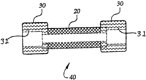
Figure 2 shows a tubular construct (20) and cuffs (30) in which the diameter
of the inner
surface (31) of each cuff (30) is approximately equal to the outer diameter of
the tubular
CA 02306346 2000-04-04
WO 99/01538 PCT/US98/13828
-8-
construct (20). A cuff (30) may be attached to each end of the tubular
construct (20) to form the
compound construct (40).
Figure 3 shows a tubular construct (20) made of one substrate material, and a
layer or film
(25) of a second substrate material within the lumen of the tubular construct
(20). This
compound substrate construct is shown in cross-sectional (left) and side
(right) views.
Figure 4 shows a compound construct (40) comprising a first tubular construct
(20) joined
to two cuffs (30), which may be joined by connectors (50) to tubing (60)
leading to a bioreactor
flow system.
Figure 5 shows a compound construct (40) comprising a first tubular construct
(20) joined
to two cuffs (30), and further joined by connectors (50) to the tubing (60) of
a bioreactor flow
system. A distensible tube (70) is inserted within the lumen of the compound
construct (40) to
apply pulsatile stretching force to the construct.
Detailed Description of the Invention
1. Definitions
In order to more clearly and concisely point out the subject matter of the
claimed
invention, the following definitions are provided for specific terms used in
the following written
description and appended claims.
Tissue-engineered construct. As used herein, a "tissue-engineered construct"
means a
three-dimensional mass of living mammalian tissue produced primarily by growth
in vitro. The
construct may include one or more types of tissue, and each tissue may include
one or more types
of cells. A tissue-engineered construct is distinguished from an explant of a
corresponding natural
tissue in that the primary the growth of the construct occurs in vitro.
Porous substrate. As used herein, a "porous substrate" means a three-
dimensional
substrate of a biocompatible material which is suitable for attachment or
adherence of mammalian
cells, and which is sufficiently porous to allow for the infiltration of
seeded cells, and the diffusion
of nutrients and waste products to and from cells adhered to the substrate,
including cells adhered
within the interior pores or interstitial spaces of the substrate. Thus, a
porous substrate has pores
or interstitial spaces interspersed through its structure, and in fluid
communication with the
exterior, such that cells may infiltrate into the interior of the substrate.
The pores or interstitial
spaces may be roughly spheroidal spaces, such as the pores in a sponge-like
material, or may be
longitudinally extended and intersecting spaces, such as the inter-fiber
spaces in a fibrous mesh
CA 02306346 2000-04-04
WO 99/01538 PCT/US98/13828
-9-
material, or may be of any other arbitrary shape. As used herein, no
distinction is made between
the "pores" of sponge-like materials, the "interstitial spaces" of fibrous
mesh materials, or the
arbitrarily shaped "spaces" of any other materials, and the term "porous"
embraces materials
characterized by any of these.
Synthetic polymer. As used herein, the term "synthetic polymer" means a non-
naturally
occurring polymer made by, for example, ex vivo synthesis, and physically
distinguishable from
naturally occurring polymers. Thus, the term is used herein merely to
distinguish synthetic
polymers, such as those described and enabled herein, from such naturally-
occurring polymers as
collagen, elastin, polysaccharides, cellulose, chitosan, and the like. A
synthetic polymer may
include one or more naturally-occurring subunits, such as naturally occurring
amino acids or
saccharide units, in an otherwise non-natural polymer (e.g., copolymers of
lysine or arginine with
lactic acid or glycolic acid).
Proteinaceous polymer. As used herein, the term "proteinaceous polymer" means
a
polymer consisting essentially of naturally-occurring or chemically modified
amino acids residues
joined by peptide linkages. Proteinaceous polymers of the invention may be
naturally-occurring
polymers which are extracted from animal tissues (e.g., collagen obtained from
connective
tissues), may be recombinantly produced polymers obtained from genetically
engineered
organisms (e.g., bacteria engineered to produce elastin), or may be produced
in vitro by chemical
synthesis. Thus, for example, as used herein, the term embraces such naturally-
occurring
proteinaceous polymers as collagen, elastin, fibronectin, laminin and the
like. A proteinaceous
polymer may also include one or more non-naturally-occurring subunits, such as
modified amino
acids (e.g., acylated, sulfonated, glycosylated, or otherwise conjugated
through reactive amino
acid side chain groups to moieties which increase hydrophilicity or provide
better cell-adhesion
characteristics), or may include non-peptide linkages joining two or more
proteinaceous
fragments (e.g., polypeptides or modified polypeptides copolymerized with
polyesters,
polyanhydrides).
Hydrophilic surface. As used herein, a "hydrophilic surface" means a surface
which is
"wettable" as that term is used in the art, or which, when subjected to a
sessile drop wettability
test, displays a contact angle with water of less than 90 . More preferably, a
hydrophilic surface
is one which displays a contact angle of less than 45 , 20 , 10 , or 5 . As
used herein, a "contact
angle" means the solid-liquid-gas contact angle where the solid is the
relevant polymer, the liquid
is water, and the gas is air. The value of the contact angle directly reflects
the surface and
CA 02306346 2000-04-04
WO 99/01538 PCT/US98/13828
-10-
interfacial energies based on Young's equation (see, e.g., Adamson, ed. (1990)
Physical
Chemistry of Surfaces, 5th Edition, John Wiley & Sons, Inc., New York, pp. 379-
420).
Distensible body. As used herein, a "distensible body" means a hollow body
comprising a
resilient material which, when subjected to repeated and sufficient
increases/decreases in pressure
within the interior of the body, can expand/contract so as to
increase/decrease in an exterior
dimension by at least 4-6%, preferably 4-10%, and more preferably 4-20%,
without rupturing. A
distensible body will have one or more openings by which it is attached to
means for increasing
the internal pressure, such as a tubing connected to a fluid pump. Examples of
distensible bodies
include distensible tubes which are substantially cylindrical in shape, and
distensible bladders
which may be substantially spheroidal or ellipsoidal in shape. Thus, for
example, the term
"distensible tube" includes substantially cylindrical devices made of a
resilient material which,
when subjected to repeated and sufficient increases in pressure within the
interior of the tube, can
distend or expand so as to increase circumferentially in diameter by at least
4-6%, preferably 4-
10%, and more preferably 4-20%, without rupturing.
Muscular. As used herein with reference to tissue engineered-constructs, the
term
"muscular" describes a tissue comprising or consisting of mammalian muscle
cells which have
grown substantially to confluence, and which can exert contractile force. In
certain preferred
embodiments, the muscle cells are smooth muscle cells. Skeletal muscle or
cardiac muscle cells,
however, may also be employed in the present invention.
Pulsatile stretch. As used herein, "pulsatile stretch" means a circumferential
stretching or
expansion of a substantially tubular object or construct, similar to the
circumferential stretching or
expansion of an artery in response to the cyclical increases and decreases in
blood pressure caused
by the beating of a heart.
Environment suitable for rg owth. As used herein, an "environment suitable for
growth" of
a particular cell type means an environment with conditions of temperature,
pressure, nutrient and
waste exchange, and gas exchange, which are permissive for the survival and
reproduction of the
cells. With respect to any particular type of cells, an environment suitable
for growth may require
the presence of particular nutrients required by that cell type, or the
presence of particular growth
factors necessary for the survival and reproduction of those cells.
CA 02306346 2000-04-04
WO 99/01538 PCT/US98/13828
-11-
II. General Considerations
The present invention provides several novel advances in methods and products
for use in
the field of tissue engineering. In particular, the present invention provides
new porous substrates
for the growth of mammalian cells which may be seeded onto and into these
substrates. In
addition, the present invention provides for new methods of producing muscular
tissue-engineered
constructs with lumens, in which a distensible body contained within the lumen
of the growing
construct applies a pulsatile force to growing tissue. This pulsatile force
mimics, in part, the
forces encountered by the cells in natural arterial and venous walls, the
alimentary canal, ureters,
the bladder, and other biological structures which include circumferentially
or peripherally
oriented rings of muscle. The use of a pulsatile force in the present
invention aids in the
organization of muscle cells into circumferential rings in the wall of the
construct, as well as the
development or maintenance of a contractile phenotype by these cells. In
addition, the present
invention provides for new growth media and methods for their use in the
production of tissue-
engineered constructs. These new growth media are believed to enhance the
production of an
appropriate extracellular matrix in the tissue-engineered construct, thus
increasing its strength.
Thus, according to one aspect of the present invention, a method for producing
a tissue-
engineered construct is provided in which a porous substrate comprising a
synthetic, polymeric,
biocompatible material is contacted or "seeded" with a suspension of a first
type of mammalian
cells to form a primary cell-seeded construct, and this cell-seeded construct
is maintained for a
first growth period in an environment suitable for growth of the cells to form
a primary tissue-
engineered construct. The porous substrate may be of essentially any size or
shape, may be a
sponge-like porous material or may be a fibrous mesh. Importantly, in this
aspect of the
invention, the substrates have hydrophilic surfaces, as described in more
detail below, which
permit cells to be seeded at a higher density, resulting in a higher final
density of cells in the final
tissue-engineered construct. The porous substrates of the invention are seeded
with cell
suspensions including at least one type of cell, but may be seeded with
suspensions comprising a
mixture of cells (e.g., hepatocytes and fibroblasts) to create a more complex
primary tissue
construct. After a first period of growth, the resulting primary tissue
construct may optionally be
seeded with a second suspension of cells including at least one cell type, and
this secondary cell-
seeded construct may be maintained for a second growth period to produce a
secondary tissue-
engineered construct. Further rounds of cell-seeding and growth may, of
course, be employed.
In addition, between any growth period and the next step of cell-seeding
(e.g., after production of
CA 02306346 2000-04-04
WO 99/01538 PCT/US98/13828
-12-
the primary tissue-engineered construct, but before production of a secondary
cell-seeded
construct), additional substrate material may be added, or the tissue-
engineered construct may be
inserted within a larger substrate. In this way, a complex organ-like
structure may be produced
by, for example, first producing a vascular tissue-engineered construct (by
one or more rounds of
cell-seeding and growth) and then embedding this in a larger substrate to
produce, for example, a
liver or other glandular tissue-engineered construct which will include an
internal, tissue-
engineered vascular system.
The present invention also provides novel methods particularly directed to the
production
of a muscular tissue-engineered construct. In these methods, a porous
substrate, comprising a
biocompatible material and defining a lumen, is contacted or "seeded" with a
suspension including
muscle cells (preferably, but not necessarily, smooth muscle cells) to form a
primary cell-seeded
construct, and this cell-seeded construct is maintained for a first growth
period in an environment
suitable for growth of the cells to form a primary tissue-engineered
construct. In addition,
however, a distensible body is provided, before or after seeding the muscle
cells, within the lumen
of the porous substrate. The distensible body is chosen to have a shape
substantially similar to the
shape of the lumen, and is capable, upon distension, of contacting the inner
surface of the
substrate (i.e., the walls of the lumen) so as to apply pulsatile stretching
forces to, and cause
distension of, the substrate. Preferably, the distensible body has outer
dimensions approximately
equal to the inner dimensions of the lumen. During the first growth period,
cyclical increases in
pressure within the distensible body are provided, thereby causing the body to
distend within the
lumen of the construct and to apply pulsatile stretch to the construct. In
addition, the primary
cell-seeded construct is preferably maintained in a growth medium which
includes certain factors,
described in more detail below, which enhance the development of the muscle
cell layer.
Optionally, after the first growth period, the resulting primary tissue-
engineered construct may be
seeded with a second suspension of cells including at least one cell type
(e.g., endothelial cells
applied to the outer and inner surfaces of the primary tissue-engineered
construct), and this
secondary cell-seeded construct may be maintained for a second growth period
to produce a
secondary tissue-engineered construct. During this second growth period, the
distensible body
may continue to be used to apply a pulsatile stretch or, if the primary tissue-
engineered construct
has sufficient strength, the distensible body may be removed and fluid flow
may be maintained
directly through the lumen, with or without additional pulsatile stretching.
As above, further
rounds of cell-seeding and growth may be employed, and the tissue-engineered
construct resulting
CA 02306346 2000-04-04
WO 99/01538 PCT/US98/13828
-13-
from any growth period may be incorporated into a larger porous substrate and
seeded to produce
a more complex organ-like construct.
In most preferred embodiments, the porous substrate is substantially tubular
or cylindrical
in shape, and the distensible body is a distensible tube. The resulting
muscular tissue-engineered
construct is characterized by circumferentially oriented rings of muscle, and
the construct can
form the basis of a vascular tissue-engineered construct or prosthesis,
preferably with an inner
lining of endothelial cells. Muscular, tubular constructs may also be produced
for esophageal,
intestinal, rectal, and ureteral prostheses.
These and other objects and advantages of the present invention are described
in more
detail in the preferred embodiments and examples below.
III. Preferred Embodiments
A. Porous Substrates for Tissue-Engineered Constructs
The porous substrates of the present invention may be any three dimensional
structure
comprising a biocompatible material which is sufficiently porous to allow for
infiltration of seeded
cells and diffusion of nutrients and waste products to and from cells adhered
to the surface,
including the inner surfaces, of the substrate. The feasibility of seeding and
culturing various cell
types on biocompatible, biodegradable substrates, including polymer films and
three-dimensional
scaffolds, has been demonstrated in the art (Takeda et al. (1995); Vacanti et
al. (1994); Mooney
et al. (1994); Cao et al. (1994); Bell (1994); Gilbert et al. (1993); Freed et
al. (1994a); Mooney et
al. (1994); Cima et al. (1991); Cima and Langer (1993); Wintermantel et al.
(1991); Mooney et al.
(1992); Freed et al. (1994b); Freed et al. (1993)). In accordance with the
present invention, the
substrate may be formed in essentially any shape including, but not limited
to, solid porous
substrates such as spheres, ellipsoids, disks, sheets or films, as well as
hollow porous substrates
such as hollow spheres or ellipsoids, and open-ended tubes. In preferred
embodiments for
muscular, tubular tissue-engineered constructs, the substrates comprise
substantially tubular or
cylindrical shapes, including tubular shapes with diameters which vary along
the length of the
substrate.
Preferably, the substrate material comprises a biodegradable or bioerodable
material, such
as one which is slowly hydrolyzed under physiological conditions. Thus,
generally, any
biocompatible, slowly hydrolyzable polymers may be employed. Preferred
substrate materials
include polymeric materials such as polyesters, polyorthoesters, or
polyanbydrides, including
CA 02306346 2000-04-04
WO 99/01538 PCT/US98/13828
-14-
polymers or copolymers of glycolic acid, lactic acid, or sebacic acid. More
generally, preferred
substrate materials include polyesters of straight chain or branched,
substituted or unsubstituted,
saturated or unsaturated, linear or cross-linked, alkanyl, haloalkyl,
thioalkyl, aminoalkyl, aryl,
aralkyl, alkenyl, aralkenyl, heteroaryl, or alkoxy hydroxy acids (e.g.,
(COOH)(CH2)õ (OH) or
(COOH)(CRR;),,(OH), where n is an integer between about 1 and 20, and each R;
and R; is
independently selected from the group consisting of -H, -OH, -SH, -NH2, the
halogens, the side
chains of the naturally occurring amino acids, and any straight chain or
branched, substituted or
unsubstituted, saturated or unsaturated, low molecular weight (e.g., C1-C14)
alkanyl, haloalkyl,
thioalkyl, aminoalkyl, aryl, aralkyl, alkenyl, aralkenyl, heteroaryl, or
alkoxy group, or a secondary
or tertiary amine substituted with such groups) or polyanhydrides of straight
chain or branched,
substituted or unsubstituted, saturated or unsaturated, linear or cross-
linked, alkanyl, haloalkyl,
thioalkyl, aminoalkyl, aryl, aralkyl, alkenyl, aralkenyl, heteroaryl, or
alkoxy dicarboxylic acids
(e.g., (COOH)(CH2)õ (COON) or (COOH)(CR;R;)õ (COOH), where n is an integer
between about
1 and 20, and each R; and R; is independently selected from the group
consisting of -H, -OH, -SH,
-NH2, the halogens, the side chains of the naturally occurring amino acids,
and any straight chain
or branched, substituted or unsubstituted, saturated or unsaturated, low
molecular weight (e.g.,
C1-C14) alkanyl, haloalkyl, thioalkyl, aminoalkyl, aryl, aralkyl, alkenyl,
aralkenyl, heteroaryl, or
alkoxy group, or a secondary or tertiary amine substituted with such groups).
Polymers including
mixtures of ester and anhydride bonds (e.g., copolymers of glycolic and
sebacic acid) may also be
employed. Thus, for example, preferred substrate materials include
polyglycolic acid polymers
(PGA), polylactic acid polymers (PLA), polysebacic acid polymers (PSA),
poly(lactic-co-glycolic)
acid copolymers (PLGA), poly(lactic-co-sebacic) acid copolymers (PLSA),
poly(glycolic-co-
sebacic) acid copolymers (PGSA), etc.
Other biocompatible biodegradable polymers useful in the present invention
include
polymers or copolymers of caprolactones, carbonates, amides, amino acids,
orthoesters, acetals,
cyanoacrylates and degradable urethanes, as well as copolymers of these with
straight chain or
branched, substituted or unsubstituted, alkanyl, haloalkyl, thioalkyl,
aminoalkyl, alkenyl, or
aromatic hydroxy- or di-carboxylic acids. In addition, the biologically
important amino acids with
reactive side chain groups, such as lysine, arginine, aspartic acid, glutamic
acid, serine, threonine,
tyrosine and cysteine, or their enantiomers, may be included in copolymers
with any of the
aforementioned materials. The currently preferred biodegradable materials are
PLA, PGA, and
CA 02306346 2000-04-04
WO 99/01538 PCT/US98/13828
-15-
PLGA polymers. See, generally, U.S. Pat. Nos. 1,995,970; 2,703,316; 2,758,987;
2,951,828;
2,676,945; 2,683,136 and 3,531,561.
Biocompatible but non-biodegradable materials may also be used in the porous
substrates
of the present invention. For example, non-biodegradable polymers of
acrylates, ethylene-vinyl
acetates, acyl substituted cellulose acetates, non-degradable urethanes,
styrenes, vinyl chlorides,
vinyl fluorides, vinyl imidazoles, chlorosulphonated olefins, ethylene oxide,
vinyl alcohols,
TEFLON (DuPont, Wilmington, DE), and nylons. See, generally, U.S. Pat. Nos.
2,609,347;
2,653,917; 2,659,935; 2,664,366; 2,664,367; and 2,846,407.
As an alternative to synthetic polymer substrates, porous substrates may be
employed
which comprise proteinaceous polymers. Such substrates are known in the art
and have been
used in the production of tissue-engineered constructs. For example, collagen
gels have been
used to produce vascular tissue constructs (Weinberg and Bell, (1986)), and
collagen sponges and
meshes are now commercially available (e.g., from Ortec International, Inc.,
New York, New
York). Such collagenous substrates, as well as similarly constructed
substrates based on elastin,
fibronectin, laminin, or other extracellular matrix or fibrillar proteins, may
be employed in the
methods and constructs of the present invention. Such proteinaceous polymer
substrates may be
in the form of fibrous meshes, as described above, or may be in the form of
non-fibrous substrates
such as sheets, films, or sponges. In addition, these substrates may include
proteinaceous
polymers which have been modified by, for example, acylating, sulfonating,
glycosylating, or
otherwise conjugating reactive groups of the amino acid side chains with other
moieties to
increase hydrophilicity and/or provide better cell-adhesion characteristics.
For example, the
proteins may be acylated with dicarboxylic acid anhydrides to increase
hydrophilicity, or may be
conjugated to cell-adhesion peptides to increase the density or avidity of
cell-seeding. Such
proteinaceous polymers have the advantage that they are completely biological
in nature and,
therefore, will have reduced immunogenicity if syngeneic to the host.
The porous substrate may comprise a randomly cross-linked material in the form
of a
sponge or, preferably, may comprise a porous mesh of fibers. For example, in
preferred
embodiments, the substrate comprises a porous mesh of fibers having a diameter
of between
approximately 5-20 gm, between approximately 10-15 m, or approximately 13
.Lm. Such
fibrous polymeric materials are known in the art and are commercially
available (e.g., fibrous PGA
polymers sold as DEXON (Sherwood Davis & Geck, Hampshire, UK), and fibrous
PLGA
CA 02306346 2000-04-04
WO 99/01538 PCT/US98/13828
-16-
polymers sold as VICRYL (Ethicon, Edinburgh, Scotland) which have been
approved by the
U.S. Food and Drug Administration for clinical use (Freed et al. (1994)). The
physical
characteristics and degradation rates of these polymers are known in the art
(Gilding and Reed
(1979)). The fibers may be solid or hollow, and may comprise a multiplicity of
materials (e.g., a
solid fiber of two materials, or a hollow fiber of one material and a core of
another).
When a porous substrate is formed of a mesh of fibers, adjacent substantially
parallel
fibers, or adjacent substantially parallel portions of fibers, are preferably
separated, on average, by
approximately 20-200 m or, more preferably, approximately 50-100 pm, to
define pores or
interstitial spaces which have similar dimensions. When a porous substrate is
formed of a sponge-
like material, the pores are preferably, on average, approximately 20-200 m
or, more preferably,
approximately 50-100 m, in each cross-sectional dimension. The pores of the
porous substrate
will define a void volume, as that term is known in the art. To allow for a
high density of cell
seeding within the pores or interstitial spaces of the substrate, the porous
substrate of the present
invention is characterized by a void volume of greater than at least 80%,
preferably 90% or, more
preferably, greater than 95%. Most preferably, the void volume is about 97%.
In one aspect of the present invention, improved tissue-engineered constructs
are provided
by employing a porous substrate with a hydrophilic surface. Without being
bound to any
particular theory of the invention, it is believed that the hydrophilic
surface aids in the attachment
or adherence of certain cell types, including smooth muscle cells, to the
substrate. Such a
hydrophilic surface preferably comprises a multiplicity of hydrophilic
chemical groups such as
carboxyl, hydroxyl, thiol, amine, sulfonyl, guanidine, and amide groups. When
the porous
substrate comprises a polyester, polyorthoester or polyanhydride material, a
hydrophilic surface
may conveniently be prepared by hydrolyzing the outer surface of the fibers
(e.g., by treatment
with a base) to cause ester or anhydride bonds accessible at the surface to be
hydrolyzed to
carboxyl and/or hydroxyl groups. These groups may be further derivatized, if
desired, to thiol,
sulfonyl, guanidine, amine or amide groups by standard organic chemical
techniques, and cell
adhesion peptides may also be bound to the surface. For example, Barrera and
co-workers
(Barrera et al. (1993)) have synthesized a copolymer of lactic acid and lysine
that allows for the
covalent attachment of cellular adhesion peptides to the polymer backbone. The
peptide arginine-
glycine-aspartic acid (RGD), which is a cell-binding domain of fibronectin, as
well as several other
cell adhesion molecules (Massia and Hubbell (1990)), have been covalently
bound by their N-
CA 02306346 2000-04-04
WO 99/01538 PCT/US98/13828
-17-
termini to the lysine moieties of this copolymer. Preferred cell-adhesion
peptides for use in the
present invention include the sequences RGD and REDV (which is preferred for
binding
endothelial cells).
The hydrophilicity of the surface of the substrate material may be
conveniently analyzed by
measuring the contact angle of water drops on the surface of a film of the
material using the
sessile drop method (e.g., employing a Video Contact Angle System, ASC, Inc.).
A hydrophilic
surface is one in which the contact angle is less than 90 . Preferably,
however, the hydrophilic
surface has a contact angle of less than 45 , 20 or 10 . In most preferred
embodiments, the
contact angle is less than 5 .
As noted above, in preferred embodiments the substrate comprises a
biodegradable
material such that, after a sufficient period of growth, the resulting tissue-
engineered construct is
substantially free of any remaining substrate material. For example, the
degradation of a PGA
substrate material having fiber diameters of approximately 13 tm was measured
without cultured
cells in phosphate buffered saline at 37 C. Under such conditions, PGA
undergoes bulk-
hydrolysis that appears to have first order kinetics in two stages.
Approximately 50% of the mass
degraded within 1-4 weeks. Even after many weeks (e.g., 3-8 weeks), however,
traces of the
matrix material may still be observed microscopically. By varying the
thickness of the fibers, as
well as their chemical composition, one of ordinary skill in the art can
readily produce
biodegradable polymeric fibers, as described above, having essentially any
desired degradation
characteristics. In addition, to reduce the mass of the substrate material,
and therefore its
degradation time, without reducing the surface area initially available for
cell adherence, hollow
fibers, fibers with a core of more readily degradable material, or fibers with
a core filled with a
biocompatible solution, may be employed. In general, it is preferred that a
substrate of
biodegradable material is employed such that, when the tissue growing on the
construct has
reached a density of approximately 1-3 x 10' cells/cc, approximately 70-100%
of the substrate
material is substantially degraded.
Finally, it should be noted that the degradation products of some substrate
materials may
have some adverse effects on cell growth even if the substrate material itself
is biocompatible.
Thus, for example, the hydrolytic degradation of polymers of organic acids
(e.g., PGA, PGLA)
releases free acids which, at the least, lower the pH in the local environment
and may also have
other physiological effects. Therefore, it may be desirable to include within
a substrate material a
CA 02306346 2000-04-04
WO 99/01538 PCT/US98/13828
-18-
neutralizing agent which will, at least partially, offset the effects of
substrate degradation. For
example, copolymers of organic acids and bases may be produced such that the
degradation
products tend to titrate or buffer each other. In the case of polymers of
organic acids, a base such
as lysine or arginine (or any other biocompatible base) may be included in a
copolymer (e.g., a
glycolic acid-lactic acid-lysine copolymer). Alternatively, if a hollow fiber
is employed, the core
may be filled with an alkaline solution or alkaline degradable material to
offset the increase in
acidity caused by fiber degradation.
B. Variations on Substrate Structures
As noted above, the porous substrates of the present invention may assume
essentially any
shape. In particularly preferred embodiments, however, tubular substrates are
utilized. In
addition, "compound" substrates comprising more than one substrate material
are also useful in
many embodiments. Thus, for example, a compound substrate may be produced
which comprises
a first porous substrate material joined to a second porous substrate
material, in which the two
materials differ in some characteristic such as biodegradability, pore size,
void volume, or
hydrophilicity. Alternatively, a porous substrate may be joined to a non-
porous substrate, such as
a film, to form a compound substrate in which the two materials may differ not
only in their
porosity, but also in other characteristics such as biodegradability or
hydrophilicity. Such
compound substrates may be seeded in one portion (e.g., a porous portion) with
one type of cells,
and in another portion (e.g., a non-porous portion) with a different type of
cells. The different
portions may be seeded with cells simultaneously, or at different times (e.g.,
after one or more
growth periods). In addition, the compound substrate can be formed after one
or more rounds of
cell seeding and growth, by adding a new substrate portion to a primary (or
later) tissue-
engineered construct.
In a preferred embodiment for producing muscular, tubular tissue-engineered
constructs, a
compound substrate is employed. Thus, referring to Figure 1, a rectangular
piece of porous mesh
material (10) having a length (1) and width (w) is rolled along its length to
form a substantially
tubular porous substrate (20), with an outer surface (22), and an inner
surface (21) defining a
lumen. The edges along the length (1) of the mesh (10) are joined in any
appropriate manner (e.g.,
by sewing with uncoated PGA suture (Davis & Geck, Inc., Manati, P.R.), or by
chemical
bonding) to form the tubular construct (20). The construct may be of arbitrary
length, but porous
substrates of 1-20 cm are currently contemplated as being most useful. The
width of the substrate
CA 02306346 2008-07-02
-19-
material is also arbitrary, but is chosen to produce a tubular substrate with
an inner lumen having
a diameter useful for the intended purpose. For vascular tissue constructs, it
is currently
contemplated that inner lumens of 2-10 mm or, preferably, 3-6 mm- will be most
useful. For
esophageal, intestinal, or rectal constructs, correspondingly larger lumens
would be employed.
The thickness of the substrate (i.e., the distance between the inner (21) and
outer (22) surfaces) is
chosen depending upon the desired thickness of the resulting tissue engineered
construct. For
vascular tissue constructs, a thickness of between 0.25-2.5 mm or, preferably,
about 0.5-2.0 mm
is currently contemplated as being most useful. As will be obvious to one of
skill in the art, the
tubular substrate (20) need not be formed by rolling a flat mesh to form a
tube but, rather, can be
produced as a single piece by, for example, weaving or extrusion.
Next, as the tubular porous construct (20) is preferably made of a
biodegradable material,
additional porous tubular portions or "cuffs" (30) made of a non-biodegradable
material are
optionally but preferably added to each end of the first construct to
facilitate attachment of the
construct to the bioreactor system. Thus, referring to Figure 2, two
substantially tubular cuffs
(30) made of a non-biodegradable material, such as a porous Dacron vascular
graft material (Bard
Vascular Systems Division, Haverhill, MA), are attached to the ends of the
first tubular construct
(20) by any appropriate means (e.g., suture or chemical bonding) to form a
compound construct
(40). Note that the inner surface (31) of the cuffs (30) defines a diameter
which is preferably
chosen to be approximately equal to the diameter of the outer surface (22) of
the porous substrate
tube (20). The cuffs (30) are preferably chosen to be porous so that they may
also be cell-seeded
and form a substantially continuous layer of cells with those seeded onto the
central portion (20)
of the construct (40). Importantly, however, as the biodegradable substrate of
the central portion
(20) dissolves during cell culture and growth, the non-degradable substrate
material of the cuffs
(30) remains to add strength to the ends of the tissue construct. This
strength is helpful in
attaching the construct to the flow apparatus described below, but the cuff
portion may be
removed at a later time (e.g., for implantation in vivo) if desired.
More complex substrate structures are also contemplated. For example, the
porous mesh
material (10) need not be uniform in composition, such that the inner surface
(21), the outer
surface (22), and/or the substrate material between these surfaces, differ in
some characteristic
such as biodegradability, pore size, void volume, or hydrophilicity. Thus,
when used for the
production of muscular, tubular tissue-engineered constructs, it is
contemplated that a substrate
material which degrades more slowly, has smaller pores, and/or has lower void
volume may be
CA 02306346 2000-04-04
WO 99/01538 PCT/US98/13828
-20-
preferred for one or more surfaces. In particular, if intralumenal flow is
desired (with or without
the presence of a distensible tube or sheath and pulsatile stretching force),
it may be desirable to
have the inner surface (21) of the tubular construct degrade more slowly, have
smaller pores,
and/or have a lower void volume. Alternatively, a substrate film (25) which is
non-porous or
slightly porous may be inserted within the lumen of a tubular construct (20)
and contacted with or
affixed to the inner surface (21) to form a compound substrate with an inner
film and outer
porous portion, as shown in Figure 3. For example, a tubular porous mesh of
PGA or PGLA
fibers having diameters of 5-20 gm, as described above, may be provided with
an inner film of
PGA, PGLA, or a protein (e.g., collagen, elastin, fibronectin, laminin,)
having a thickness of 5-50
or, preferably 10-30 m. The desired thickness of the inner film depends, at
the least, upon the
material from which it is made, the culture conditions, and the desired length
of time before the
film substantially degrades. Alternatively, or in addition, such films may be
added to the outer
surface (22) of the tubular construct (20).
C. Cell-Seeding and Growth in Tissue-Engineered Constructs
A number of different cell types or combinations thereof may be employed in
the present
invention, depending upon the intended function of the tissue-engineered
construct being
produced. Thus, for example, smooth muscle cells and endothelial cells may be
employed for
muscular, tubular tissue-engineered constructs (e.g., vascular, esophageal,
intestinal, rectal, or
ureteral constructs); hepatocytes and bile duct cells may be employed in liver
tissue-engineered
constructs; pancreatic islet cells may be employed in pancreatic tissue-
engineered constructs;
thyroid, parathyroid, adrenal, hypothalamic, pituitary, ovarian, testicular,
or salivary secretory
cells may be employed in corresponding glandular tissue-engineered constructs;
cardiac muscle
cells may be employed in heart tissue-engineered constructs; renal cells may
be employed in
kidney tissue-engineered constructs; chondrocytes may be employed in
cartilaginous tissue-
engineered tissue constructs; and epithelial, endothelial, fibroblast and
nerve cells may be
employed in tissue-engineered constructs for the great variety of tissues in
which these cells are
found. More generally, any cells may be employed which are found in the
natural tissue to which
the tissue-engineered construct is intended to correspond. In addition,
progenitor cells, such as
myoblasts or stem cells, may be advantageously employed to produce their
corresponding
differentiated cell types in a tissue-engineered construct.
CA 02306346 2000-04-04
WO 99/01538 PCTIUS98/13828
-21-
Thus, for example, natural arteries are comprised of endothelial, smooth
muscle, and
fibroblast cells organized into three layers: the intima, the media and the
adventitia. The intima is
composed primarily of endothelial cells and has three parts: the endothelium,
an intermediate
layer, and the internal elastic lamina. The media of small arteries consists
of 25 to 40 layers of
circumferentially disposed smooth muscle fibers between layers of connective
tissue, while the
media of veins contain relatively fewer (e.g., 5-20) layers of smooth muscle.
Fibroblasts appear
primarily in the adventitia in vivo and are not major components of the normal
intimal or medial
layers. Therefore, a vascular tissue-engineered construct will preferably
include each of these cell
types. Smooth muscle cells, for example, may be seeded onto a porous substrate
to form a
primary cell-seeded tissue construct which is allowed to grow for a first
period to form a primary
tissue-engineered construct. A low percentage of fibroblasts may also be
included in this initial
construct to increase the strength of the resulting construct. Endothelial
cells may then be seeded
onto the inner surface or lumen and, optionally, onto the outer surface of the
construct to form a
secondary cell-seeded construct. After a second growth period, this will
produce a secondary
tissue-engineered construct having a layer of smooth muscle cells (and,
optionally, fibroblasts)
between layers of endothelial cells.
Preferably, the cells are obtained from a live donor and cultured as a primary
cell line. In
particular, if the tissue-engineered construct is intended to be implanted
into a living host, the cells
are preferably harvested from the intended host or a histocompatible donor,
thereby minimizing or
eliminating the possibility of tissue rejection. For example, the required
cells may be obtained
from a biopsy of the patient. Thus, in the case of a patient requiring a
coronary by-pass
procedure, a biopsy of an artery (e.g. subclavian, axillary, brachial, radial,
iliac, ulnar, femoral,
anterior or posterior tibial) or peripheral vein (e.g., cephalic, basilic,
saphenous, femoral) may be
used to obtain arterial smooth muscle, endothelial and fibroblast cells.
Alternatively, in the case
of a patient requiring, for example, a liver, pancreatic, ureteral,
esophageal, intestinal, rectal or
other tissue-engineered implant, appropriate cells may be obtained by biopsies
of these tissues. It
should also be noted that, although not necessarily preferred, biopsies from
tissues or organs
which do not correspond to the intended implant, but which are phenotypically
similar, may be
employed. For example, smooth muscle cells derived from an artery may be
employed in
producing the smooth muscle layers of a venous, esophageal, intestinal,
rectal, cardiac or ureteral
tissue-engineered construct.
CA 02306346 2000-04-04
WO 99/01538 PCT/US98/13828
-22-
To obtain cells from a donor, standard biopsy techniques known in the art may
be
employed. Briefly, a desired tissue is surgically removed and the tissue is
minced or
homogenized, optionally with protease (e.g., trypsin or collagenase)
treatment, and a suspension
of dissociated cells, or small aggregates of cells, is prepared. Optionally,
the cells may then be
cultured in vitro in a standard cell growth medium until a suitable number or
density of cells are
obtained. Although cells may be passaged many times in such cultures, such
passaging often
causes a loss of differentiated phenotype and, therefore, it is preferred that
the number of passages
be limited to fewer than 5 or, more preferably, fewer than 3. Most preferably,
the cells are not
passaged at all.
Alternatively, cells may be employed which are derived from an established
cell culture
line, either derived in a laboratory or purchased from commercial sources
(e.g., ATTC, Rockville,
MD). Typically, such cell lines have lost some degree of differentiation and,
therefore, they are
generally not preferred. When established cell lines are employed, fetal cell
lines or progenitor
cell lines may be more desirable because such cells are generally more robust.
These cells may
also be grown in vitro in a standard cell growth medium until a suitable
number or density of cells
are obtained.
In another embodiment of the invention, cells are employed which have been
genetically
manipulated by the introduction of exogenous genetic sequences, or the
inactivation or
modification of endogenous sequences. Thus, for example, genes may be
introduced to cause the
cells to make proteins which are otherwise absent or defective in the host.
Alternatively,
production of scarce, but naturally occurring and desirable proteins, such as
elastin, may be
enhanced by appropriate genetic manipulations of the seeded cells. When
implanted into a host,
tissue-engineered constructs bearing such cells may serve as a production and
delivery system for
proteins which are otherwise absent, defective, or insufficient in the host.
Thus, for example,
genetically engineered endothelial cells that secrete tissue plasminogen
activator have been seeded
onto various synthetic grafts by Shayani and coworkers (Shayani et al.
(1994)), and Chen (Chen
et al. (1994)) has demonstrated the feasibility of adenovirus-mediated gene
transfer into the
endothelial cells of autologous vein grafts as a possible method to improve
patency.
Alternatively, repression of gene expression may also be used to modify
antigen
expression on the surface of seeded cells and tissue constructs, thereby
modifying the host's
immune response so that cells are not recognized as foreign. Thus, for
example, cells incapable of
producing one or more MHC proteins, or incapable of loading MHC molecules with
antigenic
CA 02306346 2000-04-04
WO 99/01538 PCT/US98/13828
-23-
peptides, may be employed to reduce the likelihood of tissue rejection. In
such cases,
immunosuppression may not be needed when a non-autologous tissue-engineered
construct is
implanted into a host.
In accordance with the present invention, mammalian cells are seeded onto and
within a
porous substrate from a suspension so that, preferably, they are evenly
distributed throughout the
substrate at a relatively high density. Preferably, the cell suspensions
comprise approximately
1x10⁴ to 5 x 10⁷ cells/ml of culture medium, preferably 2 x 10⁶ to 2 x 10⁷
cells/ml, and more
preferably about 5 x 10⁶ cells/ml. The optimal concentration of cells in a
suspension may, of
course, vary according to cell type, the propensity of the cells to form
aggregates, the growth rate
of the cell type, their binding affinity for the substrate used, and the
substrate material used. The
suspension may be formed in any physiologically acceptable fluid which does
not damage the cells
or impair their binding ability (e.g., a standard cell growth medium such as
DMEM supplemented
with 10% fetal bovine serum).
The cells may be seeded onto and within the porous substrate constructs of the
invention
by any standard method. For example, in one embodiment, the substrate is
seeded by submersion
into a cell suspension for a fixed period of time, and then the substrate is
removed from the
suspension and unbound cells are washed away. Alternatively, the substrate may
be seeded with
cells using a syringe or other sterile delivery apparatus. In a currently
preferred embodiment, the
cell suspension is dripped onto the substrate and subsequently the substrate
is rotated in, for
example, a rotating vessel. A tubular substrate, for example, as used in
making a muscular,
tubular tissue-engineered construct (e.g., a vascular construct), may be
rotated about its lumenal
axis during or after cell seeding to promote even distribution of the cells
onto the surface of the
substrate. After allowing a period of time for the cells to bind (optionally
incubating the cell-
seeded substrate in growth medium for a period), the cell-seeded substrate may
be immersed in
culture medium.
The "seeding time," or the time between initially contacting the mammalian
cells with the
substrate and later adding medium, may be varied significantly. Seeding times
of one hour or
more have been employed in the prior art. In the present invention, however,
particularly when
employing the hydrophilic, synthetic polymeric substrates described and
disclosed herein, it has
been found that substantially shorter seeding times, from 10-30 minutes or,
more preferably,
about 20 minutes, yield high densities of individually seeded cells with
reduced formation of cell
aggregates. This seeding time is to be distinguished from the "growth periods"
discussed below.
CA 02306346 2000-04-04
WO 99/01538 PCT/US98/13828
-24-
As noted above, the substrates of the present invention may be seeded with
suspensions
comprising a multiplicity of cell types. Thus, for example, a mixture of two
or more cell types
(e.g., smooth muscle cells and fibroblasts, or smooth muscle cells and
endothelial cells) may be
seeded onto a substrate simultaneously, or one or more cell types can be
seeded first, followed by
seeding with one or more additional types before cell-seeded substrate is
placed under suitable
conditions for a growth period. In either case, this may be regarded as a
single "seeding"
although several cell types may be seeded in one or more steps. Thus, as used
herein, a "primary
cell-seeded construct" is a substrate which has been subjected to a first
seeding with at least one
cell type, but possibly more than one cell type, but which has not yet been
maintained under
suitable conditions for a growth period.. During the first growth period, the
cells of the primary
cell-seeded construct grow and reproduce to yield a "primary tissue-engineered
construct" in
which the cells may or may not have yet reached confluence. This primary
tissue-engineered
construct may then be seeded a second time, again with one or more suspensions
comprising one
or more cell types, to form a "secondary cell-seeded construct." After
maintaining the secondary
cell-seeded construct under suitable conditions for a second growth period,
during which the cells
from the second seeding may grow and reproduce, the resulting construct is
referred to herein as
a "secondary tissue-engineered construct." Thus, for example, a vascular
tissue-engineered
construct may be produced by seeding smooth muscle cells onto the outer
surface of a tubular
porous substrate to form a primary cell-seeded construct which is maintained
for a first growth
period to form a primary tissue-engineered construct, and this construct may
then be seeded with
endothelial cells (and, optionally, fibroblasts) on the lumenal (and,
optionally, outer) surface to
form a secondary cell-seeded construct, which is maintained under suitable
conditions for a
second growth period to form a secondary tissue-engineered construct.
Similarly, any number of
additional constructs (tertiary, etc.) comprising various cell layers or
admixtures, can be
engineered according to the present invention (e.g., by inserting a vascular
tissue-engineered
construct into a larger substrate which is seeded with, for example,
hepatocytes to form,
ultimately, a vascularized liver tissue-engineered construct).
Suitable growth conditions and media for cells in culture are well known in
the art. Cell
culture media typically comprise essential nutrients, but also optionally
include additional elements
(e.g., growth factors, salts and minerals) which may be customized for the
growth and
differentiation of particular cell types. For example, "standard cell growth
media" include
Dulbecco's Modified Eagles Medium, low glucose (DMEM), with 110 mg/L pyruvate
and
CA 02306346 2000-04-04
WO 99/01538 PCTIUS98/13828
- 25 -
glutamine, supplemented with 10-20% Fetal Bovine Serum (FBS) or 10-20% calf
serum (CS) and
100U/ml penicillin. Other standard media include Basal Medium Eagle, Minimal
Essential Media,
McCoy's 5A Medium, and the like, preferably supplemented as above
(commercially available
from, e.g., JRH Biosciences, Lenexa, KS; GIBCO, BRL, Grand Island, NY; Sigma
Chemical Co.,
St. Louis, MO).
For use in the methods of the present invention, several variations on
standard cell growth
media have been developed. In particular, when growing smooth muscle cells, it
has been found
that the inclusion of the streptomycin should be avoided, as this commonly
used antibiotic tends
to inhibit the development of the desired phenotype in response to externally
applied physical
forces, such as the pulsatile force of the invention. In addition, for growing
any cells which
normally produce a substantial collagenous extracellular matrix, an "enhanced
cell growth
medium" has been developed which comprises standard cell growth medium, as
described above,
supplemented with 1-10 mM, preferably 5 mM, HEPES buffer; 0.01-0.1 g/L,
preferably 0.02-0.06
g/L, Vitamin C; 0.01-0.1 g/L, preferably 0.02-0.06 g/L, proline; 0.01-0.1 g/L,
preferably 0.02-
0.06 g/L, glycine; 0.01-0.1 g/L, preferably 0.02-0.06 g/L, alanine; and 0.5-
5.0 4g/L, preferably
1.0-3.0 g/L, of a copper salt (e.g., CuSO4). Because Vitamin C has a half-
life of only 6-8 hours
at 37 C in culture medium, Vitamin C is preferably replenished daily to
enhance collagen
synthesis by the cells. In addition, proline, glycine, and alanine are
provided in excess to provide
adequate amounts of these amino acids for the synthesis of collagen and other
extracellular matrix
proteins such as elastin. Copper ions are a necessary co-factor for elastin
synthesis and, therefore,
a source of copper ions (e.g., CuSO4) is preferably included in media used to
grow elastin-rich
tissues. For the growth of endothelial cells, it is preferred that CS be used
rather than FBS. In
addition, growth factors, such as acidic fibroblast growth factor (aFGF),
basic fibroblast growth
factor (bFGF), platelet-derived growth factor (PDGF), transforming growth
factor 0 (TGF-0), or
vascular endothelial cell derived growth factor (VEGF), may be employed at
suitable
concentrations (i.e., I-10 ng/ml) to enhance cell growth or differentiation or
the secretion of
extracellular matrix proteins.
Cells are cultured under sterile conditions in an atmosphere of 5-15% or,
preferably, 10%
CO2 and 90-100% or, preferably, 100% humidity in culture medium at or near the
body
temperature of the species of origin of the cells or the intended host (i.e.,
body temperature
f5 C, preferably f2 C). Thus, for example, human cells may be cultured at 32-
43 C, more
preferably 35-39 C, and most preferably 37C. Cell viability may be determined
by standard
CA 02306346 2000-04-04
WO 99/01538 PCT/US98/13828
-26-
methods (e.g., trypan blue exclusion) known in the art, or by measuring cell
attachment to, and
the extent of proliferation on, the substrate. Quantitative assessment of in
vitro cell attachment
and viability may also be assessed using scanning electron microscopy,
histology, and the
incorporation of radioisotopes (e.g., 3H thymidine) according to art known
methods.
To further enhance the attachment of cells to the substrate and/or to each
other, various
proteins or growth factors may be provided. For example, collagen, elastin,
fibronectin, or
laminin, may be provided to the substrate or to the growing constructs to
promote cell adhesion.
Thus, overlaying collagen on a material such as a polyanhydride substrate can
increase adhesion
of cells such as hepatocytes. Similarly, the substrate or construct can be
impregnated with growth
factors such as aFGF, bFGF, PDGF, TGF-13, VEGF, and various other angiogenic
and/or other
bioactive compounds that may be incorporated directly into the substrate or
otherwise contacted
with the growing cells (e.g., by addition to the cell culture medium).
Multiple growth factors
have been studied for their mitogenic effects on endothelial and smooth muscle
cells (D'Amore
and Smith (1993)). For example, aFGF, bFGF, PDGF have been found to stimulate
smooth
muscle cell proliferation, while bFGF and VEGF stimulate aortic endothelial
cell growth. Basic
FGF and VEGF have also been shown to bind to the subendothelial extracellular
matrix and
basement membrane, and are potent angiogenic factors (Edelman et al. (1991);
Rogelj et al.
(1989)).
D. Applying Pulsatile Stretch to Muscular Tissue Constructs
In another aspect of the present invention, a method for producing a muscular
tissue-
engineered construct is provided in which a distensible body is inserted
within the lumen of a
porous substrate to provide pulsatile stretch to seeded muscle cells. Thus, a
substantially tubular
porous substrate may be provided which defines a lumen, and a distensible tube
is inserted within
that lumen either before or after the porous substrate is seeded with muscle
cells. While the
muscle tissue is growing on and/or within the substrate, a pump in
communication with the
interior of the distensible body may then provide cyclic increases in pressure
(e.g., by pumping a
fluid or gas) to cause the distensible body to distend within the lumen of the
porous substrate,
contacting the inner surface of the substrate, and imparting a pulsatile
stretching force to the
substrate and growing muscle tissue. Without being bound to any particular
theory of the
invention, it is believed that this pulsatile stretch may enhance the
orientation of the muscle cells
into circumferential rings of muscle around the lumen, and may also enhance
cell-cell adhesion,
CA 02306346 2000-04-04
WO 99/01538 PCT/US98/13828
-27-
the formation of extracellular matrix, and the development and maintenance of
an appropriate
smooth muscle cell contractile phenotype.
In preferred embodiments for producing a vascular tissue construct, a
distensible tube is
distended in a cyclic manner which mimics a pulse of the organism from which
the seeded cells
are derived. The pulse rate may be chosen to mimic the pulse rate of the adult
organism, or the
higher pulse rate of the fetal organism. Thus, for example, a pulse rate of
approximately 60-
90/min, typically about 75/min, would mimic a resting pulse of a human adult.
A pulse rate of
approximately 140-160/min would mimic a human fetal pulse rate. In addition,
higher pulse rates
may be generally preferred as they may provide a greater stimulus for
development of a
contractile phenotype and mechanical strength in muscular tissue. In addition,
for a vascular
construct, the degree of pulsatile stretch induced in a cell-seeded construct
or a tissue-engineered
construct, as measured by the induced change in diameter of the construct, is
preferably chosen so
as to mimic that seen in a natural artery, but without applying excessive
stretch which would
disrupt the growing tissue. Thus, for example, after cell-seeding and during
the early part of the
growth period in which the cells are reaching confluence, a relatively low
pulsatile stretch may be
applied which causes the diameter of the construct to increase 2-10%, more
preferably 2-6% with
each pulse. Higher levels of pulsatile stretch at this early stage may disrupt
or tear the tissue, and
result in perforations in the vascular tissue construct. Later, after the
cells have reached
confluence, the tissue has thickened, and the construct has begun to assume an
arterial histology
(e.g., after 3-8 weeks), the cyclical increases in pressure within the
distensible tube may be
increased so that a pulsatile stretch of 6-10% or even 10-20% may be applied
to the construct.
As the pulsatile stretch seen in natural arteries varies from approximately 5%
to approximately
20% (depending upon the diameter and location of the artery), it is expected
that pulsatile
stretches of 5-20%, or somewhat exceeding 20%, will be useful in producing
vascular tissue
constructs.
Similarly, the application of a pulsatile stretching force may be used in the
production of
other, non-vascular, but muscular, tubular tissue constructs. In each case,
the diameter of the
construct is chosen so as to approximate the diameter of the corresponding
natural tissue or
organ, and the pulsatile stretching force is chosen to approximate the
corresponding natural
forces. Thus, esophageal, intestinal or rectal tissue constructs may be
produced in which the
diameter of the construct approximates the diameter of a section of the
esophagus, intestine or
rectum, and in which the pulsatile force approximates the forces caused by
peristaltic waves in a
CA 02306346 2000-04-04
WO 99/01538 PCT/US98/13828
-28-
corresponding section of the alimentary canal. Alternatively, a ureteral or
other muscular, tubular
tissue construct may be produced in which the diameter of the construct and
the pulsatile force
approximates the corresponding natural diameters and forces. In the case of a
bladder construct,
a distensible body may be employed which approximates the shape of the lumen
of the bladder,
and pulsatile stretch may be applied which approximates the internal pressures
experienced by a
natural bladder (e.g., a urinary bladder, or gall bladder).
E. Applying Intralumenal Flow to Muscular, Tubular Tissue Constructs
Although the use of a distensible tube within the lumen of a growing muscular,
tubular
tissue construct is preferred in some embodiments, such a tube is not
necessary to practice the
present invention. Indeed, in order to better mimic the conditions of
intralumenal flow and
pulsatile force found in natural muscular, tubular structures, it may be
preferred that a distensible
tube is not employed. For example, after a suitable growth period, if the
tissue forming the walls
of a tubular construct has achieved sufficient strength and has formed a
relatively fluid-tight seal
around the lumen, culture medium may be pumped directly through the lumen
(after removing the
distensible tube, if present). Alternatively, if a substrate is employed, as
described above, in which
the inner surface is substantially less porous than the outer surface (e.g.,
having a void volume less
than 25%, preferably less than 10%, and most preferably less than 5%), or in
which a substantially
non-porous film of substrate material is present on or adjacent to the inner
surface, a distensible
tube may not be needed, and fluid may be pumped directly through the lumen.
Preferably, the
inner surface is capable of resisting distension such that it increases in
internal diameter by
approximately 0.5-2.0%, preferably about 1.5%, per each 100 mm Hg of pressure
applied
internally.
In an alternative embodiment, a sheath or "sleeve" is provided which surrounds
the
exterior of the tissue construct to provide external mechanical support for
the construct, and
thereby prevent or inhibit disruption of the tissue by intralumenal flow and
pressure, and/or
prevent or impede fluid flow from the interior of the lumen through the walls
of the construct.
Such a sleeve may be porous or non-porous, distensible or rigid. Preferably,
however, the sleeve
comprises a distensible, non-porous material. In addition, it is preferred
that the inner dimensions
of the sleeve approximate the outer dimensions of the tissue construct such
that the tissue
construct contacts the inner surface of the sleeve during the application of
intralumenal flow
and/or pulsatile stretching force. The substrate may be placed within the
sleeve prior to cell-
CA 02306346 2000-04-04
WO 99/01538 PCT/US98/13828
-29-
seeding but, preferably, the substrate is placed within the sleeve after cell-
seeding. Preferably, the
sleeve is capable of resisting distension such that it increases in internal
diameter by approximately
0.5-2.0%, preferably about 1.5%, per each 100 mm Hg of pressure- applied
internally.
Intralumenal fluid flow may begin at relatively low pressures and be increased
as the tissue
construct grows. Ultimately, it is preferred that the intraluminal flow be
increased to levels which
mimic or exceed the pressures and shear forces in the corresponding natural
muscular structure.
Thus, for example, internal pressures for arterial and venous vascular tissue
constructs may be
subjected to pressures of 60-150 mm Hg, 150-200 mm Hg, or even > 300 mm Hg to
mimic
normal and/or elevated blood pressures although, as noted, lower pressures are
advisable at the
early stages of tissue growth to avoid disruption of the tissue. Similarly,
vascular constructs may
be subjected to shear forces of 5-30 dynes/cm2, or even 30-60 dynes/cm2, to
mimic normal and/or
elevated shear forces in the circulatory system, with lower levels preferably
used initially.
As noted above, pulsatile stretching forces may also be included in the
intralumenal flow.
These forces may, for example, be employed to mimic the natural pulsing of
blood circulating in
arteries and veins; the peristaltic passage of food, chyme or feces through
the alimentary canal; or
the internal pressures of a filled bladder. As before, the pulsatile force may
be relative low
initially, with the force increasing to physiological levels as the tissue
construct more fully
develops.
F. Substrates for Use in Tissue-Engineering
In another aspect, the present invention provides substrates, including films
and porous
constructs, which are useful not only in tissue engineering, but also in
tissue culture generally. As
described above, these substrates may be formed of various biodegradable,
biocompatible,
synthetic polymeric materials (e.g., polyesters or polyanhydrides, optionally
copolymerized with
organic bases such as the basic amino acids), or proteinaceous polymers (e.g.,
collagen, elastin,
fibronectin, laminin). Importantly, the substrates of the present invention
are synthetic or
proteinaceous polymers having hydrophilic surfaces which promote cell-seeding.
Such
hydrophilic surfaces may be produced by hydrolyzing the surface of the
substrate material to
create free hydrophilic groups on the surface, or by otherwise modifying the
surface with
acylating, sulfonating, glycosylating, or other conjugating groups to increase
hydrophilicity
and/or provide better cell-adhesion characteristics.
CA 02306346 2000-04-04
WO 99/01538 PCTIUS98/13828
-30-
Thus, the substrate material may comprise a synthetic polymer with
hydrolyzable bonds,
such as polyesters or polyanhydrides, in which the surface of the substrate is
hydrophilic.
Preferred substrate materials include polyesters of straight chain or
branched, substituted or
unsubstituted, saturated or unsaturated, linear or cross-linked, alkanyl,
haloalkyl, thioalkyl,
aminoalkyl, aryl, aralkyl, alkenyl, aralkenyl, heteroaryl, or alkoxy hydroxy
acids (e.g.,
(COOH)(CH2)õ(OH) or (COOH)(CR,RR)õ(OH), where n is an integer between about 1
and 20,
and each R; and R, is independently selected from the group consisting of -H, -
OH, -SH, -NH2,
the halogens, the side chains of the naturally occurring amino acids, and any
straight chain or
branched, substituted or unsubstituted, saturated or unsaturated, low
molecular weight (e.g., C1-
C14) alkanyl, haloalkyl, thioalkyl, aminoalkyl, aryl, aralkyl, alkenyl,
aralkenyl, heteroaryl, or alkoxy
group, or a secondary or tertiary amine substituted with such groups) or
polyanhydrides of
straight chain or branched, substituted or unsubstituted, saturated or
unsaturated, linear or cross-
linked, alkanyl, haloalkyl, thioalkyl, aminoalkyl, aryl, aralkyl, alkenyl,
aralkenyl, heteroaryl, or
alkoxy dicarboxylic acids (e.g., (COOH)(CH2)n(COOH) or (COOH)(CRR;)õ (COOH),
where n is
an integer between about 1 and 20, and each R; and R; is independently
selected from the group
consisting of -H, -OH, -SH, -NH2, the halogens, the side chains of the
naturally occurring amino
acids, and any straight chain or branched, substituted or unsubstituted,
saturated or unsaturated,
low molecular weight (e.g., C1-C14) alkanyl, haloalkyl, thioalkyl, aminoalkyl,
aryl, aralkyl, alkenyl,
aralkenyl, heteroaryl, or alkoxy group, or a secondary or tertiary amine
substituted with such
groups). Polymers including mixtures of ester and anhydride bonds (e.g.,
copolymers of glycolic
and sebacic acid) may also be employed. Thus, for example, preferred substrate
materials include
polyglycolic acid polymers (PGA), polylactic acid polymers (PLA), polysebacic
acid polymers
(PSA), poly(lactic-co-glycolic) acid copolymers (PLGA), poly(lactic-co-
sebacic) acid copolymers
(PLSA), poly(glycolic-co-sebacic) acid copolymers (PGSA), etc.
Although the manner in which the hydrophilic surface is produced is irrelevant
to the
present invention, such surfaces may conveniently be formed by hydrolysis with
bases (e.g.,
NaOH, KOH, LiOH), acids (e.g., H2S04, trifluoro-acetic acid (TFA), HCl, HF),
catalysts (e.g.,
imidazoles, glycolytic enzymes) or other methods (e.g., plasma treatment). In
a presently
preferred embodiment, a polyester (e.g., PGA, PLA, PGLA) or polyanhydride
(e.g., PSA) or
mixed polyester-polyanhydride, is briefly treated with an alkaline solution
(e.g., 1 N NaOH for 1
minute) to hydrolyze ester and/or anhydride bonds at the surface, thereby
creating free carboxyl
and/or hydroxyl groups on the surface. Very brief treatments, or treatments
with weak bases,
CA 02306346 2000-04-04
WO 99/01538 PCT/US98/13828
-31-
acids or catalysts, leads to only partial hydrolysis of the surface (i.e.,
surface accessible ester and
anhydride bonds remain). Longer or more vigorous treatments lead to greater
hydrolysis of
surface accessible ester and anhydride bonds, and some dissolution such that
the substrate,
whether a film or a fiber, becomes thinner. At some point, an equilibrium is
reached, in which
further hydrolysis causes free monomer (or small polymer) units to be formed
which diffuse away
into the solution while exposing previously surface-inaccessible ester and/or
anhydride bonds.
After this equilibrium point is reached, further treatment does not result in
increased
hydrophilicity but, rather, leads to continued dissolution and thinning of the
substrate. For
example, using a PGA fiber having a diameter of 13 m, after approximately 1
minute in 1 N
NaOH solution, up to 10% of the repeating unit of the polymer had been
hydrolyzed on the
surface, causing a decrease in fiber diameter of about 0.65 m or less.
In accordance with the present invention, the hydrophilic surface which is
produced on a
synthetic polymer substrate may be characterized in several ways. In one
preferred embodiment,
the hydrophilicity is defined by the contact angle of the substrate as
measured by the sessile drop
technique (see, e.g., Adamson, ed. (1990) Physical Chemistry of Surfaces, 5th
Edition, John
Wiley & Sons, Inc., New York, pp. 379-420). In the case of a film, the contact
angle may be
measured directly. In the case of a fiber, the contact angle may be measured
using a similarly-
treated film of the same material (i.e., the contact angle of a PGA fiber
treated in I N NaOH for I
minute is assumed to be the same as the contact angle of a PGA film treated
for 1 minute in I N
NaOH). In preferred embodiments, the substrate displays a contact angle with
water of less than
450
, more preferably less than 20 or 10 , and most preferably less than 5 .
Alternatively, the hydrophilicity of the substrates of the present invention
may be defined
by the density of hydrophilic groups on the surface. Many techniques are known
in the art for
conducting such measurements. In one preferred embodiment, the surface density
of hydrophilic
functional groups can be determined using X-ray photoelectron spectroscopy
(XPS), Using a
PGA film, for example, surface hydrolysis will increase the ratio of oxygen to
carbon from 1:1 in
the polyester to 1.5:1 in a theoretically completely hydrolyzed surface. Thus,
by using XPS, one
can estimate the fraction of bonds which have been hydrolyzed by calculating
the ratio of oxygen
to carbon atoms at the surface. Less preferred (because the measurements may
extend below the
surface of the substrate) are nuclear magnetic resonance (NMR) techniques
which can distinguish
between different bond types (e.g., ester versus hydroxyl and carboxyl). In
addition, many other
techniques are known in the art, including those which first derivatize the
surface functional
CA 02306346 2000-04-04
WO 99/01538 PCT/US98/13828
-32-
groups for ease of measurement, and these may also be employed. Irrespective
of the means
which are employed for determining the hydrophilicity of the surface,
preferred surfaces for the
present invention have a density of hydrophilic groups (e.g., carboxyl,
hydroxyl, thiol, amine,
sulfonyl, guanidine, amide) approximately 5-20 pmol/cm2, more preferably
approximately 10
pmol/cm2. Thus, for example, a fibrous PGA substrate subjected to 1 N NaOH for
1 minute was
found by XPS to have a surface density of hydroxyl and carboxyl groups of
about 10 pmol/cm2,
representing hydrolysis of approximately 10% of the surface accessible ester
bonds.
As an alternative to synthetic polymer substrates, porous substrates may be
employed
which comprise proteinaceous polymers. Such substrates are known in the art
and have been
used in the production of tissue-engineered constructs. For example, collagen
gels have been
used to produce vascular tissue constructs (Weinberg and Bell, (1986)), and
collagen sponges and
meshes are now commercially available (e.g., from Ortec International, Inc.,
New York, New
York). Such collagenous substrates, as well as similarly constructed
substrates based on elastin,
fibronectin, laminin, or other extracellular matrix or fibrillar proteins, may
be employed in the
methods and constructs of the present invention. Such proteinaceous polymer
substrates may be
in the form of fibrous meshes, as described above, or may be in the form of
non-fibrous substrates
such as sheets, films, or sponges. In addition, these substrates may include
proteinaceous
polymers which have been modified by, for example, acylating, sulfonating,
glycosylating, or
otherwise conjugating reactive groups of the amino acid side chains with other
moieties to
increase hydrophilicity and/or provide better cell-adhesion characteristics.
For example, the
proteins may be acylated with dicarboxylic acid anhydrides to increase
hydrophilicity, or may be
conjugated to cell-adhesion peptides to increase the density or avidity of
cell-seeding. Such
proteinaceous polymers have the advantage that they are completely biological
in nature and,
therefore, will have reduced immunogenicity if syngeneic to the host.
In one set of preferred embodiments, a porous substrate for use in tissue
culture (including
tissue engineering) comprises a biocompatible, synthetic or proteinaceous
polymer material, as
described above, and is further characterized by a void volume of greater than
90%, preferably
greater than 95%, and most preferably greater than 97%.
In some embodiments, a porous substrate for use in tissue culture (including
tissue
engineering) comprises a porous mesh of biocompatible, synthetic or
proteinaceous polymer
fibers having diameters of between approximately 5-20 m, preferably
approximately 10-15 p.m.
In a related embodiment, the porous substrate comprises a porous mesh of
fibers in which
CA 02306346 2000-04-04
WO 99/01538 PCT/US98/13828
-33-
substantially parallel fibers are separated by approximately 20-200 p.m,
preferably approximately
50-100 m. Similarly, a porous substrate is provided in which the substrate
has an average pore
size of less than 200 m, preferably less than 175 m, and most preferably
less than 150 m.
In some embodiments, a porous substrate for use in tissue culture (including
tissue
engineering) comprises a porous mesh of biocompatible, synthetic or
proteinaceous polymer
fibers having a surface area per unit weight of approximately 1-5 cm2/mg,
preferably about 1-3
cm2/mg, and most preferably about 2 cm2/mg. Thus, for example, PGA fibers
having diameters of
5, 13 and 20 .im have surface areas per unit weight of approximately 5.2, 2.0
and 1.3 cm2/mg,
respectively. In addition, given a density of substrate material of
approximately 1.5g/cm3 (for
PGA and similar polymers), and a preferred void volume for a fibrous mesh of
approximately 90-
97%, the density of the mesh is preferably about 0.15-0.045 g/cm3. Therefore,
for fibers having
diameters of 5-20 m, and surface area per unit weight of 1-5 cm2/mg, the
surface area per unit
volume is approximately 45-750 cm2/cm3, preferably about 75-250, and most
preferably about
150 cm2/cm3.
G. Muscular, Tubular Tissue Constructs with Physiological Strengths
In another aspect, the present invention provides muscular, tubular tissue-
engineered
constructs, including vascular constructs, which may be used medically as
prosthesis for the repair
or replacement of damaged natural structures, or which may be used for in vivo
or in vitro tests as
models of natural structures. Significantly, the muscular, tubular tissue
constructs of the present
invention have significantly higher cell density and significantly higher
strength than the prior art
constructs. Thus, for example, the present invention provides a tissue-
engineered muscular,
tubular vascular construct of living mammalian tissue defining a tubular
structure with walls and a
lumen passing therethrough. In the construct, smooth muscle cells are oriented
circumferentially,
or in rings, around the lumen. It is believed that the application of
pulsatile stretching forces
during the growth of the tissue construct greatly enhances the ability of the
smooth muscle cells to
orient circumferentially (in opposition to the pulsatile force) and to
maintain a contractile
phenotype.
In preferred embodiments, the cell density of smooth muscle cells within the
walls of the
construct is at least 107 cells/cc, preferably at least 108 cells/cc, and most
preferably about 3 x 10⁸
cells/cc. Densities up to 109 cells/cc may also be employed. It is believed
that the use of porous
substrates with large void volumes and hydrophilic surfaces greatly enhances
the initial seeding
CA 02306346 2000-04-04
WO 99/01538 PCT/US98/13828
-34-
density of cells on primary cell-seeded constructs, and that this initially
higher density leads to a
higher final density. It is also believed that the use of an enhanced growth
medium, as described
herein, rich in Vitamin C, copper ions, and certain amino acids, greatly
enhances the ability of the
cells to develop into a dense tissue and to deposit a strong extracellular
matrix.
In preferred embodiments, the tubular construct is capable of withstanding an
internal,
sustained (e.g., for at least I hour, but preferably several weeks) or
pulsatile pressure of at least
100 mm Hg, preferably at least 110 mm Hg, and most preferably, at least 120 mm
Hg, without
rupturing (i.e., tearing of the walls resulting in macroscopic perforations
and fluid leakage from
the lumen). Employing the methods of the present invention, muscular tubular
constructs have
been produced which are capable of withstanding > 2,000 mm HG for sustained
periods, but
constructs capable of withstanding at least 130-150 mm Hg, preferably at least
150-175 mm Hg,
and more preferably at least 175-200 mm Hg of internal pressure without
rupturing will have
utility in many applications. It is believed that the application of pulsatile
stretching forces during
the growth of the construct, in combination with the hydrophilic substrates,
large void volumes,
higher seeding densities and/or enhanced growth medium, permits the production
of the high
strength muscular, tubular tissue constructs of the present invention.
Similarly, in preferred embodiments, the muscular, tubular construct is
capable of
withstanding internal, sustained or pulsatile shear forces of at least 5-10
dynes/cm2, preferably at
least 10-20 dynes/cm2, and most preferably at least 20 -30 dynes/cm2, without
rupturing. It is
contemplated that muscular, tubular constructs resisting shear forces as high
as 30-60 dynes/cm2
may be produced according to the presently disclosed methods. Again, it is
believed that the
application of pulsatile stretching forces during the growth of the construct,
in combination with
the hydrophilic substrates, large void volumes, higher seeding densities
and/or enhanced growth
medium, permits the production of the high strength muscular, tubular tissue
constructs of the
present invention.
Further, in preferred embodiments, the muscular, tubular construct is capable
of retaining
sutures of 4-0 size that are sewn 1 mm from the cut edge of the construct with
a force of greater
than 50 grams, more preferably with a force of greater than 75 grams, and most
preferably with a
force greater than 100 grams. It is contemplated that muscular, tubular
constructs with these
suture retention strengths may be produced according to the presently
disclosed methods.
Further, in preferred embodiments, the muscular, tubular constructs
demonstrates static
and dynamic compliances which are comparable to those observed for the
corresponding native
CA 02306346 2000-04-04
WO 99/01538 PCT/US98/13828
- 35 -
tissue. For native blood vessels, static and dynamic compliances are in the
range of 2-25%
change in diameter over a pressure range of 100 mm Hg. Thus, static an dynamic
compliances for
the constructs in the range of 2-25% change in diameter over a pressure range
of 100 mm Hg,
and most preferably 2-10% change in diameter over a pressure range of 100 mm
Hg, may be
produced according to the presently disclosed methods.
Further, in preferred embodiments, the muscular, tubular construct
demonstrates cell
densities per cubic cm that are comparable to native tissues. For native blood
vessels, cell
densities are reported in the range of 1-3 x 10' cell/ml. Thus, muscular,
tubular constructs with
observed cell densities of greater than 1 x 10⁷ cells/ml, or more preferably
of greater than 5 x 10⁷
cells/ml, or most preferably greater than 1 x 10⁸ cells/ml, may be produced
according to the
presently disclosed methods.
The vascular tissue-engineered constructs of the invention may be
distinguished from
naturally occurring arteries by at least one of the following characteristics:
(1) they are produced
from cultured cells grown in vitro; (2) they may contain residual substrate
material interspersed
with the tissue; (3) they may lack an adventitia; (4) they may lack an
intermediate layer of the
intima; (5) they may lack the internal elastic lamina of the intima; (6) they
may lack fibroblasts
in the intima; and (7) they may lack fibroblasts in the medial layer.
Examples
Preparation of Polymeric Substrates for Cell Growth
A textile process was developed by the Langer laboratory at MIT and Albany
International Research Company (Mansfield, MA) to produce a non-woven mesh out
of fine PGA
fibers. The unprocessed PGA has a weight average molecular weight (Mw) of 68.9
kD and a
number average molecular weight (Mn) of 25.1 kD, as measured by gel permeation
chromatography (Freed et al. (1994)). The mesh is formed from a multifilament
yarn that is
produced by polymer extrusion, with a tenacity of 4.5-5.3 grams per denier.
The yarn is crimped,
cut, carded into a lofty web, and needled to form a nonwoven mesh using barbed
needles. Heat
setting further increases the dimensional stability of the mesh. The mesh has
a 97% void volume
and a thickness ranging from 0.5 to 1.0 millimeters. The individual PGA fibers
in the mesh are
approximately 13 microns in diameter and are separated by distances of 50-100
microns. In vitro
studies have demonstrated that this mesh degrades to approximately 30% of its
original mass over
eight weeks in tissue culture conditions.
CA 02306346 2000-04-04
WO 99/01538 PCT/US98/13828
-36-
Polyglycolic acid (available commercially in pellets from Birmingham Polymers,
Incorporated, Birmingham, AL) may be cast into flexible films of 10-50 microns
thickness by
either heating above the melting point in pressurized platens, or by-solvent
casting from a solution
in hexafluoroisopropanol. PGA films provide a good surface for the attachment
and growth of
both endothelial cells and smooth muscle cells.
Preparation of a Porous Substrate with a Hydrophilic Surface
A porous substrate with a hydrophilic surface is prepared from polyglycolic
acid (PGA)
mesh (Albany International Research Co, Mansfield, MA) by modifying the
surface chemistry to
increase hydrophilicity. The modified surface chemistry greatly enhances the
wettability of the
substrate, and greatly improves the number of cells which may be deposited on
the surface during
seeding. The PGA substrate material is treated as follows:
- Wash PGA mesh in hexane for 30 minutes.
- Wash in dichloromethane for 30 minutes.
- Wash in diethyl ether for 30 minutes.
- Lyophilize PGA mesh overnight to remove all traces of organic solvents.
- Place PGA mesh in ethanol.
- Remove PGA mesh to distilled water.
- Remove PGA mesh to a 1.0 normal solution of NaOH, use tweezer to agitate the
mesh,
keep in NaOH solution for 1.0 minutes.
- Remove to distilled water, use tweezer to agitate, to wash out base
solution.
- Repeat washes in distilled water until the wash solution remains at pH 7Ø
- Lyophilize overnight to dry, and then assemble into tubular substrate in the
bioreactor
system.
In one set of experiments in which smooth muscle cells were seeded onto
modified or
unmodified PGA mesh, the seeding density of the smooth muscle cells was 3.0 x
105 cells/mg of
modified PGA mesh. Unmodified PGA substrate (i.e., not hydrolyzed to increase
hydrophilicity)
was capable of binding only half as many cells under identical seeding
conditions. Scanning
electron microscopy analyses of the smooth muscle cells on the PGA substrates
showed that the
cells were attached and spread out on the surface. On partially hydrolyzed PGA
substrates, cells
were present both as cell aggregates and individual cells. Conversely, the
cells on the surface of
unmodified PGA substrates existed primarily as cell aggregates. These results
indicated that the
surface hydrolyzed PGA substrates attached more cells than the unmodified PGA
substrates.
CA 02306346 2000-04-04
WO 99/01538 PCTIUS98/13828
-37-
Preparation of a Porous Substrate for a Vascular Tissue-Engineered Construct
Surface-modified PGA mesh is rolled into tubes with inner diameters of
approximately 3-6
mm. The lengths of the tubes are on the order of 1-10 cm. The tubes are sewn
together with
uncoated PGA suture (Davis & Geck, Inc., Manati, P.R.). The ends of these
tubular porous
substrates are then sewn to porous Dacron vascular grafts having approximately
5 mm internal
diameters (Bard Vascular Systems Division, Haverhill, MA), using uncoated
Dacron suture
(Sherwood-Davis & Geck, St. Louis, MO). The Dacron graft at the ends of the
tubular porous
substrate is also seeded with smooth muscle cells during the cell-seeding
process, described
below. The purpose of the Dacron graft is to provide a non-degradable
interface between the
degradable porous substrate and the plastic and glass of the flow system. The
porosity of the
Dacron allows dense incorporation of smooth muscle cells into the Dacron
graft, thus forming a
fluid-tight seal between the engineered tissue and the rest of the
bioreactor's flow system.
Referring to Figure 4, the tubular porous substrate (20) is sutured to the
Dacron grafts (30) to
form the compound construct (40). The non-degradable Dacron graft is sutured
to plastic
connectors (50) on either end of the substrate using uncoated Dacron suture.
Plastic connectors (50) with Pharmed tubing (60) are assembled on either end
of the
Dacron grafts. All of these connections are made such that the inner lumen of
all the various
tubings is approximately the same (e.g., 3-6 mm), to minimize turbulence when
fluid flow is
applied to the inner lumen of the tissue-engineered construct. However, for
the first growth
period, it is not recommended to apply flow directly through the inside of the
porous substrate
because the application of flow and pressure to the lumen of the substrate may
result in leakage
through the substrate and disruption of the adherence and confluence of the
tissue growing
thereon. Rather, for the first growth period, a highly distensible silicone
tube (Patter Products,
Beaverton, MI) is inserted through the lumen of the substrate and the various
connectors.
Application of a pressure of approximately 300 mm Hg to the interior of the
tube results in an
increase in outer diameter of approximately 5%. By placing the distensible
tube within the lumen
of the substrate, it is possible to apply a known pulsatile circumferential
stretch to the tissue-
engineered construct during the first growth period.
Bioreactors for Tissue-Engineered Constructs
The porous substrate construct of the present invention may be placed within a
glass
bioreactor for cell-seeding and tissue growth. Bioreactors are made entirely
of glass and are
individually blown, having a volume of approximately 200 cc. A small stir bar
is added to each
CA 02306346 2000-04-04
WO 99/01538 PCT/US98/13828
-38-
bioreactor. For use in producing vascular tissue-engineered constructs,
bioreactors are produced
with inner glass connector arms for attachment to the connectors and
distensible silicone tubes
described above. Glass connector arms on each end of the bioreactor have inner
diameters of, for
example, 3 mm and outer diameters of, for example, 5 mm. The entire bioreactor
reactor
assemblies are sterilized with ethylene oxide, and allowed to out-gas for a
minimum of 3 days, to
remove any residual cytotoxic ethylene oxide gas. The porous substrates are
placed within the
bioreactors and cell-seeded. After seeding, the bioreactors are placed in a
standard tissue culture
incubator for the time required to assemble the remaining components of the
system.
Major components of the flow system for a vascular tissue-engineered construct
(with
distensible tube for applying pulsatile stretch) are as follows:
- Pharmed tubing, 1/8" (3.1 mm) inner diameter (PGC Scientifics, Gaithersburg,
MD)
- Bel-O-Just pulsatile piston pump (Gorman-Rupp Industries, Bellville, OH)
- Pulse dampener (compliance chamber) (Cole-Parmer Instrument Co, Niles, IL)
- Tissue culture flask, which functions as a gas-permeable, flexible fluid
reservoir for the
flow system (Baxter)
- Pressure transducer (Argon Medical, Texas)
- Pressure display monitor (Hewlett-Packard, Texas)
Under sterile conditions, Pharmed tubing is connected to the fluid reservoir,
compliance
chamber, and the pressure transducer. The fluid reservoir is filled with PBS
to which antibiotics
are is added (as a precaution, in case the flow system leaks). The flow system
assembly and four
bioreactors were then placed in a glovebox incubator. The glovebox system is
designed to
function as a tissue culture incubator, with controlled temperature,
humidification, and gas
atmosphere. However, the glovebox is also an airtight system, which is
sterilizable, and which
can be accessed with a minimal introduction of contaminating outside
atmosphere. The glovebox
assembly is particularly important in view of the fact that the medium in
which the tissues are
cultured contained a minimum of added antibiotics.
Major components of the glovebox incubator are as follows:
- Acrylic glovebox (PGC Scientifics, Gaithersburg, MD)
- Digital Proportional temperature controller (Cole-Parmer Instruments, Niles,
IL)
- Cast-aluminum hot plate (Cole-Parmer, Niles, IL)
- Germicidal UV lamp (PGC Scientifics, Gaithersburg, MD)
CA 02306346 2000-04-04
WO 99/01538 PCT/US98/13828
-39-
- Direct-reading gas flow meter (Cole-Parmer, Niles, IL)
- Four-position magnetic stirrer (Bellco Glass, Vineland, NJ)
The bioreactors are attached to the flow system inside the glovebox in a
sterile fashion.
The Pharmed tubing is attached to the pulsatile piston pump outside the
glovebox, and pulsatile
perfusion of the four bioreactors is initiated. Pressure in the system is
monitored continuously,
using a pressure transducer that is in contact with the pumped fluid. The
atmosphere in the
glovebox is maintained at 100% humidity using a pan of water with a large
surface area. CO2
concentration is maintained at 10%, with a balance of room air. The gas flow
rates to the
glovebox are adjusted in order to provide adequate gas turnover and oxygen
supply to the
cultured tissues. Glovebox temperature is maintained at 37 C. The glovebox is
accessed only as
required for sampling and medium changes, and is re-sterilized after each
access using the
germicidal UV lamp.
Preparation of Primary Vascular Tissue Constructs
Cells for a vascular tissue-engineered construct are sourced from explants of
bovine
thoracic aorta obtained from a local abattoir on ice. Aortas are placed in
phosphate buffered
saline (PBS) supplemented with penicillin and streptomycin (Pen/Strep). Aortas
are incised
longitudinally, and the inner surface (endothelial surface) is washed with
copious amounts of PBS
with antibiotics, in order to reduce the incidence of bacterial contamination,
and also to reduce the
chance of fibroblast contamination. Endothelial cells are obtained by scraping
the lumenal surface
with a scalpel blade, and rinsing the cells into tissue culture flasks
containing DMEM with 10%
calf serum.
Smooth muscle cells are obtained from the medial layer of calf thoracic aortas
in the
following fashion: the intimal layer of the aorta is stripped away with
forceps, and the outer
adventitia is removed along with the outer media. The remaining middle portion
of the media is
then laid down in a petri dish, with the previously-endothelial side down, and
the tissue is scored
at one centimeter intervals. Sufficient DMEM with Pen/Strep and 15% FBS is
then added to
cover the bottom of the dish, without causing the tissues to float above the
surface. Tissues are
cultured for seven to ten days, and smooth muscle cells migrate off the
tissues to form a confluent
monolayer in the dish at the end of that culture period. The tissues are
removed after seven to ten
days, and the cells cultured for a total of 2-3 passages. Smooth muscle cell
identity and purity are
CA 02306346 2000-04-04
WO 99/01538 PCTIUS98/13828
- 40-
confirmed by visual appearance and by immunostaining for smooth muscle cx-
actin. Cells are
cryopreserved until needed for use in tissue-engineered vessels.
Smooth muscle cells are brought up from cryopreservation and grown in DMEM
with 15% FBS.
Smooth muscle cells are routinely used before passage 5, and preferably are
used at least before passage
10. In addition, the cells are also preferably shown to be mycoplasma-free.
The smooth muscle cells are
removed from confluent or sub-confluent culture by trypsinization (0.05%
trypsin, 0.02% EDTA),
centrifuged to a pellet and gently re-suspended to a single cell suspension in
1-2 ml of fresh standard cell
growth medium, for a cell concentration of approximately 2-5 x 10⁶ cell/mi.
Substrate films and three-dimensional porous substrates of PGA are sterilized
with
ethylene oxide gas and out-gassed for a period of at least three days prior to
seeding. The re-
suspended cells are pipetted onto the polymeric substrate ((preferably pre-
wetted if not
sufficiently hydrophilic) and allowed to attach over at least 15 minutes,
preferably about 30
minutes, and then additional fresh medium is added to the culture. The
substrate is rotated at the
speed of 0.66 rpm in 10% CO2 at 37 C to evenly distribute the cells onto the
substrate.
Preparation of Secondary Vascular Tissue Constructs
Bovine aortic endothelial cells are isolated from aortae prepared as described
above.
Briefly, the intima of bovine aorta is isolated by scraping with a scalpel
blade and digesting the
cell layer with 0.1% collagenase/0.1% soybean trypsin inhibitor/0.5% BSA-
Fraction V .
(Worthington Biochemical Co., Freehold, NJ, and Integren Co., Purchase, NY)
for 15 minutes to
separate the cells. The cells are then spun down, resuspended in DMEM (Gibco,
Grand Island,
NY) with Pen/Strep and 1.0% CS, and cultured for a total of 2-3 passages.
Endothelial cell
identity and purity are confirmed by visual appearance and by immunostaining
for von Willebrand
factor. Cells are cryopreserved until needed for use.
Endothelial cells are brought up from cryopreservation and the cells are grown
in DMEM,
supplemented with 10% CS (Sterile Systems, Logan, UT), L-glutamine, and
penicillin 10,000
U/ml, until they reach sub-confluence. The endothelial cells are trypsinized,
spun down, and re-
suspended to a single cell concentration of 1-5 x 10⁶ cells/cc.
To seed the endothelial cells onto a tubular primary vascular tissue
construct, already
bearing growing smooth muscle tissue, the bioreactor is disconnected from the
pulsatile flow
system in sterile fashion. The bioreactor is removed to a tissue culture hood,
and the medium is
drained. Preferably, to enhance endothelial cell binding, a protein solution
containing either
CA 02306346 2000-04-04
WO 99/01538 PCT/US98/13828
-41 -
fibronectin, collagen type IV, laminin, or a mixed gel of basement membrane
proteins (all from
Sigma Biosciences, St. Louis, MO) is pipetted onto the inner and outer
surfaces of the smooth
muscle cell tube. The bioreactor is placed on a bottle roller or other
rotational device and rotated
for 20-30 minutes in the tissue incubator, and then returned to the tissue
culture hood. The
construct is again removed from the incubator, drained, and one end of the
primary tissue
construct is occluded and the endothelial cell suspension is injected through
the other end into the
lumen of the tube. Cells are also seeded onto the outer surface of the
construct. The tubular
construct is then sealed and slowly rotated over at least 3 0 minutes,
preferably 1-12 hours, or for
a time sufficient to allow optimal cell seeding. The lumen is then gently
rinsed and the secondary
cell-seeded construct is returned to culture with culture medium in incubators
at 37 C with a
10% C02 atmosphere.
Culturing a Tubular Construct with Pulsatile Stretching
A pulsatile flow system was developed for use in producing muscular, tubular
tissue-
engineered constructs. A flexible, distensible tube made, for example, of
silicone is inserted into
the lumen of a tubular porous substrate preferably before or, optionally,
after the substrate has
been seeded with smooth muscle and/or endothelial cells. For this purpose, a
silicone tube was
manufactured having an inner diameter of 0.109 inches, an outer diameter of
0.125 inches, and a
wall thickness of 0.008 inches, and which increased approximately 1.5% in
outer diameter for
each 100 mm Hg of pressure applied internally. Thus, referring to Figure 5, a
distensible tube
(70) is inserted within the lumen of a compound construct (40), passing
through the connectors
(50) and tubing (60), and is connected to a pump circuit. The cell-seeded
constructs with the
distensible tube are maintained in culture medium (or "enhanced" medium) in a
bioreactor.
Pressure is applied to the lumen of the tubular constructs in a continuous or
pulsatile fashion by
causing the distensible tube to distend under pressure from within. Initially,
pressures are chosen
such that the lumen of the construct is distended only 4-6% in diameter. Over
a period of weeks,
as the cells replicate and the constructs become stronger, the pressures and
flows applied to the
vessels may be gradually increased to the appropriate physiologic range. Rates
of flow and
pressure increase are adjusted to maximize the transmural and shear forces
applied to the vessel
without causing gross structural damage to the tissue. Using such silicone
distensible tubes,
cyclic pressures of 270/-31 mm Hg (i.e., the "diastolic" pressure being
negative) have been useful
in growing bovine and porcine vascular tissue constructs.
CA 02306346 2000-04-04
WO 99/01538 PCT/US98/13828
-42-
Growth Culture Maintenance
During the weeks-long culture period needed for producing tissue-engineered
constructs,
the medium in each bioreactor is preferably replenished (50% volume) twice per
week. Thus, an
equivalent complete volume of fresh medium is supplied each week. Each day,
Vitamin C which
is freshly reconstituted from the dry form is added to each bioreactor. After
a period of two to
four weeks, the FBS content of the enhanced medium is decreased from 15% to
5%, in order to
stimulate differentiated function and a contractile phenotype of the smooth
muscle cells. Other
additives in the enhanced medium recipe remain the same.
Measurement of Burst Strengths and Compliances
Muscular, tubular engineered constructs are harvested from bioreactors after
an
appropriate culture period of, for example, eight weeks, and are attached to a
perfusion system
which provides static or dynamic pressures directly to the lumen of the
vessel, without an
interposed silicone tube. Static pressures of up to 300 mm Hg are applied in
static fashion
manually with a syringe, and pulsatile pressures up to 300/200 mm Hg at a
pulse rate of 60-165
beats per minute. Using this method, static and dynamic compliances have been
measured in the
range of 2-25% change in initial outer diameter over pressure ranges of 100 mm
Hg. After
measurement of compliances, the burst strength of the tubular construct is
determined by applying
increasing static pressures to the lumen of the construct manually using a
syringe, in increments of
1.0 psi (approximately 50 mm Hg), until the vessel tears or ruptures. The
measured rupture
strengths of the constructs are in the range of 600-2,800 mm Hg, and vary with
the conditions
under which the construct is cultured.
Suture Retention Strength
Tubular engineered constructed are secured using a suture tie to a stopcock,
which is in
turn fastened to a syringe pump. The syringe pump is set to withdraw the
engineered construct
from a calibrated force transducer at a known rate of speed, less than 1 mm
per second. A silk
suture, 4-0 preferably, is threaded through one wall of the construct at a
distance of 1 mm from
the cut end of the construct. The 4-0 suture is attached to the calibrated
force transducer, and the
syringe pump then withdraws the construct away from the transducer until the
suture tears out of
the tubular construct. The measured force exerted on the 4-0 suture is
monitored continuously,
and the force at which the suture tears out is the suture retention strength.
Using this technique,
we have measured suture retention strengths for tubular constructs of 30-150
grams, depending
on the culture conditions used to grow the construct.
CA 02306346 2000-04-04
WO 99/01538 PCT/US98/13828
-43-
Measurement of Cell Density
Tubular engineered constructs are harvested from bioreactors and are rinsed
with
phosphate buffered saline (PBS). After excess PBS buffer was removed, the
accurate weight of
the wet tissue was measured (-j 10 mg). The tissue was placed in a cryovial (2
ml) and
lyophilized. The dry weight of the tissue was measured. The tissue was
digested in a papain
solution containing 25 l papain (Sigma, 28 mg/ml), 50 l EDTA (stock 0.5M to
final 5 mM), 4.4
cysteine HCl (5 mM) in 5 ml PBS at 60 C water bath overnight until most of the
tissue was
dissolved. The solution was cooled to room temperature and sonicated for 30
seconds. The
DNA content was determined by measuring the fluorescence intensity of a dye
(Hoechst 33258)
upon binding to DNA (lLX = 365 nm, 1eT1= 458 nm). Calf thymus (10 pg/ml) was
used as a DNA
standard. The number of cells was calculated based on a constant of 8.5 pg
DNA/smooth muscle
cell. Using this technique, cell densities of tubular engineered constructs
have been measured in
the range of 8 - 14 x 107 cells/ml, depending on the culture conditions used
to grow the vessel.
Pharmacolojzic reactivity of functional vessels
Segments of neo-artery three mm in length were assessed for reactivity to
pharmacologic
agents using techniques previously reported. Briefly, segments were placed in
physiological saline
bubbled with 95% 02 and 5% C02, and mounted on tungsten wires in conventional
myographs
connected to a pen recorder. Freshly excised segments of rabbit abdominal
aorta were used as
controls. Vessels were maintained at a resting tension of four grams for 30
minutes prior to
testing. Vessels were exposed to indomethacin 10-5 M, LNNA 10-4 M,
norepinephrine 10-6 M,
prostaglandin Fla 10-5 to 10-4 M, papavarine 10-6 to 10-5 M, serotonin 10-6 to
10-5 M, endothelin- 1
10-7 M and potassium 30-60 mM. Vessel segment showed reproducible constriction
to
prostaglandin F2a,i serotonin, and endothelin-1, as well as relaxation to
papavarine. In some
experiments, the magnitude of the constriction response was augmented by prior
exposure to
indomethacin. Magnitude of constriction was on the order of 5-10% of control
values, but the
presence of reactivity demonstrates the presence of a functional, muscular
tissue.
CA 02306346 2008-07-02
-44-
References
Barrera et al. (1993) J. Am. Chem. Soc. 115:11010-11011.
Bell (1994) Jour. Cellular Biochem, 56, 147-149.
Cao et al. (1994) Transplantation Proc. 26(6), 3390-3391.
Chen et al. (1994) Circulation 89, 1922-1928.
Circa et al. (1991) Biotechnol. and Bioendg. 38, 145-158,
Cima and Langer (1993) Chem. Eng. Prog. 6, 46-54.
Connolly et al. (1988) Trans. ASAIO 34, 1043-1046.
D'Amore and Smith (1993) Growth Factors, 8, 61-75.
Edelman et al. (1991) Biomaterials 12, 619-626.
Freed et al. (1993) Jour. Cell. Biochem. 51, 257-264.
Freed et al. (1994a) Jour. Biomed. Mat. Res. 28, 891-899.
Freed et al. (1994b) Bio/Technology 12, 689-693.
Gilbert et at. (1993) Transplantation 56(2), 423-427.
Gilding and Reed (1979) Polymer 20, 1459-1464.
Greisler et al. (1988) Circulation 78 (suppl I), 16-112 (1988).
Langer and Vacanti (1993) Science 260, pp. 920-926.
Massia and Hubbell (1990) Ann. N.Y. Acad. Sci. 589, 261-270.
Mooney et al. (1992) Mat. Res. Soc. Symp. Proc. 252, 345-3 52.
Mooney et al. (1994) Cell Transplantation, 3(20), 203-210.
Mooney et al. (1994) Transplantation Proc. 26(6), 3 425-3426.
Rogelj et al. (1989) Jour. Cell Biol. 109, 823-831.
Shayani et al. (1994) Jour. Surg. Res. 57, 495-504,
Takeda et al.. (1995) Transplantation Proc. 27(1), 635-636-
Vacanti et al, (1994) Transplantation Proc. 26(6), 3434-343 5.
Weinberg and Bell (1986) Science 231, 397-400.
Wintermantel et al. (1991) ASAIO Trans. 37, M334-M336.