Note: Descriptions are shown in the official language in which they were submitted.
CA 02315172 2000-06-14
WO 99/30766 PGT/US98n6678
1
Field Of The Invention
This invention relates to methods and apparatus for administering cardioplegia
to
the aorta during cardiac surgery. The devices include a cardioplegia occluder
that can
include various features such as a cutting blade, a blade guard, a flange,
radiopaque
markers and an occluder aligner to properly position the distal end of the
device within the
aorta. Once the cardioplegia occluder is in its proper position, the occluder
is expanded to
occlude the aorta downstream of the infusion port and cardioplegia solution is
then
introduced through the infusion port to arrest the heart. The infusion port
can alternately
be used to aspirate cardiopiegia or embolic debris or other unwanted material
from the
aorta.
Background
Currently, the most common method of temporarily occluding the ascending aorta
aad arresting the heart during open heart surgery utilizes a mechanical cross
clamp and a
cardioplegia cannula. Once the chest cavity has been opened, access to the
hear' and to
the adjacent vessels is provided. The ascending aorta is partially dissected
zom the
surrounding tissue and exposed. Arterial and venous cannulas are inserted and
sutured
into place. The cannulas are connected to the cardiopulmonary bypass machine,
and
bypass blood oxygenation is established.
At this point, the heart must be arrested and isolated from the rest of the
circulatory
system. A mechanical cross clamp is positioned between the cardioplegia
cannula and the
aortic cannula and is actuated. The aorta is completely collapsed at the clamp
site, thus
stopping flow of blood between the coronary arteries and the innominate
artery, and the
oxygenated.bypass blood is shunted around the heart. Once the vessel occlusion
has been
completed, cardioplegia solution is introduced through the cardioplegia
cannula to arrest
the heart. The surgeon may now proceed with the desired operation
Other less common means of occluding the aorta include percutaneous balloon
catheter occlusion, direct aortic balloon catheter (Foley) occlusion, aortic
balloon catheter
occlusion, aad an inflating diaphragrn occluder (Hill - occlusion trocar). The
percutaneous balloon catheter is inserted typically from the femoral artery
feed through
the descending aorta, across the aortic arch into position in the ascending
aorta. Once in
the ascending aorta, the balloon occluder is inflated and flow stopped.
CA 02315172 2004-07-08
2
As a simple replacement for the mechanical cross clamp, a Foley catheter
may be placed through an additional incision site near the standard cross
clamp site.
Once inserted, the Foley catheter balloon is inflated and flow is stopped.
Similarly, an
aortic balloon catheter is placed directly into the aorta. This catheter
replaces the
standard aortic cannula by delivering the CPB blood back to the arterial
circulatory
system. The occluder balloon is located on the catheter proximal to CPB blood
exit
port on the cannula. The occlusion trocar is desired to offer similar features
as the
aortic balloon occlude cannula and would be used in place of the standard
aortic
cannula. However, it relies on an inflatable diaphragm to occlude the vessel.
The use of a balloon to occlude an artery has been disclosed by Gabbay,
U.S. Patent No. 5,330,451. The Gabbay device included a perfusion cannula
having
a proximal balloon occluder and a distal intra-aortic balloon to divert blood
to the
carotid arteries. The Gabbayperfusion cannula is disclosed for use during open
heart
surgery in order to prevent complications associated therewith.
Moreover, Peters, U.S. Patent No. 5,433,700, discusses a method for
inducing cardioplegic arrest using an arterial balloon catheter to occlude the
ascending aorta. The Peters method includes the steps of maintaining systemic
circulation using peripheral cardiopulmonary bypass, venting the left side of
the
heart, and introducing a cardioplegic agent into the coronary circulation.
This
procedure is said to prepare the heart for a variety of surgical procedures.
Disclosures of similar endovascular occlusion catheters can be found in
Machold et
al., U.S. Patent No. 5,458,574, Stevens, International
Application No. PCT/US93112323, Stevens et al., International Application No.
PCT/US94/12986, Nasu, U.S. Patent No. 5,425,708 and Grinfeld et al., U.S.
Patent
No.5,312,344.
Each of the existing methods of blocking aortic blood flow and arresting the
heart carries with it some undesired aspects. The mechanical cross clamp
offers
simplicity and reliably consistent operation. However, the physical clamping
action on
the vessel has been linked to many adverse body responses. Barbut et al.
("Cerebral
Emboli Detected During Bypass Surgery Are Associated With Clamp Removal,"
Stroke, 25(72):2398-2402 (1994), noted the majority of embolic events
(release) is
associated with the actuation and release of the cross clamp during coronary
bypass
graph surgery. The clamping action may be responsible for breaking up and
freeing
atherosclerotic buildup on the vessel walls. In addition, the potential for
vascular
damage, like aortic dissections, may also incur during the clamp application.
CA 02315172 2000-06-14
WO 99/3076b PCTNS98I26678
3
The percutaneous balloon catheter occluder has a distinct drawback in that it
must
be placed with visionary assistance. Fluoroscopy is typically used to position
the device in
the aorta. This added equipment is not always readily available in the
surgical suite. In
addition, the catheter placement up to the aorta may also create additional
vascular trauma
and emboli generation.
The use of a Foley catheter to occlude the aorta requires an additional
incision site
to place the device. The extra cut is an additional insult site and requires
sutures to close.
Generation of emboli and the potential of aortic dissection directly
associated with just the
incision may potentially outweigh the benefits of using the catheter.
The aortic balloon occluder cannula addresses many of the deficiencies of the
previous devices. Placement is easy to visualize, no extra cuts are required,
and there is no
need for the potentially traumatic cross clamp. ~iowever the currently-
available aortic
balloon occluders suffer from problems of migration within the ascending aorta
because
the cannulas on which the balloons are mounted are typically flexible tubes as
disclosed
by Grinfeld et al. and Nasu. Attempts to solve the migration problem include
balloon
designs with a large "footprint" in the distal region of the cannula. (See
Nasu, supra)
This large footprint balloon is a Iess than adequate solution because it
encroaches into the
already limited area of the ascending aorta in which surgical access is
available. Further,
use of each of these aortic occluding balloons requires a cardioplegia cannula
to be
inserted through an additional incision site to arrest the heart. A need
exists for an
aortic cannula having both a balloon occluder which can isolate the ascending
aorta from
peripheral vasculature without substantial migration of the occluder into the
ascending
aorta, thereby reducing or eliminating the need for aortic cross-clamping, and
an
associated cardioplegia infusion port which eliminates the need for a separate
incision for
a cardioplegia cannula. Existing devices are inadequate for this purpose.
Summary Of The Ln_vention
The ,present invention relates to medical devices and their methods of use,
and
particularly cardioplegia occluders. The cardioplegia occluders comprise a
cannula having
an occluder to isolate the ascending aorta from peripheral vasculature during
cardiac
surgery and an infi~sion port for administering cardioplegia to arrest the
heart. The
infusion port can alternately be used to aspirate cardioplegia or embolic
debris or other
unwanted material from the aorta. The devices of the present invention may
include
various features such as a cutting blade, a blade guard, a flange, radiopaque
markers and
an occluder aligner to properly position the distal end of the device within
the aorta.
CA 02315172 2000-06-14
WO 99/30766 p~~g~8
4
In one embodiment, the device includes a substantially rigid cannula adapted
to
enter the aorta with a proximal end that receives cardioplegia solution into a
cardioplegia
lumen and delivers it to an infusion port in the distal region of the cannula.
An occluder,
mounted on the distal region of the cannula, expands away from the cannula
upon
activation to substantially occlude the aorta downstream from the infusion
port. During
use, the occluder isolates the ascending aorta from the peripheral
vasculatm~e. The
substantially rigid nature of the cannula inhibits migration of the occluder
into the
ascending aorta, thus overcoming problems associated with other currently
available aortic
balloon cannulas. In certain embodiments, the occluder is an inflatable
balloon. In other
embodiments, the occluder is a foau~filled, self-expanding t~iloon. Certain
balloon
embodiments also include a lumen which can be used to inflate the balloon or
alternately
can be used to apply negative pressure to deflate the balloon. Other
embodiments include
an aspiration lumen which terminates at the infusion port so that the infusion
port can
alternately be used to deliver cardioplegia solution or aspirate embolic
debris and other
unwanted material from the aorta. Another embodiment further includes an
occluder
aligner to help position the distal end of the cannula within the aorta and to
stabilize the
position of the occluder during expansion. In another embodiment, the device
includes a
cannula associated with a cutting blade which is adapted to cut through the
wall of the
aorta to allow introduction of the cannula. The proximal end of the cannuia is
adapted to
receive cardioplegia solution into a cardioplegia lumen and deliver it to an
infusion port in
the distal region of the cannula. An occluder mounted on the distal region of
the cannula
expands away from the cannula upon activation to substantially occlude the
aorta
downstream from the infusion port. During use, the occluder isolates the
ascending aorta
from the peripheral vasculature. Certain embodiments also include a blade
guard which
moves when pressed against the aorta to allow the blade to cut through the
wall of the
aorta and then repositions to prevent the blade from cutting. Other
embodiments further
include an occluder aligner, a lumen which can be used to inflate the or
deflate the balloon
or an aspiration lumen which terminates with the infusion port.
The .methods of the present invention include administering cardioplegia to
the
aorta during cardiac surgery using a cardioplegia occluder as described above.
An
incision is made in the aorta, and the distal end of the cannula is inserted
thmugh the
incision. The occluder is expanded to occlude the aorta and thereby isolate
the ascending
aorta from peripheral circulation without substantial migration of the
occluder within the
ascending aorta. Cardioplegia solution is then infused through the infiision
port to unrest
the heart. In embodiments that include a cutting blade, the step of making the
incision in
the aorta is performed by the cutting blade. In embodiments that include an
aspiration
CA 02315172 2004-07-08
lumen, the method further includes the step of aspirating cardioplegia and
embolic debris from the aorta by applying negative pressure to the aspiration
lumen.
5 The present invention, for example, provides a cardioplegia occluder for
delivering cardioplegia solution to the aorta during cardiopulmonary bypass,
comprising:
a substantially rigid cannula having a distal region with an outer
surface, a distal end adapted to enter the aorta; said distal end having a
longitudinal center axis, a proximal end adapted to receive cardioplegia
solution, said proximal end having a longitudinal center axis, the distal end
being curved relative to the longitudinal center axis of the proximal end, a
cardioplegia lumen which extends distally from said proximal end and
terminates and communicates with an infusion port in said distal region for
delivery of cardioplegia solution to the aorta; and
an occluder having a longitudinal center axis, said occluder mounted on
the distal region of the cannula and expandable between a contracted
condition and an expanded condition, wherein the occluder, when contracted,
is closely associated with the outer surface of the cannula, and when
expanded upon activation, substantially occludes the aorta downstream of the
infusion port; and
wherein, during use, said occluder isolates the ascending aorta from
the peripheral vasculature without substantial migration of the occluder
within
the ascending aorta.
The present invention, for example, also provides a further embodiment of a
cardioplegia occluder for delivering cardioplegia to the aorta during
cardiopulmonary bypass, comprising:
a cannula having a distal region with an outer surface, a distal end
adapted to enter the aorta, said distal end having a longitudinal center axis,
a
proximal end adapted to receive cardioplegia solution, said proximal end
having a longitudinal center axis, a cardioplegia lumen which extends distally
from said proximal end and terminates and communicates with an infusion
port in said distal region for delivery of cardioplegia solution to the aorta;
and
CA 02315172 2004-07-08
5a
an occluder having a Dongitudinal center axis, said occluder mounted on
the distal region of the cannula and expandable between a contracted
condition and an expanded condition, wherein the occlude, when contracted,
is closely associated with the outer surface of the cannula, and when
expanded upon activation, substantially occludes the aorta downstream of the
infusion port; and
a cutting blade adapted to cut through the wall of the aorta, and
wherein, during use, said occluder isolates the ascending aorta from
the peripheral vasculature.
The present invention also provides a cardioplegia occluder, wherein the
longitudinal center axis of the proximal end may be at an angle of
approximately 90° relative i:o the longitudinal center axis of the
distal end.
The present invention further provides a cardioplegia occluder, wherein said
occluder may be an inflatable balloon having an outer surface surrounding a
chamber. The balloon may cover a portion of said distal region of the
cannula.
The present invention also provides a cardioplegia occluder, wherein said
occluder may be mounted distal to the infusion port.
The present invention further provides a cardioplegia occluder, wherein said
cannula may further comprise a lumen which extends distally from said
proximal end of the cannula and terminates and communicates with an
inflation port inside the chamber of said balloon.
The present invention also provides a cardioplegia occluder, wherein said
distal region of said cannula may be tapered.
The present invention additionally provides a cardioplegia occluder, wherein
said occludes after expansion may assume a generally spherical shape, a
generally conical shape or a generally elliptical shape.
CA 02315172 2004-07-08
5b
The present invention further provides a cardioplegia occluder, wherein said
occluder may be circumferentially disposed about the distal region of the
cannula so that the cannula runs approximately through said longitudinal
center axis of the occluder.
The present invention also provides a cardioplegia occluder, wherein said
occluder may be circumferentially disposed about the distal region of the
cannula so that the cannula runs through a region displaced laterally from
said
longitudinal center axis of the occluder.
The present invention additionally provides a cardioplegia occluder, further
comprising a cannula open at said distal end, said distal end having a lumen,
and said occluder, when contracted, is located inside the lumen in the distal
end of the cannula, and wherein, during use, said occlude expands out the
distal end of the cannula to substantially occlude the aorta downstream of the
infusion port.
The present invention also provides a cardioplegia occluder, wherein a
balloon occluder may have a first region of first expansion capacity and a
second region of second expansion capacity, the first expansion capacity
being less than the second expansion capacity, and wherein, during use, said
second region expands preferentially and to a greater extent than said first
region. The present invention further provides a cardioplegia occluder,
wherein said first region of said balloon occluder may be made of a flexible
material of first thickness and said second region is made of said flexible
material of second thickness, the first thickness being greater than the
second
thickness. The present invention additionally provides a cardioplegia
occluder, wherein said first region of said balloon occluder may be made of a
material of first modulus and said second region is made of a material of
second modulus, the first modulus being greater than the second modulus.
The present invention also provides a cardioplegia occluder, wherein a
CA 02315172 2004-07-08
SC
balloon occluder may be made of an elastic material, the balloon occluder
further comprising a protective material covering the outer surFace of said
balloon occluder
The present invention further provides a cardioplegia occluder, wherein said
occluder may be a foam-filled, self-expanding balloon.
The present invention also provides a cardioplegia occluder, wherein said
occluder may further comprise:
an annular-shaped balloon having an inner circumference and an outer
surface;
and
a flexible, fluid-impermeable membrane bonded to the outer surFace of
the balloon and covering the area circumscribed by the inner circumference of
the annular balloon.
The present invention additionally provides a cardioplegia occluder, wherein
said cannula further comprises an occluder aligner.
The present invention also provides a cardioplegia occluder, wherein said
occluder aligner may comprise:
a longitudinally deformable region; and
an end sleeve which slides relative to the distal end of the cannula and
is coupled to the longitudinally deformable region and to the occluder,
wherein, during use, the occluder expands and the end sleeve moves
proximally, thereby compressing the longitudinally deformable region. The
present invention further provides a cardioplegia occluder, wherein said
longitudinally deformable region may be a spring. The present invention also
provides a cardioplegia occluder wherein said longitudinally deformable region
may be a flexible tube.
The present invention also provides a cardioplegia occluder, wherein an
occluder aligner may further comprise:
CA 02315172 2004-07-08
Sd
a steering wire carried by the cannula, displaced from the longitudinal
center axis of the cannula and attached at a first end in the distal region of
the
cannula, and
wherein, during use, said steering wire is manipulated to move the
distal end of the cannula. The present invention also provides a cardioplegia
occludes, wherein an occludes aligner may further comprise:
a steering sleeve slidably mounted on the cannula and coupled to said
steering wire, and
wherein, during use, said steering sleeve is manipulated to move the
distal end of the cannula.
The present invention further provides a cardioplegia occludes, which may
further comprise a flange associated with the cannula. The present invention
also provides a cardioplegia occludes, wherein said flange may be mounted
on the cannula, and wherein, during use, as the distal end of the cannula is
inserted into the aorta, the flange contacts the surface of the aorta and
prevents insertion of the cannula beyond a predetermined depth. The present
invention additionally provides a cardioplegia occludes wherein said flange
may slidably receive the cannula. The present invention further provides a
cardioplegia occludes, wherein said flange may further include a directional
indicator, and wherein, during use, said directional indicator indicates the
direction of said distal end of said cannula.
The present invention additionally provides a cardioplegia occludes, which
may further comprise a flange sleeve adapted to receive the cannula. The
present invention also provides a cardioplegia occludes, wherein said flange
sleeve may have a sharpened distal end adapted to cut through the wall of the
aorta. The present invention cardioplegia occludes, wherein said flange
sleeve may further comprise a flange stop having a bottom surface and
mounted on the flange sleeve, and
wherein, during use, said bottom surface of said flange stop contacts
the outer surface of the aorta preventingfurther movement of said flange
sleeve into the aorta.
CA 02315172 2004-07-08
Se
The present invention also provides a cardioplegia occluder, wherein said
cannula may further comprises a radiopaque marker band.
The present invention further provides a cardioplegia occluder, wherein said
cannula may further comprise an aspiration lumen, which extends distally
from the proximal end and terminates and communicates with said infusion
port, and wherein, during use, said infusion port can alternately deliver
cardioplegia solution or aspirate embolic debris and other unwanted material
from the aorta.
The present invention additionally provides a cardioplegia occluder, wherein
said cutting blade may further include a blade guard. The present invention
also provides a cardioplegia occluder, wherein said blade guard may slidably
receive the cutting blade and may move when pressed against the aorta to
allow the blade to cut through the wall of the aorta and then repositions to
prevent the blade from cutting.
The present invention further provides a cardioplegia occluder, wherein said
blade guard may be a retractable obturator which is slidably received through
said cutting blade and may move when pressed against the aorta to allow the
blade to cut through the wall of the aorta and then repositions to prevent the
blade from cutting.
The present invention also provides a cardioplegia occluder, wherein a cutting
blade may be mounted on the distal end of said cannula.
The present invention additionally provides a cardioplegia occluder, wherein
said cannula may further comprise:
a cutting blade lumen which opens at the distal end of the cannula and
is adapted to receive said cutting blade. The present invention further
provides a cardioplegia occluder, wherein said cutting blade lumen may
extend distally from the proximal end of the cannula. The present invention
CA 02315172 2004-07-08
5f
also provides a cardioplegia occluder, wherein said cutting blade lumen may
enter the cannula at an angle.
The present invention further provides a cardioplegia occluder, wherein a
blade guard may further comprise a spring coupled to the blade guard.
The present invention additionally provides a cardioplegia occluder wherein
the distal end may be curved relative to the longitudinal center axis of the
proximal end. The present invention also provides a cardioplegia occluder
l0 wherein the longitudinal center axis of the proximal end may be at an angle
of
approximately 90° relative to the longitudinal center axis of the
distal end.
The present invention further provides in relation to the further occluder
embodiment a cardioplegia occluder, wherein said occluder may be an
inflatable balloon having an outer surface surrounding a chamber. The
present invention also provides a cardioplegia occluder, wherein said balloon
may cover a portion of said curved distal region of the cannula.
Brief Description Of Drawincts
Reference is now made to a brief description of the drawings, which are
intended to illustrate a cardioplegia occluder for use herein. The drawings
and
detailed description which follow are intended to be merely illustrative and
are
not intended to limit the scope of the invention as set forth in the appended
claims.
FIG. 1 depicts an embodiment of a cardioplegia occluder with a cannula
having three lumens.
FIG. 2 depicts a lateral cross-section of the distal region of the embodiment
of
FIG. 1.
F1G. 3 depicts another embodiment of a cardioplegia occluder with a cutting
blade and a retractable blade guard.
CA 02315172 2004-07-08
Sg
FIG. 4 depicts a lateral cross-section of the distal region of the embodiment
of
FIG. 3.
FIG. 5 depicts an embodiment of a cannula with a side channel having a
cardioplegia occluder.
FIG. 6 shows the cardioplegia occluder inserted into the aorta via a minimally
invasive chest port.
FIG. 7 depicts a lateral cross-section of an embodiment having an L-shaped
cannula with infusion ports proximal to the occluder.
FIG. 7A depicts a lateral cross-section of an embodiment having an L-shaped
cannula with an infusion port at the distal end of the cannula.
FIG. 8 shows a lateral view of an embodiment with a separately insertable
balloon cannula, a separately insertable filter cannula and a separately
insertable cutting blade.
zo
FIG. 9 depicts a lateral cross-section of an embodiment with an angled
retractable cutting blade.
FIG. 10 depicts a lateral cross-section of an embodiment with a spring-
mounted retractable cutting blade and a curved distal region of the cannula
which can serve as a blade guard.
r
FIG. 10A depicts a lateral cross-section of an embodiment where the end of
the distal region is sharpened to form a cutting blade and the blade guard is
a
retractable obturator received through the cutting blade.
CA 02315172 2000-06-14
WO 99/30766 PCT/US98/26678
6
FIG. 11 shows a lateral cross-section of an embodiment with a balloon cannula
slidably inserted in a flange sleeve where the distal end of the flange sleeve
is sharpened to
form a cutting blade.
FIG. 12 shows the embodiment of FIG. 11 where the balloon cannula and the
S expanded occluder have advanced beyond the distal end of the flange sleeve
and into the
vessel.
FIG. 13 shows lateral cross-section of an embodiment with an exposed cutting
blade and a canZZUla with a collapsed occluder positioned inside the flange
sleeve.
FIG. 13A shows the embodiment of FIG. 13 where the cannula and the expanded
occluder have advanced beyond the end of the flange sleeve and into the
vessel, and the
cutting blade is retracted inside the distal end of the cannula.
FIG. 14 depicts a lateral cross-section of an embodiment partially inserted
into a
vessel where the embodiment includes a detachable intermediate flange
containing a
cannula with a collapsed occluder and an exposed cutting blade.
FIG. 14A depicts the embodiment of FIG. 14 where the canuula and the expanded
occluder have advanced beyond the end of the flange and into the vessel and
the cutting
blade is retracted.
FIG. 15 shows a lateral crosg~section of an embodiment having flange mounted
on
the cannula and a steering wire coupled to the distal end of the cannula where
the occluder
is in a collapsed condition.
FIG. 15A shows the embodiment of FIG. 15 where the steering wire has been
manipulated to curve the distal end of the cannula and the occluder is in an
expanded
condition.
FIG. 16 depicts a lateral cross -section of an embodiment having flange and a
hinged distal cannula region where the hinge is in a closed condition and the
occluder is in
a collapsed condition.
FIG. 16A depicts the embodiment of FIG. 16 where the hinge is in an open
condition creating an infusion port, and the occluder is in an expanded
condition.
FIG. 17 is a lateral cross-section of an embodiment having a flange with a
directional indicator, a cannula with three lumens, a cutting blade and
radiopaque marker
bands, where the cannula is inserted through an 18 French incision.
FIG. 18 is a top elevation of the embodiment of FIG. 17 showing the alignment
of
the directional indicator of the flange with the distal region of the cannula.
FIG. 19 shows a lateral elevation of an embodiment with radiopaque marker
bands
and an occluder asyZametrically disposed about the distal end of the cannula
CA 02315172 2000-06-14
WO 99130766 p~~Sggn~~g
7
FIG. 19A shows the embodiment of FIG. 19 where the bottom region of the
asymmetrically disposed occluder is preferentially expanding when compared to
the top
region.
FIG. 20 shows the front view of the embodiment of FIG. 19, showing the
preferential expansion of the bottom region of the occluder as the occluder
goes from a
collapsed condition to an expanded condition.
FIG. 21 shows an embodiment of an occluder that is an asymmetric polyurethane
balloon.
FIG. 22 is a lateral view of the embodiment of FIG. 21.
FIG. 23 shows an embodiment of an asymmetric occluder that is a balloon with a
thick region and a thin region where the asymmetric configuration of the
balloon is shown
in a collapsed condition, and when expanded, the balloon becomes symmetric.
FIG. 24 shows an embodiment of an symmetric occluder that is a balloon with a
higher shore region and a lower shore region where the symmetric configuration
of the
1 S balloon is shown in a collapsed condition and, when expanded, the balloon
becomes
asymmetric.
FIG. 25 depicts an embodiment where the occluder is a balloon with walls of
varying thickness.
FIG. 26 depicts an embodiment with a three-lumen cannula having a curved
distal
cannula region.
FIG. 27 is a front view of the embodiment of FIG. 26.
FIG. 28 is a lateral cross-section of the embodiment of FIG. 27 shown through
section line 28-28.
FIG. 29 is a front view of the distal region of the cannula of the embodiment
of
FIG. 26 showing the closed distal end.
FIG. 30 is a top elevation of the embodiment of FIG. 29.
FIG. 31 a lateral view of the embodiment of FIG. 29 with a partial
cross'section.
FIG. 32 is a bottom elevation of the embodiment of FIG. 29 showing the closed
distal end.
FIG. 33 is a back elevation of the embodiment of FIG. 29.
FIG. 34 is a lateral cross-ysection of the embodiment of FIG. 29 shown through
section line 34-34.
FIG. 35 is an embodiment showing a self expanding occluder with a Nitinol
frame,
a balloon seal and an impermeable membrane.
35' FIG. 36 shows an embodiment of a cannula poised to receive the occluder of
FIG.
35.
CA 02315172 2000-06-14
WO 99/30766 p~~sg~~~8
8
FIG. 37 shows the occluder of FIG. 35 inserted thmugh the side port of the
cannula
of FIG. 36.
FIG. 38 shows an embodiment of an occluder where the balloon has excess
balloon
material.
FIG. 39 shows a lateral cross-section of an embodiment of an occluder where
the
balloon is stored inside the distal end of the cannula when the balloon is in
its collapsed
condition and expands out the end of the cannula.
FIG. 40 shows a lateral cross-section of an embodiment of an occluder where
the
balloon includes an elastic line that is used to pull the collapsed balloon
back into the end
of the cannula.
FIG. 41 shows a lateral cross-section of an embodiment of an occluder where
the
balloon is shown in a collapsed, partially expanded and fully expanded
condition.
FIG. 42 depicts a lateral cross-section of an embodiment of an occluder where
the
balloon is an elastic material covered by a protective layer.
FIG. 43 depicts a lateral view of an embodiment of an occluder that is a
funnel-
shaped balloon expanding out the side of the distal end of the cannula.
FIG. 44 depicts a lateral cross~section of an embodiment having an occluder
aligner with a spring and an end sleeve shown with the occluder in a collapsed
condition.
FIG. 44A depicts the embodiment of FIG. 44 with the occluder in an expanded
condition.
FIG. 45 shows the embodiment of FIG. 44 also having a cutting blade.
FIG. 46 shows a lateral crossr section an embodiment having a steering wire
and a
flexible tube occluder aligner where the occluder is in a collapsed condition.
FIG. 46A shows the embodiment of FIG. 46 in an expanded condition where the
steering wire has been manipulat~l to elevate the cannula tip.
FIG. 46B is an enlarged view of the distal end of the embodiment of FIG. 46A.
FIG. 47 depicts a cardioplegia occluder positioned inside the aorta upstream
from a
blood cannula having a side channel housing a separately insertable filter
cannula, both
upstream from a diverter.
FIG. 47A depicts a cardioplegia occluder having a separately insertable filter
canuula positioned inside the aorta upstream from a blood cannula which is
upstream from
a diverter.
FIG. 48 depicts a cardioplegia occluder which is upstream from a filter
catmula
which is upstream from a blood cannula which is upstream from a diverter.
CA 02315172 2004-07-08
9
Detailed Descria~tion
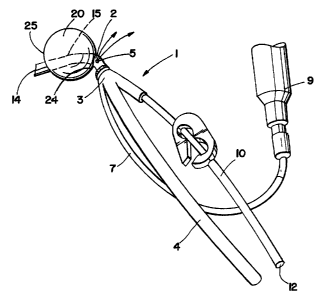
FIG. 1 depicts an embodiment of a cardioplegia occluder 1 for delivering
cardioplegia to the aorta during cardiopulmonary bypass where the distal
region 2 of the
substantially rigid cannula 3 is curved to facilitate self centering inside
the aorta. The distal
end of the cannula 14 is adapted to enter the aorta.
In this embodiment, a spherical occluder 20 is circumferentially disposed
about the
outer surface 15 of the distal region of the cannula forming a chamber 21 with
an inner
surface 22, an outer surface, a proximal end 24 and a distal end. In some
embodiments,
the occluder is an inflatable balloon. In other embodiments, the balloon is
foam-filled, so
that the occluder may be inserted in a contracted condition, for instance,
within a sleeve or
under negative pressure, and when released from the sleeve or the negative
pressure, will
automatically expand to the predetermined shape. Although FIG. 1 and FIG. 2
depict the
occluder as spherical, in other embodiments, it is conical, elliptical or
funnel shaped. In the
embodiment of FIG. 1 and FIG. 2, the occluder is an inflatable balloon
covering a portion
of the curved distal region of the cannula. In certain embodiments, the
occluder is
circumferentiafly disposed about the distal region of the cannula so that the
cannula runs
through the longitudinal center axis of the occluder. In other embodiments,
the occluder is
circumferentialfy disposed about the distal region of the cannula so that the
cannula runs
through a region displaced laterally from the longitudinal center axis of the
occluder.
The cannula is typically a rigid or semi-rigid, preferably transparent tube
having a
proximal end adapted to receive cardioplegia solution and a cardioplegia lumen
which
extends distally from the proximal end and terminates and communicates with an
infusion
port in the distal region for delivery of cardioplegia solution to the aorta.
The occlude,
which has a longitudinal center axis, is mounted on the distal region of the
cannula. The
occluder is expandable between a contracted condition and an expanded
condition,
wherein the occluder, when contracted, is closely associated with the outer
surface of the
cannula, while the occluder expands upon activation to substantially occlude
the aorta
downstream of the infusion port. During use, the occluder isolates the
ascending aorta
from the peripheral vasculature without substantial migration of the occluder
into the
ascending aorta. Because of the substantially rigid condition of the cannula,
the balloon
may have a relatively small footprint where it is coupled to the distal region
of the cannula
without substantial migration of the occluder into the ascending aorta.
CA 02315172 2000-06-14
WO 99/30766 PCT/US98/26678
The embodiment shown in FIG. 1 and FIG. 2 has three lumens within the cannula.
Other embodiments may have more or fewer lumens. In some embodiments, certain
lumens are separate, non-communicating channels. In certain embodiments, the
lumens
are generally substantially cylindrical, semi-rigid and preferably
transparent. In FIG. 1
5 and FIG. 2, a cardioplegia Iumcn 4 is adapted to receive cardioplegia
through its proximal
end and deliver it to an infusion port 5 at its distal end. The infusion port
5 is proximal to
the occluder, so that when the occluder is in an expanded condition,
cardioplegia infuses
to a region upstream from the occluded aorta. Another lumen 7 is adapted to
receive fluid
through its proximal end and deliver it to an inflation port 8 at the distal
end of the lumen
10 where it terminates and is in fluid communication with the chamber 21 of
the occluder.
When the occhuier is contracted, it is closely associated with the cannula's
outer surface
15. When fluid is delivered to the chamber of the occluder through the
inflation port, the
occluder expands away from the cannula, as depicted in FIG. 1 and FIG. 2. In
one
embodiment, the pressurized fluid used to fill the chamber of the occluder is
saline
solution and in another embodiment, it is gas. In another embodiment, negative
pressure
may be applied to the lumen 7 to contract a foam-filled balloon. An aspiration
lumen 10
has a proximal end 12 adapted to couple to an aspirator, and extends distally
from the
proximal end and terminates and communicates with the infusion port 5. In
embodiments
having an aspiration lumen, the infusion port can alternately deliver
cardioplegia solution
or aspirate embolic debris and other unwanted material from the aorta.
FIG. 3 and FIG. 4 depict another embodiment of the cardioplegia occluder 1
where
the distal end 16 of the cannula 10 is open forniing a cutting blade lumen to
receive the
cutting blade 30. The distal end 31 of the cutting blade, which when exposed,
protrudes
beyond the in the distal end of the cannula, has a sharpened tip 32 adapted to
cut through
the wall of the aorta. The embodiment shown in FIG. 3 and FIG. 4 includes a
retractable
blade guard 33 which is inserted into the distal end 16 of the cannula. The
blade guard 33
is adapted to slidably receive the cutting blade 30. During use, the blade
guard moves
when pressed against the aorta to allow the blade to cut through the wall of
the aorta, and
then the blade guard repositions to prevent the blade from cutting. In the
embodiment
shown in FIG. 3 and FIG. 4, the proximal end 34 of the cutting blade guard is
coupled to
the distal end of a spring 35. The proximal end of the spring 36 is coupled to
the inner
surface of the cannula. When the spring is at its compressed length, as
depicted in FIG. 3,
the retractable blade guard is retracted exposing the cutting blade 31. When
the spring is
at its extended length, the retractable blade guard covers the sharpened tip
of the cutting
blade as depicted in FIG. 4.
CA 02315172 2000-06-14
WO 99/30766 PGT/US98/26678
11
The cardioplegia occluder depicted in FIG. 3 and FIG. 4 is placed on the
aorta,
upstream from the brachiocephalic artery. When pressure is applied to the
cardioplegia
occluder, the surface of the aorta pushes on the retractable blade guard,
compressing the
spring and exposing the sharpened tip of the cutting blade which cuts through
the wall of
the aorta to create an incision for introduction of the distal end of the
cannula. The distal
end of the cannula, with the occluder in a contracted condition, is introduced
through the
incision made by the cutting blade. Such an embodiment can be introduced
through a site
that is a maximum of 18 French. During insertion, aspiration can be effected
through the
aspiration lumen to remove intravascular debris or air introduced into the
aorta during
incision. The curved distal end of the canttula is positioned at the desired
location inside
the aorta, and the occluder is expanded by introducing fluid through the lumen
7. Once
the occluder is fully expanded, blocking the blood supply to the aorta in the
region distal
to the occluder, cardioplegia solution may be introduced through the infusion
port to the
region upstream from the occluder to stop the heart. Cardiac surgery, may then
be
performed. Alternately, negative pressure can be applied to the proximal end
of the
aspiration lumen to remove cardioplegia and embolic debris from the aorta. In
embodiments that do not include a cutting blade, the incision is made
manually, and the
distal end of the cannula is inserted as previously described. Following
surgery, the flow
of cardioplegia solution is stopped, negative pressure is applied to the
lumen, the occluder
contracts, the cardioplegia occluder is removed through the incision initially
created for its
insertion and the incision is closed.
FIG. 5 shows another embodiment where a blood cannula 56 has a channel 57
located laterally that is adapted to receive a cardioplegia occluder 58. When
the occluder
20 is expanded inside the aorta 41, cardioplegia solution can be delivered
upstream of the
occluder through the infusion port 59. This embodiment is one example of an
integrated
configuration of a blood cannula and a cardioplegia occluder for use in a "one-
stick"
application, meaaing that only one incision need be made.
Human anatomy including the rib cage with deployed cardioplegia occluder is
depicted in FIG. 6. The cardioplegia occluder 1 is disposed through a chest
access port 40
and thereafter enters the aorta 41 behind the sternum 45 at a location 42
upstream from the
brachiocephalic artery 43. The rib cage is depicted generally by numeral 44.
The
cardioplegia occluder 1 is shown deployed within the aorta 41. The concept of
port access
allows a surgeon to ewer the aorta via a port for a minimally invasive
approach. By
accessing the aorta directly, the device is deployed without the need for
visual guidance,
e.g., fluoroscopy, ~hocardiography. This device would obviate the need for a
steraotomy
CA 02315172 2000-06-14
WO 99/30766 PCT/US981166~8
12
procedure which is generally associate with conventional coronary artery
bypass grafting
surgery.
The cardioplegia occluder may be constructed to sit in either direction once
introduced in the aorta by varying the location of the infusion port. In one
embodiment,
depicted in FIG. 7, an L-shaped cardioplegia occluder 1 is constructed to sit
inside the
aorta with occluder 20 downstream from the incision site 55, with the occluder
20
mounted distal to, or downstream from, the infusion ports 5. The cardioplegia
occluder
optionally includes seating bumps 50 to enhance sealing with the interior of
the aorta. In
another embodiment shown in FIG. 7A, a J-shaped cardioplegia occluder 1 is
constructed
to sit inside the aorta 41 so that the occluder 20 is mounted proximal to, but
still
downstream from, the infusion port 5 which is located at the distal opening 14
of the
cannula. These cardioplegia occluders can be inserted through a pre-slit
section of the
aorta, or a cutting blade can be mounted on the distal end of the cannula and
advanced
through the aortic wall.
An integrated, multiple component port access cardioplegia occluder is
depicted in
FIG.B. The system includes a cutting blade 60 having a preshaped configuration
61, a
sharp tip 62, and position limners 63. The cannula 3 includes a suture plate
70, a kink-
resistant shaft 71, an opening 72 to receive cardioplegia infusion solution
into the
cardioplegia lumen and a hemostasis valve 73. The balloon cannula 80 includes
an
occluder 81, an inflation port 82 and a lumen 83 and is adapted to receive a
filter mesh 500
through the lumen. The cannula 3 is adapted to receive the cutting blade 60
through the
infusion port 72, and to receive the occlusion device 80 through the
hemostasis valve 73.
In use, a port access point or window is opened on the patient's chest. Tissue
from the
port to the aorta is dissected. The cutting blade and cannula are advanced
through the
aortic wall. A purse string sutures) may be required to aid in wound closure
and to secure
the device. At the desired location, the cutting blade is advanced through the
aortic wall
and the cannula is pushed with the cutting blade. Once inside the vessel, the
cannula is
secured and the cutting blade is removed. At this point, the occluder (and any
filter) may
be advanced and expanded. Cardioplegia and other fluids may then be circulated
through
the cardioplegia lumen.
The distal end of the cannula may assume various designs to assist the surgeon
in
positioning the cardioplegia occluder in the aorta. In one embodiment,
depicted in FIG. 9,
a lumen 90 is adapted to receive the cutting blade 110. The cutting blade
lumen 90 enters
the distal region of the cannula 3 at an angle. A substantially straight
cutting blade 110 is
, introduced into the lumen 90 so that the sharp tip 111 of the blade
protrudes beyond the
.opening 91 at the distal end of the cutting blade lumen. In use, this
embodiment allows for
CA 02315172 2000-06-14
WO 99/30766 PCT/US98/26678
13
a single stick motion whereby the cutting blade pierces the wall of the aorta
creating an
incision and the distal end of the cannula, with the occluder in a collapsed
condition, is
advanced through the incision. A flange 100 mounted on the cannula presses
against the
exterior surface of the aortic wall preventing further movement of the cannula
into the
vessel at the point where the cannula is positioned in the desired location
within the aorta.
The cutting blade is then retracted and the occluder 20 is expanded to block
the flow of
arterial blood. An advantage of this embodiment is that it has no moving parts
other than
the retractable cutting blade. In other embodiments, the cutting blade lumen
extends
distally from the proximal end of the cannula.
The embodiment depicted in FIG. 10 has a retractable cutting blade 112
slidably
inserted into a cutting blade lumen 92 within the distal end of the cannula 3.
The proximal
end 114 of the cutting blade is coupled to a spring 120 and to an activator
line 130. The
activator line can be made of material such as wire. The proximal end of the
spring is
coupled to a stop 121 formed inside the cutting blade Lumen. When the
activator Line 130
is pulled, the spring 120 compresses and the sharp tip 111 of the cutting
blade 112 is
retracted into the distal end of the cutting blade lumen 92 which then serves
as a blade
guard. When the activator line 130 is released, the spring 120 expands and the
sharp tip
111 of the device is exposed to allow incision into a vessel. The embodiment
also
includes infusion ports 101 for introduction of cardioplegia solution upst~am
from the
occluder 20.
FIG. l0A shows another embodiment where the blade guard is a retractable
obturator 140. In this embodiment, the distal end 114 of the cannula is sharp,
thus forming
the cutting blade, and is used to create the initial incision into the aorta.
The retractable
obturator 140 is slidably received through the cutting blade. In the
embodiment of FIG.
10A, the retractable obt<uator is coupled on its proximal end to a spring 120
and to an
activator line 130. The spring is coupled on its proximal end to a stop 121
formed inside
the cutting blade lumen. During use, the obturator can be moved by pulling on
the
activator line to expose the sharp distal end 114 of the cannula which is used
to cut
through the wall of the aorta. When the activator line is released, the
obturator moves
back to prevent the blade from cutting.
FIG. 11 depicts a flange sleeve 105 adapted to receive the cannula. In some
embodiments, the flange sleeve is substantially cylindrical. In other
embodiments, the
flange sleeve may have a different shape on cross section such as square,
rectangular,
oblong or other shapes. The flange sleeve has a sharpened distal end 116
adapted to cut
through the wall of the aorta, an inner surface 108, an outer surface 109, a
proximal end
117, a distal end and a longitudinal center axis. The lumen 118 of the flange
sleeve 106
CA 02315172 2000-06-14
WO 99/30766 PCT/US98/266~8
14
runs along the longitudinal center axis and communicates with openings at the
proximal
117 and distal 116 ends of the sleeve. This embodiment also includes a flange
stop 107,
with a top surface 125, which faces the proximal end of the flange sleeve, and
a bottom
surface 126, which faces the distal end 116 of the flange sleeve. The flange
stop 107 is
mounted on the flange sleeve. The perimeter of the flange stop can be
substantially
circular, or shaped so that a region of the perimeter includes a protrusion or
notch in the
plane of the flange stop, where the protrusion or notch indicates the
direction of the tip 128
of the cutting age 116 of the flange sleeve. In the embodiment of FIG. 11, the
portion of
the flange sleeve distal to the bottom surface 126 of the flange stop 125 and
proximal to
the cutting edge 116 at the distal end of the sleeve is of a length 119 that
will position the
cutting edge 116 of the flange sleeve at a predetermined depth inside the
aorta when the
bottom surface 126 of the flange stop contacts the outer surface 46 of the
aorta thus
preventing further movement of the flange sleeve into the aorta. FIG. 11 shows
the
cannula 3 retracted inside the lumen of the flange sleeve. When in the
retracted state, the
occluder 20 is in a contracted condition. When in use, the cutting edge 116 of
the flange
sleeve is pressed into the outer surface of the wall of the aorta 46, while
the cannula 3 is in
the retracted state and the occluder 20 is in a conh~acted condition. The
cutting edge 116
of the flange 105 is advanced into the aorta until the flange stop 107
contacts the outer
surface of the wall of the aorta 46. In the next step, as depicted in FIG. 12,
the cannula 3
is advanced beyond the cutting edge 116 of the flange until the distal end of
the cannula is
situated at the pr~etermined position within the aorta 41. The occluder 20 is
then
expanded to prevent blood flow downstream in the aorta. In this embodiment,
the distal
end of the cannula is semi-rigid and preformed to assume a substantially
curved condition
when released from the flange. When retracted inside the flange, as depicted
in FIG. 1 lA,
the semi-rigid distal end of the cannula 3 generally conforms to the shape of
the flange
sleeve lumen which is straight.
In another embodiment, depicted in FIG. 13, the flange 105 includes a flange
sleeve 106 with an inner surface 108, an outer surface 109, a proximal end
117, a distal
end 129, and a longitudinal center axis. The lumen 118 of the flange sleeve
106 runs
along the longitudinal center axis and communicates with openings at the
proximal 117
and distal 129 ends of the sleeve. This embodiment also includes a
substantially flat
flange stop 107, with a top surface 125, which faces the proximal end of the
flange sleeve,
and a bottom surface 126 which is flush with the distal end 129 of the flange
sleeve. The
bottom surface 126 of the flange stop is adapted to press against the outer
surface 46 of the
aorta. FIG. 13 also shows the cannula 3 partially retracted inside the lumen
118 of the
~nge sleeve. When in the retracted state, the occluder 20, which is disposed
about the
CA 02315172 2000-06-14
WO 99/30766 . PCT/US98/Z6678
distal region of the cannula 3, is in a contracted condition. In this
embodiment, the distal
end 145 of the cannula includes a cutting blade lumen having a retractable
cutting blade
146 with a sharpened cutting edge 147 at its distal end. The cutting blade 146
slidably
inserts inside the cutting blade lumen and protrudes beyond the distal end 145
of the
5 cannula 3. When in use, the flange 105 is positioned with the bottom surface
126 of the
flange stop 107 pressing against the outer surface of the wall 46 of the aorta
and the
cannula 3 and cutting blade 146 are in the retracted state inside the lumen
118 of the
flange sleeve 106 proximal to the distal opening 129 of the sleeve. The
cannula 3 and the
cutting blade 146 are pushed through the lumen 118 of the flange sleeve beyond
the distal
10 opening 129 so that the sharpened cutting edge 147 of the cutting blade 146
cuts into the
wall of the aorta foaming an incision as depicted in FIG. 13. Once the
incision is formed,
the cannula 3 is advanced beyond the distal opening 129 of the flange sleeve
106, as
depicted in FIG. 13A, so that the distal end of the cannula and the occluder
20 are
introduced into the aorta 41 to the predetermined depth and position. In this
embodiment,
15 the semi-rigid distal end of the cannula is preformed to assume a curved
shape once it is
released from the lumen of the flange. As the cannula is advanced beyond the
distal
opening 129 of the flange into the aorta, the cutting blade 146 slidably
retracts within the
cannula so that is does not protrude beyond the distal opening 146 of the
cannula. Once
the cutting blade has been deployed to create the initial incision, it is
desirable to retract it
inside the cannula or otherwise guard the sharpened tip so that the sharp edge
of the blade
does not scrape or cut the inner surface 47 of the wall of the aorta opposite
the incision
site. The occluder 20 may then be expanded to occlude arterial flow downstream
in the
aorta
In another embodiment, depicted in FIG. 14, the flange 105 includes a flange
sleeve 106 with a proximal end 117, a distal end 129, and a longitudinal
center axis. The
lumen 118 of the flange sleeve 106 runs along the longifi~dinal center axis
and
communicates with openings at the proximal 117 and distal 129 ends of the
sleeve. This
embodiment. also includes a substantially flat tear-away flange stop 150, with
a top
surface 151, which faces the proximal end of the flange sleeve, and a bottom
surface 152,
which is flush with the distal end 129 of the flange sleeve. The tearaway
flange stop 150
is disposed about the outer surface of the flange sleeve 106 at the distal end
129 of the
sleeve. The bottom surface 152 of the tear--away flange stop is adapted to
press against
the outer surface 46 of the aorta to limit the initial insertion depth into a
vessel. FIG. 14
also shows the cannula 3 partially retracted inside the lumen 118 of the
flange sleeve.
When in the retracted state, the occluder 20 is in a contracted condition. A
cutting blade
160 is adapted to slidably insert inside a lumen within the cannula. In this
embodiment,
CA 02315172 2000-06-14
WO 99130766 PCT/US981Z6678
16
the distal end 161 of the cutting blade is sharpened 161 to cut through the
wall of the aorta.
When in use, the cannula 3, with the sharpened cutting edge 161 of the canula
insertion
device 160 exposed, is advanced through the wall of the aorta. until the
bottom surface 152
of the teas away flange stop 150 presses against the outer surface of the wall
of the aorta.
As depicted in FIG. 14A, the cutting blade 160 is then retracted within the
distal end of the
cannula 3 as the tearaway flange is removed and the cannula is advanced into
the lumen
of the aorta until the bottom surface 126 of the permanent flange stop 107
presses against
the outer surface 46 of the wall of the aorta By this process, the distal end
of the cannula
and the occluder 20 are introduced into the aorta 41 to the desired depth and
position. In
this embodiment, the semi-rigid distal end of the cannula is preformed to
assume a curved
shape once it is released from the lumen of the flange. The occluder 20 may
then be
expanded to occlude arterial flow downstream in the aorta.
As described previously, in certain embodiments, the distal region of the
cannula
may be prefonmed to a desirai shape to allow the cannula to be positioned at
the desired
depth and orientation within the aorta. In other embodiments, the distal
region of the
cannula may be mechanically activated by an occluder aligner to allow proper
positioning
of the occluder within the aorta. FIG. 15 depicts an embodiment with one form
of
occluder aligner that includes a cannula 3 with an inner surface 170, an outer
surface 171,
a proximal end (not shown), a distal end 145 and a longitudinal center axis.
The lumen
172 of the cannula runs along the longitudinal center axis and communicates
with
openings at the proximal and distal 145 ends of the cannula. The cannula also
includes a
flange stop 107 disposed about the outer surface 171 of the distal region of
the cannula.
The occluder aligner of this embodiment includes a steering wire 130 carried
by the
cannula, displaced from the center axis of the cannula and attached on a first
end 131 in
the distal region of the cannula, in the case of this embodiment, to the inner
surface 170 of
the distal region. When in use, as depicted in FIG. 15 and FIG. 15A, the
cardioplegia
occluder 1 is advanced through an incision in the wall of the aorta 41 until
the bottom
surface 126 of the flange stop 107 presses against the external surface of the
wall 46 of the
aorta. At this point, as shown in FIG. 15, the occluder 20 is in a contracted
condition. The
steering wire 130 is then manipulated, as depicted in FIG. 15A, to move the
distal end of
the cannula into a curved condition, so that the distal opening 145 of the
cannula points
downstream within the aorta 41. In one embodiment, the occluder is aligned by
pulling on
the steering wire. In another embodiment, the steering wire is fabricated from
a material
that shortens upon application of a predetermined electrical input. When this
predetermined electrical input is applied to the steering wire, the wire
shortens by a
predetermined length, pulling the distal end of the cannula into the
predetermined position.
CA 02315172 2000-06-14
WO 99/30766 PCT/US98/26678
17
In another embodiment, a control circuit containing a memory storage device
controls the
electrical input to be applied and the timing of the application and
discontinuance of the
electrical input, so that the change in length of the wire may be programmed.
Once the
occluder 20 is properly aligned within the aorta, the occluder may be expanded
to occlude
arterial flow downstream in the aorta.
FIG. 16 depicts another cannula that is mechanically activated to facilitate
proper
positioning of the occluder within the aorta. This embodiment includes a
cannula 3 with
an inner surface 170, an outer surface 171 and a longitudinal axis. The
cannula is divided
into two segments, a proximal portion 185 and a distal portion 186, flexibly
coupled to one
another. In the embodiment shown in FIG. 16, the flexible coupling is a hinge
180. In the
closed condition, as depicted in FIG. 16, the distal end of the proximal
portion 185 and the
proximal end of the distal portion 186 align at a circumferential region 181,
so that the
cannula assumes a substantially cylindrical shape. In other embodiments, the
cannula on
cross-section can be rectangular, square, oblong or other shapes. In the open
condition, as
depicted in FIG. 16A, the distal portion 186 rotates about the hinge so that
the longitudinal
axis 188 of the distal portion 186 is about a 90° angle to the
longitudinal axis 187 of the
proximal portion 185. In the closed condition, the lumen 172 of the cannula
runs along
the longitudinal center axis and communicates with openings at the proximal
and distal
145 ends of the cannula. The cannula also includes a flange stop 107 disposed
about the
outer surface 171 of the distal region of the cannula, and a cutting blade 160
which
slidably inserts within the lumen 172 of the cannula when the cannula is in
the closed
condition. When in use, as depicted in FIG. 16, the cutting blade 160
protrudes beyond
the distal end 145 of the cannula 3 which is in the closed condition with the
occluder
contracted. The presence of the cutting blade in the lumen of the cannula
helps maintain
the cannula in a closed position. The sharp distal end 161 of the cutting
blade 160 is
advanced through the wall of the aorta 41 creating an incision, and the
canuula 3 is
advanced into the aorta until the bottom surface 126 of the flange stop 107
presses against
the external surface of the wall 46 of the aorta. The canula insertion device
is then
removed causing the hinge to open as depicted in FIG. 16A, and the cannula
assumes the
open condition with the distal portion 186 of the cannula pointing downstream
in the aorta.
In some embodiments (not shown), the canuula opens with the assistance of a
spring
loaded hinge. The occluder 20 may then be expanded to occlude arterial flow
downstream
in the aorta. Cardioplegia solution may then be introduced through the
proximal portion
185 of the cannula for delivery through the fluid port 189 upstream of the
occluder.
FIG. 17 depicts an embodiment where the distal region of the cannula 3 is
taper~l
210. The embodiment of FIG. 17 also shows, a curved region 212, distal to the
tapered
CA 02315172 2000-06-14
WO 99/30766 PCT/US98126678
18
region. In this embodiment, the tapered region, on cmss~-section, as depicted
in FIG. 18,
is substantially elliptical. As also depicted in FIG. 18 from a top elevation,
the long
diameter of the ellipse of the tapered region cross-section lies directly
above the curved
region 212 of the cannula. This embodiment also includes a flange which is
slidably
received by the cannula. The flange in this embodiment has a directional
indicator. As
can be seen in the top elevation of FIG. 18, the flange assumes the shape of a
polygon. In
other embodiments, the flange can be other shapes such as rectangular, oblong,
or
triangular. The flange includes a hole 204 that is substantially elliptical,
having an inner
circumference 202. The hole is placed off axis from the center of the polygon.
The long
diameter of the elliptical hole is perpendicular to the directional edge 203
of the polygon
perimeter of the flange. The distance from the directional edge 203 to the
nearest point on
the inner circumference of the hole 204 is greater than the distance from the
edge 201
opposite the directional edge to the point on the inner circumference nearest
that opposite
edge. The inner circumference 202 of the hole in the flange is greater than
the
circumference of the outer surface 211 of the distal end of the tapered region
210 of the
cannula, but less than the circumference of the outer surface 211 of the
proximal end of
the tapered region 210 of the cannula. The flange is disposed about the
tapered region of
the cannula. The distal end of the tapered region is adapted to slidably
insert in the hole of
the flange and the proximal portion of the tapered region sfidably inserts in
the flange up
to the location where the circumference of the outer surface 211 of the
tapered region of
the cannula is substantially equal to the inner circumference 202 of the hole
in the flange,
at which location the flange is no longer free-floating, and locks into
position on the
tapered region. The tapered condition of the cannula assists in sealing the
cannula to the
flange. Since the hole 204 of the flange and the cross-section of the tapered
region are
both elliptical in shape, the flange will always be oriented in the same
position on the
cannula when it locks into place; that is, the directional edge 203 will
always point toward
the curved region 212 of the cannula, which assists the surgeon in knowing
which way the
occluder is pointing in the aorta. In other embodiments, the tapered region
210 and the
hole 204 of the flange may assume other shapes on cross~section, such as
rectangular or
triangular, In some embodiments, the directional edge is identified by a
specific color.
The embodiment of FIG. 17 also includes marker bands 220 around the outer
surface 211
of the curved region 212 of the cannula in the most proximal and most distal
locations
where the occluder 20 contacts the cannula. The marker bands are made of
n3diopaque
material such as metal~olymeric alloy so that the surgeon can identify the
position of the
occluder.
CA 02315172 2000-06-14
WO 99/30766 PGT/US98/26678
19
For the cardioplegia occluder to fimctioa properly, the occluder must be
adapted to
occlude aortas of varying diameters. Moreover, the internal surface of the
aorta may have
varying surface features creating additional challenges to fashioning
occluders that will
conform to the topography of the inner surface of the vessel and form a
complete seal.
The challenge of occluding aortas of varying diameter is further compounded in
embodiments with fixed flanges. To overcome such obstacles, in certain
embodiments,
the occluder is a balloon having a first region of first expansion capacity
and a second
region of second expansion capacity where the first expansion capacity is
greater than the
second expansion capacity. During use, the second region expands
preferentially and to a
greater extent than the first region. These embodiments can thus compensate
for
insertions where the distal end of the cannula does not lie directly in the
center of the aorta
and by thus compensating creates effective sealing. In some embodiments, the
varying
expansion capacity is created by forming the first region from a flexible
material of
different thickness that the flexible material used to create the second
region. In other
embodiments, the first region is of a different modulus (durometer) than the
second
region. In other embodiments, the occluder is adapted to occlude aortas of
varying
diameters by asymmetrically mounting the balloon on the distal region of the
cannula.
The embodiment shown in FIG. 19, which demonstrates this last case, has an
occluder 20
that is a preformed asymmetric balloon where the "long" side 230 has less
capacity to
expand than does the "short" side 231. The flange 107, as described in
previous
embodiments, will hold the curved portion 212 of the caanula at a
predetermined distance
below the region of the wall of the aorta closest to the flange. In aortas of
varying
diameters, the distance between the curved portion of the cannula and the wall
opposite
the flange will necessarily vary. To facilitate occlusion in these varying
conditions, the
short side 231 has a greater capacity for expansion, as depicted in FIG. 19A,
than does the
long side 230, so that upon inflation by a common fluid source, the short side
231 will
preferentially expand over the long side 230. FIG. 20 is a front elevation of
the
embodiment of FIG. 19A showing how the short side 231 preferentially expands
over the
long side 230 to occlude aortas of smaller 240, intermediate 241, and larger
242 diameters
even though the flange 107 fixes the depth of the cannula within each vessel.
There are several methods to achieve varying capacities for expansion in given
regions of the balloon occluder. Typically, it is desired to achieve a
preferential expansion
zone as depicted in FIG. 21 where a balloon occluder 20 is asymmetrically
disposed about
a cannula, and the occluder has a region 251 that has a greater capacity to
expand when
compared to another region 250. FIG. 22 is a lateral elevation of the
embodiment of FIG.
21. These asymmetric balloons, which can be fabricated from polyurethane,
typically .
CA 02315172 2000-06-14
WO 99130766 PCT/US98/26678
inflate to a more symmetric shape as depicted in FIG. 23, where varying
balloon wall
thickness is used to control expansion characteristics. A thin region 252 of
the balloon
will expand first, reaching a certain level of strain/elongation 252', then a
thicker region
253 will stretch to its expanded condition 253'. The expanded balloon is
symmetrically
5 disposed about the cannula.
FIG. 24 depicts another embodiment where balloon materials with differing
expansion capacities are used to create a balloon which is asymmetric upon
expansion. In
this embodiment, a region of soft material 255, e.g., one of lower modulus and
usually
lower durometer, expands more freely 255' than does a region of harder
material 254, e.g.,
10 one of higher modules and usually higher durometer, which expands less
freely 254'.
It is also important that the occluder not pmlapse at the locations where the
occluder surface is not in contact with the inner surface of the aorta when
the occluder is
expanded. Such prolapse can cause the occluder to not seal properly.
Increasing thickness
in these non-contact regions can reduce the risk of pmlapse and can otherwise
control
15 occluder length and shape. FIG. 25 depicts an embodiment where the balloon
occluder
has regions where the balloon material is thin 256 and sidewall regions where
the balloon
material is thick 257. When the balloon expands, the thin regions 256, which
ultimately
contact the inner wall of the aorta, expand more freely to their expanded
condition 256'.
The thick sidewall regions 257, which do not contact the inner surface of the
aorta and are
20 thus at risk of.pmlapse, expand less freely to their expanded condition 25T
and, due to
their thickness, are more robust. The overall average balloon length from
location 260 to
location 261 is reduced from the length that would otherwise result if the
sidewalls were
not made of thicker material. Thus, a prolapse-resistant balloon occluder with
a small
"footprint" (area of contact on the distal region of the catheter), can be
fabricated. This
small footprint occluder, when used with the substantially rigid cannula
allows the
occluder to isolate the ascending aorta from peripheral vasculature without
substantial
migration of the occluder into the ascending aorta.
FIG.. 26 depicts an embodiment of a cardioplegia occluder 1 where the
substantially rigid cannula 3 includes three lumens 4, 10 and 7, a flange 107
and a
spherical occluder 20. The infusion port 5 is shown proximal to the occluder.
Certain
embodiments of the cannula are made of clear polycarbonate acrylic, ABS or
stainless
steel. In one embodiment, the region of the cannula proximal to the flange is
made of
clear polycarbonate, acrylic or ABS, and the region of the cannula distal to
the flange is
made of stainless steel. The plastic region and the stainless steel region are
insert~nolded
at the junction. In the preferred embodiment, (i) the length of the cannula
from the
proximal end to curved portion of the distal region is in the range of 5-10
inches, most
CA 02315172 2000-06-14
WO 99!30766 PCT/US98I26678
21
preferably 7.5 inches, (ii) the width of the distal region from the beginning
of the point of
curvature to the distal end (distance A in FIG. 26) is in the range of 0.25-
0.75 inches, most
preferably 0.45-0.50 inches; and (iii) the distance between the flange and the
distal end
(distance B in FIG. 26) is the range of 3/8 inch to 1.0 inch, and most
preferably '/. inch.
FIG. 27 is a front elevation of the embodiment of FIG. 26. FIG. 28 is a
lateral cmss-
section of the embodiment of FIG. 27 shown through section line 28-28. Here,
the
pathways of the three lumens are depicted in greater detail. The lumen 7 is
shown
communicating with the inflation port 8 which opens into the chamber of the
occluder 20.
The cardioplegia lumen 4 is shown communicating with the infusion port 5 which
opens
into the region of the aorta upstream of the occluder. The aspiration lumen 10
also
communicates with the infusion port. FIG. 29 is a front elevation of the
distal region of
the cannula 3 of the embodiment of FIG. 26 with the occluder removed. In this
figure, the
closed distal end 14 of the cannula can be seen. FIG. 30 is a top elevation of
the
embodiment of FIG. 29, showing the relative locations of the lumen 7 that is
used to
inflateJdeflate the occluder, the cardioplegia lumen 4 and the aspiration
lumen 10 as they
enter the region of the cannula just proximal to the flange. FIG. 31 is a
lateral view of the
embodiment of FIG. 29 with a partial cross section of the curved region of the
cannula.
The occluder mounting zones 270 are shown on either side of the cross-section
region.
This view shows the relationship between the infusion port S, shown proximal
to the
occluder mounting zones, and the inflation port 8 which opens in the region
between the
occluder mounting zones and thus communicates with the chamber of the
occluder. FIG.
32 is a bottom elevation of the embodiment of FIG. 29. FIG. 33 is a back
elevation of the
embodiment of FIG. 29, again showing the relative locations of the infusion
port 5 and the
inflation port 8. FIG. 34 is a lateral cmss-section of the embodiment of FIG.
29 shown
through the section line 34-34.
FIG. 35 is an embodiment showing a self~xpaading occluder 320 with a hollow
Nitinol frame 300, a balloon seal 301 and a fluid-impermeable membrane 302.
The
occluder is an annular-shaped balloon having an inner circumference and an
outer surface
and a flexible, fluid-impermeable membrane bonded to the outer surface of the
balloon
and covering the area circumscribed by the inner circumference of the annular
balloon.
FIG. 36 shows a canuula 3 with an occluder side port 310, a flange stop 107
and a fluid
port 311. FIG. 37 shows the self-expanding occluder 320, which has been
inserted into
the occluder side port 310 while in a collapsed condition after the distal
region of the
cannula has been inserted into the aorta 41. Once properly positioned, the
balloon seal
301 is inflated through the hollow Nitinol frame 300 and the occluder expands,
occluding
he vessel.
CA 02315172 2000-06-14
WO 99/30766 p~~gggn~~g
22
In some applications it is desirable to provide occluder constructions with
enhanced stability and/or increased expandability. FIG. 38 depicts an
overlapping balloon
occluder 321, fabricated with excess balloon material, which allows the
occluder to inflate
to a larger size while stretching and elongating to a lesser extent. A portion
of the
occluder in its expanded condition 321' is also shown. This embodiment may
also include
thicker regions of the balloon wall to control the inflation profile.
In certain embodiments, the cannula is open at the distal end and the distal
end has
a lumen where the occluder, when contracted, is stored as shown in FIG. 39.
This figure
depicts an expanding balloon occluder 322. Upon expansion, the balloon
advances out of
the distal end of the cannula. As the balloon is inflated, more balloon
material is available
to expand, thus permitting occlusion of larger sized vessels once the balloon
reaches its
expanded condition 322'.
FIG. 40 and FIG. 41 depict a cannula with an open distal end for storage of a
contracted balloon occluder 323. The balloon can be retracted upon deflation
into the
distal end 14 of the cannula 3 by pulling on an elastic line 330 which passes
through the
lumen of the cannula. The elastic line 330 is coupled to the proximal end 331
of the
balloon and the distal end 332 of the balloon, so that when the balloon is
fully expanded
323", the elastic line is fully stretched. Upon deflation, the elastic line
contracts and the
distal end 331 of the balloon moves closer to the proximal end 332 of the
balloon. The
deflated balloon 332 may then be pulled into the distal end 14 of the cannula
by pulling on
the elastic line 330. FIG. 41 depicts the balloon in its initial contracted
condition 323, a
deflated condition 323' and a fully expanded condition 323", where the elastic
line is not
shown.
In some applications it may be advantageous to cover the occluder with a
protective layer. FIG. 42 shows a balloon occluder 20 disposed about the
distal end of a
cannula 3. The balloon 325 itself is made of an elastic material and its outer
surface is
covered by a protective material 326. In some embodiments, the protective
layer itself has
elastic capacity. In other embodiments, the protective layer is internal to
the balloon so
that the external surface of the protective Iayer is covered by the balloon
material.
FIG. 43 depicts an embodiment where a funnel-shaped. occluder 328 made of
elastic matexial is deployed through a side opening 340 of the cannula 3. The
funnel-
shaped occluder 328 can occlude vessels of varying sizes due to its shape.
Occluder aligners, which were described previously for manually aligning the
distal end of the canaula, can also be used to provide position stability to
extending
occluders. In some applications, an expanding occluder will "rock" out of
position during
.expansion if the distal region of the cannula is not positioned along the
center longitudinal
CA 02315172 2000-06-14
WO 99/30766 p~~sggn~~g
23
axis of the aorta. Certain embodiments therefore include cannulas with
occluder aligners
of various designs to stabilize the position of the occluder and distal
cannula during
occluder inflation. One embodiment includes a longitudinally defonnable region
and an
end sleeve which slides relative to the distal end of the cannula and is
coupled to the
longitudinally deformable region and to the occluder. During use, the occluder
expands
and the end sleeve moves proximally, thereby compressing the longitudinally
defonmable
region. FIG. 44 and 44A demonstrate this embodiment, where the longihidinally
deformable region is a spring. FIG. 44 shows the distal region of the
cardioplegia
occluder 1 where the occluder 20 is in the collapsed condition. The spring 400
is coiled
about the distal region 401 of the cannula inside the occluder chamber. The
proximal end
402 of the spring is coupled to the region of the cannula inside the occluder
chamber just
distal to the proximal end of the occluder 403. The end sleeve 404 is disposed
about the
distal region of the cannula. The proximal end 405 of the end sleeve is
coupled to the
distal end of the spring 400. The end sleeve 400 is coupled to the distal end
of the
occluder in a region 406 of the end sleeve just distal to the proximal end of
the sleeve.
The end sleeve includes a seal 407 near the distal end of the sleeve adapted
to sutrouad the
distal region of the cannula 3 so that this distal cannula region slidably
inserts in the seal.
The seal is adapted to prevent fluid in the occluder chamber from escaping
from the
occluder. In this embodiment, the occiuder aligner includes an end stop 408 to
prevent the
end sleeve from sliding off the distal end of the cannula 3 during use. FIG.
44 also shows
the location of the inflation port 8 inside the occluder chamber. FIG. 44A
shows the
embodiment of FIG. 44 where the occluder is in the expanded condition and the
proximal
end 405 of the end sleeve has moved along the distal region 401 of the cannula
toward the
proximal end of the occluder 403 and the spring 400 has compressed.
FIG. 45 depicts an embodiment of a cardioplegia occluder 1 that includes an
occluder aligner where the distal end of the end sleeve 404 of the occluder
aligner is a
sharpened edge 420 that serves as a cutting blade. in use, the sharpened edge
420 creates
the initial incision into the aorta and the cannula with the collapsed
occluder is advanced
into the lumen of the vessel. The occluder is expanded and the end sleeve 406
slides
proximally along the distal region 401 of the cannula retracting the sharpened
edge 420.
In this embodiment, the longitudinally deformable region of the occluder
aligner is a
flexible tube.
An occluder aligner with a steering sleeve slidably mounted on the caanula and
coupled to a steering wire is depicted in FIG. 46. In this embodiment, the
steering sleeve
455 is disposed about the region of the cannula 3 proximal to the occluder 20,
so that the
~nula slidably inserts in the steering sleeve. The distal end 453 of the
steering wire is
CA 02315172 2004-07-08
24
coupled to the inner surface of the distal region of the cannula in the area
where the occluder is
coupled to the cannula. The steering wire 454 is carried by the cannula and is
displaced from
the longitudinal center of the cannula. In some embodiments, the steering wire
passes through
a hole or slot in the cannula which is distal to the region of the cannula
which is inserted into the
vessel. The proximal end 455 of the steering wire is coupled to the steering
sleeve. In use,
during occluder expansion, the steering sleeve is manipulated to move the
distal end of the
cannula. The steering sleeve can be moved along the cannula to elevate the
distal end 450 as
depicted in FIG. 46A and 46B. Steerable occluder aligners can be designed so
that the distal
end of the cannula is positioned at the center point of the largest vessel in
which the
cardioplegia occluder is to be used. When used in smaller vessels, the tip
will lie below the
centerline and can be rotated up by pulling the steering sleeve distally.
As described previously, the cardioplegia occluder can be used in conjunction
with other
cardiopulmonary bypass equipment or other cardiac surgical equipment including
blood
cannulas, filter cannulas and diverters in various combinations as integrated
systems or as
separately insertable devices. In certain embodiments, a "one-stick" method
is.used, meaning
that one incision is made into the aorta to insert the various pieces of
equipment in either their
integrated or separately insertable configurations. In other embodiments, "two-
stick" or "three-
stick" (two or three aortic incision) methods are used. In some embodiments,
the occluder is
mounted on the blood cannula instead of the cardioplegia cannula. TABLE 1,
located at the end
of the Detailed Description section, is provided to assist in describing the
various combinations.
FIG. 47 shows a two-stick embodiment with a blood cannula 600 (adapted to
receive
separately insertable filter 500 through a channel thereof) inserted through
one incision and a
separate cardioplegia occluder 1 inserted through a second incision. The
filter is carried through
side channel 601 of the blood cannula. Either a modular filter cannula as
shown (see U.S.
patent no. 5,846,200 issued Dec. 8, 1998, for more details) or an integral
filter cannula (see
U.S. patent no. 5,769,816 issued June 23, 1998 and PCT application no.
PCT/US1998/007558
(lNT'L Publication no. W01998l046297: PCT publication date October 22, 1998),
for more
details) can be used. In this embodiment, a diverter 700 has been inserted in
the region of the
aorta 41 Where the aorta intersects the brachiocephalic artery 43, the left
subclavian artery and
the left common carotid artery. In all cases described herein, whether one-,
two- or three-stick
and whether the various cannulas are integrated, separately insertable or
certain cannulas are
absent, the diverter may be (i) absent, (ii) inserted only for the purpose of
conducting the
cardiac surgery, then
CA 02315172 2000-06-14
WO 99/30766 PGT/US98/26678
removed at the completion of the surgery, or (iii) permantntly installed in
the aorta. The
embodiment of FIG. 47 allows the cardioplegia occluder 1 to occlude the aorta
distal to
the infusion ports 5 where cardioplegia solution is introduced to stop the
heart.
Downstream from the occluder 20, the filter 500 traps embolic debris and other
unwanted
5 material that is a byproduct of the surgical activity. Downstream from the
filter, the blood
cannula supplies blood from a heart lung machine to the aorta for circulation
through the
peripheral vasculature. The diverter 700, which is permeable to blood, further
inhibits
embolic material and other unwanted debris 800 from entering the cerebral
vasculature by
diverting it past the left common carotid artery and the brachiocephalic
artery, which
10 communicates with the right common carotid artery.
FIG. 47A shows an embodiment of a two-stick model where the cardioplegia
occluder 1 is adapted to receive the filter 500 through a channel thereof, and
the blood
cannula 600 is inserted through a separate incision. A diverter is present,
but as previously
described, the diverter may be installed permanently, inserted only for the
purpose of
15 surgery or absent altogether in all one-stick, two stick or three-stick
methods. Other
embodiments of the two-stick method include (i) an integrated cardioplegia
occluder and
blood cannula with a separate filter, either inserted through a filter cannula
or separately
inserted, (ii) a separately inserted cardioplegia occluder, a separately
inserted blood
cannula and no filter cannula, {iii) a blood cannula occluder with a filter
inserted through a
20 channel in the cannula as shown in FIG. ~, or mounted on the cannula and a
cardioplegia
cannula inserted thmugh a separate incision, and (iv) a blood cannula occluder
and a
cardioplegia occluder inserted thmugh a separate incision and no filter.
FIG. 48 depicts a three-stick method with a separately inserted cardioplegia
occluder 1, a separately inserted filter cannula 501 and a separately inserted
blood cannula
25 600. In this embodiment, the diverter 702 is present, but any of the three
diverter
configurations could be utilized. In another embodiment, the filter is
separately inserted
without the use of a filter cannula, as shown in FIG. 5(f.
In other embodiments, a one-stick method is used. In one embodiment, depicted
in FIG. 51, the cardioplegia occluder and blood cannula are integrated 900,
and the filter
separately inserted through a channel in the cannuia. In other embodiments,
the filter may
be mounted on the cannula or absent. Again, each combination has three
possible diverter
configurations.
In certain embodiments of the one--, two- and three-stick methods described
above, the cardioplegia occluder may be replaced by a separate balloon cannula
and a
3S cardioplegia cannula. In such cases, the balloon cannula and the
cardioplegia cannula can
CA 02315172 2000-06-14
WO 99/30766 PCT/US98/26678
26
be separately inserted or can be integrated with one another or each
integrated with the
filter cannula or the blood cannula
CardioplegiaFilterBlood
occluder CannulaDescription
(BC)
UN>rSTTCKi
Integrated CPO/BC; filter separately
(la) + + + inserted through caturula
(FIG. 51) or mounted ~ cannula
(lb) + + lntc
grated CPO/BC
,1,w0-STTCK~
_
+ + + F~tw' iasertod thmugh CPO (FIG.
47A) or mounted on CPO
2b + + + Filter inserted through BC channel
(FIG. 4'n or mounted on BC
Intel CPO/BC; filter through filter
2c + + + capnula or separately
+ + Separately inserted CPO and BC
Occluder on BC; filter inxrtod through
2e CP + BCO blood caMUle occluder
(BCO) (FIG. 49) or mounted on BCO,
caMioplegia (CP)
cannula separately inserted
2f CPI - BCO Occluder on BC, CP cannula
separately inserted
Filter separately inserted through
(3a) + + + filter cerrnula or without
cannula
..~ w ~.v,muw wwi muuwau~ Wp rr8$ rnrpe pp~blC V8118n1s 88 t0 8 dlVer'Ier'.
The dIVeriCt may
be (i) absent, (ii) inserted only for the purpox of c~ducting the cardiac
surgery, drerr removed at the completion
of the surgery, or (iii) permao~tlY in~alled in the aorta.
While particular devices and methods have been described for using the
cardioplegia occluder, once this description is known, it will be apparent to
those of
ordinary skill in the art that other embodiments and alternative steps are
also possible
without departing from the spirit and scope of the invention. Moreover, it
will be apparent
that certain features of each embodiment as well as features disclosed in each
reference
incorporated herein, can be used in combination with devices illustrated in
other
embodiments. Accordingly, the above description should be consr<ued as
illustrative, and
not in a limiting sense, the scope of the invention being defined by the
following claims.