Note: Descriptions are shown in the official language in which they were submitted.
CA 02320296 2000-08-09
WO 99/60397 PCTNS99/09322
LIQUID ANALYSIS CARTRIDGE
FIELD OF THE INVENTION
This invention relates to microfluidic cartridges for analysis of liquid
samples, and in
particular to cartridges having a convoluted sample storage channel and to
cartridges having a
flow cytometric measuring region.
BACKGROUND OF THE INVENTION
With the advent of micro-machining technology, microfluidic devices have
proliferated (for example, U.S. Patent No. 5,637,469 to Wilding et al., U.S.
Patent No.
4,983,038 to Ohki et al., U.S. Patent No. 4,963,498 to Hillman et al., U.S.
Patent No.
5,250,263 to Manz et al., U.S. Patent No. 5,376,252 to Ekstrom et al., E.P.
Patent Publication
038150181, and Petersen, E. (1982)Proc. oftheIEEE, vol. 70, No. 5, pp. 420-
457). A
practical limitation for particle-containing liquids such as blood is the
sedimentation of
particles within the device. Following loading the liquid in the device,
appreciable particle
sedimentation can occur within the time required to position the device in a
measurement
apparatus. For example, if the sample flow is slowed or stopped, blood cells
can measurably
settle out of plasma within 20 seconds. Without a sample management method and
apparatus
for sedimentation mitigation, quantitative analysis, especially using more
than one analysis
method sequentially, is impractical. Moreover, if samples are first collected
and then
transported to a measurement apparatus, as in a clinical setting or in field
sampling, particle
sedimentation can make accurate analysis impossible.
Microfluidic devices having sample storage reservoirs are known in the art
(for
example, E.P. Patent Publication 038150181). Because ofparticle sedimentation,
these
devices are useful only for samples without particles. Flow cytometric
microfluidic devices
are also known in the art (for example, U.S. Patent No. 4,983,038 to Ohki et
al.). Flow
cytometric measurements are specifically applicable to particle-containing
liquids. However,
without sedimentation mitigation the measurements can be performed only
immediately
following sample collection.
CA 02320296 2000-08-09
WO 99160397 PCTNS99/09322
SUMMARY OF THE INVENTION
The present invention provides an apparatus and method for storing a particle-
containing liquid. The storage apparatus comprises a fluidic convoluted flow
channel having
a plurality of particle capture regions therein. Particle capture regions are
bends in the
channel that provide local gravitational minima. When sample flow is arrested
(i.e. stopped
or slowed) during operation or storage, each of the particles sediments in the
nearest particle
capture region. Unlike a storage reservoir, the particles do not aggregate in
a single clump.
Because the particles are locally captured in a plurality of regions, it is
possible to rapidly and
effectively reconstitute the sample following sedimentation. The storage
channel is
preferably spatially periodic, where the term spatially periodic channel is
used herein for a
channel having a substantially constant number of particle capture regions per
unit volume.
Spatial periodicity facilitates sample reconstitution. The storage channel is
more preferably
an isotropic spatially periodic channel, where the term isotropic is used
herein for a channel
suitable for storing a particle-containing liquid regardless of channel
orientation.
The particles can be resuspended by either a continuous or a reversing flow.
For
resuspension by continuous flow, the arrested sample flow is re-started and
particles rejoin
the sample fluid. The leading edge and trailing edge of the sample storage
segments are
discarded, but the middle segment is resuspended to a homogeneous mixture
identical to the
original sample. For the suspension by a reversing flow, a plurality of
resuspension cycles
are employed. Each resuspension cycle includes a dispense portion to sweep a
volume of the
stored sample, and an aspirate portion to sweep the volume in the opposite
direction. Flow
rates, swept volume and number of cycle are tailored to the sample fluid.
This invention further provides a fluidic analysis cartridge having a
convoluted
storage channel therein. The cartridge contains a sample inlet, a convoluted
sample storage
channel in fluidic connection with the inlet, an analysis channel, having an
analysis region, in
fluidic connection with the storage channel, and a valve interface positioned
between the
storage channel and the analysis region. The inlet includes an inlet shut-off
interface to
prevent leakage of the stored sample through the inlet. The cartridge further
includes a
resuspension pump interface to resuspend a sedimented sample by sweeping the
sample from
the storage channel in a continuous or reversing flaw. The convoluted storage
channel
CA 02320296 2000-08-09
WO 99/60397 PCTNS99/09322
enables accurate analysis of particle-containing samples. The sample analysis
region
provides for detection by any means known in the art, for example optical,
electrical, pressure
sensitive, or flow sensitive detection. For electrical detection, the
cartridge can include an
electrical interconnect. For optical detection, the cartridge can include a
window positioned
over the analysis region. The optical analysis can employ optical absorption,
fluorescence,
luminescence or scattering. Particularly useful are absorption and flow
cytometric analyses.
A plurality of analysis channels can be included in a single cartridge. The
analysis
channels can be joined to reagent inlets to mix the sample with reagents such
as diluents,
indicators and lysing agents. The reagents can be fed into the cartridge using
a pump, for
example a syringe pump. The reagent can alternatively be stored in a reservoir
in the
cartridge. For microscale channels, having laminar flow, mixing of the reagent
with the
sample is predominantly diffusional mixing. A mixing channel can be positioned
between
the reagent inlet and the analysis region to allow mixing and reaction of the
reagent with the
sample. The cartridge can include additional valves and pumps for flow
management. The
1 S analysis cartridge can be a self contained disposable cartridge having an
integral waste
storage container to seal biological and chemical waste. The storage container
can include a
vent to release gases during fluid loading. The cartridge can have alignment
markings
thereon to facilitate positioning in an analysis instrument.
This invention further provides a disposable fluidic hematology cartridge and
a
method for using the cartridge. The hematology cartridge has both an
absorption measuring
channel and a flow cytometric measuring channel. The cartridge can include a
convoluted
storage channel. It can further include reagent inlets, mixing channels, a
waste storage
container, and valves and pumps. The flow cytometric measuring channel
preferably has a
means for forcing particles in the sample fluid into single file. This can be
accomplished
with a constricted flow passage. It is preferably accomplished using a sheath
flow assembly.
This invention further provides a sheath flow assembly. The sheath flow
assembly
includes a sample channel and first and second sheath fluid channels
positioned on either side
of and converging with the sample channel. The assembly also includes upper
and lower
sheath fluid chambers positioned above and below and converging with the
sample channel.
CA 02320296 2000-08-09
WO 99/60397 PCT/US99/09322
4
The sheath fluid channels provide hydrodynamic focusing in the widthwise
direction, and the
sheath fluid chambers provide hydrodynamic focusing in the depthwise
direction. Because
the assembly provides hydrodynamic focusing, geometric focusing is not
required. It is not
necessary for the sample channel to contract in either the widthwise or
depthwise direction.
Contracting channels can also be employed.
A sample analysis instrument for use with a fluidic analysis cartridge is
further
provided. The instrument includes a cartridge holder, a flow cytometric
measuring apparatus
positioned for optical coupling with a flow cytometric measuring region on the
cartridge, and
a second measuring apparatus positioned to be coupled with a second analysis
region on the
cartridge. The cartridge holder can include alignment markings to mate with
cartridge
alignment markings. It can also include pump mechanisms to couple with pump
interfaces
on the cartridge and valve mechanisms to couple with valve interfaces on the
cartridge.
The convoluted storage channel provides one means for resuspending particles
sedimented during sample storage. This invention also provides analysis
cartridges having a
storage reservoir and an alternative resuspension means. The resuspension
means can be an
ultrasonic vibrator acoustically coupled to the reservoir or a mechanical
agitator either
positioned within the reservoir or mechanically coupled to the reservoir.
The flow cartridges of this invention can be formed by any of the techniques
known
in the art, including molding, machining and etching. They can be made of
materials such as
metal, silicon, plastics and polymers. They can be formed from a single sheet,
from two
sheets, or, in a preferred embodiment, from a plurality of laminated sheets.
This invention
further provides a method of fabricating a laminated fluidic flow channel. In
the method,
flow elements are formed in rigid sheets and abutting surfaces of the sheets
are bonded
together. The term rigid sheet is used herein for a substantially inelastic
sheet. A rigid
material still exhibits flexibility when produced in thin sheets. The flow
elements can include
fluid channels within the plane of the sheet, vias (holes) to route the fluid
to the next layer,
analysis regions, pump interfaces and valve interfaces. The flow elements can
be formed by
methods including machining, such as die cutting or laser ablating, and
molding. The sheets
CA 02320296 2000-08-09
WO 99/60397 PCT/US99/09322
can be bonded together by the use of an adhesive or by welding. They can
alternatively be
held together with mechanical compression.
BRIEF DESCRIPTION OF THE DRAWING
FIG. 1, comprising FIGS. lA-B, is an analysis cartridge with a convoluted
storage
5 channel in (A) plan view and (B) cross section.
FIG. 2, comprising FIGS. 2A-B, shows convoluted storage channels with particle
sedimentation for (A) an anisotropic storage channel and {B) an isotropic
storage channel.
FIG. 3, comprising FIGS. 3A-D, are isotropic spatially periodic channels.
FIG. 4, comprising FIGS. 4A-B, is a pinch valve (A) unactuated and (B)
actuated.
FIG. 5 is a syringe pump interface.
FIG. 6 is a plan view of a sheath flow assembly.
FIG. 7, comprising FIGS. 7A-G, shows the individual sheets which are laminated
together to form the sheath flow assembly of FIG. 6.
FIG. 8 shows a reagent channel joining the sample channel.
1 S FIG. 9 shows a convoluted mixing channel following the junction of a
reagent
channel with the sample channel.
FIG. 10, comprising FIGS. l0A-B, illustrates mixing of a particle-containing
sample
with a reagent in (A.) an anisotropic mixing channel and (B) an isotropic
mixing channel.
FIG. 11 is a schematic drawing of an analysis cartridge having a convoluted
storage
channel and a plurality of mixing and analysis channels.
CA 02320296 2000-08-09
WO 99/60397 ~ PCT/US99/09322
6
FIG. 12 is a plan view of an analysis cartridge having a convoluted storage
channel, a
plurality of reagent inlets, a convoluted mixing channel, a plurality of
analysis regions, a
plurality of valve and pump interfaces, and a waste storage channel.
FIG. 13, comprising FIGS. 13A-G, shows the individual sheets which are
laminated
S together to form the analysis cartridge of FIG. 12.
FIG. 14 is a sample analysis instrument for use with a fluidic cartridge.
DETAILED DESCRIPTION OF THE INVENTION
This invention is further illustrated by the following preferred embodiments.
In the
drawings, like numbers refer to like features, and the same number appearing
in more than
one drawing refers to the same feature. The members of the flow systems of
this invention
are fluidically connected. The term "between" refers to the fluidic
positioning, which does
not necessarily correspond to the geometric positioning. The terms "top",
"bottom" and
"side" refer to the orientation in the drawings, which is not necessarily the
orientation of the
members in operation.
Figure 1 shows the flow system contained within the cartridge of this
invention. The
term cartridge is used herein for a fluidic device which is preferably, but
not necessarily,
disposable and which can be coupled with measurement, pumping, electronic,
fluidic or other
apparatus. It includes sample inlet 10, convoluted sample storage channel 20,
resuspension
pump interface 40, sample analysis region 30 and valve interface 50. The flow
system is
preferably a microfluidic flow system. The term microfluidic channel is used
herein for fluid
elements dimensioned so that flow therein is substantially laminar. In a
laminar flow system
turbulence is negligible. To maintain laminar flow in the storage channel,
preferably the
width of the channel is less than 2000 ~cm and the depth of the channel is
less than 300 ,um.
To prevent clogging by particles, the dimension must be greater than the
largest particle
dimension, typically greater than 25 ,um.
The sample inlet has an inlet shut-off interface to prevent the loaded sample
from
leaking out of the cartridge. In the illustrated embodiment the sample inlet
comprises a
CA 02320296 2000-08-09
WO 99/60397 PCT/US99/09322
7
septum. A hypodermic needle is used to inject the sample through the septum.
Upon
removal of the needle, the septum forms a shut-off to keep the sample in the
flow system.
Alternatively, the sample inlet can be a non-sealing inlet such as a capillary
or a channel
which mates with a sample conduit. If the inlet does not have an integral shut-
off interface, it
can be combined with a separate valve interface.
The resuspension pump interface is used for reconstituting a sedimented sample
following stop flow or storage. The pump can provide continuous or reversible
flow. For
continuous flow resuspension, the leading edge and trailing edge of the sample
storage
segment must be discarded, but the sample segment in the middle is resuspended
to a
homogeneous mixture identical to the original sample. Significant operating
parameters are
the resuspension flow rate and the resuspension time. Reversible flow
resuspension uses a
plurality of dispense/aspirate cycles. In this protocol, in each cycle the
sedimented sample is
swept through the channel in dispense mode and then swept back in aspirate
mode. The
swept volume is typically 1-4 periods of the spatially periodic channel. The
aspirated volume
is typically equal to the dispensed volume. The significant operating
parameters are the
resuspend swept volume, the number of resuspension cycles and the resuspension
flow rate.
For either protocol, the resuspension parameters are specific to the particle
ladened fluid
under consideration and the geometry of the storage channel. Suitable
resuspension flow
rates and times can be calculated or determined empirically.
To calculate the required flow rate, V, the channel geometry and fluid
properties are
considered. For substantially rectangular geometries, the critical flow rate
is a function of the
width W and depth D of the channel and of the effective viscosity ,ue~. of the
particulate
suspension according to:
2D2WT~rit
V = Equation 1
3~e./~'~
By extrapolation of the data in Alonso et al. (1989), Biorheology 26, 229-246,
the critical
wall shear stress, i~r;" for cell suspension maintenance is estimated to be
0.14 Pa. As shown
by Eq. l, for greater channel dimensions the critical flow rate is greater.
For a channel 50
CA 02320296 2000-08-09
WO 99/60397 PCT/US99/09322
~m x 100 ~cm in cross-section, the critical flow rate is 0.008 ~cl/s. For a
300 ~cm x 1000 ~m
channel, the critical flow rate is 2.8 ~cl/s.
The valves and pumps of this invention can be entirely incorporated in the
cartridge,
or the cartridge can include only valve and pump interfaces, and the remainder
of the valve
and pump mechanisms can be external to the cartridge. A pump (valve) comprises
a pump
(valve) interface and a pump (valve) mechanism. The interface is that portion
which is
directly connected to flow elements, and the mechanism is the exterior
portion. The cartridge
can be inserted in measurement apparatus comprising valve and pump mechanisms.
Upon
loading the cartridge in the apparatus, the valve and pump mechanisms engage
the valve and
pump interfaces. The valve can be either normally open or normally closed.
They can be
manually or automatically actuated.
Sedimentation in convoluted storage channels is illustrated in FIG. 2. When
the flow
is arrested the particles sediment in the nearest particle capture region,
which are bends at
gravitational potential minima. The gravity vector is illustrated in the
drawings. The
channels contain a plurality of particle capture regions so that the particles
cannot aggregate
in a single clump. The illustrated convoluted channels are spatially periodic.
The term
spatially periodic channel is used herein for a channel having a substantially
constant number
of particle capture regions per unit volume. This facilitates recreating a
homogeneous sample
upon resuspension. The illustrated embodiments are spatially periodic in a
conventional
geometric sense, having repeating units of length ~.. Alternatively, the
channel can be
randomly convoluted but nonetheless have a substantially constant number of
particle capture
regions per unit volume.
The channel of FIG. 2A is suitable for storing particle-containing liquid in
the
illustrated orientation. If it were aligned along the channel axis, i.e.
rotated so that the inlet
and outlet were at the top, all of the particles would accumulate in the
bottom capture region
and would be difficult to resuspend uniformly. This type of spatially periodic
channel is
referred to herein as anisotropic because the suitability for storage depends
on orientation.
This anisotrophy can be disadvantageous. To prevent clumping the cartridge
must be
carefully handled to ensure that it is never aligned along the channel axis.
CA 02320296 2000-08-09
WO 99/60397 PCT/US99/09322
9
The channel of FIG. 2B can be used for storage at any orientation and is thus
referred
to herein as an isotropic storage channel. Isotropic channels are preferred
because it is not
necessary to maintain a particular orientation during handling. Further
examples of isotropic
spatially periodic channels are shown in FIG. 3. The channel of FIG. 3A has
the same
S structure as the channel of FIG. 2B but with more repeated units. The
channel of FIG. 3B is
similar but with rounded corners. This can be advantageous for manufacturing
and assembly.
The channels of FIGS. 3C and D are referred to as "omega" channels, angular in
FIG. 3C and
rounded in FIG. 3D. Omega channels are similar to the square wave channel of
Fig. 2A
except that bringing the bases of the square wave toward one another adds
additional capture
regions, and thereby makes the channel isotropic. Figure 3 shows a few
examples of storage
channels; numerous other isotropic spatially periodic channels can be
utilized. In the
following schematic drawings square waves are used as a generic illustration
of convoluted
channels. Other embodiments may be preferred and in particular isotropic
channels may be
preferred.
This invention also provides a structure containing an isotropic storage
channel. The
structure is any solid material with a channel formed therein. The structure
can be a
disposable cartridge or a permanently installed element of a measurement or
reaction
instrument. It can be a microscale channel dimensioned for laminar flow or a
macroscale
channel dimensioned for turbulent flow. One embodiment is a bioreactor wherein
reagents,
which can include cells, are incubated in the channel followed by resuspension
of particles.
A preferred embodiment of valve interface 50 is shown in FIG. 4. Figure 4A
shows a
cross-section of the valve in the open position and FIG. 4B shows the valve in
the closed
position. Channel 21, running orthogonal to the plane of the paper, has walls
formed by sheet
162B, and top and bottom forced by sheets 162A and C. Elastic seal 51 fits
within an
opening in sheet 162A. The fluid element containing sheets are sandwiched
between upper
cartridge case 130 and lower cartridge case 131. The valve mechanism includes
valve pin
150 which is made of a rigid material, for example metal or plastic. The valve
pin is guided
by an opening in upper case 130. When actuated, the pin presses against seal
51, which
extrudes into the channel, thereby closing it. Note that although it is termed
a pinch valve,
the channel itself is not pinched closed. The valve mechanism can be
incorporated into the
CA 02320296 2000-08-09
WO 99/60397 PCT/US99/09322
cartridge or it can be a separate element. Seal 51 is made of a deformable
material such as
silicone, urethane, natural rubber or other elastomers. In the illustrated
embodiment, the
channel is formed with three separate sheets, 162 A-C; it can instead be
formed in fewer than
or in more than three sheets. The pinch valve of FIG. 4 is an example of a
valve that can be
5 used with the analysis cartridge. Other valves can instead be used.
An embodiment of resuspension pump interface 40 is shown in cross-section in
FIG.
5. Channel 22A, running orthogonal to the plane of the paper, has walls formed
within sheet
164B and bottom formed by sheet 164C. Fluid communication via 22 is a circular
hole in
sheet 164A allowing fluid flow from 140 to 22A. Elastic seal 41 fits between
sheet 164A and
10 upper cartridge case 130. The pump mechanism includes cannula 140, which is
preferably
connected to a syringe pump, not shown. The cannula can be inserted into seal
41 to
introduce fluids into channel 22A. The cannula can be essentially a needle
with a polished
tip to avoid damaging the seal. In the resuspension procedure, a fluid such as
saline or water
is it injected into the channel through the cannula, and it sweeps the sample
fluid through the
channel. To reverse the flow, the saline in extracted through the cannula. The
syringe pump
interface can be used both as a pump, one- or two-directional, and as a
reagent inlet. The
entire pump, interface and mechanism, can be incorporated in the cartridge, or
only the
interface can be incorporated and the mechanism can be separate.
The sample analysis region provides for detection by any means known in the
art, for
example optical, electrical, pressure sensitive, or flow sensitive detection.
More then one
analysis means can be employed in a single analysis region, for example
optical and
electrical. For electrical detection, the cartridge can include an electrical
interconnect. The
cartridge can be electrically connected to electrical measuring apparatus. For
optical
detection, the cartridge can include a window positioned over the analysis
region for optical
coupling with measuring apparatus such as light sources and photodetectors.
The windows
can be inserted glass or, if the channel is formed in transparent sheets, the
sheets themselves
can serve as windows. The optical detection can be absorption, luminescent,
fluorescent or
scattering based. The cartridge can comprise a plurality of sample analysis
regions. One of
the analysis regions can provide a filling status gauge to indicate that the
storage channel is
filled. The gauge can be based on optical absorption measurement, pressure
measurement,
CA 02320296 2000-08-09
WO 99/60397 PCT/US99/09322
11
conductivity measurement, flow measurement or any measurement that indicates
the presence
of a fluid in the gauge. For absorption measurement, visual observation of
filling status may
be used.
In a preferred embodiment, the analysis region is a flow cytometric analysis
region.
Preferably a sheath flow assembly is positioned along the analysis channel
before the flow
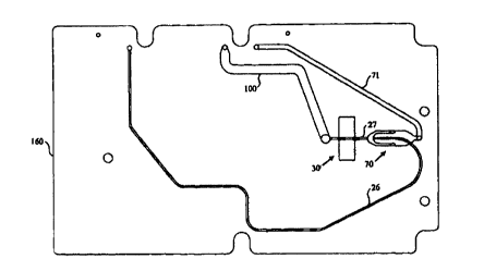
cytometric analysis region. Figures 6 and 7 illustrated a preferred embodiment
of the sheath
flow assembly. The assembly comprises seven sheets, 166A-G, which are
laminated together
to form the fluidic elements of analysis cartridge 160. The analysis channel,
comprising core
stream channel 26 and sheathed stream channel 27, is connected to the
convoluted storage
channel {not shown). In sheath flow assembly 70, first and second sheath fluid
channels,
jointly labeled as element 72, are positioned on either side of and converge
with channel 26.
In this embodiment the diameter of the sheathed portion is greater than the
core portion of the
analysis channel. The sheath fluid channels extend into layers 166C and E, and
are labeled
as elements 75 and 76. The sheath fluid channels provide hydrodynamic focusing
of particles
in channel 27 in the widthwise direction. Upper and lower sheath fluid
chambers 73 and 74
are formed in sheets 166B and F. When assembled, they are positioned above and
below and
converge with channel 26. The sheath fluid chambers provide hydrodynamic
focusing in the
depthwise direction. To minimize layer to layer depthwise discontinuities in
the region
where the sheath fluid channels and chambers converge with the analysis
channel, the
downstream edges are staggered. The edge of channels 7S and 76 are slightly to
the right of
the edge of channel 72. Sheath fluid is conducted to the sheath flow assembly
through sheath
fluid channel 71. Vias 77 in sheets 166C-E connect channel 71 with the sheath
fluid
chambers. The sheath fluid chambers communicate fluid to the sheath fluid
channels. In
typical hydrodynamic focusing operation, the ratio of sheath flow to core
stream 26 flow is
around 130:1.
Following hydrodynamic focusing, flow cytometric measuring is performed in
analysis region 30. The analysis region includes window recesses 31 and 32 in
sheets 166C
and E positioned above and below the focused sample. The window recesses
accommodate
glass inserts. In lieu of recesses, sheets 166C and E can themselves serve as
windows. In the
remaining sheets, optical clearing holes 33 allow optical access to the
analysis region. The
CA 02320296 2000-08-09
WO 99/60397 PCT/US99/09322
iz
sheets in FIG. 7 are sandwiched between an upper case and a lower case. Layers
166A and G
can be incorporated in the case. The illustrated embodiment also includes
waste storage
container 100. It is connected with flow channel 23 through vias 101 and to a
case mounted
storage container through vial 102.
One embodiment of the sheath flow assembly has been illustrated. Other sheath
flow
assemblies known in the art can be utilized, for example U.S.P.N. 4,983,038.
Because this
sheath flow assembly of the present invention provides both widthwise and
depthwise
hydrodynamic focusing, geometric focusing is not required. Although not
necessary, the
analysis channel can decrease in width and/or depth and in a downstream
direction. Two-
dimensional hydrodynamic focusing can also be achieved using the device of
U.S. Patent
Application 08/823,747, filed March 26, 1997. In lieu of hydrodynamic focusing
the flow
channel can be constricted in the analysis region to provide single file
particles, as described
in single file, as described in U.S. Patent No. 5,726,751.
Another preferred embodiment of the sample analysis region is an absorption
analysis
region. For increased sensitivity using an absorbance-based assay the optical
pathlength, i.e.
the channel depth, in the absorption measurement region is increased. For
decreased
sensitivity to factors such as intermittent sample stream perturbations,
optical window quality
and optical measurement apparatus lens defects, the effective illumination
area of the
detection region can be increased by increasing the channel width. There is a
design trade-off
between increasing the channel width and depth and minimizing the volume of
the
microfluidic system. This balance can be determined for a specific assay, a
specific set of
Iight sources, detectors and optics, and the required accuracy and resolution.
The cartridge can also include an inlet for mixing a reagent with the sample
fluid prior
to sample analysis, as shown in FIG. 8. The term "reagent" refers to any fluid
that joins the
sample fluid. It can be, for example, a diluent, a lysing agent, an indicator
dye, a fluorescent
compound, a fluorescent standard bead for flow cytometric calibration, or a
reporter bead for
flow cytometric measurement (U.S. Patent No. 5,747,349). Between storage
channel 20 and
analysis region 30, reagent channel 80 joins analysis channel 24. The reagent
channel is
connected to pump interface 40A and reagent inlet 60. In a preferred
embodiment the pump
CA 02320296 2000-08-09
WO 99/60397 PCT/US99/09322
13
and the inlet are combined in a syringe pump. The cartridge includes valve
interface 50 to
separate the storage channel from the reagent inlet.
When the flow channels are microchannels having laminar flow therein, mixing
between the reagent and the sample is predominantly diffusional mixing. The
streams can
join in side-by-side flow, as described in U.S. Patent No. 5,716,852 and U.S.
Serial No.
08/829,679 filed March 31, 1997, or in a layered flow for more rapid mixing,
as described in
U.S. Serial No. 08/938,584 filed September 26, 1997 and U.S. Serial No.
08/938,585 filed
September 26, 1997. In order to allow for mixing and reaction prior to
analysis, a mixing
channel can be included, as shown in FIG. 9. Mixing channel 90 is positioned
between the
reagent inlet and the analysis region. The geometry of mixing channel 90 is
selected to allow
mixing and reaction between the sample and reagent streams. The mixing channel
can be
convoluted in order to achieve the desired time delay within a compact space.
Alternatively,
active mixing methods can be employed, including ultrasonic, mechanical,
sonic, flow
induced, etc.
1 S In the embodiment of FIG. 9 the mixing channel is illustrated as a square
wave. For a
particle-containing sample, it may be desired to allow diffusional mixing
between smaller
species within the sample and reagent streams without allowing particles in
the sample screen
to gravitationally settle into the reagent stream. Figure 10 shows the effect
of channel
geometry on gravitational mixing. A square wave channel is illustrated in FIG.
10A. The
particle-containing sample stream enters mixing channel 90 through channel 24
and reagent
stream enters through channel 80. In the upper half of the mixing channel the
sample stream
is gravitationally above the reagent stream and particles tend to settle into
the reagent stream.
In the lower half of the mixing channel this is reversed and particles settle
back into the
sample stream. This reversal of top and bottom for the sample stream and
reagents stream
can be used more effectively in an isotropic channel as illustrated in FIG. l
OB. In a spatially
periodic isotropic channel the gravitational top and bottom of the channel
interchange within
each repeating unit. This counteracts the effect of gravity on the particles
in the sample
stream. The isotropic spatially periodic channel is therefore useful for
sedimentation
mitigation as well as sedimentation resuspension.
CA 02320296 2000-08-09
WO 99/60397 PCT/US99/09322
14
The cartridge can provide for more than one analysis region, in series or in
parallel.
Multiple parallel analysis regions are illustrated schematically in FIG. 11.
The device of FIG.
11 comprises sample inlet 10, storage channel 20, resuspension pump interface
PI1 (Pump
Interface 1), and analysis regions 30A-C. At junctions Jl, J3, J5, J6 and at
the end of the
storage channel, fluid from the sample storage channel can be directed to
analysis channels
24A-D and to waste storage container 100. Note that in this embodiment the
resuspension
pump is fluidically connected to the storage channel in the middle of the
channel rather than
at the beginning of the channel . Preferably the sample segment between J1 and
J3 flows
through valve V3 for analysis, the sample segment between J3 and J5 flows
through valve
V2 for analysis and the segment between J5 and J6 flows through valve V1 for
analysis.
The cartridge further includes pump interfaces PI2-PIS, valve interfaces V1-
V5,
reagent channels 80A-C, sheath flow assembly 70, waste storage container 100,
and vents
110A-C. In a preferred embodiment, the sample inlet is a septum, the pump
interfaces are
syringe pump interfaces and the valve interfaces are pinch valve interfaces.
The vents are
made of gas permeable plugs having a reduced permeability when wet. The
storage and
mixing channels are illustrated as square waves but are preferably isotropic
spatially periodic
channels. The sheath flow assembly is preferably as illustrated in FIGS. 6 and
7. Analysis
region 30C is a filling status gauge providing visual indication of proper
sample load.
Analysis region 30A is an absorption measurement region, optically coupled
with
measurement apparatus comprising both a green and a blue LED and a
photodetector.
Analysis region 30B is a flow cytometric analysis region optically coupled
with a
measurement apparatus comprising a diode laser and a plurality of
photodetectors at various
optical axis and collection cone angles.
The cartridge of FIG. 11 can be used for hematology. A single cartridge can
determine the red cell count, the total hemoglobin, and the white cell count
and
characterization. The analysis requires only 15 ~cl of sample, and the waste
fluid is contained
within the cartridge for safe operation and disposability. The sample is
loaded into the
storage channel through inlet 10. At J1 the potentially contaminated leading
edge of the
sample flows in bypass channel 25, having a larger diameter than channel 20.
Air in the
channel escapes through vent 110A. The next segment of the sample fills the
storage
CA 02320296 2000-08-09
WO 99/60397 PCT/US99/09322
channel. Valve V4 is open and the sample flows to filling status indicator
30C. Vent 110C
allows air to escape during sample loading. Excess sample flows into sample
load bypass
storage 115. The cartridge can be stored or transported prior to analysis. For
measurement
the cartridge is inserted into a measurement instrument having a cartridge
holder and valve
5 and pump mechanisms, which engage the valve and pump interfaces on the
cartridge. The
pump mechanisms comprise syringe pumps wherein the syringes are filled with
reagents. P1
is filled with an inert driving fluid, P2 is filled with diluent, P3 is filled
with a soft lysing
agent, P4 is filled with a Drabkin lysing reagent and P5 is filled with a
sheath fluid.
After insertion in the measurement apparatus, the sample is resuspended and
10 analyzed. The entire measurement, including sample resuspension, can be
performed in less
than two minutes. The procedure for operating the analysis cartridge of FIG.
11 for
hematology is outlined in Tables 1-3. For each time interval from tl through
t17, Table 1
describes the procedure, Table 2 gives the elapsed time, and Table 3 gives the
status of valves
and pumps fluidically connected to the cartridge and the status of optical
measurement
15 apparatus optically connected to the cartridge. In the first analysis time
interval, tl, air is
purged from resuspension pump interface PI1 through valve VS into waste
storage container
100. In t2 the reagent and sheath fluid channels are purged and wet. In t3 the
optical path in
absorption measurement region 30A is calibrated using the blue LED. In t4 the
total
hemoglobin sample segment between Jl and J3 is resuspended by alternating
dispense and
aspirate cycles using P1. In t5 the total hemoglobin assay is performed by
mixing the blood
with Drabkin reagent to lyse the red blood cells, and measuring the absorption
in analysis
region 30A. To create a bubble-free mixture in the analysis region, air is
purged from
channels 24A and 80A. Preferably the sample fluid and the reagent reach J2
simultaneously.
Mixing channel 90A is designed to allow formation of the cyanomethahemoglobin
complex.
. Following hemoglobin absorption assay, flow cytometric analysis is
performed. In
time intervals t6, t7 and t8 the channels used in flow cytometric analysis are
purged. To
protect optical surfaces in the cytometric region from direct contact with the
sample, sheath
fluid is pumped through the region during the purge. The sheath flow is set to
a low ratio to
minimize fluid accumulation in the waste storage container during priming
stages. In t9 the
RBC sample segment between JS and J6 is resuspended. In t10 and tl l the
optical
CA 02320296 2000-08-09
WO 99/60397 PCT/US99/09322
16
measuring apparatus is aligned and the flow is stabilized. In t12 and t13 the
RBC flow
cytometric assay is performed. In tl4 the WBC sample segment between J3 and J5
is
resuspended. In tl S a soft lysing reagent is added to the sample and time is
allowed for
mixing and reaction in mixing channel 90B. In t16 and tl7 the WBC assay is
performed.
The total elapsed time is 1.75 minutes. Following analysis, the cartridge is
disposed of.
Drawings of a preferred embodiment of the hematology cartridge are shown in
FIGS.
12 and 13. Figures 13A-G show the seven sheets, 167A-G, which are laminated
together to
form cartridge 160 shown in FIG. 12. This is a three-dimensional fluidic
structure wherein
channels in different layers appear to overlap in FIG. 12 but are in fact
separated by sheets
167C and E. Vias in intervening sheets connect flow elements in different
layers. Three-
dimensional structures can be more compact and rugged than two-dimensional
structures.
Registry of the laminated sheets to the case is accomplished with holes 170 in
the sheets. The
case has pins that fit within holes 170. For measurement, the cartridge is
inserted into a
measurement instrument including a cartridge holder. The outer case of the
cartridge (not
1 S shown) has alignment markings thereon for optical and fluidic alignment
with the
measurement apparatus. In this embodiment, the alignment markings are
kinematic
alignment markings comprising a pit, a groove and a flat. The cartridge holder
has
corresponding pins. The shape of the cartridge is designed for engagement with
the cartridge
holder, and thus in itself comprises an alignment marking.
Sample is introduced through inlet 10 and stored in channel 20. The sample
leading
edge flows into bypass channel 25. The bypass channel is fluidically connected
to a case-
mounted waste storage container (not shown). Syringe pump interfaces 40A-E and
pinch
valve interfaces 50A-D control sample management in the cartridge. The syringe
pump
interfaces are also reagent inlets. When valve 50D is open sample flows
through channel
24D to filling status gauge 30C. For total hemoglobin assay lysing reagent is
introduced
through syringe pump interface 40D and the mixture flows through analysis
channel 24A to
absorption analysis region 30A. For RBC assay, valve 50A is opened, diluent is
introduced
through syringe pump interface 40B, and the red blood cells are
hydrodynamically focused in
sheath flow assembly 70 and counted in flow cytometric analysis region 30B.
For WBC
assay, valve 50B is opened, a soft lysing agent, which masks red blood cells
and platelets, is
CA 02320296 2000-08-09
WO 99/60397 PCT/US99/09322
17
introduced through syringe pump interface 40C, mixing and reaction occur in
mixing channel
90, the sample is hydrodynamically focused in sheath flow assembly 70 and
analyzed in flow
cytometric analysis region 30B. Waste fluid from all three analysis regions
flows into waste
storage container 100, which is fluidically connected with a case-mounted
storage container
having a vent therein. This waste storage container is a channel. It can
alternatively or in
addition be a fixed or expandable reservoir.
In this embodiment, storage channel 20 and mixing channel 90 are formed in
sheet
167D. After cutting the sheet to form the channels, peninsulas of sheet
material remain
around the channels. The peninsulas are not well supported and can flop around
during
laminate assembly. A less floppy channel can be formed using two or more
layers, with
alternating loops of the channel formed in different layers.
The cartridge has been illustrated with particular mixing and measurement
configurations. It can also provide filtering, diffusion based filtering as
described in U.S.
Serial No. 08/663,916 filed June 14, 1996, simultaneous particle separation
and chemical
reaction as described in U.S. Serial No. 08/938,585 filed September 26, 1997,
valueless
microswitching as described in U.S. Patent No. 5,726,404, diffusion-based
chemical sensing
as described in U.S. Patent No. 5,716,852, U.S. Serial No. 08/900,926 and U.S.
Serial No.
08/936,093 and adsorption-enhanced differential extraction as described in
U.S. Serial No.
08/876,038. The channel can also include fluidic elements for extraction,
electrophoresis,
electro-chemical reactions, chromatography and ion exchange reactions.
The cartridge can be fabricated from any moldable, machinable or etchable
material.
The term machining as used herein includes printing, stamping, cutting and
laser ablating.
The cartridge can be formed in a single sheet, in a pair of sheets sandwiched
together, or in a
plurality of sheets laminated together. The teen "sheet" refers to any solid
substrate, flexible
or otherwise. The channels can be etched in a silicon substrate and covered
with a cover
sheet, which can be a transparent cover sheet. In a laminated embodiment, the
channel walls
are defined by removing material from a first sheet and the channel top and
bottom are
defined by laminating second and third sheets on either side of the first
sheet . Any of the
layers can contain fluid channels. In some cases the channel is simply a hole
(or fluid via) to
CA 02320296 2000-08-09
WO 99/60397 PCT/US99/09322
t$
route the fluid to the next fluid laminate layer. Any two adjacent laminate
layers may be
permanently bonded together to form a more complex single part. Often fluidic
elements that
have been illustrated in two separate layers can be formed in a single layer.
Each layer of a laminate assembly can be formed of a different material. The
layers
are preferably fabricated from substantially rigid materials. A substantially
rigid material is
inelastic, preferably having a modulus of elasticity less than 1,000,000 psi,
and more
preferably less than 600,000 psi. Substantially rigid materials can still
exhibit dramatic
flexibility when produced in thin films. Examples of substantially rigid
plastics include
cellulose acetate, polycarbonate, methylmethacrylate and polyester. Metals and
metal alloys
are also substantially rigid. Examples include steels, aluminum, copper, etc.
Glasses, silicon
and ceramics are also substantially rigid.
To create the fluidic element in the sheets, material is removed to define the
desired
structure. The sheets can be machine using a laser to ablate the material from
the channels.
The material can be removed by traditional die cutting methods. For some
materials
chemical etching can be used. Alternatively, the negative of the structure
desired can be
manufactured as a mold and the structure can be produced by injection molding,
vacuum
thermoforming, pressure-assisted thermoforming or coining techniques.
The individual layers, assemblies of layers, or molded equivalents are bonded
together using adhesives or welding. Alternatively, mechanical compression
through the use
of fasteners such as screws, rivets and snap-together assembly can be used to
seal adjacent
layers. Layers can be assembled using adhesives in the following ways. A rigid
contact
adhesive (for example, 3M1151) can be used to join adjacent layers. A solvent
release
adhesive may be used to chemically bond two adjacent players. An ultraviolet
curing
adhesive (for example, Loctite 3107) can be used to join adjacent layers when
at least one
layer is transparent in the ultraviolet. Precision applied epoxies, thermoset
adhesives, and
thermoplastic adhesives can also be used. Dry coatings that can be activated
to bond using
solvents, heat or mechanical compression can be applied to one or both
surfaces. Layers can
be welded together. For welding the layers preferably have similar glass
transition
CA 02320296 2000-08-09
WO 99/60397 PCTNS99/09322
19
temperatures and have mutual wetting and solubility characteristics. Layers
can be welded
using radio frequency dielectric heating, ultrasonic heating or local thermal
heating.
The device of FIGS. 12 and 13 was fabricated as follows. Layers 167A and G
were
made of 4 mil mylar and layers 167C and E were made of 2 mil mylar. Layers
167B, D and
F were made of 2 mil mylar with 3M1151 on both sides (4 mil inclusive). The
adhesive had
cover sheets thereon. With the cover sheets still on the adhesive, the sheets
were laser
ablated to machine flow elements therein. The cover sheets were removed and
the individual
laminate was assembled with the aid of an alignment jig.
This invention further includes a sample analysis instrument for use with an
analysis
cartridge, in particular a hematology analysis cartridge. The instrument has a
cartridge
holder, a flow cytometric measuring apparatus position to be coupled with a
flow cytometric
measuring region on the cartridge, and a second measuring apparatus positioned
to be
coupled with a second measuring region on the cartridge. The flow cytometric
measuring
apparatus comprises a light source, preferably a laser, and at least one
photodetector. The
photodetectors can be positioned for measuring small angle scattering, large
angle scattering
or fluorescence. The apparatus can also include optical elements such as
focusing and
collection lenses, wavelength filters, dichroic mirrors and polarizers. The
second measuring
apparatus can be any measuring apparatus including optical, electrical,
pressure sensitive and
flow sensitive apparatus. Absorption measuring apparatus comprising a light
source and a
photodetector is preferred. Preferably the light source is positioned on a
first side of the
cartridge holder and the photodetector is positioned on the opposite side.
A measurement instrument is shown schematically in FIG. 14. It comprises
cartridge
holder 190, flow cytometric measurement apparatus 180B and absorption
measurement
apparatus 180A. Cartridge 160, shown in phantom, slides into the cartridge
holder. The
measurement apparati are positioned to be optically coupled with flow
cytometric analysis
region 30B and absorption analysis region 30A. This instrument also includes
pump and
valve mechanism manifold 141. The pump mechanisms are syringe pumps which
couple to
pump interfaces on the cartridge via cannulas 140. The manifold can also
include reagent
CA 02320296 2000-08-09
WO 99/60397 PCT/US99/09322
reservoirs to refill the syringe pumps for multiple assays. The valve
mechanisms activate
valve pins 150, which couple to valve interfaces on the cartridge.
Preferably the cartridge holder has alignment markings thereon to mate with
corresponding markings on the cartridge. The alignment markings can be the
shape of the
5 holder, protruding pins, notches, rods, kinematic mounts, two stage
kinematic mounts as
described in U.S. Patent Application 08/736,336, filed October 23, 1996, or
any other feature
that facilitates positioning of the cartridge. In lieu of or in addition to
cartridge alignment, the
instrument can include optical steering elements, such as mirrors, to align
the measuring
apparatus with the analysis region. The analysis instrument can further
include valve and
10 pump mechanisms which couple with valve and pump interfaces on the
cartridge.
All references cited herein are incorporated by reference in their entirety.
Preferred embodiments described above are intended to be illustrative of the
spirit of
this invention. Numerous variations and applications will be readily apparent
to those skilled
in the art. The range and scope of this patent is defined by the following
claims.
CA 02320296 2000-08-09
WO 99/60397 PCTNS99/09322
21
x
,
..
w
a~
w
0
o ,o ,o
U
v v
~23 a i o
:x o 0 0
U
~, ~ x
0
. ,
" ~ r-. . v
~ 0 0
_ o w
..fl ~ ~ ~ ~
U U
, .,.r
, cc N
d
O ~
O ~ O V
G
. U U ~,
U
. ~ . . .
it ' ~
U ~ O ~ + ..O CT
O O 00
~ O
~' .~ N .~.~ O 'O
O ...~
.C
+, ~ O
E"'' ~ O O ~ .'' O"
N O ~
~ O, t3, j~
cd ~ ~ ''~ CC
~.Or O O O O ~ ~
''' ~ ~ O
~
o N
N M ~ ~ ~ 0 b
N ~ ~
b
_ ~l '' O ~ ~ ~ N
ts, '' b O
U 'O
E-H q~ O o ~ p~ O
O O
.-, ~ ~' ~
... ~
~, . M .t", b
p "O , ~ ' O
r'-~ .- m'-~
4, .-.
O ~ ~ ~ "O
C ~
s., .~. O O 47
w .-~ ~
o '~
o
a~ . ,
o ~ '" ~ ~' '~
~ 'o
~ c ~ 0
a ~ 4c
d
O ~''~ ~ 4.~V ~ ~ O O .~ 'L~
7 ' ~v~ O ~ O '~ ~ v~ O
O
cn ~ ~ , ~, O v ~ ~ ~ U
~' ~ w ~
~
c i. ..,Q t'L
d .
y.., sr
~ ~ Q ~ ' ~ it
w ~
Wp ~, ~~ ., p v ~ o y
. A
U s. fU.,T _ ~ by U ~ N U 10....N.~ s.,
~ . .-.. cd Y
~
~'
O r.r C~ cV ...r 1~ it ~ ;= r r~ ~ t.r..i
cd
~ i v ~ Gy,~ U ~ '- ~ ~
O O O
b V _c 7,
~ v~ ' ~ d N ~
~
o o 3
~ ~ n 3
'"
~ C/1 4r rN ~ V7
..
.. .,
,
' " a, a~ ~ ~ U U
s
a. o ~ , o , , >, >,
~'"
~,
a. ~ ~ n
a.
cd cG ~ ~ ~ ~ ~ V ~ ~ ~ .~ y .b
~ x
~ G4 f~ ~3 U~ U~ ~ U ~ U U o U U U o
. H H ~ a ~ , ~ A ~ ~ v v
~ ~ ~
A E c ~ G ~ ~ ~
n
N M d' v1 V (~- o0
..rr..~,..~,~ ...~ .r ... ,..
~n O v1
CA 02320296 2000-08-09
WO 99/60397 PCT/US99/09322
22
N N
t~ N O l~
N OMO M
l~ N M
.-. ~O O
V ~n ~t
O
M M N
M ~ I~
'~ O
N
O
~O O
M ~p
r-' O
O ~1 N M
M
O
N
.4: M l'~ V7
N
O
d' O
pp N d'
,'_' O
N M 00
N M
'~ O
N ~ ~n
N M
O
O O~ N
V1 .-, ..~ M
O
M 01 v7
O
N ~D O
M
+' O
M d' I~
N O
~" O
N
O
O
N id TJ "d
~-. ~ N
N
~
_
N N
f3. CL N
.. ~ cd ~_
~ _c~t
~ ~ LT.~ f3a
H m, '....'.yr
... ',~:.
O
CA 02320296 2000-08-09
WO 99/60397 23 PCT/US99/09322
...
k X >C X U O U U U k
.,
JC ~C ~C ~C U O U U U k
>C ~C ?< U O U U U >C
7G >C ~t >C O U U U U >C
>t ?G >t ?C O U U U U ~C
~x x x ~ o U U U U ?C
>C ?t >C >G O U U U U
.~
~G 5C 5C 7G O U U U U >G 5C >G
' ~C ~G x ~ O U U U U
>C ~C x O O U U U
M
N
?C ~t >G >C U O U U U
0
a~
>C x ~G x O U U U U
>C ~G U U O U U
U U O U U
>C U U U U U
~C ~G >C ~f O O O U U
>C >C (~ U U U O
A a > ; .~ N
W ~ N ~ i.wH
t3, G, C"~l ~
cd O o t',
~1 '~ a ~ O
~ N M .
~ ~ >
C'.. .:~, f3,~ Q > >~ G." II
.,
fz.C).~ ~ 7
~ _ ~ A b
N Q. ~" Q, d ~ id ~ b ~ :: ~ a w N
..' ~ > ""' v~ v~ -.
'~
. ~ ' i .~ > > a~ .
~ .
, a ~ . f~ ~ U U GO ia.~ ~ v
~
~ ~ . ~ H ~ ~ ~ H ~ ~ o ~ ~ v
~
~
H c c A 3 r~ m A c7 as
~n o ~n o
rw' --~ N
SUBSTITU T E SHEET (RULE 26)