Note: Descriptions are shown in the official language in which they were submitted.
CA 02324602 2000-09-19
WO 99/47046 PCT/US99/06026
MULTIDIMENSIONAL DETECTION AND CHARACTERRIZATION OF PATHOLOGIC TISSUES
FIELD OF THE INVENTION
The present invention relates to the detection and characterization of medical
pathologies in human and animal bodies. More particularly, the present
invention relates to
the detection and identification of cancer in organs or tissues.
BACKGROUND OF THE INVENTION
There are no available systems today, for medical or non-medical applications,
to
detect and characterize distinct features within an object understudy, such as
cancerous
lesions and tumors in a human body. Presently, only imaging systems are
available, such as
imaging systems based on x-ray, mammography, computed tomographic (CT) scans,
or
magnetic resonance imaging (MRI). All of these imaging systems simply provide
images
of pathologies within a human body; they do not characterize any features.
In addition, each of these imaging technologies has significant drawbacks. For
example, x-rays, mammography, and CT scans all use ionizing radiation and
therefore
present certain health risks to a patient, such as cell mutations. Also, both
CT scans and
MRI involve procedures that are relatively expensive, which hampers their
widespread use.
Moreover, both MRI and CT scans require the expertise of highly trained
personnel for
extended periods of time to operate the devices and to interpret the results.
Furthermore,
each of these imaging technologies requires that the patient lie still,
sometimes for an
extended period of time. This restriction on movement may not only
inconvenience the
patient, but also discards information that could potentially be discovered
from the
movement of tissues within the patient. As to mammography, it is particularly
uncomfortable for the patient since it requires that the breast be compressed
to allow more
uniform tissue density, better x-ray penetration, and tissue stabilization.
More importantly,
methods such as mammography rely on two-dimensional images, thus disguising
three-
dimensional structure information which can be critical for diagnosis.
As an alternative to the above-mentioned imaging technologies, the medical
community has looked to ultrasound for providing a safe, low-cost, high-
resolution imaging
tool. However, conventional ultrasound (ultrasonic B scanning) has certain
limitations. In
conventional ultrasound analysis, a small array of less than approximately
1000 elements is
moved by hand in contact with the object under study. In fact, most current
ultrasound
arrays have only 256 elements. The array sends out waves that reflect from
tissues back to
the same array. Trained technicians and physicians are needed to conduct the
ultrasound
imaging procedure and to interpret the results. This reliance solely on the
reflected waves
- I -
SUBSTITUTE SHEET (RULE 26)
CA 02324602 2000-09-19
WO 99/47046 PCT/US99/06026
results in two major drawbacks. First, ultrasonic B scans do not provide
information on the
properties of the materials themselves; rather, they provide information on
the reflectivity of
the boundaries between different types of materials. Second, the array is
incapable of
capturing radiation except that which is reflected back to the hand-held
sensing array.
Considerable information exists, however, in the transmitted waves, which is
not captured
or used in conventional ultrasonic B scans.
There is thus a need for an apparatus and method that provides detection and
characterization of medical pathologies in a human body. More generally, there
exists a
need to detect and characterize distinct features within an object under
study.
SUMMARY OF THE INVENTION
The present invention provides construction and use of multidimensional field
renderings for high-resolution detection and characterization of distinct
features within a
three-dimensional object. More particularly, the invention provides
construction of such
multidimensional field renderings for high-resolution detection and
identification of
medical pathologies in human and animal bodies, especially high-resolution
detection and
identification of cancer in organs or tissues. The present invention also
provides detection
and characterization of other medical pathologies including pathologies of
musculoskeletal
systems, digestive systems, and the alimentary canal, in addition to
atherosclerosis,
arteriosclerosis, atherosclerotic heart disease, myocardial infarction, trauma
to arterial or
veinal walls, and cardiopulmonary disorders.
The present invention provides construction of a multidimensional field
rendering
that describes the physical details of any three-dimensional object under
study. By
correlating the information contained in such a multidimensional field with
information
regarding known details of general objects under study by using a trained
evaluation
system, the present invention provides detection and characterization of the
structures that
exist in the object under study. For example, the present invention provides a
system based
on ultrasound which, when it is used to observe a human breast, correlates a
catalog of
known morphologies and acoustic characteristics of tissue types that are known
to exist in
breast tissue with the multidimensional field derivation of physical
properties; then the
system of the present invention detects and characterizes various tissues
including
fibroadenoma, fat, fibroglandular tissue, and benign versus malignant lesions
or tumors.
The present invention provides a method and apparatus that allows for the
detection
and characterization of features within an object under study. The invention
uses an array
of radiation sources and an array of radiation detectors to collect scattered
radiation
regarding the object under study. In one preferred embodiment, the source
array and
detector array are configured as a single integrated unit. In another
preferred embodiment,
-2-
SUBSTITUTE SHEET (RULE 26)
CA 02324602 2000-09-19
WO 99/47046 PCT/US99/06026
the radiation sources and detectors are the same physical devices; they
operate in one time
period as radiation emitters and in another time period as detectors. In yet
another preferred
embodiment of the invention, the arrays comprise large numbers of sources and
detectors,
preferably with more than 5000 detectors. With a sufficient number of such
sources and
detectors, the present invention provides for construction of a three-
dimensional rendering
of numerous physical quantities to describe the object and therefrom derive
interpretations.
The radiation sources emit radiation of a specific waveform, either within a
predetermined
frequency range or at a predetermined frequency, which is propagated within
the object
under study and subsequently scattered by features within the object under
study.
Generalized scattering includes reflection (backscattering), transmission
(forward
scattering), and diffraction, which may occur in any or all directions from
the features
within the object under study. All these types of secondary waves constitute
the wave
signal returned from the object under study.
In a preferred embodiment, the radiation sources and detectors cover a large
solid
angle, thereby substantially enclosing the object under study. As a result, a
large fraction of
all these types of secondary waves are detected by the radiation detectors.
The resolution
depends on the product of the number of sources and the number of detectors,
which defines
the number of resolution elements into which the volume occupied by the object
under
study may be divided.
In a preferred embodiment of the invention, the radiation is ultrasound
radiation,
although the invention generally encompasses the use of any radiation,
including
electromagnetic and acoustic radiation. In more specific embodiments of the
invention, the
object under study is tissue or an organ, or other part of an animal body such
as the human
body. By using a sufficiently large number of detectors and sources, a high
resolution
multidimensional field is provided in accordance with the present invention.
In another
embodiment of the present invention, the sources are modulated to have
different phases,
which permits focusing or scanning of the radiation.
In accordance with another embodiment of the present invention, the radiation
is
sufficiently focused and is used to destroy features within the object, such
as cancerous
lesions within human or animal tissue.
In accordance with the present invention, the data collected by the radiation
detectors are then used to construct a rendering of a multidimensional field,
represented
herein as 47[r,t:0(r,t)], that represents physical characteristics of the
object under study.
The vector r represents the position coordinate of a particular volume element
("voxel"); "t"
is the time' and "0" is a list of the physical parameters associated with the
field at that
voxel. In general, the field and each physical parameter are both spatially
and time
dependent. The multidimensional field comprises estimates of the values for
this set of
-3-
SUBSTITUTE SHEET (RULE 26)
CA 02324602 2000-09-19
WO 99/47046 PCTIUS99/06026
parameters that individually represent physical characteristics of the object
under study.
These parameter values, taken together, characterize the properties of
features within the
object under study. In the case of medical applications, this characterization
results in the
identification of focal regions, their probability of pathology, such as
malignancy, and
associated probabilities of frequency distribution and error rate.
As an illustration, consider those embodiments of the invention where the
radiation
is ultrasound radiation and the object under study is a human organ. In this
illustration,
-7[r,t:O(r,t)] may describe, for example, the sound speed, sound absorption,
tissue pressure,
density, shear modulus, elasticity, etc., of the organ as functions of
frequency. The field
values are stored electronically into a computer-readable medium, such as a
floppy disk,
random access memory, or hard memory disk. This allows subsequent processing
of the
stored field values.
In accordance with a preferred embodiment of the present invention, the
construction of the rendering of the multidimensional field,7[r,t:8(r,t)] from
the detected
data, which comprise elements from a description of the waveform of the
detected radiation
at the location of each detector, is accomplished with an optimal signal
processing
technique.
In one embodiment of the invention, this is accomplished with matched-field
processing, in which the field rendering is constructed so as to produce model
detector data
that matches the actual detector data, and which may be achieved through an
iterative
technique. In this iterative technique, the shape of the object under study is
first estimated.
This can be achieved in a number of different ways, using existing techniques,
such as using
the transmission-only radiation detected and developing the initial estimate
with
conventional computer tomographic techniques. In the embodiment where the
object under
study is human or animal tissue, organ, or other body part, this initial
estimate is referred to
as an "anatomic" construction.
An initial estimate of the multidimensional field 47, `[r,t: (r,t)] is then
calculated by
injecting physiological data to produce a "physiological" construction. This
proceeds by
using a pattern-recognition algorithm, such as an expert system, to analyze
the
morphological features of the anatomic construction and thereby to assign an
initial,
nominal estimate of the multidimensional field. This nominal estimate is based
solely on
average values that structures in the object are expected to have based on
their
morphologically based identification by the pattern-recognition algorithm. The
pattern-
recognition algorithm achieves this initial assignment by comparing the
morphological
features of the anatomic construction with a database of stored morphological
features, such
as elongation, flatness, jaggedness, etc.
-4-
SUBSTITUTE SHEET (RULE 26)
CA 02324602 2000-09-19
WO 99/47046 PCT/US99/06026
At this point, the physical characteristics of the object contained in the
estimated
field &75`[r,t:0(r,t)] are input into a wave-propagation code, together with
the information
concerning the characteristics of the radiation that was initially generated.
Such a wave-
propagation code can be used to generate the waveform of the radiation that
would be
expected at the locations of the detectors based on this information. Such
wave-propagation
codes can be rather complex, but exist in the prior art. Once these signal
data have been
generated based on the estimated field, they are compared with the actual
signal data that
was received by the detectors. If the difference between the two sets of
signal data is at the
level expected for noise in the system, then the estimated field is taken to
be the actual
constructed field rendering, i.e. 9[r,t: (r,t)] &7` S`[r,t: (r,t)].
If, however, the comparison with the actual signal data shows that the
difference
between the signals generated by the estimated field and the actual signals is
greater than
the expected noise in the system, then a correction to the estimated field is
calculated. This
correction is determined by using a wave-propagation code to generate a
refinement field
G'[r,t:0(r,t)] from the difference in actual signals and signals that would be
produced by the
estimated multidimensional field. This refinement field is then used to modify
(e.g., by
adding to) the estimated field to produce a new estimated field, which is then
used to
calculate a new set of detected signals. The process is iterated until the set
of signals
generated from the estimated multidimensional field `[r,t: (r,t)] converge to
a converged
multidimensional field 42[r,t:O(r,t)], which generates waveform signals that
are within the
noise level of the actual set of detected signals.
Once the multidimensional field 9[r,t:9(r,t)] has been calculated, it is
interpreted so
as to characterize the object under study. This is done, for example, with the
use of an
expert system, neural net, or other trained evaluation system that has been
taught to take the
values calculated for -7[r,t:0(r,t)] at every voxel and to reach a
determination of what the
identified features in the object under study are. The specific features of
the
multidimensional field that are relevant for the interpretation method
executed by the
trained evaluation system depend both on what the object under study is and on
what is to
be learned about the object.
For example, in the embodiment of the invention where human or animal tissue
or
an organ is studied with ultrasonic radiation and the goal is to identify
diagnostic
parameters suggestive of the existence of cancer, there are at least seven
identification
methods that are used to extract information from the multidimensional field
so as to allow
the trained evaluation system to draw such interpretations.
In a first identification method, the converged multidimensional field will
contain
the sound speed and sound absorption of the tissue or organ at every voxel.
The trained
-5-
SUBSTITUTE SHEET (RULE 26)
CA 02324602 2000-09-19
WO 99/47046 PCT/US99/06026
evaluation system will then classify the type of tissue present at every voxel
based on this
information, and will identify cancerous tissue based on these particular
physical properties.
In a second identification method, a Hough transformation of the
multidimensional
field is used to identify closed volumes in the tissue, which are indicative
of the existence of
neoplasms.
In a third identification method, the existence of angiogenesis and other
anomalies
of the circulatory system are identified by examining three-dimensional blood
flow with the
Doppler effect.
In a fourth identification method, the tissue pressure is extracted from the
multidimensional field and correlated with the localization of an enclosed
volume, as well
as any distinct results from the other identification methods.
In a fifth identification method, the Doppler effect is used to analyze the
effects of
external vibrations on the tissue, which produce characteristic results in the
multidimensional field to allow the identification of tissue shear modulus. In
a related
method, microcalcifications and tissue elasticity, which are also suggestive
of cancer,
produce a characteristic Doppler signature.
In a sixth identification method, information regarding the electrical
impedance of
tissue is extracted from the multidimensional field. This information is
related to the
existence of tumors.
In a seventh identification method, the multidimensional field rendering is
constructed at two different times and compared either to study changes over
time in the
tissue or to allow the use of interferometric techniques to improve the
resolution.
The actual identification of medical pathologies such as cancerous tissue
preferably
uses more than one of these identification methods in conjunction. With the
use of multiple
identification methods, the reliability of the evaluation is improved. In this
way, for
example, a human breast may be examined with ultrasonic radiation to identify
cancerous
tissue at the desired resolution. In other embodiments, the invention is used
to identify
cancer in other organs, such as the prostate, colon, lung, etc.
OBJECTS AND ADVANTAGES OF THE INVENTION
It is thus an object of the invention to produce an apparatus and method for
sensing
the spatial, or spatial and temporal, properties and determining the physical
and/or
biological nature of materials in a substantially enclosed volume.
It is another object of the invention to perform sensing operations that
uniquely
identify physical properties in contiguous, highly resolved volume elements
throughout the
sensed media.
-6-
SUBSTITUTE SHEET (RULE 26)
CA 02324602 2000-09-19
WO 99/47046 PCT/US99/06026
It is yet another object of the invention to provide a disease-detection
system
specifically designed to find small, subtle indicators of early pathology,
including cancer,
vascular disease, etc.
It is still a further object of the invention to produce a class of physics-
based
diagnostic devices that probe the subject environment, observe the response of
the contents
to the probing disturbance, and then diagnose the implications of the measured
data.
An advantage of the invention is that it provides detection and identification
of
tissue anomalies where the object under study is animal or human tissue or an
organ. In
particular, the invention detects and identifies cancerous tissue in animal
and human organs.
The invention also provides detection and identification of other medical
pathologies in
systems including cardiovascular, musculoskeletal, or digestive systems. The
disease states
that may be characterized include trauma, infection, neoplasms, and disorders
of various
biochemical pathways.
It is a further advantage of the invention that it provides construction of
the
multidimensional field rendering in three spatial dimensions by using of all
scattered
radiation, which includes radiation reflected, transmitted or diffracted by
the object under
study or by features within the object under study.
An additional advantage of the invention is that it permits the complete use
of
Doppler shifted data, since there is no limitation to Doppler shifts that lie
in a single plane
of examination.
Other objects and advantages may occur to those of skill in the art after
reading the
detailed disclosure and figures. The invention is not limited to those objects
and advantages
recited above, but encompasses all objects and advantages that would occur to
those of skill
in the art in light of the disclosure.
BRIEF DESCRIPTION OF THE DRAWINGS
FIG. 1(a) and FIG. 1(b) show cross-sectional views of the detection and
identification apparatus for one embodiment of the invention with a geodesic
dome
configuration, which is a geometrical construction appropriate for
investigation of the
human breast. FIG. 1(c) is a plan view of one embodiment of the radiation
source and
detector arrays.
FIG. 2 is a block diagram of an arrangement of the radiation sources and
detectors in
one embodiment of the invention.
FIG. 3 is a histogram that illustrates the relationship between the number of
sequences and the sequence length in the use of linear maximal length sequence
modulation
of the radiation sources and detectors.
-7-
SUBSTITUTE SHEET (RULE 26)
CA 02324602 2000-09-19
WO 99/47046 PCT/US99/06026
FIG. 4 is an example of a modulation sequence of the radiation sources and
detectors using Walsh Function modulation.
FIG. 5 is a flowchart illustrating a method for constructing the rendering of
the
multidimensional field,9[r,t:0(r,t)] from the data collected by the radiation
detectors.
FIG. 6 is a flowchart illustrating in detail a method for injecting
physiological data
into the initial construction of rendering of the multidimensional
field,?[r,t:0(r,t)] based on
the morphological characteristics of object under study.
FIG. 7 is a graph showing the relationship between the ultrasonic complex
sound
velocity (sound speed and absorption) and the type of tissue that is found in
the human
breast.
FIG. 8 is a flowchart illustrating in detail a method for changing the
physiological
data in the iterative part of the construction of the rendering of the
multidimensional field
'[r,t:0(r,t)].
FIG. 9 shows a perspective view of morphing ultrasound fields. In part (a) and
part
(c), a representation of features in tissue are displayed at an initial time;
in part (b) and part
(d), the features are shown at a later time. No cancer growth is detected in
part (b), but
cancer is detected in part (d).
DETAILED DESCRIPTION OF THE INVENTION
The invention is described in detail in the following. Although reference is
sometimes made to a specific embodiment of the invention wherein the radiation
that is
used is ultrasound radiation, it will be appreciated by those of skill in the
art that the
invention is not so limited. The invention encompasses the use of any type of
radiation,
including both acoustic and electromagnetic radiation, and reference to the
specific
embodiment of ultrasound radiation is not intended to be limiting. Reference
is also made
in the following detailed description to application of the method on a human
patient to
diagnose cancer. Such reference is again not intended to be limiting and
represents only a
preferred embodiment of the invention.
The invention relates both to an apparatus and a method that can be used to
allow
the detection and characterization of features within an object under study.
The invention
achieves this detection and characterization by constructing a rendering of a
multidimensional field,9[r,t:O(r,t)] at every volume element ("voxel") that
can be resolved.
Here, r represents the position coordinate of a voxel, and O is a list of the
physical
parameters that are associated with the field at that voxel. In principle,
9[r,t:9(r,t)] can
include information on whatever physical properties are desired for the
application of
interest. On perhaps the simplest level, the multidimensional
field,9[r,t:O(r,t)] may contain
information for construction of a visual image of the object under study. For
some
-8-
SUBSTITUTE SHEET (RULE 26)
CA 02324602 2000-09-19
WO 99/47046 PCT/US99/06026
applications, however, it is more useful for 2[r,t: (r,t)] to be constructed
to describe other,
more relevant physical properties of the object.
The versatility of the general multidimensional field 9[r,t:O(r,t)] may be
illustrated
with an example of one embodiment of the invention, where the object under
study is a
human breast, the goal of studying the object is to identify cancerous tumors,
and
ultrasound radiation is used. If all that is desired is an image of the
breast, showing its
internal structures, then 42[r,t: (r,t)] can be constructed as a one-
dimensional scalar field
that contains a single quantity, such as the density, of the object at every
point, i.e. 0 = {p}.
The detection and/or identification of tumors is, however, more readily
accomplished by an
examination of quantities such as the sound velocity, sound absorption, and
tissue pressure.
In this case, &Rr,t: (r,t)] can be constructed as a field containing the
values of these
quantities at every voxel, i.e. = {v, A, P}, where v is the sound velocity,
A is the sound
absorption, and P is the tissue pressure.
1. Apparatus
In order to generate the radiation that is used in the invention, there must
exist
radiation sources. Although it is within the scope of the invention to use a
single radiation
source, it is preferred that there be multiple sources of the radiation. In
instances where a
single radiation source is used, collection of sufficient appropriate data to
construct the
multidimensional field rendering of the object under study may require that
the source be
large and/or in motion. In general, the sources are configured so as to
minimize the volume
of the object under study that is not reached by radiation emitted from the
sources. The
radiation sources are adapted to emit radiation within a predetermined
frequency range or at
a predetermined frequency. In the embodiment where the sources provide
ultrasound
radiation, the sources are arranged so that there are few or no uninsonified
regions in the
volume of interest.
In those embodiments of the invention where there are a multiplicity of
radiation
sources, the sources can be arranged in a wide variety of geometric
configurations. These
different configurations allow for the measurement of properties of
differently shaped
objects. In particular, these different configurations allow for the
achievement of one
objective of the invention, which is to provide a large solid angle 0 over
which the object is
studied. This is desirable in order to obtain the greatest level of
information possible by
substantially enclosing the object under study. Preferably, the coverage of
the apparatus is
0 z 21t sr.
Illustrations of such configurations can be made for the study of certain
anatomical
features of human beings, which is one embodiment of the invention. For
example, where
the object understudy is a human breast, a modified hemispherical arrangement
of radiation
sources will provide large coverage (0 = 27t). If the object under study is a
human limb,
-9-
SUBSTITUTE SHEET (RULE 26)
CA 02324602 2000-09-19
WO 99/47046 PCT/US99/06026
then a cylindrical arrangement can provide Q > 21t. Alternative arrangements
include the
use of concentric cylinders, or portions of concentric cylinders, where the
object under
study is an organ inside the human body, and this can also provide a
sufficient solid angle
with 0 > 21t. In one example, a small internal cylinder is inserted into the
alimentary canal
of the patient, and a larger external cylinder or cylindrical arc is
positioned outside the
patient near the object under study. In this manner, for example, the
requisite large solid
angle coverage is achieved when analyzing the human prostate. Other array
configurations
include ellipsoids as well as cones and truncated cones.
It will be readily appreciated that these are merely examples of the types of
configurations that can be made for different objects under study. It is also
within the scope
of the invention to combine these examples to create additional configurations
adapted to
other objects, and the invention is not limited by the configuration. For
example, in the case
of study of the human breast, it is preferable to have additional coverage
beyond a
hemisphere, such as the geodesic-dome configuration that is shown in FIG.
1(b), because
tumors frequently manifest themselves in the upper outer quadrant going
towards the axilla
and in the axillary lymph nodes (under the arms). In other embodiments of the
invention,
an appropriate configuration is used to detect and identify cancers in other
parts of the body,
such as the prostate, liver, lung, or portion of the alimentary canal.
In one embodiment of the invention, the sources emit radiation that is uniform
in
phase, although in alternative embodiments, the phase of the radiation emitted
by each
source may be varied. This is achieved by individual encoding of each
individual source as
described below. This variation of phase may be structured so as to focus the
radiation in
particular areas of the object under study or it may be structured so as to
scan the entire
object under study.. In cases where the phase variation is structured so as to
focus narrowly,
it allows the possibility of confocal microscopy of the object under study.
Furthermore, if
the radiation is sufficiently focused, it may be of an intensity that can be
used to destroy
well-defined features within the object under study if this is a desired
objective for the
particular application of the invention. For example, the focused radiation
could be used to
destroy cancerous tissue discovered in an organ without invasion of the body
by a surgical
instrument.
The invention also comprises a multiplicity of radiation detectors. These
detectors
are also placed so as to cover a large solid angle, again preferably greater
than 21t sr, and
can be arranged in the same types of geometric patterns that were described
above in
relation to the sources to achieve this coverage. In most applications, the
geometric pattern
used for the detectors will be similar to the geometric pattern used for the
sources, but the
invention is not so limited. The invention also encompasses embodiments where
the
number of detectors greatly exceeds the number of radiation sources.
-10-
SUBSTITUTE SHEET (RULE 26)
CA 02324602 2000-09-19
WO 99/47046 PCT/US99/06026
It will readily be appreciated by those of skill in the art that the spatial
resolution of
the device is directly related to the number of sources and detectors that are
used, as well as
to the frequency of the radiation. The product of the number of sources and
the number of
detectors should be at least as large as the number of voxels needed to
achieve the desired
resolution. Although there is no fixed limit on the number of sources and
detectors,
provided the appropriate number are used to achieve the desired resolution,
the invention
encompasses the use of considerably more detectors than used in the prior art.
The
resolution is governed principally by the Nyquist criterion, which states in
its simplest form
that in order to prevent undesired aliasing, one must sample a signal
spatially at least twice
in a wavelength. Thus, full sampling of the object under study is achieved by
placing
detectors closely spaced at the Nyquist half-wavelength limit, as dictated by
the frequency
of the radiation to be used.
In embodiments of the invention applied to the identification of cancer in
tissue, for
example, it is preferred that the resolution be sufficient to detect small
neoplastic masses
with diameters less than 3 mm. In the case of identification of cancer in the
human breast,
an embodiment of the invention uses approximately 250 ultrasound sources and
approximately 4,000,000 ultrasound detectors. At an operating frequency of 5
MHZ, this
would achieve a resolution of approximately 0.3 mm for a hemisphere with a
diameter of 20
cm, corresponding to the volume occupied by a breast. In other embodiments of
the
invention, the frequency of the radiation and the required resolution may
dictate a need for
considerably fewer sources and detectors, or for very detailed studies could
require even
greater numbers of sources and detectors.
In different embodiments of the invention, the detectors can measure either
the
longitudinal or transverse waves ("shear waves" in the particular case of
ultrasound), or,
preferably, both the longitudinal and transverse waves. Different embodiments
also
correspond to which components of the waveform are measured by the detectors.
In
particular, the amplitude, phase, and/or frequency of the detected waves
is/are measured,
although it is preferable that all three be measured since this provides more
information that
can be used to construct the rendering of the physical multidimensional field
42[r,t: (r,t)].
It is also preferable that the detectors be configured so as to detect both
waves that have
been reflected from the object under study and transmitted through the object
under study.
Again, this is preferred so as to extract as much information as possible from
the detected
radiation, and such configurations are more readily achieved when the arrays
of sources and
detectors are arranged so that the solid angle 0 z 2,t.
Referring to FIG. 2, there is shown a detailed block diagram of a preferred
arrangement of the radiation sources and corresponding detectors. A master
controller
("Input") generates the modulation strategy and a master detector ("Output")
controls what
-11-
SUBSTITUTE SHEET (RULE 26)
CA 02324602 2008-02-06
comes out of the detectors. A modulator coupled to a radiation source
generates a
modulating signal. The radiation source, in turn, generates a modulated
waveform signal
based on the modulating signal received from the modulator. Each modulator
generates a
distinctive modulating signal, thereby causing each radiation source to
generate a distinctive
modulated waveform signal. It should be noted that one modulator may be
coupled to a
plurality of radiation sources generating a plurality of distinctive
modulating signals for the
coupled radiation sources.
There exist many digital coding methods and designs for modulating waveforms
for
the purpose of encoding waves, depending upon the desired correlation
properties. See
generally K. Sam Shamugam, Digital and Analog Communication Systems (John
Wiley &
Sons, New York, 1979). Two examples of digital switching modulation schemes
are given
here. I.,inear maximal length (LML) codes have excellent autocorrelation for
time resolution,
and the Walsh functions (WF) have superior cross-correlation for spatial
resolution. An
encoding scheme is provided for each. The waveform signals generated from the
sources are
transmitted, reflected or refracted though the object and arrive at a
plurality of detectors.
Each detector includes at least one matched filter designed to decode one
distinctive
modulated signal. If the emphasis is to be placed on temporal resolution,
coding sequences
such as LML codes may be used for modulation. Alternatively, if the emphasis
is on spatial
resolution, then orthogonal sequences such as Walsh sequences may be used. An
example of
the radiation source-detector design scheme for each type is detailed in the
following
descriptions.
Using the LML sequence modulation, described, for example, in K. Metzger, Jr.
and
R. J. Bouwens. An Ordered Table of'Primitive Polynomials Over GF [2] of'Degree
2 to 19
for Use with Linear Maximal Sequence Generators, Cooley Electronics
Laboratory,
University of Michigan Tech. Memo No. 107 (1972), each sequence of modulated
signals is
generated by means of a multiple stage digital shift register with a feedback
mechanism,
based on a primitive polynomial of degree N, where N is a positive integer.
The length of the
sequence is related to the degree of the polynomial, which determines the
number of distinct
sequences that can be generated. The relationship between the number of
distinct sequences
and the length of sequence is depicted in FIG. 3. For instance, for a sequence
length of 2047,
the number of available distinct sequences is 60. Therefore, for a scheme
where its sequence
length is 2047, it is possible to generate 60 distinct waveform signals from
60 radiation
sources.
The modulating signals from the modulators are input to the radiation sources.
Upon receiving the modulating signals, the radiation source preferably shifts
phases of its
carrier waveform in accordance with the modulating signals. In alternative
embodiments,
the amplitude or frequency of the carrier waveform can be changed based on the
modulating
-12-
CA 02324602 2008-02-06
signals. The carrier waveforms are then emitted from the radiation sources and
scattered,
transmitted, reflected or refracted by various internal parts within the
object. Eventually, the
waveforms arrive at the detectors.
Each detector includes at least one matching filter capable of decoding the
arrived
waveform signals. In other words, a matching filter is provided to decode each
distinct
sequence identifying the individual radiation source. Therefore, knowing the
locations of the
radiation sources and the locations of the detectors, the path through which
the waveforms
traveled can be estimated. Further, using the phase shifts, speed changes,
amplitude changes
and other information extracted from the decoded waveform signals by comparing
against
the original waveform signals allow internal structures of the object to be
characterized.
In another embodiment, if more than 60 radiation sources are required to cover
the
object at a desired resolution, the repetitive sequences can be reduced to
increase the
resolution. For instance, if 240 radiation sources are required, then four
groups of 60
radiation sources can be cycled on and off reducing corresponding repetitive
sequence to 16
but increasing the resolution.
In an alternative embodiment, instead of the LML sequence modulation described
above, the Walsh Function (WF) modulation scheme, described, for example, in
H.
Harmuth, Sequency Theory: Foundations and Applications, Advances in
Electronics and
Electron Physics (Academic Press, 1977), utilized to generated the modulating
signals. The
WF modulation takes on the values +1 and -1 but may be linearly transformed to
assume
digital values of I and 0 and then transformed back to their original form for
analysis.
The WF modulation is also implemented with a set of shift registers with
feedback.
The WF modulation also has a sequence length of integer N, but there are many
sequences
for any value of N compared with the LML sequence modulation. Thus more
transmitters
may be accommodated by the WF modulation than that of the LML sequence
modulation.
This characteristic of WF modulation allows for design techniques such as a
subsampling of
the WF modulation signals to select the most desirable in them of on-off
ratios and etc. The
WF modulations also have a short length N. Thus they are used in periodic
sequences,
usually with many repetitions for each pulse. An exemplary WF modulation
sequence with
32 possible modulation sequences with length of 5 is illustrated in FIG. 4.
There are a number of specific detectors that may be used in different
embodiments
of the invention. For example, the detector array may be constructed of
silicon micro-
electro-mechanical systems (MEMS) detectors, piezoelectric detectors, or
deformable
dielectric detectors, similar to liquid-crystal arrays. The detector array may
alternatively be
constructed with PVDF receiver arrays responding to a powerful piezoelectric
transmitter.
- 13 -
CA 02324602 2000-09-19
WO 99/47046 PCT/US99/06026
The conventional array that is presently used in ultrasound medicine is made
of
piezoelectric emitters, which also serve as detectors.
In different embodiments of the invention, the detectors may be integrated
into a
micro-electro-mechanical chip that contains one or more sensors. The chip
itself may
contain signal processing elements. In one alternative embodiment of the
invention, the
radiation sources and detectors exist as a single integrated unit. This is
realized, for
example, with the use of PZT elements, which can both emit and receive
ultrasonic
radiation. An example of a separate embodiment where the radiation sources and
detectors
are individually separate elements, but constructed as a single unit, is
provided in FIG. 1(c).
In this example, referring to parts (a), (b), and (c) of the figure, the
apparatus has been
configured for examination of the human breast. As shown in a cross-sectional
view in
FIG. 1(b), a housing 100 is shaped in a modified hemispherical form. In
particular, there
are extensions 102 beyond the hemisphere 100 that will protrude onto the side
of the
patient's torso so as to acquire information including the sides of the chest
wall under the
arms. The housing 100 is divided into a plurality of segments 105. Each
segment 105 may
comprise a silicon chip having a plurality of endpoints 110 and a region 115
disposed
between the endpoints 110. In the illustrated embodiment, each segment 105 is
triangular
in shape, with a length of 2 cm per side; each segment 105 has a wave source
120 at each
endpoint 110 of the triangle, and each region 115 disposed between the
endpoints 110 has
20,000 detectors, each 0.1 mm per side. The segments 105 are used to form the
housing
100 in the form of a geodesic dome.
The detector elements themselves may comprise microaccelerometers that are
capable of measuring pressure waves by detecting a change in force as a
function of time.
Alternatively, each detector may comprise a capacitive element having one
plate floating on
a deformable dielectric medium. As the waves arrive at the detectors after
scattering off of
and through the object under study, they will cause movement of one of the
capacitive
plates, causing a change in potential. Another alternative is the use of
piezoelectric
materials that produce electric signals when mechanically deformed.
The invention may also include a contact to match the impedance between the
object
to be studied and the arrangement of detectors and sources. Where ultrasound
radiation is
used, ultrasonic contact with the object under study can be provided in one
embodiment of
the invention by placing a liquid or gel in contact with the object. In
another embodiment
of the invention, the entire object under study may be immersed in water,
which provides a
matched-impedance ultrasonic contact. In yet another embodiment of the
invention, the
liquid or gel is placed inside a thin membrane; where the object under study
is a human
organ, this embodiment has the advantage that the patient does not get wet or
greasy from
the procedure. In a further embodiment, the matched-impedance contact is
provided by a
-14-
SUBSTITUTE SHEET (RULE 26)
CA 02324602 2008-02-06
plurality of individual contact elements that adapt to the contour of the skin
surface and
extend from each source and detector to a thin low-impedance deformable
membrane that
clings to the object under study. This embodiment of the invention is
advantageous because it
allows for interstitial spaces between the plurality of contacts into which
biopsy and other
probes may be inserted without interfering with the operation of the
apparatus.
There are, additionally, a number of improvements to the apparatus that remain
within the scope of the invention. For example, in one embodiment of the
invention a
mechanism as used to agitate the object under study. This may be done in one
embodiment of
the invention by applying a vibration source to the body that vibrates at low
frequencies, i.e.
less than 500 Hz. This agitation causes motion of features within the object
under study so
that the Doppler effect can be used as an additional means to obtain data such
as frequency
shifts. If such frequency-shift Doppler data are to be collected and used, it
is necessary as an
alternative embodiment of the invention that the detectors detect the
frequency of received
radiation, in addition to the phase and amplitude. These additional data can
then be
incorporated into the multidimensional field rendering that is constructed,
thereby providing a
better opportunity to characterize the object under study. In particular, the
Doppler effect is
useful in this context because microcalcifications in tissue produce well-
defined high contrast
with the surrounding tissue when external vibrations are applied. This is
described in C. M.
Sehgal et al., Visualization of.Breast Calcification by Acoustic Resonance
Imaging,
Radiology Supplement, 1 150 (1999). Near vibration resonance frequencies of 70-
250 l-iz, the
Doppler signal for such microcalcifications is 5-6 times greater than for
surrounding tissue,
permitting good discrimination of such features. Microcalcifications have also
been
detectable by Doppler shifting without using vibration. This is described in
detail for particles
having the size of many breast calcifications in Chelfouh N. et al.,
Characterization of
urinary calculi: in vitro study of "twinkling artifact " revealed by color
flow sonography,
AJR Am. J. Roentgenol. 171 (4): 1055-60 (1998). The use of further vibration
resonance
frequencies may invest a further improvement in the detection and/or
characterization
afforded by the "twinkling artifact."
The use of the Doppler effect is not restricted, however, to instances where
the
object under study has deliberately been agitated in order to produce motion.
In that case, a
coherent vibration is established and construction of the multidimensional
field will require
an examination for collective motion of adjacent voxels. There are objects,
however, in
which there is natural motion within the object itself ofcertain features.
This is, for
example, the case in the particular embodiment of the invention where the
object under
study is a human organ such as the breast. Here, there is natural fluid motion
of blood
through vessels and the identification of this blood flow is useful in the
identification of
-15-
CA 02324602 2000-09-19
WO 99/47046 PCT/US99/06026
cancerous tumors. The full three-dimensional velocity field shows the
enclosing nature of
angiogenesis surrounding the tumor. The Doppler effect can thus be used in
this
embodiment to characterize the vascularization of tissues. The improved
agitation
discussed above may also induce collective motions of fluids as in, for
example, the ducts
of the breast.
It will be understood by those of skill in the art that the Doppler effect is
dependent
on the direction of motion. The aspects of the present invention directed at a
field
construction in three dimensions, coupled with large solid angle coverage of
the object
under study, therefore make the present invention highly advantageous over
traditional two-
dimensional imaging techniques. This is because such two-dimensional
techniques are
unable to use Doppler information for any component of motion orthogonal to
the two-
dimensional imaging plane. A three-dimensional Doppler characterization of an
enclosed
volume may thus produce better volumetric depiction of surrounding
vasculature, as well as
improved localization of microcalcifications by either "twinkling artifact" or
by vibration-
induced acoustic resonance imaging.
In another embodiment of the invention, the rendering of the multidimensional
field
47[r,t: (r,t)] is constructed for the entire body of a patient. In this so-
called "wet suit"
embodiment, a suit shaped in the form of a human body, much like a SCUBA suit,
is lined
with water or another low-impedance material, and radiation sources and
detectors are
positioned throughout the wet suit. In this embodiment, the object under study
may be the
entire human body, although such an arrangement may also be used to study
isolated parts
of the human body, and large solid angle coverage of the body is achieved with
an array of
sources and detectors that effectively covers the entirety of the body.
As will be evident to one of skill in the art, it is necessary that the arrays
of sources
and detectors be controlled so as to provide the requisite incident radiation
and collect the
appropriate data needed to construct the multidimensional field rendering, as
well as to
generate conclusions that can be drawn from the multidimensional field. This
can be
achieved with a computer that is suitably programmed. This computer may be
used not
only to control the timing, amplitude, frequency, and phase of radiation
emitted by the
sources, but also used to run the field-construction and cancer-detection
algorithms
described below. Alternatively, separate computers may be used for the control
of the
sources and detectors and for the field-construction and cancer-detection
algorithms.
Construction of the multidimensional field 42[r,t:O(r,t)] is computationally
intensive. Thus,
a variety of stand-alone processors, such as a parallel processor, an
application-specific
integrated circuit (ASIC), or a digital signal processing (DSP) chip, to
perform the
necessary calculations are suitable. In other embodiments, distributed
processors, such as
dial-up services, or university or other institutional intranet hookups may be
used.
-16-
SUBSTITUTE SHEET (RULE 26)
CA 02324602 2000-09-19
WO 99/47046 PCT/US99/06026
2. Construction of the multidimensional field 9[r.t:O(r,t)1
In order to detect and identify three-dimensional features in an object, such
as
pathologies in tissues, particularly cancer, from the measurements of
transmitted and
reflected wave signals received by the array of detectors, it is first
necessary to construct a
rendering of the multidimensional field -~[r,t:0(r,t)] from the collected
data. Several
methods for constructing the multidimensional field rendering are described
below, and a
particular iterative algorithm is described in detail. While the description
below refers to a
particular embodiment wherein the detected wave signals are ultrasonic wave
signals, it will
readily be appreciated by those of skill in the art that the method is more
generally
applicable to detected radiation of any type, including acoustic and
electromagnetic
radiation.
In this method, the objective is to construct a rendering of a
multidimensional field
42[r,t:0(r,t)], where 0 is the set of physical properties that will
satisfactorily account for the
data that have been received by the detectors. The method is displayed in
flowchart form
in FIG. 5. The initial step 10 is to collect the data from the detectors. This
set of data is
represented by the set {S } and contains the amplitude and/or phase of the
detected waves,
depending on the embodiment of the invention, for every point i where a
detector is present
in the array. In a specific alternative embodiment, the data also include the
frequency of the
radiation at every point i where a detector is present; these data can then be
used in the
construction of 47[r,t:0(r,t)] by using information concerning the Doppler
shift of radiation
as it is scattered off portions of the object in which there is motion. This
motion can arise
from the natural motion of features within the object, such as the movement of
blood in an
organ, or can arise from the purposeful agitation of the object by such
methods as applying
vibration sources directly to the object, as was described above. By
collecting data
regarding frequency shifts, the Doppler shift data are used to include the
velocity of features
in the object under study as one of the parameters denoted by 0 for the
multidimensional
field.
The second step 12 in the method of analyzing the generated data is to
formulate an
initial estimate &7[r,t:0a(r,t)] of the multidimensional field that would
produce the observed
data. In performing this step of the method, it is possible to rely on
reconstruction
techniques that merely provide a representation of the three-dimensional shape
of the object
under study, including the shape of internal features. This is preferably done
by generating
a spatial representation of a single physical quantity, GO, of the object. For
example,
although this invention ultimately makes use of both the reflected and
transmitted waves in
the analysis, it is possible to rely solely on transmitted waves to generate
the initial estimate
of the shape, such as by using standard computerized tomography techniques.
Moreover,
this initial shape estimate may be made by assuming that the path of the
radiation through
-17-
SUBSTITUTE SHEET (RULE 26)
CA 02324602 2008-02-06
the object has been along straight-ray paths. While this is certainly not
completely accurate
for cases such as ultrasound propagation through the human breast because the
path of the
wave is affected by breast structure, it is a reasonable approximation for the
initial shape
estimate. This can be performed at low-frequency with decreased resolution.
The low-
frequency solution can then be integrated in one embodiment of the invention
as the
frequency is stepped to higher frequency, thus achieving the desired
resolution.
With these assumptions, the information that is received by the detectors can
be
described as the integral of the sound speed and sound attenuation along the
straight-ray
path taken by the radiation wave. The straight-ray path is that path that
would be taken by
the radiation wave with no reflection, scattering, or diffraction by the
object under study. If
this information is collected for each radiation source, then there is
sufficient information to
permit solving the resulting system of simultaneous equations. See, for
example, Gabor T.
Herman, linage Reconstruction from Projections: The Fundamentals
of'Comnputerized
Tomography (Computer Science and Applied Mathematics).
Different embodiments of the invention may include additional steps in forming
the
initial shape estimate ol[r,t : O(r,t)], such as using image-processing
techniques to smooth
the shape estimate or to introduce edge completion. Conventional image-
processing
techniques are described for example, in John C. Russ, The Image Processing
Handbook,
Third Edition (1998). In conventional image processing where an image is
dependent on two
spatial dimensions, smoothing is accomplished by having a moving window (e.g.
3 x 3)
operate on a given image to calculate an average of pixel values within the
window. The
pixel value of the moving window is replaced with the average value. This is
then repeated
for the entire image, thereby smoothing the image. In one embodiment of the
invention, an
analogous method is used to smooth the initial shape estimate '~'; [r,t :
O(r,t)], which is
instead dependent on three spatial dimensions. In that case, the shape is
smoothed by having
a moving box (e.g. 3 x 3 x 3) that calculates an average over voxels,
replacing the voxel
value of the moving window with the average value. These methods are
especially useful
when images contain speckles, but care should be used because there is a
tendency to lose
information if there is too much smoothing.
In another embodiment of the invention, edges of the initial shape estimate
q V t : O(r, t)] are completed. Conventional edge-completion methods operate
by extracting
a gradient from the image by computing the difference between neighboring
pixels, and
thresholding the gradient. Specifically, if the change is greater than a
certain value, then
that location is designated as the edge. The same principle can readily be
applied to detect
edges in the initial shape estimate ~[r,t : O(r,t)]. In this case, gradients
are extracted from
-18-
CA 02324602 2000-09-19
WO 99/47046 PCT/US99/06026
neighboring voxels and if the change is greater than a certain value, then the
location is
designated as an edge.
Often, edges detected by edge-detection algorithms fail to form closed
boundaries.
These edges are connected to form continuous boundaries by conventional
methods. The
conventional methods that are applied when the image is dependent on two
spatial
dimensions include first grouping edges that touch each other and then growing
from that
initial group to include edges found to be along the same direction by a few
pixels from the
initial group, and so on. An analogous method can be applied to complete edges
of the
initial shape estimate 47[r,t: 0(r,t)], which is dependent on three spatial
dimensions. In this
case, surfaces that touch each other are initially grouped. From this initial
group are
progressively grown surfaces that extend in the same direction by a small
number of voxels
from the initial group. The introduction of edge completion by adapting these
existing
image-processing techniques is useful because the features of edge detection
are relevant to
the proper identification of the malignant properties of a mass. This is
discussed below in
greater detail below with respect to the steps that can be included in
identifying cancerous
tissue.
At this point in the method, the multidimensional field 9[r,t:O(r,t)] has been
estimated solely on the basis of the observed waveforms of the detected
signals, and
contains a representation of the shape of the object and any features within
the object; it is
therefore described as an "anatomical" estimate of the object under study.
The third step in the method 14 is to inject physiological data, so as to
refine the
estimate and make it more realistic as a "physiological" representation of the
object under
study. Details of this step are represented in flowchart form in FIG. 6. The
first substep 30
in injecting physiological data is to segment the field according to its three-
dimensional
structure in physical space.
In order to segment the initial three-dimensional shape estimate 4
[r,t:@0(r,t)] into
separate physical regions, it is necessary to examine the physical quantity 00
that is used to
represent the shape, particularly to ascertain where there are abrupt changes
in 40. Initially,
separate regions are defined according to a criterion such as whether the
quantity is high or
low. This is done for each individual voxel that exists in the physical space
occupied by the
object.
It will be understood that once the full set of voxels is designated with such
regions,
the individual voxels may then be aggregated into segments. For example, if
ten
neighboring voxels are designated as one region, then the ten voxels should be
grouped into
a single segment. In this aggregation step, regions are also merged and split
appropriately.
Thus, for instance, if one voxel is designated as one region, 100 voxels are
designated as the
same region, but one intermediate voxel is designated as a different region,
all 102 voxels
-19-
SUBSTITUTE SHEET (RULE 26)
CA 02324602 2000-09-19
WO 99/47046 PCT/US99/06026
should nonetheless be aggregated into a single segment. After this
segmentation has been
completed, the initial shape estimate qr,t:00(r,t)] will be defined into
separate physical
regions that have individually defined shapes.
This is then followed by substep 32 of labeling the individual segments. This
labeling can be achieved by using a trained evaluation system, such as an
expert system or
neural network, that relies on stored knowledge of the structures expected in
the object
under study. One aspect of this labeling may involve the use of a
morphological
identification database that the trained evaluation system relies upon, the
generation of
which is described below. For example, in the embodiment whereby cancer is
identified in
breast tissue, the expert system will have stored the three-dimensional
morphological
features of the expected structures that exist in the breast. The
morphological identification
database may define structures according to whether they are elongated, flat,
jagged, etc.
By examining the morphology of each segmented image and comparing it to the
stored
catalog of structures in the morphological identification database, the expert
system will
assign a label to each isolated segment of the field that corresponds to that
structure. This
morphological database is generated on the basis of analyzing a population,
although in
alternative embodiments the database is generated for a specific individual.
This labeling of the individual segments, substep 32, is essentially a pattern-
recognition algorithm where the trained evaluation system is used to identify
the segmented
field based on stored morphological data. In any specific implementation of
this pattern-
recognition algorithm, it is necessary to ensure that the trained evaluation
system is making
reliable assignments. This is done by preliminary training of the evaluation
system with an
appropriate set of certifiable data that accounts for relevant risk factors,
which is then
encoded before the system is used to evaluate real data. For example, a number
of sample
segmented patterns may be provided to a set of radiologists to evaluate the
accuracy of the
expert system's assignment. In this process, images such as two-dimensional
slices of the
three-dimensional shape of the segments are provided to the radiologists for
identification.
Based on the identifications performed by them, this information is used to
train the
evaluation system's pattern recognition algorithm. In separate embodiments of
the
invention, the feature recognition techniques described in detail in the
subsection below
devoted to the interpretation of the multidimensional field are also used to
perform the
identification of the initial shape estimate.
The final substep 34 in injecting physiological data is actually to assign
nominal
physiological values to each segment. These physiological values can be any
relevant
quantities that are to be generated in the final multidimensional field
91[r,t:@(r,t)]. In one
embodiment where ultrasound is used to identify medical pathologies in tissue,
these
physiological quantities can be the sound speed and sound absorption. These
quantities are
-20-
SUBSTITUTE SHEET (RULE 26)
CA 02324602 2008-02-06
more conveniently discussed in terms of a complex sound velocity v, in which
i(v) is the
sound speed and (v) is related to the sound absorption constant, each of which
may vary
within the object under study. Thus, in this particular embodiment of the
invention, the
multidimensional field 4r,t : O(r,t)]ofconcern describes this complex sound
velocity,
[r,t : v(r,t)], at every voxel in the object under study.
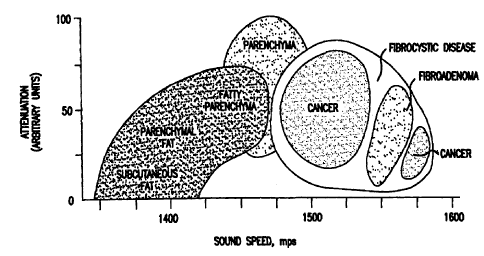
The assignment of a nominal value of v can be accomplished by reference to a
predetermined mapping between a morphologically assigned segment and a complex
sound
velocity. As will be understood by those skilled in the art, and as
demonstrated in FIG. 7 for
the case of acoustic waves, there is in reality variation in the complex sound
velocity for
different tissues, which is why the assignment at this step in the method is
merely nominal.
This figure is shown for illustration purposes in two dimensions (sound speed
and
absorption), although it will be readily apparent to those of skill in the art
that the relationship
may be generalized to arbitrarily many dimensions provided each of those
additional
dimensions bears on the identification of a particular tissue.
It will also be readily appreciated that similar relationships may be
constructed for
different types of medical pathologies and involving different physical
parameters, and the
invention encompasses the use of all such relationships in this context. In an
even broader
sense, this relationship can be expanded to include even the raw data, such as
phase changes
and amplitude changes, although there is, of course, a possibility that some
of these quantities
will then be highly correlated with each other. After the injection of the
physiological data
according to this method, a new estimate of the three-dimensional field, now
termed a
"physiological" estimate, ;, "' f r,t : O(r,t)] has been calculated.
The next step 16 of the method is the first step in the iterative portion of
the method.
At this step, the existing model of the object under study is used to
calculate what detector
data would be produced if the model multidimensional field corresponded
exactly to the
actual field of the object under study. In the particular embodiment where the
waves are
ultrasonic waves, this can be accomplished by using any appropriate sound-wave
propagation
codes. For those embodiments of the invention directed at the use of
ultrasonic radiation,
appropriate codes are available at various United States National
Laboratories, including the
Lawrence Livermore National Laboratory, Oak Ridge National Laboratory, Los
Alamos
National Laboratory, and Sandia National Laboratory. Other ultrasonic
propagation codes are
available at the Institut Francais Du Petrole in France, the National Oceanic
and Atmospheric
Administration, [See R. M. Jones, et al., HARPO: A Versatile three-dimensional
Hamiltonian
ray-tracing program for acoustic waves in an ocean with irregular bottom, NOAA
Special
Report P387-I 72573/1...1:., (1986)], and from Computational Fluid Dynamics
Research in
Huntsville, Alabama. A widely available code is the LINUX Ocean Acoustics and
Seismic
-21 -
CA 02324602 2000-09-19
WO 99/47046 PCT/US99/06026
Exploration Synthesis Package. A full version of this code is available for
licensing
through the MIT Technology Licensing Office, Massachusetts Institute of
Technology, Five
Cambridge Center, Kendall Square, Room NE25-230, Cambridge MA 02142-1493. One
feature in common with existing ultrasound propagation codes is that they
contain a
complex body of algorithms that can be used to input the estimated field 4z
t[r,t:@(r,t)], as
well as both the configuration of sources and detectors and the
characteristics of the
ultrasound waves that were generated by the sources, so as to calculate the
waveform of
ultrasonic waves that will arrive at each detector. In alternative
embodiments, more
sophisticated features of such codes can be used to model the effects of noise
and
uncertainty on the propagated waves. The result of this step in the method is
to produce an
estimate of the detected signals {S("") that can be compared with the actual
detected signals
{Silo,) .
The next step 18 in the method is to calculate the difference between the
estimated
signals and the actual signals: {E;} = {5(0) - S(")}. This error set is then
tested in step 20 to
determine whether the difference between the estimated signals and the actual
signals is less
than the noise. If so, then the iteration is deemed to have converged to its
final value for
construction of the rendering of the multidimensional field 4[r,t:4(r,t)]. If
not, then the
iteration proceeds for an additional step. To determine whether the error
field is within the
noise, the following quantity is compared against some predetermined threshold
T:
E s -s( ) 1 2
VP)+VV
where VS; is the variance for the set IS}.
If the estimated field fails to produce a set of estimated detected signals
that is
consistent with the actual signals, at least within the noise level, then the
physiological data
are changed in order to improve the estimate in step 24. The method of
changing the
physiological data is shown in greater detail in FIG. 8. In this step, the
method begins with
the set of error signals at substep 40, which is used in an ultrasound
propagation code at
substep 42 to produce a correction field2[r,t: (r,t)]. Although in one
embodiment, the
same ultrasound propagation code will be used as was used at step 16, this is
not a
requirement of the invention, and a different ultrasound propagation code may
be used in
different embodiments. The previously assigned segmentation of the field is
now used to
refine the correction field at substep 44 so that2[r,t:O(r,t)] = 0 outside the
segments. Inside
the previously established segments, the correction field is averaged to
produce the
refinement field c2[r,t:0(r,t)]> E 6[r,t:0(r,t)].
In the final step 26 of the basic method, a new multidimensional field
-22-
SUBSTITUTE SHEET (RULE 26)
CA 02324602 2008-02-06
,7n+lest rr,t : O(r,t)] is calculated by adding the refinement field to the
previous
multidimensional field:
IeSLIr,t:O(r,t)] = 9õeS`1r,t:O(r,t)]+e[r,t:O(r,t)]
The method then proceeds iteratively with this new multidimensional field used
at step 16 to
calculate the detected signals as if it were the actual field using some
available ultrasound
propagation code. The method iterates until the error field is such that it is
deemed to be
within the level of noise as defined by the threshold T. It will be readily
appreciated by those
of skill in the art that the ability of the method to converge and the speed
of convergence
will depend significantly on the quality of the initial estimate 9[r,t :
O0(r,t)].
There are several different embodiments of this invention that will be
understood by
those of skill in the art. In one embodiment, there is cycling between
different estimators at
steps 16 or 42 in order to improve the estimate of the multidimensional field
qs` Ir, t : O(r, t)] and to combine the results of such estimators with
techniques such as
Bayesian or Kalman filtering, covariance intersection, or some form of fuzzy
combination.
See, for example, James V. Candy, Signal Processing: The Model-Based Approach
(McGraw Hill, 1986).
There are also alternative methods that can also be used to construct the
multidimensional field. All such methods can be broadly categorized as falling
into one of
two classes. In the first class, into which the method described in detail
above falls, the
method begins with an initial approximation that is progressively improved. In
the second
class of methods, the system is permitted to vary essentially randomly and
individual
multidimensional field constructions that develop during the process are
evaluated to
determine which best reproduces the observed data. An example of such a method
is a
genetic algorithm.
The genetic algorithm is a model of machine learning that derives its behavior
in an
attempt to rnirnic evolution in nature. See, for example, Melanie Mitchell, An
Introduction
to Genetic Algorithms (Complex Adaptive Systems, 1996). This is done by
generating a
population of individuals represented by chromosomes, in essence a set of
character strings
that are analogous to the base-four chromosomes of DNA. The individuals in the
population
then go through a process of simulated "evolution." The genetic algorithm is
widely used in
multidimensional optimization problems in which the character string of the
chromosome
can be used to encode the values for the different parameters being optimized.
In practice,
therefore, an array of bits or characters to represent the chromosomes, in
this case the
multidimensional field OYEr,t : O(r,t)], is provided; then bit manipulation
operations allow
the implementation of crossover, mutation, and other operations.
- 23 -
CA 02324602 2000-09-19
WO 99/47046 PCT/US99/06026
When the genetic algorithm is implemented, it is trained in a manner that
involves
the following cycle: the fitness of all individuals in the population is first
evaluated; then, a
new population is created by performing operations such as crossover, fitness-
proportionate
reproduction, and mutation on the individuals whose fitness has just been
measured; finally,
the old population is discarded and iteration is performed with the new
population. One
iteration of this loop is referred to as a generation. In the present
invention, a number of
randomly generated multidimensional fields V[r,t:O(r,t)] are used as the
initial input. This
population of fields is then permitted to evolve as described above, with each
individual
field being tested at each generation to see if it can adequately reproduce
the observed data.
This is done in precisely the same manner as described above, with a wave-
propagation
code being used to generate a set of data (Si(")) that is compared with the
actual observed
data {S;(O'}. The genetic algorithm may be used as an alternative embodiment
of the
invention to generate the multidimensional field 9[r,t:O(r,t)].
It will readily be understood by those of skill in the art that the most
appropriate
technique to use in constructing the multidimensional field rendering will
depend greatly on
the speed of the technique in light of the computational capacity of the
hardware and
software that is used to perform the computations. While the invention has
been described
in detail with specific examples of how to construct the multidimensional
field, the
invention is not so limited and encompasses alternative schemes to do so.
3. Interpretation of the Multidimensional Field 421r.t:0(r,tH
As a result of determining the multidimensional field Z[r,t:O(r,t)], an
interpretation
is made of the physical characteristics that make up the field 9[r,t:
(r,t)]. The
interpretations that can be drawn are related directly to the types of
physical quantities that
have been calculated in 47[r,t: (r,t)] and the existence of a trained
evaluation system that
can take that information to develop interpretations. Without intending to
limit the scope of
the invention, this can best be illustrated by describing the embodiment where
the
multidimensional field describes the complex sound velocity for ultrasound
waves that have
been scattered off of the tissues within a human breast, v(r,t). The
relationship between the
complex sound velocity and the interpretation of what features exist in the
breast is shown
by FIG. 7. Given the complex sound velocity, this relationship is used to
classify tissue
types. There is no a priori reason to expect that this relationship will be of
a simple
functional form that can be reduced to an equation, and therefore a mapping
from the
complex sound velocity onto this interpretation requires the use of more
sophisticated
techniques. These can include the use of neural nets; stochastic optimization,
wherein the
shape of curves encompassing certain interpretations is changed by using
methods such as
steepest descent or simulated annealing; or evolutionary methods, wherein that
shape is
-24-
SUBSTITUTE SHEET (RULE 26)
CA 02324602 2008-02-06
changed by the genetic algorithm described above for evolving the
multidimensional field. See
generally, Stanley R. Deans, The Radon Transform and Some gf Its Applications
(Krieger
Publishing, 1993).
One method of interpreting the multidimensional field involves probabilistic
estimation. The basic concept of probabilistic estimators is to designate the
regions of the
voxels in terms of probabilities of certain tissue classifications given a
certain input value of
6J[r,t : O(r,t)]. For instance, given a set of values for a particular voxel,
the probability that
that voxel represents a particular interpretation is first determined. The
voxel is then identified
as the region with the highest probability. Test information is used to
generate the basic
probabilities. For example, in the embodiment where ultrasound is used to
study breast tissue,
there is a probability of certain types of tissue or tissue pathologies based
on the calculated
complex sound velocities for the individual voxels.
In more specialized stochastic estimation schemes, some regions are given more
weight
than others in order to reduce the rate of false positives or negatives. It
will readily be
understood that in different embodiments of the invention, it is more
desirable to minimize
either the rate of false positives or the rate of false negatives. For
example, in the particular
embodiment where cancer is to be identified in tissue, some level of false
positives may be
acceptable since a positive identification of cancer will be followed by
additional medical
procedures; however, it is desirable that the level of false negatives be
minimized.
In an alternative embodiment, a neural net is used to interpret the
multidimensional
field. See, for example, Simon S. Hagkin, Neural Networks - A Comprehensive
Foundation
(Prentice Hall, 1998). In that case, it is necessary to train the neural net
to be able to perform
the identification accurately and consistently. A typical neural network
includes a plurality of
nodes, and each node has a weight value associated with it. One layer is an
input layer having a
plurality of input nodes, and another layer is an output layer having a
plurality of output nodes,
with at least one layer therebetween. In this example, the input nodes receive
the
multidimensional field values q[r,t : e(r,t)] (the complex sound velocity in
the exemplary
embodiment) and the output node generates an interpretation designation
(tissue type in the
exemplary embodiment). In other words, given an input comprising the
multidimensional field
values of one voxel, the input is combined (added, multiplied, subtracted in a
variety of
combinations and iterations depending upon how the neural network is initially
organized), and
then the interpretation is generated accordingly.
In order to train the neural net, the output values are compared against the
correct
interpretation with some known samples. If the output value is incorrect when
compared
against such a test interpretation, the neural net modifies itself to arrive
at the correct output
-25-
CA 02324602 2008-02-06
value. This is achieved by connecting or disconnecting certain nodes and/or
adjusting the
weight values of the nodes during the training through a plurality of
iterations. Once the
training is completed, the resulting layer/node configuration and
corresponding weights
represents a trained neural net. The trained neural net is then ready to
receive unknown
multidimensional-field data and designate interpretations for each voxel.
Classical neural nets
include Kohonen nets, feed-forward nets, and back-propagation nets. The
different neural nets
have different methods of adjusting the weights and organizing the respective
neural net during
the training process.
In the embodiment of the invention where ultrasound is used to study breast
tissue, a
second method based on Hough transformation is used to increase the
probability that a
cancerous tumor has been found. See, for example, Stanley R. Deans, The Radon
Transform
and Some of Its Applications (Krieger Publishing, 1993). The basic concept in
this embodiment
is that closed volumes are identified in the tissue since such closed volumes
are indicative of
the existence of neoplasms. This information is then coupled with a complex
sound velocity
that is suggestive of cancer in order to increase the reliability of the
interpretation that the
identified neoplasm is a malignancy.
A closed volume may be represented as an ellipsoid where r = (x,y,z):
I z z
(X X" + (Y Y" + z-z =1
A B C
It will be apparent to those of skill in the art that for a fixed (x0 , Yo ,
Z0, A, B, C), each
(x, y, z) that satisfies the equation defines a point on the surface of an
ellipsoid. However, it
is also true that every point on the surface of an object will define a curve
in the six-
dimensional space where (x0, yo, zo, A, B, C) are the variables. If the object
is true
ellipsoid, then all of these curves will intersect at a single point.
Deviations from a true
ellipsoid manifest themselves as curves that do not pass through the point. In
order to
identify a closed volume, therefore, it is simply necessary to examine the
clumping of curves
defined by mapping each point into this six-dimensional space. Whenever
clumping of the
curves is detected, there is a likelihood that a closed volume exists in real
three-dimensional
space.
Once a closed volume has been recognized, it is useful also to calculate the
quasifractal dimension of the surface of the volume. This calculation may
increase the
probability that cancer has been identified because the surface properties of
tumors differ,
with benign tumors generally being smooth, unlike malignant tumors. Thus, if
the
quasifractal dimension is near 2, then the surface is smooth and the volume is
unlikely to be
-26-
CA 02324602 2008-02-06
cancerous. If the quasifractal dimension is greater than 2, then the surface
is fuzzy, and there
is a greater probability that the closed volume that has been identified is
cancerous.
Another useful characteristic to extract from the multidimensional field, and
a third
method that can be used for the identification of cancer, is the existence of
angiogenesis.
This is useful because an identification of regional increased blood flow
serves as an
indication that this is due to tumor recruitment. See Louvar et al.,
Correlation ofcolor
Doppleiflow in the prostate with tissue microvascularity, Cancer 1998 July 1:
83 (1): 135-
40. The relevant information needed to make this identification is extracted
from the
multidimensional field when the radiation detectors have been designed to
detect frequency
since this then allows the use of the Doppler effect, as described above, to
recognize the
motion of fluids, particularly blood, in the organ.
Specifically, the multidimensional field in this instance will include a
component
that contains the frequency shift or velocity of the features throughout the
organ. The trained
evaluation system can then identify those regions where the fluid velocity is
pronounced and
patterned in a geometrical configuration that matches other tumor indicators
such as the
closed volume discussed above, thereby identifying angiogenesis. The ability
of such a
system to identify angiogenesis is enhanced greatly by the three-dimensional
nature of the
field construction of the invention as well as the large solid angle coverage
of the invention.
In other, two-dimensional imaging systems, the Doppler information is limited
because it is
unable to use information derived from any component of motion that is
orthogonal to the
plane of the two-dimensional image.
Confidence in the identification of angiogenesis may be increased by
correlating the
Doppler information from the multidimensional field with information in the
multidimensional field related to tissue pressure as a fourth method of cancer
identification.
Tissue pressure is another relevant component in the identification of
malignant tumors, and
can be determined from an examination of subharmonic signal amplitude. The
existence of a
correlation between the subharmonic signal amplitude in ultrasonic studies
with tissue
pressure has been demonstrated in W. T. Shi et al., Effects of'Pressure
Changes on
Harmonic and Subharmonic Response o, f' US Contrast Microhubbles, Radiology
Supplement, 1154 (1999).
The Doppler sensitivity of the system to motion is also important in the
identification of cancer for another reason, which serves as a fifth method of
cancer
identification. When vibrations are applied to the tissues at a known
frequency as described
above, the invention permits recognition of the tougher tissues that are
associated with the
early tendency of cancer to attach to its surrounding normal tissues. In
particular, the
Doppler effect is useful in this context because microcalcifications in tissue
produce well-
defined high contrast with the surrounding tissue when external vibrations are
applied. This
-27-
CA 02324602 2008-02-06
is described in C. M. Sehgal et al., Visualization of Breast Calcification by
Acoustic
Resonance Imaging, Radiology Supplement, 1 150 (1999). Near vibration
resonance
frequencies of 70-250 Hz, the Doppler signal for such microcaleifieations is 5-
6 times
greater than for surrounding tissue, permitting good discrimination of such
features.
In a sixth method of cancer identification, the electrical impedance in tissue
is
extracted from the multidimensional field. The electrical impedance of tissue
may be used as
a factor to identify tumors, as described in Jeremy C. Hebden et al.,
Acoustically modulated
electrical impedance tomography, Proceedings of the SPIE, 1231: 7-14 (1990).
In a seventh method of cancer identification, the resolution can be improved
by
comparing the measurements of the object under examination made at two
different times.
The rendering of the multidimensional field o4r,t : O(r,t)] is constructed for
each set of
measurements, and the fields are compared. This will require storing of at
least the original
raw data, registration of the original raw data to the new raw data, and
comparing the
original and new raw data for the purpose of ascertaining changes between the
two different
times. Although it is possible in principle to store the original raw data
directly, this would
require a large data storage capacity. Alternatively, the original raw data
can be stored by
performing compression on the data.
Both lossy and lossless compression methods are available. For accuracy,
lossless
compression, such as by using the Joint Photographic Expert Group (JPEG)
standard can be
used, and for efficiency, lossy compression can be used. In particular, a
fractal compression
method can be used when a very high compression ratio is desired. The fractal
compression
method may represent the optimal compression method because it provides more
accuracy
in compressing images containing natural scenes (e.g., the human breast).
Conventional
fractal compression methods may be readily adapted to the multidimensional
field, which is
dependent on three dimensions. In this method, each component of the field is
compressed
by dividing it into a plurality of uniformly sized blocks. Another method is
to divide the
components of the field using a quad-tree partition. The field is split into
large blocks, and
these blocks are split into smaller blocks if a suitably accurate match cannot
be found.
Matches are then attempted for these subblocks, and if none can be found,
these subblocks
are in turn divided into subblocks. The process continues until a minimum
block size is
reached, when a match may be made.
Each tissue volume contains ducts, blood vessels, calcifications, and other
artifacts
that change very little, even over extended periods of time. However, the
original data may
not exactly match the new data due to changing of the overall shape, change in
physical
condition of the subject, etc. One method to ameliorate this problem is
morphing of the
-28-
CA 02324602 2000-09-19
WO 99/47046 PCT/US99/06026
original data to the conditions present when the new data were collected.
Here, image
morphing is the conventional image morphing method that blends a source image
to a
destination image by warping one or both images through a plurality of
intermediate
images. This is illustrated in FIG. 9, which is a perspective view of features
in tissue. In
parts (a) and (c) of the figure, the features have been constructed for
observations at an
initial time, and in parts (b) and (d) of the figure, the features have been
constructed for
observations made at a later time. In each case, there has been some change in
the overall
shape of the tissue sample. Differences manifest themselves, however, when the
fields are
morphed onto each other by using those artifacts that change little over time
as landmarks.
In part (b) of the figure, there is no significant change after the morphing,
leading to the
conclusion that there is no cancer growth. In part (d), however, there are
significant
changes that are apparent after morphing, leading to the conclusion that there
has been
anomalous change in the tissue sample.
After the multidimensional field is reconstructed for the original data, it
can be
compared with the multidimensional field generated for the new data. The
difference
between the two could be achieved by a simple subtraction of one from the
other.
Alternatively, the phase of the returned ultrasound signals could be used to
perform
interferometry to locate any changes. Also, if the two fields are generated
from data taken
relatively close in time (perhaps separated by only a few milliseconds), then
the
interferometry technique may be used to improve the resolution of the
invention.
Each of these cancer-identification techniques can be evaluated independently
by the
trained evaluation system, such as an expert system computer program or self-
learning
neural network. The various factors may be combined with a variety of means of
sensor
fusion. As an illustration, when the trained evaluation system assigns a
probability that
cancer has been identified based on a number of independent factors, the
reliability of the
assignment can be increased by calculating a product of probabilities. For
example if pk is
the probability that a given feature is cancer based on identification
technique k, then the
probability P that the feature is cancer based on the use of multiple
techniques is
P=1-1 (1 Pk)
Further variations on the method exist. This includes modifying the emitted
radiation in the sources so as to focus on areas that have been identified as
suspicious, and
thereby analyze them more closely. The method may be integrated with other
techniques
that are known, such as mammography. In this instance, the reliability of the
identification
is again improved by calculating a product of probabilities.
-29-
SUBSTITUTE SHEET (RULE 26)
CA 02324602 2000-09-19
WO 99/47046 PCT/US99/06026
Although the invention has been particularly shown and described with
reference to
embodiments of the invention directed at the detection and characterization of
medical
pathologies, it will be understood by those of skill in the art that the
method and apparatus
described may also readily be applied to non-medical applications. In
particular, the
method and apparatus may be used to detect and characterize distinctive
features within an
object. This may be exemplified, without limitation of the invention, by an
apparatus and
method used for the detection and characterization of defects in aerospace
devices. By
providing a matched-impedance contact between the aerospace device and arrays
of sources
and detectors, the invention is used to irradiate or insonify the aerospace
device. Data
collected after the radiation has been scattered (which includes reflection,
diffraction, and
transmission of the radiation) is then be used to construct a multidimensional-
field
rendering of the aerospace device. This multidimensional-field rendering is
then used to
detect and characterize features, such as defects, within the aerospace
device. Each of these
steps is carried out in the manner particularly and distinctly described above
with respect to
medical applications.
While the invention has been particularly shown and described with reference
to
preferred embodiments thereof, it is well understood by those skilled in the
art that various
changes and modifications may be made in the invention without departing from
the spirit
and scope of the invention.
25
35
-30-
SUBSTITUTE SHEET (RULE 26)