Note: Descriptions are shown in the official language in which they were submitted.
CA 02327702 2000-10-05
WO 99/51149 PCTIUS99/07652
- 1 -
STRUCTURES AND METHODS FOR CREATING
CAVITIES IN INTERIOR BODY REGIONS
FIELD OF THE INVENTION
The invention relates to structures and
procedures, which, in use, form cavities in interior
body regions of humans and other animals for
diagnostic or therapeutic purposes.
BACKGROUND OF THE INVENTION
Certain diagnostic or therapeutic
procedures require the formation of a cavity in an
interior body region.
For example, as disclosed in U.S. Patents
4,969,888 and 5,108,404, an expandable body is
deployed to form a cavity in cancellous bone tissue,
as part of a therapeutic procedure that fixes
fractures or other abnormal bone conditions, both
osteoporotic and non-osteoporotic in origin. The
expandable body compresses the cancellous bone to
form an interior cavity. The cavity receives a
filling material, which provides renewed interior
structural support for cortical bone.
This procedure can be used to treat
cortical bone, which due to osteoporosis, avascular
necrosis, cancer, or trauma, is fractured or is
prone to compression fracture or collapse. These
conditions, if not successfully treated, can result
in deformities, chronic complications, and an
overall adverse impact upon the quality of life.
A demand exists for alternative systems or
methods which, like the expandable body shown in
CA 02327702 2006-05-29
60895-1598
- 2 -
U.S. Patents 4,969,888 and 5,108,404, are capable of
forming cavities in bone and other interior body
regions in safe and efficacious ways.
SUMMARY OF THE INVENTION
The invention provides new tools for
creating cavities in cancellous bone. The tools
carry structures that cut cancellous bone to form
the cavity.
In one embodiment, the structure comprises
a filament, which can be formed as a loop or as an
array creating a brush. Manipulation of the filament
when inside bone cuts cancellous bone to create a
cavity. In another embodiment, the structure
comprises a blade that cuts cancellous bone by
either lateral movement, rotational movement, or
both. In another embodiment, the structure
comprises a transmitter of energy that cuts
cancellous bone to create the cavity.
The invention also provides directions for
using a selected tool according to a method
comprising the steps of deploying the tool inside
bone and manipulating the structure to cut
cancellous bone and form the cavity. The method for
use can also instruct filling the cavity with a
material, such as, e.g., bone cement, allograft
material, synthetic bone substitute, a medication,
or a flowable material that sets to a hardened
condition.
CA 02327702 2006-05-29
60895-1598
- 2a -
According to one aspect of the present invention,
there is provided an apparatus for treating bone comprising
a cannula having an axis establishing a percutaneous path
leading to inside bone, a shaft adapted to be deployed
inside bone by movement within and along the axis of the
cannula, and a cavity forming structure carried by the shaft
comprising a surface which directly contacts and shears
cancellous bone in response to rotating the shaft within and
about the axis of the cannula.
According to another aspect of the present
invention, there is provided an apparatus for treating a
vertebral body comprising a cannula having an axis that
establishes a percutaneous path leading into bone, a shaft
having an axis and a distal end portion adapted to be
deployed inside the bone by movement within and along the
axis of the cannula, a cavity forming structure carried by
the shaft and adapted to be extended in situ radially from
the shaft and comprising a surface which directly contacts
the cancellous bone, and a controller coupled to the cavity
forming structure and being operable to control extension of
the cavity forming structure to extend the cavity forming
structure in situ radially from the shaft in synchrony with
movement of the shaft to cause the surface to form a cavity
wholly within the vertebral body in the cancellous bone.
According to still another aspect of the present
invention, there is provided an apparatus for treating bone
comprising an elongated shaft sized for deployment inside a
cortical bone structure by passage through a percutaneous
path, the shaft having a proximal end portion and a distal
end portion, the distal end portion being sized for
placement within a cancellous bone volume inside the
cortical bone structure, the shaft including a material
enabling advancement and rotation of the distal end portion
CA 02327702 2006-05-29
60895-1598
- 2b -
within the cancellous bone volume in response to
manipulation of the proximal end portion, a cavity forming
structure carried by the distal end portion of the shaft,
the cavity forming structure being sized for retraction and
advancement through at least one opening in the shaft
between a retracted position essentially fully withdrawn
within the shaft and an advanced position projecting outside
the distal end portion of the shaft, the cavity forming
structure forming, when in the advanced position, a curved
surface that defines at least a portion of a loop having a
dimension capable of forming a cavity in the cancellous bone
volume to receive a volume of filling material, and a
controller on the proximal end portion coupled to the cavity
forming structure and being operable to control retraction
and advancement of the cavity forming structure through the
at least one opening in synchrony with rotation of the
distal end portion within the cancellous bone volume to
create the cavity.
Features and advantages of the inventions are set
forth in the following Description and Drawings, as well as
in the appended Claims.
BRIEF DESCRIPTION OF THE DRAWINGS
Fig. 1 is a side view of a rotatable tool having a
loop structure capable of forming a cavity in tissue, with
the loop structure deployed beyond
CA 02327702 2000-10-05
WO 99/51149 PCT/US99/07652
- 3 -
the associated catheter tube;
Fig. lA is an enlarged end view of the tool
shown in Fig. 1;
Fig. 2 is a side view of the tool shown in
Fig. 1, with the loop structure retracted within the
catheter tube;
Fig. 3 is a side view of the tool shown in
Fig. 1, with the loop structure deployed beyond the
catheter tube to a greater extent than shown in Fig.
1;
Fig. 4 is a side view of the tool shown in
Fig. 1 inserted within a guide sheath for deployment
in a targeted treatment area;
Fig. 5 is a side view of another rotatable
tool having a brush structure capable of forming a
cavity in tissue, with the brush structure deployed
beyond the associated drive tube;
Fig. 5A is an enlarged end view of the tool
shown in Fig. 5;
Fig. 6 is a side view of the tool shown in
Fig. 5, with the brush structure retracted within
the drive tube;
Fig. 7 is a side view of the tool shown in
Fig. 5, with the brush structure deployed beyond the
catheter tube to a greater extent than shown in Fig.
5, and with the brush structure being rotated to
cause the associated bristles to flare outward;
Fig. 8 is a side view of the tool shown in
Fig. 7, with the brush structure deployed beyond the
catheter tube to a greater extent than shown in Fig.
7, and with the brush structure still being rotated
to cause the associated bristles to flare outward;
Fig. 9 is a side view of an alternative
tool having an array of bristles carried by a
flexible shaft, which is capable of forming a cavity
CA 02327702 2000-10-05
WO 99/51149 PCT/US99/07652
- 4 -
in tissue;
Fig. 10 is a side view of the tool shown in
Fig. 9 as it is being deployed inside a cannula;
Fig. 11 is the tool shown in Fig. 9 when
deployed in a soft tissue region bounded by hard
tissue;
Fig. 12 is a side view of a tool having a
rotatable blade structure capable of forming a
cavity in tissue;
Fig. 13 is a side view of an alternative
curved blade structure that the tool shown in Fig.
12 can incorporate;
Fig. 14 is a side view of an alternative
ring blade structure that the tool shown in Fig. 12
can incorporate;
Fig. 15 is a side view of the ring blade
structure shown in Fig. 14 while being introduced
through a cannula;
Fig. 16 is a side view of a rotating tool
capable of forming a cavity in tissue, with an
associated lumen to introduce a rinsing liquid and
aspirate debris;
Fig. 17 is a perspective side view of a
tool having a linear movement blade structure
capable of forming a cavity in tissue, with the
blade structure deployed beyond the associated
catheter tube in an operative position for use;
Fig. 18 is an end view of the tool shown in
Fig. 17, with the blade structure shown in its
operative position for use;
Fig. 19 is an end view of the tool shown in
Fig. 17, with the blade structure shown in its rest
position within the catheter tube;
Fig. 20 is a side view of the tool shown in
Fig. 17, with the blade structure shown in its rest
CA 02327702 2000-10-05
WO 99/51149 PCT/US99/07652
- 5 -
position within the catheter tube, as also shown in
an end view in Fig. 18;
Fig. 21 is a side view of the tool shown in
Fig. 17, with the blade structure deployed beyond
the associated catheter tube in an operative
position for use, as also shown in an end view in
Fig. 18;
Fig. 22 is a side view of a tool having a
linear movement energy transmitter capable of
forming a cavity in tissue, with the energy
transmitter deployed beyond the associated catheter
tube in an operative position for use;
Fig. 23 is a top view of a human vertebra,
with portions removed to reveal cancellous bone
within the vertebral body, and with a guide sheath
located for postero-lateral access;
Fig. 24 is a side view of the vertebra
shown in Fig. 23;
Fig. 25 is a top view of the vertebra shown
in Fig. 23, with the tool shown in Fig. 1 deployed
to cut cancellous bone by rotating the loop
structure, thereby forming a cavity;
Fig. 26 is a top view of the vertebra shown
in Fig. 23, with the tool shown in Fig. 5 deployed
to cut cancellous bone by rotating the brush
structure, thereby forming a cavity;
Fig. 27 is a side view of the vertebra
shown in Fig. 23, with the tool shown in Fig. 17
deployed to cut cancellous bone by moving the blade
structure in a linear path, thereby forming a
cavity;
Fig. 28 is a side view of the vertebra
shown in Fig. 23, with the tool shown in Fig. 22
deployed to cut cancellous bone using an energy
transmitter, which is both rotatable and movable in
CA 02327702 2000-10-05
WO 99/51149 PCTIUS99/07652
- 6 -
a linear path, thereby forming a cavity;
Fig. 29 is a side view of the vertebra
shown in Fig. 23, after formation of a cavity by use
of one of the tools shown in Figs. 25 to 28, and
with a second tool deployed to introduce material
into the cavity for therapeutic purposes;
Fig. 30 is a plan view of a sterile kit to
store a single use cavity forming tool of a type
previously shown; and
Fig. 31 is an exploded perspective view of
the sterile kit shown in Fig. 30.
The invention may be embodied in several
forms without departing from its spirit or essential
characteristics. The scope of the invention is
defined in the appended claims, rather than in the
specific description preceding them. All embodi-
ments that fall within the meaning and range of
equivalency of the claims are therefore intended to
be embraced by the claims.
DETAILED DESCRIPTION OF THE PREFERRED EMBODIMENTS
The systems and methods embodying the
invention can be adapted for use virtually in any
interior body region, where the formation of a
cavity within tissue is required for a therapeutic
or diagnostic purpose. The preferred embodiments
show the invention in association with systems and
methods used to treat bones. This is because the
systems and methods which embody the invention are
well suited for use in this environment. It should
be appreciated that the systems and methods which
embody features of the invention can be used in
other interior body regions, as well.
I. Rotatable Cavity Forming Structures
A. Rotatable Loop Structure
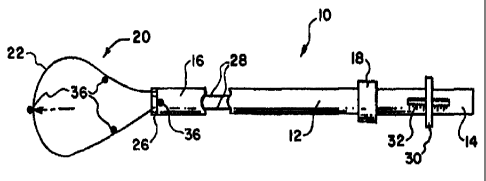
Fig. 1 shows a rotatable tool 10 capable of
CA 02327702 2000-10-05
WO 99/51149 PCT/US99/07652
_ 7 _
forming a cavity in a targeted treatment area. The
tool 10 comprises a catheter tube 12 having a
proximal and a distal end, respectively 14 and 16.
The catheter tube 12 preferable includes a handle 18
to aid in gripping and maneuvering the tube 12. The
handle 18 can be made of a foam material secured
about the catheter tube 12.
The catheter tube 12 carries a cavity
forming structure 20 at its distal end 16. In the
illustrated embodiment, the structure 20 comprises
a filament 22 of resilient inert material, which is
bent back upon itself and preformed with resilient
memory to form a loop.
The material from which the filament 22 is
made can be resilient, inert wire, like stainless
steel. Alternatively, resilient injection molded
inert plastic or shape memory material, like nickel
titanium (commercially available as NitinolTM'
material), can also be used. The filament 22 can,
in cross section, be round, rectilinear, or an other
configuration.
As Fig. 1A shows, the filament 22 radiates
from slots 24 in a base 26 carried by the distal end
16 of the catheter tube 12. The free ends 28 of the
filament 22 extend through the catheter tube 12 and
are connected to a slide controller 30 near the
handle 18.
As Fig. 2 shows, sliding the controller 30
aft (arrow A) retracts the filament 22 through the
slots 24, which progressively decreases the
dimensions of the loop structure 20. As Fig. 2
shows, in its farthest aft position, the filament 22
is essentially fully withdrawn and does not project
a significant distance beyond the distal end 16 of
the catheter tube 12.
CA 02327702 2000-10-05
WO 99/51149 PCT/US99/07652
- 8 -
As Fig. 3 shows, sliding the controller 30
forward (arrow F) advances the filament 22 through
the slots 24. The loop structure 20 forms, which
projects beyond the distal end 16 of the catheter
tube 12. As it is advanced progressively forward
through the slots 24, the dimensions of the loop
structure 20 progressively increase (compare Fig. 1
to Fig. 3). The controller 30 can include indicia
32, through which the physician can estimate the
dimensions of the loop structure 20.
In use (see Fig. 4), the catheter tube 12
is carried for axial and rotational movement within
a guide sheath or cannula 34. The physician is able
to freely slide the catheter tube 12 axially within
the guide sheath 34 (arrow S in Fig. 4). As Fig. 4
shows, when fully confined by the guide sheath 34,
the loop structure 20, if projecting a significant
distance beyond the distal end 16, is collapsed by
the surrounding sheath 34. When free of the guide
sheath 34, the loop structure 20 springs open to
assume its normal dimension. Thereafter, the
physician can operate the controller 30 to alter the
dimension of the loop structure 20 at will.
When free of the guide sheath 34, the
physician is also able to rotate the deployed loop
structure 20, by rotating the catheter tube 12
within the guide sheath 34 (arrow R in Fig. 4). As
will be described in greater detail alter, rotation
of the loop structure 20 slices or cut through
surrounding tissue mass.
The materials for the catheter tube 12 are
selected to facilitate advancement and rotation of
the loop structure 20. The catheter tube 12 can be
constructed, for example, using standard flexible,
medical grade plastic materials, like vinyl, nylon,
CA 02327702 2000-10-05
WO 99/51149 PCT/US99/07652
- 9 -
polyethylenes, ionomer, polyurethane, and
polyethylene tetraphthalate (PET). The catheter
tube 12 can also include more rigid materials to
impart greater stiffness and thereby aid in its
manipulation and torque transmission capabilities.
More rigid materials that can be used for this
purpose include stainless steel, nickel-titanium
alloys (NitinolTM' material), and other metal alloys.
The filament 22 preferably carries one or
more radiological markers 36. The markers 36 are
made from known radiopaque materials, like platinum,
gold, calcium, tantalum, and other heavy metals. At
least one marker 36 is placed at or near the distal
extremity of the loop structure 20, while other
markers can be placed at spaced apart locations on
the loop structure 20. The distal end 16 of the
catheter tube 12 can also carry markers. The markers
36 permit radiologic visualization of the loop
structure 20 and catheter tube 12 within the
targeted treatment area.
Of course, other forms of markers can be
used to allow the physician to visualize the
location and shape of the loop structure 20 within
the targeted treatment area.
B. Rotatable Brush
Fig. 5 shows an alternative embodiment of
a rotatable tool 38 capable of forming a cavity in
a targeted treatment area. The tool 38 comprises a
drive shaft 40, which is made from stiffer materials
for good torsion transmission capabilities, e.g.,
stainless steel, nickel-titanium alloys (NitinolTM
material), and other metal alloys.
The distal end 42 of the drive shaft
carries a cavity forming structure 44, which
comprises an array of filaments forming bristles 46.
CA 02327702 2000-10-05
WO 99/51149 PCT/US99/07652
- 10 -
As Fig. 5A shows, the bristles 46 extend from
spaced-apart slots 48 in a base 50 carried by the
distal end 42 of the drive shaft 40.
The material from which the bristles 46 is
made can be stainless steel, or injection molded
inert plastic, or shape memory material, like nickel
titanium. The bristles 46 can, in cross section, be
round, rectilinear, or an other configuration.
The proximal end 52 of the drive shaft 40
carries a fitting 54 that, in use, is coupled to an
electric motor 56 for rotating the drive shaft 40,
and, with it, the bristles 46 (arrows R in Figs. 7
and 8). When rotated by the motor 46, the bristles
spread apart (as Fig. 7 shows), under the influence
of centrifugal force, forming a brush-like structure
44. The brush structure 44, when rotating, cuts
surrounding tissue mass in the targeted treatment
area.
The free ends 58 of the bristles 46 extend
through the drive shaft 40 and are commonly
connected to a slide controller 60. As Fig. 6 shows,
sliding the controller 60 aft (arrow A in Fig. 6)
shortens the distance the bristles 46 extend from
the base 50. As Figs. 7 and 8 show, sliding the
controller 60 forward (arrow F in Fig. 8) lengthens
the extension distance of the bristles 46. Using the
controller 60, the physician is able to adjust the
dimension of the cutting area (compare Fig. 7 and
Fig. 8).
The array of bristles 46 preferably
includes one or more radiological markers 62, as
previously described. The markers 62 allow
radiologic visualization of the brush structure 44
while in use within the targeted treatment area. The
controller 60 can also include indicia 64 by which
CA 02327702 2000-10-05
WO 99/51149 PCT/US99/07652
- 11 -
the physician can visually estimate the bristle
extension distance. The distal end 42 of the drive
shaft 40 can also carry one or more markers 62.
The drive shaft 40 of the tool 38 is, in
use, carried for axial and rotational movement
within the guide sheath or cannula 34, in the same
manner shown for the tool 10 in Fig, 4. The
physician is able to freely slide the drive shaft 40
axially within the guide sheath to deploy it in the
targeted treatment area. Once connected to the
drive motor 56, the drive shaft 40 is free to rotate
within the guide sheath 34 to form the brush
structure 44.
Fig. 9 shows an alternative embodiment of
a rotatable tool 138 having an array of filaments
forming bristles 140, which is capable of forming a
cavity in a targeted treatment area. The tool 138
includes a flexible drive shaft 142, which is made,
e.g., from twisted wire filaments, such stainless
steel, nickel-titanium alloys (NitinolT"" material),
and other metal alloys.
The bristles 140 radially extend from the
drive shaft 142, near its distal end. The bristles
140 can be made, e.g., from resilient stainless
steel, or injection molded inert plastic, or shape
memory material, like nickel titanium. The bristles
140 can, in cross section, be round, rectilinear, or
an other configuration.
As Fig. 10 shows, the tool 138 is
introduced into the targeted tissue region through
a cannula 144. When in the cannula 144, the
resilient bristles 140 are compressed rearward to a
low profile, enabling passage through the cannula.
When free of the cannula 144, the resilient bristles
140 spring radially outward, ready for use.
CA 02327702 2000-10-05
WO 99/51149 PCT/US99/07652
- 12 -
The proximal end of the drive shaft 142
carries a fitting 146 that, in use, is coupled to an
electric motor 148. The motor 148 rotates the drive
shaft 142 (arrow R in Fig. 11) , and, with it, the
bristles 140.
As Fig. 11 shows, when deployed inside an
interior body cavity with soft tissue S (e.g.,
cancellous bone bounded by hard tissue H (e.g.,
cortical bone), the physician can guide the tool 138
through the soft tissue S by allowing the rotating
bristles 140 to ride against the adjoining hard
tissue H. The flexible drive shaft 142 bends to
follow the contour of the hard tissue H, while the
rotating bristles 140 cut adjoining soft tissue S,
forming a cavity C.
In the illustrated embodiment, the drive
shaft 142 carries a pitched blade 151 at its distal
end. The blade 151 rotates with the drive shaft
142. By engaging tissue, the blade 151 generates a
forward-pulling force, which helps to advance the
drive shaft 142 and bristles 140 through the soft
tissue mass.
In the illustrated embodiment, the bristles
140, or the cannula 144, or both include one or more
radiological markers 153, as previously described.
The markers 153 allow radiologic visualization of
the bristles 140 while rotating and advancing within
the targeted treatment area.
C. Rotatable Blade Structure
Fig. 12 shows an alternative embodiment of
a rotatable tool 106 capable of forming a cavity in
a targeted treatment area. The tool 106, like the
tool 38, comprises a generally stiff drive shaft
108, made from, e.g., stainless steel, nickel-
titanium alloys (NitinolTM material) , and other metal
CA 02327702 2000-10-05
WO 99/51149 PCT/US99/07652
- 13 -
alloys, for good torsion transmission capabilities.
The distal end of the drive shaft 108
carries a cavity forming structure 110, which
comprises a cutting blade. The blade 110 can take
various shapes.
In Figs. 12 and 13, the blade 110 is
generally L-shaped, having a main leg 112 and a
short leg 116. In the illustrated embodiment, the
main leg 112 of the blade 110 is pitched radially
forward of the drive shaft axis 114, at a small
forward angle beyond perpendicular to the drive
shaft. The main leg 112 may possess a generally
straight configuration (as Fig. 12 shows), or,
alternatively, it may present a generally curved
surface (as Fig. 13 shows). In the illustrated
embodiment, the short leg 116 of the blade 110 is
also pitched at a small forward angle from the main
leg 112, somewhat greater than perpendicular.
In Fig. 14, the blade 110 takes the shape
of a continuous ring 126. As illustrated, the ring
126 is pitched slightly forward, e.g., at an angle
slightly greater than perpendicular relative to the
drive shaft axis 114.
The material from which the blade 110 is
made can be stainless steel, or injection molded
inert plastic. The legs 112 and 116 of the blade
110 shown in Figs. 12 and 13, and the ring 126 shown
in Fig. 14, can, in cross section, be round,
rectilinear, or an other configuration.
When rotated (arrow R), the blade 110
cuts a generally cylindrical path through
surrounding tissue mass. The forward pitch of the
blade 110 reduces torque and provides stability and
control as the blade 110 advances, while rotating,
through the tissue mass.
CA 02327702 2000-10-05
WO 99/51149 PCT/US99/07652
- 14 -
Rotation of the blade 110 can be
accomplished manually or at higher speed by use of
a motor. In the illustrated embodiment, the proximal
end of the drive shaft 108 of the tool 106 carries
a fitting 118. The fitting 118 is coupled to an
electric motor 120 to rotate the drive shaft 108,
and, with it, the blade 110.
As Fig. 15 shows, the drive shaft 108 of
the tool 108 is deployed subcutaneously into the
targeted tissue area through a guide sheath or
cannula 124. Connected to the drive motor 120, the
drive shaft 108 rotates within the guide sheath 34,
thereby rotating the blade 110 to cut a cylindrical
path P in the surrounding tissue mass TM. The blade
110 can be advanced and retracted, while rotating,
in a reciprocal path (arrows F and A), by applying
pushing and pulling forces upon the drive shaft 108.
The blade 110 can also be withdrawn into the cannula
124 to allow changing of the orientation of the
cannula 124. In this way, successive cylindrical
paths can be cut through the tissue mass, through
rotating and reciprocating the blade 110, to thereby
create a desired cavity shape.
The blade 110, or the end of the cannula
124, or both can carry one or more radiological
markers 122, as previously described. The markers
122 allow radiologic visualization of the blade 110
and its position relative to the cannula 34 while in
use within the targeted treatment area.
D. Rinsing and Aspiration
As Fig. 16 shows, any of the tools 10, 38,
106, or 138 can include an interior lumen 128. The
lumen 128 is coupled via a Y-valve 132 to a external
source 130 of fluid and an external vacuum source
134.
CA 02327702 2000-10-05
WO 99/51149 PCT/US99/07652
- 15 -
A rinsing liquid 136, e.g., sterile saline,
can be introduced from the source 130 through the
lumen 128 into the targeted tissue region as the
tools 10, 38, or 106 rotate and cut the tissue mass
TM. The rinsing liquid 136 reduces friction and
conducts heat away from the tissue during the
cutting operation. The rinsing liquid 136 can be
introduced continuously or intermittently while the
tissue mass is being cut. The rinsing liquid 136
can also carry an anticoagulant or other anti-
clotting agent.
By periodically coupling the lumen 128 to
the vacuum source 134, liquids and debris can be
aspirated from the targeted tissue region through
the lumen 128.
II. Linear Movement Cavity Forming Structures
A. Cutting Blade
Figs. 17 to 21 show a linear movement tool
66 capable of forming a cavity in a targeted
treatment area. Like the tool 10, the tool 66
comprises a catheter tube 68 having a handle 70 (see
Fig. 20) on its proximal end 72 to facilitate
gripping and maneuvering the tube 68.
The catheter tube 68 carries a linear
movement cavity forming structure 74 at its distal
end 76. In the illustrated embodiment, the structure
56 comprises a generally rigid blade 78, which
projects at a side angle from the distal end 76 (see
Figs. 17 and 21). The blade 78 can be formed from
stainless steel or cast or molded plastic.
A stylet 80 is carried by an interior track
82 within the catheter tube 68 (see Figs. 18 and
19). The track 82 extends along the axis of the
catheter tube 68. The stylet 80 is free to move in
a linear aft path (arrow A in Fig. 20) and a linear
CA 02327702 2000-10-05
WO 99/51149 PCTIUS99/07652
- 16 -
forward path (arrow F in Fig. 21) within the track
82. The stylet 80 is also free to rotate within the
track 82 (arrow R in Fig. 17).
The far end of the stylet 80 is coupled to
the blade 78. The near end of the stylet 80 carries
a control knob 84. By rotating the control knob 84,
the physician rotates the blade 78 between an at
rest position, shown in Figs. 19 and 20, and an
operating position, shown in Figs. 17, 18, and 21.
When in the at rest position, the physician can push
or pull upon the control knob 84 to move the blade
78 in a linear path within the catheter tube (see
Fig. 20). By pushing on the control knob 84, the
physician can move the blade 78 outside the catheter
tube 68, where it can be rotated into the operating
condition (see Fig. 21). When in the operating
position, pushing and pulling on the control knob 84
moves the blade in linear strokes against
surrounding tissue mass.
In use, the catheter tube 68 is also
carried for sliding and rotation within the guide
sheath or cannula 34, in the same manner shown in
Fig. 4. The physician is able to freely slide the
catheter tube 68 axially within the guide sheath 34
to deploy the tool 66 in the targeted treatment
site. When deployed at the site, the physician can
deploy the blade 78 in the operating condition
outside the catheter tube 68 and slide the blade 78
along tissue in a linear path. Linear movement of
the blade 78 along tissue cuts the tissue. The
physician is also able to rotate both the catheter
tube 68 within the guide sheath 34 and the blade 78
within the catheter tube 68 to adjust the
orientation and travel path of the blade 78.
The blade 78 can carry one or more
CA 02327702 2000-10-05
WO 99/51149 PCTIUS99/07652
- 17 -
radiological markers 86, as previously described, to
allow radiologic visualization of the blade 78
within the targeted treatment area. Indicia 88 on
the stylet 80 can also allow the physician to
visually approximate the extent of linear or
rotational movement of the blade 78. The distal end
76 of the catheter tube 68 can also carry one or
more markers 86.
B. Energy Transmitters
Fig.22 shows an alternative embodiment of
a linear movement tool 90 capable of forming a
cavity in a targeted treatment area. The tool 90 is
physically constructed in the same way as the linear
movement tool 66 just described, so common reference
numerals are assigned.
However, for the tool 90 shown Fig. 22, the
far end of the stylet 80 carries, not a cutting
blade 78, but instead a transmitter 92 capable of
transmitting energy that cuts tissue (shown by lines
100 in Fig. 22). A connector 94 couples the
transmitter 92 to a source 96 of the energy, through
a suitable energy controller 98.
The type of energy 100 that the transmitter
92 propagates to remove tissue in the targeted
treatment area can vary. For example, the
transmitter 92 can propagate ultrasonic energy at
harmonic frequencies suitable for cutting the
targeted tissue. Alternatively, the transmitter 92
can propagate laser energy at a suitable tissue
cutting frequency.
As before described, the near end of the
stylet 80 includes a control knob 84. Using the
control knob 84, the physician is able to move the
transmitter 92 in a linear path (arrows A and F in
Fig. 22) between a retracted position, housed with
CA 02327702 2000-10-05
WO 99/51149 PCT/US99/07652
- 18 -
the catheter tube 68 (like the blade 78 shown in
Fig. 20), and a range of extended positions outside
the catheter tube 68, as shown in Fig. 22).
As also described before, the catheter tube
68 of the tool 90 is, in use, carried for sliding
and rotation within the guide sheath or cannula 34.
The physician slides the catheter tube 68 axially
within the guide sheath 34 for deployment of the
tool 90 at the targeted treatment site. When
deployed at the site, the physician operates the
control knob 84 to linearly move and rotate the
transmitter 92 to achieve a desired position in the
targeted treatment area. The physician can also
rotate the catheter tube 68 and thereby further
adjust the location of the transmitter 92.
The transmitter 92 or stylet 80 can carry
one or more radiological markers 86, as previously
described, to allow radiologic visualization of the
position of the transmitter 92 within the targeted
treatment area. Indicia 88 on the stylet 80 can also
allow the physician to visually estimate the
position of the transmitter 92. The distal end 76 of
the catheter tube 68 can also carry one or more
markers 86.
III. Use of Cavity Forming Tools
Use of the various tools 10 (Figs. 1 to 4),
38 (Figs. 5 to 8), 138 (Figs. 9 to 11), 106 (Figs.
12 to 15), 66 (Figs. 17 to 21), and 90 (Fig. 22)
will now be described in the context of deployment
in a human vertebra 150.
Fig. 23 shows the vertebra 150 in coronal
(top) view, and Fig. 24 shows the vertebra 150 in
lateral (side) view. It should be appreciated,
however, the tool is not limited in its application
to vertebrae. The tools 10, 38, 138, 106, 66, and 90
CA 02327702 2000-10-05
WO 99/51149 PCT/US99/07652
- 19 -
can be deployed equally as well in long bones and
other bone types.
As Figs. 23 and 24 show, the vertebra 150
includes a vertebral body 152, which extends on the
anterior (i.e., front or chest) side of the vertebra
150. The vertebral body 152 includes an exterior
formed from compact cortical bone 158. The cortical
bone 158 encloses an interior volume of reticulated
cancellous, or spongy, bone 160 (also called
medullary bone or trabecular bone).
The vertebral body 152 is in the shape of
an oval disk. As Figs. 23 and 24 show, access to
the interior volume of the vertebral body 152 can be
achieved. e.g., by drilling an access portal 162
through a side of the vertebral body 152, which is
called a postero-lateral approach. The portal 162
for the postero-lateral approach enters at a
posterior side of the body 152 and extends at angle
forwardly toward the anterior of the body 152. The
portal 162 can be performed either with a closed,
minimally invasive procedure or with an open
procedure.
Alternatively, access into the interior
volume can be accomplished by drilling an access
portal through either pedicle 164 (identified in
Fig. 23). This is called a transpedicular approach.
It is the physician who ultimately decides which
access site is indicated.
As Figs. 23 and 24 show, the guide sheath
34 (earlier shown in Fig. 4) is located in the
access portal 162. Under radiologic or CT
monitoring, a selected one of the tools 10, 38, 66,
or 90 can be introduced through the guide sheath 34.
A. Deployment and Use of the Loop Tool in
a Vertebral Body
CA 02327702 2000-10-05
WO 99/51149 PCTIUS99/07652
- 20 -
When, for example, the loop tool 10 is
used, the loop structure 20 is, if extended,
collapsed by the guide sheath 34 (as shown in Fig.
4), or otherwise retracted within the catheter tube
12 (as Fig. 2 shows) during passage through the
guide sheath 34.
Referring to Fig. 25, when the loop tool 10
is deployed outside the guide sheath 34 in the
cancellous bone 160, the physician operates the
controller 30 in the manner previously described to
obtain a desired dimension for the loop structure
20, which can be gauged by radiologic monitoring
using the on-board markers 36. The physician
manually rotates the loop structure 20 through
surrounding cancellous bone 160 (as indicated by
arrows R in Fig. 25). The rotating loop structure 20
cuts cancellous bone 160 and thereby forms a cavity
C. A suction tube 102, also deployed through the
guide sheath 34, removes cancellous bone cut by the
loop structure 20. Alternatively, the catheter tube
12 can include an interior lumen 128 (as shown in
Fig. 16) to serve as a suction tube as well as to
convey a rinsing liquid into the cavity as it is
being formed.
Synchronous rotation and operation of the
controller 30 to enlarge the dimensions of the loop
structure 20 during the procedure allows the
physician to achieve a create a cavity C of desired
dimension. Representative dimensions for a cavity C
will be discussed in greater detail later.
B. Deployment and Use of the Brush Tool
in a vertebral Body
When, for example, the brush tool 38 is
used, the physician preferable withdraws the
bristles 46 during their passage through the guide
CA 02327702 2000-10-05
WO 99/51149 PCT/US99/07652
- 21 -
sheath 34, in the manner shown in Fig. 6.
Referring to Fig. 26, when the brush tool
38 is deployed in cancellous bone 160 free of the
guide sheath 34, the physician advances the bristles
46 a desired distance (as shown in Fig. 5), aided by
radiologic monitoring of the markers 62, or the
indicia 32 previously described, or both. The
physician connects the drive shaft 40 to the motor
56 to rotate the bristles 46, creating the brush
structure 44. As Fig. 26 shows, the rotating brush
structure 44 cuts cancellous bone 160 and forms a
cavity C. The suction tube 102 (or a lumen 128 in
the drive shaft 40, as shown in Fig. 16) introduces
a rinsing fluid (with an anticoagulant, if desired)
and removes cancellous bone cut by the brush
structure 44. By periodically stopping rotation of
the brush structure 44 and operating the controller
60 (previously described) to increase the forward
extension of the bristles 46, the physician able
over time to create a cavity C having the desired
dimensions.
C. Deployment and use of the Linear Tools
in a Vertebral Body
When, for example, one of the linear
movement tools 66 or 90 are used, the physician
preferable withdraws the blade 78 or the transmitter
92 into the catheter tube 68 in the manner shown in
Fig. 20, until the distal end 76 of the catheter
tube 68 is free of the guide sheath 34.
Referring to Fig. 27, using the blade tool
66, the physician operates the stylet 80 forward
(arrow F) and aft (arrow A) to move the blade 78 in
a linear path through cancellous bone 160. The blade
78 scrapes loose and cuts cancellous bone 160 along
its path, which the suction tube 102 removes. A
CA 02327702 2006-05-29
60895-1598
- 22 -
cavity C is thereby formed. Synchronous rotation
(arrow R) and linear movement (arrows F and A) of
the blade 78 allow the physician to create a cavity
C having a desired dimension.
Referring to Fig. 28, using the energy
transmitting tool 90, the physician rotates (arrow
R) and pushes or pulls upon the stylet 80 (arrows F
and A) to position the energy transmitter 92 at
desired locations in cancellous bone 160. The
markers 86 aid the location process. Transmission by
the transmitter 92 of the selected energy cuts
cancellous bone 160 for removal by the suction tube
102. A cavity C is thereby formed. Through
purposeful maneuvering of the transmitter 92, the
physician achieves a cavity C having the desired
dimension.
D. Deployment of Other Tools into the
Cavity
Once the desired cavity C is formed, the
selected tool 10, 38, 66, 90, 106, or 138 is
withdrawn through the guide sheath 34. As Fig. 29
shows, an other tool 104 can now be deployed through
the guide sheath 34 into the formed cavity C. The
second tool 104 can, for example, perform a
diagnostic procedure. Alternatively, the second
tool 104 can perform a therapeutic procedure, e.g.,
by dispensing a material 106 into the cavity C, such
as, e.g., bone cement, allograft material, synthetic
bone substitute, a medication, or a flowable
material that sets to a hardened condition. Further
details of the injection of such materials 106 into
the cavity C for therapeutic purposes are found in
U.S. Patents 4,969,888 and 5,108,404.
CA 02327702 2006-05-29
60895-1598
- 23 -
E. Bone Cavity Dimensions
The size of the cavity C varies according
to the therapeutic or diagnostic procedure
performed.
At least about 30% of the cancellous bone
volume needs to be removed in cases where the bone
disease causing fracture (or the risk of fracture)
is the loss of cancellous bone mass (as in
osteoporosis). The preferred range is about 30% to
90% of the cancellous bone volume. Removal of less
of the cancellous bone volume can leave too much of
the diseased cancellous bone at the treated site.
The diseased cancellous bone remains weak and can
later collapse, causing fracture, despite treatment.
However, there are times when a lesser
amount of cancellous bone removal is indicated. For
example, when the bone disease being treated is
localized, such as in avascular necrosis, or where
local loss of blood supply is killing bone in a
limited area, the selected tool 10, 38, 66, 90, 106,
or 138 can remove a smaller volume of total bone.
This is because the diseased area requiring
treatment is smaller.
Another exception lies in the use of a
selected tool 10, 36, 66, 90, 106, or 138 to improve
insertion of solid materials in defined shapes, like
hydroxyapatite and components in total joint
replacement. In these cases, the amount of tissue
that needs to be removed is defined by the size of
the material being inserted.
Yet another exception lays the use of a
selected tool 10, 36, 66, 90, 106, or 138 in bones
to create cavities to aid in the delivery of
therapeutic substances.
CA 02327702 2006-05-29
60895-1598
- 24 -
In this case, the cancellous bone may or
may not be diseased or adversely affected. Healthy
cancellous bone can be sacrif iced by significant
compaction to improve the delivery of a drug or
growth factor which has an important therapeutic
purpose. In this application, the size of the
cavity is chosen by the desired amount of
therapeutic substance sought to be delivered. In
this case, the bone with the drug inside is
supported while the drug works, and the bone heals
through exterior casting or current interior or
exterior fixation devices.
IV. Single Use Sterile Kit
A single use of any one of the tools 10,
38, 138, 106, 66, or 90 creates contact with
surrounding cortical and cancellous bone. This
contact can damage the tools, creating localized
regions of weakness, which may escape detection.
The existence of localized regions of weakness can
unpredictably cause overall structural failure
during a subsequent use.
In addition, exposure to blood and tissue
during a single use can entrap biological components
on or within the tools. Despite cleaning and
subsequent sterilization, the presence of entrapped
biological components can lead to unacceptable
pyrogenic reactions.
As a result, following first use, the tools
may not meet established performance and
sterilization specifications. The effects of
material stress and damage caused during a single
use, coupled with the possibility of pyrogen
reactions even after resterilization, reasonably
justify imposing a single use restriction upon the
CA 02327702 2000-10-05
WO 99/51149 PCT/US99/07652
- 25 -
tools for deployment in bone.
To protect patients from the potential
adverse consequences occasioned by multiple use,
which include disease transmission, or material
stress and instability, or decreased or
unpredictable performance, each single use tool 10,
38, 66, 90, 106, or 138 is packaged in a sterile kit
500 (see Figs. 30 and 31) prior to deployment in
bone.
As Figs. 30 and 31 show, the kit 500
includes an interior tray 508. The tray 508 holds
the particular cavity forming tool (generically
designated 502) in a lay-flat, straightened
condition during sterilization and storage prior to
its first use. The tray 508 can be formed from die
cut cardboard or thermoformed plastic material. The
tray 508 includes one or more spaced apart tabs 510,
which hold the tool 502 in the desired lay-flat,
straightened condition.
The kit 500 includes an inner wrap 512,
which is peripherally sealed by heat or the like, to
enclose the tray 508 from contact with the outside
environment. One end of the inner wrap 512 includes
a conventional peal-away seal 514 (see Fig. 31), to
provide quick access to the tray 508 upon instance
of use, which preferably occurs in a sterile
environment, such as within an operating room.
The kit 500 also includes an outer wrap
516, which is also peripherally sealed by heat or
the like, to enclosed the inner wrap 512. One end
of the outer wrap 516 includes a conventional peal-
away seal 518 (see Fig. 31), to provide access to
the inner wrap 512, which can be removed from the
outer wrap 516 in anticipation of imminent use of
the tool 502, without compromising sterility of the
CA 02327702 2000-10-05
WO 99/51149 PCT/US99/07652
- 26 -
tool 502 itself.
Both inner and outer wraps 512 and 516 (see
Fig. 31) each includes a peripherally sealed top
sheet 520 and bottom sheet 522. In the illustrated
embodiment, the top sheet 520 is made of transparent
plastic film, like polyethylene or MYLARTM material,
to allow visual identification of the contents of
the kit 500. The bottom sheet 522 is made from a
material that is permeable to EtO sterilization gas,
e.g., TYVECT"" plastic material (available from
DuPont).
The sterile kit 500 also carries a label or
insert 506, which includes the statement "For Single
Patient Use Only" (or comparable language) to
affirmatively caution against reuse of the contents
of the kit 500. The label 506 also preferably
affirmati=vely instructs against resterilization of
the tool 502. The label 506 also preferably
instructs the physician or user to dispose of the
tool 502 and the entire contents of the kit 500 upon
use in accordance with applicable biological waste
procedures. The presence of the tool 502 packaged
in the kit 500 verifies to the physician or user
that the tool 502 is sterile and has not be
subjected to prior use. The physician or user is
thereby assured that the tool 502 meets established
performance and sterility specifications, and will
have the desired configuration when expanded for
use.
The kit 500 also preferably includes
directions for use 524, which instruct the physician
regarding the use of the tool 502 for creating a
cavity in cancellous bone in the manners previously
described. For example, the directions 524 instruct
the physician to deploy and manipulate the tool 502
CA 02327702 2000-10-05
WO 99/51149 PCT/US99/07652
- 27 -
inside bone to cut cancellous bone and form a
cavity. The directions 524 can also instruct the
physician to fill the cavity with a material, e.g.,
bone cement, allograft material, synthetic bone
substitute, a medication, or a flowable material
that sets to a hardened condition.
The features of the invention are set forth
in the following claims.