Note: Descriptions are shown in the official language in which they were submitted.
CA 02386328 2007-07-17
50270-7
1
SPINAL IMPLANT AND CUTTING TOOL PREPARATION
ACCESSORY FOR MOUNTING THE IMPLANT
10
FIELD OF THE TNVENTION
Generally, the present invention relates to spinal implant devices, surgical
tools and associated techniques for promoting spinal fusion.
BACKGROUND OF THE I]WENTION
It is known that when an intervertebral disc degenerates or is damaged,
there is often a compression of the disc and a reduction in the normal
intervertebral
height. Typically, this condition results in abnormal motions that become a
source
of pain.
In order to treat pathologies of this type, the disc is often stabilized to
eliminate the abnormal motions caused by disc disorders or injuries.
Generally,
one approach is to prevent articulation between the two vertebrae situated on
eacb
side of the damaged disc by bone fusion. This fusion fixes the vertebrae to
each
other, eliminating the relative mobility causing the pain. Various spinal
implants to
promote fusion between adjacent vertebrae have been proposed. It has been
proposed to interconnect the two vertebrae by a kind of rigid U-shaped
stirrup,
which restores the discal height with a bone graft material disposed inside
the
stimip. However, one drawback of this proposal is its diminishing
effectiveness
over a period of time.
CA 02386328 2002-04-03
WO 01/28465 PCT/US00/41216
2
Another proposal for promoting spinal fusion includes implanting a spinal
cage to interconnect the adjacent vertebrae; the spinal cage includes a
cylindrical
member provided with a series of openings and provided with anchoring points.
This implant is placed in a recess formed in the intervertebral disc and
penetrates
the opposite cortical endplates of the two vertebrae, which were previously
hollowed out to receive the implant. This penetration forms openings in the
sub-
chondral endplates to place spongy bone of the vertebrae in contact with bone
graft
material placed inside the implant, facilitating bone fusion. U.S. Patent No.
5,015,247 provides one example of this approach.
Yet another proposal for spinal fusion comprises inserting hollow tubular
implants having a generally ovoidal external shape into the intervertebral
space.
However, these implants require both annular ribs to inhibit axial
displacement and
longitudinal ribs or teeth to prevent rotation of the implant about its
longitudinal
axis. One example of this approach is found in U.S. Patent No. 5,683,463
issued to
Godefroy et al. In another example in U.S. Patent No. 5,888,224 issued to
Beckers
et al., a rotatable implant for spinal fusion is disclosed. The rotatable
implant
requires a linking connector to inhibit longitudinal rotation. Other rotatable
implants are described in U.S. Patent No. 5,607,424 issued to Tropiano.
However, one drawback of these proposed implants is their lack of support
of the cortical bone tissue, particularly bearing against the peripheral wall
of the
vertebral bodies. This contributes to their diminishing effectiveness in
maintaining
normal disc height over a period of time.
Proper performance of a spinal implant of this type requires balancing the
need to promote fusion between the spongy bone and the need to form a reliable
load bearing relationship with the stronger cortical bone. As a result, the
spinal
implant must be neither engaged too far into the openings provided in the
cortical
endplates to provide a sufficiently dense load bearing surface, nor
insufficiently
inserted, in which case the bone fusion between the two vertebrae would be
adversely affected by a poor anchorage. Thus, there is a demand for devices
and
techniques that facilitate attaining the proper balance between fusion and
load
support.
CA 02386328 2007-07-17
2- ~9-2001 = = 3 US004121 E
Beckers et al. in U.S. Patent No. 5,888,224 discloses and implant for an
intervertebral space consisting of an essentially cuboid body with a device
for
gripping by a tool.
Pisharodi in U.S. Patent No. 5,697,977 describes an apparatus for aligning
adjacent vertebrae for example for the reduction of spondylolisthesis. The
apparatus
includes a solid body having sides that arch from one end to the other or a bi-
planar,
bi-convex body.
In FR 2742044 there is illustrated a circular threaded implant for threaded
insertion into an intervertebral space.
In EP 0834295 there is illustrated a substantially cuboid implant having upper
and lower curved surfaces and cavities formed in the implant body.
Thus, in light of the above described problems, there is a continuing need for
advancements in the treatment of spinal deformities, including improved spinal
implants and devices relating to spinal fusion and for surgical methods to
treat spinal
deformities. The present invention is such an advancement and provides a wide
variety of benefits and advantages.
Ci TR.CTiTT 1TF QF7FFT
AMENDED.qHFFT
CA 02386328 2002-04-03
WO 01/28465 PCT/US00/41216
4
SUMMARY OF THE INVENTION
The present invention relates to spinal implants, surgical tools and the use
thereof. Various aspects of the invention are novel, nonobvious, and provide
various advantages. While the actual nature of the invention covered herein
can
only be determined with reference to the claims appended hereto, certain forms
and
features, which are characteristic of the preferred embodiments disclosed
herein,
are described briefly as follows.
According to one form of the invention, the spinal implant comprises a
body having a central part arranged to allow arthrodesis and at least one
terminal
part for bearing against the cortical bone of the vertebral endplates. The
central
part is adapted to penetrate the vertebral endplates, transversely projecting
from the
terminal bearing part. Thus the invention achieves a separation between the
end
parts constituting the load bearers, and the intermediate part of the implant
which
permits fusion. In addition, the central part may include at least one cavity
for
receiving a bone graft material.
In another form of the present invention, an implant for insertion between a
first
vertebra having a first cortical bone endplate and a second vertebra having a
second
cortical bone endplate includes two terminal parts. The first terminal part
defines a first
bearing surface to bear against the first cortical bone endplate and a second
bearing
surface opposite the first surface to bear against the second cortical bone
endplate. The
second terminal part opposes the first terminal part and defines a third
bearing surface to
bear against the first cortical bone endplate and a fourth bearing surface
opposite the
third surface to bear against the second cortical bone endplate. The implant
has an
elongated central part defining an upper projection extending past the first
and third
surfaces, and a lower projection extending past the second and fourth
surfaces. These
projections correspondingly pass through openings in the first and second
cortical bone
endplates when the first and third surfaces bear against the first cortical
bone endplate
and the second and fourth surfaces bear against the second cortical bone
endplate. The
terminal parts are dimensioned to facilitate restoration of the natural
geometry of the
intervertebral space (lordosis, kyphosis, and parallel discs). Thus, the first
and second
CA 02386328 2002-04-03
WO 01/28465 PCTIUSOO/41216
surfaces may be separated by a first distance, and the third and fourth
surface may be
separated by a second distance greater than the first distance to accommodate
a natural
curvature of the spine.
In a further form of the present invention, an implant with two terminal parts
also
5 has an elongated central part that includes a pair of longitudinal walls
defining a cavity.
The walls define a first edge projecting past the first and third surfaces and
a second edge
projecting past the second and fourth surfaces. The first and second edges
correspondingly penetrate the first and second cortical bone endplates when
the first and
third surfaces bear against the first cortical bone endplate and the second
and fourth
surfaces bear against the second cortical bone endplate.
According to another form, the bearing surfaces of the terminal end parts
are defined by flanges extending from opposing ends of the implant along its
longitudinal axis. Preferably, the bearing surfaces are generally flat for
bearing
against the cortical bone of the vertebral endplates of the two adjacent
vertebrae. It
is also preferred that openings be cut into the cortical endplates in their
central
regions corresponding to the length of a central part of the implant along the
longitudinal axis and leaving a region of the cortical bone endplates around
the
periphery of the openings. The length of the remaining peripheral endplate
corresponds to the length of the bearing surfaces along the longitudinal axis.
When the implant is placed in position, the edges of the walls of the central
part
engage the openings cut in the cortical endplates and consequently do not
substantially bear against the remaining peripheral portion of the endplates.
A
cavity may be defined by the central part that holds bone graft material in
contact
with the spongy bone of the two vertebrae. In contrast, the bearing surfaces
of the
flanges are disposed adjacent the edges of the openings of the cortical
endplates
and bear against the remaining portions of the endplates to establish a strong
load-
bearing relationship. Thus, both bone fusion and support are distinctly
accommodated by different parts of the implant structure, which permits
obtaining
a satisfactory support of the vertebral bodies on the implant and an excellent
arthrodesis.
CA 02386328 2002-04-03
WO 01/28465 PCT/US00/41216
6
Yet another form of the present invention includes a cutting tool accessory to
prepare the cortical endplates of two adjacent vertebrae for insertion of an
implant.
This tool comprises a proximal handle connected to an elongated shaft
configured to
rotate about a longitudinal axis of the tool. The tool also includes a first
non-cutting
portion with the shaft extending therethrough and being configured to rotate
relative
thereto. A cutting portion is fixed to the shaft to rotate therewith and is
positioned
distal to the first non-cutting portion. The cutting portion includes a first
pair of
generally parallel opposing faces and a second pair of opposing faces each
extending
between the first pair of faces. The second pair of faces each defines a
number of
cutting teeth. A second non-cutting portion is fixed to the cutting portion
that
includes a distal head. The first non-cutting portion, the cutting portion,
and the
second non-cutting portion have a rotatably selectable alignment that presents
a
generally constant height corresponding to the intervertebral space defined
between
the cortical bone endplates to facilitate insertion therein. Once inserted,
the cutting
portion may be rotated out of this alignment to cut a first opening in the
first cortical
bone endplate and a second opening into the second cortical bone endplate. The
cutting portion and the non-cutting portions may be arranged to provide
uniform,
symmetrical cutting of these openings with a predetermined length
corresponding to
the dimensions of a given implant device.
In an additional form, a technique of spinal fixation includes cutting
adjacent vertebrae and inserting an implant therebetween to promote fusion and
provide suitable support. The implant may be inserted by anterior or posterior
surgical approaches. The cutting may be performed by the cutting tool of the
present invention and may include initially inserting the tool so that a first
pair of
faces are in contact with a respective one of the first and second cortical
bone
endplates, turning a handle to rotate the cutting portion to remove cortical
bone
with cutting teeth defined by a second pair of faces, and withdrawing the
tool. The
tool may be used to form openings readily positioned in the central region of
the
adjacent vertebrae leaving a portion of the cortical bone endplates about the
openings. The insertion of the implant may include positioning the implant of
the
CA 02386328 2002-04-03
WO 01/28465 PCT/US00/41216
7
present invention between the first and second vertebrae and turning the
implant
about one quarter of a turn.
In another form of the present invention, there is provided an implant for
insertion between a first and a second vertebrae, each vertebra having a
cortical bone
endplate surface. The implant includes a first terminal part that defines a
first bearing
surface substantially planar and adapted to bear against a first cortical
endplate
surface and an opposite second bearing surface substantially planar and
adapted to
bear against a second cortical bone endplate surface; a second terminal part
opposite
the first terminal part, wherein the second terminal part defines a third
bearing surface
adapted to bear against the first cortical bone endplate surface and a fourth
bearing
surface adapted to bear against the second cortical bone endplate surface; and
an
elongated body extending from the first terminal part to the second terminal
part. The
implant has an elongated central part that defines a longitudinal axis and has
an upper
surface arcuate along the longitudinal axis and a lower surface arcuate along
the
longitudinal axis. In preferred embodiments, the implant includes a cavity for
receiving bone osteogenic material to promote bone fusion between adjacent
vertebrae. The implant also includes anti-expulsion features such as, for
example, at
least one ridge transverse to the longitudinal axis. Preferably the implant
includes a
plurality of ridges proximate to the first and second bearing surfaces to
inhibit
expulsion of the implant. Furthermore, the second terminal part can be curved
to
facilitate insertion of the implant into the prepared intervertebral space.
In another form the present invention provides an implant for insertion
between a first vertebra and a second vertebra, where the first vertebra has a
generally vertically extending first peripheral wall and a first cortical bone
endplate
and the second vertebra has a generally vertically extending second peripheral
wall
and a second cortical bone endplate. The implant comprises: A first terminal
part
defining a first bearing surface adapted to bear against a portion of the
cortical
bone endplate proximate to the first peripheral wall and an opposite second
bearing
surface adapted to bear against a portion of the second cortical bone endplate
proximate to the second peripheral wall; an elongated body extending from said
first terminal part, the body defining a longitudinal axis and having an upper
CA 02386328 2002-04-03
WO 01/28465 PCT/US00/41216
8
surface and a lower surface, wherein the first upper surface and the second
lower
surface are arcuate along the longitudinal axis; and a second terminal part
opposite
the first terminal part and having a insertion face extending from the upper
surface
to the lower surface wherein the insertion face is provided to ease insertion
of the
implant between the first vertebra and the second vertebra.
In yet another form of the present invention, there is provided a tool for
insertion between a first vertebra having a cortical bone endplate and a
second
vertebra having a second cortical bone endplate. The insertion tool includes a
proximal handle connected to an elongated shaft configured to rotate about a
longitudinal axis of the tool; an outer sleeve adjacent to the handle and the
shaft
extending through the sleeve and configured to rotate relative thereto; a
cutting
portion fixed to the shaft to rotate therewith, the cutting portion including
a pair of
generally parallel opposing arms, each arm having a first arcuate cutting edge
and an
opposite second arcuate cutting edge; and a non-cutting portion fixed to the
cutting
portion and distal to the handle, wherein the non-cutting portion is
configured to align
the cutting portion between the first cortical bone endplate of the first
vertebra and the
second cortical bone endplate of the second vertebra. The insertion tool also
can
include a stop adapted to bear against the first or second vertebra to limit
the depth of
insertion of the tool within the intervertebral space. The tool preferably
includes a
cavity provided between the first and second arms of the cutting portion. The
cavity
provides a receptacle for receipt of bone debris generated during the scraping
procedure. The cutting portion and the non-cutting portion of the tool are
adapted to
position first and second cutting edges between the first and second vertebrae
such
that rotation of the cutting head removes substantially equal amounts of bone
from the
first and second endplates of the adjacent vertebrae.
One object of the present invention is to provide a spinal implant device to
promote fusion between adjacent vertebrae. Further objects, features, aspects,
forms,
advantages and benefits shall become apparent from the description and
drawings
contained herein.
CA 02386328 2002-04-03
WO 01/28465 PCT/US00/41216
9
BRIEF DESCRIPTION OF THE DRAWINGS
Figure 1 is a perspective view, to a larger scale, of one embodiment of a
spinal implant of the present invention.
Figure 2 is a perspective view of one embodiment of a cutting tool of the
present invention for mounting the implant of Figure 1.
Figure 3 is a perspective view of a distracting wedge or "distracter" used for
carrying out a surgical method in accordance with the present invention.
Figure 4 is a partial elevation view diagrammatically illustrating the
insertion of the cutting tool between two vertebrae.
Figure 5 is a partial elevation view showing the cutting tool after rotation
of
the cutting portion through 90 and penetrating the cortical endplates of the
two
adjacent vertebrae.
Figure 6 is a partial sectional view taken in a sagittal plane showing the
implant of Figure 1 positioned in an intervertebral disc for achieving a
spinal
fusion.
Figure 7 is a perspective view of a spinal segment in the intervertebral disc
in which two implants have been installed.
Figure 8 is a perspective view of an alternative embodiment of a spinal
implant
according to the present invention.
Figure 9A is an elevated side view of the spinal implant of Figure 8.
Figure 9B is an elevated end view of the spinal implant of Figure 8.
Figure 10 is a perspective view of an alternative embodiment of a cutting tool
for use with the present invention.
Figure 11 is a partial view illustrating the cutting head of the cutting tool
of
Figure 10.
Figure 12 is a perspective view of one embodiment of the cutting tool of
Figure
10 received within a guide sleeve.
Figure 13 is an elevated side view in partial section of one embodiment of a
cutting tool of Figure 10 received within the intervertebral space of adjacent
vertebrae
V1 andV2.
CA 02386328 2002-04-03
WO 01/28465 PCT/US00/41216
Figure 14 is an elevated side view in partial section of the cutting tool of
Figure
10 rotated 90 degrees within the intervertebral space of adjacent vertebrae
V1' and
V2'.
Figure 15 is a perspective view in partial section of one embodiment of a
spinal
5 implant received within the intervertebral space of adjacent vertebrae V1'
and V2'.
Figure 16 is a top elevated view of two implants depicted in Fig. 8 positioned
on
an endplate of a vertebral body.
CA 02386328 2002-04-03
WO 01/28465 PCTIUSOO/41216
11
DETAILED DESCRIPTION OF THE INVENTION
For the purposes of promoting an understanding of the principles of the
invention, reference will now be made to the embodiments illustrated herein
and
specific language will be used to describe the same. It will nevertheless be
understood that no limitation of the scope of the invention is thereby
intended. Any
alterations and further modifications in the described processes, systems or
devices,
and any further applications of the principles of the invention as described
herein, are
contemplated as would normally occur to one skilled in the art to which the
invention
relates.
Figures 1, 6, and 7 depict spinal cage implant 1. Implant 1 is adapted to be
inserted in a cavity provided in a damaged intervertebral disc 2 (Figure 7),
so as to
restore the normal height of the intervertebral space between the two
vertebrae V 1
and V2 adjacent to the disc 2, for example, the lumbar vertebrae L3, L4 as
depicted in
Figure 7. Figure 1 shows implant 1 disposed along its longitudinal axis 52.
Implant 1
comprises a hollow body 3 of elongate shape, having a central part 3c formed
by two
parallel longitudinal walls 4 arranged to permit the arthrodesis and, at the
two
opposite ends of the central part 3c, in the direction of the implantation of
the implant
1, two terminal parts 5, 6 for bearing against the cortical bone 14 of
vertebral cortical
bone endplates 15, 16 of the two vertebrae (Vl, V2) adjacent to the disc 2
(see, for
example, Figure 6).
The longitudinal walls 4 define therebetween a cavity 7 and are provided
with openings 11. Terminal parts 5, 6 are disposed opposite each other along
axis
52 and each includes a terminal face defining a centrally located threaded
hole l la.
Holes 11 a are configured for engagement by a threaded shaft of an
insertion/manipulation accessory (not shown) to facilitate placement of
implant 1
between vertebrae V 1, V2. The cavity 7 is provided to receive a graft G of
spongy
bone or any other material favoring bone fusion, for example, a synthetic
ceramic.
Terminal part 5 has two opposite bearing surfaces 12 transverse to axis 52
which are so dimensioned that they are separated by distance dl. Terminal part
6
has two opposite bearing surfaces 13 transverse to axis 52 which are so
CA 02386328 2002-04-03
WO 01/28465 PCTIUSOO/41216
12
dimensioned that they are separated by distance d2. Distances dl, d2 are
adapted
to the geometry of the intervertebral space to be restored. When d2 is greater
than
dl as depicted, implant 1 is preferred for fusion of vertebrae in a region of
the
spine having a natural curvature. Distance 1 corresponds to a maximum width
dimension of body 3. In one embodiment, the width of body 3 is generally
constant along axis 52 and is about the same as distance dl to facilitate a
proper fit
between vertebrae V 1, V2 as will become more apparent hereinafter.
The terminal parts 5, 6 are extended by load-bearing flanges 8, 9, namely,
two load bearing flanges 8 for part 5 and two load bearing flanges 9 for part
6. In
the depicted embodiment, flanges 8, 9 form parallel bars perpendicular to the
longitudinal walls 4, and define generally flat surfaces 12, 13 for bearing
against
the cortical bone 14 of the vertebral endplates 15, 16.
In the illustrated embodiment, the longitudinal walls 4 have a substantially
trapezoidal contour of which the small base corresponds to the terminal part 5
and
the large base corresponds to the terminal part 6. The walls 4 constituting
the
central part 3c of the implant 1 are so shaped as to transversely project from
the
terminal bearing parts 5, 6, as can be seen in Figures 1 and 6. Thus, the
walls 4 can
penetrate respective openings 19, 21 of the vertebral endplates 15, 16 whose
edges
bear against the bearing surfaces 12, 13 of the flanges 8, 9 (Figure 6).
Flanges 8, 9
are shaped to define step projections 17a, 18a to further facilitate
penetration of
openings 19, 21. Projections 17a, 18a also provide a lip to maintain alignment
of
bearing surfaces 12,13 with cortical bone endplates 15, 16 about the periphery
of
the openings as shown in Figure 6. Walls 4 also include tapered edges 4a to
facilitate penetration. Correspondingly, central part 3c has upper projection
3a and
lower projection 3b defined by edges 4a. Notably, edges 4a and projections 3a,
3b
transversely project away from surfaces 12, 13.
Implant 1 may be placed in position in the intervertebral disc 2 after
preparation with cutting tool accessory 22, which will now be described with
reference to Figures 2, 4, and 5. Preferably, tool 22 is made from a metallic
material suitable for surgical use. Tool 22 has a milling cutter 23 including
central
cutting portion 24 and two non-cutting portions 31, 36 arranged at opposite
ends of
CA 02386328 2002-04-03
WO 01/28465 PCT/US00/41216
13
central cutting portion 24. Non-cutting portions 31, 36 have a height
corresponding to the intervertebral space and permitting uniform, symmetrical
cutting of a predetermined length through a central region of both vertebral
endplates 15, 16. Preferably, the geometry of portions 24, 31, 36 is
determined for
preparing the intervertebral space with the geometry of implant 1 to restore
the
natural lordosis of the intervertebral space; and correspondingly, the
distances
represented by h and d 1 are approximately equal.
Central cutting portion 24 has a trapezoidal shape with two generally
smooth, longitudinal faces 25 opposed to each other. Faces 25 are configured
to
facilitate insertion into the intervertebral space, being generally separated
from
each other by distance h. Central cutting portion 24 also has cutting faces 26
extending between faces 25. Cutting faces 26 define a number of uniformly
spaced-apart cutting teeth 28. Teeth 28 each extend along a generally straight
path
that is slanted at an oblique angle relative to the longitudinal axis 54 of
too122.
Preferably, central cutting portion 24 is made from a suitable metallic
material that
has been machined to form teeth 28.
Non-cutting portion 31 is fixed to the distal end of central cutting portion
24. Portion 31 extends from central cutting portion 24 to provide a distal
head in
the form of a parallelepiped-shaped bar. Portion 31 has a first dimension
substantially the same as distance h to be generally coextensive with faces 25
of
central cutting portion 24. Portion 31 also has opposing faces 32 separated
from
each other by a distance H as shown in Figure 5. Preferably distance H is
approximately equal to distance d2 when too122 is being utilized to install
implant
1.
Too122 also includes a shaft or shank 33 connected to a proximal actuating
handle 34. Shank 33 is fixed to central cutting portion 24 and non-cutting
portion
31. Shank 33 extends from the small end face 27 remote from the non-cutting
head 31 and terminates in the handle 34 which permits rotating the cutting
portion
24 about the longitudinal axis 54 of too122.
Non-cutting portion 36 has a rectangular shape with generally planar faces
37, 38. Portion 36 may be inserted between two consecutive vertebrae during
CA 02386328 2002-04-03
WO 01/28465 PCT/USOO/41216
14
rotation of central cutting portion 24. Portion 36 extends in the direction
toward
the handle 34 by a tubular part 40 and through block 40a which terminates in
the
vicinity of the handle 34. Non-cutting portion 36 is provided with lateral
stops 39
capable of being put into abutment against the sides of the vertebrae (V 1,
V2) after
insertion therebetween. Non-cutting portion 36 encloses shank 33. Shank 33 is
configured to rotate relative to portion 36. Correspondingly, when handle 34
is
turned, shank 33, central cutting portion 24, and non-cutting portion 31
rotate
together about axis 54 with non-cutting portion 36 preferably remaining
stationary.
It should be noted that the partial view of Figures 4 and 5 does not show
handle 34
and depict a cutaway view of non-cutting portion 36 with shank 33 projecting
therefrom.
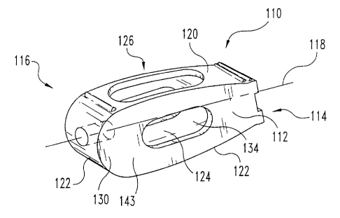
Figures 8, 9A and 9B depict an alternative embodiment of a spinal implant of
the present invention. Spinal implant 110 is adapted to be inserted into a
cavity
provided between adjacent vertebrae, for example, between adjacent lumbar
vertebrae, to promote fusion of the vertebrae and restore normal disc space
height.
Implant 110 comprises an elongate body 112 defining a longitudinal axis 118, a
first
terminal part 114, and a second terminal part 116. At least one of terminal
parts 114
and 116, preferably both, includes opposed upper and lower bearing surfaces
adapted
to bear against cortical bone surfaces of adjacent vertebrae. In one
embodiment
depicted in Figure 9B, implant 110 is provided to have a substantially
rectangular
cross-section when viewed along the longitudinal axis 118.
Elongate body 112 includes central portion 113 that extends from first
terminal
part 114 to second terminal part 116. Body 112 includes an upper surface 120
and an
opposite lower surface 122 defining cavity 124 therebetween. Upper surface 120
and
lower surface 122 are adapted to substantially mate with the natural curvature
of
corresponding facing endplate surfaces of adjacent vertebrae. Thus, the convex
curvilinear configuration of upper surface 120 and lower surface 122
facilitates
locating the implant approximately in the middle of the vertebra body. As a
result,
the spinal implant is neither engaged too far into the openings provided in
the cortical
endplates to provide a sufficiently dense load-bearing surface, nor
insufficiently
inserted, in which case the bone fusion between the two vertebrae would be
adversely
CA 02386328 2002-04-03
WO 01/28465 PCT/US00/41216
affected by a poor anchorage. In another form, upper surface 120 and lower
surface
122 are provided as arcuate surfaces along longitudinal axis 118. The arcuate
surfaces inhibit expulsion, particularly posterior expulsion, of the
surgically implanted
spinal implant by providing an implant that has a maximum height that is
greater than
5 the height of the surgically prepared entrance in the posterior vertebrae
body walls
into the intervertebral space.
Further, upper bearing surface 120 includes at least one opening 126 extending
into cavity 124. Similarly, lower bearing surface 122 includes at least one
opening
(not shown) into cavity 124. The perimeter of both the upper and lower bearing
10 surfaces is substantially continuous and uninterrupted. Cavity 124 is
provided to
receive a graft of osteogenetic material, such as spongy bone or other
material
favoring bone growth, including synthetic bone media. Therefore, the
curvilinear
configuration of upper surface 120 and lower surface 122 and their associated
openings 126 allow interpenetration of the cancellous bone revealed in the
surgically
15 prepared intervertebral space of adjacent vertebrae. Interpenetration of
the cancellous
bone of the vertebra enhances the intimate contact and interdiffusion of
osteogenic
material initially deposited in cavity 124 with the cancellous bone tissue and
greatly
enhances the potential for bone growth.
Elongate body 112 can also include sidewalls 130 and 132 extending from
upper surface 120 to lower surface 122. Sidewalls 130 and 132 can include
openings
134 providing communication into cavity 124 to further enhance interdiffusion
of the
osteogenic material in cavity 124 with cancellous bone tissue.
Referring to Figure 9B, in one embodiment upper surface 120 and lower surface
122 have a substantially uniform height from sidewall 130 to sidewall 132 in a
direction transverse to longitudinal axis 118.
First terminal part 114 includes a first bearing surface 138, an opposite
second
bearing surface 140, and a terminal face 142 extending therebetween.
Preferably first
bearing surface 138 and second bearing surface 140 include substantially
planar
surfaces 141 and 146, respectively, adapted to engage surfaces of cortical
bone
endplates on adjacent vertebral bodies. When inserted within the prepared
intervertebral space, first bearing surface 138 and second bearing surface 140
bear
CA 02386328 2002-04-03
WO 01/28465 PCT/US00/41216
16
against cortical bone tissue proximate to the posterior wall of the vertebral
bodies.
The implants can sustain the compressive forces associated with normal
activity and
resist receding into the sponge-like cancellous bone tissue of the vertebral
body. The
desired disc height can be maintained for an extended time period while bone
fusion
progresses. First bearing surface 138 and second bearing surface 140 are
separated by
a distance d3 selected to restore normal disc space height and natural
lordosis.
Further, in a preferred aspect, first bearing surface 130 and second bearing
surface
140 are substantially planar surfaces extending substantially parallel to
longitudinal
axis 118. It will be appreciated that implant 110 can be adapted to be
inserted in
intervertebral spaces of vertebrae other lumbar vertebrae. Therefore, distance
d3 can
be varied to accommodate varying disc heights and natural lordosis.
In preferred embodiments, first and/or second bearing surface 138 and 140
include anti-expulsion features 148, for example, ridges, teeth, and other
projections,
adapted to inhibit the expulsion of implant 110 from the intervertebral space.
In one
embodiment, the anti-expulsion surface features include a ridge transverse to
longitudinal axis 118. In a preferred form, the anti-expulsion features are
adapted to
minimize the force needed to insert implant 110 into prepared intervertebral
space, yet
inhibit expulsion of implant 110. Examples of such preferred forms include
ratchet-
shaped ridges or teeth that have an apex pointing toward the first terminal
end. When
thus configured, the ratchet-shaped ridges or teeth chisel deeper into the
cortical bone
tissue in response to a posteriorly directed expulsive force.
Terminal face 142 includes tool-engaging portion 150. Tool-engaging portion
150 can be provided with a variety of features adapted to engage an insertion
tool for
insertion of implant 110 into the intervertebral space. For example, tool-
engaging
portion 150 can include a variety of indents and openings, which may or may
not be
threaded, to engage correspondingly configured features on an insertion,
manipulation
accessory (not shown) to facilitate implantation and/or rotation of implant
110 in the
intervertebral space. In the preferred embodiment of Figures 8 and 9, tool-
engaging
portion 150 includes a longitudinally extending threaded bore 151 and a
driving
indent 153.
CA 02386328 2002-04-03
WO 01/28465 PCT/USOO/41216
17
Second terminal part 116 is opposite first terminal part 114. Second terminal
part 116 can include third bearing surface 154, opposing fourth bearing
surface 156,
and an insertion face 152 extending therebetween. Third bearing surface 154
and
fourth bearing surface 156 are adapted to bear against surfaces of cortical
bone
endplates proximal to the anterior wall of adjacent vertebral bodies. In
preferred
forms, third bearing surface 154 and fourth bearing surface 156 are provided
as
curved surfaces that can abut correspondingly curved surfaces of cortical bone
prepared using a cutting/insertion tool (described below). Third and fourth
bearing
surfaces 154 and 156, respectively, are separated by a distance, d4. In the
preferred
illustrated embodiment, distance d4 is selected to be greater than 0 to
restore desired
anterior disc height of vertebrae, V 1 and V2 and maintain a desired
angulation
between the vertebrae. While third and fourth bearing surfaces 154 and 156 are
shown as curved surfaces, it is understood that these bearing surfaces can be
provided
in a variety of shapes including convex or ogival in either the horizontal or
vertical
plane or both, or substantially planar as depicted for the first and second
bearing
surfaces 138 and 140, respectively.
Further, third and fourth bearing surfaces 154 and 156 can include anti-
expulsion features 157 as described for first and second bearing surfaces 138
and 140.
The anti-expulsion features are preferably provided in a configuration to ease
insertion of implant 110 into the prepared intervertebral space while
inhibiting
expulsion of the implant.
Second terminal part 116 includes insertion face 152 extending between upper
surface 120 and lower surface 122. Insertion face 152 is adapted to minimize
the
force needed to insert spinal implant 110 into a prepared cavity in the
intervertebral
space between adjacent vertebrae. In one form, insertion face 152 is provided
as a
curved surface. In alternative configurations, insertion face 152 can be
provided as a
convex surface. Further, insertion face 152 can include one or more openings
providing communication with cavity 124 of body 112 to facilitate
interdiffusion of
osteogenic material with bony tissue and thus promote bone growth of adjacent
vertebrae V 1 and V2.
CA 02386328 2002-04-03
WO 01/28465 PCT/US00/41216
18
Preferably implant 110 is made as a single, integral piece. Implant 110 is
made
of physiologically acceptable material having the requisite strength to
withstand the
compressive force exerted on the spinal column during normal activity.
Examples of
such acceptable material include titanium, composites, ceramics, bone,
stainless steel
and surgical steel.
Implant 110 may be inserted into an intervertebral space after preparation of
the
endplate of adjacent vertebrae using cutting tool 180, which will now be
described
with reference to Figures 10-12. Cutting tool 180 includes a cutting head 182,
shaft
184 defining a longitudinal axis 186, and handle-engaging portion 188.
Cutting head 182 is attached to the distal end of shaft 184. Cutting head 182
includes a first arm 190 and a second arm 192 extending generally parallel to
longitudinal axis 186. Opposed first arm 190 and second arm 192 include two
generally smooth, longitudinal faces 202 and 204. Faces 202 and 204 are
configured
to facilitate insertion of cutting head 182 into the intervertebral space, and
are
generally separated from each other by a distance d5.
First and second arms 190 and 192 each include first arcuate cutting edge 194
and a second opposite arcuate edge 196. Thus, cutting head 182 includes a
total of
four cutting edges. First cutting and second cutting edges 194 and 196,
respectively,
are provided in a configuration to substantially conform to arcuate upper and
lower
surfaces of implant 110. Further, first and second arms 190 and 192 and their
included first and second cutting edges 194 and 196 are adapted to cut and
remove a
portion of cortical bone tissue on opposing endplates of adjacent vertebrae V
1 and
V2, while substantially retaining the natural concave curvature of the
endplates. The
cutting edges 194 and 196 have a length d6 selected to avoid cutting the
anterior and
posterior portions of the endplates and the vertebral wall of vertebrae
selected for
treatment. The cavity thus prepared with cutting tool 180 provides contact
with the
graft material in implant 110 and the spongy bone of the two vertebrae. The
bearing
surfaces of implant 110 are disposed adjacent the edges of the openings of the
cortical
endplates and bear against the remaining portions of the endplates to
establish a
strong load bearing relationship.
CA 02386328 2002-04-03
WO 01/28465 PCT/USOO/41216
19
First arm 190 and second arm 192 are generally opposed and define a cavity
198 therebetween for receipt of bony debris generated during the cutting
operation.
The bony debris collected from the cutting operation can be saved and packed
in the
cavity 124 of implant 110 to promote vertebral fusion. Proximal end of first
arm 190
and second arm 192 attach to the distal terminus of shaft 184. Opposite ends
of first
arm 190 and second arm 192 attach to non-cutting portion 200.
Non-cutting portion 200 of cutting head 182 is fixed to the distal end of
first
arm 190 and second arm 192. Preferably, non-cutting portion 200 has a first
dimension transverse to the longitudinal axis substantially the same as
distance d5 to
be generally co-extensive with faces 202 and 204 of arms 194 and 196. Non-
cutting
portion 200 also is adapted to align faces 202 and 204 an equal distance from
opposed
endplate surfaces of adjacent vertebrae to facilitate removal of equal amounts
of
cortical bone tissue from adjacent vertebrae. Further, non-cutting portion 200
is
adapted to inhibit removal of cortical bone from the anterior cortical bone
surfaces of
adjacent vertebrae. While the non-cutting portion is depicted as a cylindrical
abutment, it is understood that alternative configurations are also included
within this
invention. Such alternative configurations include spherical, semispherical,
frustoconical and the like.
Shaft 184 is rotatably received within sleeve 206. Sleeve 206 includes stop
208
adapted to bear against a vertebral body when the cutting edge is inserted
into the
intervertebral space. Preferably, stop 208 is adapted to inhibit interference
with the
inter-spinal processes and associated nerve bodies. In one embodiment, stop
208 is
adapted to engage a single vertebral body.
Handle-engaging portion 188 is attached to the proximate end of shaft 184.
Handle-engaging portion 188 is adapted to releasably engage a variety of
handles
known in the art (not shown) to facilitate rotation of shaft 184 and cutting
head 182.
Alternatively, it is understood that cutting tool 180 can include a handle
fixedly
attached to the proximal end of shaft 184.
Cutting tool 180 can be provided for use in conjunction with guide sleeve 210
illustrated in Figure 12. When used with a guide sleeve, cutting tool 180 can
be
slideably received within the guide sleeve to protect nerve tissue and related
spinal
CA 02386328 2002-04-03
WO 01/28465 PCT/US00/41216
processes and orientated with respect to the disc space. A variety of guide
sleeves
suitable for use with this invention are known and commonly used in surgical
procedures. Guide sleeve 206 can include a variety of structural features
adapted to
facilitate distraction of the vertebrae and fixation of the selected vertebrae
and
5 associated instruments for performing spinal surgery. Such structural
features can
include, for example, insertion fins 212, pins (not shown) and clamps (not
shown).
The implant and associated surgical instruments described and/or disclosed in
this application can be provided as a surgical kit. The surgical kit can
include a
number of implants as described herein including implants having varying
dimensions
10 for use with patents of varying ages and sizes, and for implantation
adjacent to
different vertebrae of the spine. The associated surgical instruments
including the
cutting tool, distracters and guide sleeve are configured and sized to
facilitate the
implantation of the varying sized implants.
Various non-limiting embodiments of a spinal fixation or fusion procedure of
15 the present invention are next described. One procedure is characterized
by: (a)
Cutting the vertebrae V 1 and V2 and disc 2 with tool 180 to prepare for
implantation
of implant 110, and (b) Inserting implant 110 between vertebral bodies V 1'
and V2'.
Another more detailed procedure for fusing two vertebrae together is described
in
terms of the procedural stages A-H as follows:
20 (A) The surgeon reveals the vertebrae in need of fusion using known
surgical
techniques. The surgeon then separates the dural sleeve forming an
extension of the bone marrow if the procedure is in the lumbar region
and then carries out a discectomy to provide a space for implant 110 in
the disc space.
(B) The surgeon inserts between the two vertebral bodies V1, V2 from the
rear (posterior), two distracters known in the art. Distracters may be
inserted laterally with respect to the cavity provided by the discectomy
and then turned 90 so as to spread apart the vertebral bodies and to
restore disc height. If a lordotic angle is intended, the distracters may
include tapered surfaces intended to establish the desired angulation.
Next, one of the distracters is removed.
CA 02386328 2002-04-03
WO 01/28465 PCTIUSOO/41216
21
(C) The surgeon then inserts cutting tool 180 between vertebral bodies V 1
and V2 so that the faces 202 and 204 are in contact with the vertebral
endplates as shown in Figure 13. When the cutting head 182 is correctly
positioned in the central region of the cortical endplates, stop 208 abuts
the outer surface of V 1 or V2, and non-cutting portion 200 is proximal to
the interior cortical bone wall of V 1 and V2.
(D) Next, the surgeon rotates handle 188, causing cutting head 182 to rotate
about longitudinal axis 186. Typically, the surgeon rotates handle 188
through only a partial rotation to engage cutting edges 194 and 196 with
the cortical bone of the adjacent endplates and then changes direction to
generate an oscillating cutting action. Cutting action continues until the
proper amount of vertebral endplate is removed. When non-cutting
portion 200 is correctly positioned between interior cortical bone
portions of adjacent vertebrae V 1 and V2, first cutting edge 194 and
second cutting edge 196 cut equally through endplates 244 and 246.
Remaining portions of endplates 246 and 248 bear against non-cutting
portion 200 and non-rotating shaft 206. Bony debris generated by the
cutting of cortical bone is received in cavity 198 between first arm 190
and second arm 192.
(E) Then, the surgeon withdraws cutting tool 180 from the intervertebral
space. Bony debris residing in cavity 198 can then be collected and
packed inside cavity 124 of implant 110.
(F) The surgeon then implants implant 110, previously filled with either
osteogenic material or bony debris, between endplates 244 and 246 from
the posterior of vertebral bodies V 1 and V2. Implant 110 is positioned
such that arcuate upper surface 120 and lower surface 122 engage cut
portions of endplate 244 and 246, while remaining uncut portions of
endplates 244 and 246 bear against bearing surfaces 138 and 140. In
addition, bearing surfaces 154 and 156 on the second terminal portion of
implant 110 contact the non-cut interior cortical bone surfaces of
adjacent vertebrae. Implant 110 may be presented flat, so that sidewalls
CA 02386328 2002-04-03
WO 01/28465 PCTIUSOO/41216
22
134 and 136 contact cut portions of endplates 244 and 246. Thereafter,
the surgeon turns implant 110 through a quarter of a turn about its
longitudinal axis 118 so as to place it in the position with walls 134 and
136 perpendicular to the cortical endplates and its arcuate upper surface
120 and lower surface 122 in contact with the cut portion of endplates
260 and 262 as shown in Figure 15. When implant 110 is in its final
position, in which it is stabilized, a bone graft or other osteogenic
material is in contact with the spongy portion, promoting bone fusion.
(G) The surgeon then removes the second distracter and repeats the
preceding sequences of stages (A) through (G) to mount a second spinal
implant 110 by placing it in position generally parallel to the first spinal
implant 110 as illustrated in Figure 16.
In other embodiments, it is envisioned that the described stages may be
altered, deleted, combined, repeated, or re-sequenced, as would occur to those
skilled
in the art. By way of a non-limiting example, the procedure according to the
present
invention may utilize one or more different tools to prepare the spine for
fixation by
the implantation of the present invention. In another example, the tools of
the present
invention may be utilized to prepare a surgical site for an implant.
The scope of the invention is not intended to be limited to the ascribed
embodiment and may also include variants within the scope of the accompanying
claims. For example, bearing surfaces 138, 140, 154, and 156 may have any
shape,
such as curved or cylindrical shaped, with endplates 244 and 246 being
correspondingly cut to allow placing the bearing surface in a suitable
position.
Further, these bearing surfaces may be interconnected in pairs to constitute a
single
member. Further, the bearing surfaces may also include a variety of structural
features adapted to inhibit expulsion of implant 110 from the intervertebral
space.
Likewise, the body 112 of implant 110 may include any shape, preferably
retaining edges that project from the endplate contacting portions. In
particular, the
body may have a multitude of cells residing within the interior cavity 124.
Also, it
should be generally noted that the implant 110 and tool 180 of the present
invention
can be adapted to a geometry of the spine with respect to lordosis, kyphosis,
or
CA 02386328 2002-04-03
WO 01/28465 PCT/USOO/41216
23
parallel vertebral endplates. Thus, the present invention includes application
to
adjacent vertebrae other than the lumbar vertebrae. Correspondingly, the
implant and
the cutting portion of the tool may have a different shape, such as a
cylindrical
geometry, other than the general teardrop geometry depicted. Also, instead of
using
the disclosed implant 110, the spinal space prepared by tool 180 can be filled
with any
other material as would occur to those skilled in the art.
According to other embodiments, the implants described herein can be partly
or totally constituted by porous rehabitable or other resorbable materials
favoring
osteointegration. Such embodiments include: (a) an implant according to the
above-
illustrated geometry made entirely of a resorbable or rehabitable material;
(b) an
implant in which the whole of the central part is made of a resorbable or
rehabitable
material; or (c) an implant in which the periphery of the central part is of a
metallic or
other material and the inside part is of a material favoring osteointegration
that may
be in an initially solid, pasty, or liquid state. Preferably, the implant 110
of the
present invention is provided in a biocompatible metal, such as titanium or
surgical
steel.
The present invention contemplates modifications as would occur to those
skilled in the art. It is also contemplated that processes embodied in the
present
invention can be altered, rearranged, substituted, deleted, duplicated,
combined, or
added to other processes as would occur to those skilled in the art without
departing
from the spirit of the present invention. In addition, the various stages,
steps,
procedures, techniques, phases, and operations within these processes may be
altered,
rearranged, substituted, deleted, duplicated, or combined as would occur to
those
skilled in the art. All publications, patents, and patent applications cited
in this
specification are herein incorporated by reference as if each individual
publication,
patent, or patent application was specifically and individually indicated to
be
incorporated by reference and set forth in its entirety herein.
While the invention has been illustrated and described in detail in the
drawings
and foregoing description, the same is considered to be illustrative and not
restrictive
in character, it is understood that only the preferred embodiments have been
shown
CA 02386328 2002-04-03
WO 01/28465 PCT/US00/41216
24
and described and that all changes and modifications that come within the
spirit of the
invention are desired to be protected.