Note: Descriptions are shown in the official language in which they were submitted.
WO 01/41867 CA 02393535 2002-06-06 PCT/US00/32987
-1-
ADAPTIVE ELECTRIC FIELD MODULATION OF NEURAL SYSTEMS
This application claims the benefit of provisional application Serial No.
60/169,280,
filed December 7, 1999, which is hereby incorporated by reference in its
entirety.
BACKGROUND OF THE INVENTION
Numerous attempts have been made to suppress epileptic seizures in human
patients
with indirect electrical stimulation at sites remote from the epileptic focus,
including
cerebellum (Cooper et al., 1976; Van Buren et al., 1978), thalamus (Cooper et
al., 1985;
Fisher et al., 1992), and vagal nerve (Murphy et al., 1995; McLachlin, 1997).
Surprisingly,
there has been far less investigation of the technology required to directly
control an epileptic
focus electrically. It has been shown that direct current injection into
tissue could suppress
evoked (Kayyali and Durand, 1991 ) or spontaneous (Nakagawa and Durand, 1991;
Warren
and Durand, 1998) epileptiform activity in brain slices. Even simple periodic
pacing of a
neuronal network with direct electrical stimulation (Kerger and Schiff, 1995)
can reduce
seizure-like events. In addition, there is some evidence that nonlinear
control schemes might
be useful in manipulating epileptiform activity (Schiff et al., 1994). In each
of these cases,
the stimulation was applied in the form of short current pulses directly into
the tissue that
evoke neuronal firing. Recently, it was demonstrated that steady state (DC)
electric fields
oriented parallel to pyramidal cells were capable of suppressing epileptic
seizure activity in in
vitro hippocampal brain slices (Gluclanan et al., I 996x). Such f eld
application led to nearly
complete suppression of neuronal activity, yet due to a combination of
polarization effects
(electrode and tissue) and neuronal adaptation, this effect was transient.
DESCRIPTION OF THE DRAWINGS
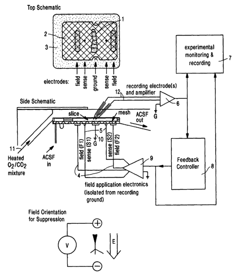
Fig: 1. (A) is a top view schematic drawing of a perfusion chamber used to
adaptively
modulate the neuronal activity of an isolated neural system. (B) is a side
view schematic of
the same chamber. The brain slices-rest on a nylon mesh just below the Zipper
surface of the
perfusate of artificial cerebrospinal fluid (ACSF), and the atmosphere above
the perfusate is
warmed to the bath temperature of 35°C and saturated with 95% OZ-5 %
CO~. An electric
field is imposed on the slice by a set of A~-AgCI electrodes embedded in the
floor of the
WO 01/41867 CA 02393535 2002-06-06 pCT/US00/32987
-2-
chamber. The potential difference applied between parallel plate electrodes F1
and F2 is
feedback controlled so that the averafe field measured at sensing electrodes
S1 and S2 is
proportional to a program voltage. An additional pair of electrodes, G, are
used as recording
ground.
Fig. 2. Power spectral density (PSD) for recorded activity and applied f eld
stimulus
in the case for which the stimulus was a low frequency random signal (A) and
for which the
stimulus was a typical feedback control signal (B). For display purposes, the
stimulus PSD
was vertically scaled such that its amplitude matched that of the recorded
activity PSD at low
frequencies. In both cases, the stimulus PSD falls off quickly ( J' ) for
frequencies, f, above
about 4 Hz, in contrast to the neuronal activity PSDs, which have significant
spectral power
up to approximately 350 Hz. Also shown are the PSDs of the recorded neuronal
activity after
removal of an estimate of the stimulus artifact. These signals are
indistinguishable from the
original recording for frequencies above ~2 Hz. In (B) the raw signal lies
slightly below the
processed signal for low frequencies. These results indicate that the applied
field during
control is not simply masking the neuronal activity in the recording process
during control.
The stimulus artifact accounts for less than 5% of the RMS recorded signal
amplitude.
Fig. 3. Adaptive control of seizure activity using applied electric fields. In
each panel,
the main trace is the raw extracellular potential recording. Insets are
tracings of activity,
filtered to illustrate the high frequency activity, shown at expanded scales.
In each case, a
dashed line is used to demarcate when control is turned on. A,B: Examples of
seizure
suppression from separate experiments using electric fields applied as a
negative feedback
parameter. Electrographic seizures are observed as an increase in high
frequency activity
atop large low-frequency deflections (Traynelis and Dingledine, 1988). In B,
seizures occur
interspersed among frequent short network bursts (Rutecki et al., 1985). C:
Example of
seizure induction achieved using positive feedback.
Fig. 4. Event detection results for a single 90 minute recording, with
different electric
field stimuli applied. The lower trace indicates feedback gain (G, left axis)
or amplitude (A,
right axis) of the applied stimulus. Greek letters indicate type of stimulus:
baseline (no
letter); full-wave feedback control (a.); half wave rectified feedback control
((3); constant
amplitude suppressive field (y); low frequency noise (8); suppressive half
wave rectified low
WO X1/41867 CA 02393535 2002-06-06 pCT/US00/32987
-3-
frequency noise (s); positive feedback control (~). Two types of event
detection were used to
identify synchronous neuronal activity from the recorded field potentials.
"RMS events" were
detected from variations in the RMS power in the frequency band 100-350 Hz.
"DC events"
were detected by threshold detection after low pass filtering the recordings
at 10 Hz. The
character of both types of events, as quantified by their average and maximal
amplitudes as
well as their duration, was visibly changed from baseline when control was
applied. No
events of either type were observed during the final and longest (16 minutes)
application (a~)
of full-wave control.
Fig. 5. Traces and spectrograms of activity with and without control for same
experiment as Fig. 4. (A) Activity (lower trace) and applied field (upper
trace) from the final
application of full-wave control (oc~) from Fig. 4 and the baseline preceding
it. (B,C) A 15
second long trace and spectrogram of a seizure-like event (B) and of activity
during control
(C) from A. The upper traces in B and C are the activity, high-pass filtered
at 100 Hz. The
spectrograms (B, C, D) are calculated in overlapping vertical frequency bins
50 Hz tall from
I S 25-350 Hz, and in overlapping horizontal time windows 0.05 s wide. (D)
Spectrogram for
longer period illustrating contrast between baseline and controlled activity.
Fig. 6. Examples of activity during non-feedback electric licld stimulus for
the same
recording as Fig. 4. For each sct, the upper-trace is of the recorded
activity, while the applied
field is shown in the lower trace. (A) Application of constant-amplitude (DC)
suppressive
freld (4y). (B) Application of fill-wave low frequency noise field (4c~). (C)
Application of
half wave rectified low frequency noise field (4E). In each case, large
neuronal events are
observed, though the full-wave noise field did have the effect of breaking up
the seizure-like
events into shorter durations.
Fig. 7. Comparison of power spectral density (PSD) of recorded activity during
control (lines with symbols) as compared to baseline (lines without symbols).
The control
corresponds to the final control application in Fig. 4, and the baseline
corresponds to the final
baseline application. PSDs were calculated in overlapping 1.64 s (2~~ point)
windows. The
power averaged over the windows is shown in A, while the window to window
variance of
power is shown in B. For both measures, the controlled activity falls well
below that of the
baseline activity.
WO 01/41867 CA 02393535 2002-06-06 pCT/US00/32987
-4-
Fig. 8. Statistics of the RMS power of recorded activity in the frequency band
100-350 Hz, calculated in 1.64 s windows, for baseline (squares), full-wave
control (circles)
and half wave rectified control (triangles). Statistics correspond to all
applications
independent of gain for the recording of Fig. 4. The normalized histogram and
cumulative
probability are shown in (A) and (B). It is clear that the baseline activity
has many windows
with much higher power than either type of control. These windows correspond
to the first
phase of the seizures. The inset in A is the normalized histogram of power
calculated with
logarithmically spaced bins (power, abscissa; frequency, ordinate) for
baseline (boxes) and
full-wave control (circles). From this plot, it is observed that deviations to
both high and low
power are eliminated during full-wave control. The windows with extremely low
power
correspond to the latter phase of the seizures and the recovery times
following them. The
power variance vs. average power is plotted in (C) for these three conditions.
The two types
of control are statistically well distinguished from that of the baseline
activity.
Fig. 9. Examples of network activity when control is released. In each panel,
the
inset is the activity for the full control period, indicated in gray, plus the
baseline periods
before and after. The trace in (A) cor-esponds to the same experiment as
Figure 3A, with
half wave rectified control. The network oscillates between excitation similar
to seizure
onset and being suppressed by the controller. When control is released, this
activity proceeds
immediately into a full seizure-like event. B,C Traces from another experiment
in which
half wave rectified control (B) was compared to non-rectified control (C). For
half wave
rectification, seizures were observed very soon (0-3 s) after control was
released, as
compared to 12-18 s for non-rectified control. The time base for the insets is
the same, and
indicated in (A). The inset vertical scale is half that of the main traces.
DESCRIPTION OF INVENTION
The present invention relates to devices and methods for modulating the
neuronal
activity of a neural system comprising neurons; such as a brain, brain
regions, or any in vivo
or ifs vitro collection of neurons. In particular, the present invention
involves the use of
applied electric fields to modulate the behavior of a target neural system. In
preferred
embodiments, the polarity and magnitude of the applied electric field is
varied according to
WO 01/41867 CA 02393535 2002-06-06 pCT/US00/32987
-5-
information gathered from the modulated neural system, or any other desired
source chosen
to provide feedback, to modulate the strength of the applied electric field.
In such
embodiments, preferably a sub-threshold stimulus is administered to modulate
to the neural
system. The methods and devices of the present invention can be used to treat
diseases of the
nervous system, to restore neuronal function, paralysis, and motor and sensory
deficits, to
produce prosthetic devices that interact and modulate neuronal activity, to
enhance or
suppress neuronal activity and associated phenotypes, and the like.
A preferred method of the present invention relates to modifying the neuronal
activity
of a neural system comprising neurons, comprising one of more of the following
steps, in any
order: measuring the neuronal activity, or other behavior, of a neural system;
and applying an
oriented electric field to said neural system effective to modify the neuronal
activity of the
neural system, wherein the magnitude and polarity of said applied electric
field is changed in
response to the measured neuronal activity.
A neural system in accordance with the present invention can be any ensemble
of one
or more neurons, and/or other excitable cells, such as muscle, heart, retinal,
cochlear, tissue
culture cells, stem or progenitor cells, including cell-electrode interface
devices and the like.
Cells can be coupled electrically, chemically, or combinations thereof. The
neural system
can be an entire brain, ganglia, nerve, etc., or it can be a region or portion
of it. Any animal
source of material is suitable, including neural systems of invertebrates,
such as mollusks,
arthropods, insects, etc., vertebrates, such as mammals, humans, non-human
mammals, great
apes, monkeys, chimpanzees, dogs, cats, rats, mice, etc. In the examples, a
specific region of
a mammalian brain is dissected out and placed in a chamber where its activity
is modified.
However, physical isolation of a target brain region is unnecessary; the
activity modulation
can be perfornled in situ, as well. Preferred target regions include, but are
not limited to,
neocortex, sensory cortex, motor cortex, frontal lobe, parietal lobe,
occipital lobe, temporal
lobe, thalamus, hypothalamus, limbic system, amygdala, septum, hippocampus,
fornix,
cerebellum, brain stem, medulla, pons, basal ganglia, globus pallidum,
striatum, spinal cord,
ganglion, cranial nerves, peripheral nerves, retina, cochlea, etc.
In one step of a prefen-ed method, the neuronal activity of the neural system
is
measured. By the term "neuronal activity," it is meant any measurable physical
behavior,
output, or phenotype of the system. For example, neurons typically display
variations in their
WO X1/41867 CA 02393535 2002-06-06 pCT/US00/32987
-6-
membrane potential, such as action potentials, depolarizations, and
hyperpolarizations. These
changes in the membrane potential car be utilized as a measure of neuronal
activity, e.g., by
monitoring intracellularly in a single neuron, or extracellularly, the
electrical activity of a
single neuron or the activity of an ensemble of neurons. Behaviors, or other
products of a
neural system (e.g., hormones, growth factors, neurotransmitters, ions, etc.)
can also be
detected, and used as a feedback signal to determine the magnitude and
strength of the
modulating applied field. For instance, if a purpose is to elicit movement of
a limb, then the
neuronal activity can be limb motion. The neuronal activity which is measured
or assessed
can be a subset of the total activity observed in the system, e.g., a
particular frequency band
of the full neural signal. In the examples, hippocampus slices were monitored
for neuronal
activity. Although the measuring electrode detected various types of activity,
including
spontaneous neuronal firing, slow burst activity, and background noise, as
well as fast
frequency epileptic seizures, it was desired to modulate only the latter.
Thus, for these
purposes the neuronal activity can be considered to be only the events of
interest, e.g., the
epileptic seizures.
Methods for measuring and recording neuronal activity can be accomplished
according to any suitable method. In preferred embodiments of the invention,
the neuronal
activity is monitored cxtracellularly by measuring the extraccllular
electrical potential of a
target population of neurons. Such measurements can reveal complex spikes or
burst
activity, sharp or slow waves, epileptiform spikes or seizures, arising from
one or more
neurons in the neural system.
The neuronal activity can be measured by recording the neural system's
electrical
potential in the extracellular space. The electrodes used to measure the field
potential
produced by the neural system are referred to as "measuring electrodes" or
"recording
electrodes." One or more electrodes can be used to measure the field
potential. In preferred
embodiments, two or more electrodes are utilized. The field potentials
recorded at a given
extracellular site will depend on a variety of factors, including the location
of the electrodes)
with respect to the soma and dendritic layers, the architecture of the neural
system, the
perfusion solution, etc.
The measuring electrodes can detect the field potential from the applied field
as well
as the activity generated by the neural system. There are a number of methods
that can be
WO 01/41867 CA 02393535 2002-06-06 pCT~S00/32987
used to distinguish the neuronal activity from the applied fields. For
example, in i~~ vitro
hippocampal slices, a pair of differential electrodes, aligned as closely as
possible to the
isopotential of the applied field, were used as measuring electrodes. They are
"differential"
in the sense that an active electrode is placed in the tissue, preferably near
the cell body layer
of the target neurons, while the reference electrode is placed preferably in
the bath external to
the tissue. The values obtained from each electrode can be electronically
subtracted from
each other, reducing background noise. For in vivo use, the differential
measuring electrodes
can be placed at the same isopotential with respect to the applied field. The
electrodes can be
as close to the target population as possible, without damaging it. Other
methods to reduce
noise and the artifact from the applied field can be used as well, either
alone, or in
combination with the differential electrodes, including filtering and post-
processing of the
measured signal.
Recording from the electrodes can be performed routinely. For instance,
measurements can be made with an AC amplifier if the frequency and number of
extracellular bursts are of interest. It can be equipped with filters to cut
off frequencies below
and above a particular range (band-pass filter) and amplify the signal in
preferred ranges, e.g.,
50-1000 Hz, preferably, 100-500 Hz. A DC amplifier can also be used, if slower
potential
changes are of interest.
A method in accordance with the present invention also involves applying an
oriented
electric field to the neural system effective to modify the neuronal activity
of the neural
system, preferably where the magnitude and polarity of said applied electric
field is changed
in response to the measured neuronal activity. Preferably, the applied field
is oriented in a
particular direction with respect to the somatic-dendritic axis of the neurons
in the neural
system. Most preferably, the field is parallel to the somatic-dendritic axis.
Changing the
strength of the applied field in response to a measured activity of the neural
system can also
be refen-ed to as "adaptive modulation" since the strength of the applied
field is adjusted
based on an activity value of the neural system (e.g., electrical activity,
motor activity, such
as limb motion, etc.). A function of the applied electric field is to modify
the neuronal
activity of the neural system. The electric field is thus applied to the
neural system in an
amount adequate to change the neuronal behavior of the neural system. Any
amount of field
which changes the neural system's behavior is an effective applied field. It
is believed that a
WO 01/41867 CA 02393535 2002-06-06
PCT/US00/32987
_g_
mechanism that underlies adaptive modulation is the ability of the applied
field to alter the
neuron's excitability by changing its threshold; however, the invention is not
bound nor
limited to any theory, explanation, or mechanism of how it works.
In preferred methods of the present invention for in vitoo applications, two
pairs of
electrodes can be used in the field application step. A pair of "field
electrodes" can be used
to produce the applied field. A second pair of electrodes, "sensing
electrodes," can be used to
measure or sense the field generated by the "field electrodes." The sensing
and field
electrodes can comprise the same materials described above for the measuring
electrodes. In
certain applications, however, such as in vivo applications, a field can be
applied without
sensing electrodes.
In preferred embodiments of the invention, the effective amount of applied
field is
sub-threshold with respect to the field potential experienced by the neural
system. By the
teen "sub-threshold," it is meant that the amount of applied field or current
does not reliably,
with 100% probability, initiate new action potentials within the neural
system. In contrast,
the application of a supra-threshold stimulus reliably, with a high degree of
probability,
results in neuronal firing. A sub-threshold potential is, for example, less
than l00 mV/mm,
preferably 50 mV/mm and less, more preferably, 25 mV/mm and less, such as 20
mV/mm, 15
mV/mm, or 10 mV/mm. The sub-threshold potential refers to the potential
generated at the
level of the target neurons. The amount of potential actually produced by the
field electrodes
is less important that the field perceived by the target neurons. It is the
generated field sensed
by the neurons that determines whether a stimulus is sub- or supra-threshold.
In response to the applied electric field, the activity of the neural system
can be
modified in any desired manner, e.g., the activity can be suppressed, reduced,
decreased,
diminished, eliminated, counteracted, canceled out, etc., or it can be
enhanced, increased,
augmented, facilitated, etc. To determine whether the activity of the system
has been
modified, preferably the same neuronal activity measured in the measurement
step is re-
measured. Most preferably, the measurement of the neuronal activity is
performed
simultaneously and continuously with the applied field.
Any effective electrodes can be used for the recording, sensing, and field
electrodes,
including, e.g., metal, steel, activated iridium, platinum, platinum-iridium,
iridium oxide,
titanium oxide, silver chloride, gold chloride, etc., where the electrode can
be insulated by
WO 01/41867 CA 02393535 2002-06-06 pCT/US00/32987
-9-
glass or lacquer, as well as silicon microelectronics, including tetrode or
other multielectrode
arrays or bundles, multichannel and ribbon devices. Typically, the electrodes
can have
relatively large tips with low resistance to detect activity from a number of
neuronal elements
within the neural system. Smaller tipped electrodes can be used for monitoring
activity from
single neurons or smaller populations. Activity can be measured from one or
more
electrodes, preferably two or more. In some cases, it may be desired to record
from several
regions of the neural system in order to characterize its activity. Recordings
of intracellular,
extracellular, or a combination thereof, can be analyzed separately, or
together. The
electrodes can be AC- or DC-coupled.
For certain purposes, iridium oxide type electrodes may be preferred since
they are
relatively nontoxic to cells, as well as being effective carriers of high
current and charge
densities. An activated iridium or iridium alloy wire can be used, or a metal
substrate, such
as noble metal (e.g., Au, Pt, or PtIr), ferrous steel alloy, stainless steel,
tungsten, titanium, Si
microprobe, etc., or other suitable substrate, can be coated with a film of
iridium oxide to
I S produce an effective electrode. Any suitable method to prepare the coating
can be used,
including, but not limited to, an activation process (e.g., Loeb et al., J.
Nezrro. Sci. Methods,
63:175-183, 1995; Anderson et al., IEEE Ti~cans. Bionzed. End., 36:693-704,
1989) to form
activated iridium oxide films (AIROFs), thermal decomposition (Robblea et al.,
Mnt. Res.
Soc. Svmp. Proc., 55:303-310, 1986) to forn~ thenoal 11'ld1Ll171 OXI(le films
(TIROFs), reactive
sputtering (15) to form sputtered iridium oxide films (SIROFs),
electrodcpositing (Kreider et
al., Sensors and Actuc~tons, B28:167-172, 1995) to form electrodeposited
iridium oxide films
(EIROFs), etc.
As described herein, it has been found that adaptive modulation of a neural
system
can be used to modify its neuronal activity. In preferred embodiments, this is
achieved by
characterizing the neuronal activity and then using a feedback algorithm to
determine the
field magnitude necessary to modulate its activity. Neuronal activity can be
characterized by
various measurements, depending upon the particular activity that is being
assessed. When
electrical activity is a determinant, then measurements can include, e.g.,
local field polarity
and magnitude (e.g., -10 mV), burst activity, burst amplitude, burst
frequency, power in a
predetermined frequency band of activity, non-burst activity, single or small
population firing
rate, amplitude or phase of periodic activity, such as theta rhythm, root-mean-
square (RMS),
WO 01/41867 CA 02393535 2002-os-06 pCT/jJS00/32987
-10-
variance, etc. In general, any :,unable measure of neuronal activity can be
used as the
feedback stimulus for the applied field. The feedback stimulus can also be
determined by
multiple measurements, e.g., electrical activity, limb motion, cochlear
activity, etc.
In the examples, the neuronal activity, after appropriate filtering, was
characterized by
the RMS fluctuations of the measured signal, serving as the feedback stimulus.
An electric
field was subsequently applied in proportion to the RMS. Specifically, the
instantaneous
RMS activity (e.g., the last 0.25 sec of activity) was low pass filtered with
a time constant T
to yield A~. This value was compared with a threshold value, as determined by
the long time
average of the RMS (e.g., the last 30 seconds of activity). The magnitude of
the applied field
was then derived by calculating the di fference between the A r and the
threshold multiplied by
a gain factor. Any suitable methods and/or algorithm for determining field
strength and
polarity can be used, e.g., linear and nonlinear proportional feedback,
proportional - integral -
differential feedback, etc.
The values for instantaneous activity and threshold can be selected
empirically, e.g.,
based on the activity characteristics of the system and the neuronal activity
that is to be
controlled. The goal is to choose a time scale that distinguishes the activity
of interest from
the baseline activity of the system. When a timescale for the threshold (e.g.,
the last 30
seconds of total activity) and instantaneous (e.g., last 0.25 sec of total
activity) activity
determinations are selected, the difference between such values should permit
detection of
the onset of the activity of interest.
A gain factor can be chosen such that the output of the applied field is
adequate to
modulate the neuronal activity that is being monitored. It can be empirically
derived, based
on previous performance of the neural system and various considerations,
including, e.g.,
magnitude of the onset of the event which is being assessed, magnitude of the
applied field
necessary to modulate the neural system, characteristics of the field
electrodes, characteristics
of the neural system environment, etc. In the experiments described herein, a
gain was
chosen such that a typical difference between Ar and the threshold yielded a
field in the range
of order of IOmV/mm. Successful control was achieved for the same experiment
with gains
differing by an order of magnitude indicating that the choice of gain was not
critical.
WO 01/41867 CA 02393535 2002-os-06 PCT/US00/32987
-)< )< -
The applied field can utilize the full feedback signal ("full-wave control"),
or, it can
be half wave rectified. When half wave rectification is used, a field is
applied only when the
instantaneous activity (or the calculated A z) is above (or below) the
threshold value. In the
examples described below, a field was applied only when there was a positive
difference
between the instantaneous activity and the threshold. Thus, half wave
rectification indicates
that the field is applied in only one direction. For full-wave control, a
field is applied
continuously when there is any difference between the instantaneous activity
(or calculated A_
r) and the threshold value. The outcome of half wave rectification is the
application of a field
in only one direction, while full-wave control results in both negative and
positive applied
f elds, depending upon the sign of the difference between instantaneous
activity and
threshold. As a result, full-wave control can involve the administration of
both excitatory and
suppressive si;~nals, while half=wave rectification involves only one kind of
signal, either
excitatory or suppressive, depending upon the direction of the applied field.
The experiments
described below show that full-wave control was generally superior to half
wave rectification
for seizure suppression, for reducing withdrawal seizures, and for obtaining a
more regular
baseline of neuronal activity.
Full-wave control may also be desirable to avoid substantial electrode and
tissue
polarization which occurs when half wave rectification is used. In the latter
case, the
electrodes may need to repolarized between field applications, e.g., by
applying bias currents
to the electrodes.
In general, the duration and intensity of the applied field can be determined
by the
measured activity. If the purpose is to eliminate neuronal activity, then
preferably a field
potential, or current, is applied until the activity level is reduced below a
threshold level. At
this point, the field can be discontinued until activity is observed again.
The applied field is
preferably not a stationary field, such as the fields described in Gluckman et
al., .l.
Neurophys., 76:4202-4205, 1996; U.S. Pat. No. 5,800,459. See, also, U.S. Pat.
Nos.
5,797,965 and 5,522,863.
Activity can also be augmented, induced, or initiated. In the examples,
reversing the
field potential converted sporadic bursts into a full-blown seizure. In this
case, the feedback
stimulus is positive feedback, where the applied field is used to enhance
activity, e.g., by
producing depolarization toward threshold and/or recruiting more neurons into
the activity.
WO 01/41867 CA 02393535 2002-06-06 PCT/LJS00/32987
-12-
Here the sign of the gain factor is switched so that a negative field is
applied when the RMS
activity goes above threshold, forcing the network to become more excitable.
The ability to
create activity in vitro and in vivo is useful in variety of ways. It can be
used to create animal
models for epilepsy or electroconvulsive therapy (ECT) and for testing agents
which
modulate these brain behaviors for therapeutic, prophylactic, and research
purposes. It can
also be used to induce ECT in humans for therapeutic purposes.
In some instances, a neural system will exhibit ongoing neuronal activity,
such as
spike activity varying in amplitude and frequency. This information can be
processed in any
suitable way to serve as a threshold stimulus for the applied field. For
instance, the activity
in a certain frequency band can be of particular interest because it indicates
that certain state
of the neural system has been reached, such as epilepsy. It therefore may be
desired to apply
the electric field only when the system becomes epileptic. This can be
accomplished by
processing the measured neuronal activity, and applying the field when a
predetermined
threshold of activity is reached. For example, the long-term average of
spontaneous or non-
1 S epileptic activity can be determined and used as the stimulus threshold,
where no field is
applied unless the long-term average, or a function of the average, is
exceeded. A particular
characteristic of neural activity can also be compared to a matched filter
using a temporal,
spectral, or wavelet filter, or a nonlinear filter, and its output compared
with a threshold.
The methods and devices of the present invention are useful in any endeavor in
which
it is desired to modify the behavior of a neural system. In general, an
applied field in
accordance with the present invention can be utilized to modulate any neural
activity,
including, e.8., synchronized firing, oscillatory firing, pulsating activity,
and any in-phase
activity of a neural system. Because of such ability to augment or reduce
neuronal activity of
a neural system, the invention is useful for modulating many kinds of output
which arise from
neural systems, including motor, sensory, emotional, behavioral, etc.
For example, the methods and devices of the present invention are useful for
treating
brain diseases characterized by aberrant neuronal activity. Epilepsy, for
instance, is a brain
disorder characterized by recurrent seizures, affecting 1-2% of the
population. In this
disease, the pattern of neuronal discharge becomes transiently abnormal. In
the examples, an
in vitro slice preparation is utilized to illustrate how epilepsy can be
treated in accordance
with the present invention. When perfused in a high potassium concentration,
these networks
WO 01/41867 CA 02393535 2002-06-06 pCT/iJS00/32987
-13-
show a broad range of interictal-like and epileptiform activity, from network
wide
synchronous events to local and propagating events. Application of the
adaptive electric field
can be used to suppress the epileptiform activity, effectively treating and
controlling the brain
disorder.
S A modulatory effect can be achieved analogously irr situ. For instance, to
treat a
patient having epilepsy, a device can be utilized which simulates the pair of
field electrodes
used in the in vitro method. The field electrodes can be positioned in any
arrangement which
is effective to produce a modulatory field. They can be in contact with brain
tissue or
associated meninges, e.g., by inserting, through an occipital entrance hole,
one, or more, long
flat electrode strips that contacts the long axis of the hippocampus surface
in the temporal
horn of the lateral ventricle. A round electrode (e.g., a single depth
electrode with one or
more suitable high current contacts) can also be utilized, e.g., by placing it
within the long
axis of the hippocampus in order to produce a radial electric field.
Electrodes can also be
external to the brain, e.g., on the scalp. The electrode strip preferably
produces an effective
electric field. Useful electrode strips include non-polarizing biocompatible
electrodes
embedded in silastic sheets with sealed electrode-lead connections, similar to
those used for
cochlear implants, e.g., a Clarion Cochlear Implant, comprising iridium oxide
electrodes
sealed within a curved silastic silicone elastomer sheath. In another
embodiment, a sheet
comprising multiple electrodes can be placed over the neocortex in the
subdural,
subarachnoid, or epidural spaces, or within the sulci of the brain. Thin
electrodes can also be
inserted into brain tissue. In general, any types or combinations of
electrodes, such as those
mentioned above, can be used.
In addition to epilepsy, any brain disorder that displays abnormal activity,
such as
oscillatory or pulsating activity, can be treated analogously. Such diseases,
include,
schizophrenia, depression (unipolar or bipolar), Parkinson's disease, anxiety,
obsessive-
compulsive disorder (OCD), etc., where the electric field is applied to the
particular brain
region exhibiting the abnormal activity, e.g., cortex, hippocampus, thalamus,
etc.
Parkinson's disease is characterized by decreased activity in cells that
produce dopamine.
Patients with the disease experience tremors, rigidity, and difficulty in
movement. Patients
with Parkinson's disease can be treated by applying an electric field in an
amount effective to
ameliorate one or more symptoms of the disease. Preferably, the applied field
is sub-
WO 01/41867 CA 02393535 2002-06-06 pCT/US00/32987
-14-
threshold. The field electrodes can be placed in any suitable region of the
brain, such as the
thalamus or basal ganglia. The electrodes can be of the same in situ type
described above for
treating epilepsy. The amount of applied field can be changed in response to
an electrical
activity in the brain, or in response to a manifestation of such electrical
activity. For instance,
S the field can be applied until one or more symptoms are eliminated, such as
tremors or
difficulty in initiating movement. In such case, the field can be operated
manually by the
patient, or the behavior can be monitored automatically by feedback sensors
either within the
brain or placed strategically along the body to sense the behavioral output.
A method of the present invention also relates to restoring or repairing a
brain
function. These functions include, e.g., sensory functions, such as vision,
hearing, smell,
touch, and taste, motor activity and function, somatic activity and function,
etc. For
instance, the method can be useful to treat a condition where an animal (e.g.,
a human) has
lost its vision due to a peripheral defect, such as the loss of an eye, but
the visual cortex is
largely intact. The present invention can be used to restore vision by
creating patterned
activity in the brain using an applied field. For example, devices can be used
to capture
images (e.g., light intensity, wavelength, etc.), process the information, and
use the
information as a feedback stimulus to the visual cortex, or a subservient
pathway, modulating
the on-going cortical activity analogously to how epileptic activity was
induced from non-
epileptic activity as described above and below. Similar strategies can be
applied to restoring
other lost functions, e.g., hearing or touch to the auditory or somatosensory
cortex,
respectively.
The present invention also relates to a field-producing device for modifying
the
neuronal activity of a neural system comprising neurons. Such device is not a
voltage-clamp
device, or a patch-clamp, as used to clamp the activity of single neurons, or
parts thereof. A
field-producing device can comprise one or more of the following components:
(a) field
electrode means for applying an external electric field to a neural system;
(b) field application
electronic means for generating an external field to a neural system, which is
operably
connected to (a) field electrode means; (c) measuring means for monitoring the
neural
activity of the neural system; (d) measurement electronics means for recording
neural
activity, which is operably connected to (d) measuring electronic means; (e)
feedback
controller means for determining the amount of external field to apply to the
neural system,
W~ 01/41867 CA 02393535 2002-06-06 pCT/LJS00/32987
-15-
which is operably connected to (b) field application means and (c) measuring
means; (f)
sensing means for detecting the external field produced by the field electrode
means; (g)
sensing electronic means for recording the field produced by the field
electrode means, which
is operably connected to (f) sensing electrode means and (b) field application
means. The
device can be used for in vitro applications, or as as iu vivo prosthetic
devices for treating
brain disorders, such as epilepsy and Parkinson's disease, and restoring brain
function. In the
latter case, the (f) sensing electrodes and (g) electronics are optional.
Fig. 1 illustrates an i~ vitro field-producing device. In this example, the
(b) f eld
application electronic means and (g) sensing electronic means are bundled
together, along
with an isolation stage. The (d) measuring electronic means is an amplifier of
the type
typically used to record extracellular and intracellular neuronal activity.
The (e) feedback
controller means in the example is a computer loaded with the appropriate
software for taking
data in from the recording electronics and outputting a signal, derived from
feedback
algorithm, to the field electronics. Fig. 1 also contains a computer ("user
interface 7) for
recording and displaying information from the various components of the device
The device preferably is for applying a sub-threshold field. It can further
comprise a
power source for generating the applied f eld (e.8., a direct or inductive
source); exten~al
feedback sensors for detecting behavioral output, etc.
For in vioo applications, various methods can be used to place the electrodes
the in
target tissue, including, visually, stereotactically, endoscopically,
ultrasonically, x-rays (such
as CT scan), nuclear magnetic resonance, electrical activity, ctc.
In addition to identifying characteristics to be used in calculating a
feedback stimulus,
an additional parameter that can be varied is the choice of the activity that
is being measured.
Thus, for instance, the feedback stimulus activity can be measured
intracellularly from one or
more neurons, or extracellularly, capturing field potential from single
neurons or a neuronal
population. Additionally, the feedback stimulus can be remote or external to
the neural
system. Thus, the feedback stimulus can be recorded at the site of field
application (e.8.,
using measuring electrodes placed in the tissue), at site remote from the
field application, or
using a behavioral feedback stimulus, such as movement of a limb when motor
activity is
modulated, or the ability to experience a sensation when sensory activity is
modulated.
W~ 01/41867 CA 02393535 2002-06-06 pCT~S00/32987
-16-
The present invention also relates to methods of identifying pharmacological
agents
which modulate the neuronal activity of a neural system comprising neurons,
comprising one
or more of the following steps in any order, e.8., measuring the neuronal
activity of a neural
system; applying an oriented electric field to said neural system effective to
modify the
neuronal activity of the neural system, wherein the magnitude and polarity of
said applied
electric field is changed in response to the measured neuronal activity; and
administering an
agent which modulates the neuronal activity of the neural system. Such a
method is
especially useful for identifying agents that can be used therapeutically
and/or
prophylactically in brain disease. Any agent can be administered to the neural
system,
including, e.8., neurotransmitter agonists and antagonists (such as,
serotonin, dopamine,
GABA, glutamate), sympathomimetics, cholinergics, adrenergics, muscarinics,
antispasmodics, hormones, peptides, genes (sense and antisense, including
genetic therapy),
metabolites, cells (e.8., where neural grafting is being used as a modulatory
therapy),
sedatives, hypnotics, anti-epileptics (e.8., acetazolamide, amphetamine,
carbamazepine,
chloropromazine, clorazepate, dextroamphetamine, dimenhydrinate, ephedrine,
divalproex,
ethosuximide, magnesium sulfate, mephenytoin, metharbital, methsuximide,
oxazepam,
paraldehyde, pamethadione, phenacemide, Phenobarbital, phenslaximidc,
phenytoin,
primidonc, trimethadione, valproate, etc.), hormones, peptides, etc.
In an in vitro method and device of the present 111Ve17t1o11, a slice of rat
brain tissue
obtained from the hippocampus of the temporal lobe is perfuscd with an
oxygenated
physiological perfusate fluid ("ACSF" or artificial cerebrospinal fluid) in an
interface-type
perfusion chamber (e.8., Hass-style) comprising an inlet 9 and outlet 10 for
continuously
replacing the perfusate. A heated oxygen/carbon dioxide gas (95% oxygen, 5%
carbon
dioxide at 35°C) is provided through inlet I I. The top of the chamber
can be open, or
covered.
The anatomy of the brain tissue includes layers of pyramidal neurons of the
Cornu
Ammonis (CA) regions. In order to induce seizures, the ACSF perfusate is
replaced through
the inlet 9 with a high potassium solution, comprising 8.5 mM potassium and
141 mM
chloride. The elevated potassium produces epileptic activity characterized by
events in the
form of spontaneous burst firings and seizure-like events within the two
regions (CA3 and
CA1 respectively) at opposite ends of the Cornu Ammonis. Seizure-like activity
can also be
WO 01/41867 CA 02393535 2002-os-06 pCT/US00/32987
-17-
produced by other treatments, including, penicillin, low magnesium, kainic
acid lesions, or
any one of the epileptogenic compounds. Additionally, naturally-occurring and
induced
mutants which result in aberrant brain activity, including mutants produced by
genetic-
engineering, e.g., in channel genes and receptor genes, can be used as a
source of brain tissue.
S The brain tissue slice labeled by reference numeral 1 in Fig. 1 is supported
on a nylon
mesh 2 submerged in artificial cerebrospinal fluid the perfusate within a
chamber formed by
an annular wall 3. A pair of parallel spaced Ag-AgCI field electrode plates 4
(Fl, F2) are
placed on the floor of the chamber, positioned in such a manner to produce an
electric field
parallel to the soma-dendritic axis. The field electrodes 4 are spaced apart
from each other,
for example by 1.8 cm. An electric field is established between the electrodes
4 in the
perfusion chamber within which the tissue slice 1 is submerged in the
perfusate fluid. A pair
of ground electrodes 10 (G) are positioned on the floor of the chamber. A pair
of Ag-AgCI
sensing electrodes 5 (S1, S2), placed 12 mm apart, are shown in Fig. 1 for
sensing the field
produced by the electrodes 4 and to feedback control the field in the chamber.
Micropipette
measuring electrodes 12 (above the chamber) are used to measure neuronal
activity
extracellularly. The electronics are set up so that the potential between S 1
and S2 is edual to
a gain (of 1 or 0. I ) times the program potential (from the computer or a
wavefor» generator).
The measuring electrodes 12 are adjacent to the pyramidal cell layer of the
brain
tissue slice 1 at a position along a field isopotential to minimize recording
artifact by means
of differential amplification. Such positional arrangement of the electrodes
12 allows for
continuous recording of neuronal activity in the brain tissue slice I despite
relatively
substantial changes in the electric field established between the electrodes
4.
The potential measured through the measuring electrodes 12 are filtered
through the
recording amplifier 6 and directed to the user interface for monitoring and
parameter control
7 and the feedback controller 8. The monitoring and parameter control 7 can
accept input
from the recording electrode 6 and the feedback controller 8, and display and
record such
input. Based on the measured activity from the recording electrodes 12, an
electric field is
externally imposed on the brain tissue slice 1 by applying a potential
difference to the
electrodes 4 through the field application electronics 9. The amount of
generated field is
determined by the feedback controller 8 which accepts information from the
recording
(measuring) electrode electronics G about the activity of the neural system,
and using a
W~ 01/41867 CA 02393535 2002-06-06 pCT/iJS00/32987
-18-
selected algorithm (either as soft~.vare, hardware, or a combination),
generates a signal to the
field electronics 9. This signal to the field electronics 9 results in the
application of a field by
the field electrode means 4. The field application electronics 9 comprises an
amplifier circuit
through a 4-probe feedback technique which applies a potential (or current)
between the field
S electrodes 4 in order to set the field between the sensing electrodes 5
equal to the amplifier's
program voltage times a gain (gain = 1 or 0.1). Built into this circuit is a
layer of ground
isolation stage that allow its potentials to float from those of the recording
system.
The electronics used to control the field can comprise an input stage A, a
standard
summing amplifier with a switchable gain of either 1.0 or 0.1 and a low pass
frequency of
l OkHz. The output of A is sent both to a monitoring stage B, and to an
isolated output stage
C. The monitoring stage B can be composed of a unity gain non-inverting
amplifier which
acts as a buffer to a monitoring channel for recording the summed input. The
output stage C
can be a circuit utilizing the Analog Devices AMPOI instrumentation amplifier
and a OP37
op-amp which provides the feedback stabilized field via the Ag-AgCI electrode
plates in a
1 S chamber D. This stage can be separately powered by rechargeable batteries
in order to isolate
this circuit from measurement ground. Unity gain buffers (e.8., from an AD712
op-amp)
used to minimize the current through sensing plates S 1 and S2.
WO 01/41867 CA 02393535 2002-os-06 pCT/US00/32987
-19-
EXAMPLES
Materials and Methods
Tissue preparations. Sprague-Dawley rats weighing 125-150 gm were anesthetized
with diethyl-ether and decapitated in a accordance with a George Mason
University Animal
Use Review Board approved protocol. Hippocampal slices 400 m t h i ck were
prepared with
a tissue chopper, cut either transversely or longitudinally with respect to
the long axis of the
hippocampus, and placed in an interface type perfusion chamber at 35
°C. After 90 min of
incubation in normal artificial cerebrospinal fluid (ACSF: 155 mM Na+, 136 mM
CI-, 3.5 mM
K+, 1.2 mM Ca~T, l.2 mM Mg'~, 1.25 mM PO:~'-, 24 mM HCO,-, 1.2 mM SO.~'-, and
10 mM
dextrose), the perfusate was replaced with elevated potassium ACSF (8.~ mM
[K'] and 141
mM [C1-]) and the slices were allowed another 30 min incubation time. In some
experiments,
transverse slices were further cut so as to isolate just the CA1 region, and
then allowed to
incubate longer until seizures were observed.
E.aperime~ttal crpparcrtus tend electronics. A schematic of the experimental
system is
shown in Figure 1. A uniform electric field was introduced by passing current
between a pair
of large Ag-AgCI plates embedded in the chamber floor relatively far from the
slice ( 17 mm
plate separation). A 4 electrode technique was employed, where a separate pair
of electrodes
was used to sense the field in addition to the pair of field producing
electrodes (Cole, 1972).
This eliminated effects from the slow polarization known to occur even in
"nonpolarizing"
Ag-AgCI electrodes. Field application electronics were used that control the
current between
the field plates such that the potential difference between the sensing
electronics equals an
input voltage signal and such that the potential of the plates float with
respect to signal
ground (defined by a pair of Ag-AgCI plates near the chamber midline). The
input voltage
signal to the field electronics was computer-generated, and low pass filtered
(< 30 kHz) in
order to eliminate artifacts from the digital to analog conversion.
Electroplysiological recorcliyigs: Synchronous neuronal population activity
was
monitored by measuring the extracellular potential in the cell body layer of
the CA1 region.
Extracellular recordings were made with paired saline filled micropipette
electrodes (1-4
M ) and a differential DC coupled amplifier (Grass Model P16). In order to
produce a
feedback system, measurement of neuronal activity must be perfomed
simultaneously with
WO 01/41867 CA 02393535 2002-os-06 PCT/US00/32987
-20-
the applied field. Two approaches to minimizing artifact from the field in the
recordings
were used. First, the micropipette electrodes were aligned as close as
possible to an .
isopotential of the applied field. Alignment was achieved by applying a
sinusoidal field and
adjusting the position of the reference electrode so as to minimize the field
artifact. This
allowed us to measure neuronal activity in the presence of relatively large
(50-100 mV/mm)
fields with high resolution and without saturating the recording amplifiers.
Second, since
some stimulus artifact persists in our measurements, we additionally
restricted the frequency
content of the applied field to be distinct from that of the measured activity
of primary
interest.
Feedback algorithm. For feedback purposes we characterized the neuronal
activity
associated with seizures as the RMS of the recorded activity measured within a
frequency
band of 100-500 Hz, averaged over a time which varied from 0.1-1.5 s. The
applied field
was proportional to the positive difference between this RMS activity and a
threshold value.
The threshold was set by an average (~30-3000 s) of the measured RMS power.
The
frequency content of the applied field was restricted to less than 10 Hertz.
For practical
purposes, a maximal (saturation) field amplitude was enforced. In some
applications, the
output field was half wave rectified (i.e. when the RMS was below threshold,
no field was
applied). Both the gain and the threshold were set empirically. In general,
optimal control
was found with a moderate gain which could be estimated by ~(50 millivolts/mm)
/ (peak
recorded power of a seizure).
Field strengths are presented in units of mV/mm, with positive field
correspondingly
aligned with the primary dendrite-soma axis to produce a suppressive effect,
as illustrated at
the bottom of Figure 1. Gains are presented in arbitrary units, with positive
gain
corresponding to negative feedback mode.
Analysis methods
Seizure-like events in these slices are characterized from extracellular field
potential
recordings by an extended burst of high frequency (100-350 Hz) activity
accompanied by a
relatively large (0.2-5 mV) low frequency (0.01-1 Hz) negative potential shift
which typically
lasts many seconds. Three methods were used to characterize neuronal activity
from the field
WO 01/41867 CA 02393535 2002-06-06 PCT/US00/32987
-21-
potential recordings. First, events were detected from the high frequency
activity in the field
potentials. The RMS power in the frequency band 100-300 Hz was calculated from
the field
potential recordings with a time constant of 0.1-0.5 s, then analyzed with a
simple threshold
crossing event detection scheme. These "RMS events" were then characterized by
their
average and maximum power and duration. Second, events were detected from the
low
frequency deflection in the field potentials. The field potential recordings
were low-pass
filtered with a cutoff at 10 Hz, and threshold crossing again applied. These
"DC events"
were characterized by their average and maximum potential shift, as well as
duration. We
note that because these analyses are based on distinct or separate frequency
bands, they are
independent measures. Finally, spectral methods were used to characterize
average frequency
content of the neuronal activity during different types of stimuli.
Prior to each of the above-mentioned analyses, the linear component of the
stimulus
artifact was calculated from the cross-correlation coefficient between the
field-potential
recordings and the stimulus. The stimulus artifact accounted for less than 5%
of the RMS
deviations in the field-potential recordings.
Results
Electric fields are known to modulate neuronal activity and even transiently
suppress
seizure-like activity (Gluckman, et. al., 1996a). Our objective in this work
was to demonstrate
that, when applied in a feedback fashion, that control of seizure-like network
behavior could
be achieved for extended periods of time.
Field Characteristics
Critical to performing these experiments was our ability to record neuronal
activity
independent of the applied time-varying electric field stimulus with miJ~inud
field stintcdation
artifact ire the recoc°dihg. We achieve this with the use of DC
differential recordings from
paired electrodes aligned to be nearly on the same isopotential of the applied
field. We
further restricted our applied field to have frequency content in a band
distinct from that of
the signal in which we were interested. This distinction is illustrated in
Figure ?. Power
spectra for recorded activity and applied field are shown for both the case
where the applied
WO 01/41867 CA 02393535 2002-os-06 pCT/US00/32987
-22-
field is noise (2A) and the case where the field is a typical feedback signal
(2B). In addition,
we have post-processed our rec;ordin~ to eliminate the residual artifact,
which typically
constitutes less than 5% of the RMS field-potential variations. The power
spectra for the
processed signals is also shown in these plots, and is indistinguishable from
the unprocessed
signals except at low (<3 Hz) frequencies. These results indicate that the
applied field during
control is not simply masking the neuronal activity in the recording process
during control.
Since the applied field was restricted to have frequency content below 10 Hz,
it only changes
the character of the field potential recordings at the lowest frequencies.
Overview of control phenomena
There is a characteristic low frequency negative potential shift of the tissue
associated
with these seizure-like events iyz vitro (Traynelis and Dingledine, 1988) that
is quite similar to
the slow low frequency potential shifts observed during in vitro seizures
(Wadman et al.,
1992). Typical seizure-like events in these slices exhibited durations of
order 5-25 seconds
and inter-event intervals of order 40 seconds, and low frequency (0.0I-1 Hz)
potential shifts
of order 0.2-5 mV. Recording to recording variations in the morphology and
amplitude of
DC deflection can be attributed to the details of the measurement electrode
location with
respect to both the origin of the seizure and to the position of the reference
electrode.
Seizz~~-e Suppressio~t: In Figure 3A and 3B we show examples that illustrate
how an
electric field can be used to adaptively suppress seizure-like activity within
the CAI.
Suppression is achieved by using negative feedback. In both cases the high
frequency
activity, towards which the suppression algorithm is directed, is
significantly attenuated. The
DC shift was completely eliminated (3A) during suppression for some slices,
while it was
partially retained (3B) for others. During control, some non-zero level of
network activity is
still observed from the field potentials (third inset in each). We have
documented successful
suppression in 20 of 30 seizing slices with which we applied adaptive control.
Control can often be maintained for prolonged periods of time. To date, the
longest
we have maintained control is 16 minutes in a slice otherwise exhibiting
seizures
approximately every 40 seconds. Since the amplitude, duration and interval
between of the
WO 01/41867 CA 02393535 2002-os-06 PCT/US00/32987
-23-
events slowly change over the course of an hour (see Figure 4), 16 minutes is
near the limit
for reliable suppression testing in this system.
Seizure Enhc~racerne»t: Positive feedback, set by changing the sign on the
gain which
reverses'the applied field polarity, can be used to either enhance seizures or
even create
S seizures where none were observed beforehand. In Figure 3C, we show an
example of the
characteristic population burst-firing events seen in high [K+] hippocampal
slices (Rutecki et
al., 1985) in the uncontrolled state. With positive feedback control, the
adaptively applied
field now enhances the brief network bursts into large seizure-like events
with the substantial
low frequency potential shifts characteristic of seizures. We have documented
seizure
generation in all 4 non-sizing slices with which we applied positive feedback
control.
Comparison of parameters: a single experiment
Detailed event extraction results for a 90-minute recording from a single
experiment
is shown in Figure 4. In this experiment, we compared the application of
negative feedback
both with and without half wave rectification of the applied field at various
gains, application
of a constant amplitude suppressive field and random waveform fields, as well
as positive
feedback control. From this experiment, we extracted events both from the RMS
power in
the frequency band 100<f<350 Hz, which we term "RMS events," and events from
the low
frequency (f<10 Hz) potential shifts, which we teen "DC events."
The type of stimulus applied is indicated in the lower trace, where the height
of the
blocks indicate either the gain (G, left axis) used in the proportional
feedback routine, or the
amplitude (A, right axis) of the waveform applied. The Greek letters indicate
the type of
stimulus applied, as indicated in the figure caption. Baseline recordings of 1-
4 minutes were
made between stimuli. In the upper plots are shown the duration, maximum and
average
deflections (DC or RMS power) of all events extracted either from the RMS
power ("RMS
events", upper trace for each pair) or low frequency deflections ("DC events")
as a function
of time. Values for all extracted events are plotted. For the maximum and
average.
deflections, the horizontal lines correspond to the trigger threshold for
defining an event. As
expected, the maximum deflections are always greater than or equal to the
trigger threshold.
In contrast, the average deflection need not be larger than the trigger
threshold. Therefore,
WO 01/41867 CA 02393535 2002-06-06 pCT/US00/32987
-24-
the trigger threshold provides a logical dividing line between large and small
events in the
average deflection plots. In the duration plots, a horizontal line at 3
seconds is plotted as a
rough threshold for distinguishing seizure-like episodes from smaller burst-
like events.
Feedback Suppression: Negative (i.e. suppressive) feedback, indicated by a
negative
gain, was applied with both full-wave (a) and half wave ((3) rectification.
Even cct the
smallest gain used (a~, (3~), all six types of event characteristics are
distinct from the baseline
crctivit~~ (black) for both detection schemes. At the intermediate gain used,
no DC events
were observed during the non-rectified control (a,), while only short, low
power RMS events
were observed. For half wave rectified control at comparable gain ((3~),
short, small events
were observed from both the DC and the RMS event extraction. alt the highest
gain usecl,~or
iron-rectifred control (a~, starting cat time 3960 s), no DC or RMS events
vrere detected
throughoast the l6 minutes of control cappliccztio~t.
Examples of activity for this experiment with and without control are shown in
Figure
5. The upper pair of traces (A) correspond to the measured field potential
(lower) and
applied field (upper) starting 2 minutes prior to the last application of non-
rectified control
(a~). The baseline activity, without control, is characterized by large
seizure-like events that
start with a burst of high frequency activity, which are accompanied by a
large low frequency
potential shifts. Details of one of these events are shown in the trace of B
at an expanded
scale (15 s), high-pass filtered at 100 Hz, along with a spectrogram of the
activity covering
frequencies from 25-350 Hz. The power associated with these seizures can be
observed in the
spectrogram to start at high frequencies (near 120 Hz) and progress toward
lower frequencies,
a characteristic known as a 'spectral chirp'. Similar spectral chirps have
been observed to be
spectral signature of human seizures (Schiff, et. al., 2000). The neuronal
activity following
the seizure-like events in our experiments, as measured by the RMS power, is
depressed
across all frequencies.
Expanded views for recorded neuronal activity during control are shown in
Figure SC
with the same scales as B. Although the RMS power fluctuates during control
(C), it never
approaches the level observed in baseline (B). Note that the color scale is
logarithmic. This
behavior continues throughout the 16-minute of this control application (Fig.
4, a3), where
WO X1/41867 CA 02393535 2002-06-06 pCT/US00/32987
-25-
the fluctuation are never large enough to trigger the RMS event detection. A
spectrogram
corresponding to a longer period (150 s) crossing from baseline to control is
shown in D.
Throughout the control period, the RMS power activity lacks both the
characteristic highs
and lows observed during non-controlled activity. We note that this power
reduction/stabilization occurs across all frequencies displayed (25-350 Hz),
whereas the
applied field was constrained to have frequency content only below ~10 Hz. The
RMS
amplitude of the applied field averaged over the full control period was ~4.8
mV/mm, and
typically much smaller than the allowed maximum of 17.5 mV/mm.
Suppression with constant field: A relatively large suppressive constant (DC)
field
(16.7 mV/mm) was applied starting at time 900 s (Fig. 4, y). As was observed
in earlier
work (Gluclcman, et. al. 1996a), this had the effect of suppressing the large
seizure like events
observed with no field. However, the effect had limited duration, as a large
seizure-like event
was observed 276 seconds after initiation of the field, as shown in Figure 6A.
This is in
contrast to the 600 s period of control initiated at time t=1400 s, during
which no large events
were observed (Fig. 4, ocz).
Stimulation with low freyuencv noise: One hypothesis might be that any low
frequency field might elicit a similar suppressive effect on the neuronal
activity. We have
tested various non-adaptive periodic and random signals. Although such signals
do tend to
modulate neuronal activity, we have observed little effective suppressive
effect on seizures.
Examples of a random signals were used in the experiment of Figure 4.
Application cS
corresponds to a full-wave (suppressive and enhancing) random field, while E
corresponds to
a half wave rectified (only suppressive) random field. Each was restricted to
have frequency
content belov~ 1 Hz. Examples of activity from each of these applications are
shown in
Figure 6B,C. The fill-wave random field (6B) did have the overall effect of
breaking up the
seizures in time and decreasing their duration as measured by the RMS event
extraction (Top
of 4). However, the maximum amplitude of those events as measured in the RMS
was
typically larger than baseline, and comparable findings were reflected in the
low frequency
deflections (DC events). The half wave rectified field (6C) had little effect
at either
amplitude used.
WO 01/41867 CA 02393535 2002-os-06 PCT/US00/32987
-26-
Positive Feedback control: We applied a positive feedback for a short duration
during this experiment. During this time, two events were observed, both of
which were
relatively large as measured from the average and maximum deflection for both
RMS and DC
detection methods (Fig. 4, l,~), as compared to the baseline events nearby in
time.
Statistics z~sirrg power spectra: The character of the neural activity during
control can
be further quantified from the average power spectra. Spectra from the last
control
application in Figure 4 and the baseline recording following it are shown in
Figure 7A.
These averages were calculated by averaging the spectra of I.63 s (2'4=16384
points,
recorded at 10 kHz) half overlapping windows. The standard deviation of power
as a function
of frequency, which represents window to window power variations, is shown in
7B. For
both of these measures, the curve for the controlled activity (line with
symbols) lies well
below that of the baseline activity.
Although our objective was to suppress the seizure-like events, the control
law we
used (the algorithm) was designed to limit the RMS power of recorded neural
activity in a
frequency band from 100-500 Hz. We can therefore quantify the success of this
controller by
investigating the statistics of the RMS power integrated over the frequency
band 100-350 Hz,
again for overlapping 1.63 s windows. The power above about 250 Hz is
negligible (Fig. 5).
This measure should be independent of stimulus artifact, since the power
associated with the
stimulus is confined to frequencies below 10 Hz (Figure 2). Non»alized
histograms of this
integrated power are shown in Figure 8A, for the baseline recordings
(squares), during full-
wave feedback control (a,, circles) and half wave rectified control (~,
triangles) for the whole
recording of Figure 4. The distributions for all three conditions are
populated primarily with
windows of low power. The windows with high power are of great interest, since
we
associate high power in this frequency band with the first portion of the
seizure-like events.
To highlight the tails of these distributions, we compute the cumulative
probability, shown in
Figure 8B. This distribution, C(p), can be understood to be the fraction of
windows with
power greater than p. From it, we observe that the maximum power observed
during baseline
is roughly 4 times higher than observed during control. In addition, roughly
3% of the
windows during baseline activity have higher power than the maximum observed
during
either type of control.
WO X1/41867 CA 02393535 2002-06-06 pCT/US00/32987
-27-
The high-frequency burst of activity in the uncontrolled seizure-like events
is usually
followed by a quiet, refractory-like period. During full-wave control, the
objective of the
control algorithm was to maintain a target level of activity by either
suppressing or exciting
the network. In order to further illustrate the controller's efficacy, we show
in the inset of 8A
the normalized histogram of power for baseline (squares) and full-wave
feedback (circles,
thick line) control computed with logarithmic bins (power, abscissa;
frequency, ordinate).
From this graph, it is clear that such excursions to low power are also
curtailed during full-
wave control. Half wave rectified control (not shown) also decreased these
excursions, but to
a lesser extent.
The window-to-window variance of the integrated power is plotted vs. the
average
power in Figure 8C for each of these conditions (baseline, control, and
rectified control). We
use the variance as a measure of the width of the distribution. The baseline
activity is clearly
differentiated statistically from both types of controlled activity using
either the mean or
variance as measures.
Release phenomena
The character of the activity during control varied from experiment to
experiment. It
depended both on variations in the network activity as well as our choice of
parameters for
the controller. In some cases, (Figure 3A), during control, the network-
controller system
would be in a cyclic state. The network would begin to become more excited and
then the
controller would apply a field, causing the neural activity to become quiet.
The field would
then decrease, and the cycle would repeat. In these cases, large seizure-like
events were
observed nearly immediately when the controller was turned off. An example of
such a
seizure following release is illustrated in Figure 9A, for the same control
run as Fig. 3A. The
upper trace is the recorded field potentials, while the lower trace is the
applied field. In other
cases, the amount of intervention by the controller cycled on a longer time
scale (of order a
minute), often reaching a point at which no field would be applied for a few
seconds. In
those cases, the activity when control was released depended on the phase of
this cycle. If
the controller was actively suppressing when shut off, then a seizure would
progress (Fig.
9B). Otherwise, one would appear later, but within a few seconds of release.
WO 01/41867 CA 02393535 2002-os-06 PCT/US00/32987
-28-
In the majority of these experiments only half wave rectified control was
used. This
has the effect of only suppressing activity when it is above the threshold. If
we use the full
proportional feedback control signal (full-wave control), the effect is not
only to suppress
when the activity level is too high, but to also excite when the activity
level is too low. In the
two longer experiments (2 slices from 2 rats) in which we compared full-wave
to half wave
rectified control with similar parameters, upon release the network was
consistently quiet for
a period comparable to roughly half the baseline inter-event interval. An
example of full-
wave release is shown in Figure 9C for comparison with half wave release of 9B
in the same
network. During this experiment, designed to contrast the network responses to
these
different control algorithms, we alternated solely between rectified and non-
rectified control
(with baseline in-between) at constant gain. The intervals between turning off
control and the
next event were 0.1-6 s for rectified control (3 applications) and 14-17 s (4
applications) for
full signal control. Application of a Student's t test estimates these
distributions to be
different with greater than 95'% significance. Similar results were observed
for the
experiment of Figure 4.
Results Summary
Clear suppression of the seizure-like activity compared to the baseline
activity during
was achieved using feedback control through electric field stimulation in 20
of 30 seizing
slices (4 whole transverse slices, 21 cut transverse slices, and 5 CAl
longitudinal slices;
prepared from 21 rats). Half wave control was applied in all, and full wave
control was
applied in 5, of the successful suppression applications. We analyzed 5
experiments in detail
as described for the experiment in Figures 4-8. In each of those experiments,
the RMS power
and power fluctuation in the frequency band 100-350 Hz during control was
significantly
lower than during baseline recordings, as in Figure 8C. In each, there were
clear differences
in the character (duration, average and maximum power) of the events as
extracted from the
RMS power, and 4 out of 5 revealed clear differences from events extracted
from the DC
deflections. In 6 experiments (6 slices from 6 rats), we maintained control
for periods of at
least 5 minutes without breakthrough seizures before parameters were changed.
In addition,
we generated seizures in non-seizing slices by applying positive feedback in 4
experiments
(4 slices from 4 rats).
WO 01/41867 CA 02393535 2002-os-06 PCT/LTS00/32987
-29-
Control Failure
We were not always successful in controlling seizures, and the reasons for
failure
appear multifactorial. Procedural and equipment problems often played a role.
Specifically,
failure to closely align the reference electrode on the same isopotential of
the applied field as
the measurement electrode played a role in at least 3 of the outright
failures, and prevented
detailed analysis from at least another 3 experiments. The formation of large
air bubbles
deformed the electric field in one experiment. In three other cases, control
parameters
(especially the filter settings) were not found which would suppress the
seizures and not
respond to the background activity. This would occur for example when the
events had very
little of the high frequency signature at seizure initiation, so suppressive
field was not applied
until too late.
More interesting are some of the dynamical failures to control. In some cases
of half
wave control, the activity level would be modulated by the field, but would
continue to
increase until the controller would saturate at the maximum allowed field
amplitude. The
seizure would then be free to break through, as observed with constant field
application
(Figure GA). After these 'breakthrough' seizures, the RMS activity would then
decrease, and
the field would return to zero. Breakthrough seizures could often be
eliminated by increasing
the maximum field amplitude. In four of the complete failures, breakthrough
seizures were
observed within one typical seizure interval of initiation of control. In four
of the successful
experiments, breakthrough seizures either were only observed after 3-7 minutes
(3-10 seizure
intervals) of control, or appeared as relatively small events compared to the
uncontrolled
activity. In at least three of the cases for which we failed to control the
activity, subsequent
multiprobe measurements of activity indicated that the seizures were
initiating at points
distant from where we were controlling, and were propagating toward the
microelectrode.
For further aspects of neurophysiology, reference is made to Kandel and
Schwartz, 4'~,
Edition, and, Fundamentals of Neuroscience, Zigmond et al.
Without further elaboration, it is believed that one skilled in the art can,
using the
preceding description, utilize the present invention to its fullest extent.
The preceding
preferred specific embodiments are, therefore, to be construed as merely
illustrative, and not
limitative of the remainder of the disclosure in any way whatsoever.
WO 01/41867 CA 02393535 2002-06-06 pCT/US00/32987
-30-
The entire disclosure of ail patents and publications, cited above and in the
figures are
hereby incorporated in their entirety by reference.
From the foregoing description, one skilled in the art can easily ascertain
the essential
characteristics of this invention, and without departing from the spirit and
scope thereof, can
make various changes and modifications of the invention to adapt it to various
usages and
conditions.
W~ X1/41867 CA 02393535 2002-06-06 pCT/US00/32987
-31-
Refef-ences
Adair, R (1991) Constraints on biological effects of weak extremely-low-
frequency
electromagnetic fields. Phys. Rev. A 43:1039-1048.
Arieli A, Sterkin A, Grinvald A, Aertsen A (1996) Dynamics ofongoing activity:
explanation
of the large variability in evoked cortical responses. Science 273: 1868-1871
'
Barbarosie M, Avoli M (1997) CA3-driven hippocampal-entorhinal loop controls
rather than
sustains In Vitro limbic seizures, J. Neurosci. 17: 9308-9314.
Bawin SM, Abu-Assal ML, Sheppard AR, Mahoney MD, Adey WR (1986x) Long-term
effects of sinusoidal extracellular electric fields in penicillin-treated rat
hippocampal
slices. Brain Res. 399: 194-199.
Bawin SM, Sheppard AR, Mahoney MD, Abu-Assal M, Adey WR (1986b) Comparison
between the effects of extracellular direct and sinusoidal currents on
excitability in
hippocampal slices. Brain Res. 362: 350-354
Belair J, Glass L, an der Heiden U, Milton, J. (eds) (1995). Dynamical Disease
Mathematical Analysis of Human Illness. (Woodbury: AIP Press).
Bragdon AC, Kojima H, Wilson, WA (1992). Suppression of interictal bursting in
hippocampus unleashes seizures in entorhinal cortex: a proepileptic effect of
lowering
[K+]~, and raising [Ca~+]~,. Brain Research 590: 128-135
Borck C, Jefferys, JGR (1999). Seizure-Like Events in Diinhibited Ventral
Slices of Adult
rat hippocampus. J. Neurophysiol. 82:2130-2142
Chan CY, Nicholson C (1986) Modulation by applied electric fields of Purkinje
and stellate
cell activity in the isolated turtle cerebellum. J. Physiol. (Lond.) 371: 89-
114
Chan CY, Houndsgaard J, Nicholson C (1988) Effects of electric fields on
transmembrane
potential and excitability of turtle cerebellar Purkinje cells In Vitro. .I.
Physiol.
(Lond.) 402: 751-771
Cole KS (1972) Membranes ions and impulses (Berkeley: Univ. California Press).
Cooper IS, Amin I, Riklan M, Waltz .1M, Poon TP ( 1976) Chronic cerebellar
stimulation in
epilepsy. Arch. Neurol. 33: 559-570.
Cooper IS, Upton AR (1985) Therapeutic implications of modulation of
metabolism and
functional activity of cerebral cortex by chronic stimulation of cerebellum
and
thalamus. Biol Psychiatry 20: 81 1-813.
Fisher RS, Uematsu S, Krauss GL, Cysyk B.l, McPherson R, Lesser RP, Gordon B,
Schwerdt
P, Rise M (1992) Placebo-Controlled pilot study of centromedian thalamic
stimulation in treatment of intractable seizures. Epilepsia 33, 841-851.
Ghai RS, Bikson M, Durand, DM (2000) Effects of applied electric fields on low-
calcium
epileptiform activity in the CA1 region of rat hippocampal slices. J.
Neurophysiol 84,
274-80.
WO 01/41867 CA 02393535 2002-06-06 PCT/US00/32987
-32-
Gluckman BJ, Neel EJ, Netoff TI, Ditto WL, Spano ML, Schiff SJ (1996a)
Electric field
suppression of epileptifonn activity in hippocampal slices. J. Neurophysiol.
76: 4202-
4205.
Gluckman BJ, Netoff TI, Neel EJ, Ditto WL, Spano ML, Schiff SJ (1996b)
Stochastic
resonance in a neuronal network from mammalian brain. Phys. Rev. Lett., 77:
4098
4101.
Jefferys .IGR (1981) Influence of electric fields on the excitability of
granule cells in guinea-
pig hippocampal slices. J. Physiol. (Lond.) 319: 143-152.
Jerger K, Schiff SJ (1995) Periodic pacing an In Vitro epileptic focus, J.
Neurophysiol. 73:
876-879.
Kayyali H, Durand D (1991) Effects of applied currents on epileptifonn bursts
In Vitro.
Exper. Neurol. 113: 249-254.
Lesser RP, Kim S.H., Beyderman L., Miglioreti D.L., Webber W.R.S., Bare M.,
Cysyk B.,
Krauss G., Gordon B (1999). Brief bursts of pulse stimulation tern~inate
aftcrdischarges caused by cortical stimulation. Neurology 53: 2073-2081.
Llinas RR, Ribary U, Jeanmonod D, Kronberg E, Mitra PP ( 1999) Thalamocortical
dysrhythmia: A neurological and neuropsychiatric syndrome characterized by
magnetoencephalography. Proc. Nat. Acad. Sci. 96: 15222-15227.
McLachlan RS (1997) Vagus nerve stimulation for intractable epilepsy: A
review. Journal of
Clinical Neurophysiology 14: 358-368.
Murphy JV, Hornig G, Schallert G (1995) Left vagal nerve stimulation in
children with
refractory epilepsy. Arch. Neurol. 52: 886-889.
Nakagawa M, Durand D (1991) Suppression of spontaneous epileptifonn activity
with
applied cun-ents. Brain Res. 567: 241-247.
Rushton WAH (1927) The effect upon the threshold for nervous excitation of the
length of
nerve exposed, and the angle between current and nerve. J. Physiol. (Load.)
63: 357-
377.
Rutecki PA, Lebeda FJ, Johnston D (1985). Epileptiforn~ activity induced by
changes in
cxtracellular potassium in hippocampus, J. Neurophysiol. 54: 1363-1374
Schiff SJ, Jerger K, Duong DH Chang, T., Spano, ML, Ditto WL (1994).
Controlling chaos
in the brain. Nature 370: 615-620.
Schiff SJ, Colella D, Hughes E, Conry J, Creekmore JW.,
Marshall A., Bozek-Kuzmicki M., Weinstein SL, Benke G., Gaillard WD, and
Jacyna
GM. (2000) Brain Chirps: Spectrographic Signatures of Epileptic
Seizures. Clinical Neurophysiology 111, 953-958.
Staley KJ, Longacher M, Bairns JS, Yee A (1998) Presynaptic modulation of CA3
network
activity. Nat. Neurosci. 1: 201-209.
Terzoulo CA, Bullock TH (1956) Measurement of imposed voltage gradient
adequate to
modulate neuronal firing. Proc. Nat. Acad. Sci. 42: 687-694.
WO 01/41867 CA 02393535 2002-06-06 PCT/US00/32987
-33-
Tranchina D, Nicholson CA (1986) Model for the polarization of neurons by
extrinsically
applied electric fields. Biophys. J. 50: 1139-1159.
Traynelis SF, Dingledine R (1988) Potassium-induced spontaneous electrographic
seizures in
the rat hippocampal slice. J. Neurophysiol. 59: 259-276.
Van Buren JM, Wood JH, Oakley J, Hambrecht F (1978) Preliminary evaluation of
cerebellar
stimulation by double-blind stimulation and biological criteria in the
treatment of
epilepsy. J. Neurosurgery 48: 407-416.
Velasco M, Velasco F, Velasco AL, Boleaga B, Jimenez F, Brito F, Marquez I
(2000)
Subacute electrical stimulation of the hippocampus blocks intractable temporal
lobe
seizures and paroxysmal EEG activities. Epilepsia 41: 158-169.
Wadman WJ, Juta AJ, Kamphuis W, Somjen GG (1992) Current source density of
sustained
potential shifts associated with electrographic seizures and with spreading
depression
in rat hippocampus. Brain Res. 570: 85-91.
Warren RJ, Durand D (1998) Effects of applied currents on spontaneous
epileptiform activity
induced by low calcium in the rat hippocampus. Brain Research 806: 186-195.