Note: Descriptions are shown in the official language in which they were submitted.
CA 02451807 2003-12-23
WO 03/001966 PCT/US02/19619
CAPSULE AND iVIETHOD FOR TREATING OR DIAGNOSING THE
INTESTINAL TRACT
Field of the Invention:
This invention relates to a device and method for mapping,
diagnosing and treating the intestinal tract using a capsule passing through
the intestinal tract. Further, this invention relates to a capsule tracking
system for tracking a capsule's location, including for tracking a
1 o corresponding diagnosis or treatment, along the length of an intestinal
tract.
The invention also relates to various treatment and diagnosis methods and
devices that may be used with such a capsule and in such a tracking system.
One of such devices and methods concerns influencing and/or measuring the
electrical behavior of the intestinal tract.
Background of the Invention:
Different areas of the intestinal tract have varying degrees of surgical
accessibility. For example, there has been great difficulty in diagnosing and
treating disorders in the human small intestine because of the length of the
2o small intestine (typically about 21 feet or 7 meters), and its
inaccessibility.
Also certain regions of the colon have proven difficult to access for
treatment. Accordingly, it would be desirable to provide a less or minimally
invasive device for diagnosing or treating difficult to access portions of the
intestinal tract, such as, the small intestine and colon.
Swallowable telemetry capsules have been used in a number of
treatment and diagnostic applications. Some swatlowable capsules have
been proposed to deliver medication to specific areas of the intestinal tract
where the release of the medication is actuated by an external 1ZF signal
received by the capsule. The signal actuates an electromechanical device
3o within the capsule to release the medication. Similarly, some capsules have
been proposed to acquire samples from the intestinal tract where actuation of
an electromechanical sampling device is remotely controlled and the capsule
CA 02451807 2003-12-23
WO 03/001966 PCT/US02/19619
is then retrieved when excreted. Other capsules have been proposed, for
example, to take pictures or video images, or measure pH, pressure or
temperature. An autonomous capsule with electrodes has been proposed to
provide electrical stimulation while moving through the GI tract to restore
motor evacutgry function of the GI tract. Such a device has been proposed
to propel a capsule through the gut.
Telemetry treatment and/or diagnostic capsules with mapping
capabilities have been proposed to identify a target treatment site on a three-
dimensional map of the intestinal tract. Generally, the proposed systems
l0 include capsules that transmit RF signals to externally located antennas.
The
relative amplitudes of the RF signals received by the antennas are used to
determine relative location of the capsule based on the correlation between
the capsule to antenna distance and RF amplitude (signal strength).
According to these proposed systems, using four or more antennas and
triangulation techniques, the location of the capsule in two or three-
dimensional space is determined based on RF amplitude. From the location
information, a map of the capsule's path in space may be created. In
subsequent passes of the capsule through the intestinal tract, the capsule is
used for treatment or diagnosis purposes at a target location. In addition, it
has been proposed to use video images in combination with such RF
determined spatial information to identify a target location in first and
subsequent capsule passes.
A capsule with a mechanical cogwheel has been proposed to
calculate the small bowel length and small bowel transit velocity. The
device relies on the turning of the cogwheel by contact with the intestinal
wall during small bowel transit to calculate centimeters of travel.
Many disadvantages are inherent in the current capsule tracking
techniques. Tracking systems using RF amplitude data from signals
transmitted through body tissue have a high degree of error and inadequate
resolution for accurate intestinal tract mapping. (With lcm intestinal
diameters and substantial overlap of intestines, an accurate resolution is
necessary.) The resolution problems are due to a number of possible
CA 02451807 2003-12-23
WO 03/001966 PCT/US02/19619
inaccuracies, which are compounded because RF signal strength over
distance varies in a non-linear fashion. RF signal is directional, and thus
its
strength varies with the direction of the signal or the orientation of the
coil
transmitter with respect to the fined coil receiver. Thus, without any change
in location, a change in orientation may cause a dramatic change in RF
amplitude at the antenna. Further, RF transmission is absorbed by tissue,
particularly at higher frequencies. Thus the larger coils that would be
required to transmit lower frequency RF signals, constrain the ability to
miniaturize an optimal device.
In addition to RF resolution issues, due to movement and shifting of
the intestinal organs within the abdomen, 3D mapping may not repeatably
identify a precise location within the intestines when a subsequent capsule is
passed through the tract. The intestinal organs tend to shift with the filling
or emptying of the various portions of the digestive system, and they tend to
move with peristalsis. A patient's abdomen also moves with respiration and
change in patient position. Thus, given the intestinal shifting along with the
intestine's small diameter and overlap, the 3D tracking system may identify
the wrong portion of the intestinal tract when a later capsule passes through.
Therefore, it would be desirable to provide a tracking system that accurately
2o and repeatably identifies a desired location in the intestinal tract so
that a
location identified by a first capsule is substantially the same as a location
identified by a subsequently passed capsule. It would also be desirable to
provide a capsule and tracking system that does not rely on RF transmission
amplitude data for accurate tracking.
As noted above, telemetry capsules have been used in therapeutic and
diagnostic applications. Such therapeutic and diagnostic devices have
typically involved providing medication to a location in the intestinal tract
alone or in combination with sampling the fluids of the intestinal tract. The
pH, temperature and pressure have also been measured. It would be
desirable to provide capsules with new diagnostic and treatment modalities,
particularly in a manner that would combine the treatment with tracking and
3
CA 02451807 2003-12-23
WO 03/001966 PCT/US02/19619
diagnostic capabilities, to treat difficult to access regions of the
intestinal
tract.
One clinically significant condition that has been challenging to treat
in the intestines is bleeding. Location of bleeding in the intestinal tract is
very difficult to identify and requires surgical intervention to correct if it
persists. Therefore, it would be desirable to provide a method and device for
identifying a location of intestinal bleeding and for treating the location in
a
less invasive manner.
Another diagnostic/therapeutic area of interest is in identifying
to blockages or other diseased portions of the intestine and the ability to
biopsy
the specific location where there is such a blockage or disease. It would also
be of interest to assist a surgeon in specifically marking a site for surgery
prior to surgical intervention for easier identification of the site.
Another clinically significant parameter is the transit time of
materials through the intestines. Current techniques in measuring transit
time involve ingesting a material that reacts with the contents of the colon
such that the patient's breath gives off a detectable gas at such time. This
technique is not very precise and does not provide information on, e.g.,
which particular portion of the tract is responsible for transit
abnormalities.
2o Some patients have segmental diseases where a segment of the intestine does
not have adequate motility. Thus, velocity of travel of materials through
various portions of the intestine would be of interest in determining where
there may be segmental disease.
Motility disorders in some situations relate to abnormalities in the
periodic, coordinated contractile activity of the smooth muscles associated
with the intestinal tract. Various organs of the intestinal tract such as the
stomach, small intestine and colon contain cells that are believed to govern
the organs' periodic contractile behavior. In healthy humans, in certain
intestinal tract regions, these cells generate and propagate rhythmic
electrical
signals. In general, several types of electrical potential activities have
been
observed in the intestinal tract. Consistent slow wave or pacesetter
potentials have been observed and higher frequency spike activity has been
4
CA 02451807 2003-12-23
WO 03/001966 PCT/US02/19619
observed. The pacesetter potentials are continuously propagating, relatively
low frequency, cyclic depolarizations of the smooth muscle lining, The
higher frequency spike bursts tend to correspond with smooth muscle
contractile activity including segmentation and peristalsis. In general, when
the spike burst activity occurs, it appears to be at a fixed time delay with
respect to the slow wave potentials. It is believed that when the pacesetter
potentials are combined with a chemical or neural excitation of the cells,
smooth muscle contractile activity may occur and that the pacesetter
potentials control and coordinate the frequency and direction of the
contractions.
Accordingly, it would be of interest to provide a means for observing
the electrical activity such as, for example, the vagal nerve activity, the
electromyogram, or of the intestinal smooth muscle layers, etc., to determine
whether the electrical activity is abnormal, indicating possible disease.
is Electrical stimulation of the gastrointestinal tract has been proposed
to treat motility related disorders and other gastrointestinal diseases. The
electrical stimulation has been proposed in a number of forms, such as, e.g.,
pacing; electrical contractile stimulation or other stimulation; e.g., to
treat
nausea. Electrical pacing of the intestinal tract is generally defined as
2o periodic electrical stimulation that captures and/or controls the frequency
of
the pacesetter potential or slow wave activity of the intestinal organ
(including in a retrograde direction). Electrical contractile stimulation
generally refers to stimulation that directly causes or results in muscular
contraction associated with the intestinal tract.
Z5 In some disease states, dysrhythmias of the intestinal tract pacesetter
potentials may be present. Electrical pacing of pacesetter potentials has
been proposed to induce regular rhythms for the pacesetter potentials with
the intent of inducing regular or controlled intestinal tract contractions.
Pacing has also been suggested to cause retrograde propagation of pacesetter
3o potentials. Also, electrical contractile stimulation of the intestinal
tract has
been proposed to induce peristalsis.
5
CA 02451807 2003-12-23
WO 03/001966 PCT/US02/19619
Many currently proposed intestinal tract electrical stimulation
procedures are relatively invasive and require accessing the intestinal tract
through the abdomen, e.g., in an open or a laparoscopic procedure. The
devices used typically require implanting permanent leads, electrodes and a
pacemaker within the body. Therefore, it would be desirable to provide a
less invasive device for electrically stimulating the intestinal tract,
particularly in combination with a system for tracking the device and
delivering the treatment to an identified location.
to Summary of the Invention:
The present invention provides a capsule having diagnostic and/or
treatment capabilities, and a system for tracking the capsule through the
intestinal tract. One embodiment of a tracking system provides an improved
system for determining the coordinates of a capsule in three-dimensional
space. According to this embodiment, an acoustic signal is transmitted
between a capsule as it is passing through the intestinal tract, and a
location
external a patient's body. As such an acoustic transmitter or transmitters are
located either at the capsule or location external to the patient's body and
the
acoustic receivers) or sensors) are located at the other of either the capsule
or location external a patient's body. The velocity of an acoustic signal
through tissue is predictable (ultrasound transmits through tissue at about
1540 meters per second). Using the amount of time the signal takes to travel
to the receivers) and the signal velocity, the relative capsule distances) to
the locations) external the patient's body is determined. Also, it should be
noted that the transit time of the acoustic signal is linearly proportional to
the
distance traveled.
In one preferred embodiment, a capsule passing through the intestinal
tract transmits an acoustic signal through the body to a plurality of
externally
located acoustic sensors. The relative capsule distances to the sensors are
3o determined using the amount of time the signal takes to travel to the
receiver. Triangulation of the comparative distances will result in a location
of the capsule in space (for example, on a Cartesian coordinate system).
6
CA 02451807 2003-12-23
WO 03/001966 PCT/US02/19619
According to a preferred embodiment, a reference signal is used to
identify the time of acoustic signal origination. In one variation, reference
signal may be in the form of an RF reference signal delivered from the
capsule to an external sensor where the capsule emits the acoustic signal. In
this variation, the RF reference signal is delivered at predetermined time
from the emission of the acoustic signal. The RF signal, which travels at the
speed of light, is received by the sensors relatively instantaneously. The RF
signal is used by the sensor/ receiver to determine when the acoustic signal
was transmitted. Alternatively, in another variation, an external,
1o telemetrically delivered electromagnetic control signal may be used to
trigger
the emission of the acoustic signal from the capsule, thereby providing a
time reference. Where the acoustic transmitter is at located externally of the
patient, the reference signal, for example, may also be a trigger signal that
triggers emission of the acoustic signal from and external transducer. In
t 5 various other embodiments, the reference signal may utilize other
communication media to provide a reference signal. For example, an infra-
red link or a distributed resistive link could be used. According to these
alternative embodiments, signals may be transmitted either to or from the
capsule.
20 Another embodiment provides a tracking system that tracks a
capsule's linear position along the intestinal tract length or a portion
thereof.
As the capsule moves through the tract, it senses diagnostic information.
The tracking system correlates sensed diagnostic information with the
capsule's corresponding linear position when the information is sensed.
25 From the diagnostic information, a location along the length traveled is
identified for treatment or therapeutic functions, which also include acting
on the intestinal tract for a therapeutic purpose, e.g., to mark the location
for
surgical intervention. A location along the length may also be identified for
further diagnosis, including using subsequently passed capsules.
3o In a subsequent pass of a capsule, the capsule's linear position is
monitored until it reaches the position along the length identified by a
previous capsule. At that location, the subsequent capsule then provides,
7
CA 02451807 2003-12-23
WO 03/001966 PCT/US02/19619
treatment, further diagnosis, or marking. Because the intestinal tract length
is relatively constant; the tracking system provides a means for locating a
portion of the intestinal tract that is relatively independent of intestinal
tract
shifting or movement. Thus, the system also provides repeatable tracking
independent of the location of the sensors or pods on the patient. The system
of this embodiment thus allows for subsequent passes of the capsule where
the sensors or pods have been repositioned, for example in a later treatment
cycle. In a preferred embodiment, the sensors are provided with the ability
to actively locate each other in a three dimensional coordinate system. This
allows the sensors to re-calibrate to determine their relative location when
they have moved due to respiration, or other patient movement. Because the
location of the capsule in a preferred embodiment of the tracking system
depends on the relative location of the sensors, re-establishing the relative
sensor location on a regular basis compensates for sensor movement during a
procedure using tracking.
Preferably, the position of a capsule along a length of the intestinal
tract is determined by first identifying the capsule's 3-dimensional position
over time, for example, on a Cartesian coordinate system created by the
pods. The tracking system includes a processor that monitors the signals
2o from the pods and that uses incremental change in position over time to
convert the 3D capsule location information to linear travel distance
measurements. The linear travel distance measurements are then used to
derive the capsule's position along the length of the intestinal tract portion
of
interest. Preferably the tracking system uses acoustic transmission time from
the capsule to external sensors to determine the capsules 3D coordinates as
described herein. An initial location of the capsule is preferably first
identified, such as, when it reaches the pylorus. Such position may be
determined by a number of means such as by determining capsule movement
indicative that the capsule is moving from the stomach into the small
3o intestine, including, for example change in location, or acceleration.
Alternatively a capsule's initial location may be determined, for example by
8
CA 02451807 2003-12-23
WO 03/001966 PCT/US02/19619
pressure, which changes when the capsule passes through the pylorus, or pH,
which changes when~the capsule enters the duodenum.
Another feature of the invention provides a system to compensate for
variations in capsule location determinations along the length of the
intestinal tract that are due to intestinal smooth muscle contractions and
corresponding foreshortening of the intestinal tract. For example, pressure
may be measured to determine the relative relaxation/contraction of the tract
and the corresponding foreshortening. The determination of capsule location
may be a factor of such pressure. Another feature of the invention provides a
to filter that detects and filters out capsule movement not corresponding to
actual movement along the length of the tract. For example, by observing
the orientation and type of movement, movement that is not statistically
related to movement along the intestinal length may be filtered out.
Another feature of the invention is a capsule having a plurality of
15 acoustic transducers to provide information concerning directional
orientation of the capsule.
Although the linear tracking system may not require sensing of
additional parameters to determine location, the linear tracking is used as a
diagnostic tool when combined with other sensed information to provide a
2o diagnostic linear map of the intestinal tract or a portion thereof (such as
the
small intestine.) Further, the tracking system is preferably combined with
both diagnostic and treatment functions. In use, after a diagnostic capsule
provides a diagnostic linear map of the intestinal tract, a treatment capsule
is
passed through intestinal tract portion. The treatment capsule that travels
25 through the intestinal tract is monitored by the tracking system for its
relative
linear position until it reaches a position along the intestinal tract length
to
be treated. The mechanism for providing the treatment is then actuated,
typically by a telemetrically delivered control signal.
A number of capsules may be used as a combined diagnostic and
30 treatment system. For example, a first capsule obtains information on the
capsule position along the intestinal length and corresponding diagnostic
information (if desired, a diagnostic linear map of the tract). Another
CA 02451807 2003-12-23
WO 03/001966 PCT/US02/19619
capsule may then be passed through the tract to provide treatment and/or
diagnosis at a desired location along the length of the tract. Once the length
of the tract has been mapped, any number of subsequent capsules may be
passed through to further obtain diagnostic information or to provide
treatment. Using this technique a clear map of diagnostic information vs.
length of intestine may be obtained. Additional capsules may be used at a
later time using the same map for additional diagnosis, treatment or follow
up. Also a combination of capsules may be swallowed in a spaced apart
sequence where more than one capsule is in the digestive system at one time.
to A diagnostic capsule may sense a number of parameters such as, for
example, pH for assessing acidity levels in the intestinal tract, electrical
activity, electrical impedance, optical parameters for detection of specific
reflected or transmitted light spectra, e.g. blood, objects or obstructions in
the intestinal tract, pressure for intestinal tract manometric data
acquisition
~ 5 and various diagnostic purposes such as determining effectiveness of
stimulation, blockages or constrictions, etc., etc. An acoustic transducer,
far
example, piezoelectric crystals, may be used for performing diagnostic
ultrasound imaging of the intestinal tract etc. Also, a temperature transducer
may be used. Also, from the positional information over time, capsule
2o transit time, velocity, and acceleration may be calculated and used to
identify
locations or segments of the intestine Where there are motility disorders
(such as segmental diseases).
A treatment capsule with the described tracking system subsequently
passing through the identified portion to be treated will be signaled to
z5 provide treatment. The treatment capsule may include but does not require
any diagnostic sensors. The treatment capsules may perform one or more of
a number of treatment functions. Such treatment may take several forms or
combinations that may include, for example, delivering an electrically
stimulating signal, treating bleeding with ablation, clotting agents or
30 coagulants, active or passive drug delivery or gene therapy treatment at
specific portions of the tract, an inflatable element for performing balloon
plasty of the intestinal tract, for placing a stent (e.g. for strictures), a
self
CA 02451807 2003-12-23
WO 03/001966 PCT/US02/19619
expanding stmt delivery system, tissue biopsy or content sampling devices,
or marking devices, (e.g. staining, marking or tattooing ink, such as India
ink, methylene blue or purified carbon powder; radiopaque dye; or magnetic
devices) e.g., for locating a portion of the tract for surgery, etc.
One embodiment of the capsule system includes a sensor for
detecting the presence of blood. For example, an optical sensor or a
chemical sensor may be provided that senses the presence of blood. The
capsule is passed through the intestine and the location of the capsule along
the length of the tract where the blood is sensed is identit7ed. A treatment
capsule having bipolar electrodes is then passed through the intestinal tract
until it reaches the identified length of the tract where bleeding is
occurring.
An external power source is coupled to an RF coil within the capsule to
deliver a current through the electrodes to ablate or cauterize the bleeding
tissue. Alternatively, a site where bleeding is present may be treated using a
~ 5 subsequently passed capsule having a balloon tamponade, I.e. an inflatable
member that uses compression and/or a thrombogenic substance coated on
the inflatable member to help cause hemostasis.
Another embodiment of the capsule system comprises a diagnostic
capsule that includes a sensor (such as a pressure sensor) that identifies a
2p blockage, stricture or narrowing of the intestine. The location of the
capsule
along the length of the intestine is tracked. The sensed blockage is
correlated to the capsules linear position along the intestinal tract. The
tracking system tracks the linear position of a treatment capsule as it passes
through the tract until it reaches the location of the blockage. An externally
?5 transmitted telemetric signal causes a balloon plasty capsule to deploy an
expandable member that dilates the intestinal passage. In one variation, a
variable size balloon may be used to determine the extent of a blockage. In
this variation, for example, a balloon may be inflated at the suspected
blockage area. The balloon is gradually deflated until it passes through the
30 blocked area. The diameter of the balloon when the balloon is able to pass
through the constricted site may, e.g., be used to determine extent of the
blockage. The diameter of the balloon may be approximated from the
CA 02451807 2003-12-23
WO 03/001966 PCT/US02/19619
volume of inflation medium in the balloon. In another variation a balloon
may be provided with an expandable support structure over the balloon such
as a stmt. The stmt may be deployed within the intestinal tract when the
balloon is expanded and thereby provide additional radial support of the
intestinal wall.
Another embodiment of the capsule system provides a diagnostic
capsule for which position and corresponding diagnostic information are
tracked along the length of the intestinal tract. A location for surgical
intervention is identified based on the diagnostic information and a second
t o capsule is passed through the tract. When the second capsule reaches the
linear position of the location for surgical intervention, a telemetric signal
is
delivered from an external device that triggers the release of a marker within
the tract at the desired location. Such marker may include, for example a
radiopaque marker that may be located with an x-ray system during a
15 procedure, a fluorescing compound that is used to identify the location
(e.g.,
fluorescein), or a dye that stains through the wall of the intestine (e.g.
staining, marking or tattooing ink, such as India ink, methylene blue or
purified carbon powder, radiopaque dye). The markers may assist a surgeon
in a laparoscogic or open procedure where such imaging systems are used
2o during the procedure or where visualization is possible, e.g. of a stain.
In an alternative embodiment, a capsule may be used to mark a
location in the intestinal tract by affixing itself to the intestinal wall at
an
identified location. Such capsule may include deployable anchor
mechanisms where an actuation mechanism causes the anchor to deploy.
25 For example, an external telemetric command signal may trigger the release
of such anchor. Such anchor may be provided in a number of forms
including an expandable member, or other wall engaging mechanism. The
capsule may also be provided with a light emission source such as a laser or
an IR source, that emits light to enable location of the capsule, preferably
30 when the capsule is affixed to the intestinal wall.
Another embodiment of the treatment capsule system is an ingestible
capsule that will electrically stimulate a predetermined portion of the
12
CA 02451807 2003-12-23
WO 03/001966 PCT/US02/19619
intestinal tract. Electrical stimulation is generally defined herein to mean
any application of an~ electrical signal or of an electromagnetic field to
tissue
of the intestinal tract for a therapeutic purpose or to obtain diagnostic
information. According to this embodiment, electrical signals are delivered
to intestinal tract tissue by at least one electrode, preferably a bipolar
electrode pair, or one or more selected electrode pairs coupled to the capsule
that electrically stimulates the intestinal tract as the capsule passes
through it.
The electrodes deliver a signal that is designed to cause desired therapeutic
effect, for example, a smooth muscle response, i.e., stimulation or inhibition
to of contraction or peristaltic motion. The electrodes may deliver the
electrical
stimulation to the smooth muscle by contacting, for example, the tissue that
forms the intestinal lining or the mucosal tissue of the intestinal tract.
In one preferred treatment method, the electrical stimulation signal
entrains a slow wave signal of a portion of the intestinal tract smooth muscle
that is clinically absent, weak, of an undesirable frequency, sporadic or
otherwise not optimal. Also, the capsule may transmit other electric stimuli.
In one embodiment the electrical stimulus is designed to trigger the spike
burst electrical activity of the smooth muscle associated with smooth muscle
contractions. The stimulating signals may also be designed to inhibit the
2o inherent smooth muscle pacing potentials, to reduce smooth muscle
contractions. The signals may also be designed to disrupt the natural
waveform and effectively alter the existing or inherent pacing.
The stimulation electrodes provide stimulation either by way of a
preprogrammed generator or one that is programmed while the capsule is in
the intestine, e.g., based on sensed parameters or response to stimulation. In
one embodiment, the capsule acts as a slave to an external device providing
master stimulation signals that are received by the capsule and delivered to
the tissue.
The stimulation capsule of the present invention may include a
plurality of electrodes that may be utilized for forward or backward
electrical
stimulation, e.g., where the order in which a series of electrode pairs are
activated can cause peristalsis to move in a directional manner. A plurality
13
CA 02451807 2003-12-23
WO 03/001966 PCT/US02/19619
of electrode or bipolar electrode pairs may be provided. Such electrodes,
electrode pairs or combination of electrodes or electrode pairs may be
selected for delivering stimulation pulses, (either preprogrammed or
programmed while the electrodes are deployed in the intestine) to optimize
various parameters, e.g. impedance, current density, optimal tissue contact.
etc.
The capsule is swallowed or alternatively delivered endoscopically to
a predetermined portion of the intestinal tract. The capsule is sized and has
a
conformity such that it can then readily pass through the intestinal tract.
For
example, the capsule may pass from the stomach to the small intestine to the
colon and exit from the intestinal tract through a bowel movement,
permitting its recovery if desired. Also, the capsule may, in general, move
with the food material as it passes through the intestinal tract.
The capsule is preferably provided with RF or other signal
transmission capabilities, e.g., light. The signal transmission may be used in
a number of manners. As described above, the system may have RF signal
transmission capabilities that enable determination of a location of the
capsule by providing a reference for the time of the acoustic signal
initiation.
The signal transmission capabilities may also be used for telemetric
communication between the capsule and an external device, e.g., to
communicate data to the external device or to receive additional capsule
programming information, command signals, or stimulation signals from the
external device.
The capsule may be used to sense electrical parameters. For example
the capsule electrodes can be used to sense native pacesetter potential (slow
wave activity) as well as spike burst activity which corresponds to muscular
contractions. The electrodes may also be used to determine tissue
impedance. By recording the electrically sensed signals and combining that
information with tracking inforniation a comprehensive knowledge of the
electrical behavior of the intestinal tract can be gained. Information such as
absence of slow wave activity, slow wave frequency, presence of spike burst
activity, number of spike burst events per slow wave, and spike burst
i4
CA 02451807 2003-12-23
WO 03/001966 PCT/US02/19619
frequency can assist the clinician in detection and
pinpoint location of
various disorders such as intestinal neuropathy, tachyarrhythmia,
ileus, etc.
Preferably the electrical characteristics are correlated
to the capsule's
movement along the length of the tract to provide a
diagnostic linear map of
the intestinal tract.
A number of capsules may be passed through in series
so that the
capsules follow each other in short spaced time intervals.
A first capsule
provides diagnostic information correlated to the capsule's
position along the
len'th of the intestine. A subsequent capsule may provide
electrical
stimulation based on the sensed conditions. A number
of capsules may be
passed through, each time obtaining diagnostic information
or providing
treatment according to the linear map.
The electrical stimulation capsule may be provided
with one or more
sensors for sensing various conditions in the intestinal
tract. Also, the
information obtained by the sensors may by communicated
via telemetry to a
control or locating device that evaluates the sensed
information and sends a
control signal to the capsule in response, instructing
the capsule to perform a
particular function or may provide such stimulation
signals to the capsule to
be delivered through the electrodes on the capsule.
The capsule may
2o combine the electrical stimulation feature with other
therapeutic or
diagnostic capsule functions such as, for example,
drug delivery, biopsy or
other material sample recovery, etc. Finally, the sensed
parameter may be
used to ascertain whether or not the stimulated portion
is contracting in
response to electrical stimuli received from the capsule.
For example, the
pressure or change in pressure within the tract at
a particular location may be
indicative of a contractive response to electrical
stimulation..
As an alternative to relying on the tracking system
described herein,
an electrical stimulation capsule may respond to the
sensed information by
performing a function, such as, for example, by initiating,
altering or ceasinj
3o delivery of stimulation signals upon sensing of electrical
activity, pressure or
pH conditions that identity the location of the capsule
or condition of the
intestinal tract at the location.
CA 02451807 2003-12-23
WO 03/001966 PCT/US02/19619
(n a variation, the inventive capsule includes an encasing.: at least a
portion of which is dissolvable in fluids in the intestinal tract. The
encasing
may selectively dissolve depending on the pH of the tract. For example, the
encasing may dissolve in the small intestine where the pH is substantially
neutral in comparison to the acidic stomach conditions. Dissolving the
encasing may release a component contained within the capsule for erample,
so that encased electrodes are erposed or deployed at a desired location.
Another feature of the invention is a capsule having the capability of
functioning regardless of the directional orientation in the intestinal tract.
Ip In a preferred embodiment, the capsule and method described above
are used in stimulating the small intestine. One variation of this embodiment
provides for small intestine pacing.
Additional features of the invention will appear from the following
description in which the preferred embodiments are set forth in detail in
conjunction with the accompanying drawings.
Detailed Description of the Drawings:
Figure 1 illustrates the tracking system of the present invention
positioned on a user.
?p Figure ? is a side partial cross-sectional view of a pod of the tracking
system of Fig. 1.
Figure 3A and 3B are partial cross-sectional views of a first
embodiment of a capsule of the present invention with tracking capabilities,
used with the tracking system of the present invention.
Figure 4 illustrates the electronic circuitry of the capsule illustrated in
Figure 1.
Figure 5 illustrates a schematic of the electronics of the recorder of
the tracking system of the present invention.
Figure 6 illustrates the pods such as the one illustrated in Fig. 2 set up
;o in an x, y, z Cartesian coordinate system. '
Figure 7 illustrates the location of a capsule on the x, y, z Cartesian
coordinate system of Fig. 6.
16
CA 02451807 2003-12-23
WO 03/001966 PCT/US02/19619
Figures 8A-G illustrate a timing dia~~ram of signal emission and
reception of an exerriplary tracking system of the present invention.
Figure 8A illustrates the emission of the RF reference signal.
Figure 8B illustrates the emission of an ultrasound signal from the
capsule.
Figure 8C illustrates the timing of the reception of the RF reference
signal by the Pods.
Figure 8D illustrates the timing of the reception of the ultrasonic
signal at the first Pod.
Figure 8E illustrates the timing of the reception of the ultrasonic
signal at the second Pod.
Figure 8F illustrates the timing of the reception of the ultrasonic
signal at the third Pod.
Figure 8G illustrates the timing of the reception of the ultrasonic
signal at the fourth Pod.
Figure 9 illustrates a partial cross-sectional view of a second
embodiment of a capsule of the present invention.
Figure 10 illustrates a partial cross-sectional view of a third
embodiment of a capsule of the present invention.
z0 Figures 1 1A illustrates an example of the length of a gastrointestinal
system.
Figure 11B illustrates an example of a map of pH as sensed in
relation to the linear position of a capsule along the length of the tract of
Fib re 11 A.
Figure 11C illustrates an example of a map of pressure as sensed in
relation to the linear position of a capsule along the length of the tract of
Figure 11 A .
Figure 11D illustrates an example of a map of electrical activity as
sensed in relation to the linear position of a capsule along the length of the
tract of Figure 1 1A.
Figure l3 illustrates a partial cross-sectional view of a fourth
embodiment of a capsule of the present invention.
CA 02451807 2003-12-23
WO 03/001966 PCT/US02/19619
Figure 13 illustrates the electronic circuitry for the capsule of Figure
l2, including ablation electronics.
Figure 14 illustrates the electronic circuitry for an external power
source for the ablation function of the capsule of Figure l2.
Figure 15 is a partial cross-sectional view of a fitth embodiment of a
capsule of the present invention having a dissolvable encasing containing a
deployable stimulation electrode.
Figure 16 is a side elevational view of the capsule shown in Figure
1 ~ with the encasing dissolved and the deployable stimulation electrode
to deployed.
Figures 17A, 1 7 B and 17C are graphs showing the programmable
pacing parameters of the capsule shown in Figures 15 and 16.
Figure 13 is a side elevational view of a sixth embodiment of the
capsule of the present invention.
15 Figure 19 is a cut away view of a seventh embodiment of a capsule of
the present invention and showing stimulation electrodes wrapped about the
capsule and encapsulated in a dissolvable encasing that is partially cut away.
Figure 20 is a partial cross sectional view of the embodiment of
Figure 19 with the electrodes deployed.
?o Figure 21 is a partial cross sectional view of an eighth embodiment
of a capsule of the present invention with pressure sensing capabilities.
Figure 22 is an enlarged cross sectional view of a portion of the
capsule shown in Figure 21.
Figure 23 illustrates alternative electronic circuitry that may be used
25 with the stimulation capsule.
Detailed Description of the Preferred Embodiments
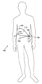
Referring to Figure 1, there is illustrated a tracking system 160 of the
present invention positioned on a patient. The tracking system 160
3o comprises an external recorder 10~; tour pods 101, 102, 103 and 104
respectively, containing both acoustic and EVI emitter/receivers; and a
capsule l 10 that is swallowable or otherwise positionable to move within an
i3
CA 02451807 2003-12-23
WO 03/001966 PCT/US02/19619
intestinal tract. The recorder 10~ is secured to the external abdomen of the
patient. The pods 101, 102, 103 and 104 are adhered to the skin of the
patient and have an acoustic transmittin~lcoupling material, e.g., a gel
layer,
interfacing between the skin of the patient and the pods f O l, 102. 103, 104.
As illustrated in Figure 2, the pod l01 comprises an outer plastic
casing 106 enclosing an acoustic transducer 107a and an RF coil 108a. The
casin'; 106 has an interfacing wall 106a for interfacing with the skin of a
patient. An adhesive layer 109 is formed on a portion of the interfacing wall
106x, for adhering the pod 101 to the patient's skin while a remaining
tp portion of the interfacing wall 106a is exposed to the patient's skin. The
acoustic transducer 10 7a is attached to the wall 106a within the casing 106
adjacent the exposed portion of the wall 106a in a manner that allows the
acoustic or ultrasonic energy to transmit through the interfacing wall 106a.
On the opposite side of the acoustic transducer 107a, an acoustic backing
15 material 107m is provided that absorbs the acoustic energy transmitted in
the
direction towards the backing material 107m. Typically a gel or other
acoustically transmittinglcoupling material is placed on the outside of the
exposed portion of the interfacing wall 106a. The output of the acoustic
transducer 107a is coupled to wires 100a that are coupled to the recorder 10~
2p through the wire conduit 100 extending out of the casino 106. The RF coil
108a is coupled through wires 100b also extending through wire conduit 100
to recorder 10~. Pods 102, 103, and 104 are similarly constructed.
As illustrated in Figures 3A and 3B, a first embodiment of a capsule
110 comprises a liquid impermeable and airtight capsule body 11 I. In
25 general, the capsule of the present invention is sized so that it is
capable of
being ingested for passage through the intestinal tract. For adult human use,
a preferred embodiment of the capsule is to be sized so that it has a length
ranging from about 1.5 to 2.5 cm and having a diameter of about 8 mm or
less. For children and lamer and smaller animals, the capsule can be
3p appropriately sized. The capsule body t l 1 contains and protects the
enclosed circuitry from body fluids while passing through the intestinal
tract.
At least a portion of the capsule body 1 I l is constructed of an ultrasound
t9
CA 02451807 2003-12-23
WO 03/001966 PCT/US02/19619
transmitting material that is compatible for use in the human body such as,
for example, a medical grade plastic, e.°., polyethylene. A radiopaque
marker 1 l 1 a is embedded in the plastic casing so that in the event it is
necessary to locate the device via an external imaging source, its location
may be identified. A dissolvable encasing (not shown) may surround the
capsule body 1 11. The encasing may be formed of a suitable dissolvable
material such as, for example, a soluble gelatin or enteric coating that is
dissolvable in the body fluids contained in the stomach or intestinal tract.
Such materials may be selectively dissolved based on the pH condition so
that the encasing dissolves after the capsule 110 has passed through the
highly acidic stomach and into the more neutral small intestine. The capsule
body 111 includes a generally hemispherical back end 131 and a generally
hemispherical front end 132. The back end 131 includes an inner end
surface 131a. The front end 132 includes an inner end surface 132a. The
~5 overall conformation of the ingestible capsule 110 is cylindrical in shape
forming a substantially smooth outer capsule surface.
The capsule 110 includes an RF coil 13~ for transmitting and
receiving RF signals, and an acoustic transducers 136a, 136b, and 136c
located within the capsule body 111. The acoustic transducers 136a and
2o 136b are located against the inner end surfaces 132a and 131a respectively
with an acoustic transmitting/coupling material filling any gap beriveen the
transducers 136a and 136b and the end surfaces 132a, 131a in a manner so
that the transducers can transmit acoustic, preferably ultrasonic waves
through the capsule body 111 to the surrounding tissue or material. Acoustic
25 transducer 136c is cylindrical in shape, extending around an inner
circumference of the capsule. An acoustic transmitting/coupling material
similarly fills any gap between the acoustic transducer 136c and the inner
wall of the capsule body 111. The acoustic transducers 136a-c are arranged
in combination to transmit acoustic si,nals relatively omni-directionally.
3o The transducer 136a comprises a piezoelectric crystal 137 located
between electrode plates 135 that when energized cause the crystal to
oscillate at an ultrasonic frequency (preferably between 100kHz and SVIHz).
?o
CA 02451807 2003-12-23
WO 03/001966 PCT/US02/19619
An acoustic backing material 139, such as, oxide particles in a flexible
polymer, e.g., an epoxy matrix tungsten powder, is placed on the back of the
transducer 136a to absorb any acoustic transmissions in a direction opposite
to the end surface 132a. The acoustic transducers 136b and 136c are
constmcted in a similar manner to transducer 136a and of similar materials.
Other configurations of an acoustic transducer or transducers may be used to
provide relatively omni directional acoustic signal transmission. The RF coil
13~ and the acoustic transducers 136a, 136b and 136c are electrically
coupled to the electronics 113 which is powered by battery 114.
l0 An elongate member 115 is affi~ced to the back end 131 of the
capsule body l 11. First and second bipolar electrodes 116, 117 are located
on the elongate member 115, the second bipolar electrode 117 being
electrically opposite of the first electrode 116. The elongate member 11~ is
preferably formed of an elastically behaving material such as a Ni-Ti alloy.
The capsule body 111 also includes a pH sensor 133 on the capsule
body 111. The pH sensor 133 is formed with dissimilar metals such as, e.g.,
silver chloride and antimony that sense differences in pH and convert the
sensed result into a calibrated electrical signal. The pH sensor is coupled to
the electronics 113 by electrical conductors.
Referring now to Figure 4, the electronic circuitry 113 of the capsule
110 is illustrated. The electronic circuitry 113 is a chip that includes a
number of optional connectors, and, as such, may be used in a number of
different diagnostic or therapeutic capsule configurations. The electronic
circuitry 113 of the capsule 110 comprises, a microprocessor or controller
122 for controlling the operations of the electronic circuitry, an internal
clock 121, and battery device 114 such as a pair of lithium iodine batteries,
for powering the various components of the circuit 113. As such, the
controller 122 and battery device 114 are coupled to each of the major
components of the circuit as would be known to one of ordinary skill in the
art.
The controller l22 is coupled to ROVl 123, which contains the
program instructions for the controller 12~ and any other permanently stored
2!
CA 02451807 2003-12-23
WO 03/001966 PCT/US02/19619
inforn~ation that allows the microprocessorlcontroller 122 to operate. The
controller l22 addresses memory in a location in RONt l23 through address
bus 123a and the ROM 123 provides the stored program instruction to the
controller 122 via data bus 123b.
The electrode plates 138 of the acoustic transducerl36a are powered
throush oscillator 137a controlled by the controller 122 to produce a desired
acoustic Gvave output. Similarly, electrode plates of acoustic transducers
136b and 136c are powered through oscillators 137b and 137c, respectively,
controlled by the controller 122. The controller 122 controls the RF coil 135
that acts either to deliver an RF tracking signal or as a telemetry device for
communicating data to the recorder LOS. The RF coil 135 delivers signals to
or receives signals from the RF coils 108a-d (Fig. 5) in the pods 101, 102,
103, and 104. For tracking purposes, controller 122 will respectively, at
fired time intervals, order the transmission of an RF signal and an acoustic
15 signal using the RF coil 135 and at least one of acoustic transducers 136a-
136c. The controller's commands will incorporate a preset time interval
between the RF signal transmission and acoustic signal initiation. Such time
interval (which could be zero) will be factored in at the recorder 105 to
determine acoustic wave transmission time. In the preferred embodiment,
the capsule's acoustic transducers 136a-136c transmit the acoustic signals
immediately, or a defined time after the RF reference signal. The acoustic
transducer 136a will emit a first signal a predetermined time after the RF
signal, the second and third acoustic transducers 136b and 136c will emit
second and third signals respectively at predetermined times after the RF
~5 signal and sufficiently spaced in time from the other signals so that the
acoustic signals may be differentiated. Alternatively, the second and third
acoustic signal may be referenced from second and third differentiated RF
signals.
When the RF coil 13S is receiving an external telemetry signal, the
3p buffered oscillator 119 is disabled. Telemetry signals received on RF coil
13S are detected in a detector circuit 119a and communicated to
CA 02451807 2003-12-23
WO 03/001966 PCT/US02/19619
microprocessor 122. The detector circuit l 19a is preferably selected based
on the modulation used for the telemetry signals.
One or more sensors, e.~., 127a (pressure), 127b (pH), 127c (optical).
127d (temperah.ire). and 116, 117(electrodes) may be coupled to controller
122 through A/D converters (with amplifiers) 126a, 126b, 126c, 126d, 126e
which convert a representative analog electrical signal into a digital signal.
Suitable sensors of these types are generally known in the art and may be
located within, on. or external to the capsule body 111. The electrodes I l6,
117 used to deliver the stimulation are also used to sense electrical activity
0 or impedance as described in ft~rther detail herein.
The controller 122 is coupled to RAM 120 via an address bus 120a
for addressins a location in RANI 120 and a bi-directional data bus 1206 for
delivering information to and from RA1~I 120. The R.AI~I 120 includes event
memory 124 that temporarily stores data recorded by sensors 127a-127d and
t.5 electrodes 116, 117. RAM 120 also includes a programmable memory 12~
which may be programmed, for example, via telemetry while the capsule 110
is within the intestinal tract, to provide treatment protocols. The data
stored
in the event memory 124 may be sent to external coils 108a-d (Fig. 5)
intermittently as data bursts via telemetry through the RF coil 135, as
20 opposed to continuously in order to save battery power. The data stored in
the programmable memory 125 may include specifications for the electrical
stimulation operating modes (e.g. waveform, type of stimulation: for pacing,
inducing contraction or other type) and various procedure parameters (e.g.,
~,vhen to deliver a drug or electrical stimulation). Such programming may be
25 done in response to sensed information or it may be done automatically by
an external controller or as desired by a treating physician, etc.
Controller 122 is coupled to a buffered oscillator 119 that provides
an RF signal to be emitted from the RF coil 13~. The RF signal is preferably
at about 1001:Hz to about ~~IHz so that the signal is efficiently transmitted
3o through tissue.-The controller 122 controls the oscillator 1 l9 and
provides
data for example, various sensed data such as pressure, pH, impedance,
electrical activity, etc.. to be modulated with the RF signal to be delivered
23
CA 02451807 2003-12-23
WO 03/001966 PCT/US02/19619
through RF coil 13~. The controller l22 may also be coupled through
stimulation driver 1 L8 and coupling capacitors 116a, 1 17a to bipolar
stimulating electrodes 116, 1 l 7, respectively. Electrical stimulation may be
provided in a manner similar to that described herein with reference to the
stimulating electrodes 16a-c, l7a-b, 56. 57, 66, 67, 86, and 87 of Figures 15-
22. The stimulation modes and parameters can be preprogrammed or set by
an external device that telemetrically communicates the parameters.
The battery 114 has its output supplied to a DC-to-DC converter l30
to provide a higher voltage, which is utilized for electrical stimulation
pulses. The DC-to-DC converter 130 is conventional and provides an output
voltage of 1~ to 20 volts. Further the circuit 113 may include one or more
drivers 128a, 128b, 128c, 123d that drive various devices, for example,
diagnostic or therapeutic electromechanical devices, such as controlling
valves, solenoids, etc, for, e.g., drug delivery, biopsy, content sampling, or
a
is marker release, etc. The controller 122 provides a signal to a driver 128a-
128d based on a preset program in ROM 123, on sensed parameters stored in
RAM 120, and/or on a telemetrically received signal from the recorder 105
or RF coils 108a-d in the pods, 101-104. The circuit may also include a
stepping driver 129 coupled to a stepper motor for example for rotating an
2o imaging device (e.g., diagnostic ultrasonic device) or actuating a biopsy
device, etc.
Referring now to Figure 5, a schematic of the electronic circuitry 140
of the recorder 10~ of the present invention is illustrated. The electronic
circuitry 140 of the recorder 105 comprises: a microprocessor or controller
25 142 for controlling the operations of the electronic circuitry, an internal
clock 141, and power source such as a battery 147 for powering the various
components of the circuit 140. The controller 142 and battery device 147 are
coupled to each of the major components of the circuit in a manner known to
one of ordinary skill in the art.
30 The electronic circuitry 140 is coupled to the pods 101, 102, 103 and
104, which respectively include RF coil sensors lOS a-d and acoustic
transducers 107 a-d that send and receive si4~nals to and from the capsule
2a
CA 02451807 2003-12-23
WO 03/001966 PCT/US02/19619
1 l0. The details of the coupling of the transducer 107a and 108a are
illustrated in Fig. 5. 'The transducers 107b-d and coils 108b-d are coupled in
a similar manner not shown. The output of the RF coil 108a is coupled
through a demodulator 15~ to the controller 142. The demodulator 15~
demodulates the information carried by the RF signal received by the RF coil
108a. Such information may include, for example, telemetrically delivered
sensed data. Also, the RF coil 108a may emit an. RF reference signal. The
controller 142 controls the output of the RF coil 108a, which communicates
with the capsule 1 l0. The controller 142 is coupled to an oscillator 1~6 that
provides a carrier signal, preferably having a characteristic frequency in the
range of 100kHz to SivIHz so that it may be efficiently transmitted through
tissue to the capsule. The controller 142 provides data to be modulated with
the RF signal, for example, commands to the capsule 110 to provide
treatment, treatment parameters, etc. The controller 142 controls the output
of acoustic transducer 107a through oscillator 1~7, which provides the
oscillating frequency to the transducer when the pod is pinging another pod,
i.e., when the pods are sending signals to calibrate the pods and identify
their
locations on the coordinate system. The controller 142 also receives the
representative acoustic signal from the transducer 107a through automatic
?o gain control device 158 which brings the voltage or current levels within a
predefined range, and though filter 159.
The controller 1=12 is further coupled to ROM 143, which contains
the program instructions for the controller 142 and any other permanently
stored information that allows the microprocessor/controller 142 to operate.
The controller 142 addresses memory in ROM 143 via address bus 143a and
the ROM 143 provides the stored program instruction to the controller I42
via data bus 143b.
The controller 142 is coupled to RAM 1:14 via address bus 144a and
bi-directional data bus 144b. The RAM l44 comprises event memory 14~
3o that temporarily stores data sent via telemetry frum the capsule 1 l0 to
the RF
coils 108 a-d in the pods l01-l04 until the data is downloaded onto a
computer using external data port l ~0. For tracking purposes, the RAM 144
CA 02451807 2003-12-23
WO 03/001966 PCT/US02/19619
is also used to store the data concerning lag times between the RF signal and
acoustic signals received by transducers l07 a-d, and RF coils 103 a-d in th a
pods 10l-104. The RAVI 144 also comprises a programmable memory 146,
which is used to specify operation modes (e.g. waveform, type of
stimulation: for pacing, inducing contraction or other type) and various
procedure parameters that may be transmitted to the capsule 110 through RF
coils 108a-d via telemetry. The recorder 10~ also includes a display 151 to
show recorded data, sensed parameters, treatment parameters, and staW s of
device (e.g., capsule position, battery charge status, etc.). The recorder 10~
io also includes a data input device l~? such as a keyboard, pad or input
screen
for inputting new parameters, programming the capsule, changing the
treatment scheme, viewing various data or turning the device on or off. The
input is coupled through a buffer 1 ~4 to the controller 142. The controller
142 is coupled to a speaker 1~3 for providing audible information such as an
I 5 alert.
In Figures 6 and 7, the pods 101,102,103, and 104 are set up in an
Cartesian (x,y,z) coordinate system. The origin of the coordinate system is
defined as the location of pod 101. The y-axis is defined as the line that
passes through pod 101 and pod 102. The x-y plane is defined as the plane
2o that intersects pods 101, 102 and 103. The z-axis is perpendicular to the x-
y
plane. Pod 104 is located off of the x-y plane. Thus, the coordinates of the
pods in this defined coordinate system are:
Pod 101: (0, 0, 0)
Pod 102: (0, y~, 0)
z5 Pod 103: (x;, y;, 0)
Pod 104: (xa, y,, z,~)
where the pod coordinates y2, x;, y;, x.~, y~, and z.~ are initially unknown.
Once the pods are placed as illustrated in Figure 1, the coordinates of
3o the pods are initially determined in the following manner. As illustrated
in
Figure 6, the distances d,2, d,;, di:~, d_3, d,:,, and d;., represent the
distances
between pods 101 and 102, 10l and 103, 101 and 104, 102 and 103, 102 and
10=1, and l03 and 104, respectively. The pods, which can both emit and
receive electromagnetic and acoustic (including ultrasound) signals, will
26
CA 02451807 2003-12-23
WO 03/001966 PCT/US02/19619
sense time-lads between the RF and acoustic signals sent between the pods
along the distances d',z, d,3, d,.,, d_3, dz.,, and d3~, i.e., the pods will
ping each
other. The pods communicate with a processor located in the recorder that
calculates the distance and determines the coordinates. The time-lays are
multiplied by the velocity of sound to calculate the distances (d,=, d,3,
d,,~,
d_3, dz~, and d3.~) between the pods.
Under Pythagoras' Theorem the following six
equations relate the coordinates of the pods and the distances
between them:
(xz-xi)z + (Yz-Yi)z + (zz-zi)z = dizz (1)
(x~-xi)z+ (Y~-YOz+ (z~-z~)z = daz (')
(Xa-xOz + (Y~-YOz + (za-zOz = diaz
(x3-XO)? + (Y3'Y2)~ + (Z3-Z2)~ = dz3~
(x~-xz)z + (Y.~-Y2)z + (z~-zz)' = dzaz
1J (X4-X3)- + (y~-y3)~ ~ (Z.1'z3)~ = d3J2
The pod coordinates x,, y,, z, ,xz, z~. and z3 are defined as having
the value of 0. Thus, plugging in the known pod coordinates, the equations
can be rewritten as:
?o Yzz = d~,z (1 ~)
X~z~Y~z = dog (2')
x~z+Y.~,+z.~z = diaz (3,)
X32-(Y3'YZ)~ = d23~
Xaz+(Ya-Yz)z+zaz = dzaz (5')
(X~-x3)z+(Y-~-Y3)z+z.~z = d3az (6')
With these six equations, and the determined distances, d~z, d,3, d,~,
dz3, dz:~, and d;,~, the six pod coordinates, yz, x3, y;, x:,, y:~, and za may
be
solved. Single solutions for all the coordinates may be obtained by setting
3o the following position restrictions: yz > 0; x3 > 0; and z.~ > 0. In other
words,
pod 101 should be placed on the right side of the user, pod 102 on the left
37
CA 02451807 2003-12-23
WO 03/001966 PCT/US02/19619
side, pod 103 on the lower abdomen, and pod l 04 on the upper abdomen as
illustrated in Figure 1.
The determination of the solutions for the six pod coordinates yz, x;,
y;, x.,, y.~, and z.~ are described below:
Equation ( I') gives:
y,-di, (1")
l0 Pluggin~~ ( 1 ") into (4') and subtracting (4') from (2') gives:
z
Y3 = (dn' ~ di3z - d,3 ) / (2 d~z) (2")
Plugging (2") back into (2') gives:
x; _ (di3z - y~z )o.s
(3 ")
where y; has been solved above.
Plugging (1') into (5') and then subtracting (~') from (3') gives:
Ya = (dizz T d~az- d~az) / (2 d~z) (4")
Subtracting (6') from (3') gives:
'-0 x.~ _ (dmz - d3az + x~z + y;z _ ? y3 Y:~ )/ (' X~)
where x;, y; and y~ have been solved above.
Plugging (4") and (5") into (3') gives:
Z4 - ( dl4z - x~z - yaz ) o.s
(6")
where x.~ and ya have been solved above.
The pod coordinates are determined whenever the pods are re-
positioned. The pod coordinates may also be re-established at regular
intervals to account for movement and thus relative change in pod position.
As illustrated in Figures 7 and 8A-G, using the coordinates of the
pods, the location of the capsule in space may be determined as follows. The
range-finding capability of the pods measure the distances between the
capsule 110 and each pod. As illustrated in Figures 8A-B, the capsule 110
?s
CA 02451807 2003-12-23
WO 03/001966 PCT/US02/19619
emits an RF si~~nal 205 and a sychronized ultrasonic signal 206 that is
emitted a predetermined time interval after the RF signal 205 is emitted. In
the preferred embodiment the ultrasound signal 206 is emitted immediately
following the RF signal 205. In this drawing, for illustrative purposes the
signal emitted from transducer 136a is illustrated. Second and third acoustic
signals emitted from the second and third transducers 136b and 136c would
be similar to the si~Tnal emitted from transducer 136a except that they
preferably emitted after the first signal 206 and at predetermined time
intervals from the RF signal 205. The signals from the additional acoustic
to transducers 136b and 136c may also alternatively have different waveforms
as that of the first signal 206. Figure 8C illustrates the timing of when the
RF
signal 205 is received at the pods. Figures 8 D-G illustrate the timing of
when the ultrasound signal 206 is respectively received at pods 101, 102,
103, and 104. Because the RF signal 205 travels at the speed of light, it is
received by the pods 101, 102, 103 and 104 at a relatively negligible time
delay in comparison to the ultrasonic signal which travels generally at about
1540 meters per second in human tissue. The distances c,, c,, c~, and c:~
represent the distances between the capsule and pods 101, 102, 103, and 104,
respectively. The pods 101, 102, 103 and 104 receive the ultrasound signal
?0 206 transmitted from the capsule 110 at varying times depending on the
distances c;, c_, c;, and ca respectively. Such time lags may be represented
as
illustrated, for example, in Figure 8 as t,, ta, t;, and 4 corresponding to
distances c,, cz, c;, and c,~, respectively. The time-lags will then be
multiplied by the velocity of sound to calculate the distances (c1, cZ, c;,
and
c.,) between the capsule 110 and each pod.
Using Pythagoras' Theorem the following equations relate the
coordinates of the capsule (xn, yn, zn) and pods, and the distance between
them:
(xn-xO + (yn-Yl)~ 1 (Zn-ZI)~ - cn
3U (xn_x?) ' (yn'y_)~ T ~Zn'Z2)~ - ~'~
(xn-x3), 1 (Yn'Y3)~ T (Zn'Z3)~ - 03
~xnW.~O ~' ~Yn-Y4)~ T (Zn-Z.i)~ - 0~- (10)
29
CA 02451807 2003-12-23
WO 03/001966 PCT/US02/19619
These four equations may be solved to obtain a single solution- for the three
coordinates of the capsule, x", yn, and zn.
According to one embodiment, a three-dimensional or four
dimensional map of the capsule's trip through the intestinal system can be
generated by measuring the capsule's coordinates at fixed time intervals.
Alternatively, linear travel distance measurements can be made by
using Pythagoras' Theorem. Incremental linear distances can be calculated
and then summed to obtain a total linear travel distance (L):
L- ~o U'~n+1-xn) ~"' (yn+1-yn) + (Zn+1-Zn) ]~!_ '
where m is equal to the number of incremental distances and where (xn, yn,
zn) and (xn+~,yn+~,zn+i) are consecutive capsule coordinate measurements
used to measure incremental linear distances traveled. In this manner a
linear map of the capsule's position along the intestinal tract may be
obtained. Such a map shows the position of the capsule along the tract
independent of actual 3D spatial orientation. Thus, errors based on intestinal
shifting, peristaltic motion, patient positioning, and change in pod location
are reduced without requiring additional sensed information. Retrograde
peristaltic motion can occur in the small intestine. An algorithm may be
used to cancel out any backtracking travel measurements when calculating
the linear distance traveled by the capsule. As described below using an
additional acoustic transducer, (e.g., located on the opposite end of the
capsule) and obtaining the same positional information may provide
25 information on capsule orientation and direction of capsule movement.
Preferably, the additional transducer will deliver a signal at time intervals
between the acoustic signals of the first transducer. The signals from the
additional transducer may have a different waveform to differentiate the
signal from signals corresponding to the first transducer. The orientation
30 information may provide additional information that is used to cancel out
retrograde capsule movement.
CA 02451807 2003-12-23
WO 03/001966 PCT/US02/19619
Referring to Figures 1 lA-D, an example of a linear map of an
intestinal tract and corresponding maps of sensed information are illustrated.
Figure 1 1 A illustrates an example of a linear map of a gastrointestinal
tract.
Figure 11 B illustrates an example of a map of pH sensed by a capsule in
relation to its linear position along the length of the tract of Figure 1 IA.
Figure 1 l C illustrates an example of a map of pressure sensed by a capsule
in relation to its linear position along the length of the tract of Figure I
IA.
Figure 1 ID illustrates an example of a map of electrical activity sensed by a
capsule in relation to its linear position along the length of the tract of
Figure
I 1A. These maps may be plotted from sensed information on a display
screen in the illustrated format or as othenvise may be desirable by a user.
The parameters shown in the maps in Figures 11 B-D may be
determined by a capsule having sensing capabilities. As the capsule passes
through the intestinal tract and its location along the length is determined,
other parameters relating to the condition of the intestinal tract may be
sensed periodically or continuously. The sensed conditions may be sent via
telemetry to one or more pod receivers. This may occur independently from
the time of the RF reference signal transmission and the acoustic signal
transmission so that the telemetry signal is independent of the coordinate
2o determining RF reference signal. The sensed information is mapped along
the length of the intestine by the tracking system as described above. A
linear map of sensed information is overlaid on the linear map of the
intestine so that unusual parameter values, or areas to be treated may be
determined. Upon a second pass of a capsule, the area or portion of the tract
to be treated may be located along the length of the linear map created from
the first capsule pass. The second capsule uses a similar method to
determine its position along the length of the tract and its linear travel
position is compared to the linear travel position of the first capsule. Thus,
when the capsule has traveled the appropriate position alone the tract, the
3o segment of the tract may then be treated. Treatment may be triggered by a
telemetric signal sent to the capsule when the recorder and external
controller have calculated the appropriate linear position.
31
CA 02451807 2003-12-23
WO 03/001966 PCT/US02/19619
Referring now to Figure 9, there is illustrated a second embodiment
of a treatment capsule of the present invention. Capsule 170 comprises a
capsule body 171 including an electronic circuit 113 and battery 174 coupled
to the electronic circuit 1 l3. An RF coil 175 and acoustic transducers 176a-
c operate in a similar manner as RF coil 135 and transducers 136a-c
described herein. The capsule further comprises a compressed gas source
165 and an inflatable balloon 167 externally fixed to the capsule body 171.
The gas source 165 is in fluid communication with a valve 166 that opens
into a chamber 168 in the balloon 167. The chamber 168 of the balloon 167
1o further is in fluid communication with a valve 169 that opens to a gas exit
port 172 that is in fluid communication with the intestinal tract. The valves
are coupled through drivers 128a, 128b in electronic circuit 113. The
operation of the valves 166, 169 is controlled by the controller 122 in the
electronic circuit. 113. In use, the capsule is delivered after a diagnostic
t 5 capsule using an optical sensor has been passed through the intestinal
tract to
obtain a map of optically sensed parameters along the length of the tract.
After a blockage site along the length has been determined, the capsule 170
is ingested. Using the RF coil 175 and acoustic transducers 176a-c of the
tracking system described above, the tracking system identifies when the
2o capsule 170 has reached the blocked site. The tracking system sends a
telemetric control signal to the RF coil 175 that instructs the controller 122
to inflate the balloon 167. The controller activates valve 166 through driver
128a which opens to allow compressed gas from the gas source 165 to fill
the chamber 168 of the balloon. The inflation of the balloon 167 expands
2s the intestinal wall at the site of the balloon 167 to open the blockage.
The
controller 122 then opens the valve 169 through driver 128b to allow the gas
to escape from the chamber 168 through the gas exit port 172 and into the
intestinal tract. The controller may release the gas upon an external
telemetrically delivered command that is initiated by, for example, a
30 physician who is observing the capsule and balloon under fluoroscopy, to
determine if and when a blockage has been opened. Alternatively, the
balloon may be preprogrammed to expand for a predetermined amount of
32
CA 02451807 2003-12-23
WO 03/001966 PCT/US02/19619
hme. The expandable member may be used for a variety of diagnostic or
treatment purposes, for example, pressure sensing, openine partial
blockages, measuring the openings of partially blocked or constricted areas,
providing hemostasis, delivering therapeutic substances that are coated on
the balloon 167, or affixing a capsule in an identified location to mark the
location in the intestine. An expandable support member such as a stmt may
be provided on the balloon for placement within a strict«re upon expansion
of the balloon. Alternatively, the capsule may be provided with a self
expanding support stnicture such as a self expanding stmt.
Figure 10 illustrates a third embodiment of a treatment capsule of the
present invention. Capsule 180 comprises a capsule body 181 including an
electronic circuit 113 and battery 184 coupled to the electronic circuit 113.
An RF coil 185 and acoustic transducers 186a-c operate in a similar manner
as RF coil 13~ and transducer 136a-c described herein. The capsule further
comprises a pump 187 filled with a dye such as, e.g., fluorescein or
methylene blue to provide a surgeon with identification of a site for sureery.
Such marker may include, for example a radiopaque marker that may be
located with an active x-ray system during a procedure, a radioactive
material that may be interrogated by a passive system, a fluorescing
compound that is used to identify the location, or a dye that stains through
the wall of the intestine. The compounds may assist a surgeon in a
laparoscopic or open procedure where such imaging systems are used during
the procedure or where visualization, e.g., of a dye or stain is possible. The
pump is coupled to a valve 189 by a conduit 188. The pump 187 and the
valve 189 are controlled by the controller 1?2 in the electronic circuitry 113
through drivers 128c and 128d. In use, the capsule 180 is delivered after a
diagnostic capsule having a diagnostic sensor has been passed through the
intestinal tract to obtain a map of sensed parameters along the length of the
tract. After a site along the length of the tract has been identified for
surgical
intervention, the capsule 130 is ingested. Using the RF coil 18~ and acoustic
transducers lS6a-c of the tracking system described above, the tracking
system identifies when the capsule 180 has reached the identified site. The
33
CA 02451807 2003-12-23
WO 03/001966 PCT/US02/19619
tracking system sends a telemetric control signal to the RF coil l8~ that
instructs the controller l22 to activate the pump 157. The controller
activates
the pump 187 through driver 128c. The controller also activates valve 189
through driver 1234 which opens to allow dye from the pump 187 to exit the
pump through conduit 183 and valve 189 and be sprayed onto the adjacent
intestinal wall. The dye thus marks a location for surgical intervention.
The capsule 180 may also be used to release a ~~as into the intestinal
tract at a given location where e.g. a blockage or other anatomical feature is
believed to exist. Usin~~ fluoroscopy, the anatomy may be observed.
Similarly, using a capsule such as capsule 180. a fluid such as a radiopaque
fluid may be released near a contriction or other area to be imaged where
pump 187 pumps the fluid into the intestinal tract through a conduit 188 and
valve 189.
Figure 12-14 illustrate a fourth embodiment of a treatment capsule of
t5 the present invention. Capsule 210 comprises a capsule body 211 including
an electrocautery ablation circuit 213, an electronic circuit 113, and a
battery
214 coupled to the electronic circuit 113. The capsule 210 also comprises an
elongate member 225 with a larger area return electrode 227 located thereon.
The elongate member 225 and electrodes 226, 227 are constructed in a
3o manner similar to elongate member 15 and electrodes 16a, 16b, and 16c
described with respect to Figures 1~-16 herein. . A small area ablation
electrode 226 is located on the capsule body 211, preferably in the form of a
ring. A thermocouple sensor 127d is located on the capsule body 211
immediately adjacent to the ablation electrode 226 so that the sensor can
25 sense the temperature of tissue that is being treated by the ablation
elelctrode
226 and provide a feedback loop to an external controller 142 that regulates
the power delivered to the ablation electrode 226. An RF coil 215 and
acoustic transducers 216a-c operate in a similar manner as RF coil 13~ and
transducers 136 a-c described herein. In this embodiment, the RF coil 2l~
30 operates at a frequency of about l LIHz.
As illustrated in Figure 13, the ablation electronics include, an
ablation coil 221, electrodes 226, 227, and an ablation circuit 213 including
a
34
CA 02451807 2003-12-23
WO 03/001966 PCT/US02/19619
capacitor 222. The ablation coil 221 that is tuned to a Frequency of about
250kHz, thus the coils 21~ and 221 receive different frequencies, enabling
them to distinguish between a telemetry signal and an ablation power signal.
An external variable power generator 230 (Figure 14) supplies an RF signal
at 2~OkHz through power transmitter coil 231. The ablation signal received
by the ablation coil 22l and parallel capacitor 222 (which together form a
rimed circuit to separate the ablation signal from the telemetry signal) is
then
delivered to electrodes 226, 227.. The ablation electrode 226 has a
considerably smaller area than the return electrode 227 so that the current
density is greater at the ablation electrode 226 where the ablation current is
to be focused on the adjacent tissue. The thermocouple sensor 127d
provides an electrical signal representative of the temperature of the
adjacent
tissue, through the A/D converter 126d of the capsule circuit 113. The signal
is converted to a digital signal that is provided to the controller 122 of the
t5 circuit 113. The signal is telemetrically delivered to the controller 142
of the
recorder 105 in a manner as described herein.
As illustrated in Figure 14, the power is controlled by the controller
142 of the recorder 10~ which is coupled to the power generator 230 by way
of connector 233. The controller 142 in the recorder electronics 140 will
?0 regulate the power output to the ablation electronics based on feedback
information as sensed by the thermocouple 127d on the capsule body 211
and delivered via telemetry from the capsule RF coil 215. The regulation of
the power is significant in this embodiment as the RF ablation signal
strength may vary with distance from the capsule, the type of the tissue being
25 treated, the impedance of the tissue being treated. Thus, the temperature
feedback loop is intended to prevent over or under heating of the tissue. In
addition, the treatment is initiated by a user by activating a switch 234
coupled to the power generator 230.
In use, the tracking system is used in a manner as described above. A
30 location to be treated along the length of the intestinal tract is first
identified
by a first capsule passing through the tract. Preferably the capsule will have
an optical, chemical or other means for deternuning a location where
CA 02451807 2003-12-23
WO 03/001966 PCT/US02/19619
bleeding is occuring. This location is identified in a subsequent pass oFthe
ablation capsule 210'and the user horns the ablation power on when the
appropriate location is identified to ablate or cauterize the tissue that is
bleeding. In a variation of the embodiment, a site where bleeding is present
may be treated using a subsequently passed capsule having a balloon
tamponade, i.e. an inflatable member that uses compression and/or a
thrombogenic substance coated on the intlatable member to help cause
hemostasis. A capsule embodiment having an inflatable member is
described herein with reference to Figures Z 1 and 2?.
Figures 1~-16 illustrate a fifth embodiment of the capsule of the
present invention. The capsule 10 comprises a treatment and sensing device
that may be used with the tracking system. The capsule 10 is used to sense
electrical parameters of the intestinal wall and/or to treat the intestinal
tract
by electrically stimulating the intestinal wall. The capsule 10 comprises a
t5 liquid impermeable and airtight capsule body 11. The capsule body 11
contains electronic circuitry 113, battery 11=1, RF coil 135 and acoustic
transducers 136a-c as described above with reference to Figs. 3A and 3B.
The capsule body 11 protects the enclosed circuitry from body fluids while
passing through the intestinal tract. The capsule body 11 is formed of a
2o material that is compatible for use in the human body, for example, a
medical grade plastic or polymer.
An elongate member 15 is affired to an end of the capsule body 11.
Electrodes 16a, 16b and 16c are located on the elongate member 15. Two
second, larger area electrodes 17a and 17b extend around the width of the
25 capsule body 11, Electrodes 16a-c may be selected in a number of
combinations to form electrode pairs to deliver stimulation to the intestinal
wall (or alternatively to sense electrical activity of the intestinal wall).
Additionally, one or more of electrodes 17a and/or 17b may be utilized to
work with one or more of electrodes 16a-16c where current density will be
3o concentrated at the smaller electrodes) 16a, 16b, and/or 16c. The capsule
electronics may include logic to select which electrodes should deliver
stimulation pulses for optimal stimulation. The electronics may similarly
36
CA 02451807 2003-12-23
WO 03/001966 PCT/US02/19619
control which electrodes may be used to sense electrical activity of the
intestinal wall. Alternatively, an external processing unit may determine
optimal electrode selection that is communicated to the capsule by a
telemetry command signal.
In one preferred embodiment, the capsule I I may be used for
stimulation and subsequent measurement of electrical parameters. This
function may be used for diagnostic purposes, for example, to determine if
the intestinal wall is properly conducting electrical pulses or if the wall at
a
particular location is an electrically hypo-active or "dead"' area. In a
preferred embodiment, the capsule electrodes are electrically configured so
that a plurality of adjacent electrode pairs can be used where a first pair
stimulates the intestinal wall at a first location and the second pair then
detects signals at a second location that are propagated from the original
stimulation signal. Accordingly, in a variation of one embodiment, to
determine if the intestinal wall is electrically abnormal, e.g., is
electrically
hypo-active, electrodes 17a and 17b are used to deliver a stimulation signal
and an electrode pair formed from at least two of electrodes 16a-c are used to
sense resulting signals propagated in an orad direction. In a variation of
another embodiment, signal propagation in the aborad direction, i.e., from
the back of the capsule to the front assuming the front of the capsule is
oriented in a direction away from the mouth is determined using an electrode
pair formed from at least two of electrodes 16a-c are used to deliver a
stimulation signal and electrodes 17a and l7b sense resulting propagated
signals.
25 As illustrated in Figure 15, a dissolvable encasing 12 surrounds the
elongate member 15, the electrodes 16a-c, and at least a portion of the
capsule body 11. When encapsulated by the encasing 12, the elongate
member 1 ~ is in a coiled or compressed position.
The encasing 12 is formed of a suitable dissolvable material such as,
30 for example, a soluble gelatin or enteric coating that is dissolvable in
the
body fluids contained in the intestinal tract. Such materials may be
selectively dissolved based on the pH condition so that the encasing 12
37
CA 02451807 2003-12-23
WO 03/001966 PCT/US02/19619
dissolves after the capsule 10 has passed through the highly acidic stomach
and into the more neutral small intestine.
The elongate member L S is preferably formed of a material that has
elastic properties such as a Ni-Ti alloy, which permits it to be compressed
into the initial configuration and to release into its elongate state when the
encasing 12 has dissolved. As shown in Fib. 16, the elongate member l~
extends into its elongate form when the encasing 12 has dissolved.
The capsule body 11 is provided with a front portion 11 a and a back
portion 1 1b of reduced diameter. The encasing 1? is bonded to the back
portion 1 1b by suitable means such as an adhesive. The diameter of the back
portion 1 1b is reduced by a sufficient amount so that the thickness of the
encasing 12 forms a substantially smooth outer capsule surface in
conjunction with the outer surface of the front portion 1 la of the capsule
body 11. The overall conformation of the ingestible capsule 1 I is cylindrical
~ 5 in shape having a generally hemispherical end surface 23 on the front
portion
1 la and a generally hemispherical end surface 24 on the back portion 1 1b.
Dissolvable encasing 12 also has a generally hemispherical end surface 12a.
It is desirable that the elongate flexible member 1~ have an extremity
which has a curved configuration so as to ensure that the stimulation
?p electrodes 16a-c are maintained in close proximity to the wall of the
intestinal tract as the capsule 10 moves through the intestinal tract as
hereinafter described. The electrode 17 is formed of a conducting layer of a
suitable metal such as gold deposited on the surface of the capsule body 1 I.
Alternatively, the additional electrodes 16b and 16c may be carried by
?5 additional elongate members constructed and secured to the capsule body 11
in a similar manner as elongate member 1 ~.
The electronic circuitry 113 shown in Figure 4 is capable of
producing various types of programmable waveforms. Figures 17A and 17B
illustrate examples of stimulation waveforms that may be used in stimulating
3o the smooth muscle layer of the intestinal tract. Figure 17A illustrates a
waveform design for stimulating the intestinal tract. In a preferred
embodiment, the waveform 300 has a pulse amplitude of between 1 and 30
38
CA 02451807 2003-12-23
WO 03/001966 PCT/US02/19619
mA, a pulse width of between 0.5 and 300 ms, and a frequency of about
between 8 to 12 cycles per minute (this corresponds to a repetition period of
between 5 to 7.5 seconds). Figure 17B illustrates an alternative wavefonn
design for stimulating the intestinal tract. The waveform 400 utilizes bursts
of pulses rather than a single pulse. The burst repetition rate is selected,
preferably, to be between about 8 to 12 cycles per minute (this corresponds
to a burst repetition period of between 5 to 7.5 seconds). The duration of a
pulse in this example is between about 300~s and 20 ms, and has an
amplitude of about 1-30 mA. The frequency of the burst pulses during a
t0 burst period are about 50 to 100 Hz corresponding to a pulse repetition
period of l0 to 20 ms. The burst duration can vary from about 0.6 ms to 1
second. As is well known to those skilled in the art, there are many different
types of electrical stimulation programs and strategies which can be utilized
for providing electrical stimulation parameters through the circuitry 113, the
principal focus being providing electrically stimulating parameters for the
intestinal tract, preferably the small intestine.
Figure 18 illustrates a sixth embodiment of a capsule of the present
invention. Stimulation capsule 50 is generally constructed in a similar
manner as capsule 110. Capsule 50 comprises first bipolar electrode 56 and
a second, electrically opposite bipolar electrode 57 on a capsule body 51 in
longitudinally spaced apart positions. The electrodes 56, 57 are connected
by conductors to the electronics 113 within the capsule body 51. According
to this embodiment, various electrical stimulation parameters, including
those described herein, may be used.
A seventh embodiment of the capsule is shown in Figures 19 and 20.
Capsule 60 comprises a stimulation electrode deployment mechanism
consisting of a loop 76 formed of an elastic material wrapped about the
capsule body 61. Bipolar stimulating electrodes 66 and 67 are carried by the
loop 76 and are connected to the electronic circuitry 1 l3 in the capsule body
61 by conductors (not shown) extending through the hollow tubular member
forming the loop 76. As shown in Figure l9, a dissolvable encasing 62 is
provided over the capsule body 61. This encasing 62 can be formed of the
39
CA 02451807 2003-12-23
WO 03/001966 PCT/US02/19619
same material as the encasing 12 in the embodiment shown in Figure I ~.
When encasing 62 is dissolved, the loop 76 will expand to the ovoid looped
configuration shown in Figure 20, bringin~~ the stimulation electrodes 66 and
67 into contact with the wall of the intestinal tract as the capsule 60
travels
through the intestinal tract. The loop 76 allows the electrodes 66, 67 to be
positioned behind (orad to) the capsule 60 regardless of its orientation in
the
intestinal tract. As the capsule 60 moves through the intestinal tract the
loop
76 will be in contact with the wall of the tract. The friction forces of the
loop 76 drag;ing along the wall will cause the loop 76 to shift such that the
electrodes 66, 67 are generally behind (orad to) the capsule. In this regard,
a
contraction stimulated by the electrodes 66, 67 will tend to result in forward
(aborad) movement of the capsule as the stimulated contraction propagates
along the intestinal tract.
Figures 21 and 22 illustrate an eighth embodiment of a capsule of the
~ 5 present invention. Capsule 80 includes an expandable member. In Figures
21 and 22, an inflatable member with pressure sensing capabilities is
illustrated. Electronic circuitry 113 is located in the capsule body 81. A
pressure transducer 127x, also located in the capsule body 81 is coupled to
circuitry 113. The pressure transducer 127a comprises a commercially
2o available silicone or other suitable plastic bridge pressure transducer
that
measures hydrostatic pressure to determine changes in pressures as described
below.
An elongate member 85 is affixed to an end of the capsule body 81.
Bipolar stimulation electrodes 86, 87 are located in a spaced apart
25 relationship, rearvvardly on the elongate member 85. Conductors 95 extend
through the flexible elongate member 85 connecting the electrodes 86,' 87 to
the electronics 1 l3. Opposing ends 92a, 92b of an inflatable balloon 92 are
mounted forwardly of the electrodes 86, 87 on the flexible elongate tubular
member 8~ by a suitable adhesive (not shown). A balloon inflation/deflation
30 lumen 9=1 is provided in the flexible elongate member 8~ and extends from
the capsule body 81 to an inflation port 93 that opens into the interior of
the
balloon 9? as show m in Figure 22. The balloon intlation/detlation lumen 94
a0
CA 02451807 2003-12-23
WO 03/001966 PCT/US02/19619
is coupled to the pressure transducer 127a so that
compression pressures
sensed by the balloon 92 will be supplied to the pressure
transducer 127a as
the pressure of the gas in the balloon 92 and the lumen
94 chan;es.
The capsule 80 includes a dissolvable encasing (not
shown) of the
s same type as the encasing 12 shown in Figure l~. Similar
to the encasing
shown in Fig. 1 ~. st:ch an encasing would enclose
the t7exible elongate
member 8~ including the inflatable balloon 92 and electrodes
86, 87 and
would dissolve, e.~~. in the small intestine releasing
the elongate member 8~
as illustrated in Figs. 21 and 22.
to A balloon inflator is provided within the capsule 80
comprising a
small canister 97 of compressed C0~ or other suitable
gas. The canister 97 is
coupled to the lumen 94 through a valve connection
98. The operation of the
valve 98 is controlled by the electronics 113 through
a driver 128a, b, c
or d.
,
When the flexible elongate member 85 is deployed upon
dissolving of the
~5 encasing, the electronics 113 cause the valve 98 to
open and inflate the
balloon 92.
Alternatively, the balloon 92 can be pre-inflated with
a gas or fluid
before enclosure within the encasing. In this case,
the inflation canister 97
and valve 98 may be eliminated. The balloon 92 is formed
of a gas
20 impermeable material so that it will remain inflated
over substantial periods
of time. The balloon may be formed, for example, of
polyurethane, PET,
nylon or polyethylene.
In a preferred operation and use, the capsules shown
in the various
embodiments in Figures l2 and 18-22, are used in conjunction
with the
25 circuitry shown in Figure 4 or Figure 13 in small intestine
electrical
stimulation. A small intestine suited for treatment
using the capsule may be
diseased and incapable of adequate contractile activity.
For example the
nerves of the small intestine may be compromised due
to gastric or diabetic
neuropathy. Because of such a disorder, the patient
may have a motility
30 disorder that would be advantageously treated using
small intestine electrical
stimulation.
=n
CA 02451807 2003-12-23
WO 03/001966 PCT/US02/19619
The stimulator capsule may also be used to measure other electrical
characteristics such as EMG or impedance as described herein with respect
to the electronic circuitry 113 show in Figure 4. A patient wishing to treat a
motility disorder ingests a capsule of the present invention near the
g beginning, midway, or following the in;estion of food. A capsule when
ingested will travel throu~~h the esophagus into the stomach. Where a
dissolvable encasing is utilized for encapsulating the elongate member and
electrode(s), the encasing is readily dissolved by the fluids within the
stomach or duodenum, permitting the t7exible elongate member carrying the
vg stimulation electrode to be deployed.
The capsule is preferably used with the tracking system described
herein where treatment is triggered by an external (telemetry) signal from the
tracking device. A first capsule may be delivered and an electrical parameter
of the intestine may be mapped with respect to the length of the intestine. A
t5 second capsule may be delivered and used to provide electrical stimulation
at
an identified location along the length of the tract. An external signal to
the
capsule signals when to begin and end stimulation.
The electrical stimulation capsule may also be used independent of
the tracking system. In a variation of the embodiment, the capsule can be
?o programmed to begin emitting electrical stimuli to one or more stimulation
electrodes 16a-c, and/or 17, within a predetermined time after ingestion, for
example, within one to one and one-half hours after ingestion into the
stomach, at which time it is most probable that the capsule would have
passed into the duodenum along with food material passing from the
25 stomach. As an alternative, a single capsule may stimulate and measure the
electrical parameters. The capsule may sense electrical parameters and when
a clinically undesirable electrical parameter is detected, the capsule may
provide an appropriate electrical stimulation in response.
Such a system would have the advantage of not requiring external
30 gear such as the recorder and pods. Also, the capsule may be constructed to
sense when it is in the duodenum, for example with a pH sensor or a
pressure sensor. Also, the electronics 113 can be triggered to commence at
CA 02451807 2003-12-23
WO 03/001966 PCT/US02/19619
the time the encasing is dissolved and the stimulation
electrode is erposed to
body tluids. Alternatively, electrical stimuli can
be triggered by the
electronics l 13 to commence within a predetermined
time after the encasino
dissolves. In such case, the capsule is enclosed in
a gel material that
dissolves after it leaves the stomach when it reaches
the small intestine.
'Vhen triggered, electronic circuitry 113 initiates
electrical stimuli to the
small intestine of the patient, at periodic intervals,
such as, for example
using one or more waveforms like those shown in Fi~ures
17A and 17B.
Alternative electronic circuitry 313 illustrated in
Figure 23 may be
used with any of the stimulation capsules illustrated
herein. According to an
alternative embodiment, the electronic circuitry 313
is used in a simplified
stimulation system. According to a preferred embodiment
of the system,
prior to each stimulation pulse or burst of pulses
the capsule receives basic
instructions. The instructions may be a trigger signal
to trigger a stimulation
pulse or burst of pulses with predetermined stimulation
parameters, such as
amplitude and pulse width, to be emitted by the capsule.
The instructions
may also include information regarding the stimulation
parameters for the
pulses to be emitted. The instructions to trigger and/or
specify a stimulation
pulse or burst of pulses to be delivered to the intestinal
wall are
telemetrically delivered to the electronic circuitry
313.
The electronic circuit 313 is simplified and includes
a microprocessor
312, ROM 31~, RAM 316, a clock 311, a telemetry coil
33~, a battery 314 a
dc-do converter for stimulation 330, a telemetry detection
circuit 317, and a
pacing driver 318. The microprocessor 312 is coupled
to the ROM 315,
which contains program instructions for the microprocessor
312 and any
other permanently stored information that allows the
microprocessor 312 to
operate. ROM 315 may also contain default and standard
stimulation
parameters. The microprocessor 312 addresses memory
in a location in the
ROM 3 l5 through address bus 31 ~a and the RONI 3 I5
provides the stored
3o program instmctions to the microprocessor 312 via data
bus 31 fib. The
microprocessor is coupled to the RAM 316 via an address
bus 316a for
addressing a location in the RAM 316 and a bi-directional
data bus 316b for
43
CA 02451807 2003-12-23
WO 03/001966 PCT/US02/19619
delivering information to and from the RAM 316. The R.AM 3l6 may be
used by the microprocessor 3 l2 to store custom stimulation parameters sent
via telemetry prior to a series of stimulation pulses or bursts of pulses, or,
just before each stimulation pulse or burst of pulses. RAM 3l6 may also
temporarily store an identification code to specify the already stored
default,
standard or custom stimulation parameters to be used for stimulating the
intestinal wall.
The trig;er signals for each stimulating pulse or burst of pulses and
the stimulation parameter instructions are supplied through the telemetry coil
335 to the microprocessor 312 and are then delivered through the pacing
driver 318 in real time to the intestinal wall (through electrodes as
described
herein). Thus, the capsule itself does not direct the stimulation or the
intestinal wall but receives directions from an external source and delivers
stimulation accordingly and in real time to the intestinal wall.
t 5 The embodiment of Figure 23 could be further simplified by
replacing the microprocessor 312, ROM 315, RAM 316, and clock 311 with
logic gates or a state machine. In such variation, some or all of the
stimulation parameters may be preset and stored in the hard~,vare in the
capsule. For example, stimulation amplitudes could be stored as 5 different
2o states in a simple state machine. The telemetry instruction signal could
then
consist of a simple pulse train that would represent the trigger signal as
well
as encode one of the five stimulation amplitudes while using an otherwise
fixed stimulation pattern.
The electrical pulses provided by the electronics 113 through the
z5 electrode' pairs 16a-c, 17 (as selected)(Fig 15, 16); 56, 57 (Fig. 18); 66,
67
(Fig 19, 20); 86, 87 (Fig. 21); and 116, 117 (Fig 3A) may be used to create
peristaltic contractions in the wall to cause movement of food material along
with the capsule in the intestine. In an alternative embodiment where it is
desired to retard motility in the small intestine, inhibition of peristaltic
3o contractions by electrical stimulation may be effected by delivering
electrical
pulses designed to inhibit or interfere with the inherent electrical
potentials,
resulting in failure of nornial peristaltic contractile activity.
CA 02451807 2003-12-23
WO 03/001966 PCT/US02/19619
In certain situations with respect to motility disorders, it may be
desirable to supply synchronized stimulating pulses to the wall of the small
intestine by the use of multiple pairs of stimulating electrodes such as, for
example, a plurality of pairs similar to electrodes 16a-c carried on the
flexible elongate tubular member secured to the capsule as shown in Figure
12 and synchronizing the pulses in forivard (aborad) or reverse (orad)
directions in order to cause forward or reverse stimulation of the intestinal
tract.
As the capsule passes along the intestinal tract, it continues to supply
successive stimuli through the intestine. The rapidity of movement of food
material through the small intestine can be controlled by the stimulating
parameters such as frequency or amplitude of the signals utilized for
supplying electrical stimuli or pulses to the intestinal tract. The capsule
may
provide certain stimulation patterns in the small intestine until it reaches
the
colon. (This may be determined by sensed electrical or other parameters, or
by a predetermined time interval). At this time the electrical stimuli can be
terminated or alternatively they can continue to be generated at the same or
different parameters as the capsule passes through the colon until it exits
from the body through the rectum in a bowel movement.
'Vhere it is necessary for the patient to ingest a capsule each time
food is ingested by the patient, the patient can have additional capsules on
hand and ingest a capsule with each meal.
The electrode configuration preferably comprises two separate
electrical elements forming electrically opposite bipolar electrodes.
05 However, a monopolar or unipolar construction with a remote return is also
contemplated by the invention. Spacing of the bipolar electrode elements
from one another will preferably be about 5 mm. Electrodes formed on an
elongate member will preferably be constructed from a metal wire or strip
wound in a helical manner around the elongate tail portion. The electrode
;p metal will preferably be cotTOSion resistant and biocompatible such as
Gold,
Platinum, Titanium, etc. A helical windings pattern is preferred to provide an
electrode that is more tlexible than a solid cylinder, and thereby allow the
d5
CA 02451807 2003-12-23
WO 03/001966 PCT/US02/19619
elongate tail to be more easily wound or compressed
for cuntainment in the
dissolvable portion of the capsule. An alternative
construction is
contemplated where the electrode is embedded in an
insulatin~~ polymer with
an insulated lead extending within or along the elongate
member into the
capsule body.
By varying the spacin~~ between the stimulation electrodes
or the size
of the electrodes, it is possible to change the current
density passing through
the wall of the intestine during stimulation. .~. device
may be provided where
electrodes may be selected to maximize these parameters.
For example a
plurality of electrode pairs may be provided from which
the optimal pair of
electrodes may be selected. Also individual electrodes
may be configured to
form a pair of bipolar electrodes upon selection.
The electrical pulses or pulse train supplied to the
stimulation
electrodes can be at suitable stimulation intervals
as for example, in the case
of pacing type electrical stimulation, every few seconds
up to ten seconds in
the small intestine or several hours in the colon.
In connection with the electrical stimulation functions
described
herein, it is often desirable to measure the pressures
which are created by
peristalsis of the intestinal contractions. Referring
to Figures 21 and 22, this
?p can be readily measured by sensing the compressive
forces exerted on the
balloon 93 with transducer 91. By sensing such pressures
and supplying the
information by telemetry to the external recorder 10~,
it is possible to
ascertain the efficacy of the stimulation being applied
to the particular
portion of the intestinal tract and if necessary to
adjust the electrical
2~ stimulation parameters to create the desired contractile
forces being sensed
by the balloon and the pressure transducer. For example,
if the sensed
pressure indicates suboptimal contractile response,
the stimulation
parameters may be adjusted, e.g., telemetrically. If
the existence of
contractions is detected, the stimulation electrodes
may be turned off. This
3p may also serve to conserve battery power.
One method of use of a capsule of the present invention
is in small
intestine electrical stimulation. Electronic circuitry
is disposed within the
X16
CA 02451807 2003-12-23
WO 03/001966 PCT/US02/19619
capsule and creates electrical stimuli for causin;; peristaltic motion of the
small intestine for causing pacing of peristaltic motion in the small
intestine.
Other effects on the electrical, chemical, and,for neural systems of the
intestinal tract may be achieved with electrical stimulation. One example
includes an electrical stimulus that is usLd to interfere with the natural
pacesetter potential and thus prevent organized intestinal tract contractile
activity from occurrin;.
The present invention provides an improved method and device for
tracking an autonomous capsule as well as a method and device for tracking
and diagnosinj the gastrointestinal tract, preferably using a tracking device.
Various modifications and combinations are contemplated by this invention
and may be made without departing from the scope of the invention.
For example, in another embodiment of the tracking system, the
direction of the ultrasound signal used for locating the capsule is reversed.
In this embodiment, the capsule receives the ultrasound signals generated by
the pods and retransmits the signals on the RF carrier back to the pods or
external monitor. In this way, the capsule position may be located by
measuring the time delay from transmission of the ultrasound signals) by
the pods) to their reception by the capsule. Rather than activating all pods
?o simultaneously, each pod may be sequentially activated to transmit
ultrasound. Accordingly, the pod to capsule path is identified by the time of
transmission from a particular pod. When a single pod is activated in this
way for transmission, all the remaining pods may also be switched to receive
the ultrasound signal from the transmitting pod. This allows the pod-to-pod
?5 delay times to be measured, so that the relative position of the pods can
be
determined on an ongoing basis.
If simultaneous transmission from all pods is desired, the ultrasound
signals from each pod may be separated by using a variety of methods. For
example, each pod may generate a unique ultrasound frequency allowing the
;p signals to be separated by filtering.
In one variation, for e~cample, a continuous wave signal with
amplitude modulation may be used rather than a narrower pulse. In such
47
CA 02451807 2003-12-23
WO 03/001966 PCT/US02/19619
variation, time delays may be measured by measurin' the phase of the
received signals relative to the transmitted signal.
Alternative reference signals may be used to establish when the
acoustic signal is transmitted. For example, an infra-red link or a
distributed
resistive link may be used. Infra-red links may be constructed using light
emitting diodes with an infra-red wavelength chosen to minimized the
effects of tissuelli~ht attenuation. The light transmitters and sensors may be
on the capsule and/or at the external location for one or two way signal
transmission. The light may be modulated with a high frequency carrier in a
to similar manner to an RF link. The modulated light signal can then be
detected after it has passed through the tissue using a light sensor or
sensors.
A distributed resistive link may be used to directly couple an electrical
carrier signal through the body to an external sensor or sensors, or
alternatively or additionally from an external transmitter to electrode
sensors
t 5 coupled to the capsule. A small high frequency carrier, typically 100kHz
or
above, is preferably chosen for the carrier frequency to prevent any muscle
stimulation by the carrier. The sensor en tl:e capsule or at the e:cte~a:
location would then detect the high frequency carrier signal, which would be
attenuated by the distributed resistive divider formed by the conductive body
2o tissue. To transmit or receive the signal to or from an external location,
the
e:cte:,lal source or sensor would be coupled into the body via nvo skin
electrodes, spaced at some distance apart. Electrodes on the capsule would
be used to receive (or transmit) such carrier signal. The high frequency
carrier would preferably be modulated in the same way as an RF link, using
z5 amplitude, frequency or other modulation schemes as are well known in the
art. Preferably, the various signals e.g., going to or from the capsule, would
be placed on different carrier frequencies to allow for easy separation via
filtering, of the outgoing and incoming signals.
Further, as an alternative to using an externally detectable signal such
;0 as an RF signal, as a reference signal to establish the time at which the
acoustic pulse is emitted, the ultrasound transmitters and receivers may be
confi_rured to establish such transmission times and thus the location of the
as
CA 02451807 2003-12-23
WO 03/001966 PCT/US02/19619
capsule. Based on the differential time between two ultrasound receivers
receiving an ultrasound pulse from a capsule, the possible location of the
capsule may be defined by a paraboloid plane between the two receivers.
Usine more than two receivers, additional such paraboloid planes
representing possible locations may be determined. The intersection of the
planes provides information from which the actual location of the capsule
may be derived. By filtering out impossible locations (e.g.. by knowing
points that would lie outside a patient's body, e.~., based on pod placement
on a patient, or by adding additional pods for additional location
1U information), the actual location of the capsule may be determined.
Accordin_ to one variation, the differential distance is determined by
multiplying the differential time bet~.veen the reception of the ultrasound
signal at one pod and the reception at the other pod times the speed of sound
in tissue. The possible location of the capsule based on the derived
differential distance is represented by a paraboloid plane between the two
pods. 'Vhen a third acoustic reference receiver is added, the detected
differential time bet<veen receiver one and three and the differential time
between receivers two and three provide additional paraboloid planes of
possible capsule locations. Two paraboloid planes intersect in a paraboloid
3o or ellipsoid line; intersection with a third paraboloid plane defines one
or
more points of possible capsule locations. Strategic positioning of the
acoustic reference receivers, use of additional receivers and/or exclusion of
invalid mathematical solutions (e.g. outside of the patient's body) may
enable a single solution to be obtained for capsule location.
35 The foregoing embodiments and variations of the invention are
illustrative and not contemplated to be limiting, having been presented by
way of example. Numerous other variations and embodiments, as would be
apparent to one of ordinary skill in the art, are contemplated as falling
within
the scope of the invention as defined by the claims and equivalents thereof.
~o
=t9