Note: Descriptions are shown in the official language in which they were submitted.
CA 02474954 2004-07-30
WO 03/063801 PCT/US03/03321
Treatment of Tumor Cells For Use
In Immunotherapy of Cancer
to
This application claims priority from U.S. Provisional Application Serial No.
60/354,094, filed February 1, 2002, which is hereby incorporated by reference
in its entirety.
FIELD OF THE INVENTION
The invention relates to compositions comprising a tumor cell treated for
preservation, sterility, or both. The tumor cell compositions are particularly
suitable for
immunotherapeutic vaccine. Haptenized tumor cell preparations are especially
advantageous.
BACKGROUND OF THE INVENTION
In blood transfusion, bone marrow transplantation, immunotherapeutic vaccine
preparation, or other cell preparations ex vivo, one of the principal problems
encountered is that
of the preservation of cells. It is critical to be able to preserve cells,
under good conditions of
viability, for time periods compatible with clinical production and storage,
and to make it
possible to analyze cell preparations. The most commonly used method of long-
term
preservation of cells is to freeze and subsequently thaw them. However, during
the freezing of
cells, lysis of cells and loss of cell integrity may occur. This problem can
be even more
complex when the cells have been modified or altered prior to preservation,
and when the cells
are obtained by proteolytic digestion of a tissue or tumor specimen.
Preservation of cells under less extreme conditions, for example on ice (about
0°C), refrigerated (about 4°C), or at room temperature, prior to
use, is also difficult.
CA 02474954 2004-07-30
WO 03/063801 PCT/US03/03321
Immunotherapy
The preservation of cells, especially their antigenicity, is important is in
immunotherapy of cancer using tumor cells. The aim of the immunotherapy is to
evoke an
immune response to the tumor, or to vaccinate against new tumors, by
administering tumor
cells to the cancer patient. The tumor cells in the composition should contain
antigens that are
also present in the tumor to be treated, so that the immune response elicited
against the antigens
in the composition is effected against the tumor. Generally, the cells are
recovered from
tumors, suspended in a cryopreservation medium and frozen until used for the
vaccine
preparation. When needed, the cells are thawed, and then stored at
temperatures ranging from
about 0°C (on ice) to room temperature until administration.
Immunotherapy regimens using unmodified intact tumor cells prepared from
tumors taken from the patient, i. e. , autologous tumor cells, have been
extensively described in
the literature (see, e. g. , Berd et al. , Cancer Research 1986;46:2572-2577;
Hoover et al . ,
Cancer 1985;55:1236-1243; and U.S. Patent No. 5,484,596 to Hanna et al.).
Alternative
vaccine compositions based on disrupted cells have also been suggested
including, e.g., tumor
membranes (see, e.g., Levin et al., In: Human Tumors in Short Term Culture:
Techniques and
Clinical Applications, P. P. Dendy, Ed., 1976, Academic Press, London, pp. 277-
280) or
tumor peptides extracted from tumors (see, e.g., U.S. Patent No. 5,550,214 to
Eberlein, and
U.S. Patent No. 5,487,556 to Elliot et al.). The tumor cells can also be
modified in some
manner to alter or increase the immune response (see, e. g. , Hostetler et al.
, Cancer Research
1989;49:1207-1213, and Muller et al., Anticancer Research 1991;11:925-930).
Haptenized Tumor Cell Vaccines
One particular form of tumor cell modification that has a pronounced effect on
immunotherapy is coupling of a hapten to the tumor cells. An autologous whole-
cell vaccine
modified with the hapten dinitrophenyl (DNP) has been shown to produce
inflammatory
responses in metastatic sites of melanoma patients. Adjuvant therapy with DNP-
modified
vaccine produces markedly higher post-surgical survival rates than those
reported after surgery
alone. U.S. Patent No. 5,290,551 to Berd discloses and claims vaccine
compositions
comprising haptenized melanoma cells. Melanoma patients who were treated with
these cells
developed a strong immune response. This response can be detected in a delayed-
type
2
CA 02474954 2004-07-30
WO 03/063801 PCT/US03/03321
hypersensitivity (DTH) response to haptenized and non-haptenized tumor cells.
More
importantly, the immune response resulted in increased survival rates of
melanoma patients.
Haptenized tumor cell vaccines have also been described for other types of
cancers, including lung cancer, breast cancer, colon cancer, pancreatic
cancer, ovarian cancer,
and leukemia (see International Patent Publication Nos. WO 96/40173 and WO
00/09140, and
U.S. Patent No. 6,333,028, and the associated techniques and treatment
regimens optimized
(see International Patent Publication Nos. WO 00/38710, WO 00/31542, WO
99/56773, WO
99/52546, and WO 98/14206). For example, it has been shown that the addition
of human
serum albumin (HSA) increases the stability of haptenized tumor cell
preparations (see WO
00/29554 and U.S. Patent No. 6,248,585).
It has also been found that haptenization of tumor cell extracts such as
plasma
membranes and peptides can yield potent immunotherapy vaccines (see
International Patent
Publication Nos. WO 96/40173 and WO 99/40925, both by Berd et al.).
For haptenized vaccines, the search for storage conditions that preserve the
stability of the haptenized cells or extracts also have to take into account
that some
haptenization reactions may alter or affect the cell viability or integrity.
Previous work has
suggested that if no measures are taken to increase the stability of
haptenized melanoma vaccine
preparations, they might have a cell integrity duration of less than four
hours after hapten
modification. Also, some haptens or hapenization procedures render the cells
more fragile than
others. For example, while preparations of DNP-modified cells can be stable
for at least 18
hours when stored at 4°C, some procedures for sulfanilic acid (SA)
conjugation render the cells
more fragile, and the SA-modified cells may in some cases only be stable for
less than 2 hours
at 4°C.
However, whether utilizing modified or unmodified tumor cells, in order to
elicit a successful immune response against the tumors of the patient after
administration, the
amount and antigenicity of the antigens in the tumor cell composition should
be retained during
preparation and storage of the composition. The tumor antigens should also
remain associated
with the cells.
Thus, there is a need in the art for an effective treatment for cells to be
stored
and preserved prior to delivery as an immunotherapy vaccine. There is also a
need for a
treatment that preserves the antigenicity of such vaccines prior to
administration, and methods
for designing tumor cell preparations and formulations to obtain optimal
immune response.
The present invention advantageously addresses these and other needs in the
art.
3
CA 02474954 2004-07-30
WO 03/063801 PCT/US03/03321
SUIVEVIARY OF THE INVENTION
The present invention advantageously provides a method of treating tumor cells
for their preservation and/or storage prior to use in anti-tumor vaccines.
Thus, in a first
embodiment, the invention provides a method of treating a tumor cell
comprising exposing the
tumor cell to a preserving agent such as, for example, ethanol, isopropanol,
or
paraformaldehyde, for a period of time and at a concentration effective to
stabilize the tumor
cell until administration to the patient. The tumor cell may be modified or
unmodified. One
type of modified cells that are suitable for use in the present invention are
haptenized cells, or
cells intended for haptenization.
The invention also provides a method of preserving tumor cells, which method
comprises contacting the tumor cells with ethanol at a concentration effective
to preserve the
tumor cells, whereby the tumor cells are better preserved than the same type
of tumor cells
incubated in control medium without ethanol for the same period of time and at
the same
temperature. The concentration of ethanol can be within the range of about
22.5 % to about
75 % by volume, more preferably about 37.5 % by volume. The method may
comprise
contacting the tumor cells with ethanol for a period of about 2 minutes to
about 24 hours at a
temperature within the range of about 0°C to about 20°C, more
preferably for a period of about
10 minutes at a temperature of about 4°C. In a preferred embodiment,
the tumor cell
preservation comprises preservation of antigenicity. Alternatively, the tumor
cell preservation
comprises preservation of the number of cells. The method of the invention can
be used on
tumor cells selected from, for example, melanoma cells, ovarian cancer cells,
colorectal cancer
cells, small cell lung cancer cells, kidney cancer cells, breast cancer cells,
and leukemia cells.
More preferably, the cells are melanoma cells or ovarian cancer cells. In a
particular
embodiment, the tumor cells are conjugated to a hapten. The hapten may be
selected from
DNP, TNP, and sulfanilic acid, or combinations thereof.
In addition, the invention provides a composition comprising tumor cells for
use
in a vaccine and a concentration of ethanol effective to preserve the tumor
cells. Preferably,
the concentration of ethanol is within the range of about 22.5 % to about 75 %
by volume, more
preferably about 37.5 % by volume. The temperature of the composition can be
within the
range of about 0°C to about 20°C, more preferably about
4°C. In preferred embodiments, the
concentration of ethanol is effective to preserve the antigenicity of the
tumor cells and/or the
number of tumor cells. The tumor cells may be, for example, melanoma cells,
ovarian cancer
4
CA 02474954 2004-07-30
WO 03/063801 PCT/US03/03321
cells, colorectal cancer cells, small cell lung cancer cells, kidney cancer
cells, breast cancer
cells, or leukemia cells. Preferably, the tumor cells are melanoma cells or
ovarian cancer cells.
In a particular embodiment, the tumor cells are conjugated to a hapten. The
hapten may, for
example, be selected from DNP, TNP, and sulfanilic acid, or combinations
thereof.
The invention also provides for a tumor cell vaccine comprising (i) dead
autologous tumor cells; and (ii) an adjuvant, wherein the vaccine is
essentially free of live
autologous tumor cells of the same tumor type. Preferably, the antigenicity of
the autologous
tumor cells is no less than the antigenicity of live autologous tumor cells of
the same tumor
type. The tumor cells can be, for example, melanoma cells, ovarian cancer
cells, colorectal
cancer cells, small cell lung cancer cells, kidney cancer cells, breast cancer
cells, and leukemia
cells. Preferably, the tumor cells are melanoma or ovarian cancer cells. In
one embodiment,
the tumor cells are conjugated to a hapten. The hapten can be, for example,
DNP, TNP, or
sulfanilic acid, or a mixture thereof.
The invention also provides for a method for treating cancer in a subject,
comprising administering a vaccine comprising an adjuvant and autologous tumor
cells which
have been treated to render them dead, wherein the vaccine is essentially free
of live autologous
tumor cells of the same tumor type. In one embodiment, the tumor cells have
been treated with
ethanol, preferably ethanol within the range of about 22.5 % to about 75 % by
volume, more
preferably about 37.5 % by volume. The tumor cells can be conjugated to at
least one hapten.
The hapten can be at least one hapten selected from the group consisting of
DNP, TNP, and
sulfanilic acid. For example, the tumor cells can comprise a first fraction of
tumor cells
conjugated to DNP, and a second fraction of tumor cells conjugated to
sulfanilic acid.
The present invention will be further explained by the Drawings, Detailed
Description, and Examples.
BRIEF DESCRIPTION OF TIIE DRAWINGS
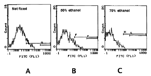
FIGURE 1. This figure shows flow cytometry evaluation using an anti-HLA
class I antibody of ethanol-treated, bi-haptenized, melanoma cells. Three
parts of a 0% (A;
control), 50% (B), or 70% (C) ethanol solution was added to one part mixed-
haptenized tumor
cell suspension (see Example 2).
FIGURE 2. This figure shows flow cytometric analysis of unmodified cells (A)
and ethanol-treated and sulfanilic acid (SA)-modified melanoma cells (B).
Forward light
scatter, an indication of cell diameter, was measured.
5
CA 02474954 2004-07-30
WO 03/063801 PCT/US03/03321
FIGURE 3. This figure shows a flow-cytometric comparison between
unmodified and fixed (A), unmodified unfixed (B), DNP-modified and fixed (C),
and SA-
modified and fixed melanoma cells (D). An antibody against HLA class I antigen
was used in
the analysis.
FIGURE 4. This figure displays flow cytometry histograms showing the effect
of various concentrations of ethanol on cells. An antibody against HLA class I
antigen was
used in this analysis.
FIGURE 5. This figure shows the number of preserved cells in various
ethanol-preserved preparations of mixed-haptenized melanoma cells, after
certain periods of
incubation at 4°C.
FIGURE 6. This figure shows the number of preserved cells in three
preparations of mixed-haptenized melanoma cells after up to 7 days of
incubation at 4°C.
FIGURE 7. This figure shows the antigen-preservation of mixed-haptenized
ethanol-fixed melanoma vaccine, by flow-cytometric analysis using antibodies
directed against
the haptens DNP and SA (A and B, respectively), the melanoma-associated
antigens S 100 and
GD3 (C and D, respectively), and HLA class I antigen (E). (F) is a control.
Ethanol-treated
cells were frozen for up to two months, and then thawed for analysis.
FIGURE 8. This figure shows inhibition of proliferation of mixed-haptenized
and ethanol-fixed melanoma cells. The proliferation of various preparations of
unmodified cells
were compared to cells that had been fixed, and to cells that had been both
mixed-haptenized
and fixed.
FIGURE 9. This figure shows the delayed-type hypersensitivity response
(DTH) measured in patients immunized with DNP-modified melanoma cells to DNP-
modified
tumor cells (A) and unmodified tumor cells (B). The DTH response to ethanol-
fixed cells was
compared to that of untreated or "fresh" cells for both types of cells.
DETAILED DESCRIPTION
As described herein, the present invention contemplates tumor cell
preparations
and vaccines in which the tumor cells are dead and, e. g. , permeable to
Trypan Blue or other
supravital agents, and have a substantially retained or improved antigenicity
as compared to a
vaccine comprising live and/or Trypan Blue-excluding cells. Such vaccines may
or may not be
haptenized. The preparation of such tumor cell vaccines include a treatment
step wherein the
treatment leads to permeabilized or dead cells while at least retaining
antigen expression or
6
CA 02474954 2004-07-30
WO 03/063801 PCT/US03/03321
display on the tumor cell surface. Advantageously, the treatment also has an
additional benefit,
such as leading to improved sterility, purity, or preservation of the
vaccines. Exemplary but
non-limiting treatments include very high doses of radiation (e.g., 100,000
cGy) which can be
bactericidal; heating (e.g., to >_60°C or greater) to kill certain
bacteria or viruses; treatment
with alcohols such as ethanol or isopropanol that can be bactericidal while
maintaining antigen
display; treatment with other chemicals than alcohols, e.g., paraformaldehyde,
which is known
to maintain antigen display; and purification on polymyxin columns to remove
endotoxins.
While it is often desirable to remove treatment agents such as alcohols from
the tumor cell
vaccine after the treatment step, the treatment agent can also be a
pharmaceutically acceptable
agent which can remain in the vaccine. Examples of such agents are
preservatives such as,
e.g., sodium azide or merthiolate. The experimental parameters of the
treatment step,
including concentration of agent, length of exposure to the tumor cells, and
optional
purification, can be determined by routine experimentation. For example, the
optimization and
evaluation techniques used for ethanol treatment, described in detail herein,
can be used for
other agents as well.
Thus, the present invention advantageously provides new preservation methods
which stabilizes tumor cells, including modified tumor cells such as
haptenized cells, for
storage. The preserved cells are preferably stored at between about 0°C
(on ice) and 20°C (at
room temperature) prior to delivery to the patient. In one embodiment, the
method for the
preservation and/or storage of tumor cells comprises contacting the cells with
an optimized
concentration of ethanol. After ethanol treatment, most or all of the
preserved cells are dead,
and the tumor cell composition essentially free of live cells. The
preservation method of the
invention is suitable for treatment of any tumor cell, such as, e. g. ,
haptenized or non
haptenized tumor cells derived from melanoma, ovarian cancer, small cell lung
cancer, colon
cancer, leukemia, or lymphoma.
After the preservation treatment step, the cells may be used for preparing a
tumor cell vaccine for administration to a patient in need thereof. The
preservation method of
the invention is particularly advantageous for such applications, since
preserved cell can be
maintained a longer time in solution without losing antigenicity or vaccine
potency, thus
permitting a longer period of time for quality assurance (QA) and quality
control (QC) of the
vaccine before administration to the patient.
Yet another advantage of the method of the invention using, e.g., ethanol
treatment, is its bactericidal effect. Bacterial contamination can be a
problem when preparing
7
CA 02474954 2004-07-30
WO 03/063801 PCT/US03/03321
vaccines or other medications from tissues. The anti-bacterial effect of
treatment with ethanol,
isopropanol, irradiation, heat, etc., treatment can therefore improve
sterility of tumor cell
vaccines, or even obviate the necessity for additional treatment steps to
sterilize tumor cell
preparations.
Cells treated with the optimized concentration of preserving agent remain
substantially intact and preserve antigen display on the tumor cell surface,
as determined by
flow cytometry, to a greater extent than that of control cells that have not
been treated with the
agent. For example, greater than 10% of ethanol-treated tumor cells are
preserved during
storage for a three-day period at about 4°C, as compared to the initial
number of cells after
ethanol treatment. Preferably, greater than about 25 % of the cells are
preserved; more
preferably, more than 50 % of the cells are preserved, and, even more
preferably, 75 % of the
cells are preserved. For SA-modified tumor cells not treated with ethanol,
typically 90 % of the
cells can be lost, i.e., lysed, after 2-4 hours incubation at 4°C.
Preferably, the preservation of
tumor cells treated with ethanol is greater than the preservation of the same
kind, number, and
concentration of tumor cells incubated in control medium without ethanol for
the same period
of time and at the same temperature.
The treatment step may result in loss of cells, but the remaining cells are
substantially intact and retain their display or accessibility of relevant
cell surface antigens.
Moreover, they are stable for at least 3 days at 4°C, and the shelf-
life of the treated cells can be
extended by freezing. This preparation has the following advantages over prior
art preparations
of modified or urunodified tumor cells: (1) treatment prolongs the shelf life;
(2) irradiation is
not necessary; and (3) cell counting is made easier because differentiation
between "dead" and
"live" tumor cells is moot. The opportunity to exclude irradiation of tumor
cell vaccines is a
particularly attractive feature of the invention, since irradiation has been a
technically
cumbersome and economically burdensome necessity in previous procedures to
render the cells
non-proliferative. Essentially all treated cells of the invention take up
Trypan Blue or other
supravital dyes to some extent but have substantially intact membranes,
preserved shape, and
retain surface antigens.
Furthermore, according to the present invention, autologous tumor cell
vaccines
comprising dead or non-Trypan Blue excluding cells, or consisting wholly of
dead cells or
Trypan Blue excluding cells are equally effective, in some cases even better,
in eliciting an
immune response against a tumor as tumor cell vaccines comprising live cells.
, See, e.g.,
Examples 6, 9, and 10. Thus, according to one embodiment, the invention
provides tumor cell
8
CA 02474954 2004-07-30
WO 03/063801 PCT/US03/03321
vaccines wherein substantially all cells are dead or permeable to Trypan Blue,
and essentially
free of live, Trypan Blue-excluding cells, as well as methods of preparing
such vaccines and
treating cancer patients with such vaccines.
The various aspects of the invention will be set forth in greater detail in
the
following sections, directed to suitable media and formulations for preserving
haptenized tumor
cells. This organization into various sections is intended to facilitate
understanding of the
invention, and is in no way intended to be limiting thereof.
Definitions
The following defined terms are used throughout the present specification, and
should be helpful in understanding the scope and practice of the present
invention.
The term "about" or "approximately" means within an acceptable error range for
the particular value as determined by one of ordinary skill in the art, which
will depend in part
on how the value is measured or determined, i. e. , the limitations of the
measurement system.
For example, "about" can mean within 1 or more than 1 standard deviations, per
the practice in
the art. Alternatively, "about" can mean a range of up to 20 % , preferably up
to 10 %, more
preferably up to 5 % , and more preferably still up to 1 % of a given value.
Alternatively,
particularly with respect to biological systems or processes, the term can
mean within an order
of magnitude, preferably within 5-fold, and more preferably within 2-fold, of
a value.
If not otherwise stated, the concentration of a liquid in a liquid mixture is
given
as percentage of the liquid in the total volume ( % v/v) of the mixture, i. e.
, "by volume" . For
example, a 3:1 mixture between 50% ethanol and HBSS would lead to a 37.5% v/v
ethanol
solution, or 37.5 % ethanol by volume.
A "formulation" refers to an aqueous medium or solution for the preservation
of
haptenized tumor cells, which is preferably directly injectable into an
organism. An aqueous
buffer will include salts or sugars, or both, at about an isotonic
concentration. The formulation
may further comprise ethanol, as described herein.
"Human serum albumin" or "HSA" refers to a non-glycosylated monomeric
protein consisting of 585 amino acid residues, with a molecular weight of 66
kD. Its globular
structure is maintained by 17 disulphide bridges, which create a sequential
series of 9 double
loops (Brown, "Albumin structure, function and uses", Rosenoer, V.M. et al.
(eds.), Pergamon
Press:Oxford, pp. 27-51, 1977). HSA may also be called human plasma albumin.
9
CA 02474954 2004-07-30
WO 03/063801 PCT/US03/03321
A "live" cell means a cell that has an intact cell, plasma, or "outer"
membrane
as assessed by exclusion of a supravital dye such as Trypan Blue. A live cell
may be capable of
growth or maintenance, and division or multiplication, or attenuated, i. e. ,
incapable of division
and multiplication. A cell can be rendered attenuated by, for example,
irradiation.
"Dead" cells means cells that do not exclude supravital dyes such as Trypan
Blue, propidium bromide, or ethidium bromide, as assessed in an exclusion
experiment (see,
e.g., Methods In Analysis Of Apoptosis And Cell Necrosis by Darzynkiewicz Z.,
In: The
Purdue Cytometry CD-ROM Vol 3, J. Parker, C. Stewart, Guest Eds.; J. Paul
Robinson,
Publisher, Purdue University, West Lafayette, 1997). Dead cells are incapable
of division or
multiplication. A "dead" cell can be prepared by, e. g. , ethanol treatment of
a live cell. A dead
cell may appear intact, e. g. , by microscopic inspection, meaning that the
cellular shape
resembles that of a live cell. A "fixed" cell is one example of a dead cell.
A "lysed" cell is no longer intact, meaning that the cellular shape does not
resemble that of a live cell.
The "total" number of tumor cells in a preparation means the sum of live and
dead tumor cells in the preparation.
A "preserved" cell is a cell which is not lysed. A preserved cell cari be live
or
dead. The cell may or may not exclude Trypan Blue, but retains its
antigenicity over time
better than a cell which is not similarly preserved. "Preservation" of cells
can be expressed as
the percentage of cells remaining after a certain period of time following
ethanol treatment of
the cells according to the method of the invention. Thus, about 90 % of the
cells being
preserved over a period of 1 day (i. e. , 24 hours) means that the number of
"non-lysed" cells in
the preparation after 1 day storage is about 90% of the number of "non-lysed"
cells in the
preparation just after ethanol treatment.
Treatment with ethanol can lead to "fixed" cells. Ethanol-treatment can
therefore also be termed "fixation".
"Antigenicity" means the ability of a tumor cell to evoke an immune response
directed to the tumor cell. Generally, antigenicity is higher for a tumor cell
that comprises
tumor-specific antigens than a tumor cell which does not comprise, or
comprises a lower
amount of, tumor-specific antigens. Antigenicity can be measured by, for
instance, DTH-
testing, or by measuring the number of tumor cell-associated antigens using,
e. g. , FACS
analysis with antibodies directed against the tumor-associated antigens.
CA 02474954 2004-07-30
WO 03/063801 PCT/US03/03321
The term "cell recovery" or "cell recovery rate" is a measure of how many
cells
are substantially intact, has a shape corresponding to or resembling that of a
live cell, and/or
has preserved antigenicity, after a certain period of storage or incubation.
When calculating
cell recovery, the number of cells at a certain time point or after a certain
preparation step is
related to the number of cells at a reference time point or prior to the
preparation step in
question.
The phrase "pharmaceutically acceptable" refers to molecular entities, at
particular concentrations, and compositions, that are physiologically
tolerable and do not
typically produce an allergic or similar untoward reaction, such as gastric
upset, fever,
dizziness and the like, when administered to a human or non-human animal.
Preferably, as
used herein, the term "pharmaceutically acceptable" means approved by a
regulatory agency of
the Federal or a state government, or listed in the U.S. Pharmacopoeia or
other generally
recognized pharmacopoeia for use in humans or non-human animals.
A "subject" is a human or a non-human animal who may receive haptenized
tumor cells formulated in a composition of the invention. Non-human animals
include
domesticated pets, such as cats and dogs; farm animals, such as horses, cows,
pigs, sheep, and
goats; laboratory animals, such as mice, rats, guinea pigs, and rabbits; etc.
An "anti-tumor response" is at least one of the following: tumor necrosis,
tumor
regression, tumor inflammation, tumor infiltration by activated T lymphocytes,
activation of
tumor infiltrating lymphocytes, delayed-type hypersensitivity (DTH) response,
or a clinical
response. Clinical response criteria for anti-tumor response resulting from
treatment according
to the present invention include complete, partial, or mixed response, as well
as stable disease.
Other clinical responses that may be observed following treatment according to
the invention is
prolongation of time to relapse, or prolongation of survival.
A "formulation" refers to an aqueous medium or solution for the preservation
or
administration, or both, of haptenized tumor cells, which is preferably
directly injectable into
an organism. The aqueous medium can include salts or sugars, or both, at about
an isotonic
concentration.
A "vaccine composition" is a composition as set forth previously further
comprising an adjuvant, including an immunostimulatory cytokine or lymphokine.
The terms "vaccine", "immune therapy" and "immunotherapy" are used herein
interchangeably to administration of a composition comprising a tumor cell
preparation
(preferably haptenized) to treat a cancer, e. g. , after surgical resection of
the tumor.
11
CA 02474954 2004-07-30
WO 03/063801 PCT/US03/03321
"Efficacy of an immunotherapy" is the degree to which the immunotherapy
elicits am anti-tumor response in an individual subject, or the percentage of
subjects in which
an anti-tumor response develops as a result of treatment. Preferably efficacy
is determined by
composition to controls that harbor the spontaneous tumor but receive either
no therapy, sham
therapy, or an alternative therapy.
A "tumor cell preparation" refers to isolated or purified tumor cells for
inclusion
in a composition. "Hapten modified" means that the tumor cells are chemically
coupled
(conjugated) to a hapten, as that term is understood immunology.
As used herein, a "bi-haptenized", "mufti-haptenized", or "mixed haptenized"
tumor cell preparation means a composition comprising two or more tumor cell
preparations, in
which each tumor cell preparation is differently haptenized.
The term "differentially haptenized" as used herein refers to mixture of at
least
two haptenized tumor cells, wherein a first cell was haptenized under a
particular condition or
using a particular reagent and a second cell was haptenized under a different
condition or using
a different reagent. The conditions or reagents may differ so that, for
example, different amino
acids are haptenized on the proteins of the first and second tumor cells,
and/or that the hapten
attached to the first cell is different from the hapten attached to the second
cell.
The term "treat" means to attempt to elicit an anti-tumor response against
cells
of the tumor, i. e. , the cancer. An anti-tumor response includes, but is not
limited to, increased
time of survival, inhibition of tumor metastasis, inhibition of tumor growth,
tumor regression,
and development of a delayed-type hypersensitivity (DTH) response to
unmodified tumor cells.
As used herein, the term "control" generally describes a cell or cells not
treated
with ethanol. More preferably, a control describes a composition which in
essentially all other
aspects than ethanol treatment has been exposed to the same conditions, and is
stored in the
same buffered medium and additional components.
Treatment With Ethanol or Other Agents
As noted above, and demonstrated in the Examples, infra, it has been
unexpectedly discovered that exposure of tumor cells to an appropriate
concentration of a
preserving agent such as ethanol in a buffered cultured medium, preferably
HBSS, greatly
increases cell preservation and antigenicity. This is especially advantageous
for tumor cells for
use in immunotherapy vaccine preparations. Accordingly, depending on the
specific tumor
cells to be stored, and their modification, if any, one of ordinary skill in
the art can test for the
12
CA 02474954 2004-07-30
WO 03/063801 PCT/US03/03321
optimum concentration of ethanol or other preserving agent for, as well as the
duration of, such
a treatment step, as exemplified infra. Such a concentration can be one that
yields an increase
in cell preservation relative to a control for stored tumor cells. In
addition, such a
concentration can be one that retains the amount of antigen-displaying cells
relative to a control.
Preferably, the increase in preservation of the number of cells is
statistically significant. In a
specific embodiment, the yield of intact cells after treatment is at least
about 10%, more
preferably at least about 20 % , and even more preferably, at least about 50 %
. In a preferred
embodiment, the cells are then stored in 1 % HSA in HBSS.
After treatment, the cells are stable or preserved in that at least about 30%,
preferably at least about 50 % , and even more preferably at least about 80 %
, of the treated cells
can be present after about 3 days of storage at 4°C, and have
substantially retained antigen
content. See also Table 2 in the Examples. By contrast, in one example, about
90 % of SA-
modified cells not exposed to the preserving agent ethanol were lost (i. e. ,
lysed) during 4 hours
of storage at 4°C. In experiments using SA-modified cells, the recovery
of total cells
(including dead cells) is rarely more than 30% after 4 hours storage at
4°C. Thus, preservation
of antigen-displaying or antigen-associated cells can be substantially
improved by treatment
with an agent such as ethanol. Preferably, the preservation of a tumor cell
subjected to
treatment with an agent is greater than the same kind of tumor cells incubated
in control
medium without the agent for the same period of time, at the same temperature.
The following is a description of one treatment according to the invention,
using
ethanol as preserving agent. Tumor cells suspended in a suitable medium, such
as, but not
limited to, HBSS, and are kept on ice, at about 0°C to 10°C, or
at about 4°C. Optionally, the
medium contains HSA at a concentration of, for example, 1 % (weight to
volume). Next,
ethanol is added to the cells at a suitable final concentration (see below).
In one embodiment, 3
ml of ice-cold ethanol solution (50 % v/v) are added per each ml of tumor cell
suspension. The
ethanol can be added to each tube while vortexing at low speed. The tubes are
thereafter
incubated in the presence of ethanol. Suitable incubation time and temperature
can be
determined experimentally for different tumor cell preparations. For example,
it has been
found that a 10 minute incubation at 4°C is suitable for mixed-
haptenized cells (see Examples
1-3). The cells are thereafter pelleted by centrifugation, e.g., by spinning
at 1100 RPM for 7
minutes. The supernatant is aspirated to remove the ethanol-containing
supernatant, and the
cells washed in medium. For example, 5x 10⁶ cells can be resuspended in 10 ml
HBSS + 1 %
HSA, and pelleted again by spinning at 1100 RPM for 7 minutes. This washing
procedure can
13
CA 02474954 2004-07-30
WO 03/063801 PCT/US03/03321
be repeated if necessary. After washing, the cells are pelleted, the
supernatant aspirated, and
the cells resuspended in the desired medium. For example, Sx 106 cells can be
resuspended in
2 ml Hanks + 1 % HSA (se also "Formulations", below). Preferably, the cells
are stored in a
medium suitable for administration to a subject. In another embodiment, the
cells are stored in
a medium suitable for cryopreservation, and cryopreserved (see below) until
needed.
Any ethanol concentration effective to preserve the tumor cells may be used in
this procedure, for example by varying either the ethanol concentration in the
stock solution
added to the HBSS solution, and/or by varying the amount of ethanol added to
the HBSS
solution. Generally, treatment with a solution containing more than 75 %
ethanol leads to
fixation of cells, but also to loss of antigens. Thus, according to the
invention, the cells are
preferably incubated in about 5 % to about 75 % (v/v) ethanol. More
preferably, the cell are
incubated in about 20 % to about 60 % (v/v) ethanol, or, even more preferably,
in about 25 % to
about 40 % (v/v) ethanol. In a particularly preferred embodiment, the cells
are incubated in
about 30-40 % (v/v) ethanol. In one specific embodiment, the cells are treated
in no greater
than about 52.5 % (v/v) ethanol. In another specific embodiment, the cells are
incubated in
about 37.5 % (v/v) ethanol. A suitable ethanol concentration is one that can
fix the cells,
maintain display or association of antigens, and prevent cell proliferation.
In one embodiment,
a suitable ethanol concentration has, in addition, a bactericidal effect.
The duration as well as the temperature of the ethanol treatment step may also
have an impact on the preservation of the cells. Preferably, the ethanol
exposure is conducted
at room temperature or less, preferably at 10°C or less, and even more
preferably at about 4°C
or on ice. A period of incubation for about 10 minutes is suitable for mixed-
haptenized cells.
The optimal time period for modified or unmodified cells can be determined on
a case-by-case
basis using standard parameter-optimization procedures. The most suitable time
of incubation
would depend both on the modification and the type of tumor cell, as well as
the temperature
and ethanol concentration. Preferably, the cells are incubated for at least 10
seconds,
preferably more than one minute, and, even more preferably, more than 2
minutes. In a
preferred embodiment, the cells are incubated in ethanol for no more than 24
hours, preferably
less than 1 hour, and even more preferably for about 10 minutes.
After the ethanol or other treatment step, the ethanol or other treatment
agent is
preferably, although not necessarily, substantially removed from the cells.
This may be
accomplished by, e.g., centrifugation, removal of the supernatant, and
resuspending the cells in
a suitable storage buffer as described above. As an alternative to
centrifugation, the ethanol or
14
CA 02474954 2004-07-30
WO 03/063801 PCT/US03/03321
other agent can be removed by dialysis, extraction, microfiber extraction,
filtration,
chromatography, evaporation, or other techniques known by those skilled in the
art.
Thereafter, the cells can be stored frozen, i. e. , at less than 0 ° C,
or not frozen, i. e. , at above
0°C. A tumor cell composition which is stored frozen can be stored,
e.g., at -10°C to about -
30°C, or, alternatively, in liquid nitrogen, which has a temperature of
about -196°C. In one
embodiment, the cells are first stored in a -70°C or -86°C
freezer and then transferred to liquid
nitrogen. A tumor cell composition which is stored at 0°C or higher
temperatures can be
stored in a fridge, e. g. , at between 0 ° C to about 10 ° C
such as at about 4 ° C, or at room
temperature, which corresponds to from about 15 to about 25°C.
The concentration of cells to be used during the ethanol or other treatment
step
can be determined experimentally depending on the type of cells or cell
preparation used.
However, a generally suitable concentration is between 105-10$ cells, more
preferably between
106 to 10' cells, and most preferably about 5x10⁶ cells, per milliliter
solution. The solution is
advantageously, although not necessarily, isotonic.
After ethanol or other treatment, at least the vast majority of the cells,
preferably substantially all of the cells, take up Trypan Blue. However, by
microscopic
inspection, the cells are intact anatomically and/or has a shape resembling
that of an intact cell.
Generally, the treated cells are not easily distinguishable from living cells
in the absence of
Trypan Blue. The treated cells also retain antigen display to a substantial
degree, as shown in
the Examples.
For vaccines comprising haptenized tumor cells, the ethanol or other treatment
is preferably, although not necessarily, conducted after haptenization.
m".,",.. ron~
The tumor cells used in the present invention are prepared from tumor cells,
e.g., obtained from tumors, or tissue or body fluids containing tumor cells,
surgically resected
or retrieved in the course of a treatment for a cancer. The ethanol-treated
tumor cells are
useful in the preparation of, e.g., tumor cell vaccines for treating cancer,
including metastatic
and primary cancers. If used in a tumor cell vaccine, the preserved tumor
cells should be
incapable of growing and dividing after administration into the subject, such
that they are dead
or substantially in a state of no growth. It is to be understood that "dead
cells" means a cell
which do not have an intact cell or plasma membrane and that will not divide
in vivo; and that
"cells in a state of no growth" means live cells that will not divide in vivo.
Conventional
CA 02474954 2004-07-30
WO 03/063801 PCT/US03/03321
methods of suspending cells in a state of no growth are known to skilled
artisans and may be
useful in the present invention. For example, cells may be irradiated prior to
use such that they
do not multiply. Tumor cells may be irradiated to receive a dose of 2500 cGy
to prevent the
cells from multiplying after administration. Alternatively, ethanol treatment
may result in dead
cells.
The tumor cells can be prepared from virtually any type of tumor. The present
invention contemplates the use of tumor cells from solid tumors, including
carcinomas; and
non-solid tumors, including hematologic malignancies. Examples of solid tumors
from which
tumor cells can be derived include sarcomas and carcinomas such as, but not
limited to:
fibrosarcoma, myxosarcoma, liposarcoma, chondrosarcoma, osteogenic sarcoma,
chordoma,
angiosarcoma, endotheliosarcoma, lymphangiosarcoma,
lymphangioendotheliosarcoma,
synovioma, mesothelioma, Ewing's tumor, leiomyosarcoma, rhabdomyosarcoma,
colon
carcinoma, pancreatic cancer, breast cancer, ovarian cancer, prostate cancer,
squamous cell
carcinoma, basal cell carcinoma, adenocarcinoma, sweat gland carcinoma,
sebaceous gland
carcinoma, papillary carcinoma, papillary adenocarcinomas, cystadenocarcinoma,
medullary
carcinoma, bronchogenic carcinoma, renal cell carcinoma, hepatoma, bile duct
carcinoma,
choriocarcinoma, seminoma, embryonal carcinoma, Wilms' tumor, cervical cancer,
testicular
tumor, lung carcinoma, small cell lung carcinoma, bladder carcinoma,
epithelial carcinoma,
glioma, astrocytoma, medulloblastoma, craniopharyngioma, ependymoma,
pinealoma,
hemangioblastoma, acoustic neuroma, oligodendroglioma, meningioma, melanoma,
neuroblastoma, and retinoblastoma. Hematologic malignancies include leukemias,
lymphomas,
and multiple myelomas. The following are non-limiting preferred examples of
tumor cells to
be preserved according to the present invention: melanoma, including stage-4
melanoma;
ovarian, including advanced ovarian; small cell lung cancer; leukemia,
including and not
limited to acute myelogenous leukemia; colon, including colon metastasized to
liver; rectal,
colorectal, breast, lung, kidney, and prostate cancer cells.
Tumor cell vaccines can be prepared from any of the tumor cell types listed
above. Such tumor cell vaccines can comprise preserved cells, i. e. , cells
treated with ethanol
according to the method of the invention. Preferably, the vaccine comprises
the same type of
cells as the tumor to be treated. Most preferably, the tumor cells are
autologous, derived from
the patient for whom treatment with the vaccine is intended. Vaccines
comprising tumor cells
prepared using the method of the invention can used for treatment of both
solid and non-solid
tumors, as exemplified above. Thus, the invention includes "preserved"
vaccines prepared
16
CA 02474954 2004-07-30
WO 03/063801 PCT/US03/03321
from, and intended for treatment of, solid tumors, including carcinomas; and
non-solid tumors,
including hematologic malignancies. Preferred tumor types for vaccines include
melanoma,
ovarian cancer, colon cancer, and small cell lung cancer.
The tumor cells are preferably of the same type as, most preferably syngeneic
(e.g., autologous or tissue-type matched) to, the cancer which is to be
treated. For purposes of
the present invention, syngeneic refers to tumor cells that are closely enough
related genetically
that the immune system of the intended recipient will recognize the cells as
"self", e.g., the
cells express the same or almost the same complement of HLA molecules. Another
term for
this is "tissue-type matched. " For example, genetic identity may be
determined with respect to
antigens or immunological reactions, and any other methods known in the art.
Preferably the
cells originate from the type of cancer which is to be treated, and more
preferably, from the
same patient who is to be treated. The tumor cells can be, although not
limited to, autologous
cells dissociated from biopsy or surgical resection specimens, or from tissue
culture of such
cells. Nonetheless, allogeneic cells and stem cells are also within the scope
of the present
invention.
Tumor cells for use in the present invention may be prepared as follows.
Tumors are processed as described by Berd et al. (Cancer Res. 1986;46:2572;
see also US
Patent No. 5,290,551; US Patent Applications No. 08/203,004, No. 08/475,016,
and No.
08/899,905). The cells are extracted by dissociation, such as by enzymatic
dissociation with
collagenase, or, alternatively, DNase, or by mechanical dissociation such as
with a blender,
teasing with tweezers, mortar and pestle, cutting into small pieces using a
scalpel blade, and the
like. Mechanically dissociated cells can be further treated with enzymes as
set forth above to
prepare a single cell suspension.
Tumor cells may also be prepared according to Hanna et al., U.S. Patent No.
5,484,596. Briefly, tumor tissue is obtained from patients suffering from the
particular solid
cancer from which the vaccine is to be prepared. The tumor tissue is
surgically removed from
the patient, separated from any non-tumor tissue, and cut into small pieces,
e.g., fragments 2-3
mm in diameter. The tumor fragments are then digested to free individual tumor
cells by
incubation in an enzyme solution. After digestion, the cells are pooled and
counted, and cell
viability is assessed. If desired, a Trypan Blue exclusion test can be used to
assess cell
viability.
In addition, tumor cells can be prepared according to the following procedure
(see Hanna et al., U.S. Patent No. 5,484,596). The tissue dissociation
procedure of Peters et
17
CA 02474954 2004-07-30
WO 03/063801 PCT/US03/03321
al. (Cancer Research 1979;39:1353-1360) can be employed using sterile
techniques throughout
under a laminar flow hood. Tumor tissue can be rinsed three times in the
centrifuge tube with
HBSS and gentamicin and transferred to a petri dish on ice. Scalpel dissection
removed
extraneous tissue and the tumor are minced into pieces approximately 2 to 3 mm
in diameter.
Tissue fragments are placed in a 75 ml flask with 20-40 ml of 0.14 % (200
units/ml)
Collagenase Type 1 (Sigma C-0130) and 0.1 % (500 Kunitz units/ml)
deoxyribonuclease type 1
(Sigma D-0876) (DNAase 1, Sigma D-0876) prewarmed to 37°C. Flasks are
placed in a 37°C
water bath with submersible magnetic stirrers at a speed which cause tumbling,
but not
foaming. After a 30-minute incubation, free cells are decanted through three
layers of sterile
medium-wet nylon mesh (166t: Martin Supply Co., Baltimore, Md.) into a 50 ml
centrifuge
tube. The cells are centrifuged at 1200 rpm (250xg) in a refrigerated
centrifuge for 10
minutes. The supernatant is poured off and the cells are resuspended in 5-10
ml of DNAase
(0.1 % in HBSS) and held at 37°C for 5-10 minutes. The tube is filled
with HBSS, washed by
centrifugation, resuspended to 15 ml in HBSS and held on ice. The procedure is
repeated until
sufficient cells are obtained, usually three times for tumor cells. Cells from
the different digests
are then pooled, counted. Optionally, although not necessarily, cell viability
is assessed by the
Trypan Blue exclusion test.
Tumor cells, prior to or after ethanol-treatment, can be frozen if stored for
extended persiods of time. The cells may be frozen or cryopreserved according
to any method
known in the art, either before or after any modification to the cells (e. g.
, haptenization, lysis,
etc.) has been made. For example, the dissociated cells may be stored frozen
in a freezing
medium (e.g., prepared from a sterile-filtered solution of 50 ml Human Serum
Albumin
[American Red Cross] added to 450 ml of RPMI 1640 (Mediatech) supplemented
with L-
glutamine and brought to an appropriate pH with NaOH), such as in a controlled
rate freezer or
in liquid nitrogen until needed. The cells are ready for use upon thawing.
Preferably, the cells
are thawed shortly before use, or stored for no more than a couple of days
before use.
Optionally, the cells may be washed once or twice, and then suspended in HBSS
without phenol
red.
Alternatively, the concentration of dissociated tumor cells can be adjusted to
about 5-lOx 10'/ml, or to about Sx 10' or lOx 10' cells per ml, in HBSS and/or
a freezing
medium. The freezing medium can be a plain cell growth medium such as HBSS, or
a medium
or buffer complemented with HSA, sucrose, dextran, or mixtures thereof.
Preferably, the
freezing medium is based on HBSS and complemented with either HSA/sucrose or
18
CA 02474954 2004-07-30
WO 03/063801 PCT/US03/03321
HSA/dextran. The cells can also be added in equal volume to chilled 2xfreezing
medium
containing 15 % dimethylsulfoxide (DMSO) and 4 % human serum albumin (HSA),
with or
without a suitable concentration of sucrose or dextran. The final suspension
of 2 to 4x 10'
cells/ml is placed in 1.2 ml Nunc freezer vials. In preparation for freezing,
the Nunc vials are
transferred on ice to a Cryo-Med model 990 Biological Freezer with a model 700
Controller
and a model 500 Temperature Recorder for controlled-rate freezing. Care should
be taken that
the temperature of the individual vials, including the monitor vial, is
uniform at the beginning
of the freezing process. Vials are cooled at a controlled rate of -1
°C/min to a final temperature
of -80°C. The vials are then transferred in liquid nitrogen to liquid
nitrogen storage. Suitable
HSA preparations are available commercially, from, e.g., Baxter Corp.
Mississauga, Canada).
An alternative freezing medium is a medium containing 7 % sucrose and 10 %
HSA in HBSS. The cells are stored overnight at -86°C, and then
transferred to liquid nitrogen.
Haptens
In one embodiment, the tumor cells are haptenized. For purposes of the present
invention, virtually any small protein or other small molecule that fails to
induce an immune
response when administered alone, may function as a hapten. A variety of
haptens of quite
different chemical structure have been shown to induce similar types of immune
responses,
e. g. , TNP (Kempkes et al. , J. Immunol. , 147:2467, 1991 );
phosphorylcholine (Jang et al. ,
Eur. J. Immunol., 21:1303, 1991); nickel (Pistoor et al., J. Invest.
Dermatol., 105:92, 1995);
and arsenate (Nalefski and Rao, J. Immunol., 150:3806, 1993). Conjugation of a
hapten to a
cell to elicit an immune response may preferably be accomplished by
conjugation via E-amino
groups of lysine or -COOH groups. This group of haptens include a number of
chemically
diverse compounds: dinitrophenyl, trinitrophenyl, N-iodoacetyl-N'-(5-sulfonic
1-naphthyl)
ethylene diamine, trinitrobenzenesulfonic acid, dinitrobenzene sulfonic acid,
fluorescein
isothiocyanate, arsenic acid benzene isothiocyanate, and dinitrobenzene-S-
mustard (Nahas and
Leskowitz, Cellular Immunol., 54:241, 1980). Once armed with the present
disclosure, skilled
artisans would be able to choose haptens for use in the present invention.
Hapenization
When using haptenized cells in the tumor cell composition, modification of the
prepared cells with a hapten may be performed by known methods, e.g. by the
method of
Miller and Clanian (J. Immunol. 1976;117:151). The described procedure
involves a 30
19
CA 02474954 2004-07-30
WO 03/063801 PCT/US03/03321
minute incubation of tumor cells with DNFB under sterile conditions, followed
by washing with
sterile saline or Hanks/HSA. Haptenization is also described in the Examples
(see below).
Other procedures for haptenization are known in the art (see, e. g. ,
International Patent
Publications WO 96/40173, WO 00/09140, WO 00/31542, WO 99/56773, WO 99/52546,
WO
99/40925, WO 98/14206, WO 00/295, all by Berd et al., and U.S. Patent No.
5,290,551 to
Berd, hereby incorporated by reference in its entirety).
For example, the following procedure may be used for tumor cell haptenization.
About 100 mg of DNFB (Sigma Chemical Co., St. Louis, MO) is dissolved in about
0.5 ml of
70% ethanol. About 99.5 ml of PBS is added. The solution is stirred overnight
in a 37°C
water bath. The shelf life of the solution is about 4 weeks. The cells are
thawed and the pellet
resuspended in 5 x 10⁶ cells/ml in Hanks balanced salt solution. About 0.1 ml
DNFB solution
is added to each ml of cells and incubated for about 30 minutes at room
temperature.
Similarly, other haptens such as and not limited to trinitrophenyl, N-
iodoacetyl-N'-(5-sulfonic
1-naphthyl) ethylene diamine, trinitrobenzenesulfonic acid, fluorescein
isothiocyanate, arsenic
acid benzene isothiocyanate, trinitrobenzenesulfonic acid, sulfanilic acid,
arsanilic acid,
dinitrobenzene-S-mustard and combinations thereof may be used.
The tumor cells can also be dual-haptenized, i. e. , the same tumor cell
preparation can be conjugated with two different haptens. The haptens may
comprise reactive
groups that react with different functional groups on the tumor cell, such as
different amino
acids. Such dual-haptenization is described in WO 00/38710 by Berd et al.
Alternatively, the tumor cell can be bi-haptenized or mixed haptenized, i. e.
, two
or more aliquots of a single tumor cell preparation is each coupled to a
different hapten, or the
same hapten is coupled to different functional groups, can be mixed prior to
administration, or
administered in conjunction with each other. Bi-haptenization may be conducted
as described
in the Examples.
Optionally, tumor cells can be frozen before or after haptenization, as
described
above.
Tin~.mmlnW nroe
The tumor cells treated with ethanol or another permeabilizing agent or step
according to the invention may be included in various formulations. For
example, tumor cells
may, in haptenized or unmodified form, be useful for preparing tumor vaccines.
The different
components of such a formulation may be mixed together, and then added to
tumor cells. It is
CA 02474954 2004-07-30
WO 03/063801 PCT/US03/03321
also possible to mix one or several of the components with the tumor cells and
then to add the
remaining component(s). The preparation of the formulation and its addition of
the tumor cells
are preferably performed under sterile conditions. Preferably, the tumor cells
are subjected to
ethanol or other treatment before the final formulation. However, one or more
components to
be included in the final formulation may also be present before or during the
treatment step.
The respective proportions of the components of the media according to the
invention may be adapted by persons skilled in the art. As illustrated in the
Examples, the
proportions may be modified although certain concentration ranges are
preferred.
Generally, an appropriate buffered medium is used for tumor cell formulation.
In its essence, a buffered medium is an isotonic buffered aqueous solution,
such as phosphate
buffered saline (PBS), Tris-buffered saline, or HEPES buffered saline. In a
preferred
embodiment, the medium is a buffered cell culture medium such as plain Hank's
medium (not
containing phenol red), e.g., as sold commercially by Sigma Chemical Co. (St.
Louis,
Missouri, USA). Other tissue culture media can also be used, including basal
medium Eagle
(with either Earle's or Hank's salts), Dulbecco's modified, Eagle's medium
(DMEM), Iscove's
modified Dulbecco's medium (IMDM), Medium 199, Minimal Essential Medium (MEM)
Eagle (with Earle's or Hank's salts), RPMI, Dulbecco's phosphate buffered
salts, Earle's
balanced salts (EBSS), and Hank's Balanced Salts (HBSS). These media can be
supplemented,
e.g., with glucose, Ham's nutrients, or HEPES. Other components, such as
sodium
bicarbonate and L-glutamine, can be specifically included or omitted. Media,
salts, and other
reagents can be purchased from numerous sources, including Sigma, Gibco, BRL,
Mediatech,
and other companies.
Generally, human serum albumin (HSA) is also included, as described below.
In addition, a composition or formulation of the invention may contain
components in addition
to HSA to further stabilize the haptenized tumor cells. Examples of such
components include,
but are not limited to, carbohydrates and sugars such as dextrose, sucrose,
glucose, and the
like, e.g. , at a 5 % concentration; medium to long chain polyols such as
glycerol, polyethylene
glycol, and the like, e. g. , at 10 % concentration; other proteins; amino
acids; nucleic acids;
chelators; proteolysis inhibitors; preservatives; and other components.
Preferably, any such
constituent of a composition of the invention is pharmaceutically acceptable.
21
CA 02474954 2004-07-30
WO 03/063801 PCT/US03/03321
Human Serum Albumin
In a preferred embodiment, the tumor cell formulations of the invention
comprise a concentration or amount of a protein such as, e.g., albumin, which
is effective to
stabilize the tumor cells. An amount of protein effective to stabilize the
tumor cells may be
added before and/or after ethanol treatment, or, in the case of haptenized
tumor cells, before
and/or after haptenization. In a preferred embodiment, the albumin is human
serum albumin or
HSA. HSA has been shown to stabilize solutions of proteins, including protein
antigens, and
small organic molecules such as hemin (Paige, A.G. et al., Pharmaceutical
Res., 12:1883-
1888, 1995; Chang, A.-C. and R.K. Gupta, J., Pharm. Sci., 85:129-132, 1996;
Niemeijer, N.
R. et al., Ann. Allergy Asthma Immunol., 76:535-540, 1996; and Cannon, J.B. et
al., PDA:J.
Pharm. Sci. & Tech., 49:77-82, 1995), as well as haptenized tumor cell
compositions (see WO
00/29554, corresponding to U.S. Patent No. 6,248,585).
The HSA used within the framework of the present invention may be either of
natural origin (purified HSA) or of recombinant origin (rHSA). Naturally, for
delivery of a
formulation in vivo, it is preferable to use an autologous or non-immunogenic
serum albumin.
Thus, for human therapy, HSA is desirable and preferred. However, the skilled
person can
immediately appreciate that any serum albumin can be used in the practice of
this invention,
and, more particularly, any autologous serum albumin can be used in connection
with tumor
cell vaccine for cancer treatment in any non-human animal as well. In a
specific embodiment,
a Human Serum Albumin Solution (American Red Cross), which is a 25 % HSA
solution, is
used.
Advantageously, a recombinant or natural HSA is used which meets certain
quality criteria (e.g., homogenetic, purity, stability). Thus, the
pharmacopoeias set a number
of parameters for the albumin solutions, namely a pH value, a protein content,
a polymer and
aggregate content, an alkaline phosphatase content, and a certain protein
composition. It
imposes, furthermore, a certain absorbance, the compliance with tests for
sterility, pyrogens,
and toxicity (see "Albumini humai solutio", European Pharmacocpoeia (1984),
255). The use
of an albumin composition corresponding to these criteria, although not
essential, is particularly
preferred.
Generally, the HSA formulation of the invention is made by adding HSA
powder or solution to the selected culture medium/balanced salt solution, to
achieve the desired
final concentration. The final concentration of HSA is preferably, in weight
to volume, from
22
CA 02474954 2004-07-30
WO 03/063801 PCT/US03/03321
about 0.1 % to 10 % , even more preferably from about 0.25 % to about 2 % ,
and most preferably
about 1 % .
Additional information about the use of albumin in formulations of tumor
cells,
especially haptenized tumor cells, can be found in WO 00/29554, corresponding
to U.S. Patent
No.6,248,585.
Vaccine Preparation and Administration
The compositions of the invention may be administered in a mixture with a
pharmaceutically-acceptable carrier, selected with regard to the intended
route of administration
and standard pharmaceutical practice. Dosages may be set with regard to weight
and clinical
condition of the patient. The proportional ratio of active ingredient to
carrier naturally depends
on the chemical nature, solubility, and stability of the compositions, as well
as the dosage
contemplated. The amounts to be used of the tumor cells of the invention
depend on such
factors as the affinity of the compound for cancerous cells, the amount of
cancerous cells
present and the solubility of the composition. The compounds of the present
invention may be
administered by any suitable route, including inoculation and injection, for
example,
intradermal, intravenous, intraperitoneal, intramuscular, and subcutaneous.
For example, the
composition may be administered by intradermal injection into 3 contiguous
sites per
administration on the upper arms or legs, excluding limbs ipsilateral to a
lymph node
dissection.
Tumor Cell Dose
A predetermined number or concentration of cells is included in each vaccine
dose. To prepare the vaccine dosage forms to contain the right number and/or
concentration of
cells, the cells in a tumor cell preparation can be counted by any suitable
method known in the
art. For example, cells can be counted manually using a microscope and
standard cell counting
chambers, or by using automatic cell counters such as, e.g., Beckman Coulter
cell counters.
Since the method does not require distinguishing between live and "dead"
cells, and in some
embodiments, even prefer "dead cells", Trypan Blue and other means which are
selective for
live or dead cells can be omitted. The concentration of cells can then be
adjusted by diluting
the cells with a sterile solution so that a certain volume corresponds to the
number of cells to be
injected into the patient, and this volume aliquoted into storage vials.
23
CA 02474954 2004-07-30
WO 03/063801 PCT/US03/03321
In one embodiment of the invention, the composition comprises a vaccine
comprising about 1 x 10⁴ to 1 x 10⁸, more preferably 1 x 10⁶ to about 25 x
106, even more
preferably about 2.5 x 10⁶ to about 7. S x 106, tumor cells suspended in a
pharmaceutically
acceptable carrier or diluent, such as, but not limited to, Hank's solution
(HBSS), saline,
phosphate-buffered saline, and water. In another embodiment, the tumor cell
vaccine
comprises from about Sx 104 to about Sx 106 cells, for example, Sx 104, Sx
105, or Sx 106 tumor
cells. Preferably, the tumor cells are dead and do not exclude Trypan Blue or
another
supravital dye.
Adjuvants
In preferred embodiment, a tumor cell composition may be administered with an
immunological adjuvant. While the commercial availability of pharmaceutically
acceptable
adjuvants is limited, representative examples of adjuvants include Bacille
Calmette-Guerin,
BCG, or the synthetic adjuvant, QS-21 comprising a homogeneous saponin
purified from the
bark of Quillaja saponaria, Corynebacterium parvum, (McCune et al., Cancer
1979 ;43:1619),
and IL-12.
It will be understood that the adjuvant is subject to optimization. In other
words, the skilled artisan can engage in no more than routine experimentation
and determine
the best adjuvant to use.
Immunostimulants and Combination T7zerapies
The tumor cell compositions may be co-administered with other compounds
including but not limited to cytokines such as interleukin-2, interleukin-4,
gamma interferon,
interleukin-12, GM-CSF. The tumor cells preparations of the invention may also
be used in
conjunction with other cancer treatments including but not limited to
chemotherapy, radiation,
antibodies, antisense oligonucleotides, and gene therapy. In a preferred
embodiment,
cyclophosphamide is used as adjunctive chemotherapy in treatment regimes
involving the
present tumor cell vaccines.
EXAMPLES
The following examples are illustrative of the invention, but not limiting
thereof.
24
CA 02474954 2004-07-30
WO 03/063801 PCT/US03/03321
EXAMPLE 1
Ethanol Treatment of Mixed-Haptenized Melanoma Cells
This Example describes a strategy for preparation of a bi-haptenized vaccine,
i. e. , haptenization of two different tumor cell preparations with two
different haptens, followed
by ethanol treatment to preserve the cells. One tumor cell preparation was
modified with
dinitrophenyl ("DNP") while the other tumor cell preparation was modified with
sulfanilic acid
"SA")
Materials
Wash and Thaw solution:
500 ml Hanks (HBSS, Sigma catalogue # 21-022-CV)
Add 0.5 g EDTA (Sigma catalogue # E-5134)
Adjust pH to 7.2 with 5 N NaOH
Add 2.0 ml HSA (as 25 % solution (final concentration = 0.1 % )).
Sterile filter through 0.2 ~, filter into sterile plastic bottle attached to
filtration
unit (Nalgene - Fisher catalog # 09-740-25A)
Shelf life = 30 days - store at 4°C.
Thawin
Thaw cells rapidly in water bath. Remove before last ice crystal has melted.
Dilute DMSO in Wash & Thaw solution, as follows: For each ml cells, add 0.05
ml and swirl
for 30 sec, then add 0.1 ml and swirl for 30 sec, then add 0.2 ml and swirl
for 30 sec, then add
0.4 ml and swirl for 30 sec, then add 0.8 ml and swirl for 30 sec. Allow cells
to sit at room
temperature for 5 minutes. Add 10 ml Wash & Thaw solution. Spin at 1100 RPM
for 7
minutes. Aspirate supernatant and suspend pellet in lOml Hanks Buffered Saline
Solution
(HBSS) without albumin. Spin at 1100 RPM for 7 minutes. Aspirate supernatant
and suspend
pellet in 2. ml HBSS without albumin. Do cell count as per Cell Counting
Procedure (below).
Then divide cell suspension into two 1 ml aliquots. Label one tube "DNP" and
the other "SA".
Place the "SA" tube at 4°C.
Cell Counting Procedure
1) Resuspend pellet in 2.0 ml Hanks
2) Remove 25 p,l of cell suspension using Eppendorf pipettor with sterile tip
extension. Add to 0.2 ml of Hanks solution; then add 25 ~1 of Trypan Blue
solution
3) Mix with Pasteur pipette and apply to hemacytometer
CA 02474954 2004-07-30
WO 03/063801 PCT/US03/03321
4) Count cells belonging to the following categories: a) large, Trypan-Blue (-
);
b) small, trypan-blue (-); c) dead, trypan blue (+). Count a minimum of 40
(and a maximum
of 100) large trypan-blue (-) cells. Count at least a portion of two large
squares (there are 9
large squares in the hemacytometer). It may be necessary to count all 9
squares to reach the
minimum count of 40 large cells. If there are < 40 large cells in the 9
squares, it is necessary
to re-pellet the cell suspension and follow procedure B (see below)
B- If number live tumor cells originally _ frozen is < 5x10⁶ per vial
1) Resuspend pellet in 0.5 ml Hanks
2)Add 25 pl of cell suspension to 0.2 ml of Hanks solution; then add 25 ~1 of
trypan blue solution.
3) Mix with Pasteur pipette and apply to hemacytometer
4) Count cells - a) large, trypan-blue (-); b) small, trypan-blue (-); c)
dead,
trypan blue (+). Count a minimum of 40 (and a maximum of 100) large trypan-
blue (-) cells.
Count at least a portion of two large squares (there are 9 large squares in
the hemacytometer).
If there are < 40 large cells in all 9 squares, use the count, but make a
control count.
~'.,1...,li,ta,.,o
The total number of cells (x106) _ (C x V x 10 )/(S x 100)
C= No. cells counted
V=(volume of suspension) = 2.0 or 0.5
S= No. large squares counted
DNP Modification
To the "DNP" tube add HBSS without albumin to bring the concentration of
cells (intact tumor cells (TC) + lymphocytes (LY) + dead cells) to 5 x 10⁶/ml.
For each 1.0
ml of cell suspension, add 0.1 ml of DNFB solution. Mix and incubate at room
temperature
for 30 minutes; gently mix every 10 minutes.
SA Modi,~cation
Reagents for SA Modification:
Hanks Balanced Salt Solution (HBSS)
10% sodium nitrite: 10 g sodium nitrite (Sigma S-3421 powder) + 100 ml
water; sterile filter through 0.2 ,u membrane; keep for 1 month.
O.1N hydrochloric acid - buy as Sigma 210-4 (endotoxin-free)
Sulfanilic acid: add 100 mg sulfanilic acid (Sigma - S-5643 (100g)
(anhydrous))
to 10 ml O.1N hydrochloric acid
26
CA 02474954 2004-07-30
WO 03/063801 PCT/US03/03321
SA diazonium salt: add ice-cold sodium nitrite dropwise to sulfanilic acid -
stir
for 30 sec after each drop, then add droplet to starch-iodide paper until blue
color appears
(about 15 drops) - then stop (the final concentration of sulfanilic acid
diazonium salt should be
40 mM).
Sterile filter the sulfanilic acid diazonium salt through 0.2 a membrane and
store at 4°C for no
more than 7 days.
While the DNP cells are incubating, dilute the SA diazonium salt 1:8 in Hanks
without albumin, and adjust the pH to 7.2 by dropwise addition of 1N NaOH (2-3
drops).
Sterile filter the solution through 0.2 ,u membrane. Pellet the "SA" tube by
centrifuging at
1100 RPM for 7 minutes. Aspirate supernatant. Add a quantity of the diluted
diazonium salt to
the pellet to make a cell concentration (intact TC + LY + dead) of 5 x 10⁶/ml.
Immediately
resuspend. Incubate for 5 minutes at room temperature.
As soon as the DNP and SA modifications are finished (30 minutes and 5
minutes, respectively), stop the reactions by adding 0.5 ml of the stock
solution of human
serum albumin (25 % solution) to the tube, capping, and mixing. Pellet the
cells by spinning at
1100 RPM for 7 minutes. Wash the cells twice in HBSS + 1.0 % HSA.
Ethanol treatment
After the last centrifugation, resuspend the cells in the DNP and SA tubes in
1
ml cold (4°C) HBSS with 1 % HSA. Place the tubes on ice (4°C).
Add 3 ml of ice-cold 50%
ethanol to each tube while vortexing at low speed. Incubate the tubes at
4°C for 10 minutes.
Pellet cells by spinning at 1100 RPM for 7 minutes. Aspirate supernatant,
resuspend in 10 ml
HBSS + 1 % HSA, and pellet by spinning at 1100 RPM for 7 minutes. Aspirate
supernatant
and resuspend in 2. ml Hanks + 1 % HSA. Other ethanol concentrations may be
used in this
procedure, for example by varying either the ethanol concentration in the
stock solution added
to the HBSS solution, and/or by varying the amount of ethanol added to the
HBSS solution.
Perform cell count of SA and DNP tubes. Addition of Trypan Blue is not
necessary (the cells are fixed and all will take up Trypan Blue). Count only
large cells (tumor
cells) and small cells (lymphocytes). No discrimination is made between live
tumor cells and
dead ones (most if not all cells are dead). Add a quantity of HBSS + 1 % HSA
to each tube to
make the cell concentration (large cells only) to 1 x 10⁶/ml. Mix the DNP and
SA tubes by
adding to a third tube labeled "BIHAP" as follows.
27
CA 02474954 2004-07-30
WO 03/063801 PCT/US03/03321
Vaccine Dose Volume of DNP Cells Volume of SA Cells
10x106 5 ml 5 ml
x 1 O6 2. 5 ml 2 . 5 ml
2. 5 x 1 O6 1. 25 ml 1. 25 ml
5 1.25x10⁶ 0.625 ml 0.625 ml
Pellet the BIHAP tube by spinning at 1100 RPM for 7 minutes. Aspirate
supernatant and resuspend in 0.15 ml HBSS + 1.0 % HSA. Place suspension into a
properly
labeled cryotube: a) patient's name; b) patient study number; and c) date when
cells were
cryopreserved. Keep the vaccine at 4°C until administered.
EXAMPLE 2
Optimization of Ethanol Concentration
This Example describes experiments in which the mixed-haptenized tumor cell
retention of the HLA class I antigen was measured, by flow cytometry, after
treatment with
different ethanol concentrations. Mixed-haptenized cells were prepared as
described in
Example 1. Ethanol treatment of the mixed-haptenized cells was investigated in
order to
produce a vaccine that was stable enough to allow time for quality control
testing and for
shipping, while retaining the antigenicity of the vaccine. Ethanol is known to
be an excellent
cell fixative, but high concentrations can diminish the availability of cell
surface antigens that
can be important to the effectiveness of a vaccine.
Ethanol treatment was performed as follows. After the last centrifugation,
resuspend the cells in the DNP and SA tubes in 1 ml cold (4°C) Hanks
with 1 % HSA. Place
the tubes on ice (4°C). Add 3 ml of ice-cold ethanol to each tube while
vortexing at low speed.
Incubate the tubes at 4°C for 10 minutes. Pellet cells by spinning at
1100 RPM for 7 minutes.
Aspirate supernatant, resuspend in 10 ml Hanks + 1 % HSA, and pellet by
spinning at 1100
RPM for 7 minutes. Aspirate supernatant and resuspend in 2 ml Hanks + 1 % HSA.
Flow Cytometry
Flow cytometry analysis was conducted as follows: Aliquot cells in 10x75 mm
tubes, pellet, and resuspend in 50 ~1 Hanks + HSA. Add a predetermined optimum
concentration of each antibody in a volume of 10-50 ,ul. Vortex the tubes and
incubate for 30
minutes at 4°C. Washed the cells twice in 2 ml Hanks + HSA, pellet, and
resuspend in 500
28
CA 02474954 2004-07-30
WO 03/063801 PCT/US03/03321
,ul Hanks + HSA. Maintain cells at 4°C until analysis. The analysis can
be performed with a
Coulter EPICS XL flow cytometer. Forward light scatter gates are set to
include cells and to
exclude debris. The percentage of cells binding various antibodies iss
determined by the
percentage positive in the green fluorescence channel.
The flow cytometry histograms in FIG. 1 indicated that the cell-associated
presence of the invariant region of HLA class I (detected by the monoclonal
antibody W6/32)
was greatly reduced by fixation with 70 % ethanol, i. e. , 3 ml 70 % ethanol
mixed with 1 ml
mixed-haptenized cell suspension (right panel). 100 % ethanol reduced class I
display even
further. However, reduction of the ethanol concentration to 50 % (middle
panel) preserved the
cellular display of class I. Therefore, 50% ethanol (final concentration =
38%) was chosen as
optimal concentration.
EXAMPLE 3
Retention of HLA class I Antigen After Ethanol Treatment
This Examples describes the cell recovery and antigenicity of haptenized cells
when stored. Cell counting and flow cytometry was conducted as described in
Examples 1 and
2.
As expected and as shown in the table below, bihaptenization followed by
ethanol treatment resulted in loss of melanoma cells. However, the remaining
cells appeared
intact by microscopic examination and flow cytometry~(see TABLE 1).
29
CA 02474954 2004-07-30
WO 03/063801 PCT/US03/03321
TABLE 1
Yield of Melanoma Cells (live+dead) Following Hapten Modification and Ethanol
Treatment.
PatientHapten Post-Thaw Post-Haptenization + FixationYield
* *
1 DNP 18.7 9. 8 52 %
1 SA 18.7 8. 8 47 %
2 DNP 23.1 4.2 18 %
2 SA 23.1 2.8 12
3 DNP 29.0 11.4 39
3 SA 29.0 10.6 37 %
4 DNP 11.9 3.2 27 %
4 SA 11.9 3.0 25
DNP 8. 8 5. 6 64 %
5 SA 8.8 3.2 36 %
* No. cells x 106
Flow cytometric analysis of mixed-haptenized, ethanol-treated melanoma cells
showed a consistent change in forward light scatter: the peak was more clearly
defined and
shifted to the left, as shown in the histograms in FIG. 2. This is an
indication of fixation, which
5 causes a characteristic shift in the forward light scatter peak.
Since all the mixed-haptenized, ethanol-treated cells were dead, as assessed
by
uptake of a supravital dye (trypan blue), it was important to demonstrate that
they retained
display of surface antigens. The histograms in FIG. 3 show that cell-
association with surface
class I antigen (detected by antibody W6/32) was intact and only slightly
diminished compared
with unmodified and/or non-ethanol-treated melanoma cells. FIG. 4 shows a
comparison
between (non-haptenized) unfixed cells, and cells treated with 30 % , 50 % ,
70 % , and 100
ethanol.
CA 02474954 2004-07-30
WO 03/063801 PCT/US03/03321
EXAMPLE 4:
Stability of Ethanol-Treated Cells
As expected, mixed-haptenized, ethanol-fixed melanoma cells were much more
stable than mixed-haptenized unfixed cells, of which 90% were lost after 4
hours at 4°C.
TABLE 2 and FIG. 5 show that these fixed cells could be stored for 48-72 hours
at 4°C in
Hanks + 1 % human serum albumin with minimal loss of tumor cells.
TABLE 2
Short-Term Stability of Mixed-Haptenized, Ethanol-Treated Cells.
Sample No. Oh * 24h * 48h * 72h
1 7.4 8.4 8.8 5.2
2 1.4 1.2 1.4 1.0
3 9.2 6.2 6.6 3.4
4 2.8 4.2 4.4 3.0
5 3.8 3.8 4.0 4.6
6 3.2 2.4 1.8 3.0
7 3.2 2.8 3.8 3.6
8 4.4 3.6 2.6 3.0
* No. cells x 10-6
Longer term studies, shown in TABLE 3 and FIG. 6, indicated that the loss of
tumor cells was significant only after 5 or 7 days at 4°C, although
even at 7 days the recovery
average was 57 % . From the data in TABLE 2, it was found that, in average, 95
% , 98 % , and
85 % of the cells remained after 24h, 48h, and 72h of incubation,
respectively.
31
CA 02474954 2004-07-30
WO 03/063801 PCT/US03/03321
TABLE 3
Long-Term Stability of Mixed-Haptenized, Ethanol-Treated Cells.
Sample No. Od* 3d* 5d* 7d*
9 ~ 3 4.2 2.8 1.2
3.8 4.4 2.6 3.2
11 4.2 4.6 1.4 2
* No. cells x 10'6
Mixed-haptenized, fixed vaccine stored for 3 days retained their display of
antigens and hapten modification. The three sets of histograms in FIG. 7 show
stability of cell-
associated HLA class I and the melanoma-associated antigens S-100 and GD3, and
the presence
5 of sulfanilic acid and DNP. Similar results were obtained for the antigens
HMB-45 and
MART-1. (5100: see Weiss et al., Lab Invest 49:299-308, 1983; HMB-45: see
Thomson and
Mackie, J Am Acad Dermatol 21:1280-1284, 1989; R24 anti-GD3 antibody: see
Houghton et
al., Proc.Natl.Acad.Sci.USA 82:1242-1246, 1985; MART-1: see Cole et al.,
Cancer Res.
54:5265-5268, 1994).
EXAMPLE 5
Inhibition of Proliferation of Mixed-Haptenized and Ethanol-treated Cells
This Example shows that ethanol-treatment produces attenuated or dead cells,
i.e., cells incapable of cellular proliferation (FIG. 8). The assay ("MTS Cell
Proliferation
Assay") was performed using mixed-haptenized and ethanol-treated cells
prepared as described
above.
MTS Cell Proliferation Assay
The cell Titer 96 Aqueous Non-Radioactive Cell Proliferation Assay is a
colorimetric method for determine the number of viable cells in proliferation
or
chemosensitivity assays. The Cell Titer 96 Aqueous Assay is composed of
solutions of the
tetrazolium compound (3-(4,5-dimethylthiazol-2-yl)
5-(3-carboxymethoxyphenyl)-2-(4-sulfophenyl)-2H-tetrazolium, inner salt (MTS)
and an
electron coupling reagent (phenazine methosulfate; PMS). MTS is bioreduced by
cells into a
formazan product that is soluble in tissue culture medium. The absorbance of
the formazan at
32
CA 02474954 2004-07-30
WO 03/063801 PCT/US03/03321
490 nm can be measured directly from 96 well assay plates without additional
processing. The
conversion of MTS onto aqueous, soluble formazan is accomplished by
dehydrogenase enzymes
found in metabolically active cells. The quantity of formazan product as
measured by the
amount of 490nm is directly proportional to the number of living cells in
culture.
Preparation of Media:
Prepare 10 % FCS in RPMI + Penicillin/Streptomycin. Mix 10 ml FCS (Fetal
Calf Serum) or AB, 1 ml Penicillin-Streptomycin, 1 ml Hepes buffer, 2 ml
Glutamine, 1 ml
non-essential amino acids, and 85 ml RPMI. Sterile filter through 0.2 ~
filtration unit.
Preparation of MTS:
1. Thaw MTS solution and PMS solution vials in 37 degrees Celsius water bath.
2. Pipet PMS solution into MTS vial and mix thoroughly.
3. Pipet 2 ml of combined solution into 2ml cryogenic vials
4. Store at -20 degrees Celsius, and avoid exposure to direct light.
Preparation of cells:
1. Thaw tumor cell suspensions by SOP.
2. Aliquots of a tumor cell sample may be treated to inhibit replication
and/or
metabolic activity, e.g., irradiation, haptenization, or ethanol fixation.
3. Suspend cells at 10 x 106/ml in medium.
4. Place suspension at 4 degrees Celsius until needed.
Preparation of plates:
1. Label a 96 well plate with patient name and date.
2. Using mufti pipette place 100u1 of medium in wells A1-A6 to H1-H6.
3. Using mufti pipette place 100u1 aliquots of untreated (viable) cells in
wells
B 1-B3.
4. Using mufti pipette place 100u1 aliquots of treated cells in wells B4-B6.
5. Perform a two fold dilution going from wells B1-B6 to H1-H6
6. Incubate plates at 37°C for required time (48 hours to 4 weeks).
Reading of Plates:
1. At the end of the incubation period, pipet l0ul of MTS in wells A1-A6
through
H1-H6
2. Incubate plates at 37 deg for 3 to 4 hours.
3. Place plates on ELISA plate reader, and record absorbance at 490nm.
33
CA 02474954 2004-07-30
WO 03/063801 PCT/US03/03321
The melanoma cells were not irradiated. As shown in FIG. 8, irradiation is not
necessary to abrogate the ability of the melanoma cells to grow, as either
fixation in 50
ethanol or fixation preceded by bihaptenization completely inhibited the
proliferative capacity of
melanoma cells as indicated by incorporation of MTS.
EXAMPLE 6
Elicitation of DTH by DNP-modified, Ethanol-Treated Cells
Seven patients were immunized with DNP-modified cells according to standard
procedures. Five of the patients suffered from melanoma, and two from ovarian
carcinoma.
The patients were immunized with DNP-modified melanoma or ovarian cells (not
fixed)
according to established protocols, and underwent post-vaccine DTH testing
simultaneously
with autologous tumor cells prepared in the standard fashion (i. e. , not
treated) and the same
preparation of cells that had been fixed in 50% ethanol. The cells had been
stored for a couple
of hours after ethanol treatment. DTH-testing was conducted as follows:
Approximately 1 x 10~
tumor cells (with non-fixed cells this was defined as trypan blue-excluding
tumor cells; with
fixed cells this was defined as all tumor cells - no trypan blue added) was
injected intradermally
on the patient's forearm. Control material (diluent=Hanks + HSA) was similarly
injected.
After 48 hours the patient's arm was inspected. For each injection site, the
largest diameter of
induration was measured (in millimeter) with a ruler.
The results are shown in FIGS. 9A and 9B, and in TABLE 4. In the figures,
each line represents one patient. Ethanol-fixed cells elicited DTH responses
that were
indistinguishable from those elicited by non-fixed cells, both for DNP-
modified tumor cells and
for unmodified cells (p=0.696 and 0.395, respectively).
34
CA 02474954 2004-07-30
WO 03/063801 PCT/US03/03321
TABLE 4
DTH-response elicited by Ethanol-Treated Cells.
Patient Histology Date Material* Fixed? DTH (mm)
1 Mel 3/20/01 TC UNMOD No 5
1 Mel 3/20/01 TC UNMOD Yes 4
2 Mel 3/26/01 TC UNMOD No 5
2 Mel 3/26/01 TC UNMOD Yes 5
3 Mel 4/9/01 TC UNMOD No 3
3 Mel 4/9/01 TC UNMOD Yes 5
4 Mel 4/23/01 TC UNMOD No 6
4 Mel 4/23/01 TC UNMOD Yes 0
Mel 3/ 12/01 TC UNMOD No 5
5 Mel 3/12/01 TC UNMOD Yes 5
6 Ov 3/27/01 TC UNMOD No 5
6 Ov 3/27/01 TC UNMOD Yes 6
7 Ov 5/1/01 TC UNMOD No 7
7 Ov 5/1/01 TC UNMOD Yes 5
1 Mel 3/20/01 TC-DNP No 17
1 Mel 3/20/01 TC-DNP Yes 14
2 Mel 3/26/01 TC-DNP No 8
2 Mel 3/26/01 TC-DNP Yes 7
3 Mel 4/9/01 TC-DNP No 17
3 Mel 4/9/01 TC-DNP Yes 13
4 Mel 4/23/01 TC-DNP No 7
4 Mel 4/23/01 TC-DNP Yes 7
5 Mel 3/12/01 TC-DNP No 21
5 Mel 3/ 12101 TC-DNP Yes 17
6 Ov 3/27/01 TC-DNP No 7
CA 02474954 2004-07-30
WO 03/063801 PCT/US03/03321
6 Ov 3/27/01 TC-DNP Yes 13
7 Ov 5/ 1 /O1 TC-DNP No 6
7 Ov 5/ 1 /O1 TC-DNP Yes 6
* TC UNMOD = Unmodified tumor cells; TC-DNP = DNP-modified tumor cells.
EXAMPLE 7
Clinical Study With Ethanol-Treated Cells
This Example outlines the design of a clinical study using ethanol-treated
cells.
A novel human cancer vaccine, consisting of autologous tumor cells modified
with the hapten, dinitrophenyl (DNP), has been developed. The DNP-modified
vaccine induces
unique immunological effects and shows clinical efficacy. A second-generation
vaccine
composed of autologous tumor cells, half of which have been modified with DNP
and half with
a second hapten, sulfanilic acid (SA), has also been developed. Moreover,
because the vaccine
composition is fixed with a low concentration of ethanol and frozen, it will
more readily meet
current regulatory requirements.
A phase I trial of the mixed haptenized vaccine in patients with stage IV
melanoma is conducted, testing four dosage levels. The major endpoints are the
development of
delayed-type hypersensitivity (DTH) to DNP-modified, SA-modified, and
unmodified
autologous tumor cells. Also, the development of tumor inflammatory responses
is studied.
Subsequently, a phase II trial using the lowest dose that is found to be
immunologically effective in the phase I trial is conducted. The immunological
basis of a
newly discovered phenomenon - the importance of the timing of a vaccine
"induction" dose, is
investigated. The hypothesis that the administration of an induction dose
timed optimally with
administration of low dose cyclophosphamide results in selective depletion of
suppressor T cells
that would otherwise down-regulate or abrogate the anti-tumor immune response
is tested.
Peripheral blood lymphocytes are obtained from patients at various time points
and assayed for
the presence of suppressor cells. It is then determined whether such
suppressor cells have a
characteristic phenotype, CD4+CD25+ with co-expression of CTLA4, and whether
upon
stimulation they produce the immunoregulatory cytokine, IL10. Finally, the
ability of the
suppressor cells to down-regulate in vitro T cell responses to alloantigens,
hapten-modified
tumor cells, and unmodified tumor cells, is tested. These studies provide
insights into the
36
CA 02474954 2004-07-30
WO 03/063801 PCT/US03/03321
immunobiology of human cancer vaccines and assist in the development of more
effective
immunotherapy strategies.
EXAMPLE 8
Clinical Protocol for Mixed-Haptenized, Ethanol-Treated Tumor Cell Vaccine
This Example describes a phase I-II trial of a human cancer vaccine,
consisting
of cryopreserved, irradiated autologous tumor cells, half of which have been
modified with the
hapten, dinitrophenyl (DNP) and half of which have been modified with the
hapten, sulfanilic
acid (SA). The study subjects are patients with stage IV melanoma (non-
regional metastases)
who have at least one resectable metastasis. The tumor tissue obtained is
dissociated into single
cell suspensions and cryopreserved. The yield of tumor cells (live + dead)
should be
>_ 100x 10G. After recovery from surgery, the patients receive a seven-week
course of treatment.
The DNP-modified and SA-modified cells are mixed in equal numbers, fixed with
ethanol,
aliquotted, and frozen. The vaccine is administered as follows: a) induction
dose day l, b) low
dose cyclophosphamide day 8, c) starting day 11, weekly vaccine mixed with BCG
for six
weeks, d) booster injection of vaccine mixed with BCG at 6 months. Three dose
levels of
mixed haptenized vaccine are studied. Low dose cyclophosphamide is
administered between
the first and second vaccine injections, because of its ability to augment the
development of
cell-mediated immunity to tumor-associated antigens. The patients are
evaluated for delayed-
type hypersensitivity (DTH) to autologous tumor cells and for toxicity. The
development of
tumor inflammation and tumor regression is recorded.
Eli ibili
Patients, ages 18 and above, have stage IV melanoma (non-regional metastases)
with at least one metastasis that is resectable and an estimated survival of
at least 6 months.
Patients with residual metastases following surgery as well as those who are
clinically tumor
free are included. The mass of excised tumor must be sufficient to obtain
>_100x106 tumor cells
(live + dead). Allowable metastatic sites from which tumor may be harvested
include: lymph
nodes, lung, liver, adrenal, and subcutaneous tissue. Metastatic sites that
are not allowed are:
bone, brain, or gastrointestinal tract. A sufficient number of vaccine cells
have been prepared
and frozen to administer a course of therapy, and vaccines must have passed
lot release tests.
Surgery and Tumor Acguisition
Patients undergo surgical resection of metastases by standard techniques. The
tumor tissue is hand delivered or shipped to the laboratory in sterile
isotonic medium containing
37
CA 02474954 2004-07-30
WO 03/063801 PCT/US03/03321
gentamicin 20ug/ml and maintained at 4°C. The maximum time from tumor
procurement to
initiation of vaccine protocol is 6 months.
Materials_for Vaccine Preparation
Banking Medium. 450.0 ml RPMI without phenol red (Sigma catalogue # R
7509); 50.0 ml Human Serum Albumin (25 % solution; final concentration = 2.5 %
), and 5.0
ml glutamine (Sigma Chemical Co., catalog #G6392). Adjust to pH to 7.2 with 5.
N NaOH.
Sterile filter through 0.2 a filter into sterile plastic bottle attached to
filtration unit (Nalgene
Fisher catalog # 09-740-25A).
Collagenase Solution (for making collagenase-coated lymphocytes for skin
testing). 100 ml Hanks + 1 % HA and 140 mg collagenase (Sigma catalogue # C-
0130). Mix
until completely dissolved. Sterile filter through 0.2 a filter.
Dinitrofluorobenzene (DNFB) Solution. (Reference: Miller and Claman, J
Immunol 117:1519, 1976). Place 0.5 ml of 95 % ethanol (USP grade - Pharmco
Products) into
a 50 ml beaker. Micropipet 65 pl of concentrated stock DNFB (Sigma D-1529)
into the
beaker. Mix by swirling for several minutes to get even suspension. Add 99.5
ml PBS
(Mediatech Inc., catalogue # 21-031-CV) and a sterile stirring bar to a 250 ml
beaker - then
add DNFB suspension - rinse small beaker with PBS. Cover beaker with parafilm
and stir
overnight in 370 water bath. Filter through 0.2 a filter set into sterile
plastic bottle. Cover
bottle with aluminum foil, and store at 4°C.
Enzyme Solution For Tumor Dissociation. 100 ml Wash and Thaw Solution,
140 mg collagenase (Sigma catalogue # C-0130), and gentamicin stock solution -
1. ml. Mix
until completely dissolved. Sterile filter through 0.2 a filter.
Ethanol Solution For Fixation. 100% ethanol (USP grade - Pharmco Products)
- 100 ml. Water - 100 ml. Sterile filter
Gentamicin Stock Solution (100X). 1 vial of gentamicin (40 mg/ml - 2. ml =
80 mg), 38. ml Hanks (Sigma catalog # 21-022-CV). Sterile filter through 0.2 a
filter (final
concentration of gentamicin = 2 mg/ml).
Hanks + Gentamicin For Tumor Transport And Processing. Hanks (Sigma
catalogue # 21-022-CV) - 500 ml, and gentamicin stock solution - 5. ml.
Sterile filter through
0.2 a filter.
Hanks + Gentamicin For Skin Testing. 10 ml Hanks + Gentamicin for Tumor
Transport and Processing. 10 ml Hanks - mix. Sterile filter through 0.2 a
syringe filter
38
CA 02474954 2004-07-30
WO 03/063801 PCT/US03/03321
Hanks + 0.1 % HSA. 500 ml Hanks, 2.0 ml Human Serum Albumin (25 %
solution). Sterile filter through 0.2 a filter.
Hanks + 1.0% HAS. 500 ml Hanks, 20. ml Human Serum Albumin (25%
solution). Sterile filter through 0.2 a filter.
'Hanks + EDTA (for lymphocyte separation). 500. ml Hanks (Sigma catalogue
# 21-022-CV), add 0.5 g EDTA (Sigma catalogue # E-5134), and adjust pH to 7.2
with 5. N
NaOH. Sterile filter through 0.2 a filter.
Sucrose Freezing Medium. Hanks balanced salt solution - 60 ml, Human serum
albumin (25 % solution) - 40 ml, Sucrose - 8. g. Mix to dissolve completely.
Sterile filter
through 0.2 a filter. For skin testing, dispense 0.5 ml of Sucrose Freezing
Medium per vial.
Sulfanilic Acid Diazonium Salt. Sulfanilic acid - anhydrous - Sigma - S-5643
(100g), 10% Sodium nitrite - 10 g sodium nitrite (Sigma S-3421), 100 ml water.
Sterile filter
through 0.2 a filter. Add 100 mg sulfanilic acid to 10. ml O.1N hydrochloric
acid (Sigma 210-
4 (endotoxin-free)). Add ice-cold 10% sodium nitrite dropwise to sulfanilic
acid - stir for 30
sec after each drop, then add droplet to starch-iodide paper until blue color
appears (about 15
drops) - then stop (the final concentration of sulfanilic acid diazonium salt
should be 40 mM).
Sterile filter.
Wash & Thaw Solution. 500. ml Hanks, add 0.5 g EDTA, adjust pH to 7.2
with 5. N NaOH. Add 2.0 ml 25% Human Serum Albumin (final concentration =
0.1%).
Sterile filter through 0.2 a filter.
Tumor Processing
Briefly, cells are extracted by enzymatic dissociation with collagenase and by
mechanical dissociation, frozen in a controlled rate freezer, and stored in
liquid nitrogen until
needed. Gentamicin 20 pg/ml is added to the tumor processing solution and
washed out before
the tumor cells are cryopreserved.
The tumor specimen is kept at 4°C until processing - no more than 48
hours.
Trim off and discard most of fat, connective tissue, and obviously necrotic
material. Determine
tumor weight. Add enough sterile Hanks + Gentamycin to cover bottom of a
sterile Petri dish
under the hood. Transfer the tumor tissue from the specimen container to the
Petri dish. Cut
off small sample of tumor (3-5 mm diameter) and place in vial with buffered
formaldehyde;
affix a prepared label. Mince tumor with scalpel so that pieces are 3-5 mm
diameter. Pour
minced tissue + liquid through sterile disposable filter set with sterilized
steel screen: collect
supernatant, pour into sterile tube ("TCM"). Keep at 4°C until further
processing.
39
CA 02474954 2004-07-30
WO 03/063801 PCT/US03/03321
Pipet appropriate amount of enzyme solution into disposable 125 ml or 250 ml
flask with minced tumor pieces. Cap flask tightly and place in incubator
shaker that has been
pre-warmed to 37°C. Close the cover and set speed to about 350 RPM. Set
timer on shaker
for 30 minutes. After 30 minutes, turn off shaker and remove flask. Pipet
fluid containing cell
suspension into sterile mesh; transfer cell suspension to sterile 50 ml tube
labeled "TCE" .
Keep cell suspension at 4°C until further processing. (The second
digestion may be omitted if
it appears that the only remaining tissue is connective tissue). Pipet enough
enzyme solution to
cover tissue pieces in disposable flask and place in incubator shaker for
another 30 minutes at
about 350 RPM. After 30 minutes, turn off shaker and remove flask. Pipet fluid
containing
cell suspension into sterile mesh; transfer cell suspension to sterile 50 ml
tube labeled "TCE" .
Pipet about 25 ml Hanks + Gentamycin into tumor dissociation flask; swirl
briefly, then pipet
as much of supernatant as possible through sterile mesh and add to "TCE" tube.
Keep cell
suspensions at 4°C until further processing. Add Hanks (no gentamycin)
to make volume of
about 45 ml to each TCE tube.
Pellet the TCM and TCE tubes by centrifugation at 300g (about 1100 RPM) for
7 minutes. Aspirate supernatants. Combine all resuspended TCE pellets in one
50 ml tube.
Add about 45 ml Hanks (no gentamycin) and mix. Pellet the TCE tube by
centrifugation at
300g (about 1100 rpm) for 7 minutes. Aspirate supernatant and resuspend in
Hanks (no
gentamycin). Use at least 10 ml Hanks, but more can be added if pellet is very
large.
Resuspend the TCM pellet in 10 ml Hanks (no gentamycin).
Perform cell counts of TCE and TCM tubes according to Cell Counting
Procedure. Following cell count, combine the TCE and TCM and label tubes as
TC. Then,
add enough Hanks (no gentamycin) to make volume of about 45 ml. Pellet cells
by
centrifugation at 300g (about 1100 rpm) for 7 minutes. Aspirate supernatant.
Resuspend the cells in ice-cold Banking Medium, add the appropriate volume of
20% DMSO, and mix by inverting the capped tubes. Dispense the cell suspension
into
cryovials, and keep at 4°C until ready to freeze. Freeze the cells in
the programmed freezer
and then place in liquid nitrogen bank. Cells should be maintained in the
vapor phase of liquid
nitrogen only.
Vaccine Preparation
Only if a sufficient number of mixed haptenized vaccine cells is obtained and
the
patient's vaccine passes lot release tests (endotoxin level < 100 EU/ml, 14-
day sterility testing
negative), will patients be offered entry onto the study. Briefly, the vaccine
consists of
CA 02474954 2004-07-30
WO 03/063801 PCT/US03/03321
irradiated tumor cells, half of which have been haptenized with DNP and half
with SA. The
two types of haptenized cells are mixed in equal numbers, fixed with ethanol,
and frozen.
Melanoma cells may be admixed with variable numbers of tumor-associated
lymphocytes and
trace numbers of erythrocytes. The final volume of the vaccine is 0.2 ml.
A summary of the vaccine manufacturing procedure is as follows: The required
number of autologous tumor cells will be thawed, washed, and divided into two
aliquots.
They will be irradiated to 2500 cGy. Then, one aliquot will be modified with
dinitrophenyl
(DNP) by the method of Miller and Claman (19) that we have used since 1988.
This involves a
30-minute incubation of tumor cells with dinitrofluorobenzene under sterile
conditions,
followed by washing with Hanks solution. The second aliquot will be modified
with sulfanilic
acid (SA). The method is a modification of published procedures (Bach et al.,
J. Immunol.,
121: 1460-1468, 1978; Sherman et al., J.Immunol., 121: 1432-1436, 1978; and
Collotti et al.,
J.Exp.Med., 571-582, 1969). Cells are incubated for 5 minutes at room
temperature with the
diazonium salt of sulfanilic acid under sterile conditions, followed by
washing with sterile
Hanks solution. Following hapten modification, the cells are mixed 3:1 with 50
% ethanol for a
final concentration of 37.5 % . Equal numbers of ethanol-treated DNP-modified
and ethanol-
treated SA-modified tumor cells will be mixed, washed, resuspended in
cryopreservative
(sucrose + human serum albumin) and dispensed in labeled vials. The vials are
frozen by
placing in a -86° freezer overnight, followed by transfer to and
storage in liquid nitrogen.
When a patient is ready to be treated, a vial of vaccine will be rapidly
thawed, drawn up in a
syringe, and injected intradermally within 20 minutes of thawing.
Specifically, thaw cryovials by placing in heating block at 37~0.5°
until the
contents are thawed with a few small ice crystals remaining. Gently pipet cell
suspensions into
50 ml centrifuge tubes. Dilute DMSO in Wash & Thaw Solution, as follows: For
each vial of
cells in the tube, (a) add 0.05 ml & swirl for 30 sec, then (b) add 0.1 ml &
swirl for 30 sec,
then (c) add 0.2 ml & swirl for 30 sec, then (d) add 0.4 ml & swirl for 30
sec, then (e) add
0.8 ml & swirl for 30 sec. Allow cells to sit at room temperature for 5
minutes. To each 50
ml centrifuge tube add ml Wash & Thaw solution: 10 ml for each original vial
of cells in the
tube. Pellet cells by centrifugation at 300 g (about 1100 RPM) for 7 minutes.
Aspirate
supernatants. Suspend one pellet in 10 ml Hanks + 0.1 % HSA. Then resuspend
and
consolidate all of the pellets into one 15 ml centrifuge tube with an affixed
patient label. Do
cell count as per Cell Counting Procedure.
41
CA 02474954 2004-07-30
WO 03/063801 PCT/US03/03321
Irradiation. Pellet cells by centrifugation at 300 g (about 1100 RPM) for 7
minutes. Aspirate supernatant and suspend pellet in 2 4 ml (depending on
pellet size) of Hanks
+ 0.1 % HSA. Pipet the cell suspension to cryovials, about 2. ml per cryovial,
and place in
refrigerated block. Irradiate tumor cells in cesium irradiator to 2500 cGy (at
the currently
calculated dose rate of 106.3 cGy/min, the time is 23.5 minutes).
After irradiation, pipet cells into 15 ml centrifuge tubes. Add 10 ml Hanks-no
HSA and mix. Pellet cells by centrifugation at 300 g (about 1100 RPM) for 7
minutes.
Aspirate supernatant and resuspend pellet in 10 ml Hanks-no HSA. Perform cell
count as per
Cell Counting SOP, except: Do not add trypan blue. Count large and small
nucleated cells.
Unmodified Skin Test Materials. Pipet 15x 106 tumor cells into tube labeled
"ST-UN". Pellet the "ST-UN" tube by centrifugation at 300 g (about 1100 RPM)
for 7
minutes. Aspirate the supernatant and resuspend pellet in 1. ml cold
(4°C) Hanks with 1 %
HSA. Add 3. ml of ice-cold 50% ethanol to the "ST-UN" tube while vortexing at
low speed.
Incubate the tube at 4° for 10 minutes. Pellet cells by centrifugation
at 300 g (about 1100
RPM) for 7 minutes. Aspirate supernatant and resuspend in 2. ml Hanks + 1 %
HSA.
Perform cell count of "ST-UN" tube by Cell Counting Procedure, except do not
add trypan
blue. Count only large and small cells.
Pellet "ST-UN" tube by centrifugation at 300 g (about 1100 RPM) for 7
minutes. Aspirate supernatant. Resuspend "ST-UN" pellet in volume of ml
Sucrose Freezing
Medium to make a concentration of 1 x 10⁶ large cells per 0.15 ml (6.7 x
106/ml). Dispense
"ST-UN" cells into vials, 0.15 ml/vial (3x10⁶ tumor cells/vial) Place
cryovials at 4°C until
ready for freezing.
Hapten Modification. Divide remainder of tumor cell suspension into two equal
aliquots. Label one tube "DNP" and the other "SA". Pellet both cell
suspensions by
centrifugation at 300 g (about 1100 RPM) for 7 minutes. Aspirate supernatants.
To the SA
tube, add 2. ml Hanks-no HSA and keep at 4°C until needed.
To the "DNP" tube add Hanks without albumin to bring the concentration of
cells (tumor cells + lymphocytes) to Sx 106/ml. For each 1.0 ml of cell
suspension, add 0.1 ml
of DNFB solution. Mix and incubate at room temperature for 30 minutes; gently
mix every 10
minutes.
While the DNP cells are incubating, dilute the diazonium salt of SA 1:8 in
Hanks without albumin. Adjust the pH to 7.2 by dropwise addition of 1N NaOH (2-
3 drops).
Sterile filter the solution. Pellet the "SA" tube by centrifuging at 300 g
(about 1100 RPM) for
42
CA 02474954 2004-07-30
WO 03/063801 PCT/US03/03321
7 minutes. Aspirate supernatant. Resuspend the pellet in a quantity of the
diluted diazonium
salt to make a cell concentration (intact TC + LY + dead) of Sx 106/ml.
Immediately
resuspend. Incubate for 5 minutes at room temperature.
As soon as the DNP and SA are finished (30 minutes and 5 minutes,
respectively), stop the reactions by adding 0.5 ml of the stock solution of
human serum albumin
(25 % solution) to the tube, capping, and mixing. Pellet cells by
centrifugation at 300 g (about
1100 RPM) for 7 minutes. Wash the cells twice in Hanks + 1.0 % HSA.
Ethanol Treatment. After the last centrifugation, resuspend the cells in the
DNP
and SA tubes in 1. ml ice-cold (40) Hanks with 1 % HSA. Add 3. ml of ice-cold
50% ethanol
to each tube while vortexing at low speed. Incubate the tubes at 4°C
for 10 minutes. Pellet
cells by centrifugation at 300 g (about 1100 RPM) for 7 minutes. Aspirate
supernatant,
resuspend in 10 ml Hanks + 1 % HSA, and pellet by centrifugation at 300 g
(about 1100 RPM)
for 7 minutes. Aspirate supernatant and resuspend in 2. ml Hanks + 1 % HSA.
Perform cell
count of SA and DNP tubes (do not add trypan blue). Count large and small
nucleated cells
and erythrocytes.
To determine the proportion of dead cells: Add one drop of suspension from
SA and DNP tubes to separate glass slides. Add one drop trypan blue to each
slide. Place a
cover slip over the drops. Perform a count of trypan-blue (+) and trypan blue
(-) cells by
counting 100 cells. Calculate the percentage of trypan-blue (+) cells and
record in the batch
record.
Haptenized Skin Test Materials. Remove 4x 10⁶ large cells from SA tube and
pipet into tube with affixed patient label and label "ST-SA". Remove 4x10⁶
large cells from
DNP tube and pipet into tube labeled "ST-DNP" . Pellet cells in both tubes by
centrifugation at
300 g (about 1100 RPM) for 7 minutes. Aspirate supernatants. Resuspend each in
0.60 ml
Sucrose Freezing Medium. Add 0.15 ml of ST-SA cells to each of 4 cryovials.
Add 0.15 ml
of ST-DNP cells to each of 4 cryovials. Place cryovials at 4°C until
ready for freezing.
Combining and Aliquotting SA and DNP-Haptenized Vaccine. Calculate number
of remaining SA-modified and DNP-modified tumor cells. Mix equal numbers
(maximum
possible) of remaining SA-modified and DNP-modified cells in a tube with an
affixed patient
label. Pellet cells by centrifugation at 300 g (about 1100 RPM) for 7 minutes.
Aspirate
supernatant. Calculate the total number of SA-modified + DNP-modified tumor
cells.
Resuspend the pellet in Sucrose Freezing Medium.
43
CA 02474954 2004-07-30
WO 03/063801 PCT/US03/03321
Gently mix the vaccine cell suspension. Add 0.2 ml of the vaccine suspension
to each of the pre-labeled "VACC". Freeze all of the vials by placing them
into Nalgene Cryo
1°C Freezing Container with isopropanol in -86°C freezer. Leave
vials overnight. Then
transfer to liquid nitrogen bank.
Pre-vaccine Skin-Testing
This is performed 2 weeks prior to beginning vaccine injections by the
intradermal injection of 0.15 ml of test material on the forearm. DTH is
assessed at 48h by
measuring the mean diameter of induration. Patients are tested for DTH to the
following
materials:
1 ) 1.Ox 106 autologous melanoma cells: irradiated (2500 cGy), DNP-modified,
fixed
2) 1.Ox 106 autologous melanoma cells: irradiated (2500 cGy), SA-modified,
fixed
3) 1.Ox 106 autologous melanoma cells: irradiated (2500 cGy), unmodified,
fixed
4) diluent - Hanks solution with sucrose + human serum albumin (HSA)
All skin test materials will be prepared and frozen in advance of the date of
testing. The standard operating procedure is appended. An aliquot of each
material will be
tested for sterility and endotoxin and the material will be used only if it
passes both tests (no
growth in 14-day sterility assay and endotoxin level < 100 EU/ml).
Patients who have a negative baseline DTH reaction ( < 5 mm induration) to all
three of the melanoma cell preparations will continue on the study to receive
vaccine at one of
the three study doses. Patients who have a positive baseline DTH reaction (>_
5 mm induration)
to any of the three melanoma cell preparations will be eligible to receive
vaccine only at dosage
level B (0.5x10⁶ tumor cells).
Method for Skin Test Application and Measurement. After the patient has
arrived, thaw a vial of each of the cellular materials. The thawed materials
may be stored at
4°C for 20 minutes prior to injection. The most proximal skin test
should be at least 3 cm
below the elbow crease on the ventral forearm and each injection should be
separated by at least
3 cm. If one of the patient's forearms is unusable, e.g., because of post-
surgical lymphedema,
all skin test must be done on the same arm by using medial and lateral edges
of the ventral
forearm.
44
CA 02474954 2004-07-30
WO 03/063801 PCT/US03/03321
For each cellular skin test material, draw up the contents (0.15 ml) into a .5
cc
Lo-Dose insulin syringe and inject intradermally, making sure that a wheat is
raised by the
injection. For soluble skin test materials (PPD, diluent, gentamicin) draw up
0.10 ml.
Measuring the Reactions. After 48~4 hours, inspect the skin test injection
sites.
Measure the diameters of erythema at each site, i.e., the longest diameter and
the diameter
perpendicular to this. Palpate each reaction to determine the induration.
Measure the
diameters of induration at each site; the longest diameter and the one
perpendicular to this. A
positive response is defined by mean diameter of induration >_ 5 mm.
Vaccine Administration
The left arm is the site of all vaccine injections, unless the patient has had
a left
axillary lymph node dissection; in that case the right arm will be used for
all vaccine injections.
If a patient has undergone bilateral axillary dissections, the vaccine
injections are made on the
left upper thigh. See diagram.
Vaccine ventral forearm Vaccine
#1 onl
Vaccine dorsal BCG-A only BCG-A + BCG-A + BCG-A +
#2
a erarm vaccine vaccine vaccine
Vaccine dorsal BCG-A + BCG-A + BCG-A +
#3
a erarm vaccine vaccine vaccine
Vaccine dorsal BCG-B + BCG-B + BCG-B +
#4
a erarm vaccine vaccine vaccine
Vaccine dorsal BCG-B + BCG-B + BCG-B +
#5
a erarm vaccine vaccine vaccine
Vaccine dorsal BCG-C + BCG-C + vaccine
#6 only
a erarm vaccine vaccine
Vaccine dorsal BCG-C + BCG-C + BCG-C +
#7
a erarm vaccine vaccine vaccine
Vaccine dorsal BCG-C + BCG-C + BCG-C +
#8
a erarm vaccine vaccine vaccine
On day 1, patients are injected intradermally on the ventral forearm with
mixed
haptenized vaccine without added BCG. This serves as an induction dose of
vaccine. Draw up
the vaccine suspension (0.2 ml, tumor cells in Hanks solution with sucrose and
human serum
albumin) into a .5 cc "Lo-Dose" insulin syringe and inject intradermally into
the mid ventral
forearm.
On day 8 + 1, patients will receive cyclophosphamide 300 mg/MZ as a bolus
injection over 5 minutes. The rationale is based on published evidence from
animal and clinical
studies (Hengst et al., Cancer Res, 40: 2135-2141, 1980; Berd et al., Cancer
Res, 46:2572-
CA 02474954 2004-07-30
WO 03/063801 PCT/US03/03321
2577, 1986) showing that cyclophosphamide augments the development of cell-
mediated
immunity to tumor-associated antigens. Cyclophosphamide is reconstituted with
bacteriostatic
water for injection, USP, at a dilution of 20 mg of cyclophosphamide per 1 ml
of water.
Three days later the patients is injected intradermally on the dorsal upper
arm
with vaccine mixed with BCG and this will be repeated weekly for a total of 6
weeks. The
injection of vaccines #2-7 will be made into the same limb as the induction
dose. Three dose
ranges of mixed haptenized vaccine will be studied. The method of
administration of vaccine
#2-7 is as follows: Prepare BCG by reconstituting with 1.0 ml saline for
injection (without
preservative) according to package label. Prepare 1:10, 1:100, and 1:1,000
dilutions of the
BCG in saline for injection and label as A, B, and C, respectively. After the
patient has
arrived, thaw a vial of mixed haptenized vaccine, checking for identifying
information. Add
0.1 ml of the proper dilution of BCG (see below) to the vaccine suspension.
Tmmediately draw
up the vaccine-BCG mixture into a .5 cc "Lo-Dose" insulin syringe and inject
intradermally
into three adjacent sites, separated by about 1 cm, on the upper arm.
The administration of vaccines #2 and #6 is modified to allow a better
assessment of toxicity. Vaccine #2: The administration of the vaccine is given
as described.
However, an additional dose of BCG will be administered to differentiate the
local toxicity of
BCG from the local toxicity of the vaccine-BCG combination. This is done as
follows: Add
0.1 ml of BCG dilution "A" ( 1:10) to a sterile vial. Add 0.2 ml of saline for
injection to the
vial. Mix and withdraw 0.1 ml with a 0.5 cc "Lo-Dose" insulin syringe. Inject
intradermally
about 1 cm medial to the most medial vaccine injection site. Vaccine #6: Two-
thirds of the
vaccine dose will be injected with BCG and one-third without BCG. This is done
as follows:
Gently mix the thawed vaccine suspension and draw up 0.07 ml into a 0.5 cc "Lo-
Dose"
insulin syringe. Inject the vaccine intradermally into the most lateral of the
three intended
vaccine sites. Add 0.1 ml of the proper BCG dilution ("C", 1:1,000) to the
remainder of the
vaccine suspension and inject intradermally into two sites medial to the first
injection.
Booster Injections
Patients who have not exhibited tumor progression and who have not received
other melanoma treatments in the interval will be given a booster vaccine at
the six month point
(measured from beginning the vaccine program) if sufficient cells are
available. The dose and
method of administration of the booster injections will be the same as vaccine
#7.
46
CA 02474954 2004-07-30
WO 03/063801 PCT/US03/03321
Assignment of Vaccine Dose
Patients whose baseline DTH response to autologous melanoma cells was
negative ( < 5 mm induration) are assigned to one of three vaccine dosage
levels.
A = S.Ox 104 tumor cells
B = S.Ox 105 tumor cells
C = S.Ox 106 tumor cells
A patient is assigned to one of these dosage levels according to the yield of
mixed haptenized, fixed tumor cells obtained after vaccine production. If the
yield of tumor
cells is >_ 2x 10⁶ and < 20x 106, the dose assignment is "A" . If the yield of
tumor cells is >_
20x 106 and < 55x 106, the dose assignment is "B". If the yield of tumor cells
is >_ 55x 106, the
dose assignment is "C". The three dosage levels will be tested simultaneously.
At least 6 and
no more than 14 evaluable patients will be treated at each dosage level. After
14 evaluable
patients have been treated at a given dosage level, subsequent patients are
assigned to the next
unfilled dosage level.
Patients who have a positive (>_5 mm induration) baseline DTH response to any
of the melanoma cell preparations will be eligible to receive the vaccine at
dosage level B only.
If their vaccines had been aliquotted and frozen for dosage levels A or C,
they will not receive
vaccine treatment and will be discontinued from the study. A maximum of 14
patients with
positive baseline DTH reactions will be treated.
BCG doses
The first dose (induction dose) contains no BCG. The second and third vaccines
are mixed with 0.1 ml of a 1:10 dilution of Tice BCG ("Tice-A"). The fourth
and fifth
vaccines are mixed with 0.1 ml of a 1:100 dilution ("Tice-B"). The sixth and
seventh and the
booster vaccines are mixed with 0.1 ml of a 1:1000 dilution ("Tice-C"). The
ideal vaccine
reaction is an inflammatory papule with no more than small ( < 5 mm) central
ulceration. If
reactions are larger than this, the dose of BCG is further attenuated ten-
fold.
Post-Vaccine Skin-Testing
This is performed by the intradermal injection of test material on the
forearm,
and DTH is assessed at 48h by measuring the mean diameter of induration.
Patients are tested
for DTH to the following materials:
1) 1.0x10⁶ autologous melanoma cells: irradiated (2500 cGy), DNP-modified,
fixed
47
CA 02474954 2004-07-30
WO 03/063801 PCT/US03/03321
2) 1.Ox 106 autologous melanoma cells: irradiated (2500 cGy), SA-modified,
fixed
3) 1.Ox 106 autologous melanoma cells: irradiated (2500 cGy), unmodified,
fixed
4) 5.0x 10⁶ autologous peripheral blood lymphocytes, unmodified, fixed
5) S.OxIO~ autologous peripheral blood lymphocytes - coated with collagenase,
fixed
6) diluent - Hanks solution with sucrose + human serum albumin (HSA)
7) gentamicin 1.0 p,g in 0.1 ml Hanks solution
8) PPD intermediate
All skin test materials (with the exception of PPD, which is commercially
available and approved for human testing) are prepared and frozen in advance
of the date of
testing. The volume of the cellular materials (#1-5) is 0.15 ml; the volume of
materials #6-8 is
0.10 ml. The procedure for measuring and photographing DTH reactions is as
described
above.
Tumor Inflammation
Patients are evaluated clinically to determine whether they developed
inflammation in superficial metastases (dermal and subcutaneous). This is
defined as erythema
in and/or around tumor sites that develops following vaccine treatment.
Metastases that exhibit
inflammation are photographed and are biopsied if possible.
Clinical Evaluation of Patients
Anti-tumor responses are documented. Only patients with measurable
metastases at the time of beginning vaccine treatment are assessed for
response. CT or MRI
imaging are performed every 3 months until tumor progression. Standard
definitions of
response will be used:
Complete Response (CR): Complete disappearance of all clinically detectable
disease by two observations no less than 4 weeks apart.
Partial Response (PR) : A >-50 % decrease (in bidimensional lesions) or >_30 %
decrease (in unidimensional lesions) in the total tumor size of the lesions
(as determined by the
sum of the products of the two greatest perpendicular diameters of all
measurable lesions),
which have been measured to determine the effect of therapy. The decrease is
documented by
two observations no less than 4 weeks apart. In addition, there are no
appearance of new
lesions or progression of any lesion.
48
CA 02474954 2004-07-30
WO 03/063801 PCT/US03/03321
Stable Disease (SD): A <50% decrease in bidimensional lesions or < 30%
decrease in unidimensional lesions (as defined above) or, a < 25 % increase in
any individual
lesions for a least 4 weeks.
Progressive Disease (PD): An increase of >_ 25 % of one or more measurable
lesions or the appearance of any new lesion.
EXAMPLE 9
Correlation Between Vaccine Dose and DTH Response
This Example relates to the correlation between DTH response, which is an
established indicator of clinical response in immunotherapy, and the amount of
tumor cells in
the vaccine. The Example shows that preparing immunotherapy vaccines based on
the total
number of tumor cells rather than live tumor cells enables a much better
prediction of immune
response, and thereby clinical outcome.
Tumor cell vaccines based on DNP-modified autologous melanoma cells were
prepared as described in published PCT application Nos. WO 96/40173, WO
00/29554, WO
00/09140, WO 00/38710, WO 00/31542, WO 99/56773, WO 99/52546, WO 98/14206, and
in
U.S. Patent Nos. 5,290,551; 6,248,585; and 6,333,028. Using Trypan Blue
exclusion, both
the number of live, i. e. , Trypan Blue excluding, and dead, i. e. , non-
Trypan Blue-excluding but
with a substantially intact shape, cells in each vaccine does were counted.
In the majority of the patients, the treatment schedule was as follows: On Day
0,
an induction dose of about O. Sx 106-1 x 10⁶ live, DNP-modified cells was
administered, followed
at Day 7 by an intravenous injection of about 300 mg/MZ cyclophosphamide. On
Day 10, a
tumor cell vaccine comprising about 2.Ox 106-25.Ox 106 live, DNP-modified
tumor cells was
injected intradermally. In most patients, another 5 doses of DNP-modified
tumor cells were
administered at weekly intervals.
Then, the DTH-response of each patient to unmodified autologous tumor cells
was measured as described in the aforementioned WO publications and U.S.
Patents, and in
Example 6. Briefly, about 1 x 10⁶ Trypan-Blue excluding tumor cells were
injected
intradermally on the patient's forearm. Control material (diluent=Hanks + HSA)
was
similarly injected. After 48 hours the patient's arm was inspected. For each
injection site, the
largest diameter of induration was measured (in millimeter) with a ruler.
The resulting data was plotted as DTH response (in mm) versus the number of
live tumor cells or DTH response versus the total number of tumor cells (i.e.,
both live and
49
CA 02474954 2004-07-30
WO 03/063801 PCT/US03/03321
dead cells), in each vaccine dose, and subjected to linear regression
analysis. The total number
of tumor cells, i. e. , live plus dead, yielded a better correlation to DTH
response (R=0.222;
p < 0.001), and thereby clinical response, than live cells only (R=0.033;
p=0.608).
EXAMPLE 10
Contribution of Dead Cells to Vaccine Effectiveness
Treatment of melanoma patients with a vaccine consisting of autologous tumor
cells modified with the hapten dinitrophenyl (DNP) and preceded by low dose
cyclophosphamide induces delayed-type hypersensitivity (DTH) to autologous,
unmodified
tumor cells. This DTH response is a significant predictor of survival.
The present Example describes the analysis of vaccines prepared for 284
patients who were treated following resection of regional or distant
metastases to determine
whether the dose and composition correlated with immunological response.
Briefly, regression
analysis showed no significant association between the magnitude of DTH and
the number of
intact (trypan blue-excluding) melanoma cells per dose, while vaccines
containing higher
numbers of dead tumor cells or higher proportions of dead tumor cells induced
better immune
responses. Thus, dead tumor cells contribute to the immunogenicity of the DNP-
vaccine.
Production of Autologous, DNP-Modified Vaccine
Metastatic tumor was excised, maintained at 4°C, and delivered to
the
laboratory within 48 hours of excision. Tumor cells were extracted by
enzymatic dissociation
with collagenase and DNAse, aliquotted, frozen in a controlled rate freezer,
and stored in liquid
nitrogen in a medium containing human albumin and 10% dimethylsulfoxide until
needed. On
the day that a patient was to be treated, an aliquot of cells was thawed,
washed, and irradiated
to 2500 cGy. Then they were washed again and modified with DNP by the method
of Miller
and Claman (Miller J.Immunol. 1976; 117:1519-1526). After washing, the cells
were
counted, suspended in 0.2 ml Hanks solution with human albumin, and maintained
at 4°C until
administered.
Vaccine Administration
Just prior to injection, 0.1 ml of Tice BCG was added to the vaccine. Then the
mixture as drawn up in a 1. ml syringe and injected intradermally, usually
into the upper arm,
excluding the arm ipsilateral to a lymph node dissection.
Five vaccine dosage-schedules were tested sequentially. All dosage-schedules
included the administration of low dose (300 mg/M2) cyclophosphamide, a
cytotoxic drug that
CA 02474954 2004-07-30
WO 03/063801 PCT/US03/03321
augments cell-mediated immunity when administered at the proper time in
relation to
immunization. Moreover, in all dosage-schedules, the dose of BCG was
progressively
attenuated to produce a local reaction consisting of an inflammatory papule
without ulceration.
Delayed-Type Hypersensitivity (DTH) Responses Induced by DNP Vaccine
DTH testing was performed pre-treatment and at various times post-treatment.
Test materials consisted of autologous cryopreserved melanoma cells, either
DNP-modified or
unmodified; autologous peripheral blood lymphocytes (PBL); and PPD. A positive
response
was defined as a mean diameter of induration >_5 mm, measured after 48 hours.
DTH studies have been performed in two types of patients: 1) Stage IV
melanoma with surgically-incurable metastases (N=83), and 2) Stage III or IV
post-surgical
adjuvant melanoma patients, i.e., clinically melanoma free following resection
of one or more
metastases (N=284). Almost all (99%) patients developed a large (median
diameter = 24 mm)
PPD response, which indicates that they were sufficiently immunocompetent to
respond to a
strong antigen. Most patients (95 % ), with measurable metastases or tumor-
free, also exhibited
a large (median diameter = 17 mm) DTH response to DNP-modified autologous
melanoma
cells. A much lower proportion of patients (57 % ) developed DTH to unmodified
autologous
melanoma cells, and the median diameter was 5 mm. However, this parameter is
the most
clinically meaningful because it is predictive of survival. For example, in
the post-surgical
adjuvant group, the development of a positive response to unmodified tumor
cells was
associated with significantly greater 5-year survival (71 % vs. 49%) (p <
.001, log rank test).
Effect of Vaccine Dose and Composition on Induction of DTH Responses
Table 5 shows a summary of the composition of all of the vaccines
administered. All vaccines contained intact (trypan blue-excluding) tumor
cells, dead (trypan
blue positive) tumor cells, and lymphocytes. As seen in the table, there was
considerable
variation in vaccine composition among patients. However, for a given patient
the composition
of multiple vaccines manufactured over a period time was similar. Therefore,
for all analyses,
the mean value for each patient was used.
51
CA 02474954 2004-07-30
WO 03/063801 PCT/US03/03321
TABLE 5
Composition of Vaccines
Dose Parameter median (ran
e)
No. Live Tumor Cells (x106) 6.8 (0.5-25.0)
No. Dead Tumor Cells (x106) 8.0 (0.1-71.2)
No. Live + Dead Tumor Cells 16.6 (0.5-73.0)
(x106)
% Live Tumor Cells 44% (3%-88%)
%a L m hoc tes 34 % (0-86
% )
Using linear regression analysis, it was determined whether the maximum DTH
response to autologous unmodified melanoma cells was dependent on the dose of
intact tumor
cells. No significant relationship was observed (adjusted squared multiple R =
0.000,
p=.512). Next, the effect of increasing numbers of dead tumor cells on the
development of
DTH was analyzed. Surprisingly, there was a small but significant positive
relationship
between the mean number of dead cells in the vaccines of a given patient and
that patient's
maximum DTH to unmodified melanoma (adjusted squared multiple R = 0.060, p <
001).
There was a significant inverse relationship between DTH and the proportion of
intact tumor
cells per dose (calculated as the number of intact tumor cells divided by the
total number of
tumor cells) (adjusted multiple squared R=0.063, p< .001).
These analyses were confirmed by the observation that patients whose vaccines
contained > 50% live cells developed significantly smaller DTH responses than
patients whose
vaccines contained 26-50 % or <_25 % live cells. Thus, only 37 % of patients
whose vaccines
contained >50% live cells developed DTH to unmodified melanoma, as compared
with 69%
and 65 % of patients whose vaccines contained <_25 % or 26-50 % live cells,
respectively (p <
.001, Kruskal-Wallis test).
Survival using the Kaplan-Meier method in which patients were stratified by
each of the vaccine composition parameters was also conducted. None of these
parameters had
any significant effect on relapse-free or overall survival.
IJiscussion
Our previous studies have demonstrated that the efficacy of autologous, DNP-
modified melanoma vaccine is dependent on the induction of DTH to autologous,
unmodified
52
CA 02474954 2004-07-30
WO 03/063801 PCT/US03/03321
melanoma cells. However, the intensity of the DTH response to autologous,
unmodified
melanoma cells was not primarily determined by the dose of vaccine
administered, at least
over the dosage range (0.5-25.0x10⁶) that we tested. We have defined the dose
by the number
of melanoma cells that were live, i.e. excluding the supravital dye, trypan
blue, although
rendered proliferation incompetent by irradiation and DNP modification.
There was a direct correlation between DTH and the number of dead cells per
dose. The number of dead cells per dose accounted for about 6 % of the
variation in DTH
responses. That it is biologically significant is reinforced by the
observation that DTH
responses were greater in patients whose vaccine had the lowest proportions of
live cells.
Therefore, the data shows that dead tumor cells contribute to the
immunogenicity of the DNP-
modified vaccine, and the results are applicable to other cellular human
cancer vaccines.
The present invention is not to be limited in scope by the specific
embodiments
described herein. Indeed, various modifications of the invention in addition
to those described
herein will become apparent to those skilled in the art from the foregoing
description and the
accompanying figures. Such modifications are intended to fall within the scope
of the appended
claims.
It is further to be understood that all values are to some degree approximate,
and are provided for purposes of description.
Patents, patent applications, and publications are cited throughout this
application, the disclosures of which are incorporated herein by reference in
their entireties.
53