Note: Descriptions are shown in the official language in which they were submitted.
CA 02479866 2012-07-12
CONTROL SYSTEM FOR DRIVING FLUIDS THROUGH AN
EXTRACORPOREAL BLOOD CIRCUIT
Technical Field of the Invention
[0001] This invention relates generally to the field of systems for driving
fluids through an
extracorporeal blood circuit, and specifically to non-disposable systems for
driving and
controlling fluid flow through disposable extracorporeal blood circuit kits.
Background of the Invention
[0002] Several treatments for disease require the removal of blood from a
patient, processing
the one or more components of the blood, and return of the processed
components for a
therapeutic effect. Those extracorporeal treatments require systems for safely
removing
blood from the patient, separating it into components, and returning the blood
or blood
components to the patient. With the advance of medical sciences, it has become
possible to
treat a patient's blood in closed-loop processes, returning the patient's own
treated blood
back to him in one medical treatment. An example of such processes include
external
treatment methods for diseases in which there is a pathological increase of
lymphocytes, such
as cutaneous T-cell lymphoma or other diseases affecting white blood cells. In
such methods,
the patient's blood is irradiated with ultraviolet light in the presence of a
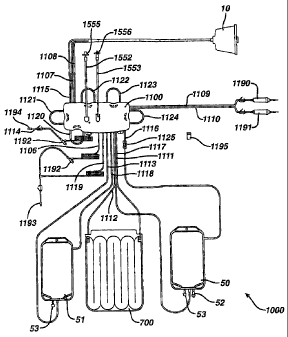
chemical or an
antibody. Ultraviolet light affects the bonding between the lymphocytes and
the chemical or
antibody that inhibits the metabolic processes of the lymphocytes.
[0003] Photopheresis systems and methods have been proposed and used which
involve
separation of buffy coat from the blood, addition of a photoactivatable drug,
and UV
irradiation of the buffy coat before re-infusion to the patient.
Extracorporeal photopheresis
may be utilized to treat numerous diseases including Graft-versus-Host
disease, Rheumatoid
Arthritis, Progressive Systematic Sclerosis, Juvenile Onset Diabetes,
Inflammatory Bowel
Disease--and-other-diseases-that-are-thou- ght to-be- T-cell-or-white blood
cell-mediated,
including cancer. Apheresis systems and methods have also been proposed and
used which
involve separation of blood into various components.
[0004] Additionally, apheresis systems and methods have also been proposed and
used which
involve separation of blood into various components, and also involve systems
pumping and
valving systems which are difficult to manufacture or operate. Prior
photopheresis and
apheresis systems and methods usually require batch processes and therefore
take several
CA 02479866 2012-07-12
hours to treat a patient or to obtain a sufficient supply of separated blood
components.
Furthermore, the systems are very complex to manufacture, especially the fluid
flow
controllers and valving systems.
[0005] In known photopheresis systems, a disposable kit is provided that is
loaded into a
permanent piece of hardware. The disposable kit contain complex tubing that is
used to carry
blood fluids to and from the various devices included in the kit, such as a
centrifuge bowl, an
irradiation chamber, and various bags for delivering and/or collecting blood
fluids. Known
disposable kits often contain a cassette, or other controller mechanism, for
controlling the
flow of blood fluids throughout the disposable kit and to and from the
patient. Disposable
kits are used only once and must be replaced or disposed after each treatment
session. In
performing a treatment process, the kit is connected to patient to form a
closed-loop system
and the various devices of the disposable kit are loaded into a permanent
piece of equipment
used to drive blood fluids throughout the disposable kit as necessary. Once
loaded, the
permanent blood drive system drives the blood fluids through the kit's fluid
circuitry.
[0006] Known permanent blood driving systems have control decks for receiving
the cassette
of the disposable cassette. In preparing for a blood treatment process, an
operator must
properly load the cassette into the deck and load the other devices of the kit
into their
appropriate positions. It is vital that the cassette be loaded properly and
not be able to move
during treatment. It is also vital to ensure that the disposable kit being
loaded onto the
permanent blood driving system is compatible with the blood driving system and
capable of
carrying out the intended treatment. However, these goals must be balanced
with the
competing goals of reducing the complexity of cassette clamping mechanisms so
as to reduce
operator loading errors and reducing kit loading time.
[0007] Another very real advancement in photopheresis systems would result if
the size,
manufacturing complexity, manufacturing costs, and tubing within the
disposable kit could
be reduced, even at the cost of a more complex blood driving system. This is
because the
blood driving system represents permanent reusable equipment, whereas a new
sterile
disposable kit must be used each time. Known disposable photopheresis kits are
difficult and
expensive to manufacture, especially the valving and pumping mechanisms within
the
cassette.
[0008] The size of existing permanent blood driving systems is another issue.
Known blood
driving systems are bulky and have a very large footprint, taking up valuable
hospital floor
2
CA 02479866 2012-07-12
space. Thus, the above goals must be achieved while maintaining, preferably
reducing, the
footprint of the permanent blood driving system.
[0009] Another deficiency in existing blood driving systems is their inability
to communicate
or receive real time data during a treatment. If a problem arises during the
treatment, either
the problem will not be detected and/or nothing can be done until after the
treatment. Thus, a
need exists for a blood driving system that can both communicate real time
data during a
treatment and respond if necessary to data inputs in real time during a
treatment process.
[0010] Additionally, prior photopheresis and apheresis systems and methods
usually require
batch processes and therefore take several hours to treat a patient or to
obtain a sufficient
supply of separated blood fragments. It is a constant object to reduce the
time it takes to
perform a complete photopheresis or apheresis treatment session. Another
object is to reduce
the amount of blood that must be drawn form a patient and processed in closed-
loop
processes per photopheresis treatment session. Yet another object to increase
the amount of
white blood cell yield or obtain a cleaner cut of buffy coat per volume of
whole blood
processed. Still another object is to reduce the costs and complexity
associated with making
the disposable kits used.
Disclosure of the Invention
[0011] These objects and others are met by the present invention. The present
invention is
directed at permanent blood driving systems for photopheresis and apheresis to
provide less
complex, easier to manufacture, and a continuous process for separation of
sufficient
fragment for treatment so as to greatly reduce the treatment time.
[0012] The invention, in one aspect, is an improved deck for driving fluids
through an
extracorporeal blood circuit kit. The kit including a cassette for controlling
fluid flow and
having at least one tab protruding from a housing of the cassette. The deck is
designed to
allow easy, quick, and reliable loading of the cassette through the use a new
cassette
clamping-mechanism. In-this-aspects the-deck-comprises: -a control-ler-a plat-
e-havi-ng-a
cassette loading area; at least one catch for slidably receiving a
corresponding tab of the
cassette, the catch positioned on the plate adjacent to the cassette loading
area; at least one
rotating clamp rotatable between an open position and a closed position, the
rotating clamp
positioned on the plate adjacent to the cassette loading area; wherein when
the rotating clamp
is in the open position, the rotating clamp does not obstruct the cassette
from being removed
from the cassette loading area; and wherein when the rotating clamp is in the
closed position
3
CA 02479866 2012-07-12
and the cassette loaded onto the cassette loading area, the rotating clamp
prohibits the
cassette from being removed from the cassette loading area.
[0013] It is preferable that the rotating clamps rotate about an axis that is
substantially
perpendicular to a top surface of the plate. It is further preferable that the
rotating clamps be
spring loaded so as to return to the closed position when rotational force is
not applied and
that the rotational clamps be operably coupled by a timing belt so that
rotation of all rotating
clamps is coordinated. Providing two catches and two rotating clamps is most
preferable.
[0014] Each rotating clamp will preferably have an angled ledge that allows
the cassette to be
lowered onto the cassette loading area of the plate while the rotating clamps
are in closed
position. The angled ledge will also prohibit the cassette from being raised
from the cassette
loading area when the rotating clamps are in the closed position. Rotation
between the open
and closed positions can be facilitated by pneumatic cylinders.
[0015] When the above claming mechanism is provided on a deck, a cassette can
be loaded
onto the deck by aligning the tabs of the cassette with the catches, slidably
inserting the tabs
into the catches, and pressing the cassette downward onto the cassette loading
area. As the
cassette is forced downward against the rotating clamps, the rotating clamps
are rotated to the
open position allowing the cassette to move below the angled ledge. When the
cassette is
below the angled ledges, the rotating clamps snap back to the closed position
locking the
cassette onto the cassette loading area. When this happens the cassette can
not be removed or
moved until the rotating clamps are moved to the open position after treatment
is complete or
until the operator does so manually. This setup provides little or no chance
for operator error
in loading the cassette and is time efficient.
[0016] Turning now to other elements of the inventive deck, it is preferred
that the deck also
have at least one compression actuator adapted to move between a raised
position and a
lowered position. When the cassette is loaded onto the cassette loading area,
and the
compression actuator is in the raised position, the compression actuator will
occlude a portion
of flexible tubing within the cassette by compressing the portion of flexible
tubing against a
housing of the cassette. As such, the compression actuators act as valves to
control and direct
fluid flow through desired fluid passageways of the kit. There are preferably
eight
compression actuators.
[0017] It is further that at least one of the compression actuators be spring
loaded so as to
return the compression actuator to the raised position when force is not
applied and that at
least one compression actuator be spring retracted so as to return the
compression actuator to
4
CA 02479866 2012-07-12
the lowered position when force is not applied. More preferably, the deck has
three
compression actuators that are spring loaded and positioned on the plate so
that when a
cassette is loaded onto the cassette loading area, the three spring loaded
compression
actuators are aligned with portions of flexible tubing within the cassette
that are connected
directly to a patient. These three compression actuators can be coupled to one
another so that
their movement between the lowered and raised positions is coordinated. It is
also preferred
that the deck have five compression actuators that are spring retracted and
positioned on the
plate so that when a cassette is loaded onto the cassette loading area, the
five compression
actuators are aligned with portions of flexible tubing within the cassette so
as to be able to
route fluids throughout the kit.
[0018] For patient safety, it is most preferable that the deck have an air
bubble detector
adapted to monitor tubes of the kit that are carrying fluids to and from a
patient when the
cassette is loaded onto the cassette loading area. When the air bubble
detector detects an air
bubble, it will take the necessary actions to prohibit flow of fluids to and
from the patient.
[0019] The deck will also preferably have at least one peristaltic pump
adjacent to the
cassette loading area for driving fluids through the kit. The peristaltic pump
will comprise a
rotor rotatably mounted about a rotor axis; a housing having a curved wall
surrounding at
least a portion of the rotor and forming a tube pumping region between the
rotor and the
curved wall; the rotor comprising at least one drive roller for progressively
compressing a
loop of tubing against the curved wall; the rotor comprising a flange above
the housing and
an angled guide extending upward from the flange for displacing the loop of
tubing toward
the flange upon the rotor being rotated in a forward direction; the flange
having an opening
with a leading edge and a trailing edge for capturing and feeding the loop of
tubing into the
tube pumping region upon the rotor being rotated in the forward direction; and
wherein the
trailing edge is higher than the leading edge. Most preferably, five
peristaltic pumps are
provided.
[0020] A hematocrit sensor for monitoring a tube of the kit that leads to a
treatment bag for
the presence of red blood cells is also preferably provided. The hematocrit
sensor can be
coupled to the controller to control the peristaltic pump that drives fluid
into the tube that
leads into the treatment bag.
[0021] In another aspect, the invention is a system for driving blood fluids
through a
disposable kit comprising: a housing having the deck described above; a
centrifuge chamber
within the housing; and an infrared communication port coupled to the
controller. Preferably,
CA 02479866 2011-06-22
the infrared communication port is adapted to transmit real time data relating
to a therapy
session being performed on the system to a remote device. Infrared
communication abilities
allow the system to be able to both transmit and receive data in real time
during a treatment
process without disturbing the treatment.
[0022] When the system is adapted to be used for photopheresis treatments, the
system will
further comprise a photoactivation chamber for receiving an irradiation
chamber of the kit.
The photoactivation chamber can be vertically oriented. It is more preferable
that a leak
detector be provided in the photoactivation chamber and that the leak detector
comprises at
least two U-shaped electrodes, a solid state switch connected to a first end
of the electrodes,
and an integrated circuit connected to a second end of the electrodes. The
leak detector is
coupled to the controller. It is still further preferable that a similar leak
detector also be
provided in the centrifuge chamber.
[0023] A means to authenticate a unique identifier associated with the kit is
also preferably
provided on the system. The authentication means is coupled to the controller.
The means to
authenticate can be a data card receiving slot.
[0024] In yet another aspect, the invention is a blood diving system having an
upright tower
configuration that reduces the footprint of the system, saving valuable
hospital floor space.
In this embodiment, the system will comprise: a controller; a base portion
having a top
having a deck for receiving and controlling a cassette for directing fluid
flow through the kit;
an upper portion atop the top; and a centrifuge chamber within the upper
portion. Placing the
centrifuge chamber above the deck reduces the footprint of the system and
provide a working
platform for the operator to place objects on.
[0025] When adapted to be used for photopheresis treatments, the system will
have a
photoactivation chamber in the base portion for receiving an irradiation
chamber of the kit.
The photoactivation chamber will be preferably vertically oriented and have a
leak detector
[0026] The system is provided with wheels for mobility and is preferably
designed to have a
height of the system is less than about 60 inches so as not to obstruct
visibility during
moving. Additionally, the system can be provided with all of the features
discussed above.
6
CA 02479866 2012-07-12
FIG. 1 is a schematic representation of an embodiment of a disposable kit for
use in
photopheresis therapy embodying features of the present invention.
FIG. 2 is an elevated perspective view of an embodiment of a cassette for
controlling
fluid flow in the disposable photopheresis kit of FIG. 1.
FIG. 3 is an exploded view of the cassette of FIG. 2.
FIG. 4 is a top view of the cassette of FIG. 2 with the cover removed and
showing
internal tubular circuitry.
FIG. 5 is a bottom view of a cover of cassette of FIG. 2.
FIG. 6 is an elevated perspective view of an embodiment of a filter assembly.
FIG. 7 is bottom perspective view of the filter assembly of FIG. 6.
FIG. 8 is an exploded view of the filter assembly of FIG. 6.
FIG. 9 is a rear perspective view of the filter assembly of FIG. 6.
FIG. 10 is schematic representation of the filter assembly of FIG. 6 coupled
to pressure
sensors and a data processor.
FIG. 11 is a front view of an irradiation chamber.
FIG. 12 is a side longitudinal view of the irradiation chamber of FIG. 11.
FIG. 13 is a side transverse view of the irradiation chamber of FIG. 11
FIG. 14 is a cut-away view of a section of the first plate and the second
plate prior to
being joined together to form the irradiation chamber of FIG. 11.
FIG. 15 is a cut-away dimensional end view of the irradiation chamber of FIG.
11.
FIG. 16 is a perspective view of the irradiation chamber of FIG. I 1
positioned within a
UVA light assembly.
FIG. 17 is an elevated perspective view of an embodiment of a permanent tower
system
for use in conjunction with a disposable kit for facilitating a photopheresis
therapy session.
FIG. 18 is a cross-sectional view of an embodiment of the photoactivation
chamber,
without a UVA light assembly, used in the tower system of FIG. 17.
FIG. 19 is a cross-sectional view of an embodiment of the centrifuge chamber
used in the
tower system of FIG. 17.
FIG. 20 is an electrical schematic of the leak detection circuit provided in
the
photoactivation chamber of FIG. 18.
FIG. 21 is an electrical schematic of the leak detection circuit provided in
the centrifuge
chamber of FIG. 19.
7
CA 02479866 2012-07-12
FIG. 22 is an elevated perspective view of an embodiment of the fluid flow
control deck
of the tower system of FIG. 17.
FIG. 23 is a perspective bottom view of the control deck of FIG. 22.
FIG. 24 is an exploded view of the control deck of FIG. 22.
FIG. 25 is a top perspective view of the control deck of FIG. 22 with the
cassette of FIG.
2 loaded thereon.
FIG. 26 is a flowchart of an embodiment of a photopheresis treatment process.
FIG. 27 is a schematic of an embodiment of the fluid flow circuit used in
performing the
treatment process of FIG. 26.
FIG. 28 is top perspective view an embodiment of a peristaltic pump.
FIG. 29 is a cross sectional side view of the peristaltic pump of FIG. 28.
FIG. 30 is a top perspective view the rotor of the peristaltic pump of FIG.
29.
FIG. 31 is a bottom perspective view of the rotor of FIG. 30.
FIG. 32 is a top view of the peristaltic pump of FIG. 28.
FIG. 33 is a top view of the peristaltic pump of FIG. 28 in a loading position
and near the
cassette of FIG. 2.
FIG. 34 is an electrical schematic of the infrared communication port circuit.
FIG. 35 illustrates an embodiment of a centrifuge bowl and a rotating frame.
FIG. 36 is a dimensional view of the bowl of FIG. 35.
FIG. 37 is an exploded view of the bowl of FIG. 36.
FIG. 38 shows a cross sectional view of the bowl of FIG. 36 along the line XIX-
XIX.
FIG. 39A shows a cross sectional view of a connection sleeve in place with a
lumen
connector of the bowl of FIG 38 along the line XX.
FIG. 39B shows another cross sectional view of a connection sleeve in place
with a lumen
connector of the bowl of FIG 38.
FIG. 40 shows a cross sectional view of the top core of the bowl of FIG. 37.
FIG. 41 shows a dimensional view of the top core and upper plate of FIG. 37.
FIG. 42 shows a bottom view of the top core of FIG. 41.
FIG. 43A shows a dimensional exploded view of the bottom core and a lower
plate of the
bowl of FIG. 37.
FIG. 43B shows an dimensional cross section view of the bottom core and a
lower plate
of the bowl of FIG. 43A attached together.
FIG. 44 shows an exploded side view of the bottom core and a lower plate of
FIG. 43A.
8
CA 02479866 2012-07-12
FIG. 45 shows a dimensional view of another embodiment of a conduit assembly.
FIG. 46 shows a dimensional view of the connection sleeve of FIG. 45.
FIG. 47 shows a dimensional view of one end of conduit assembly of FIG. 45.
FIG. 48 shows a dimensional view of an anchor end of the present invention.
FIG. 49 shows a lateral cross-sectional view of an anchor end.
FIG. 50 shows a horizontal cross-sectional view of an anchor end taken along
line XXI.
FIG. 51 illustrates a dimensional view of the rotating frame of FIG. 35.
FIG. 52 is an enlarged view of a holder for an external conduit.
FIG. 53 shows an alternative embodiment of the bowl with the cross-section
taken
similarly to that shown in FIG. 38.
FIG. 54 shows an alternative embodiment of the top core.
FIG. 55 shows an alternative embodiment of the connection sleeve.
Modes for Carrying Out The Invention
[0028] Features of the present invention are embodied in the permanent blood
driving
equipment, the disposable photopheresis kit, the various devices which make up
the
disposable kit, and the corresponding treatment process. The following written
description is
outlined as follows:
I. Disposable Photopheresis Kit
A. Cassette for Controlling Fluid Flow
1. Filter Assembly
B. Irradiation Chamber
C. Centrifuge Bowl
1. Drive Tube
II. Permanent Tower System
A Photoactivation Chamber
B. Centrifuge Chamber
C. Fluid Flow Control Deck
1. Cassette Clamping Mechanism
2. Self-Loading Peristaltic Pumps
D. Infra-Red Communication
III. Photopheresis Treatment Process
[0029] The above-outline is included to facilitate understanding of the
features of the present
invention. The outline is not limiting of the present invention and is not
intended to
categorize or limit any aspect of the invention. The inventions are described
and illustrated in
sufficient detail that those skilled in this art can readily make and use
them. However,
various alternatives, modifications, and improvements should become readily
apparent
9
CA 02479866 2012-07-12
without departing from the spirit and scope of the invention. Specifically,
while the invention
is described in the context of a disposable kit and permanent blood drive
system for use in
photopheresis therapy, certain aspects of the invention are not so limited and
are applicable to
kits and systems used for rendering other therapies, such as apheresis or any
other
extracorporeal blood treatment therapy.
1. Disposable Photopheresis Kit
[0030] FIG. 1 illustrates disposable photopheresis kit 1000 embodying features
of the present
invention. It is necessary that a new disposable sterile kit be used for each
therapy session. In
order to facilitate the circulation of fluids through photopheresis kit 1000,
and to treat blood
fluids circulating therethrough, photopheresis kit 1000 is installed in
permanent tower system
2000 (FIG. 17). The installation of photopheresis kit 1000 into tower system
2000 is
described in detail below.
[0031] Photopheresis kit 1000 comprises cassette 1100, centrifuge bowl 10,
irradiation
chamber700, hematocrit sensor 1125, removable data card 1195, treatment bag
50, and
plasma collection bag 51. Photopheresis kit 1000 further comprises saline
connector spike
1190 and anticoagulant connector spike 1191 for respectively connecting saline
and
anticoagulant fluid bags (not shown). Photopheresis kit 1000 has all the
necessary tubing and
connectors to fluidly connect all devices and to route the circulation of
fluids during a
photopheresis treatment session. All tubing is sterile medical grade flexible
tubing. Triport
connectors 1192 are provided at various positions for the introduction of
fluids into the tubing
if necessary.
[0032] Needle adapters 1193 and 1194 are provided for respectively connecting
photopheresis kit 1000 to needles for drawing whole blood from a patient and
returning blood
fluids to the patient. Alternatively, photopheresis kit 1000 can be adapted to
use a single
needle to both draw whole blood from the patient and return blood fluids to
the patient.
However, a two needle kit is preferred because of the ability to
simultaneously draw whole
blood and return blood fluids to the patient. When a patient is hooked up to
photopheresis kit
1000, a closed loop system is formed.
[0033] Cassette 1100 acts both as a tube organizer and a fluid flow router.
Irradiation
chamber 700 is used to expose blood fluids to UV light. Centrifuge bowl 10
separates whole
blood into its different components according to density. Treatment bag 50 is
a 1000mL
three port bag. Straight bond port 52 is used to inject a photoactivatable or
photosensitive
CA 02479866 2012-07-12
compound into treatment bag 50. Plasma collection bag 51 is 1000mL two port
bag. Both
treatment bag 50 and plasma collection bag 51 have a hinged cap spike tube 53
which can be
used for drainage if necessary. Photopheresis kit 1000 further comprises
hydrophobic filters
1555 and 1556 which are adapted to connect to pressure transducers 1550 and
1551 to filter
1500 via vent tubes 1552 and 1553 for monitoring and controlling the pressures
within tubes
connecting the patient (FIG. 10). Monitoring the pressure helps ensure that
the kit is
operating within safe pressure limits. The individual devices of photopheresis
kit 1000, and
their functioning, are discussed below in detail.
A. Cassette for Controlling Fluid Flow
[0034] FIG. 2 shows a top perspective view of a disposable cassette 1100 for
valving,
pumping, and controlling the movement of blood fluids during a photopheresis
treatment
session. Cassette 1100 has housing 1101 that forms an internal space that acts
as a casing for
its various internal components and tubular circuitry. Housing 1101 is
preferably made of
hard plastic, but can be made of any suitably rigid material. Housing 1101 has
side wall 1104
and top surface 1105. Side wall 1104 of housing 1101 has tabs 1102 and 1103
extending
therefrom. During a photopheresis treatment, cassette 1100 needs to be secured
to deck 1200
of tower system 2000, as is best illustrated in FIG. 25. Tabs 1102 and 1103
help position
and secure cassette 1100 to deck 1200.
[0035] Cassette 1100 has fluid inlet tubes 1106, 1107, 1108, 1109, 1110, 1111,
and 1112 for
receiving fluids into cassette 1100, fluid outlet tubes 1114, 1115, 1116,
1117, 1118, and 1119
for expelling fluids from cassette 1100, and fluid inlet/outlet tube 1113 that
can be used for
both introducing and expelling fluids into and out of cassette 1100. These
fluid input and
output tubes fluidly couple cassette 1100 to a patient being treated, as well
as the various
devices of photopheresis kit 1000, such as centrifuge bowl 10, irradiation
chamber700,
treatment bag 50, plasma collection bag 51, and bags containing saline,
anticoagulation fluid
to form a closed-loop extracorporeal fluid circuit (FIG. 27).
[0036] Pump tube loops 1120, 1121, 1122, 1123, and 1124 protrude from side
wall 1104 of
housing 1101. Pump tube loops 1120, 1121, 1122, 1123, and 1124 are provided
for
facilitating the circulation of fluids throughout photopheresis kit 1000
during therapy. More
specifically, when cassette 1100 is secured to deck 1200 for operation, each
one of said pump
tube loops 1120, 1121, 1122, 1123, and 1124 are loaded into a corresponding
peristaltic
pump 1301, 1302, 1303, 1304, and 1305 (FIG. 4). Peristaltic pumps 1301, 1302,
1303, 1304,
11
CA 02479866 2012-07-12
and 1305 drive fluid through the respective pump tube loops 1120, 1121, 1122,
1123, and
1124 in a predetermined direction, thereby driving fluid through photopheresis
kit 1000 (FIG.
1) as necessary. The operation and automatic loading and unloading of
peristaltic pumps
1301, 1302, 1303, 1304, and 1305 is discussed in detail below with respect to
FIGS. 28-33.
[0037] Turning now to FIG. 3, cassette 1100 is shown with housing 1101 in an
exploded
state. For ease of illustration and description, the internal tubular
circuitry within housing
1101 is not illustrated in FIG. 3. The internal tubular circuitry is
illustrated in FIG. 4 and will
be discussed in relation thereto. Cassette 1100 has filter assembly 1500
positioned therein
and in fluid connection with inlet tube 1106, outlet tube 1114, and one end of
each of pump
tube loops 1120 and 1121. Filter assembly 1500 comprises vent chambers 1540
and 1542.
Filter assembly 1500, and its functioning, is discussed in detail below with
respect to FIGS.
6-10.
[0038] Housing 1101 comprises cover 1130 and base 1131. Cover 1130 has top
surface
1105, a bottom surface 1160 (FIG. 5), and side wall 1104. Cover 1130 has
openings 1132
and 1133 for allowing vent chambers 1540 and 1542 of filter assembly 1500 to
extend
therethrough. Side wall 1104 has a plurality of tube slots 1134 to allow the
inlet tubes, outlet
tubes, and pump loop tubes to pass into the internal space of housing 1101 for
connection
with the internal tubular circuitry located therein. Only a few tube slots
1134 are labeled in
FIG. 3 to avoid numerical crowding. Tabs 1102 and 1103 are positioned on side
wall 1104
so as not to interfere with tube slots 1134. Cover 1130 has occlusion bars
1162 and 1162A
extending from bottom surface 1160 (FIG. 5). Occlusion bars 1162 and 1162A are
preferably
molded into bottom surface 1160 of cover 1130 during its formation.
[0039] Base 1131 has a plurality of U-shaped tube-holders 1135 extending
upward from top
surface 1136. U-shaped tube holders 1135 hold the inlet tubes, outlet tubes,
pump loop tubes,
filter assembly, and internal tubular circuitry in place. Only a few U-shaped
holders 1135 are
labeled in FIG. 3 to avoid numerical crowding. Preferably, a U-shaped holder
1135 is
provided on base 1131 at each location where an inlet tube, an outlet tube, or
a pump loop
tube passes through a tube slot 1134 on side wall 1104. Male extrusions 1136
protrude from
top surface 1136 of base 1131 for mating with corresponding female holes 1161
located on
bottom surface 1160 of cover 1130 (FIG. 5). Preferably, a male protrusion 1136
is located at
or near each of the four corners of base 1130 and near filt.,r 1500. Male
protrusions 1136
mate with the female holes 1161 to form a snap-fit and secure base 1131 to
cover 1130.
12
CA 02479866 2012-07-12
[0040] Base 1131 further comprises a hub 1140. Hub 1140 is a five-way tube
connector used
to connect five tubes of the internal tubular circuitry. Preferably, three
apertures 1137 are
located near and surround three of the tubes leading into hub 1140. Hub 1140
acts as a
centralized junction which can be used, in conjunction with compression
actuators 1240-1247
(FIG. 22), to direct fluids through photopheresis kit 1000 and to and from the
patient. In
addition to hub 1140, appropriate tube connectors, such as T-connectors 1141
and Y-
connector 1142, are used to obtain the desired flexible tubing pathways.
[0041] Five apertures 1137 are located on the floor of base 1130. Each
aperture 1137 is
surrounded by an aperture wall 1138 having slots 1139 for passing portions of
the internal
tubular circuitry therethrough. An elongated aperture 1157 is also provided on
the floor of
base 1131. Apertures 1137 are located on base 1131 to align with corresponding
compression actuators 1243-1247 of deck 1200 (FIG. 22). Aperture 1157 is
located on base
1131 to align with compression actuators 1240-1242 of deck 1200 (FIG. 22).
Each aperture
1137 is sized so that a single compression actuator 1243-1247 can extend
therethrough.
Aperture 1157 is sized so that three compression actuators 1240-1242 can
extend
therethrough. Compression actuators 1240-1247 are used to close/occlude and
open certain
fluid passageways of the internal tubular circuitry in order to facilitate or
prohibit fluid flow
along a desired path. When it is desired to have a certain passageway open so
that fluid can
flow therethrough, the compression actuator 1240-1247 for that passageway is
in a lowered
position However, when it is desired to have a certain fluid passageway closed
so that fluid
can not flow therethrough, the appropriate compression actuator 1240-1247 is
raised,
extending the compression actuator 1240-1247 through aperture 1137 or 1157 and
compressing a portion of the flexible tubular circuitry against bottom surface
1160 (FIG. 5)
of cover 1130, thereby closing that passageway. Preferably, occlusion bars
1163 and 1173
(FIG. 5) are positioned on bottom surface 1160 to align with the compression
actuators 1240-
1247 so that the portion of flexible tubing being occluded is compressed
against occlusion bar
1163 or 1173. Alternatively, the occlusion bar can be omitted or located on
the compression
actuators themselves.
[0042] It is preferable for cassette 1100 to have a unique identifier that can
communicate
with and relay information to permanent tower system 2000. The unique
identifier is
provided to ensure that the disposable photopheresis kit is compatible with
the blood drive
equipment into which it is being loaded, and that the photopheresis kit is
capable of running
the desired treatment process. The unique identifier can also be used as a
means to ensure
13
CA 02479866 2012-07-12
that the disposable photopheresis kit is of a certain brand name or make. In
the illustrated
example, the unique identifier is embodied as data card 1195 (FIG. 2) that is
inserted into
data card receiving port 2001 of permanent tower system 2000 (FIG. 17). Data
card 1195 has
both read and write capabilities and can store data relating to the treatment
therapy performed
for future analysis. The unique identifier can also take on a variety of
forms, including, for
example, a microchip that interacts with the blood drive equipment when the
kit is loaded, a
bar code, or a serial number.
[0043] Cover 1130 has data card holder 1134 for holding data card 1195 (FIG.
1). Data card
holder 1134 comprises four elevated ridges in a segmented rectangular shape
for receiving
and holding data card 1195 to cassette 1100. Data card holder 1134 holds data
card 1195 in
place via a snap-fit (FIG. 2).
[0044] Referring now to FIGS. 1 and 4, the internal tubular circuitry of
cassette 1100 will
now be discussed. At least a portion of the internal tubular circuitry is
preferably made of
flexible plastic tubing that can be pinched shut by the exertion of pressure
without
compromising the hermetic integrity of the tube. Base 1131 of cassette 1100 is
illustrated in
FIG. 4 so that the internal tubular circuitry can be viewed. Inlet tubes 1107
and 1108 and
outlet tube 1115 are provided for coupling cassette 1100 to centrifuge bowl 10
(FIG. 1).
More specifically, outlet tube 1115 is provide for delivering whole blood from
cassette 1100
to centrifuge bowl 10, and inlet tubes 1107 and 1108 are respectively provide
for returning a
lower density blood components and higher density blood components to cassette
1100 for
further routing through photopheresis kit 1000. The lower density blood
components can
include, for example, plasma, leukocytes, platelets, buffy coat, or any
combination thereof.
The higher density components can include, for example, red blood cells.
Outlet tube 1117
and inlet tube 1112 fluidly couple cassette 1100 to irradiation chamber 700.
More
specifically, outlet tube 1117 is provided for delivering an untreated lower
density blood
component, for example buffy coat, to irradiation chamber700 for exposure to
photo energy,
while inlet tube 1112 is provided for returning the treated lower density
blood component to
cassette 1100 for further routing.
[0045] Inlet tube 1111 and outlet tube 1116 couple treatment bag 50 to
cassette 1100. Outlet
tube 1116 is provided to deliver an untreated low density blood component, for
example
buffy coat, to treatment bag 50. Outlet tube 1116 has hematocrit ("HCT")
sensor 1125
operably connected thereto to monitor for the introduction of a high density
blood
component, such as red blood cells. HCT sensor 1125 is a photo sensor assembly
and is
14
CA 02479866 2012-07-12
operably coupled to a controller. HCT sensor 1125 sends a detection signal to
the controller
when red blood cells are detected in outlet tube 1116 and the controller will
take the
appropriate action. Inlet tube 1111 is provided to return the untreated low
density blood
component from treatment bag 50 to cassette 1100 for further routing. Inlet
tubes 1109 and
1110 are respectively connected to a saline and anticoagulant storage bags
(not shown) via
spikes 1190 and 1191 and are provided for delivering saline and an
anticoagulant fluid to
cassette 1100 for further routing to the patient.
[0046] Inlet/Outlet tube 1113 and outlet tube 1118 couple plasma collection
bag 50 to
cassette 1100. More specifically, outlet tube 1118 delivers a blood component,
such as
plasma, to plasma collection bag 51. Inlet/Outlet tube 1113 can be used to
either deliver red
blood cells to plasma collection bag 51 from cassette 1100 or return the blood
component(s)
that build up in plasma collection bag 51 to cassette 1100 for further
routing. Inlet tube 1106
and outlet tubes 1119 and 1114 are coupled to a patient. Specifically, outlet
tube 1114 is
provided to return treated blood, saline, untreated blood components, treated
blood
components, and other fluids back to the patient. Inlet tube 1106 is provided
for delivering
untreated whole blood (and a predetermined amount of an anticoagulant fluid)
from the
patient to cassette 1100 for routing and treatment within photopheresis kit
1000. Outlet tube
1119 is specifically provided for delivering an anticoagulant fluid to inlet
tube 1106. It is
preferable that all tubing is disposable medical grade sterile tubing.
Flexible plastic tubing is
the most preferred.
[0047] Cassette 1100 has five pump tube loops 1120, 1121, 1122, 1123, and 1124
for driving
blood fluids throughout cassette 1100 and photopheresis kit 1000. More
specifically, pump
tube loop 1121 loads into whole blood pump 1301 and respectively drives whole
blood in and
out of cassette 1100 via inlet tube 1106 and outlet tube 1115, passing through
filter 1500
along the way. Pump loop tube 1120 loads into return pump 1302 and drives
blood fluids
through filter 1500 and back to the patient via outlet tube 1114. Pump loop
tube 1122 loads
into red blood cell pump 1305 and draws red blood cells from centrifuge bowl
10 and drives
them into cassette 1100 via inlet line 1108. Pump loop tube 1123 loads into
anticoagulant
pump 1304 and drives an anticoagulant fluid into cassette 1100 via inlet tube
1124 and out of
cassette 1100 to via outlet tube 1119, which connects with inlet tube 1106.
Pump loop tube
1124 loads into recirculation pump 1303 and drives blood fluids, such as
plasma, through
treatment bag 50 and irradiation chamber700 from cassette 1100.
CA 02479866 2012-07-12
[0048] Each of peristaltic pumps 1301-1305 are activated when necessary to
perform the
photopheresis treatment therapy according to an embodiment of the method of
the present
invention which is described below in relation to FIGS. 26-27. Peristaltic
pumps 1301-1305
can be operated one at a time or in any combination. The pumps 1301-1305 work
in
conjunction with compression actuators 1240-1247 to direct fluids through
desired pathways
of photopheresis kit 1000. Apertures 1137 and 1157 are strategically located
on base 1131
along the internal tubular circuitry to facilitate proper routing. Through the
use of
compression actuators 1240-1247, the fluids can be directed along any pathway
or
combination thereof.
1. The Filter Assembly
[0049] Filter 1500, which is located within cassette 1100 as described above,
is illustrated in
detail in FIGS. 6-10. Referring first to FIGS. 6 and 7, filter 1500 is
illustrated fully
assembled. Filter 1500 comprises a filter housing 1501. Filter housing 1501 is
preferably
constructed of a transparent or translucent medical grade plastic. However,
the invention is
not so limited and filter housing 1501 can be constructed of any material that
will not
contaminate blood or other fluids that are flowing therethrough.
[0050] Filter housing 1501 has four fluid connection ports extruding
therefrom, namely
whole blood inlet port 1502, whole blood outlet port 1503, treated fluid inlet
port 1504, and
treated fluid outlet port 1505. Ports 1502-1505 are standard medical tubing
connection ports
that allow medical tubing to be fluidly connected thereto. Ports 1502-1505
respectively
contain openings 1506, 1507, 1508 and 1509. Openings 1506, 1507, 1508 and 1509
extend
through ports 1502, 1503, 1504 and 1505, forming fluid passageways into filter
housing 1501
at the desired locations.
[0051] Ports 1502, 1503, 1504 and 1505 are also used to secure filter 1500
within cassette
1100. In doing so, ports 1502, 1503, 1504 and 1505 can engage U-shaped
fasteners 1135 of
cassette 1100 (FIG. 3). Filter housing 1501 also has a prc-rusion 1510
extending the bottom
surface of housing floor 1518. Protrusion 1510 fits into a guide hole of base
1131 of cassette
1100 (FIG. 3).
[0052] Referring now to FIG. 8, filter 1500 is illustrated in an exploded
state. Filter housing
1501 is a two-piece assembly comprising roof 1511 and base 1512. Roof 1511 is
connected
to base 1512 by any means known in the art, such as ultrasonic welding, heat
welding,
applying an adhesive, or by designing roof 1511 and base 1512 so that a tight
fit results
16
CA 02479866 2012-07-12
between the two. While filter housing 1501 is illustrated as a two-piece
assembly, filter
housing 1501 can be either a single piece structure or a multi-piece assembly.
[0053] Base 1512 has chamber separation wall 1513 extending upward from a top
surface of
housing floor 1518 (FIG. 7). When base 1512 and roof 1511 are assembled, top
surface 1515
of chamber separation wall 1513 contacts the bottom surface of roof 1511,
forming two
chambers within the filter housing, whole blood chamber 1516 and filter
chamber 1517.
Fluid can not directly pass between whole blood chamber 1516 and filter
chamber 1517.
[0054] Whole blood chamber 1516 is a substantially L-shaped chamber having
floor 1514.
Whole blood chamber 1516 has a whole blood inlet hole 1519 and a whole blood
outlet hole
(not illustrated) in floor 1514. Whole blood inlet hole 1519 and the whole
blood outlet hole
are located at or near the ends of the substantially L-shaped whole blood
chamber 1516.
Whole blood inlet hole 1519 forms a passageway with opening 1506 of inlet port
1502 so that
a fluid can flow into whole blood chamber 1516. Similarly, the whole blood
outlet hole (not
illustrated) forms a passageway with opening 1507 of outlet port 1503 so that
fluid can flow
out of whole blood chamber 1516.
[0055] Filter chamber 1517 has floor 1520. Floor 1520 has elevated ridge 1521
extending
upward therefrom. Elevated ridge 1521 is rectangular and forms a perimeter.
While elevated
ridge 1521 is rectangular in the illustrated embodiment, elevated ridge 1521
can be any shape
so long as it forms an enclosed perimeter. The height of elevated ridge 1521
is less than the
height of chamber separation wall 1513. As such, when roof 1511 and base 1512
are
assembled, space exists between the top of elevated ridge 1521 and the bottom
surface of roof
1511. Elevated ridge 1521 and chamber separation wall 1513 form a trench 1524
there
between.
[0056] In order to facilitate fluid flow through filter chamber 1517, floor
1520 of filter
chamber 1517 has treated fluid inlet hole 1522 and treated fluid outlet hole
1523. Treated
fluid inlet hole 1522 is located exterior of the perimeter formed by elevated
ridge 1521 and
forms a passageway with opening 1508 of inlet port 1504 so that a fluid can
flow into filter
chamber 1517 from outside filter housing 1501. Treated fluid outlet hole 1523
is located
interior of the perimeter formed by elevated ridge 1521 and forms a passageway
with opening
1509 of outlet port 1505 so that a fluid can flow out of filter chamber 1517.
[0057] Filter 1500 further comprises filter element 1530. Filter element 1530
comprises
frame 1531 having filter media 1532 positioned therein. Frame 1531 has a neck
1534 that
forms a filter inlet hole 1533. Filter element 1530 is positioned in filter
chamber 1517 so that
17
CA 02479866 2012-07-12
frame 1531 fits into trench 1524 and neck 1534 surrounds treated blood inlet
hole 1522.
Filter inlet hole 1533 is aligned with treated fluid inlet hole 1522 so that
incoming fluid can
freely flow through holes 1522 and 1533 into filter chamber 1517. Frame 1531
of filter
element 1530 forms a hermetic fit with elevated ridge 1521. All fluid that
enters filter
chamber 1517 through holes 1522 and 1533 must pass through filter media 1532
in order to
exit filter chamber 1517 via treated fluid outlet hole 1523. Filter media 1532
preferably has a
pore size of approximately 200 microns. Filter media 1532 can be formed of
woven mesh,
such as woven polyester.
[0058] Filter chamber 1517 further comprises filter vent chamber 1540 within
roof 1511.
Filter vent chamber 1540 has gas vent 1541 in the form of a hole (FIG. 9).
Because gas vent
1541 opens into filter vent chamber 1540 which in turn opens into filter
chamber 1517, gases
that build-up within filter chamber 1517 can escape through gas vent 1541.
Similarly, whole
blood chamber 1516 comprises blood vent chamber 1542 within roof 1511. Blood
vent
chamber 1541 has gas vent 1543 in the form of a hole. Because gas vent 1543
opens into
blood vent chamber 1542 which in turn opens into whole blood chamber 1517,
gases that
build-up in whole blood chamber 1516 can escape via gas vent 1543.
[0059] FIG. 10 is a top view of filter 1500 having pressure sensors 1550 and
1551 connected
to gas vents 1541 and 1543. Pressure sensors 1550 and 1551 are preferably
pressure
transducers. Pressure sensor 1550 is connected to gas vent 1541 via vent
tubing 1552. Vent
tubing 1552 fits into gas vent 1541 so as to form a tight fit and seal.
Because gas vent 1541
opens into filter vent chamber 1540 which in turn opens into filter chamber
1517, the
pressure in vent tubing 1552 is the same as in filter chamber 1517. By
measuring the
pressure in vent tubing 1552, pressure sensor 1550 also measures the pressure
within filter
chamber 1517. Similarly, pressure sensor 1551 is connected to gas vent 1543
via vent tubing
1553. Vent tubing 1553 fits into gas vent 1543 so as to form a tight fit and
seal and pressure
sensor 1551 measures the pressure within whole blood chamber 1516. Filter vent
chamber
1540 and blood vent chamber 1542 extend through openings 1132 and 1133 of
cassette 1100
when filter 1500 is positioned therein (FIG. 2). This allows the pressure
within chambers
1516 and 1517 to be monitored while still protecting filter chamber 1500 and
the fluid
connections thereto.
[0060] Pressure sensors 1550 and 1551 are coupled to controller 1554, which is
a properly
programmed processor. Controller 1554 can be a main processor used to drive
the entire
system or can be a separate processor coupled to a main processor. Pressure
sensors 1550
18
CA 02479866 2012-07-12
and 1551 produce electrical output signals representative of the pressure
readings within
chambers 1517 and 1516 respectively. Controller 1554 receives on a frequent or
continuous
basis data representing the pressure within chambers 1516 and 1517. Controller
1554 is
programmed with values representing desired pressures within chambers 1516 and
1517.
Controller 1554 continuously analyzes the pressure data it receives from
pressure sensors
1550 and 1551 to determine whether the pressure readings are within a
predetermined range
from the desired pressure for chambers 1517 and 1516. Controller 1554 is also
coupled to
whole blood pump 1301 and return pump 1302. In response to the pressure data
received
from pressure sensors 1551 and 1550, controller 1554 is programmed to control
the speed of
whole blood pump 1301 and return pump 1302, thereby adjusting the flow rates
through the
pumps 1301 and 1301. Adjusting these flow rates in turn adjust the pressure
within whole
blood chambers 1516 and filter chamber 1517 respectively. It is in this way
that the pressure
within the lines drawing and returning blood to and from the patient is
maintained at
acceptable levels.
[0061] The functioning of filter 1500 during a photopheresis therapy session
will now be
discussed in relation to FIGS. 1, 6, and 10. While the functioning of filter
1500 will be
described in detail with respect to drawing whole blood from a patient and
returning a
component of said whole blood back into the patient after it is treated, the
invention is not so
limited. Filter 1500 can be used in connection with almost any fluid,
including red blood
cells, white blood cells, buffy coat, plasma, or a combination thereof.
[0062] Whole blood pump 1601 draws whole blood from a patient who is connected
to
photopheresis kit 1000 via a needle connected to port 1193. The rotational
speed of whole
blood pump is set so that the pressure of the line drawing the whole blood
from the patient is
at an acceptable level. Upon being drawn from the patient, the whole blood
passes into
cassette 1100 via inlet tube 1106. Inlet tube 1106 is fluidly connected to
inlet port 1502 of
filter 1500. The whole blood passes through opening 1506 of inlet port 1502
and into L-
shaped whole blood chamber 1516. The whole blood enters chamber 1516 through
inlet hole
1519 which is located on floor 1514. As more whole blood enters chamber 1516,
the whole
blood spills along floor 1514 until it reaches the whole blood outlet hole
(not illustrated) at
the other end of L-shaped whole blood chamber 1516. As discussed above, the
whole blood
outlet whole forms a passageway with opening 1507 of outlet port 1503. The
whole blood
that is within chamber 1516 flows across floor 1514, through the whole blood
outlet hole,
into outlet port 1503, and out of filter 1500 through opening 1507.
19
CA 02479866 2012-07-12
[0063] As the whole blood passes through whole blood chamber 1516, gases that
are trapped
in the whole blood escape. These gases collect in blood vent chamber 1542 and
then escape
via gas vent 1543. Pressure sensor 1551 continuously monitors the pressure
within blood
chamber 1516 through vent tube 1553 and transmits corresponding pressure data
to controller
1554. Controller 1554 analyzes the received pressure data and if necessary
adjusts the speed
of whole blood pump 1301, thereby adjusting the flow rate and pressure within
chamber 1516
and inlet tube 1106. Controller 1554 adjust the pump speed to ensure that the
pressure is
within the desired pressure range.
[0064] The whole blood then exits filter 1500 through outlet port 1503 and
passes out of
cassette 1100 via outlet tube 1115. The whole blood is then separated into
components
and/or treated as described in detail below. Before being returned to the
patient, this treated
fluid (i.e. treated blood or blood components) must be filtered. Untreated
fluids such as red
blood cells also must be filtered and will subjected to the below filtering
process. The treated
fluid is fed into filter chamber 1517 through opening 1508 of inlet port 1504.
Inlet port1504
is fluidly connected to pump loop tube 1120. The treated fluid enters filter
chamber 151'.
through inlet hole 1522 and passes through filter inlet hole 1533 of filter
element 1530. Th,
treated fluid fills filter chamber 1517 until it spills over frame 1531 of
filter element 1530,
which is secured to elevated ridge 1521. The treated fluid passes through
filter media 1532.
Filter media 1532 removes contaminants and other undesired materials from the
treated fluid
while at the same facilitating the release of trapped gases from the treated
fluid. The treated
fluid that passes through filter media 1532 gathers on floor 1520 of filter
chamber 1517
within the perimeter formed by elevated ridge 1521. This treated fluid then
passes into
treated fluid outlet hole 1523 and out of filter 1500 through opening 1506 of
outlet port 1502.
The treated fluid is then returned to the patient via outlet tube 1114, which
is fluidly
connected to outlet port 1502. The treated fluid is driven through filter
chamber 1517 and
outlet tube 1114 by return pump 1302.
[0065] Gases that are trapped in the treated fluid escape and collect in
filter vent chamber
1540 as the treated fluid flows through filter chamber 1517. These gases then
escape filter
1500 via gas vent 1541. Pressure sensor 1550 continuously monitors the
pressure within
filter chamber 1517 through vent tube 1552 and transmits corresponding
pressure data to
controller 1554. Controller 1554 analyzes the received pressure data and
compares it to the
desired pressure value and range. If necessary, controller 1554 adjusts the
speed of return
CA 02479866 2012-07-12
pump 1302, thereby adjusting the flow rate and pressure within chamber 1517
and outlet tube
1114.
B. Irradiation Chamber
[0066] FIGS. 11-16 illustrate irradiation chamber700 of photopheresis kit 1000
in detail.
Referring first to Fig. 11, irradiation chamber700 is formed by joining two
plates, a front and
a back plate having a thickness of preferably about 0.06 in. to about 0.2 in.,
which are
preferably comprised of a material ideally transparent to the wavelength of
electromagnetic
radiation. In the case of ultraviolet A radiation, polycarbonate has been
found most preferred
although other materials such as acrylic may be employed. Similarly, many
known methods
of bonding may be employed and need not be expanded on here.
[0067] The first plate 702 has a first surface 712 and a second surface 714.
In a preferred
embodiment the first plate 702 has a first port 705 on a first surface 712, in
fluid
communications with the second surface 714. The second surface 714 of the
first plate 702
has a raised boundary 726A defining an enclosure. The boundary 726A preferably
extends
substantially perpendicular from the second surface 714 (i.e. about 80-100
degrees).
Extending from the second surface 714 (preferably substantially
perpendicularly) are raised
partitions 720A. The boundary 726A surrounds the partitions 720A. One end of
each
partition 720A extends and contacts the boundary 726A.
[0068] The second plate 701 has a first surface 711 and a second surface 713.
In a preferred
embodiment the second plate 701 preferably has a second port 730 on a first
surface 711, in
fluid communications with the second surface 713. The second surface 713 of
the back plate
701 has a raised boundary 726B defining an enclosure. The boundary 726B
preferably
extends substantially perpendicular from the second surface 713 (i.e. about 80-
100 degrees).
Extending from the second surface 713 (preferably substantially perpendicular)
are raised
partitions (720B). The boundary 726B surrounds the partitions 720B. One end of
each
--------------- ----- -
partition 720A extends and contacts one side of boundary (726B).
[0069] The joining of the second surfaces of the first and second plates
results in a fluid tight
junction between boundaries 726A and 726B thereby forming boundary 726.
Partitions 720A
and 720B are also joined forming a fluid tight junction thereby forming
partition 720. The
boundary 726 forms an irradiation chamber700 and together with the partitions
720 provides
a pathway 710 having channels 715 for conducting fluid. The pathway maybe
serpentine,
zig-zag, or dove-tailed. Currently preferred is a serpentine pathway.
21
CA 02479866 2012-07-12
[0070] With reference to FIG. 11 and 12, irradiation chamber700 comprises a
serpentine
pathway 710 for conducting patient fluid, such as buffy coat or white blood
cells, from inlet
port 705 to outlet port 730, i.e., the serpentine pathway 710 is in fluid
communication with
inlet port 705 of front plate 702 and outlet port 730 of back plate 701.
Patient fluid is
supplied from cassette 1100 to inlet port 705 via outlet tube 1117. After
photoactivation and
passing through serpentine pathway 710, the treated patient fluid is returned
to cassette 1100
via inlet tube 1112 (FIGS. 1 and 4). The patient fluid is driven by
recirculation pump 1303.
Self-shielding effects of the cells is reduced while the cells are
photoactivated by irradiation
impinging upon both sides of irradiation chamber700.
[0071] Figure 11 shows pin 740 and recess 735 which align the two plates of
irradiation
chamber prior to being joined together in a sealing arrangement by RF welding,
heat impulse
welding, solvent welding or adhesive bonding. Joining of the plates by
adhesive bonding and
RF welding is more preferred. Joining of the front and back plates by RF
welding is most
preferred as the design of the raised partitions 720 and perimeter 725
minimizes flashing and
allows for even application of RF energy. Locations of pin 740 and recess 735
may be inside
serpentine pathway 710 or outside of serpentine pathway 710. Figure 2 also
shows a view of
an irradiation chamber with axis L. Rotation of chamber 700 180 degree about
axis L gives
the original configuration of the irradiation chamber. The irradiation chamber
of the present
invention has C2 symmetry about axis L.
[0072] Referring to FIGS. 11, 13, and 16, the leukocyte enriched blood,
plasma, and priming
solution are delivered through inlet port 705 of front plate 702 of
irradiation chamber700 into
channel 715, The channel 715 in the irradiation chamber700 is relatively
"thin" (e.g. on the
order of approximately 0.04" as distance between two plates) in order to
present large surface
area of leukocyte rich blood to irradiation and reduce the self-shielding
effects encountered
with lower surface area/volume ratios. The cross section shape of channel 715
is
substantially rectangular (e.g. rectangular, rhomboidal or trapezoidal) which
has as its long
side the distance between partition 720 and the distance between the plates as
its short side.
The shape of the cross section is designed for optimal irradiation of cells
passing through
channel 715. While a serpentine pathway 710 is preferred in order to avoid or
minimize
stagnant areas of flow, other arrangements are contemplated.
[0073] The irradiation chamber 700 allows efficient activation of
photoactivatable agents by
irradiation from a light array assembly, such as the PHOTOSETTE 's two banks
of UVA
lamps (758) for activation (Figure 16). The irradiation plate and UVA light
assembly (759)
22
CA 02479866 2012-07-12
are designed to be used in a setting where edge 706 is oriented downward and
edge 707
points upward. In this orientation, fluids entering input port 705 can exit
from outlet port 730
with the aid of gravity. In the most preferred embodiment, irradiation of both
sides of the
irradiation chamber takes place concurrently while still permitting facile
removal of the
chamber. UVA light assembly 759 is located within UV chamber 750 of permanent
tower
system 2000 (FIGS. 17 and18).
[0074] The irradiation chamber's fluid pathway loops to form two or more
channels in which
the leukocyte-enriched blood is circulated during photoactivation by UVA
light. Preferably,
irradiation chamber 700 has between 4 to 12 channels. More preferably, the
irradiation
chamber has 6 to 8 channels. Most preferably, the irradiation chamber has 8
channels.
[0075] Figure 14 shows cut-away views of the irradiation chamber. The channels
715 of
serpentine pathway 710 are formed by the joining of raised partition 720 and
perimeter 726
of the plates.
[0076] The irradiation chamber of the present invention can be made from a
biocompatible
material and can be sterilized by known methods such as heating, radiation
exposure or
treatment with ethylene oxide (ETO).
[0077] The method of irradiating cells using irradiation chamber 700 during
extracorporeal
treatment of cells with electromagnetic radiation (UVA) to be used in the
treatment of a
patient (such as to induce apoptosis in the cells and administer the cells
into the patient) will
now be discussed. Preferably the cells treated will be white cells.
[0078] In one embodiment of this method, a photoactivatable or photosensitive
compound is
first administered to at least a portion of the blood of a recipient prior to
the extracorporeal
treatment of the cells. The photoactivatable or photosensitive compound may be
administered in vivo (e.g., orally or intravenously). The photosensitive
compound, when
administered in vivo may be administered orally, but also may be administered
intravenously
and/or by other conventional administration routes. The oral dosage of the
photosensitive
compound may be in the range of about 0.3 to about 0.7 rng/kg., more
specifically, about 0.6
mg/kg.
[0079] When administered orally, the photosensitive compound may be
administered at least
about one hour prior to the photopheresis treatment and no more than about
three hours prior
to the photopheresis treatment. If administered intravenously, the times would
be shorter.
Alternatively, the photosensitive compound may be administered prior to or
contemporaneously with exposure to ultraviolet light. The photosensitive
compound may be
23
CA 02479866 2012-07-12
administered to whole blood or a fraction thereof provided that the target
blood cells or blood
components receive the photosensitive compound. A portion of the blood could
first be
processed using known methods to substantially remove the erythrocytes and the
photoactive
compound may then be administered to the resulting enriched leukocyte
fraction. In one
embodiment, the blood cells comprise white blood cells, specifically, T-cells.
[0080] The photoactivatable or photosensitive compound may, in the case of
some psoralens,
be capable of binding to nucleic acids upon activation by exposure to
electromagnetic
radiation of a prescribed spectrum, e.g., ultraviolet light.
[0081] Photoactive compounds may include, but are not limited to, compounds
known as
psoralens (or furocoumarins) as well as psoralen derivatives such as those
described in, for
example, U.S. Pat. No. 4,321,919 and U.S. Pat. No. 5,399,719. The
photoactivatable or
photosensitive compounds that may be used in accordance with the present
invention include,
but are not limited to, psoralen and psoralen derivatives; 8-methoxypsoralen;
4,5'8-
trimethylpsoralen; 5-methoxypsoralen; 4-methylpsoralen; 4,4-dimethylpsoralen;
4-5'-
dimethylpsoralen; 4'-aminomethyl-4,5',8-timethylpsoralen; 4'-hydroxymethyl-
4,5',8-
trimethylpsoralen; 4',8-methoxypsoralen; and a 4'-(omega-amino-2-oxa) alkyl-
4,5',8-
trimethylpsoralen, including but not limited to 4'-(4-amino-2-oxa)butyl-4,5',8-
trimethylpsoralen. In one embodiment, the photosensitive compound that may be
used
comprises the psoralen derivative, amotosalen (S-59) (Cerus, Corp., Concord,
CA). See, e.g.,
U.S. Patent Nos. 6,552,286; 6,469,052; and 6,420,570. In another embodiment,
the
photosensitive compound that may be used in accordance with the invention
comprises 8-
methoxypsoralen.
[0082] Methoxsalen is a naturally occurring photoactive substance found in the
seed of the
Ammi majus (umbelliferae plant). It belongs to a class of compounds known as
psoralens or
furocoumarins. The chemical name is 9-methoxy-7H-furo[3,2-g][1]-benzopyran-7-
one. The
formulation of the drug is a sterile liquid at a concentration of 20 mcg/mL in
a 10 mL vial.
See http://www.therakos.com/TherakosUS/pdf/uvadexpi.pdf. Toxicology studies of
extracorporeal photopheresis and different dosages of UVADEX and ultraviolet
light in
beagle dogs is located in the investigator's brochure.
[0083] Next, the portion of the subject's blood, recipient's blood, or the
donor's blood to
which the photoactive compound has been administered is treated by subjecting
the portion
of the blood to photopheresis using ultraviolet light. The photopheresis
treatment may be
carried out using long wavelength ultraviolet light (UVA) at a wavelength
within the range of
24
CA 02479866 2012-07-12
320 to 400 nm. Such a range is not limiting, however, but is merely provided
as an example.
The exposure to ultraviolet light during the photopheresis treatment may have
a duration of
sufficient length to deliver, for example, about 1-2 J/cm2 to the blood.
[0084] The photopheresis step is carried out in vitro by installing
irradiation chamber 700
into photoactivation chamber 750 of permanent tower system 2000 (FIGS. 17 and
18). In
one embodiment, when the photopheresis step is carried out in vitro, at least
a fraction of the
treated blood is returned to the subject, recipient, or donor. The treated
blood or the treated
enriched leukocyte fraction (as the case may be) may then be administered back
to the
subject, recipient, or donor.
[0085] The photopheresis process consists of three phases including: 1) the
collection of a
buffy-coat fraction (leukocyte-enriched), 2) irradiation of the collected
buffy coat fraction,
and 3) reinfusion of the treated white blood cells. This process will be
discussed below in
greater detail. Generally, whole blood is centrifuged and ~-eparated in
centrifuge bowl 10. A
total of approximately 240 ml of buffy coat and 300 ml of plasma are separated
and saved for
UVA irradiation.
[0086] The collected plasma and buffy coat are mixed with heparinized normal
saline and
UVADEX . (water soluble 8-methoxypsoralin). This mixture flows in a 1.4 mm
thick layer
through the irradiation chamber of the present invention. The irradiation
chamber 700, is
inserted in photoactivation chamber 750 of tower system 2000 between two banks
of UVA
lamps of the PHOTOSETTE (FIG. 15). PHOTOSETTE UVA lamps irradiate both sides
of this UVA-transparent irradiation chamber 700, permitting exposure to
ultraviolet A light,
yielding an average exposure per lymphocyte of 1-2 J/cm2. Following the
photoactivation
period, the cells are removed from the irradiation chamber 700.
[0087] In a preferred embodiment of the present invention the cells are
removed by the action
of gravity and any cells remaining in the chamber are displaced from the
chamber with
additional fluid selected from the group consisting of saline, plasma, and
combinations
thereof. For patients who are small such as children (e.g. under 30kg) or
patients whose
vascular system is easily overloaded with fluids the amount of additional
fluid used to was
the irradiation chamber will preferably be not more than 2X the volume of the
chamber,
preferably not more than 1X the volume of the chamber, more preferably not
more than 0.5X
the volume of the chamber 0.25X the volume of the chamber. The treated cells
volume is
reinfused to the patient.
CA 02479866 2012-07-12
[0088] For a description of similar photopheresis systems and methods, see
U.S. Patent
Application No. 09/480,893, which is expressly incorporated herein by
reference. Also
useful herein are the methods and systems described in U.S. Patent Nos.
5,951,509;
5,985,914; 5,984,887, 4,464,166; 4,428,744; 4,398,906; 4,321,919; PCT
Publication Nos.
WO 97/36634; and WO 97/36581, all of which are entirely expressly incorporated
herein by
reference.
[0089] The effective amount of light energy that is delivered to the
biological fluids may be
determined using the methods and systems described in U.S. Patent No.
6,219,584, which is
entirely expressly incorporated herein by reference. Indeed, the application
of ECP to the
various diseases described herein may require an adjustment of the amount of
light energy to
optimize the treatment process.
[0090] Furthermore, the photosensitizing agent used in the ECP process may be
removed
prior to returning the treated biological fluid to the patient. For example,
Methoxsalen
(UVADEX ) is utilized in the ECP process. Methoxsalen belong to a group of
compounds
known as psoralens. The exposure to methoxsalen or other psoralens may cause
undesirable
effects on the subject, recipient, or donor such as phototox;city or other
toxic effects
associated with psoralen and their decomposition products. Therefore, the
psoralen, psoralen
derivatives, or psoralen decomposition products that may remain in the
biological fluid may
be removed after UV exposure. A process for the removal of psoralen biological
fluids is
described in U.S. Patent No. 6,228,995, which is entirely expressly
incorporated herein by
reference.
C. Centrifuge Bowl
[0091] In a specific embodiment, the present invention relates to methods and
apparatus that
separate fluid components, such as, for example, the components of a
biological fluid by
density or weight. Biological fluids encompass fluids that comprise, exist in,
or are used in,
or delivered to living organisms. Indeed, biological fluids may comprise
bodily fluids and
their components, such as blood cells, plasma, and other fluids that comprise
biological
components, including living organisms such as bacteria, cells, or other
cellular components.
Biological fluids may also comprise whole blood or specific whole blood
components,
including red blood cells, platelets, white blood cells, and precursor cells.
In particular, it
may be desirable to remove blood from a patient for treatmnent, such as for
example,
extracorporeal treatment. It is to be understood, however, that the present
invention is
26
CA 02479866 2012-07-12
adaptable to use with various centrifugal processing apparatus, and the
specific example
given herein is merely for illustrative purposes. Other uses for the
separation techniques and
apparatus may include other medical processes such as dialysis, chemotherapy,
platelet
separation and removal, and separation and removal of other specific cells.
Additionally, the
present invention may be used to separate other types of fluids that include a
wide variety of
non-medical uses, such as, for example, oil and fluid component separation.
All components
used in the present invention should not adversely affect biological fluids or
render them
unsuitable for their intended uses, such as those described herein and may be
made of any
suitable material compatible with uses described herein including, but not
limited to plastics,
such as polycarbonate, methyl methacrylate, styrene-acrylonitrile, acrylic,
styrene,
acrylonitrile or any other plastic. Where parts of the present invention are
indicated to be
attached together and form a fluid tight seal any appropriate conventional
means of joining
the parts may be used including but not limited to, adhesives, ultrasonic
welding or RF
welding.
[0092] The present invention has several advantages over centrifuges what use
conventional
Latham bowl. The Latham bowl in the UVAR XTSTM system has one inlet port that
allows
whole blood to come into the bowl and one outlet port that allows plasma and
buffy coat to
come out. Having only two ports limits the volume of buffy coat that can be
collected per
cycle. Each cycle involves filling the bowl with whole blood; 2) spinning the
bowl to
separate whole blood into plasma, buffy coat, and red blood cells; 3)
collecting buffy coat for
treatment, 4) bringing the bowl to rest; and 5) returning collected plasma and
red blood cells.
This buffy coat collection method may be characterized as being "batch-like"
as the volume
of buffy coat required for irradiation treatment can only be collected after
several cycles of
buffy coat collection. The limited volume of collected buffy coat per cycle
results from the
accumulated red blood cells remained.inside the bowl. Thus the accumulated red
blood cells
that can only be emptied at the end of a huffy coat collection cycle is an
inherent limitation of
the Latham Bowl.
[0093] The bowl of the instant invention has three separate fluid conduits
that can be used as
an inlet port and two outlet ports. The additional fluid conduits allows for
1) reduce patient
treatment time by having continuous spinning during the entire buffy coat
collection process
without having to stop spinning the bowl for removal of accumulated red blood
cells; 2) treat
small blood volume patients; by having collected red blond cells returned to
patients
continuously, these patients may be more amenable to medical treatments
requiring the use of
27
CA 02479866 2012-07-12
the buffy coat or fractions thereof such as extracorporeal photopheresis; 3)
better separation
of different components of fractions of cells within the buffy coat due to the
increased
spinning or rotation time and 4) the ability to separate high density red
blood cells fractions
from whole blood. This centrifuge bowl also provides the opportunity for
reduced treatment
time for any medical procedure requiring buffy coat fractions to be collected
from patients
that are substantially free of red blood cells, such as extra corporeal
photopheresis.
[0094] To achieve the objects in accordance with the purpose of the present
invention, as
embodied and broadly described herein, FIGS. 35 and 36 depict specific
embodiments of the
present invention. The embodiment depicted in FIG. 35 comprises a centrifuge
bowl 10A,
conduit assembly 860A, frame 910A and stationary restraint 918A. The
centrifuge bowl 10A
is in fluid communications with external conduit 20A of conduit assembly 860A.
Lower
sleeve end 832A (FIG. 46) of connection sleeve 500A is secured to bowl 10A.
Upper sleeve
end 831A of connection sleeve 500A is secured to external conduit 20A,
connecting the
external conduit 20A to bowl 10A and providing fluid communications from
external conduit
20A to bowl 10A. The fluid communications enables fluid 800 to be supplied
through
external conduit 20A to the bowl 10A. Similarly this fluid communications also
enables
separated fluid components 810 and 820 to be removed from bowl 10A through
external
conduit 20A. Bowl 10A and frame 910A are adapted to be rotated around center
axis 11A.
[0095] Referring to FIG. 36, bowl 10A comprises outer housing 100A, connection
sleeve
500A, top core 200A, bottom core 201A, and housing floor 180A. Outer housing
100A may
be constructed of any suitable biocompatible material as previously described
for the purpose
of the illustration in FIG. 36 the outer housing 100A is constructed of clear
plastic so that
cores 200A and 201A are visible there through. Outer housing 100A is attached
to a housing
floor 180A, which in turn comprises protrusions 150A for locking bowl 10A into
a rotational
device such as rotational device 900A. Bowl 10A is preferably simplified in
construction and
is easy to manufacture by molding or other known manufacturing processes, such
that it may
be disposable or used for a limited number of treatments, and is most
preferably capable of
containing about 125 ml of fluid, such fluid possibly being pressurized. In
alternative
embodiments, the volume capacity of the bowl may vary depending upon the
health of the
patient and his or her allowable extracorporeal volume. The volume capacity of
the bowl
may also vary depending upon the use of the bowl or the )articular treatment
for which the
bowl is utilized. Additionally, to avoid contamination of biological fluids,
or exposure of
persons involved in the processing operation to the fluids, the transfer
operations are
28
CA 02479866 2012-07-12
preferably carried out within a sealed flow system, possibly pressurized,
preferably formed of
flexible plastic or similar material which can be disposed of after each use.
[0096] As is illustrated in FIGS. 36 and 37, the outer housing 100A is
substantially conical
having an upper housing end 110A, an outer housing wall 120A and a lower
housing end
190A. Outer housing 100A may be made of plastic (such as those plastics listed
previously),
or any other suitable material. Upper housing end 110A has an outer surface
110B, inner
surface 110C and housing outlet 700A providing a passage between said
surfaces. Preferably
the upper housing will also have a neck 115A formed about the housing outlet
700A. The
housing outlet 700A and neck 115A are sized to allow body 830A of the
connection sleeve
SODA to pass through while retaining sleeve flange 790A, which extends from
the body 830A
of connection sleeve 500A. In one embodiment of the present invention an o-
ring 791A may
be inserted between the sleeve flange 790A and inner surface 110C of the
housing end 110A
to ensure a fluid tight seal is provided. In an alternative embodiment of the
present invention
illustrated in FIG 53, a second sleeve flange 790B extends from the body 830A
of connection
sleeve 500B distal to the sleeve flange 790A. Both sleeve flange 790A and 790B
being
adapted to fit within neck 115A and retain o-ring 791A therebetween. A fluid
tight seal is
provided in this embodiment by the o-ring contacting body 830A and inner
surface 110C of
the housing end 110A adjacent to the neck 115A. However, connection sleeve
500A can be
secured to bowl 10A by any suitable means, including for example, a lip,
groove, or tight fit
and adhesive with a component of bowl 10A. The outer housing wall joins the
upper housing
end 110A and lower housing end 190A. Lower housing end 190A is attached to a
housing
floor 180A of greater diameter than upper end 110A. Housing floor 180A is
adapted to mate
with the lower housing end 190A and provide a fluid tight seal therewith. Any
conventional
means may be used to secure the lower housing end 190A to the housing floor
180A,
including but not limited to, adhesives, ultrasonic welding or RF welding.
Housing floor
180A may have an indentation 185A that is used to collect denser fluid 810.
The diameter of
outer housing 100A increases from upper housing end 110A to lower housing end
MA.
[0097] Outer housing 100A is adapted to rotatably connect to a rotational
device 900 (FIG.
35), such as for example, a rotor drive system or a rotating bracket 910. The
rotatable
connection may, for example, be a bearing that allows free rotation of bowl
10A. Outer
housing 100A preferably has a locking mechanism. The locking mechanism may be
one or
more protrusions 150A designed to interact with corresponding indentations in
a centrifuge
29
CA 02479866 2012-07-12
container or any other suitable interconnect or locking mechanism or
equivalent known in the
art. The locking mechanism may also comprise a key slot 160 (FIG. 51).
[0098] Referring to FIG. 37, outer housing 100A and the base 180A define an
interior
volume 710A in which cores 200A and 201A will fit when bowl 1OA is assembled.
When
fully assembled, cores 200A and 201A are fully within interior volume 710A of
outer
housing 100A, occupying a coaxial volume of interior volume 710A about axis
11A.
[0099] Referring to FIGS. 38, 40 and 44, the top core 200A and bottom core
201A are
substantially conical and respectively have upper core ends 205A, 206A; outer
core walls
210A, 211A; and lower core ends 295A, 296A. The cores 200A, 201A occupy
coaxial
volumes of interior volume 710A of bowl 10A and forming separation volume 220A
between
upper end 205A and outer wall 210A of top core 200A and outer wall 211A and
lower core
end 296A of bottom core 201A and outer housing 100A. Separation volume 220A is
that
space of interior volume 710A that is between cores 200A and 201A and outer
housing 100A.
[00100] As depicted in Figures 40 and 41 top core 200A comprises upper core
end
205A and a lower core end 295A that are joined by outer core wall 210A. The
outer core wall
210A having an outer surface 210B and inner wall surface 210C and a lower edge
210D. The
diameter of top core 200A preferably increases from upper core end 205A to
lower core end
295A. Upper core end 205A also comprises an outer surface 205B and an inner
surface
205C. Centrally located about center axis and extending perpendicularly from
the upper
surface 205B is lumen connector 481A. Lumen connector 481A has a top surface
482A and
a wall surface 482B. Top surface 482A has two passages 303B and 325D that
provide fluid
communications through the upper core end 205A with second bowl channel 410A
and first
bowl channel 420A respectively. Second bowl channel 410A is a conduit that has
a conduit
wall 325A that extends perpendicularly from the inner surface 481C of lumen
connector
481A.
__.._[0010` As shown on FIGS. 39B, 39A and 40, second bowl channel 410 has
fluid
communication with conduit channel 760A through conduit 321A having a first
end 321B
and a second end 321C that is adapted to fit into passage 325D of lumen
connector 481A. In
operation conduit channel 760A of external conduit 20A1~as fluid communication
with bowl
channel 410A. First bowl channel 420A is a second conduit that has a channel
wall 401A
that extends substantially perpendicularly from inner surface 481C of the
lumen connector
481A. As shown in FIGS. 39A, 39B and 40, first bowl channel 420A has fluid
communication with conduit channel 780A of external conduit 20A through hollow
cylinder
CA 02479866 2012-07-12
322A having a first end 322B and a second end 322C adapted to fit opening 303B
top surface
482A. As is illustrated in one embodiment of the present invention, second
bowl channel
410A is disposed within first bowl channel 420A. In an alternative embodiment
of the
present invention illustrated in FIG. 53, conduit wall 325A may be composed of
upper part
325F and lower part 325G and be fused with channel walls 401A and 402A.
[00102] Top surface 482A also has indentation 483A which provides fluid
communications with chamber 740A. When assembled, chamber 740A is defined by
lumen
mounting recess 851A less the volumes occupied by hollow cylinders 321A and
322A in the
connection junction of connection sleeve 500A and lumen connector 481A.
Chamber 740A
has fluid communication with conduit channel 770A and with separation volume
220A near
neck 115A through indentation 483A. Thus indentation 483A forms a passageway
for the
removal of second separated fluid component 820 through bowl chamber 740A.
Optionally
present on the outer surface 205B are a plurality of spacers 207A which extend
from the outer
surface and contact the inner surface 110C of the upper housing end 110A to
ensure fluid
communications between the separation volume 220A and the passageway formed by
the
indentations 483A.
[00103] In an alternative embodiment illustrated in FIGS. 53, 54 and 55,
conduits
321A and 322A may be affixed to openings 325D and 303B in the top surface 482A
of the
lumen connector 481A. Additionally, indentations 483A may form a plurality
channels in the
lumen connector 481A and be adapted to form chamber 740B when connected to
connection
sleeve 500A or 500B. Chamber 740B is adapted to have one or more surfaces 742A
that can
mate with the male end 853A of the connection sleeve 500A (male end 853A
surrounds end
861 of external conduit 20A). To facilitate the correct orientation of the
connection sleeve
500A to the lumen connector 481A the shape of the male end 853A and chamber
740B may
be nonsymmetrical or as is illustrated in FIGS 53, 54 and 55 a guide 855A may
be provided
which extends from the top surface of the lumen connector 481A and is adapted
to fit within
opening 857A of the sleeve flange 790A.
[00104] Referring back to Figures 40, the lower cc :e end 295A comprises an
upper
plate 299A having a top surface 298A, a bottom surface 297A, and an edge 299B
that
attaches and makes direct contact with lower edge 210D of the outer core wall
210A. The
edge 299B of the upper plate 299A is adapted to be joined with lower edge 210D
of outer
core wall 210A and form a fluid tight seal therewith. Extending
perpendicularly from the top
surface 298A of upper plate 299A is a channel wall 402A, having an upper end
402B and a
31
CA 02479866 2012-07-12
lower end 402C and surrounds opening 303A which is substantially in the center
of upper
plate 299A. A number of fins 403A, attached to the outside surface of channel
wall 402A
and top surface 298A, supports lumen wall 402A. The channel wall 402A is
adapted to mate
with channel wall 401A forming a fluid tight seal and providing lumen 400A.
First bowl
channel 420A is in fluid communications with conduit channel 780A of external
conduit 20A
through conduit 322A. Opening 303A provides fluid communications from lumen
400A to
separation volume 220A as will be further discussed. First bowl channel 420A
also
surrounds second bowl channel 410A.
[00105] Referring to Figures 43A, 43B and 44, botto-n core 201A comprises an
upper
core end 206A, a outer core wall 211A and a lower core end 296A. The outer
core wall
211A having an outer surface 211B, an inner wall 211C and lower edge 211D. The
diameter
of bottom core 201A preferably increases from upper core end 206A to lower
core end 296A.
Bottom core 201A also has a top surface 309A and a bottom surface 309B. Top
surface
309A has an indentation 186A (preferably generally circular) substantial in
the center of the
surface 309A of the upper core end 206A. The indentation 186A has an upper
surface 186B
and an inner surface 186C. The upper surface 186B of the indentation 186A has
therein an
opening 324D which extends through to the inner surface 186C. In an
alternative
embodiment of the present invention illustrated in FIG 53, the upper surface
186B, may also
have a recess a186D adapted to receive an o-ring and form a fluid type seal
around the lower
end of 325B of conduit wall 325A. Extending perpendicularly from inner surface
186C
around said opening 324D is conduit wall 324A having a distal end 324B. On the
top surface
309A extending from the indentation 186A to the outer surface 211B of the
outer core wall
211A are one or more channels 305A. The top surface 309A may be horizontal or
slope
upward or downward from indentation 186A. If top surface 309A slopes upward or
downward from indentation 186A to core end 206A, one skilled in the art would
be able to
adjust the shapes of upper plate 299A and upper core end 295A accordingly.
Channels 305A
may have an even depth through out the length of the chaiineT305A: I~owever,
cYiannel
305A may slope downward or upward radially from the center. One skilled in the
art would
see that if top surface 309A slopes upward or downward and channel 305A has a
constant
depth, then channel 305A slopes upward or downward accordingly.
[00106] Referring to Figures 38, the bottom surface 297A of upper plate 299A
is in
direct contact with the top surface area 309A of bottom core 201A when
completely
assembled. This contact forms a fluid tight seal between the two surface areas
forming an
32
CA 02479866 2012-07-12
opening 305B from the indentation 186A to channel 305A. A second opening 305C
from
channel 305A is formed in the outer surface 211B of outer core wall 211A. The
opening
305B provides fluid communications from indentation 186A through channel 305A
and
opening 305C to separation volume 220A (FIGS. 38 and 40). Thus fluid 800 flows
through
conduit channel 780A and subsequently passes through first bowl channel 420A.
From first
bowl channel 420A, fluid 800 then goes to through channel 305A to the
separation volume
220A.
[00107] Referring to Figures 43A and 44, the lower :ore end 296A has a lower
plate
300A, which has a top surface 300B, a bottom surface 300C and outer edge 300D.
Extending
from the bottom surface 300C of the lower plate 300 are one or more
protrusions 301A. The
outer edge 300D is adapted to be attached to the lower edge 211D of the outer
core wall
211A and provide a fluid tight seal therewith. Positioned above housing floor
180A, lower
plate 300A is circular and curves upward radially from its center (illustrated
in FIG. 44).
Alternatively, lower plate 300A can be flat. As shown in FIG. 38 when
positioned above
housing floor 180A, a volume 220C exists between lower plate 300A and housing
floor
180A. This volume 220C is in fluid communication with separation volume 220A.
Lower
plate 300A may be made of plastic or any other suitable material.
Additionally, extending
substantially perpendicularly from the lower surface 300C of lower plate 300A
is a conduit
320A. Conduit 320A has a first end 320B that extends into the space 220C
between lower
plate 300A and housing floor 180A and a second end 320C that extends above the
top surface
300B of lower plate 300A. The diameter of conduit 320A is adapted to have a
tight fit with
conduit wall end 324B. The volume inside conduit walls 324A and 325A comprises
a lumen
400B. The volume defined by lower plate 300A, inner surface 211C, and ceiling
253A of
bottom core 201A, excluding second bowl channel 410A, may comprise of air or a
solid
material (See FIGS. 43B and 44).
-- ___ [00108] _ In an alternative embodiment of the present invention as
illustrated in FIG. 53,
support walls 405A and 407A may be optionally present. Support wall 405A
extends
perpendicularly from bottom surface 309B. Support wall 407A extends
perpendicularly from
the top surface 300B of lower plate 300A and connects with support wall 405A
when the
bottom core 201A is assembled. Conduit wall 324A may be connected to conduit
320A to
form a fluid tight seal and conduits 324A, 320A may be fused respectively with
supports
walls 405A and 407A. Additionally present extending from the bottom surface
300C of
33
CA 02479866 2012-07-12
lower plate 300A are one or more orientation spacers 409A that mate within
indentation
185A.
[00109] As will be readily apparent to one of ordinary skill in the art, the
bowl 10A
will need to be balanced about center axis 11A. Accordingly, weights may be
added as part
of the device as is appropriate to facilitate the balancing of the bowl 1OA
such as weight.
408A illustrated in FIG 53.
[00110] Referring to FIG. 38, bowl 1OA is adapted -,o that outer housing 100A,
cores
200A and 201A, lower plate 300A and upper plate 299A, housing floor 180A,
external
conduits 20A and connection sleeve 500A, and lumens 400A and 400B are in
connection and
rotate together. Housing floor 180A of outer housing 100A comprises recesses
181A on its
top surface and these recesses are shaped to fit protrusion 301A of lower
plate 300A. As
shown, lower plate 300A has round protrusion 301A on its bottom surface 300C
to restrict
movement of lower plate 300A with respect to housing floor 180A. When
assembled, each
single protrusion 301A on the bottom surface of lower plate 300A forms a tight
fit with
recess 181A on housing floor 180A. Thus, when outer housing 100A is rotated,
external
conduit 20A and connection sleeve 500A, top core 200A, upper plate 299A,
bottom core
201A, lower plate 300A, housing floor 180A, and lumens 400A and 400B will
rotate
therewith.
[00111] As illustrated in FIG. 38 lumen 400A allows whole blood 800 to come
into
bowl 1OA via a first bowl channel 420A. First bowl channel 420A provides a
passageway for
inflow of fluid 800 through lumen 400A to indention 186A and then to the
separation volume
220A through channel 305A. Lumen 400A is located inside top core 200A. Lumen
400A
has a height from upper lumen end 480A and lower lumen end 402C. Lumen 400A is
formed by the connection of channel wall 401A extending from the inner surface
481C of
lumen connector 481A and channel wall 402A extending from the top surface 298A
of upper
plate 299A. Channel wall 401A is supported by a plurality of fins 251A which
are attached
to the inner wall surface 210C of the outer core wall 210A and inner surface
205C of the
upper core end 205A, and channel wall 402A is supported by a plurality of fins
403A (FIG.
40). It can readily be seen that height of lumen 400A can be adjusted by
changing the sizes
and shapes of core 200A, channel wall 401A, channel wall 402A, conduit wall
325A, and the
height of conduit wall 324A.
[00112] As illustrated in FIG. 38,lumen 400A, from upper lumen end 480A to
lower
lumen end 402C, encloses an inner lumen 400B. Lower lumen end 402C has an
opening
34
CA 02479866 2012-07-12
303A which is in fluid communication with separation volume 220A through a
number of
channel 305A. In the illustrated embodiment lumen 400A comprises first bowl
channel
420A. Second bowl channel 410A is located inside first bowl channel 420A of
the top core
200A and is enclosed therein from lumen end 480A and to lumen 402C.
Furthermore,
second bowl channel 410A forms a passageway through lumen 400B from below
lower plate
300A for the removal of a first separated fluid component 910 that gathers in
indentation
185A of housing floor 180A. Second bowl channel 410A extends from housing
floor 180A
of outer housing 100A through lumen 400B and to conduit channel 760A of
external conduit
20A.
[00113] Referring Figure 38 (shown without conduit 321C), inner lumen 400B
allows
red blood cells 810 to exit bowl 1OA via a second bowl channel 410A that
provides fluid
communication from the housing floor above indentation 185A to opening 324E.
Inner
lumen 400B has an upper conduit end 325C and a lower conduit end 324B and
comprises
two conduit walls 324A and 325A which are connected in a fluid tight manner
and form
second bowl channel 410A that has a smaller diameter than and is separate and
distinct from
first bowl channel 420A. Conduit wall 325A is supported by a fin 251A that
extends through
channel wall 401A and attaches to conduit wall 325A. Unlike lumen 400A which
has one
end near indentation 186A, lumen 400B extends beyond indentation 186A and
through
bottom plate 300A. The first conduit wall 325A has an upper end 325C which has
an
opening 325D on the top surface 482A of lumen connector 481A and a lower end
325B
having an opening 325E adapted to fit tightly with upper end 324C of conduit
wall 324A.
Upper end 324C of conduit wall 324A is higher than indentation 186A and has an
opening
324D. Conduit wall 324A also has end lower end 324B and is supported by a
plurality of
fins 252A. Lower end 324B having opening 325E is adapted to connect to conduit
320A
having opening 302A located near the center of lower plate 300A. The
connection of
openings 325E and 302A provide fluid communication between lumen 400B and the
space
220C between lower plate 300A and housing floor 180A. The space-X2WC"'between
lower
plate 300A and housing floor 180A in turn has fluid communication with
separation volume
220A.
[00114] Conduit 320A provides a tight fit with lower end 324B, providing
support for
second bowl channel 410A. Each bowl channel 420A and 410A may be made of any
type of
flexible or rigid tubing (such as medical tubing) or other such device
providing a sealed
CA 02479866 2012-07-12
passageway, possibly for pressurized or unpressurized fluid flow, and which
preferably can
be disposable and sterilizable, i.e., of simple and efficient manufacture.
1. Drive Tube
[00115] As illustrated in FIGS. 39A and 39B, conduit assembly 860A is attached
to
bowl 10A via connection sleeve 500A which is attached onto the first end 861A
of external
conduit 20A having a first conduit channel 780A, a second conduit channel
760A, and a third
conduit channel 770A. Each conduit channel has fluid communication with a
first bowl
channel 420A, a second bowl channel 410A, and a bowl chamber 740A. The three
conduit
channels are equally spaced 120 apart and equal in diameter in external
conduit 20A (See
FIG. 50). When fluidly connect to external conduit 20A and bowl 10A, conduit
channel
780A is fluidly connected with first bowl channel 420A for inflowing fluid 800
from external
conduit 20A into bowl 10A for separation. Similarly, second conduit channel
760A fluidly
connects to second bowl channel 410A for removing first separated fluid
component 810
from bowl 10A into external conduit 20A. Finally, third conduit channel 770A
connects to
bowl chamber 740A for removing second separated fluid component 820 from bowl
10A.
[00116] As is illustrated in FIG. 45, external conduit 20A has a connection
sleeve
500A on the first end 861A and an anchor sleeve 870A on the second end 862A of
external
conduit 20A. Optionally present between the connection sleeve 500A and the
anchor sleeve
870A on external conduit 20A are a first shoulder 882 and a second shoulder
884 which
extend perpendicularly from the external conduit 20A and are of a larger
diameter. Between
the connection sleeve 500A and anchor sleeve 870A (or if present the first and
second
shoulder 882, 884) are a first and second bearing rings 871A and 872A.
External conduit
20A, anchor sleeve 870A, and connection sleeve may be prepared from the same
or different
biocompatible materials of suitable strength and flexibility for use in this
type of tubing in a
centrifuge (one such preferred material is HYTREL ). The connection sleeve
500A and the
anchor sleeve 870A may be attached through any suitable means such as
adhesives; welding -
etc., however, for ease of manufacture it is preferred that the connection
sleeve 500A and the
anchor sleeve 870A be overmolded to the external conduit 20A.
[00117] Referring to FIGS. 45, 48 and 49 anchor sleeve 870A comprises a body
877B
having a first anchor end 873A and second anchor end 874A. Anchor sleeve 870A
is
attached to second conduit end 862A of external conduit 20A (preferably by
overmolding)
and increases in diameter from first collar 873A to the collar 874A. Spaced
distally from
36
CA 02479866 2012-07-12
second end 874A is a collar 886A, which extends perpendicularly from body 877B
and of a
larger diameter than the body 877B of the anchor sleeve 870A. A plurality of
ribs 877A
having a first rib end 877B between the collar 886A and second anchor end 873A
and a
second rib end 877C extending beyond the first anchor end 873A are attached to
the body
877B. The second rib ends 877C are joined together by a ring 880A, which is
also attached
to external conduit 20A. The ribs 877A run parallel to the external conduit
20A and are
preferably placed over the region where conduit channels 760A, 770A, and 780A,
are closest
to the surface of the external conduit 20A (FIG. 50). The regions where the
conduit channels
760A, 770A and 780A are closest to the outside diameter of external conduit
20A unless
reinforced tend to fail during high speed rotation. Having ribs parallel with
the conduit
channels beyond the anchor sleeve end 873A provides reinforcement to this
region and
prevents conduit failure at high speed rotation. In one aspect, the ribs
prevent the buckling of
the external conduit 20A in this region and act as structural elements to
transfer the torsional
stress to the anchor sleeve 870A.
[00118] Connection sleeve 500A comprises body 830A having an upper sleeve end
831A and lower sleeve end 832A (FIGS. 46 and 47). Lower sleeve end 832A has
sleeve
flange 790A and a plurality of protrusions 843A, which are sized to engage
indentations
484A on the wall surface 482Aof lumen connector 481A. When the bowl 10A is
assembled,
a fluid tight seal may be provided by placing o-ring 791A around body 830A and
compressing the o-ring 791A between flange 790A and housing 100A. Upper sleeve
end
831A is adapted to be secured to external conduit 20A. Referring to FIG. 46,
39A and 39B,
connection sleeve 500A is secured to bowl 10A by means of sleeve flange 790A
and is
adapted to fluidly connect conduit channels 780A, 760A, 770A of external
conduit 20A to
bowl channels 420A and 410A, and chamber 740A of bowl 10A. When assembled,
connection sleeve 500A is mounted to lumen connector 481A (FIGS. 39A and 39B).
[00119] Connection sleeve 500A preferably increases in diameter from upper
sleeve
-----
end 831A to lower sleeve end 832A and is overmolded to first conduit en 6lA of
external
conduit 20A. Connection sleeve 500A connects bowl l0A to external conduit 20A
without
use of a rotatable seal, which would otherwise normally be located between
bowl IOA and
connection sleeve 500A. The seal-less connection between bowl 10A and
connection sleeve
500A may occur as explained above or alternatively through use of, for
example, an O-ring, a
groove, or lip, grommet-type connection, welding, or a tight fit with or
without adhesive in
either bowl 10A or connection sleeve 500A.
37
CA 02479866 2012-07-12
[001201 As illustrated in Figure 46 and 39B, sleeve flange 790A has a bottom
surface
847A that contacts with top surface 482A of lumen connector 481A forming a
tight seal.
However, lumen connector 481A has a plurality of indentation 483A that
provides for fluid
communication between separation chamber 220A and bowl chamber 740A, which, in
turn
has fluid communication with conduit channel 770A. Bowl chamber 740A is
defined by
lumen mounting recess 851A and top surface 482A of lumen connector 481A,
excluding the
space occupied by hollow cylinders 321A and 322A. A plurality of protrusions
843A on the
bottom surface 847A of sleeve flange 790A engages and slides into indentations
484A on the
wall surface 482B of lumen connector 481A, thus providing a tight fit.
[00121] Connection sleeve 500A helps to secure external conduit 20A to bowl
10A,
thus fluidly connecting external conduit 20A to bowl 10A. This fluid
connection enables
fluid 800 to be supplied through external conduit 20A to bowl 10A. Similarly,
this fluid
connection also enables separated fluid components b, 820 to be removed from
bowl 1OA
through external conduit 20A.
[00122] External conduit 20A has an approximately constant diameter which
helps to
reduce the rigidity. An excessively rigid external conduit 20A will heat up
and fail more
quickly. Additionally, a constant diameter conduit is cheap/easy to
manufacture, allows easy
experimentation with connection sleeve 500A and anchor sleeve 870A sizes, and
allows
bearing rings 871A, 872A to be easily slid thereon. Preferably the movement of
bearings
871A and 872A will be constrained by first and second shoulders 882A and 884A.
External
conduit 20A may be made of any type of flexible tubing (such as medical
tubing) or other
such device providing a sealed passageway for the flow of fluids, which may be
pressurized,
into or out of a reservoir of any sort, and which preferably can be disposable
and sterilizable.
II. Permanent Tower System
[00123] FIG. 17 illustrates tower system 2000. Tower system 2000 is the
permanent
----------------------
(i.e., non-disposable) piece of hardware that receives the various devices of
photopheresis kite
1000, such as, cassette 1100, irradiation chamber 700, and centrifuge bowl 10
(FIG. 1).
Tower system 2000 performs the valving, pumping, and overall control and drive
of fluid
flow through disposable photopheresis kit 1000. Tower system 2000 performs all
of the
necessary control function automatically through the use of a properly
programmed
controller, for example a processor or IC circuit, coupled to all of the
necessary components.
While a new disposable kit must be discarded after each photopheresis therapy
session, tower
38
CA 02479866 2012-07-12
system 2000 is used over and over again. Tower system 2000 can be modified to
perform a
number of extracorporeal blood circuit treatments, for example apheresis, by
properly
programming the controller or by changing some of its components.
[00124] Tower system 2000 has a housing having an upper portion 2100 and a
base
portion 2200. Base portion 2200 has a top 2201 and a bottom 2202. Wheels 2203
are
provided at or near the bottom 2202 of base portion 2200 so that tower system
2000 is mobile
and can easily be moved from room to room in a hospital setting. Preferably,
the front
wheels 2203 are pivotable about a vertical axis to allow ease in steering and
maneuvering
tower system 2000. Top 2201 of base portion 2200 has a top surface 2204 having
control
deck 1200, best illustrated in FIG. 22, built therein (see FIG.22). In FIG.
17, cassette 1100 is
loaded onto control deck 1200. Base portion 2200 also has hooks (not
illustrated), or other
connectors, to hang plasma collection bag 51 and treatment bag 50 therefrom.
Such hooks
can be located anywhere on tower system 2000 so long as their positioning does
not interfere
with the functioning of the system during therapy. Base portion 2200 has
photoactivation
chamber 750 (FIG. 18) located behind door 751. Additional hooks (not
illustrated) are
provided on tower system 2000 for hanging saline and anticoagulant bags.
Preferably, these
hooks are located on upper portion 2100.
[00125] Photoactivation chamber 750 (FIG. 18) is provided in base portion 2200
of
tower system 2000 between top 2201 and bottom 2202 behind door 751. Door 751
is
hingedly connected to base portion 2200 and is provided for access to
photoactivation
chamber 750 and to allow the operator to close photoactivation chamber 750 so
that UV light
does not escape into the surrounding during treatment. Recess 752 is provided
to allow tubes
1112, 1117 (FIG. 1) to pass into photoactivation chamber 750 when irradiation
chamber 700
is loaded and when door 751 is closed. The photoactivation chamber is
discussed in detail
below with respect to FIGS. 16 and 18.
[00126] Upper portion 2100 is located atop base portion 2200. Centrifuge
chamber
2101 (FIG. 19) is locatedin upper portion 2100-behin' centrifuge c am er door
210
Centrifuge chamber door 2102 has a window 2103 so an operator can see in
centrifuge
chamber 2101 and monitor for any problems. Window 2103 is constructed with
glass thick
enough to withstand any forces that may be exerted on it from an accident
during
centrifugation which can rotate the centrifuge bowl at speeds greater than
4800 RPMs.
Preferably, window 2103 is constructed of shatter-proof glass. Door 2102 is
hingedly
connected to upper portion 2100 and has an automatic locking mechanism that is
activated by
39
CA 02479866 2012-07-12
the system controller during system operation. Centrifuge chamber 2101 is
discussed below
in more detail with respect to FIG. 19.
[00127] Preferably, deck 1200 is located on top surface 2204 of base portion
2200 at or
near the front of system tower 2000 while upper portion 2100 is extending
upward from base
portion 2200 near the rear of tower system 2000. This allows the operator easy
access to
control deck 1200 while simultaneously affording the operator access to
centrifuge chamber
2101. By designing tower system 2000 to have the centrifuge chamber 2101 in
the upper
portion 2100 and having the photoactivation chamber 750 and deck 1200 in base
portion
2200, an upright configuration is achieved. As such, system tower 2000 has a
reduced
footprint size and takes up a reduced amount of valuable hospital floor space.
The height of
system tower 2000 remains below sixty inches so that one view is not
obstructed when
transporting the machine around the hospital form the rear. Additionally,
having deck 1200
in a fairly horizontal position will provide the operator with a place to set
devices of
photopheresis kit 1000 during the loading of other devices, facilitating easy
loading. Tower
system 2000 is robust enough to withstand forces and vibrations brought on by
the
centrifugation process.
[00128] A monitor 2104 is provided on centrifuge chamber door 2102 above
window
2103. Monitor 2104 has a display area 2105 for visually displaying data to an
operator, such
as, for example, user interfaces for data entry, loading instructions,
graphics, warnings, alerts,
therapy data, or therapy progress. Monitor 2104 is coupled to and controlled
by the system
controller. A data card receiving port 2001 is provided on a side of monitor
2104. Data card
receiving port 2001 is provided to slidably receive data card 1195 which is
supplied with
each disposable photopheresis kit 1000 (FIG. 1). As mentioned above, data card
1195 can be
pre-programmed to store serve a variety of data to supply to the system
controller of tower
system 2000. For example, data card 1195 can be programmed to relay
information so that
the_$ystem controller can ensure: (1) that the disposable photopheresis kit is
compatible with
the blood drive equipment into which it is being loaded; (2) that the
photopheresis kit is
capable of running the desired treatment process; (3) that the disposable
photopheresis kit is
of a certain brand name or make. Data card receiving port 2001 has the
necessary hardware
and circuitry to both read data from, and write data to, data card 1195.
Preferably, data card
receiving port 2201 will record treatment therapy data to data card 1195. Such
information
can include for example, collection times, collection volumes, treatment
times, volumetric
flow rates, any alarms, malfunctions, disturbances in the process, or any
other desired data.
CA 02479866 2012-07-12
While data card receiving port 2001 is provided on monitor 2104, it can be
located anywhere
on tower system 2000 so long as it is coupled to the system controller or
other appropriate
control means.
A. Photoactivation Chamber for Receiving Irradiation Chamber
[00129] Referring now to FIGS. 16 and 18, photoactivation chamber 750 is
illustrated
in cross section. Photoactivation chamber 750 is formed by housing 756.
Housing 756 fits
within base portion 2200 of tower system 2000 behind door 751 (FIG. 17).
Photoactivation
chamber 750 has a plurality of electrical connection ports 753 provided on
back wall 754.
Electrical connection ports 753 are electrically coupled to a source of
electrical energy.
Photoactivation chamber 750 is designed to receive UVA light assembly 759
(FIG. 16).
When fully loaded into photoactivation chamber 750, electrical contacts (not
illustrated)
located on contact wall 755 of UVA light assembly 759 form an electrical
connection with
electrical connection ports 753. This electrical connection allows electrical
energy to be
supplied to UVA lamps 758 so that they can be activated. Preferably, three
electrical
connection ports are provided for each set of UVA lamps 758. More preferably,
UVA light
assembly 759 has two sets of UVA lamps 758 forming a space which irradiation
chamber
700 can be inserted. The supply of electrical energy to UVA lamps 758 is
controlled by the
properly programmed system controller using a switch. UVA lamps 758 are
activated and
deactivated as necessary by the controller during the photopheresis therapy
session.
[00130] Vent hole 757 is provided in the top of housing 756 near back wall 754
of
photoactivation chamber 750. Vent hole 757 connects to vent duct 760 which
leads out of
the back of tower system 2000. When heat generated by UVA lamps 758 builds up
in
photoactivation chamber 750 during a treatment therapy, this heat escapes
photoactivation
chamber 750 via vent hole 757 and vent duct 760. The heat exits tower system
2000 through
tower housing hole 761 located in the rear of tower system 2000, away from the
patient and
---------- --------
the operator.
[00131] Photoactivation chamber 750 further comprises tract 762 for receiving
irradiation chamber 700 and holding irradiation in an upright position between
UVA lamps
758. Tract 762 is at or near the bottom of photoactivation chamber 750.
Preferably, a leak
detector circuit 763 is provided below tract 762 to detect any fluid leaks
irradiation chamber
700 during, before, or after operation. Leak detector circuit 762 has two
electrodes patterned
in a U shape located on an adhesive backed flex circuit. The electrodes are
designed to allow
41
CA 02479866 2012-07-12
for application of a short circuit to test for discontinuities. One end of
each electrode goes to
an integrated circuit while the other end of each electrode is tied to a solid-
state switch. The
solid-state switch can be used to check for continuity of the electrodes. By
closing the switch
the electrodes are shorted to one another. The integrated circuit then detects
the short.
Closing the switch causes a situation equivalent to the electrodes getting wet
(i.e., a leak). IN
If the electrodes are damaged in any way, the continuity check will fail. This
is a positive
indication that the electrodes are not damaged. This test can be performed
each time at
system start-up or periodically during normal operation to ensure that leak
detection circuit
762 is working properly. Leak detection circuit 762 helps ensure that leaks do
not go
unnoticed during an entire therapy session because the leak detection circuit
is damaged. An
electrical schematic of leak detector circuit 762 is provided in FIG. 20.
B. Centrifuge Chamber
[00132] FIG. 19 illustrates centrifuge chamber 2101 in cross section with the
housing
of tower system 2000 removed. Rotational device 900 (also in cross-section)
capable of
utilizing 1-omega 2-omega spin technology is positioned within centrifuge
chamber 2101.
Rotational device 900 includes a rotating bracket 910 and a bowl holding plate
919 for
rotatably securing centrifuge bowl 10 (FIG. 1). Housing 2107 of centrifuge
chamber 2101 is
preferably made of aluminum or some other lightweight, sturdy metal.
Alternatively, other
rotational systems may be used within tower system 2000 such as that described
in U.S.
Patent No. 3,986,442, which is expressly incorporated herein by reference in
its entirety.
[00133] Leak detection circuit 2106 is provided on back wall 2108 of housing
2107.
Leak detection circuit 2106 is provided to detect any leaks within centrifuge
bowl 10 or the
connecting tubes during processing. Leak detection circuit 2106 is identical
to leak detector
circuit 762 described above. An electrical schematic of leak detection circuit
2106 is
provided in FIG. 21.
C. Fluid Flow Control Deck
[00134] FIG. 22 illustrates control deck 1200 of tower system 2000 (FIG. 17)
without
a cassette 1100 loaded thereon. Control deck 1200 performs the valving and
pumping so as
to drive and control fluid flow throughout photopheresis kit 1000. Preferably,
deck 1200 is a
separate plate 1202 that is secured to base portion 2200 of tower system 2000
via screws or
42
CA 02479866 2012-07-12
other securing means, such as, for example, bolts, nuts, or clamps. Plate 1202
can be made of
steel, aluminum, or other durable metal or material.
[00135] Deck 1200 has five peristaltic pumps, whole blood pump 1301, return
pump
1302, recirculation pump 1303, anticoagulant pump 1304, and red blood cell
pump 1305
extending through plate 1202. Pumps 1301-1305 are arranged on plate 1202 so
that when
cassette 1100 is loaded onto deck 1200 for operation, pump loop tubes 1120-
1124 extend
over and around pumps 1301-1305 (FIG. 25).
[00136] Air bubble sensor assembly 1204 and HCT sensor assembly 1205 are
provided
on plate 1202. Air bubble sensor assembly 1204 has three trenches 1206 for
receiving tubes
1114, 1106, and 1119 (FIG. 25). Air bubble sensor assembly 1204 uses
ultrasonic energy to
monitor tubes 1114, 1106, and 1119 for differences in density that would
indicate the
presence of air in the liquid fluids normally passing therethrough. Tubes
1114, 1106, and
1119 are monitored because these lines go to the patient. Air bubble sensor
assembly 1204 is
operably coupled and transmits data to the system controller for analysis. If
an air bubble is
detected, the system controller will shut down operation and prohibit fluid
flow into the
patient by occluding tubes 1114, 1106, and 1109 by moving compression
actuators 1240-
1242 to a raised position, thereby compressing tubes 1114, 1106, and 1119
against cassette
1100 as discussed above and/or shutting down the appropriate pump. HCT sensor
assembly
1205 has trench 1207 for receiving HCT component 1125 of tube 1116. HCT sensor
assembly 1205 monitors tube 1116 for the presence of red blood cells by using
a
photoelectric sensor. HCT sensor assembly 1205 is also operably coupled to and
transmits
data to the system controller. Upon HCT sensor assembly 1205 detecting the
presence of red
blood cells in tube 1116, the system controller will take the appropriate
action, such as
stopping the appropriate pump or activating one of compression actuators 1243-
1247, to stop
fluid flow through tube 1116.
[00137] Deck 1200 also has five compression actuators 1243-1247 and three
compression actuators 1240-1242 strategically positioned on plate 1202 so that
when cassette
1100 is loaded onto deck 1200 for operation, each of compression actuators
1240-1247 are
aligned with corresponding apertures 1137 and 1157. Compression actuators 1240-
1247 can
be moved between a lowered position and a raised position. As illustrated in
FIG. 22,
compression actuators 1243-1247 are in the lowered position and compression
actuators
1240-1242 are in the raised position. When in a raised position, and when
cassette 1100 is
loaded onto deck 1200 as illustrated in FIG 25, compression actuators 1240-
1247 will extend
43
CA 02479866 2012-07-12
through the corresponding apertures 1137 or 1157 and compress the portion of
flexible tubing
that is aligned with that aperture, thereby pinching the flexible tube shut so
that fluid can not
pass. When in the lowered position, compression actuators 1240-1247 do not
extend through
apertures 1137 and 1157 and thus do compress the flexible tubing.
[00138] Compression actuators 1243-1247 are spring retracted so that their
default
position is to move to the lowered position unless activated. Compression
actuators 1243-
1247 are independently controlled and can be raised r lowered independent of
one another.
Compression actuators 1240-1242 on the other hand are coupled together.
As.such, when
one compression actuator 1240-1242 is lowered or raised, the other two
compression
actuators 1240-1242 are also lowered in raised accordingly. Additionally,
compression
actuators 1240-1242 are spring loaded so that their default position is to
move to the raised
position. Thus, if the system loses power during a therapy session,
compression actuators
1240-1242 will automatically move to the raised position, occluding tubes
1114, 1106, and
1119 and preventing fluids from entering or leaving the patient.
[00139] Referring now to FIGS. 23 and 24, deck 1200 further includes system
controller 1210, cylinder assembly 1211, manifold assemblies 1213, pump cable
1215, pump
motor cable 1216, and timing belt assembly 1217. System controller 1210 is a
properly
programmed integrated circuit that is operably coupled to the necessary
components of the
system to perform all of the functions, interactions, decisions, and reaction
discussed above
and necessary to perform a photopheresis therapy according to the present
invention.
Cylinder assembly 1211 couples each of compression actuators 1240-1247 to a
pneumatic
cylinder. Air ports 1212 are provided on the various elements of deck 1200 as
necessary to
connect air lines to the devices and the appropriate one of manifolds 1213. As
such, air can
be provided to the devices as necessary to actuate the necessary component,
such as
compression valves 1240-1247. All of these functions and timing are controlled
by system
controller 1210. Timing belt assembly 1217 is used to coordinate the rotation
of rotating
clamps 1203. Finally, plate 1202 includes a plurality of holes 1215, 1219,
1220, 1221, and
1218 so that the various components of deck 1200 can be properly loaded into
and so that
deck 1200 can be secured to tower system 2000. Specifically, pumps 1301-1305
fit into
holes 1314, HCT sensor assembly 1205 fits into hole 1220, air bubble detector
assembly
1204 fits into hole 1219, compression actuators 1240-12'7 extend through holes
1218, and
bolts extend through holes 1221 to secure deck 1200 to tower assembly 2000.
44
CA 02479866 2012-07-12
1. Cassette Clamping Mechanism
[00140] Referring now to FIGS. 22 and 25, the method by which cassette 1100 is
loaded and secured to deck 1200 will now be discussed. In order for system
2000 to perform
a photopheresis therapy, cassette 1100 must be properly loaded onto deck 1200.
Because of
the compression actuator valving system incorporated in the present invention,
it is
imperative that cassette 1100 be properly secured to deck 1200 and not shift
or become
dislodged when compression actuators 1240-1247 occlude portions of the
flexible tubing by
compressing the flexible tubing against cover 1130 of cassette 1100 (FIG. 3).
However, this
requirement competes with the desired goals of ease in loading cassette 1100
onto deck 1200
and reducing operator errors. All of these goals are achieved by the below
described cassette
clamping mechanism.
[00141] In order to facilitate clamping of cassette 1100 to deck 1200, deck
1200 is
provided with two catches 1208 and two rotating clamps 1203 and 1223. Catches
1208 have
a slot 1228 near the middle of the top plate. Catches 1208 are secured to
plate 1202 at
predetermined positions so that the spacing between them is substantially the
same as the
spacing between tabs 1102 and 1103 on cassette 1100 (FIG. 2). Rotating clamps
1203 and
1223 are illustrated in a closed position. However, rotating clamps 1203 and
1223 can be
rotated to an open position (not illustrated) manually or through the
automatic actuation of a
pneumatic cylinder. Rotating clamps 1203 and 1223 are spring loaded by torque
springs so
as to automatically return to the closed position when additional torque is
not being applied.
Rotating clamps 1203 and 1223 are linked together by timing belt assembly 1217
(FIG. 24).
[00142] Referring now to FIG. 23, timing belt assembly 1217 comprises timing
belt
1226, torque spring housings 1224, and tension assembly 1225. Timing belt
assembly 1217
coordinates the rotation of rotational clamps 1203 and 1223 so that if one is
rotated, the other
also rotates in the same direction and the same amount. In other words,
rotational clamps
1203 and 1223 are coupled. Tension assembly 1217 ensures that timing belt 1226
is under
sufficient tension to engage and rotate the rotational clamp 1203 or 1223 that
is being
coordinated. Torque spring housings 1224 provide casings for the torque
springs that torque
rotational clamps 1203 and 1223 to the closed position.
[00143] Referring back to FIGS. 22 and 25, when ,)ading cassette 1100 onto
deck
1200, cassette 1100 is placed at an angle to deck 1200 and tabs 1102 and 1103
(FIG. 2) are
aligned with catches 1208. Cassette 1100 is moved so that tabs 1102 and 1103
slidably insert
into catches 1208. Rotational clamps 1203 and 1223 are in the closed position
at this time.
CA 02479866 2012-07-12
The rear of the cassette 1100 (i.e. the side opposite the tabs 1102 and 1103)
contacts
rotational clamps 1203 and 1223 as tabs 1102 and 1103 are being inserted in
catches 1108.
As force is applied downward on cassette 1100, rotational clamps 1103 and 1123
will be
rotated to the open position, allowing the rear of cassette 1100 to move
downward to a
position below ledges 1231 of rotational clamps 1203 and 1223. Once cassette
1100 is in this
position, the rotational clamps 1203 and 1223 spring back from the force
applied by the
torque springs and rotate back to the closed position, locking cassette 1100
in place. When in
the locked position, cassette 1100 can resist upward and lateral forces.
[00144] To remove cassette 1110 after the therapy session is complete,
rotational
clamps 1203 and 1223 are rotated to the open position either manually or
automatically.
Automatic rotation is facilitated by an air cylinder that is coupled to an air
line and system
controller 1210. Once rotational clamps 1203 and 1223 are in the open
position, cassette
1100 is removed by simple lifting and sliding tabs 1102 and 1103 out of
catches 1208.
2. Self-Loading Peristaltic Pumps
[00145] Referring to FIG. 24, peristaltic pumps 1301-1305 are provided on deck
1200
and are used to drive fluids through photopheresis kit 1000 (FIG. 1) along
desired pathways.
The activation, deactivation, timing, speed, coordination, and all other
functions of peristaltic
pumps 1301-1305 are controlled by system controller 1210. Peristaltic pumps
1301-1305 are
identical in structure. However, the placement of each peristaltic pump 1301-
1305 on deck
1200 dictates the function of each peristaltic pump 1301-1305 with respect to
which fluid is
being driven and along which pathway. This is because the placement of
peristaltic pumps
1301-1305 dictates which pump loop 1220-1224 will be loaded therein.
[00146] Referring now to FIGS. 28 and 29, whole blood pump 1301 is illustrated
in
detail. The structure and functioning of whole blood pump will be described
with the
understanding that peristaltic pumps 1302-1305 are identical. Whole blood pump
1301 has
motor 1310, position sensor 1311, pneumatic cylinder 1312, pneumatic actuator
1313, rotor
1314 (best illustrated in FIG. 30), and housing 1315.
[00147] Rotor 1314 is rotatably mounted within hc'ising 1315 and is in
operable
connection with drive shaft 1316 of motor 1310. Specifically, rotor 1314 is
mounted within
curved wall 1317 of housing 1315 so as to be rotatable by motor 1310 about
axis A-A. When
rotor 1314 is mounted in housing 1315, a space 1318 exists between rotor 1314
and curved
wall 1317. This space 1318 is the tube pumping region of whole blood pump 1301
into
46
CA 02479866 2012-07-12
which pump loop tube 1121 (FIG. 33) fits when loaded for pumping. Position
sensor 1316 is
coupled to drive shaft 1316 of motor 1310 so that the rotational position of
rotor 1314 can be
monitored by monitoring drive shaft 1316. Position sensor 1311 is operably
connected and
transmits data to system controller 1210 (FIG. 24). By analyzing this data,
system controller
1210, which is also coupled to motor 1310, can activate motor 1310 to place
rotor 1314 in
any desired rotational position.
[00148] Housing 1315 also includes a housing flange 1319. Housing flange 1319
is
used to secure whole blood pump 1310 to plate 1202 of deck 1200 (FIG. 22).
More
specifically, a bolt is extended through bolt holes 1320 of housing flange
1319 to threadily
engage holes within plate 1202. Housing flange 1319 also includes a hole (not
shown) to
allow pneumatic actuator 1313 to extend therethrough. This hole is sized so
that pneumatic
actuator 1313 can move between a raised and lowered position without
considerable
resistance. Pneumatic actuator 1313 is activated and deactivated by pneumatic
cylinder 1312
in a piston-like manner through the use of air. Pneumatic cylinder 1312
comprises air inlet
hole 1321 for connecting an air supply line. When air is supplied to pneumatic
cylinder
1312, pneumatic actuator extends upward through housing flange 1319 to a
raised position.
When air ceases to be supplied to pneumatic cylinder 1312, pneumatic actuator
retracts back
into pneumatic cylinder 1312, returning to the lowered position. System
controller 1210
(FIG. 22) controls the supply of air to air inlet hole 1321.
[00149] Curved wall 1317 of housing 1315 contains two slots 1322 (only one
visible).
Slots 1322 are located on substantially opposing sides of curved wall 1317.
Slots 1322 are
provided for allowing pump loop tube 1121 (FIG. 33) to pass into tube pumping
region 1318.
More specifically, pump inlet portion 1150 and outlet portions 1151 (FIG. 33)
of pump loop
tube 1121 pass through slots 1322.
[00150] Turning now to FIGS. 30 and 31, rotor 1314 is illustrated as removed
from
housing 1315 so that its components are more clearly visible. Rotor 1314 has a
top surface
1323, angled guide 1324, rotor flange 1325, two guide rollers 1326, two drive
rollers 1327,
and rotor floor 1328. Guide rollers 1326 and drive rollers 1327 are rotatably
secured about
cores 1330 between rotor floor 1328 and a bottom surface 1329 of rotor flange
1325. As is
best illustrated in FIG. 29, cores 1330 fit into holes 1331 of rotor floor
1328 and recesses
1332 in bottom surface 1329. Guide rollers 1326 and drive rollers 1327 fit
around cores 1330
and can rotate thereabout. Preferably, two guide rollers 1326 and two drive
rollers 1327 are
47
CA 02479866 2012-07-12
provided. More preferably, guide rollers 1326 and drive rollers 1327 are
provided on rotor
1314 so as to be in an alternating pattern.
[00151] Referring to FIGS. 29 and 31, drive rollers 1327 are provided to
compress the
portion of pump loop tube 1121 that is loaded into tube pumping region 1318
against the
inside of curved wall 1317 as rotor 1314 rotates about axis :&-A, thereby
deforming the tube
and forcing fluids to flow through the tube. Changing the rotational speed of
rotor 1314 will
correspondingly change the rate of fluid flow through the tube. Guide rollers
1326 are
provided to keep the portion of pump loop tube 1121 that is loaded into tube
pumping region
1318 properly aligned during pumping. Additionally, guide rollers 1326 help to
properly
load pump tube loop 1121 into tube pumping region 1318. While guide rollers
1326 are
illustrated as having a uniform cross-section, it is preferred that the top
plate of the guide
rollers be tapered so as to come to a sharper edge near its outer diameter.
Tapering the top
plate results in a guide roller with a non-symmetric cross-sectional profile.
The tapered
embodiment helps ensure proper loading of the tubing into the tube pumping
region.
[00152] Rotor 1314 further includes cavity 1328 extending through its center.
Cavity
1328 is designed to connect rotor 1314 to drive shaft 1316 of motor 1310.
[00153] Referring now to FIGS. 30 and 32, rotor flange has opening 1333.
Opening
1333 is defined by a leading edge 1334 and a trailing edge 1335. The terms
leading and
trailing are used assuming that rotating rotor 1314 in the clockwise direction
is the forward
direction while rotating rotor 1314 in a counterclockwise direction is the
rearward direction.
However, the invention is not so limited and can be modified for
counterclockwise pumps.
Leading edge 1334 is beveled downward into opening 1333. Trailing edge 1335
extends
upward from the top surface of rotor flange 1325 higher than the leading edge
1334. Leading
edge is provide for trailing edge for capturing and feeding pump loop tube
1121 into tube
pumping region 1318 upon rotor 1314 being rotated in the forward direction.
[00154] Rotor 1314 also has angled guide 1324 extending upward, at an inverted
angle, from rotor flange 1325. Angled guide 1324 is provi~edfor flisplacing
pump loop fube -
1121 toward rotor flange 1325 upon rotor 1314 being rotated in the forward
direction.
Preferably, angled guide 1324 has elevated ridge 1336 running along top
surface 1323 for
manual engagement by an operator if necessary. More preferably, angled guide
1314 is
located forward of leading edge 1334.
[00155] Referring now to FIGS. 28 and 33, whole blood pump 1301 can
automatically
load and unload pump lop tube 1121 into and out of tube pumping region 1318.
Using
48
CA 02479866 2012-07-12
position sensor 1311, rotor 1314 is rotated to a loading position where angled
guide 1324 will
face cassette 1100 when cassette 1100 is loaded onto deck 1200 (FIG. 25). More
specifically,
rotor 1314 is preset in a position so that angled guide 1324 is located
between inlet portion
1150 and outlet portion 1151 of pump loop 1121 when cassette 1100 is secured
to the deck,
as is illustrated in FIG. 13. When cassette 1100 is secured to deck 1200, pump
lop tube 1121
extends over and around rotor 1314. Pneumatic actuator 1313 is in the lowered
position at
this time.
[00156] Once cassette 1100 is properly secured and the system is ready, rotor
1314 is
rotated in the clockwise direction (i.e., the forward direction). As rotor
1314 rotates, pump
tube loop 1121 is contacted by angled guide 1324 and displaces against the top
surface of
rotor flange 1325. The portions of pump loop tube 1121 that are displaced
against rotor
flange 1325 are then contacted by trailing edge 1325 and fed downward into
tube pumping
region 1318 through opening 1333. A guide roller 1326 is provided directly
after opening
1333 to further properly position the tubing within tube pumping chamber for
pumping by
drive rollers 1327. When loaded, inlet portion 1150 and outlet portion 1151 of
pump loop
tube 1121 pass through slots 1322 of curved wall 1317. One and a half
revolutions are
needed to fully load the tubing.
[00157] To automatically unload pump tube loop 1121 from whole blood pump 1301
after the therapy is complete, rotor 1314 is rotated to a position where
opening 1333 is
aligned with the slot 1322 through which outlet portion 1151 passes. Once
aligned,
pneumatic actuator 1313 is activated and extended to the raised position,
contacting and
lifting outlet portion 1151 to a height above trailing edge 1335. Rotor 1314
is then rotated in
the counterclockwise direction, causing trailing edge to 1335 to contact and
remove pump
loop tube 1121 from tube pumping region 1318 via opening 1333.
D. Infra-Red Communication
[00158] Referring to FIG. 34, tower system 2000 (FIG. 17) preferably further
includes
a wireless infrared ("IR") communication interface (not shown). The wireless
IR interface
consists of three primary elements, system controller 1210, IRDA protocol
integrated circuit,
1381, and IRDA transceiver port 1382. The IR communication interface is
capable of both
transmitting and receiving data via IR signals from a remote computer or other
device having
IR capabilities. In sending data, system controller 1210 sends serial
communication data to
the IRDA protocol chip 1381 to buff the data. IRDA protocol chip 1381 adds
additional data
49
CA 02479866 2012-07-12
and other communication information to the transmit string and then sends it
to IRDA
transceiver 1382. Transceiver 1382 converts the electrical transmit data into
encoded light
pulses and transmits them to a remote device via a photo transmitter.
[00159] In receiving data, IR data pulses are received by a photo detector
located on
the transceiver chip 1382. The transceiver chip 1382 converts the optical
light pulses to
electrical data and sends the data stream to IRDA protocol chip 1381 where the
electrical
signal is stripped of control and additional IRDA protocol content. The
remaining data is
then sent to the system controller 1210 where the data stream is parsed per
the
communication protocol.
[00160] By incorporating an IR communication interface on tower system 2000
real
time data relating to a therapy session can be transmitted to a remote device
for recording,
analysis, or further transmission. Data can be sent via IR signals to tower
system 2000 to
control the therapy or allow protocols to be changed in a blinded state.
Additionally, IR
signals do not interfere with other hospital equipment, like other wireless
transmission
methods, such as radio frequency.
III. Photopheresis Treatment Process
[00161] Referring together to Fig. 26, a flow chart illustrating an embodiment
of the
invention which includes photactivation of buffy coat, and Fig. 27, a
schematic representation
of apparatus which can be employed in such an embodiment, the process starts
1400 with a
patient 600 connected by means of a needle adapter 1193 carrying a needle, for
drawing
blood, and needle adapter 1194 carrying another needle, for returning treated
blood and other
fragments. Saline bag 55 is connected by connector 1190 and anticoagulant bag
54 is
connected by connector 1191. Actuators 1240, 1241, and 1242 are opened,
anticoagulant
pump 1304 is turned on, and saline actuator 1246 is opened so that the entire
disposable
tubing set is primed 1401 with saline 55 and anticoagulant 54. The centrifuge
10 is turned on
1402, and blood-anticoagulant rruxture is pump d-1-403-t6lie centrifuge-bowl-U-
, with-the-
A/C pump 1304 and WB pump 1301 controlled at a 1:10 speed ratio.
[00162] When the collected volume reaches 150 ml 1404, the return pump 1302 is
set
1405 at the collection pump 1301 speed until red cells are detected 1406 at an
HCT sensor
(not shown) in the centrifuge chamber 1201 (Fig. 19). Packed red cells and
buffy coat have
at this point accumulated in the spinning centrifuge bowl and are pumped out
slowly at a rate,
controlled by the processor, which maintains the red cell line at the sensor
interface level.
CA 02479866 2012-07-12
[00163] The red cell pump 1305 is then set 1407 at 35% of the inlet pump speed
while
controlling 1408 the rate to maintain the cell line at the interface level
until the collection
cycle volume is reached 1409, at which point the red cell pump 1305 is turned
off 1410 and
the fluid path to the treatment bag 50 via the HCT sensor 1125 is opened by
lowering
actuator 1244, and stops when the HCT sensor 1125 detects 1411 red cells.
"Collection cycle
volume" is defined as the whole blood processed target divided by the number
of collection
cycles, for example a white blood process target of 1500 ml may require 6
cycles, and so
1500/6 is a volume of 250 ml. With whole blood continuing at 1410 to be
delivered from the
patient to the bowl and the red cell pump off, red cells will accumulate and
will push out the
buffy coat from inside the bowl 10. The red cells are used to push out the
buffy coat and will
be detected by the effluent hematocrit (HCT) sensor, indicating that the buffy
coat has been
collected.
[00164] If another cycle is needed 1412, the centrifuge 10 effluent path is
returned
1413 to the plasma bag 51 and the red cell pump 1305 rate is increased 1413 to
the inlet
pump 1301 pump rate until red cells are detected 1414, which is the beginning
of the second
cycle. If another cycle 1412 is not needed, the centrifuge 10 is turned off
1415 and inlet
pump 1301 and anticoagulant pump 1304 are set at KVO rate, 10 ml/hr in this
embodiment.
The effluent path is directed 1416 to the plasma bag 51, the red cell pump
1305 rate is set
1417 at 75 ml/min, the recirculation pump 1303 and photoactivation lamps are
turned on
1418 for sufficient period to treat the buffy coat, calculated by the
controller depending on
the volume and type of disease being treated.
[00165] When the bowl 10 is empty 1419, the red cell pump 1305 is turned off
1420
and the plasma bag 51 is emptied 1421 by opening actuator 1247 and continuing
return pump
1302. The return pump 1302 is turned off 1422 when the plasma bag 51 is empty
and when
photoactivation is complete 1423, the treated cells are returned 1424 to the
patient from the
plate 700 by means of the return pump 1302. Saline is used to rinse the system
and the rinse
is returned to the patient, completing the process 1425.
[00166] The anticoagulant, blood from patient, and fluid back to patient are
all
monitored by air detectors 1204 and 1202, and the fluid back to the patient
goes through drip
chamber and filter 1500. The pumps, 1304, 1301, 1302, 1303, and 1305, the
actuators 1240,
1241, 1242, 1243, 1244, 1245, 1246, and 1247, and the spinning of the bowl 10
are all
controlled by the programmed processor in the tower.
51
CA 02479866 2012-07-12
[00167] The process and related apparatus have significant advantages over
prior
processes and apparatus in that the invention allow buffy coat to be in the
bowl longer since
red cells are being drawn off while collecting buffy coat in the bowl while
centrifuging,
keeping more buffy coat in the bowl until the desired amount of buffy coat
cells are collected
prior to withdrawing the collected buffy cells. Platelets, leukocytes, and
other buffy coat
fractions can also be separated, or red cells can be collected rather than
returning them with
plasma to the patient as the illustrated process does.
[00168] It has been found that increasing the time that buffy coat 810 is
subjected to
rotational motion in centrifuge bowl 10 yields a "cleaner cut" of buffy coat
820. A "cleaner
cut" means that the hematocrit count (HCT%) is decreased. HCT% is the amount
of red
blood cells present per volume of buffy coat. The amount of time that buffy
coat 820 is
subjected to rotational motion in centrifuge bowl 10 can be maximized in the
following
manner. First, whole blood 800 is fed into first bowl channel 420 as
centrifuge bowl 10 is
rotating. As discussed above, whole blood 800 is separated into buffy coat 820
and RBC's
810 as it moves outwardly atop lower plate 300. Second bowl channel 410 and
third bowl
channel 740 are closed at this time. The inflow of whole blood 800 is
continued until the
separation volume 220 is filled with a combination of buffy coat 820 near the
top and RBC's
810 near the bottom of centrifuge bowl 10. By removing RBC's 810 from
centrifuge bowl
via second bowl channel 410 only, additional volume is created for the inflow
of whole
blood 800 and the unremoved buffy coat 820 is subjected to rotational forces
for an extended
period of time. As centrifuge bowl 10 continues to rotate, some of the RBC's
810 that may
be trapped in buffy coat 820 get pulled to the bottom of centrifuge bowl 10
and away from
third bowl channel 740 and buffy coat 820. Thus, when third bowl channel 740
is opened,
the buffy coat 820 that is removed has a lower HCT%. By controlling the inflow
rate of
whole blood 800 and the outflow rates of buffy coat 820 and RBC's 810, a
steady state can
be reached that yields a buffy coat 820 with an approximately constant HCT%.
[00169] The elimination of batch processing and the improved yields achieved
by the
current invention, have reduced the treatment time necessary to properly treat
patients. For
an average sized adult, 90-100 milliliters of buffy coat/white blood cells
must be captured in
order to conduct a full photopheresis treatment. In order to collect this
amount of buffy
coat/white blood cells, the present invention needs to process around 1.5
liters of whole
blood. The required amount of buffy coat/white blood cells can be removed from
the 1.5
liters of whole blood in about 30-45 minutes using the present invention,
collecting around
52
CA 02479866 2012-07-12
60% or more of the total amount of the buffy coat/white blood cells that are
subjected to the
separation process. The captured buffy coat/white blood cells have an HCT of
2% or less. In
comparison, one existing apparatus, the UVAR XTS, takes around 90 minutes to
process 1.5
liters of whole blood to obtain the sufficient amount of buffy coat/white
blood cells. The
UVAR XTS only collects around 50% of the total amount of the buffy coat/white
blood cells
that are subjected to the separation process. The HCT of the buffy coat/white
blood cells
collected by the UVAR XTS is around, but not substantially below, 2%. Another
existing
apparatus, the Cobe SpectraTM by Gambro, must process 10 liters of whole blood
in order to
collect the sufficient amount of buffy coat/white blood cells. This typically
takes around 150
minutes, collecting only 10-15% of the total amount of the buffy coat/white
blood cells that
are subjected to the separation process, and having an HCT of about 2%. Thus,
it has been
discovered that while existing apparatus and systems require anywhere from 152
to 225
minutes to separate, process, treat, and reinfuse the requisite amount of
white blood cells or
buffy coat, the present invention can perform the same functions in less than
70 minutes.
These times do not include the patient preparation or prime time. The times
indicate only the
total time that the patient is connected to the system.
53