Note: Descriptions are shown in the official language in which they were submitted.
CA 02523027 2011-06-23
1
Core/shell nanoparticles suitable for (F)RET-assays
The present application relates to lumininescent, in
particular photoluminescent nanoparticles having a core of a
metal salt or oxide, surrounded by a luminescent shell, the
synthesis of these particles and their use in (F)RET-assays,
in particular bioassays.
Background of the present application
Over the last decade, nanoparticles, i.e. particles having
sizes below 1 micrometer, have attracted a great deal of
interest in research and industry due to their unique
properties. Research and development in the optoelectronic
area have focused on luminescent particles in view of their
possible application in light emitting diodes (LED),
displays, optoelectronic devices in nanometer dimensions or
as a light source in low threshold lasers.
Among luminescent materials, a distinction is often made
between semiconductor and non-semiconductor materials.
Semiconductor nanoparticles (often referred to as "quantum
dots", such as II-VI or III-V semiconductors which may be
doped or not, are characterized by a quantum confinement of
both the electron and hole in all three dimensions which
leads to an increase in the effective band gap of the
material with decreasing crystalline size. Consequently, it
is possible to shift both the optical absorption and emission
of semiconductor nanoparticles to the blue (higher energies)
as the size of the nanoparticles gets smaller.
Water-soluble core/shell semiconductor nanocrystals are, for
instance, described in WO 00/17655.
CA 02523027 2011-06-23
2
If being compared with quantum dots, it constitutes the
particular attractivity of nanocrystalline non-semiconductor-
based luminescent materials, in particular, lanthanide-doped
metal oxides or salts, that their fluorescent emission is
relatively narrow and does not depend to a greater extent on
the host material and the size of the nanoparticles. It is
rather only the type of lanthanide metal which determines the
emission color. WO 2002/020696 assigned to the same
applicants discloses a generally applicable synthesis method
for lanthanide-doped nanoparticles of this type. These
nanoparticles can be produced in sizes (below 30 nm) no
longer interacting with the wavelength of visible light,
thereby leading to transparent dispersions, e.g., in organic
or aqueous solvents.
Other publications relating to lanthanide-doped non-
semiconductor based luminescent nanoparticles are, for
instance,:
K. Riwotzki et al.: Angewandte Chemie, Int. Ed. 40,
2001, pages 573-576 with respect to LaPO4:Ce,Tb;
K. Riwotzki, M. Haase, J. Phys. Chem. B; Vol. 102,
1998, pages 10129-10135 with respect to YVO4:Eu, YVO4:Sm and
YV04:Dy;
H. Meyssamy, et al., Advanced Materials, Vol. 11, Issue
10, 1999, pages 840-844 with respect to LaPO4:Eu, LaPO4:Ce
and LaPO4:Ce,Tb;
K. Riwotzki et al., J. Phys. Chem. B 2000, Vol. 104,
pages 2824-2828, <<Liquid phase synthesis doped
nanoparrticles: colloids of luminescent LaPO4:Eu and CePO4:Tb
particles with a narrow particle size distribution >;
M. Haase et al., Journal of Alloys and Compounds, 303-
304 (2000) 191-197, "Synthesis and properties of colloidal
lanthanide-doped nanocrystals";
Jan W. Stouwdam and Frank C. J. M. van Veggel, Nano
Letters, ASAP article, web release May 15, 20Q2, "Near-
infrared emission of redispersible Er3+, Nd3+ and Ho3+ doped
LaF3 nanoparticles"; and
CA 02523027 2005-10-19
WO 2004/096944 PCT/EP2004/004574
3
G. A. Hebbink et al., Advanced Materials 2002, 14, No.
16, pages 1147-1150, "Lanthanide (III) -doped nanoparticles
that emit in the near-infrared"
Semiconductor-based nanoparticles ("quantum dots") have
already been considered for use in bioassays. Bawendi et
al., Physical Review Letters, 76, 1996, pages 1517-1520,
report, for instance, FRET-effects in specifically labeled
biological systems. Further, WO 00/29617 discloses that
proteins or nucleic acids can be detected by means of
"quantum dots" as label in (F)RET assays. US 6,468,808 Bl
and US 6,326,144 B1 also describe biomolecular conjugates of
quantum dots and their use in fluorescence spectroscopy.
(F)RET (fluorescence resonance energy transfer) and the
related resonance energy transfer (RET) are based on the
transfer of excitation energy from a donor capable of
emitting fluorescence to an acceptor in close vicinity. With
this technique it is possible, for instance with suitable
fluorescent labels in biological systems, to determine
distances on a molecular level in the range of from about 1
to 8 nm. The energy transferred to the acceptor can relax
without emission by internal conversion (RET) and then leads
only to the cancellation (quenching) of the donor
fluorescence. Alternatively, the acceptor emits the accepted
energy also in the form of fluorescence (FRET). These
phenomena are well understood and, in the case of dipole-
dipole interaction between donor and acceptor, can be
explained by the theory of Forster (for instance, J. R.
Lakowicz, Principles of Fluorescence Spectroscopy, Kluwer
Academic Press, New York, 1990, pages 368-445). The energy
transfer reduces the intensity of the donor fluorescence as
well as its lifetime and simultaneously initiates,
sensitizes, or increases the acceptor fluorescence. The
efficiency of the energy transfer is dependent on the inverse
6th power of the intermolecular separation and decreases
proportionally to Ro6 /(Ro6 +R6). Rol the so-called Forster
CA 02523027 2005-10-19
WO 2004/096944 PCT/EP2004/004574
4
radius characterizes that distance between donor and acceptor
for which the efficiency of the energy transfer is 50%.
The F(RET) efficiency can be either determined via the
fluorescence intensity of the donor with acceptor (QDA) and
without acceptor (QD), respectively, by means of the
equation 1-(QDA / QD) or by comparing the lifetimes of the
donor in the presence (TDA) of and absence (TD) of the
acceptor probe on the basis of the equation 1-(TDA / TD).
The use of "quantum dots" in bioassays suffers, however, from
various disadvantages. Since the emission wavelengths of
fluorescent "quantum dots" depends on the size of the
particles, only a very narrow size distribution can be used.
This represents a challenge for synthesis and/or size
selection techniques. Moreover, "quantum dots" normally show
relatively low quantum efficiencies which is caused by
emission-free electron-hole pair recombinations. To overcome
this deficiency, CdSe/CdS core/shell structures have been
proposed wherein the CdS coating protects and enhances the
photostability of the luminescent CdSe core (X. Peng et al.,
J. Am. Chem. Soc. 119, 1997, pages 7019-7029).
Typically, (F)RET-based assays are conducted with organic dye
molecules, such as fluoresceine or rhodamine. For many
applications a general drawback associated with these organic
fluorescent dyes is their insufficient stability towards
incident light. Their photo-toxicity can further damage
biological material in the close environment. Other
undesirable properties are their broad emission bands and the
small stoke shifts, i.e. the difference between excitation
and emission maximum, as well as the relatively narrow
spectral excitation bands which require often the use of
several light sources and/or complicated photo systems.
Accordingly, it is one object of the present invention to
provide fluorescent inorganic materials which are
CA 02523027 2005-10-19
WO 2004/096944 PCT/EP2004/004574
particularly suitable for (F)RET-assays, in particular
bioassays, and overcome the above-mentioned disadvantages.
It is a further object of the present invention to increase
the (F)RET efficiency. A higher (F)RET efficiency increases
the sensitivity of the method and improves for instance the
signal/noise ratio.
In addition, (F)RET-based assays require donor molecules
having high quantum yields (the ratio of emitted to absorbed
protons) in order to increase the overall sensitivity of the
assay. Therefore, it is a further object of the present
invention to provide inorganic fluorescent particles having
high quantum yields, which make them also particularly
attractive for other applications than in bioassays.
According to a further object of the present invention, a
specific process for the manufacture of these fluorescent
materials is to be provided.
Finally, it is an object to provide a bioassay based on
inorganic nanoparticulate materials.
Summary of the present invention
The above technical objects have been solved by luminescent
inorganic nanoparticles comprising
(a) a core made from a first metal salt or oxide being
surrounded by
(b) a shell made from a second metal salt or oxide being
luminescent and having non-semiconductor properties.
and the process for their manufacture as laid down below.
CA 02523027 2005-10-19
WO 2004/096944 PCT/EP2004/004574
6
Brief description of the Figures
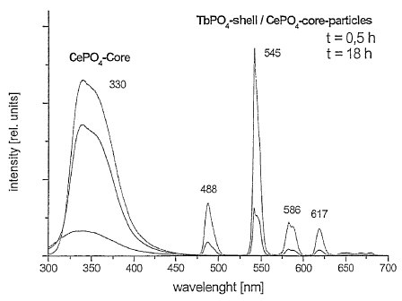
Figure 1 shows the fluorescence spectra of homogeneous CePO4
core particles and CePO4/TbPO4 core/shell particles according
to the present invention.
Figure 2 shows various images obtained by energy filtering
transmission electron microscopy of one CePO4:Tb/LaPO4
core/shell particle.
Figure 3 shows two fluorescence decay curves of CePO4/TbPO4
core/shell particles according to the invention, which were
modified and chemically coupled to fluorescein, respectively.
As reference, the fluorescence decay curve of CePO4/TbPO4
core/shell particles which were not coupled to fluorescein is
also shown.
Figure 4 shows the fluorescence decay curves of fluorescein-
coupled, homogeneous LaPO4:Ce, Tb particles (comparative
example 1).
Figure 5a shows two fluorescence spectra measured in time
gated (TGF) mode of CePO4/TbPO4 core/shell particles
according to the invention, which were not further modified
or coupled to fluorescein, respectively.
Figure 5b shows one fluorescence spectrum measured in time
gated mode of homogeneous LaPO4:Ce, Tb nanoparticles
(comparative example 1) at 520 nm and 542 nm, respectively.
Fig. 6a: Homogeneous kinase assay with (F)RET partners
coupled to one molecule
Fig. 6b: Homogeneous immunoassay with (F)RET partners
coupled to one molecule.
CA 02523027 2005-10-19
WO 2004/096944 PCT/EP2004/004574
7
Fig. 7: Competitive immunoassay with (F)RET partners
coupled to one molecule (epitope).
Fig. 8: Homogeneous saturation immunoassay with (F)RET
partners coupled to separate molecules.
Fig. 9: Homogeneous competitive immunoassay with (F)RET
partners coupled to separate molecules.
Fig. 10: Homogeneous assay with (F)RET partners coupled to
one molecule.
Fig. 11: Assay following the method of molecular beacons.
Detailed description of the present invention
I. Lumininescent nanoparticles
The luminescent, in particular photoluminescent particles of
the present invention comprise (a) a core made from a first
metal salt or oxide being surrounded by (b) a luminescent,
non-semiconductor shell made from a second metal salt or
oxide.
"Luminescence" characterizes the property of the claimed
nanoparticles to absorb energy (e.g., in the form of photons
(IR, visible, UV), electron rays, X-ray, etc.) which is then
emitted as light of lower energy. It is to be understood
that the term "luminescent" throughout the description and
the claims also includes the more specific and preferred
meaning "photoluminescent"
As "photoluminescence", we understand the capability of the
inorganic metal salt to absorb photons of a specific energy
(e.g. UV, visible) and emit light of lower energy (longer
wavelength, e.g. W, visible, IR) over a certain period of
CA 02523027 2005-10-19
WO 2004/096944 PCT/EP2004/004574
8
time. The period of light emission can correspond to life-
times of the excited state up to 10-7 or 10-8 sec, which are
typically referred to as fluorescence, but also much longer.
For lanthanide-doped salts, e.g. sulfates, phosphates or
fluorides, typically lifetimes of the excited state in the
order of milliseconds (for instance 1-20 ms) are observed.
According to the present invention it is preferred that both
shell and core material do not show semiconductor properties.
Both shell and core preferably also constitute crystalline
materials. This can be confirmed by X-ray powder diffraction
patterns.
The shape of the claimed core/shell particles can be for
instance needle like, ellipsoid or spherical, the latter two
options being preferred.
The claimed core/shell nanoparticles preferably have an
average size measured along their longest axis of 1 to 100
nm, more preferably 1 to 50 nm. Average sizes of maximally
30 nm, maximally 20 nm, maximally 10 nm, for instance 2 to 8
nm, or 4-6 nm are even more desirable. In each case, the
standard derivation is preferably less than 30%, in
particular less than 10%.
The particle size and distribution can be measured according
to techniques further described in the already-cited articles
by K. Riwotzki et al and M. Haase et al, for instance, with
transmission electromicrographs (TEM). Gel permeation
chromatography and ultra-centrifugation also allow
determining the size.
The thickness of the shell is preferably at least two
monolayers. A preferred upper limit for the shell thickness
are two diameters of the core (for non-spheroidal particles
CA 02523027 2005-10-19
WO 2004/096944 PCT/EP2004/004574
9
measured along the longest axis), more preferably one core
diameter, e.g. 2/3 thereof.
According to the first embodiment of the present invention,
the core (a) is made from a metal salt or oxide, which does
not accept energy from the shell after its electronic
excitation, in particular a non-luminescent metal salt or
oxide and (b) the shell is made from a luminescent, in
particular doped metal salt or oxide.
Throughout the present application"doping" is to be
understood in a broad sense. The upper limit of dopant to be
used should be low enough that the generated luminescence is
not reduced by concentration quenching phenomena.
Correspondingly, this upper limit depends on factors like the
type of doping ion and the distance between the dopant metal
ions in the lattice which are specific to each core material.
Preferably, the host material is substituted by the dopant in
an amount of up to 50 mol %, preferably 0,1 to 45 mol%, e.g.
0.5 to 40 mol %, or 1 to 20 mol %.
There are also no specific restrictions regarding the type of
dopant metal to be incorporated, as long as the same is
capable of converting absorbed photons to luminescent
radiation. Thus, for instance metals like Ag, Cu, Co or Mn
(for instance, in combination with zinc as host metal) can be
used. Doping with lanthanide metals is however preferred
since the luminescence of lanthanide metals is particularly
independent from its lattice environment. Generally,. the use
of bi- or trivalent dopants, in particular lanthanide dopants
is preferred. Bivalent lanthanides (+II oxidation state) are
characterized by a relatively strong absorption, but
relatively broad emission bands. For this reason, they can be
suitably used as sensitizer transferring the energy to other
luminescing metals (e.g. Eu2+ to Mn2+). The capacity of
trivalent lanthanides (oxidation state +III) to emit light in
CA 02523027 2005-10-19
WO 2004/096944 PCT/EP2004/004574
the form of relatively sharp bands makes them particularly
attractive dopants for single use although, as explained
later, also suitable combinations of trivalent lanthanides
dopant systems exist.
Suitable dopant materials for the shell include Al, Cr, Tl,
Mn, Ag, Cu, As, Nb, Ni, Ti, In, Sb, Ga, Si, Pb, Bi, Zn, Co
which, depending on the host material used, have luminescent
properties, in particular Mn, Ag, Cu, Bi, Cr, Sn, Sb and
preferably the lanthanides, in particular Ce (58), Pr (59),
Nd (60), Sm (62), Eu (63), Gd (64), Tb (65), Dy (66), Ho
(67), Er (68), Tm (69), or Yb (70) or combinations thereof.
Doping with lanthanide metals is preferred since the
luminescence of lanthanide metals is particularly independent
from its lattice environment.
From a practical point of view (type of fluorescence,
intensity, etc.) Ce, Tb, Eu, Nd, Dy, Th, Sm, Gd, Ho, Er and
Yb show the most interesting luminescence properties.
Er3+, Nd3+ and Ho3+ are of particular interest for the
telecommunication area since they emit between 1300 and 1600
nm. Ce is preferably used in combination with another dopant
material, such as Nd, Dy or Tb. Ce is known to absorb
strongly UV radiation having a wavelength of from 250 to 300
nm, but shows a fairly broad luminescence band around 330 nm
depending on the host lattice (e.g. phosphate). If used in
combination with other dopants to which the absorbed energy
can be transferred, very efficient luminescent systems can be
generated. Another attractive combination of dopant metals
is Yb and Er, which is of great importance in Er3+- doped
optical amplifiers where Er3+ is pumped indirectly via Yb3+
which has a ten times higher absorption cross section and a
much broader peak at 980 nm than Er3+. Nd3+ and Gd3+ can also
be combined.
CA 02523027 2005-10-19
WO 2004/096944 PCT/EP2004/004574
11
As indicated before, it is not only possible to use these
lanthanide metal combinations as dopants for the shell. It
is equally effective to employ as host metal that lanthanide
metal ion (e.g. Ce3+, yb3+, Nd3+) having the higher
absorption cross section and replacing a part thereof by
lower amounts of the other metal (e.g. Tb3+, Er3+, Gd3+).
For this reason, lanthanide salts (e.g. Ce3+, yb3+, Nd3+
salts) can also be used as the host material of the shell.
For applications in aqueous media as used for biological
assays, the most preferred dopants are those (e.g. Tb, Dy,
Tm, Sm) showing luminescence in the visible area in order to
minimize interaction with water which otherwise may absorb
the emitted light.
The host material for the shell is not specifically limited
and can be selected from known non-luminescent metal oxides
or salts, such as sulfides, selenides, sulfoselenides,
oxysulfides, phosphates, halophosphates, arsenates, sulfates,
borates, aluminates, gallates, silicates, germanates, oxides,
vanadates, niobates, tantalates, tungstates, molybdates,
alkalihalogenates, other halides, in particular fluorides,
phosphides, or nitrides. The use of sulfates, phosphates or
fluorides is particularly preferred.
The metals of these salts preferably belong to the main
groups 1, 2, 13, or 14, the subgroups 3, 4, 5, 6, 7, or the
lanthanides. Since most luminescent dopants are bi- or tri-
valent metal ions, it is preferred to use, as counter ion for
the shell, non-luminescent bi- or tri-valent metal atoms such
as the metals of group 2 (earth alkaline metals, such as Mg,
Ca, Sr, or Ba), or group 3 (Sc, V or La) or group 13 (e.g.,
Al, Ga or In) or Zn.
Preferred embodiments of host metal salts comprise:
phosphates of the corresponding number of metals (to
ensure charge neutrality) selected from main group 2 (e.g.
CA 02523027 2005-10-19
WO 2004/096944 PCT/EP2004/004574
12
from Mg, Ca, Sr, Ba), group 3 (e.g. Sc, Y, La), or
lanthanides (elements 58 to 71, i.e. Ce, Pr, Nd, Pm, Sm, Eu,
Gd, Tb, Dy, Ho, Er, Tm, Yb and Lu);
sulfates of the corresponding number of metals selected
from group 2 (e.g. from Mg, Ca, Sr, Ba), group 3 (e.g. Sc, Y,
La), or lanthanides (as above);
borates of the corresponding number of metals selected
from main group 2 (e.g. from Mg, Ca, Sr, Ba), group 3 (e.g.
Sc, Y, La), or group 13 (Al, Ga, In, Tl) or lanthanides (as
above);
fluorides of the corresponding number of metals selected
from group 2 (e.g. from Mg, Ca, Sr, Ba), subgroup 3 (e.g. Sc,
Y, La), or lanthanides (as above);
aluminates (e.g. A15012 or A104) of the corresponding
number of metal atoms selected from group 2 (e.g. from Mg,
Ca, Sr, Ba), group 3 (e.g. Sc, Y, La), or lanthanides (as
above);
gallates (e.g. Ga5012) of the corresponding number of
metal atoms selected from group 2 (e.g. from Mg, Ca, Sr, Ba),
group 3 (e.g. Sc, Y, La), or lanthanides (as above);
silicates (e.g. Si03 or Si04) of the corresponding
number of metals selected from group 2 (e.g. from Mg, Ca, S'r,
Ba), group 3 (e.g. Sc, Y, La), group 12 (e.g. Zn, Cd) or
lanthanides (as above);
vanadates (e.g. V04) of the corresponding number of
metal atoms selected from group 2 (e.g. from Mg, Ca, Sr, Ba),
group 3 (e.g. Sc, Y, La), or lanthanides (as above);
tungstates (e.g. W04) of the corresponding number of
metal atoms selected from group 2 (e.g. from Mg, Ca, Sr, Ba),
group 3 (e.g. Sc, Y, La), or lanthanides (as above);
molybdates (e.g. MoO4) of the corresponding number of
metal atoms selected from group 2 (e.g. from Mg, Ca, Sr, Ba),
group 3 (e.g. Sc, Y, La), or lanthanides (as above);
tantalates (e.g. Ta04) of the corresponding number of
metal atoms selected from group 2 (e.g. from Mg, Ca, Sr, Ba),
group 3 (e.g. Sc, Y, La), or lanthanides (as above); or
arsenates (e.g. As04) of the corresponding number of
CA 02523027 2005-10-19
WO 2004/096944 PCT/EP2004/004574
13
metal atoms selected from group 2 (e.g. from Mg, Ca, Sr, Ba),
group 3 (e.g. Sc, Y, La), or lanthanides (as above).
When selecting a suitable host material for a specific
dopant, it is further to be taken into account, as known in
the art, that host and dopant metal preferably should have
the same valence and similar (tolerance e.g. 200) or
identical ion diameters. Simultaneously, it typically
increases the compatibility of dopant and host metal if these
are capable of forming, with a specific anion, crystals of
the same or similar lattice type having the same or similar
lattice constant(s) (tolerance e.g. 20%).
The above criterion can often be met with Ba and La as host
material metal for the core since these metals display ion
diameters, which are very similar to those of the two-valent
(+II) lanthanides. For the same reason, La and Y salts
represent suitable host materials for tri-valent (+III)
lanthanide dopants.
Specific examples of luminescent shell materials are for
instance LiI:Eu; NaI:Tl; CsI:Tl; CsI:Na; LiF:Mg; LiF:Mg,Ti;
LiF:Mg,Na; KMgF3:Mn% A1203:Eu; BaFC1:Eu; BaFC1:Sm; BaFBr:Eu;
BaFClo,5Bro,5 : Sm; BaY2F8 :A (A = Pr, Tm, Er, Ce) ; BaSi205: Pb;
BaMg2A116O27 : Eu; BaMgA114023 : Eu; BaMgAl10017 : Eu; BaMgAl203 : Eu;
Ba2P2O7 : Ti; (Ba, Zn, Mg) 3Si2O7 : Pb; Ce (Mg, Ba) A111019;
CeO, 65Tb0 35MgA111O19 : Ce, Tb; MgAl11O19 : Ce, Tb; MgF2 : Mn; MgS : Eu;
MgS:Ce; MgS:Sm; MgS:(Sm,Ce); (Mg,Ca)S:Eu; MgSiO3:Mn;
3,5MgO.0,5MgF2=Ge02:Mn; MgWO4:Sm; MgWO4:Pb; 6MgO=As205:Mn;
(Zn,Mg)F2:Mn; (Zn4Be)SO4:Mn; Zn2SiO4:Mn; Zn2SiO4:Mn,As;
Zn3 (P04) 2: Mn; CdBO4 : Mn; CaF2 : Mn; CaF2 : Dy; CaS : A A =
Lanthanide, Bi); (Ca,Sr)S:Bi; CaWO4:Pb; CaWO4:Sm; CaSO4:A
(A = Mn, lanthanide); 3Ca3(P04)2=Ca(F,Cl)2:Sb,Mn;
CaSi03 :Mn, Pb; Ca2A12Si2O7:Ce; (Ca,Mg) Si03:Ce; (Ca,Mg) Si03 :Ti;
CA 02523027 2005-10-19
WO 2004/096944 PCT/EP2004/004574
14
2SrO.6 (B203) =SrF2:Eu; 3Sr3 (P04) 2=CaC12 :Eu; A3 (P04) 2=AC12 :Eu (A =
Sr, Ca, Ba); (Sr,Mg)2P207:Eu; (Sr,Mg)3(P04)2:Sn; SrS:Ce;
SrS:Sm,Ce; SrS:Sm; SrS:Eu; SrS:Eu,Sm; SrS:Cu,Ag; Sr2P2O7:Sn;
Sr2P207 : Eu; Sr4A114025 : Eu; SrGa2S4:A (A = lanthanide, Pb) ;
SrGa2S4 : Pb; Sr3Gd2Si60l8 : Pb, Mn; YF3 : Yb, Er; YF3 : Ln (Ln =
lanthanide); YLiF4:Ln (Ln = lanthanide); Y3A15012:Ln (Ln =
lanthanide) ; YAl3 (B04) 3 :Nd, Yb; (Y, Ga) B03 : Eu; (Y, Gd) B03: Eu;
Y2A13Ga2Ol2 : Tb; Y2Si05 : Ln (Ln = lanthanide) ; Y203 : Ln (Ln =
lanthanide); Y202S:Ln (Ln = lanthanide); YV04:A (A =
lanthanide, In) ; Y (P, V) 04: Eu; YTa04 :Nb; YA103 :A (A = Pr, Tm,
Er, Ce); YOCl:Yb,Er; LnP04:Ce,Tb (Ln = lanthanide or mixture
of lanthanides); LuVO4:Eu; GdVO4:Eu; Gd2O2S:Tb;
GdMgBs0i.o : Ce , Tb ; LaOBr : Tb ; La202 S : Tb ; LaF3 : Nd, Ce ; BaYb2F8 :
Eu ;
NaYF4 : Yb, Er; NaGdF4 : Yb, Er; NaLaF4 : Yb, Er; LaF3 : Yb, Er, Tm;
BaYF5 : Yb, Er; Ga203 : Dy; GaN:A (A = Pr, Eu, Er, Tm) ; Bi4Ge3O12;
LiNb03 :Nd, Yb; LiNb03 : Er; LiCaA1F6 : Ce; LiSrA1F6 : Ce; LiLuF4:A (A
= Pr, Tm, Er, Ce) ; Li2B407 : Mn, SiOX : Er, Al (0 < x <_ 2) ; Y203 : Ln
(Ln = lanthanides, in particular Eu) , Y202S : Eu, Y2SiOs : Eu,
Si02 : Dy, Si02 : Al , Y203 : Tb, CaSiO3 : Ln, CaS : Ln, CaO : Ln, wherein Ln
= one, two or more lanthanides.
If classified according to the host lattice type the
following preferred embodiments can also be enumerated.
1. Halides: for instance XY2 (X = Mg, Ca, Sr, Ba; Y = F,
Cl, I) , CaF2 : Eu (I I) , BaF2 : Eu; BaMgF4 : Eu; LiBaF3 : Eu; SrF2 : Eu;
SrBaF2Eu; CaBr2 : Eu-Si02; CaCI2 : Eu; CaCI2 : Eu-Si02; CaCI2 : Eu, Mn-
Si02 ; CaI2 : Eu; CaI2Eu, Mn; KMgF3 : Eu; SrF2 :Eu (II), BaF2 : Eu
(I I) , YF3, NaYF4, : MgF2 : Mn ; MgF2 : Ln (Ln = lanthanide (s)) .
2. Earth alkaline sulfates: for instance XSO4 (X = Mg, Ca,
Sr, 13a), SrSO4 : Eu, SrSO4 : Eu, Mn, BaSO4 : Eu, BaSO4 : Eu, Mn, CaSO4,
CA 02523027 2005-10-19
WO 2004/096944 PCT/EP2004/004574
CaSO4:Eu, CaSO4:Eu,Mn, as well as mixed earth alkaline
sulfates, also in combination with magnesium, e.g.
Ca,MgSO4:Eu,Mn.
3. Phosphates and halophosphates: for instance CaPO4:Ce,Mn,
Ca5 (P04) 3C1 : Ce, Mn, Ca5 (P04) 3F : Ce, Mn, SrP04 : Ce, Mn,
Srs (P04) 3C1 : Ce, Mn, Srs (P04) 3F : Ce, Mn, the latter also codoped
with Eu (II) or codoped with Eu,Mn, a-Ca3 (P04) 2 : Eu; f3-
Ca3 (PO4) 2: Eu, Mn; Ca5 (P04) 3C1 :EU; Sr5 (P04) 3C1 :Eu; Balo (P04) 6C1 :
Eu;
Balo (P04) 6C1 :Eu,Mn, Ca2Ba3 (PO4) 3Cl :Eu; Ca5 (P04) 3F:EU2+X3+;
Srs (P04) 3F: Eu2+X3+ (X=Nd, Er, Ho, Tb) ; Bas (P04) 3C1 : Eu; 13-
Ca3 (P04) 2: Eu; CaB2P209 : Eu; CaB2P209 : Eu; Ca2P207 : Eu; Ca2P207 : Eu, Mn;
Srlo (P04) 6C12: Eu; (Sr, Ca, Ba, Mg) to (P04) 6C12:Eu; LaP04: Ce; CeP04;
LaP04 : Eu, LaP04 : Ce, LaP04 : Ce, Tb, CeP04 : Tb .
4. Borates: for instance LaB03 ; LaB03 : Ce; ScB03 : Ce
YA1B03 : Ce ; YB03 : Ce ; Ca2B509C1 : Eu; xEuO'yNa2O' zB203 .
5. Vanadates: for instance YV04, YV04 : Eu, YV04 : Dy, YV04 : Sm
YV04 : Bi ; YV04 : Bi , Eu, YV04 : Bi , Dy, YV04 : Bi , Sm, YV04 : Tm,
YV04 : Bi , Tm GdV04, GdV04 : Eu, GdV04 : Dy, GdV04 : Sm GdV04 : Bi ;
GdV04 : Bi , Eu, GdV04 : Bi , Dy, GdV04 : Bi , Sm; YV04 : Eu, YV04 : Sm,
YV04 : Dy .
6. Aluminates: for instance MgA1204 : Eu; CaA1204 : Eu;
SrAl204 : Eu; BaA1204 : Eu; LaMgA111O19 : Eu; BaMgAlloO17 : Eu;
BaMgAl1o017 : Eu, Mn; CaA112019 : Eu; SrAl12019 : Eu; SrMgAllo017 : Eu;
Ba (A1203) 6 : Eu; (Ba, Sr) MgAllo017 : Eu, Mn; CaAl204 : Eu, Nd;
SrA1204 : Eu, Dy; Sr4Al1402s : Eu, Dy.
7. Silicates: for instance BaSrMgSi207 : Eu; Ba2MgSiO7 : Eu;
BaMg2Si2O7 : Eu; CaMgSi20G : Eu; SrBaSi04 : Eu; Sr2Si308. SrCl2 : Eu;
Ba5Si04Br6 : Eu; BasSi04Cl6 : Eu; Ca2MgSi2O7 : Eu; CaA12Si2O8: Eu;
Cal.5Sro.5MgSi207 : Eu; (Ca, Sr) 2MgSi207 : Eu, Sr2LIS1O4F : Eu .
8. Tungstates and molybdates: for instance X3WOG (X = Mg,
Ca, Sr, Ba) õ X2W04 (X = Li, Na, K, Rb, Cs) , XM004 (X = Mg,
CA 02523027 2005-10-19
WO 2004/096944 PCT/EP2004/004574
16
Ca, Sr, Da) as well as polymolybdates oder polytungstates or
the salts of the corresponding hetero- oder isopolyacids.
9. Germanates: e.g. Zn2GeO4
10. moreover the following classes: ALnO2:Yb, Er (A = Li,
Na; Ln = Gd, Y, Lu); Ln203:Yb, Er (Ln = La, Gd, Y, Lu);
LnAO4 : Yb, Er (Ln = La, Y; A=P, V, As, Nb) ; Ca3Al2Ge3O1.2 : Er;
Gd2O2S : Yb, Er; La2S : Yb, Er.
According to the first embodiment of the present invention,
the core material, i.e. a metal salt or oxide, does not
accept energy transfer from the luminescent shell in its
electronically excited state.
This requirement can be always met with core metal salts or
oxides having only electronic states wherein the energetic
distance between the electronic ground state and the first
electronically excited state is greater than the distance
between the first electronically excited state of the
selected luminescent shell and its ground state. Under these
circumstances the energy (e.g. UV, visible, IR) absorbed by
the shell cannot be transmitted to the core metal atoms or
anions. The localization of the energy in the shell achieved
thereby enhances surface quenching phenomena and is believed
to increase the overall (F)RET efficiency of the particle.
According to one preferred embodiment, the core salt or oxide
is non-luminescent and thus lacking absorption bands (UV-vis
or IR) to which the energy could be transferred from the
excited shell. Since non-luminescent materials are often
cheaper than luminescent materials, this is also economically
of advantage.
Preferably, the core material corresponds to the host
material of the doped shell.
CA 02523027 2011-06-23
17
Suitable anions forming the core are thus the same as
indicated above and involve, but are not limited to
phosphates, halophosphates, arsenates, sulfates, borates,
aluminates, gallates, silicates, germanates, oxides,
vanadates, niobates, tantalates, tungstates, molybdates,
alkalihalogenates, other halides, or nitrides.
Nanoparticulate metal salts of this type are disclosed in
WO 2002/020696.
The only criteria governing the selection of the core metal
atoms is their lacking capability to accept luminescence from
the shell after irradiation with photons. Preferred metal
ions, which can be used for this purpose, are the same as
mentioned above for the host material of the shell. They
include, but are not limited to metals of group 2 (earth
alkaline metals, such as Mg, Ca, Sr or Ba), metals of group 3
(such Sc, y or La), zinc, or metals of group 13 (such Al, Ga,
or In). In order to increase the aptitude of the shell
material to grow on the surface of the core material, it is
further preferred, but not absolutely necessary to select as
core material the same salt that constitutes the host of the
doped shell. If this requirement is not fulfilled, it is
preferred that the host material of the core and the shell
material belong to the same lattice type and display very
similar (tolerance e.g. 20%) or, identical lattice constants.
According to the second embodiment, (a) the core comprises a
first metal salt or oxide ("donor") which after excitation is
capable of transferring the excitation energy to (b) a second
shell-forming luminescent metal salt or oxide ("acceptor")
which emits the same as luminescence.
Suitable donor-acceptor metal combinations can for instance
be selected among the above-identified dopants, in particular
lanthanides and generally require a distance between the
electronic ground state and the first excited state of the
CA 02523027 2005-10-19
WO 2004/096944 PCT/EP2004/004574
18
donor metal which involves a higher energy than the
corresponding distance of the acceptor metal.
Examples for suitable photon energy absorbers (donors), which
can be used as core material in the second embodiment of the
invention, are lanthanide ions having relatively high
absorption cross-sections such as Ce3+, Yb3+, Nd3+ or Eu2+.
Ce3+ is preferably used in combination with Tb3+, Dy3+ or
Nd3+ as shell material metal and acceptor, e.g. in the form
of the corresponding sulfates, phosphates or fluorides.
Yb3+ salts, such as phosphates, sulfates or fluorides are
preferably combined as core material with Er3+ salts, such as
sulfates, phosphates or fluorides, respectively, as shell
material. This allows pumping Er3+ indirectly via Yb3+.
In terms of shell constitution, the acceptor atoms can be
used as high concentration dopant materials of the host
materials described in the context of the first embodiment of
the present invention. However, it is also possible that the
entire shell consists of the corresponding acceptor salt,
e.g. metal sulfate, phosphate or fluoride in order to
increase the efficiency of energy transfer from the core to
the shell.
The core material of the second embodiment may comprise the
donor metal as high concentration dopant of a host material
as described above. Alternatively and preferably, the core
consists of the corresponding donor metal salt.
The anion of the core salt can be freely selected among
compatible anions allowing the growth of the selected shell
material. Examples of suitable anions are given for the first
embodiment, sulfate, phosphate or fluoride being preferred.
One particular preferred example for the so-called second
embodiment are CePO4/TbPO4 core/shell particles.
CA 02523027 2005-10-19
WO 2004/096944 PCT/EP2004/004574
19
In accordance with the second embodiment, it is also possible
to employ vanadates, molybdates, tungstates or germanates as
core materials (donor) since the corresponding anions are
also capable of absorbing energy and transferring the same to
a suitable shell material (acceptor) which then emits the
energy as luminescence. These may also be combined with
dopant metals acting itself as luminescent centers and thus
enhancing luminescence, such as Bi3+ and/or Eu3+ for
vanadates. The core may for instance comprise or consists of
vanadates, molybdates, tungstates or germanates of metals of
group 3 (such Sc, Y or La) or metals of group 13 (such Al,
Ga, or In). It is preferably combined with lanthanide salts,
preferably phosphates, vanadates, molybdates, tungstates or
germinates as shell material wherein the lanthanide acts as
energy acceptor. Specific examples involve core/shell
combinations of the type LaVO4/EuPO4, LaVO4/NdPO4,
YVO4/DyPO4.
II. Synthesis of core/shell nanoparticles
The above-described core/shell nanoparticles of the present
invention are synthesised in a process as laid down below and
in the claims which comprises at least the following two
steps:
1. The preparation of a so-called "first mixture"
comprising nanoparticles of a first metal salt or oxide,
e.g. metal sulfate, phosphate or fluoride nanoparticles
(cores) in an organic medium.
2. Reacting said first mixture, an anion source for the
shell to be formed, in particular a phosphate, sulfate
or fluoride source, and a "second mixture" comprising
shell-forming metal ions and an organic complexing agent
CA 02523027 2005-10-19
WO 2004/096944 PCT/EP2004/004574
for said metal ions at a temperature of 50 to 350 C
until a shell has formed around said nanoparticle cores.
II.1 First process step and synthesis of core particles
The nanoparticles provided as core material and being present
in the so-called "first mixture" can be synthesized according
to processes known in the art.
Generally, wet synthesis techniques are preferred over dry
formation processes since the former allow a better control
of the particle sizes. Furthermore, the aggregation of the
formed nanoparticles can be more easily suppressed in wet
synthesis techniques.
Among the known wet synthesis techniques, for instance sol-
gel processes, the hydrothermal synthesis, or the organic
synthesis with complexing agents that regulate crystal growth
can be used. Further, it is possible to produce specifically
the fluorides in a synthesis technique described in the
already mentioned article by J. W. Stouwdam and F. C. J. M.
Van Veggel. Accordingly, LaF3 nanoparticles and other
fluorides can be prepared by heating a solution of ammonium
di-n-octadecyldithiophosphate and NaF in ethanol/water.
Subsequently, solutions of the corresponding metal nitrates
in water are added dropwise, followed by stirring the
solution two hours at 75 C and cooling to room temperature.
The disadvantage of this technique, however, is that the
generated particles still display a relatively broad particle
size distribution which necessitates further purification
steps by centrifugation.
The "hydrothermal synthesis" of lanthanide-doped phosphates
is, for instance, described in "Wet-chemical synthesis of
doped colloidal nanomaterials: particles and fibres of
LaPO4:Eu, LaPO4:Ce and LaPO4:Ce,Tb" by H. Meyssamy et al,
Advanced Materials (1999), Vol. 11, No. 10, pages 840 et seq.
CA 02523027 2005-10-19
WO 2004/096944 PCT/EP2004/004574
21
As starting materials for sulfate, phosphate or fluoride
nanoparticles, preferably metal chlorides, nitrates or
acetates are used. The reaction is performed in water as
reaction medium in an autoclave to maintain high pressures,
preferably pressures of from 10-20 bar during the reaction.
The hydrothermal synthesis results in relatively large
particles which often have a needle-like shape. Further, a
relatively broad distribution of particle sizes typically
characterizes the product. In the above-named method by H.
Meyssamy et al, the percentage of nanoparticles with
diameters of less than 25 nm is, for instance, only around
20%. These can be isolated by subsequent centrifugation
steps.
Other examples for the hydrothermal synthesis can be found in
PCT/DE 01/03433. This document discloses, on a more general
level and by means of concrete examples, the synthesis of
nanoparticulate silicates, vanadates, tungstates, molybdates,
tantalates, etc. in water under high pressures (autoclave).
Further, this document pertains to a related technique for
the synthesis of aluminates or gallates in 1,6-hexanediol
(therein also referred to as "glycothermal" synthesis).
Further, it is possible to produce optionally doped sulfates
under ambient pressure in organic media selected from polyols
and sulfoxides, which are believed to regulate crystal growth
by metal-complexing activity. This technique will be referred
to in the following as "polyol or sulfoxide synthesis".
The polyols to be used preferably have two or three hydroxy
groups and can be exemplified by glycerol, ethylene glycol or
polyethylene glycol, whereby preferably low molecular weight
polyethylene glycol is used (preferred average number of
ethylene glycol units up to 4). As sulfoxide
dimethylsulfoxide (DMSO) may be used. This synthesis
technique is preferably employed in the preparation of earth
CA 02523027 2005-10-19
WO 2004/096944 PCT/EP2004/004574
22
alkaline metal sulfates, such as magnesium, calcium,
strontium or barium sulfate as doped host material.
Preferred metal atom sources are the corresponding chlorides
and their hydrates. As starting material for the sulfate,
preferably alkali metal sulfates, ammonium sulfates or
sulfates having an organic cation are employed. The
corresponding hydrogensulfates are equally suitable.
The organic cation is preferably selected from basic N-
containing aliphatic, aromatic and aliphatic/aromatic
substances which preferably have from 4 to 30, preferably
from 4 to 20 carbon atoms. Suitable cations involve, for
instance,
= quaternary ammonium or phosphonium wherein the four
substituents can be independently selected from alkyl
having preferably from 1 to 10 carbon atoms (preferably
1 to 5) or benzyl, or
= protonated aromatic bases, such as hydrazine,
amantadine, pyridine or collidine.
Correspondingly, sulfate nanoparticles can be produced from
starting materials such as tetrabutylammonium
hydrogensulfate, tetramethylammonium sulfate, bis-
tetrabutylammonium sulfate, or triethylammonium
hydrogensulfate. Other suitable starting materials are
ammonium hydrogensulfate, ammonium sulfate, alkali metal
hydrogensulfates, amantadine sulfates, ethylenediammonium
sulfate and hydrazinium sulfate.
For doping the sulfate host material, nitrates or halides of
the corresponding dopant, in particular the corresponding
metal chloride can be used.
If hydrogensulfates are contained in the starting material,
organic bases such as imidazol are preferably added as acid
scavenger to the reaction medium. The reaction is preferably
CA 02523027 2005-10-19
WO 2004/096944 PCT/EP2004/004574
23
conducted at temperatures of from 50 to 2400C, whereby the
lower temperature range of from 50 to 100 C is preferred for
glycerol and higher temperatures in the range from 160 to
240 C, in particular 160 to 180 C are most suitable for the
other polyol or sulfoxide solvents. The particles obtained
have an average diameter in the order of 0.2 to 50 nm and are
readily dispersible in aqueous media.
Nanoparticle cores obtained by sol-gel processes, the
hydrothermal synthesis, glycothermal synthesis or the so-
called "polyol or sulfoxide synthesis" are sometimes not
dispersible in the organic medium to be used in the first
step of the claimed method, especially if the reaction medium
for the core and the method of the invention (shell
synthesis), respectively, differ considerably in terms of
polarity. For this reason, it may become necessary to
subject the nanoparticles to an after-treatment with a
suitable polar organic compound, in order to increase their
dispersibility. Preferably, this after-treatment is carried
out with the same organic medium (complexing agent) which
will be used in the shell synthesis or organic media of
similar polarity.
If for instance the shell synthesis is to be carried out in
N- or P- containing media, the after-treatment can suitably
involve subjecting particles obtained in sol-gel processes,
the glycothermal or hydrothermal synthesis or the so-called
"polyol or sulfoxide synthesis" to an after-treatment with N-
or P- containing media.
This after-treatment involves heating the nanoparticles in
the corresponding organic compound. It has the effect that
water,,or other hydrophilic residues bonded at the surface of
the nanoparticle are replaced by the polar organic compound.
For the reasons given above, the polar organic compound is
preferably selected from N- or P- containing complexing
agents for metal ions as will be described further below in
CA 02523027 2005-10-19
WO 2004/096944 PCT/EP2004/004574
24
the context of the "organic synthesis" and the second process
step. However, other functionalised polar organic compounds
may also be used.
This after-treatment is not required for sulfates, as
produced in the "polyol or sulfoxide" synthesis, if the
subsequent manufacture steps are carried out in polyols
and/or sulfoxides.
According to a further and preferred technique, hereinafter
referred to as "organic synthesis", the process for the
preparation of the nanoparticle cores comprises the steps of:
a) reacting, in an organic reaction medium comprising at
least one metal complexing agent, and optionally at
least one further solvent, a reaction medium-soluble or
-dispersible metal source and a reaction medium-soluble
or -dispersible anion source, in particular phosphate,
sulfate or fluoride source,
b) optionally removing the reaction medium from the
nanoparticulate metal salt (e.g. phosphate, sulfate or
fluoride) formed thereby, and
c) optionally recovering the nanoparticulate salt.
As "organic medium" we understand organic solvents, which,
apart from unavoidable traces, do not contain water. The
boiling point of this organic medium is preferably higher
than the reaction temperatures given below. It is e.g. from
150 to 400 C, preferably above 180 C, in particular above
210 C (at ambient pressure).
Depending on the susceptibility of the metal source to
oxidation, it is preferred to conduct the reaction under
inert gas such as nitrogen or argon.
CA 02523027 2005-10-19
WO 2004/096944 PCT/EP2004/004574
Regarding the degree of purity of starting materials, it is
recommendable to use metal salts having a purity of at least
99.9%. All reactants and the solvents used are preferably
water-free and/or are dried prior to use. However, metal
chlorides which are frequently employed as hydrates should
preferably not be subjected to a longer drying procedure
since this may enhance the formation of reaction medium-
insoluble oxychlorides.
The reaction is preferably conducted at a temperature of 50
to 350 C, e.g. 120 to 320 C, in particular 180 to 290 C. A
suitable temperature can be easily determined by a skilled
person by monitoring the reaction of the reactants at
gradually increasing temperatures thereby determining the
synthesis minimum temperature at which the reaction proceeds
with sufficient speed. For this purpose the nanoparticles
may, for instance, be precipitated from samples of the
reaction medium which allows studying the particle growth
with increasing reaction time.
Suitable reaction times can be determined in the same manner
and preferably range from 10 min to 48 hours, in particular
min to 20 hours.
After completion of the reaction, the reaction mixture can be
cooled down to room temperature. If the nanoparticles have
not yet fully precipitated during the reaction or after
cooling, it is possible to add methanol to the reaction
medium or vice versa in order to obtain maximum yields.
Without being bound to theory, it is believed that the metal
complexing agent used in the "organic synthesis" coordinates
with surface metal atoms of the nanoparticles formed and
thereby terminates their growth after the starting materials
have reacted. It is believed that this metal complexing
agent remains bound to the particle surface and in this
manner prevents or reduces agglomeration and exchange
CA 02523027 2005-10-19
WO 2004/096944 PCT/EP2004/004574
26
processes between the particles like Oswald ripening. The
organic synthesis thus leads to fairly small particles
wherein the average diameter measured at the longest axis is
preferably 1-10 nm, in particular 2-8 nm, for instance 4-6 nm
with narrow size distributions (standard deviation < 30%, in
particular <-10%). The metal complexing agent is
characterized by the presence of a polar group capable of
coordinating the metal ion and at least one second molecule
portion (less polar, preferably hydrophobic), for instance an
aliphatic, aromatic/aliphatic, or purely aromatic molecule
portion having preferably 4 to 20, in particular 6 to 14
carbon atoms.
The metal complexing agent is preferably a phosphororganic
compound or a mono-or di-substituted amine.
Among the latter, the most preferred embodiments are mono- or
dialkyl amines wherein the alkyl residue preferably has from
4 to 20, in particular 6 to 14 carbon atoms, such as dodecyl
amine or bis(ethylhexyl)amine.
As regards the phosphororganic compounds, it is preferred to
use at least one of the following substances:
a) esters of phosphinic acid
R1 0
R2- P=0
R3
b) diesters of phosphonic acid
CA 02523027 2005-10-19
WO 2004/096944 PCT/EP2004/004574
27
R~
R20 P=0
R30
c) triesters of phosphoric acid, most preferably trialkyl
phosphates such as tributylphosphate or
tris(ethylhexyl)phosphate,
R1 0
R20 P=0
/
R30
d) trialkyl phosphines, such as trioctylphosphine (TOP),
R1
R2 -P
/
R3
or
e) trialkyl phosphene oxides, such as trioctylphosphine
oxide (TOPO)
R1
R2 P=0
/
R3
CA 02523027 2005-10-19
WO 2004/096944 PCT/EP2004/004574
28
wherein R1, R2 and R3 are independently selected from branched
or linear aliphatic (preferably alkyl), aliphatic/aromatic or
aromatic residues having from 4 to 20, more preferably from 4
to 14, in particular from 4 to 10 carbon atoms. Aromatic
residues can be exemplified by phenyl and aliphatic/aromatic
residues by tolyl, xylyl or benzyl.
The use of phosphororganic compounds (a) to (c) and (e), in
particular (a) to (c) is particularly preferred.
The metal complexing agent can be the only solvent in the
organic reaction medium. It is preferably used in an amount
of at least 10 mol based on the molar amount of the metal
atom(s) used as metal source, if it represents the only
solvent. A preferred upper limit is approximately 1000 mol.
Depending on the choice of the metal complexing agent and, in
particular, the length of the hydrophobic molecule portion,
the use of larger amounts may be inconvenient as it can
hamper a complete precipitation of the nanoparticles formed.
Therefore, it is preferred to use additionally "at least one
further solvent". In this embodiment, the metal complexing
agent ("first solvent") is preferably used in a molar amount
of less than 10 mol, more preferably 0.9 to 6 mol, based on
one mol of the metal ions (as used as metal source). The
amount of the "further solvent(s)" is preferably from 5 to
100 mol, based on one mol of metal atoms (as used as metal
source).
The "further solvent(s)" should be miscible with the metal
complexing agent and have a boiling point above the synthesis
minimum temperature, preferably a boiling point above 150 C,
more preferably above 180 C, most preferably above 210 C.
Boiling points above 400 C can be undesired.
CA 02523027 2005-10-19
WO 2004/096944 PCT/EP2004/004574
29
The "further solvent(s)" can be hydrocarbon-based or have at
least one polar group. The use of the latter is preferred,
if water of crystallization is present in the metal salt
starting materials and said water is to be replaced by a
solvent which is capable of coordinating to the metal. The
"further solvent(s)" is (are) preferably selected from
= solvents having at least one ether functionality; in
particular, dialkylethers having from 5 to 10 carbon
atoms per alkyl group, such as dipentyl ether, dihexyl
ether, diheptyl ether, dioctyl ether, or diisoamyl
ether; diaryl ether or diaralkyl ether, having in total
from 12 to 18 carbon atoms, such as diphenyl ether or
dibenzylether; or mono- or polyethyleneglycol (PEG)
dialkylether (wherein each alkyl preferably has from 1
to 4 carbon atoms and the average number of PEG units
preferably is up to 10), such as diethyleneglycol
dibutyl ether, triethyleneglycol dibutyl ether, and/or
tetraethyleneglycol dimethylether;
= branched or unbranched alkanes which preferably have
from 10 to 18 carbon atoms, in particular 12 to 16
carbon atoms, such as dodecane or hexadecane; and/or
= an organic high boiling base, preferably N-containing
aliphatic base, most preferably a tri-substituted amine,
in particular trialkylamine compounds having from 5 to
carbon atoms per alkyl group, such as trioctylamine
or tris(2-ethylhexyl)amine or a N-containing aromatic
base having preferably from 3 to 20 carbon atoms, such
as imidazol.
These solvents may also be used in combination. The organic
high-boiling base may not only serve as solvent, but can also
function as acid scavenger. For instance if an acid, such as
phosphoric acid or HF is employed as anion source, then it is
preferred to use the base in an approximately equimolar
CA 02523027 2011-06-23
amount (e.g. about 0,6 to 1,4 mol) with respect to the
hydrogen(s) atom(s) of the acid.
The "cation source" can be selected from any suitable
(sufficiently reactive) metal salt and is preferably a metal
chloride, metal alkoxide (wherein the alkoxide preferably has
from 1 to 6 carbon atoms, in particular from 1 to 4 carbon
atoms), a metal nitrate or metal acetate. The use of metal
chlorides is particularly preferred. Hydrated metal salts may
also be used. However, it is preferred to remove the
crystallization water before the reaction.
The "anion source" is preferably selected from starting
materials disclosed in WO 2002/020696. For the synthesis of
nanoparticulate sulfates, phosphates, borates, fluorides,
sulfides, arsenates or silicates, the following compounds are
suitable:
a. sulfuric acid, phosphoric acid, boric acid or HF,
b. sulfide, arsenate, phosphate, borate, sulfate,
silicate or fluoride salts that are soluble or at
least dispersible in the synthesis mixture, in
particular salts having an organic cation or alkali
metal salts, or
c. esters which decompose at higher temperatures, such
as boric acid alkyl esters, sulphuric acid alkyl
esters, arsenic acid alkylesters or silicic acid
alkyl esters (e.g. tetraethyl orthosilicate)
As to option b, the cation is preferably selected from basic
N-containing aliphatic, aromatic and aliphatic/aromatic
substances which preferably have from 4 to 30, preferably
from 4 to 20 carbon atoms. Suitable cations involve, for
instance, quaternary ammonium or phosphonium as described
above or protonated aromatic bases, such as pyridine or
CA 02523027 2005-10-19
WO 2004/096944 PCT/EP2004/004574
31
collidine. For the preparation of phosphate nanoparticles,
tetrabutylammonium dihydrogenphosphate, tetramethylammonium
dihydrogenphosphate, or triethylammonium dihydrogenphosphate
may be used as anion source. Correspondingly, sulfate
nanoparticles can be produced from starting materials such as
tetrabutylammonium hydrogensulfate, tetramethylammonium
hydrogensulfate, bis-tetrabutylammonium sulfate, or
triethylammonium hydrogensulfate. For the preparation of
nanoparticles with fluorine-containing anions, triethylamine-
trishydrofluoride, tetrabutyl ammonium fluoride, tetrabutyl
ammonium hydrogendifluoride, dodecylamine hydrofluoride or
the less soluble pyridine hydrofluoride, or collidine
hydrofluoride can be used.
If the metal ion (cation source) dissolves too slowly in the
organic medium, it is preferred to dissolve the same in a
lower alcohol, preferably methanol, prior to the addition of
the metal-complexing agent and reaction solvent. Methanol and
water of crystallization are then removed by distillation and
drying, before further reactants are added.
According to the claimed process, nanoparticles obtainable
according to one of the above synthesis techniques are
provided as dispersion in an organic medium (so-called "first
mixture") .
The organic medium is preferably based on one or more polar
solvents having a boiling point of more than 120 C, in
particular more than 180 C, but less than 400 C. It is
preferably selected from "metal-complexing agents", in
particular said mono- or dialkyl amines wherein the alkyl
residues have from 4 to 20 C atoms, phosphororganic
compounds, polyols and sulfoxides. Preferably, the organic
medium contains the metal-complexing agent and optionally "at
least one further solvent" described in the context of the
organic synthesis.
CA 02523027 2005-10-19
WO 2004/096944 PCT/EP2004/004574
32
Correspondingly, it is possible and preferred to employ
nanoparticles produced in an "organic" synthesis or "polyol
orsulfoxide"in the first step of the claimed process without
isolating the same.
It should be noted that the organic medium serves as a
dispersion medium for the nanoparticle cores. Thus, due to
the ability of the organic medium to coordinate to the metal
atom, the nanoparticles are maintained in their colloidal
(non-dissolved) state before a shell can be grown thereon.
11.2. Second process step
In the second step
= the above-described first mixture,
= an anion source for the shell to be formed, in
particular a phosphate, sulfate or fluoride source, and
= a so called "second mixture" comprising shell-forming
metal ions (and their counterion) and an organic
complexing agent for said metal ions
are reacted at a temperature of 50 to 350 C until a
luminescent shell has formed around said nanoparticles.
Generally, it is preferred to keep anion source and first
mixture separate in order to avoid a premature reaction.
The second process step can be conducted according to the
following three embodiments (A), (B) and (C) :
Process (A) comprises the steps of
preparing a first mixture comprising metal salt or
oxide nanoparticles, e.g. metal sulfate, phosphate or
fluoride nanoparticles in an organic medium,
heating said first mixture to a temperature of 50
to 350 C,
CA 02523027 2005-10-19
WO 2004/096944 PCT/EP2004/004574
33
adding to this first mixture at this temperature,
dropwise and separately, an anion source for the shell
to be formed and a second mixture comprising shell-
forming metal ions and an organic complexing agent for
said metal ions, and
reacting the resulting mixture at this temperature
until a luminescent shell has formed around said
nanoparticles.
The separate, but simultaneous addition of anion source and
second mixture, for instance by means of two dropping tunnels
reduces the concentration of active starting materials for
the shell and thus increases the selectivity of the reaction
by decreasing independent particle growth from the starting
materials for the shell.
Process (B) comprises the steps of
preparing a first mixture comprising nanoparticles
of a first metal salt or oxide, e.g metal sulfate,
phosphate or fluoride nanoparticles in an organic
medium,
adding a shell-forming anion source to said first
mixture
heating the resulting mixture to a temperature of
50 to 350 C,
adding dropwise thereto a second mixture comprising
shell-forming metal ions and an organic complexing agent
for said metal ions, and
reacting the resulting mixture at this temperature
until a luminescent shell has formed around said
nanoparticles.
Process (A) and (B) tend to form more uniform particles,
which further contain a smaller percentage of independently
grown particles of shell-forming material.
Process (C) comprises the steps of
CA 02523027 2005-10-19
WO 2004/096944 PCT/EP2004/004574
34
preparing a first mixture comprising nanoparticles
of a first metal salt or oxide , e.g. metal sulfate,
phosphate or fluoride nanoparticles in an organic
medium,
combining said first mixture, an anion source for
the shell to be formed and a second mixture comprising
shell-forming metal ions and an organic complexing agent
for said metal ions, preferably by adding said first
mixture and said anion source to said second mixture,
and
heating the resulting mixture to a temperature of
50 to 350 C until a luminescent shell has formed around
said nanoparticles.
Surprisingly, it was found that a gradual addition, e.g.
dropwise, of starting materials is not absolutely required.
Although, according to process (C), the starting materials
can be combined by mixing the complete portions, the desired
core/shell material is formed with high selectivity and
little independent particle growth. Process (C) thus is more
easily handled than processes (A) and (B).
If not stated otherwise, the following preferred embodiments
apply to all three processes (A), (B) and (C).
As metal ion source any sufficiently reactive metal salt can
be used, preferably chlorides or alkoxides of the shell metal
ion. The alkoxide group preferably has from 1 to 4 carbon
atoms.
Any suitable anion source can be used as long as it is
capable of forming a shell around the core particles provided
in the first step.
Suitable anions forming the shell involve, but are not
limited to phosphates, halophosphates, arsenates, sulfates,
CA 02523027 2011-06-23
borates, aluminates, gallates, silikates, germanates, oxides,
vanadates, niobates, tantalates, tungstates, molybdates,
alkalihalogenates, other halides, nitrides, sulfides,
selenides, sulfoselenides oder oxysulfides.
It is preferred to use for the shell formation anions which
suitably react in organic media under similar or identical
conditions as described in WO 2002/020696. Examples involve
silicates, borates, arsenates, sulfides, sulfates,
phosphates, and fluorides, in particular sulfates, phosphates
and fluorides. This document also teaches which anion sources
can be used for generating the corresponding nanoparticulate
material.
As to a suitable silicate, borate, arsenate, sulfide,
sulfate, phosphate and fluoride source, reference is also
made to anion sources described above for the first step of
the claimed process, in particular those employed in the
"polyol or sulfoxide" and/or "organic" synthesis.
The anion source is preferably added as fine dispersion or
solution in at least one of the solvents described for the
"polyol or sulfoxide" or "organic" synthesis.
The anion source, in particular phosphate, fluoride or
sulfate source is preferably used in amounts of 0,75 to 3
mol, in particular 0,75 to 2, based on the stoichometrically
required molar amount for reacting with all shell-forming
metal atoms added. With binary salts (AB) the ratio B -(anion)
to A (metal) thus ranges thus from 0,75: 1 to 2:1.
Phosphate and Fluoride sources, such as phosphoric acid or HF
are preferably employed in excess amounts in the "organic"
synthesis of core or core/shell particles made from phosphate
or fluoride. The excess molar amount is preferably at least
1,05 mol, more preferably 1,1 to 2 mol, in particular 1,2 to
1,6 mol based on the stoichometrically required molar amount.
CA 02523027 2005-10-19
WO 2004/096944 PCT/EP2004/004574
36
It is similarly preferred to use sulfate sources, such as
quaternary ammonium (hydrogene)sulfate salts in excess
amounts in the "polyol or sulfoxide" synthesis of sulfate
core or core/shell particles. The excess molar amount is
preferably at least 1,05 mol, more preferably 1,1 to 3 mol,
in particular 1,2 to 2 mol based on the stoichometrically
required molar amount.
The organic complexing agent contained in the second mixture
may also be selected from the organic complexing agents
explained above in the context of the organic synthesis of
nanoparticles or the solvents described for the "polyol or
sulfoxide synthesis".
Generally, it is desirable to keep the effective
concentration of the shell-forming ions as low as possible.
In accordance with the present invention, this is achieved by
the use of this metal complexing agent. Without being bound
to theory, it is believed that only a small concentration of
reactive (uncomplexed) metal ions favors shell growth vis-a-
vis the independent formation of new particles.
According to a preferred embodiment, the organic medium used
for the first mixture and the complexing agent being present
in the second mixture represent one of the phosphororganic
compounds, mono/di-substituted amines, polyols or sulfoxides
mentioned before. It is further preferred to use the same
polar organic compound as organic medium and complexing
agent.
Moreover, it is preferred to use the aforementioned "at least
one further solvent" in the same ratio to the organic
complexing agent. This allows using lower amounts of metal
complexing agent as if it constitutes the only solvent. Then
the molar ratio of metal complexing agent and shell-forming
metal ions is again preferably 0,9:1 to 6:1.
CA 02523027 2005-10-19
WO 2004/096944 PCT/EP2004/004574
37
If the anion source for the shell material possesses acid
hydrogen atoms, it is preferred to use the above-described
bases. The above-described organic high-boiling base (e.g.
trialkylamine) is for instance preferably used as acid
scavenger for anion sources like phosphoric acid or HF under
the conditions described. This organic high-boiling base may
also be added in the synthesis of silicates, borates,
arsenates, or sulfates, typically if anion sources having
acid hydrogen atoms are employed. According to process (A) or
(B), the base is preferably added as ingredient of the
"second mixture" comprising the metal source and complexing
agent.
The total amount of solvent(s), including the metal
complexing agent can be easily determined by a skilled
person, since it is generally preferred to dissolve or
disperse all starting materials homogeneously. In process (A)
and (B) it is preferred to use approximately the same amounts
of solvents for dissolving the anion source and the metal
source (second mixture).
Generally speaking, the reaction preferably proceeds under
the same or similar conditions as discussed before under Item
II.1 for the "polyol or sulfoxide" or "organic" synthesis, if
not stated otherwise. This also applies to the use of
protecting inert gas and the drying of the reactants.
The amount of nanoparticle cores to be combined with the
remaining starting materials is not specifically limited and
primarily depends from the targeted shell thickness.
According to the process of the present invention, the
reaction medium is heated to a temperature of from 50 to 350
C, in particular 120 to 320 C until a luminescent shell has
formed around the nanoparticle cores prepared in the first
process step.
CA 02523027 2005-10-19
WO 2004/096944 PCT/EP2004/004574
38
The reaction is preferably conducted at'a temperature of from
160 to 240 C, in particular 180 to 220 C for the fluorides
and phosphates, and 160 to 180 C for the sulfates. The
formation of sulfate shells in glycerol may also allow much
lower temperatures (e.g., 50 to 100 C). A suitable
temperature can be easily determined by a skilled person by
monitoring the shell growth at gradually increasing
temperatures, thereby determining the synthesis minimum
temperature at which the reaction proceeds with sufficient
speed, but without undesired side reactions, like the
development of new particles from the starting materials
employed for the shell.
In those processes (A and B) where starting materials are
added dropwise, the addition time ranges preferably from 0,5
to 10 hours, in particular 1 to 5 hours.
Preferred reaction times range from 30 min to 48 hours, in
particular from 1 hour to 20 hours, specifically from 1,5 to
16 hours. Again, monitoring the reaction, for instance by
precipitating the nanoparticles from samples taken from the
reaction medium and studying the particle size distribution
in TEM micrographs, allows determining a suitable reaction
time. The reaction must be terminated, for instance by
cooling, as soon as Oswald ripening is observed, i.e. when
the bigger particles start to grow at the expense of the
smaller particles.
After completion of the reaction, the reaction medium is
cooled down to room temperature. This already enhances the
precipitation of the core/shell nanoparticles formed. If the
precipitation is incomplete, the addition of precipitating
solvents (e.g. methanol) to the reaction medium or vice versa
allows a complete recovery of the reaction product.
Alternatively, it is possible to distill off the excess of
organic solvents, including the organic complexing agent or
CA 02523027 2005-10-19
WO 2004/096944 PCT/EP2004/004574
39
conduct an ultra-filtration through membranes with a
preferred pore size corresponding to Dalton values in the
order of 5000 to 10000. These values correspond to a cut-off
of about 3nm which is many cases great enough to allow the
solvent passing and small enough to prevent the penetration
and loss of nanoparticles. Typically, a pressure of 2 to 5
bar is necessary for exchanging the solvents in the
corresponding ultra-filtration cells.
Further, it is preferred to wash the nanoparticles obtained,
for instance with methanol, ethanol or isopropanol.
As regards shell materials from oxides, the synthesis of
fluorescent, doped metal oxides, is for instance described in
US 6,309,701, including host metal oxide such as Y203, Zr02,
CuO, Cu02, Gd203, Pr203, La203, and mixed oxides, being doped
with at least one rare earth metal (to be understood as Sc,
Y, La and the elements 58 to 71), in particular Eu, Ce, Nd,
Sm, Tb, Gd, Ho, and/or Tm.
In the manners indicated below and in the examples, it can be
confirmed that shell growth actually has taken place.
One option involves the continuous monitoring of the reaction
by precipitating small samples and analysing their particle
size distribution, for instance in TEM micrographs. The
samples drawn in this manner will show whether shell growth
has occurred over the entire reaction time or the independent
formation of smaller particles can also be observed. EDX
analysis (energy-dispersive X-ray analysis) can prove the
total composition of the nanoparticles. XPS spectroscopy may
furnish additional information regarding the distribution of
the composition from the outer to the inner portions of the
particles, if the XPS is performed at different excitation
energies. Moreover, the luminescence spectra of core/shell
particles can often be easily distinguished from the core
CA 02523027 2005-10-19
WO 2004/096944 PCT/EP2004/004574
nanoparticles employed in the reaction as also shown in the
examples.
III. Use of core/shell particles
III.1 Use in bioassays
The core/shell particles of the present invention can be
advantageously employed in bioassays utilizing the
luminescence properties thereof. A particularly interesting
application for the present core/shell particles are (F)RET-
based assays ("(fluorescence) resonance energy transfer" as
explained above).
In biological systems (F)RET is often used to determine the
spatial vicinity of correspondingly labeled biomolecules or
molecule groups. The method can serve as proof for various
biological reactions or interactions of interest, e.g.
protein-protein interactions, antigen-antibody reactions
during immunoreactions, receptor-ligand interactions,
hybridism of nucleic acid or the binding of proteins to
nucleic acids.
The determination that (F)RET occurred proceeds via measuring
a change of intensity or a spectral change of donor or
acceptor luminescence, or via measuring changes in the decay
time of the donor luminescence.
Many applications of these techniques are described in the
literature and are also applicable to the present 'invention
which is not restricted in this respect: the determination
of specific antigens in immunofluorescence assays
(US 3,996,345; US 4,160,016; US 4,174,384; US 4,199,559), the
determination of electrostatic potentials in specific
localized areas on the surface of proteins (Yamamoto et al.,
CA 02523027 2005-10-19
WO 2004/096944 PCT/EP2004/004574
41
J. Mol. Biol. 241, 1994, pages 714-731) or high-throughput
screening processes (Boisclair et al., J. of Biomolecular
Screening 5, 2000, pages 319-328).
Moreover, (F)RET systems can also determine the absolute
distance between two biomolecules or within portions of one
biomolecule, respectively. This technique has already been
successfully applied to the protein or DNA structure analysis
(Heyduk et al., SPIE, Vol. 3256, 1998, pages 218-222), the
measurement of distances within polypeptides (Lakowicz et
al., Biophys. Chem. 36, 1990, pages 99-115), proteins (K. Cai
et al., J. Biol. Chem. 271, 1996, pages 27311-27320),
polynucleotides (Hochstrasser et al., Biophys. Chem. 45,
1992, pages 133-141 and Ozaki et al., Nucl. Acids Res. 20,
1992, pages 5205-5214) or other macromolecules, the analysis
of membranes and membrane proteins and their construction (S.
Wang et al., Biochemistry 27, 1988, pages 2033-2039), the
detection (US 4,996,143; US 5,532,129; US 5,565,332) and
quantification of amplified nucleic acids by PCR (Polymerase
Chain Reaction) (US 5,538,848; US 5,723,591), for example,
for in vitro diagnostics, genetic analysis, forensic
analysis, food and agrochemical tests or parentage tests.
The DNA or RNA is directly, i.e. without additional
separation steps, detected or quantified.
A quantitative nucleic acid determination by real time PCR
with (F)RET systems is the as TagMan assay (Applied
Biosystems Division of Perkin-Elmer Corp., Foster City, USA)
known 5'-nuclease assay (US 5,538,848; US 5,210,015; Holland
et al., Proc. Natl. Acad. Sci. USA 88, 1991, pages 7276-7280;
Lee et al., Nucleic Acids Res. 21, 1993, pages 3761-3766).
The method of molecular beacons (Tyagi and Kramer, Nature
Biotechnology 14, 1996, pages 303-306; US 5,312,728) is based
on a similar mechanism.
Recently a review on "FRET in biochemistry" was published by
S. Brakmann and N. Nobel in Nachrichten aus der Chemie, 51,
CA 02523027 2005-10-19
WO 2004/096944 PCT/EP2004/004574
42
March 2003, pages 319 - 322, who describe further
alternatives for FRET-based bioassays where the core/shell
particles of the present invention can also be employed.
Accordingly, the core/shell particles of the present
invention can be used in (F)RET-based bioassays, comprising a
first molecule group A which is labeled with at least one
energy donor (donor) and at least a second molecule group B
which is labeled with at least one energy acceptor
(acceptor), wherein
the donor comprises a molecule or particle, which can be
energetically excited by an outer radiation source and is
capable of emitting luminescence, and
the acceptor comprises a molecule or particle, which can
be excited by energy transfer from the donor under partial or
complete quenching of the donor luminescence, and
donor and/or acceptor comprise the core/shell particles
of the present invention, preferably those having an average
diameter measured along their longest axis of not more than
50 nm, in particular not more then 30 nm, etc. as described
hereinbefore.
This assay can be conducted in two manners. (F)RET-based
assays require that the acceptor is also capable of emitting
luminescence. RET systems function also if the acceptor
relaxes without emitting radiation.
Preferably, the core/shell particles of the present invention
are used as donor. Since these emit electromagnetic
radiation with stokes or anti-stokes shift after energetic
excitation, a spectroscopic distinction between excitation
source and emitted radiation is easily possible.
The core/shell particles of the present invention show a
superior behavior in bioassays of the above type since their
luminescence can be more effectively quenched. Without
wishing to be bound to theory, it is believed that the higher
CA 02523027 2005-10-19
WO 2004/096944 PCT/EP2004/004574
43
percentage of luminescent centers located at or in close
vicinity to the surface as compared to homogeneous particles
accounts for this observation. The higher susceptibility to
quenching brings about various important advantages in
(F)RET-based bioassays, such as a higher sensitivity.
The higher susceptibility to quenching can be noted in the
decay curves (intensity of luminescence versus time) as
shorter half values (half life) of the donor. In time-gated
fluorescence spectroscopy (TGF modus) an almost complete
disappearance of donor luminescence (core/shell particles) is
obtained due to a very efficient energy transfer to the
acceptor system, as explained in further detail in the
examples.
As regards preferred embodiments of the core/shell particles
to be used as donor and/or acceptor, preferably only as
donor, reference is made to Item I of the specification.
When selecting a suitable donor/acceptor pair, it is
generally recommendable to employ donor probes having a high
quantum yield. Further, it is required that the emission
spectrum of the donor probe must overlap considerably with
the absorption spectrum of the acceptor probe. A further
requirement, the appropriate alignment (approximately
parallel) of donor and acceptor transition dipole
orientations, is generally not a problem in biological
systems allowing unrestricted isotropic motion of donor and
acceptor. Further, as already mentioned, the Forster
distance is to be taken into account insofar as donor and
acceptor are preferably within 1 + 0,5 Ro (Forster distance)
from each other. The Forster distance is the distance at
which energy transfer is 50% efficient. It can be calculated,
as known in the art, from the spectral properties of donor
and acceptor.
CA 02523027 2011-06-23
44
Typical donor and/or acceptor systems other than the
core/shell particles of the present invention are organic
dyes such as fluorescein, tetramethylrhodamine, IAEDANS,
EDANS, Dabcyl, BODIPY FL, QSY 7 and QSY 9. Other commercially
available luminescent organic dyes being suitable for the
spectral range of about 350 to 750 nm and above involve Alexa
Fluor"" dyes (manufactured by Molecular Probes) or CyDyes
(Amersham Pharmacia). Among these dyes, those absorbing and
emitting at higher wavelengths (visible to near IR) are
particularly attractive since they do not damage biological
systems.
According to the present invention it is preferred to use the
core/shell particles of the present invention as donor in
combination with a suitable acceptor selected from the above
organic fluorescent dyes.
Particles having a Eu3+-doped shell can, for instance, be
combined as donor with Alexa Fluor 680 as acceptor, or Tb3+-
containing particles with Dabcyl or Fluorescein. Examples
for these Tb-containing core/shell particles are, for
instance, core/shell systems having an inert (non-
luminescent) core surrounded by a Tb3+- or Ce3+,Tb3+- doped
metal salt or oxide as shell, as well as core/shell systems
based on a Cerium (Ce3+) salt or oxide core surrounded by a
Terbium (Tb3+) salt or oxide shell.
Core/shell particles having an average diameter below 50 nm
show a smaller potential for undesired steric interactions or
sedimentation in bioassays than bigger particles. Moreover,
less impact on the kinetics of the binding reaction (for
instance, immunoreaction or DNA hybridization) of the
biochemical process to be examined is to be expected.
Two different spectroscopic modes are typically applied for
measuring the energy transfer in (F)RET-based systems (WO
87/07955; EP 242 527; EP 439 036; WO 92/01225; US 4,822,733;
CA 02523027 2005-10-19
WO 2004/096944 PCT/EP2004/004574
US 5,279,943; US 5,622,821; US 5,656,433; US 5,998,146;
US 6,239,271), i.e. time-gated fluoremetry (TGF) and/or time
resolved fluoremetry (TRF). According to TGF mode, the
fluorescent donor is excited with a pulsed light source
(e.g., laser, flashlight), followed by measuring the light
emission after a predetermined delay within a specific time
window. The relatively short delay still allows measuring
with sufficient high intensity the long-lasting luminescence
of lanthanide ions. The relatively short-lasting background
fluorescence (typically smaller than 1 s) as caused by
intrinsic autofluorescence of biological material, impurities
of solvents or surrounding biological material is almost
fully discriminated by the delay.
In contrast to the TGF mode, the TRF mode measures
luminescence as a function of time at a constant wavelength.
The donor is also excited by a pulsed light source or light
sources modulated in a different manner.
Core/shell particles having a diameter of not more than 50 nm
can be suitably used in TRF mode since for bigger particles,
a major part of the particle volume is not close enough to
the acceptor to participate in the energy transfer, thereby
lowering the intensity of the effect.
According to the present invention, at least one of the
(F)RET partners, i.e. donor or acceptor, shows a relatively
long luminescence decay time whereas the other (F)RET partner
is characterized by short decay times.
Preferably, core/shell particles having luminescence half
values ranging from 1 microsecond to 50 milliseconds, more
preferably between 100 microseconds and 10 milliseconds, are
used as donor.
If these donors are combined with conventional organic
fluorescence dyes which typically have shorter decay times,
CA 02523027 2005-10-19
WO 2004/096944 PCT/EP2004/004574
46
the donor sensitizes and prolongs the luminescence of the
acceptor beyond its intrinsic luminescence. Measuring such
systems in TGF mode allows excluding the short-lasting
intrinsic acceptor luminescence and determining the
sensitized acceptor luminescence with high sensitivity.
Other suitable acceptors can be selected from electrically
conducting materials, such as gold, silver, platinum, or
conductive metal oxides, such as In-Sn oxide (ITO) or
conductive polymers.
In order to bind the core/shell particles of the present
invention to the biological molecule(s) on which the assay is
based, the following techniques can be applied.
The binding can be generated by
= chemically modifying the core/shell particles which
typically involves the generation of "functional groups"
on the surface, said functional groups being capable of
binding to a biological molecule, and/or
= linking the optionally chemically modified surface of
the core/shell particle with covalently or non-
covalently bonded so-called "linking molecules",
followed by reacting the biomolecule(s) with the particles
obtained thereby.
The term "linking molecule" means a substance capable of
linking with the core/shell particles of the invention and
also capable of linking to an affinity molecule or a molecule
or molecule portion competing for the same binding sites of
the affinity molecule as the target molecule, e.g. an
epitope.
The term "target molecule" means an entity or group, the
presence or absence of which in a material such as a
biological sample is to be ascertained by use of the
core/shell particles of the invention.
CA 02523027 2005-10-19
WO 2004/096944 PCT/EP2004/004574
47
The term "affinity molecule" means a biomolecule which will
selectively bond to the target molecule (if present) in the
material (e.g. biological material) being analysed.
The term "functional groups" as used hereinbefore is not
restricted to reactive chemical groups forming covalent
bonds, but also includes chemical groups leading to an ionic
interaction or hydrogen bonds with the biomolecule(s).
Moreover, it should be noted that a strict distinction
between "functional groups" generated at the surface and
linking molecules bearing "functional groups" is not
possible, since sometimes the modification of the surface
requires the reaction of smaller linking molecules such as
ethylene glycol with the nanoparticle surface.
The functional groups or the linking molecules bearing them
may be selected from amino groups, carbonic acid groups,
thiols, thioethers, disulfides, guanidino, hydroxyl groups,
amine groups, vicinal dioles, aldehydes, alpha-haloacetyl
groups, mercury organyles, ester groups, acid halide, acid
thioester, acid anhydride, isocyanates, isothiocyanates,
sulfonic acid halides, imidoesters, diazoacetates, diazonium
salts, 1,2-diketones, phosphonic acids, phosphoric acid
esters, sulfonic acids, azolides, imidazoles, indoles, N-
maleimides, alpha-beta-unsaturated carbonyl compounds,
arylhalogenides or their derivatives.
Non-limiting examples for other linking molecules with higher
molecular weights are nucleic acid molecules, polymers,
copolymers, polymerizable coupling agents, silica, proteins,
and chain-like molecules having a surface with the opposed
polarity with respect to the core/shell particles. Nucleic
acids can provide a link to affinity molecules containing
themselves nucleic acid molecules, though with a
complementary sequence with respect to the linking molecule.
CA 02523027 2005-10-19
WO 2004/096944 PCT/EP2004/004574
48
As examples for polymerizable coupling agents, diacetylene,
styrene butadiene, vinylacetate, acrylate, acrylamide, vinyl
compounds, styrene, silicone oxide, boron oxide, phosphorous
oxide, borates, pyrrole, polypyrrole and phosphates can be
cited.
Linking techniques are described in further detail below:
1. The surface of the core/shell nanoparticles can be
chemically modified, for instance by the binding of
phosphonic acid derivatives having functional reactive
groups. One example of these phosphonic acid or
phosphonic acid ester derivates is imino-
bis(methylenphosphono) carbonic acid which can be
synthesized according to the "Mannich-Moedritzer"
reaction (Moedritzer and Irani, J. Org. Chem, 1966, 31,
1603). This binding reaction can be performed with
core/shell particles as directly obtained from the
preparation process of the present invention or after a
pre-treatment (for instance with trimethylsilyl
bromide). In the first case the phophonic acid (ester)
derivative may for instance displace components of the
reaction medium which are still bound to the surface.
This displacement can be enhanced at higher
temperatures. Trimethylsilyl bromide, on the other
hand, is believed to dealkylate alkyl group-containing
phosphorous-based complexing agents as used in the
process of the invention, thereby creating new binding
sites for the phosphonic acid (ester) derivative. The
phosphonic acid (ester) derivative, or linking
molecules bound thereto, may display the same
functional groups as given above.
2. A further example of the surface treatment of
core/shell nanoparticles involves heating the particles
in a diole such as ethylene glycol. It should be noted
that this treatment may be redundant if the synthesis
CA 02523027 2005-10-19
WO 2004/096944 PCT/EP2004/004574
49
of the core/shell particles already proceeded in a
diole. Under these circumstances the synthesis product
directly obtained is likely to show the necessary
functional groups. This treatment is however applicable
to core/shell particles that were produced in the above
described N- or P-containing complexing agents. If such
core/shell particles are subjected to an after-
treatment with ethylene glycol, ingredients of the
reaction medium (e.g. complexing agent) still binding
to the surface can be replaced by the diole and/or can
be dealkylated. The treatment with dioles results in
water-soluble particles. Analogously, primary alcohols
having a second functional reactive group, as indicated
above, can be used for the after-treatment. It is also
possible to replace N-containing complexing agents
still bound to the particle surface by primary amine
derivatives having a second functional group selected
from the above examples.
3. The surface of the core/shell particles of the present
invention can also be coated with silica. Silica allows
a relatively simple chemical conjugation of organic
molecules since silica easily reacts with organic
linkers, such as triethoxysilane or chlorosilane. The
particle surface may also be coated by homo- or
copolymers. Examples for polymerizable coupling agents
are N-(3-aminopropyl)-3-mercaptobenzamidine, 3-
(trimethoxysilyl)propylhydrazide and 3-
trimethoxysilyl)propylmaleimide. Other examples of
polymerizable coupling agents were already mentioned
above. These coupling agents can be used singly or in
combination depending on the type of copolymer to be
generated as nanoparticle coating.
4. According to one further surface modification technique,
core/shell particles containing oxidic transition metal
compounds can be converted by chlorine gas or organic
CA 02523027 2005-10-19
WO 2004/096944 PCT/EP2004/004574
chlorination agents to the corresponding oxychlorides.
These oxychlorides are capable of reacting with
nucleophiles, such as hydroxy or amino groups as often
found in biomolecules. This technique allows generating
a direct conjugation with proteins, for instance via the
amino group of lysine side chains. The conjugation with
proteins after surface modification with oxychlorides
can also be effected by using a bi-functional linker,
such as maleimidopropionic acid hydrazide.
5. For non-covalent linking techniques, chain-type
molecules having a polarity or charge opposite to that
of the core/shell particle surface are particularly
suitable. Examples for linking molecules which can be
non-covalently linked to core/shell nanoparticles
involve anionic, cationic or zwitter-ionic surfactants,
acid or basic proteins, polyamines, polyamides,
polysulfone or polycarboxylic acid. The hydrophobic
interaction between nanoparticle and amphiphilic reagent
having a functional reactive group can generate the
necessary link. In particular, chain-type molecules with
amphiphilic character, such as phospholipids or
derivatised polysaccharides, which can be crosslinked
with each other, are useful. The absorption of these
molecules on the surface of the core/shell particle can
be achieved by coincubation. The binding between
affinity molecule and core/shell particle can also be
based on non-covalent, self-organising bonds. One
example thereof involves simple detection probes with
biotin as linking molecule and avidine- or
strepdavidine-coupled affinity molecules.
Protocols for coupling reactions of functional groups to
biological molecules can be found in the literature, for
instance in "Bioconjugate Techniques" (Greg T. Hermanson,
Academic Press 1996). The biological molecule, in particular
affinity molecule can be coupled to the linking molecule,
CA 02523027 2005-10-19
WO 2004/096944 PCT/EP2004/004574
51
covalently or non-covalently, in line with standard
procedures of organic chemistry such as oxidation,
halogenation, alkylation, acylation, addition, substitution
or amidation. These methods for coupling a biological
molecule to the covalently or non-covalently bound linking
molecule can be applied prior to the coupling of the linking
molecule to the core/shell nanoparticle or thereafter.
Further, it is possible, by means of incubation, to effect a
direct binding of affinity molecules to correspondingly pre-
treated core/shell nanoparticles (for instance by
trimethylsilyl bromide), which display a modified surface due
to this pre-treatment (for instance a higher charge or polar
surface). The molecule groups A and B, which were labeled
with a donor or acceptor, respectively, can represent a part
of the same molecule and for instance be coupled to the same
affinity molecule. A change in the spatial distance of these
molecule groups may for instance be caused by a confirmation
change or by a cleavage of the molecule. This confirmation
change or cleavage of the molecule can be the result of an
interaction between the affinity molecule and a target
molecule.
Alternatively, the molecule groups A and B can be located on
different molecules, said molecule groups A and B each being
coupled to their own affinity molecules. A change in the
spatial distance can be brought about by an interaction of
the affinity molecules being allocated to molecule groups A
and B with a joint target molecule or with each other. This
interaction can be for instance an interaction between
proteins, such as an immunoreaction of antigen and antibody,
a hybridism of nucleic acids or the interaction between
nucleic acids and proteins.
The bioassay can be for instance a homogeneous immunoassay
for detecting an analyte in a body sample (for instance swab,
sputum, organ punctate, biopsies, secretion, liquor, bile,
CA 02523027 2005-10-19
WO 2004/096944 PCT/EP2004/004574
52
blood, lymph, urine, feces). Homogeneous assays do not
require washing or separation steps.
The bioassay using the core/shell particles of the present
invention can also be a heterogeneous assay.
The analyte (as a rule the target molecule) to be detected in
the assay can be for instance a monoclonal or polyclonal
antibody, protein, peptide, oligonucleotide, nucleic acid,
oligo- or polysaccharide, haptene or low molecular synthetic
or natural antigen.
Similarly, non-limiting examples for affinity molecules are
proteins, peptides, oligonucleotides, or other nucleic acid
molecules or related species such as PNAs or morpholinos as
well as oligo- or polysaccharides, haptenes such as biotin or
digoxine or low molecular synthetic or natural antigenes or
epitopes.
The assay can be used in solution as well as in solid phase-
based or array-based systems wherein oligo- or polynucleotide
chains or antibodies or antigens, respectively, are
immobilised on a surface.
Assays using the core/shell particles of the present
invention can be utilized in various manners.
According to one application type, the (F)RET partners are
located on the same molecule, i.e. both (F)RET partners are
bound via corresponding linking molecules (partially not
shown) with the same affinity molecule (figure 6a, 6b, 7, 10
and 11). The binding of a target molecule to the affinity
molecule induces a confirmation change of the affinity
molecule, thereby leading to a change of the spatial position
of the labels with respect to each other and thus a
measurable difference in (F)RET.
CA 02523027 2005-10-19
WO 2004/096944 PCT/EP2004/004574
53
For other applications the (F)RET partners are located on
different molecules and are each coupled to their own
affinity molecule (Fig. 8) or the analyte and the affinity
molecule (Fig. 9). The respective affinity molecules can be
selected in a manner leading to an interaction between donor
and acceptor which is produced or cancelled by the reaction
with the target molecule, thereby inducing a change of energy
transfer.
The use of core/shell nanoparticles according to the
invention in (F)RET-based bioassays will now be further
explained by means of the Figures 6 to 11.
Fig. 6a shows schematically the interaction of (F)RET
partners located on the same molecule in a homogeneous kinase
assay. The lad-nanoparticle 1 (lad = luminescent anorganic
doped) and chromophor 2 are linked by means of a peptide
sequence 3. The peptide sequence contains a kinase-specific
identification sequence 4. If the peptide sequence 3 is
phosphorylated at this position by kinase 5, the presence of
phosphate 6 changes the confirmation of the peptide sequence
3. Thus the interaction between the (F)RET partners,
nanoparticle 1 and chromophore 2 becomes measurable.
Fig 6b shows schematically a homogeneous immunoassay with
(F)RET partners located on one molecule, for which protein-
protein interactions are to be determined, for instance
antigen-antibody reactions. Nanoparticle 1 and chromophore 2
are linked by means of peptide sequence 3. The peptide
sequence contains epitope 14. If an antibody 15, which
specifically recognizes epitope 14, binds to epitope 14, the
confirmation of peptide sequence 3 is changed. Thereby the
interaction between the (F)RET partners, nanoparticle 1 and
chromophore 2, becomes measurable.
The molecule to be detected can directly bind to the affinity
molecule as described in figure 6a and 6b. However, it may
CA 02523027 2005-10-19
WO 2004/096944 PCT/EP2004/004574
54
also be indirectly responsible for the binding of a molecule
to the affinity molecule. One example of this is the
measurement of Ca2+ concentrations in living cells. For this
purpose the calcium-dependent binding of calmoduline to
myosin-light-chain kinase (MLCK) in unstriated muscles is
utilized. The calmoduline binding domain of MLCK acts as
affinity molecule and is coupled to (F)RET partners.
Depending on the Ca2+ concentration, calmoduline binds to the
binding domain and effects a confirmation change of the
detecting probe. This entails a change of the measurable
(F)RET.
Fig. 7 shows schematically a competitive immunoassay with
(F)RET partners located on one molecule which is used for
determining the concentration of analyte 26 in a body sample.
Nanoparticle 1 and chromophore 2 are linked by a linking
molecule 29 which is bound to epitope 27. Epitope 27 is
designed according to one epitope of the analyte 26 to be
detected. The affinity molecule 28 binds specifically to
epitope 27. By the addition of a sample (for instance a body
sample) containing the analyte 26 to be detected, the
affinity molecule 28 still being bound to the epitope 27 is
displaced from this epitope 27. This results in a
conformation change of affinity molecule 29 and thus in a
measurable change of the interaction between the (F)RET
partners, nanoparticle 1 and chromphore 2. This (F)RET change
is utilized for determining the concentration of analyte 26.
Fig. 8 shows schematically a homogeneous saturation-
immunoassay with (F)RET partners on different molecules. The
affinity molecules of lad nanoparticle 110 and chromophore
120, respectively, can recognize different epitopes of the
same target molecule 130, thereby leading to a measurable
energy transfer in the presence of target molecule 130. One
example of a homogeneous immunoassay where donor and acceptor
are located on different molecules is the detection of hCG
(human chorional gonadotropine) in serum. Therein donor and
CA 02523027 2005-10-19
WO 2004/096944 PCT/EP2004/004574
acceptor are coupled to antibodies that recognize different
epitopes of hCG. If hCG is present in a body sample, donor
and acceptor probes bind to the analyte. The measurable FRET
can be used to determine the concentration of the analyte in
the body sample by means of a calibration curve.
Fig. 9 shows schematically a homogeneous, competitive
immunoassay with (F)RET partners 210 and 220 located on
different molecules. One or more chromophores 220 are linked
to molecule 222 which corresponds in part or completely to
molecule 224 to be detected. The lad nanoparticle 210 is
coupled to affinity molecule 212 which interacts specifically
with molecule 222 and molecule 224 to be detected. A binding
arises between affinity molecule 212 and molecule 222 thereby
allowing (F)RET. If a sample (for instance a body sample)
containing the molecule 224 to be detected is now added
thereto, a displacement reaction occurs depending on the
concentration of molecule 224 to be detected in said sample.
This generates a measurable change, in this case a reduction
of (F)RET, which allows determining the concentration of the
molecule to be detected by means of a calibration curve.
Fig 10. shows schematically a homogeneous assay with (F)RET
partners located on one molecule. Lad nanoparticle 310 and
chomophore 320 are linked by peptide 330 as affinity
molecule. This peptide can be cleaved by enzyme 340 to be
detected. After this cleavage (F)RET can no longer be
observed.
The assay of Fig. 10 can be used for determining in a sample
or cell a specific enzyme activity, for instance of a
protease being specific for HI-virus. Both (F)RET partners
are linked by the short identification sequence of this
protease and will be separated spatially from each other by
the activity of this protease leading to peptide cleavage.
The enzyme activity to be detected may also stem from a
CA 02523027 2005-10-19
WO 2004/096944 PCT/EP2004/004574
56
restriction endonuclease. Then both (F)RET partners are
linked by nucleic acid.
Fig. 11 shows schematically an assay following the method of
molecular beacons. Molecular beacons are DNA molecules which
are capable of folding themselves by intermolecular
complementary sequences into a so-called stem-loop or hair-
pin structure. One lad nanoparticle 410 is coupled to one
terminus of DNA sequence 430. The other terminus binds to
chromphore 420 as fluorescence cancellation agent or
quencher. In the hair-pin structure both (F)RET partners 410
and 420 are arranged in close vicinity. The fluorescence of
donor 410 is therefore fully quenched. The target molecule
440 shows sequences which are complementary to the loop
region of DNA sequence 430. Since the binding of target
molecule 440 is energetically more favourable, the hair-pin
confirmation is dissolved, chromophore 420 and lad
nanoparticle 410 separate from each other and measurable
fluorescence is emitted since (F)RET does no longer cause
fluorescence quenching. The hybridism properties can be
adjusted in such a manner that one single base pair
mismatching between molecular beacon 430 and target DNA 440
does not result in an opening of the hair-pin structure. Thus
it is possible to detect even single base differences (for
instance SNPs, single nucleotide polymorphisms).
This technique shown in Fig. 11 can also be used in brand
protection and/or security marking of products. If a product
is marked with DNA (fragments) showing at both ends short
complementary structures whereof one is linked to a
core/shell particle of the present invention and the other
one to acceptors as explained above, (F)RET can be observed
in the resulting molecular beacon (hair-pin structure). As
soon as this DNA (fragment) is contacted with the
complementary structure, hybridism will dissolve the hair-pin
structure thereby preventing (F)RET. This allows a specific
indentification and protection of commercial products. Brand
CA 02523027 2005-10-19
WO 2004/096944 PCT/EP2004/004574
57
protection based on synthetic DNA identification is already
commercialized, for instance by November AG, Germany.
111.2 Other uses
Independently from their use in bioassays, the claimed
core/shell particles generally allow the (F)RET-based
measurement of nanometer distances in biological or other
systems, if being combined with a suitable luminescence
acceptor. Such measurements may for instance be of interest
for spectroscopic purposes in nanomaterial sciences.
Moreover, the claimed core/shell particles can be used for
various industrial devices and products demanding excellent
(photo)luminescence properties.
For this purpose they are typically prepared as dispersion in
fluid or solid media.
Suitable fluid media comprise for instance an organic or
aqueous dispersion medium, a coating composition, an ink or
dye, a polymer composition, or an aerosol. Suitable organic
dispersion media involve, but are not limited to toluene,
CHC13 or CH2C12.
The synthesis with N- or P- containing media/complexing
agents, as described above ensures the ready dispersibility
of the core/shell particles according to the present
invention in organic media.
The preparation of an aqueous dispersion may require an
after-treatment where residues of organic materials used in
the synthesis are replaced by solvents having one
functionality binding to the surface of the particles and one
molecule portion ensuring the necessary compatibility in
water, optionally in combination with water-miscible
solvents.
CA 02523027 2005-10-19
WO 2004/096944 PCT/EP2004/004574
58
The solid dispersion medium may be selected from a coating,
ink or dye, a polymer composition, in particular a polymer
film.
The nanoparticles as such, or typically a fluid or solid
medium containing the same, can for instance be used for
light generation, printing or marking items and materials.
Such applications are for instance light-emitting diodes,
displays, optoelectronic devices, e.g. amplifiers with nm
dimensions and light sources in zero-threshold lasers. They
can also be used as ink in printing devices, which is of
great interest in security marking of documents or money
bills.
IV. Examples
Example 1: CePO4 nanoparticle cores having a TbPO4 shell.
In a 100 ml round-bottom flask provided with a high
performance reflux condenser, a temperature probe and heating
mantel, 3,72 g (10 mmol) CeCl3 x 7H20 are dissolved in about
4 ml methanol followed by adding 40 ml tris-2-
ethylhexylphosphat (TEHP) to the resulting solution. A
vacuum is applied to the round-bottom flask in order to
remove methanol and crystallization water, firstly at room
temperature (1 to 2 hours) and then at 50 C (about 1,5
hours).
In a second flask, dry ortho-phosphoric acid (20 mmol) is
dissolved in 5 ml tetraethyleneglycoldimethylether.
Under a nitrogen atmosphere and at 50 C, 13,1 ml (30,0 mmol)
trioctylamine and 2.5 ml ortho-phosphoric
acid/tetraethyleneglycol dimethylether mixture are added to
the CeCl3 solution in TEHP. Thereafter the mixture is heated
CA 02523027 2005-10-19
WO 2004/096944 PCT/EP2004/004574
59
15 hours to 200 C. After this period of time, a clear
dispersion ("first mixture") of CePO4 particles (average
diameter 5mm) is obtained.
In a second 100 ml round-bottom flask provided with a high
performance reflux condenser, a temperature probe and a
heating mantel, 3,72 g (10 mmol) TbC13 x 6H20 are dissolved
in about 4 ml methanol followed by adding 40 ml tris-2-
ethylhexylphosphat (TEHP) to the solution. After applying
vacuum to this round-bottom flask, methanol and
crystallization water are firstly removed at room temperature
(1 to 2 hours) and then at 50 C (1,5 hours). Under a
nitrogen atmosphere and at 50 C, 13,1 ml (30 mmol)
trioctylamine and 2,5 ml (10 mmol phosphoric acid) ortho-
phosphoric acid/tetraethyleneglycol dimethylether solution as
well as 14 ml of the CePO4 dispersion (cooled down to about
20-30 C) are added to the TbC13 solution ("second mixture"),
followed by heating to 200 C over 12 hours. After cooling to
room temperature, the reaction mixture is poured into
methanol in order to precipitate the core/shell
nanoparticles. The precipitate is centrifuged (at 5500 upm)
and the resulting particles are washed with methanol and
dried.
The photoluminescence spectrum of these particles as shown in
Figure 2 confirmed their core/shell structure.
TEM measurements further indicated that the particles had an
average diameter (along their longest axis) of about 6 nm.
Figure 1 shows the fluorescence spectra of homogeneous CePO4
particles (line 1), and core/shell particles according to the
present invention (lines 2 and 3) wherein a TbPO4 shell grows
around CePO4 particles. The spectrum reflected by line 2 was
taken after a reaction time of 0,5 hours whereas line 3 shows
the fluorescence of CePO4 cores having a fully developed
CA 02523027 2005-10-19
WO 2004/096944 PCT/EP2004/004574
TbPO4 shell (reaction time 18 hours). The spectra were
recorded at the same optical density (10'3 wt-%) in i-propanol
(Xexc= 274 nm).
As seen from Figure 1, homogeneous CePO4 particles are
characterized by a strong fluorescence emission around 330nm,
but show no emission in the visible range. This situation
dramatically changes by the TbPO4 coating. Ce3+ strongly
absorbs the exciting irradiation, transfers the absorbed
energy to Tb3+ that emits the same in the form of four strong
characteristic bands located at 488 nm, 545 nm, 586 nm and
617 nm. This energy transfer results in lowered Ce emission
and strongly increases Tb emission.
Example 2: LaPO4 nanoparticle cores having a TbPO4 shell
In a 100 ml round-bottom flask provided with a high
performance reflux condenser, a temperature probe and heating
mantel, 3,2 g (8,6 mmol) LaC13 x 7H20 are dissolved in about
10 ml methanol followed by adding 39 ml tris-2-
ethylhexylphosphat (TEHP) to the resulting solution. A
vacuum is applied to the round-bottom flask in order to
remove methanol and crystallization water, firstly at room
temperature (1 to 2 hours) and then at 50 C (several hours).
In a second flask, dry ortho-phosphoric acid (20 mmol) is
dissolved in 5 ml tetraethyleneglycol dimethylether.
Under a nitrogen atmosphere and at 50 C, 11,5 ml (26,3 mmol)
trioctylamine and 2.3 ml ortho-phosphoric acid/ tetra-
ethyleneglycol dimethylether mixture are added to the LaCl3
solution in TEHP. Thereafter the mixture is heated 16 hours
to 200 C. After this period of time, a clear dispersion
("first mixture") of LaP04 particles is obtained.
CA 02523027 2011-06-23
61
In a second 100 ml round-bottom flask provided with a high
performance ref lux condenser, a temperature probe and a
heating mantel, 2,97 g (8 mmol) TbC13 x 6H20 are dissolved in
about 10 ml methanol followed by adding 35,2 ml tris-2-
ethylhexylphosphat (TEHP) to the solution. After applying
vacuum to this round-bottom flask, methanol and
crystallization water are firstly removed at room temperature
(1 to 2 hours) and then at 50 C (several hours). Under a
nitrogen atmosphere and at 50 C, 10,5 ml (24 mmol)
trioctylamine and 2,0 ml (8 mmol phosphoric acid) ortho-
phosphoric acid/tetraethyleneglycol dimethylether solution as
well as the entire amount of the LaPO4 dispersion (cooled
down to about 20-30 C) are added to the TbC13 solution
("second mixture"), followed by heating to 200 C over 16
hours. After cooling to room temperature, the reaction
mixture is poured into methanol (300 ml) in order to
precipitate the core/shell nanoparticles. The precipitate is
centrifuged (at 5500 upm) and the resulting particles are
washed twice with methanol and dried.
Reference example 1: Analysis of core/shell particle
The following reference example describes the measurement of
chemical composition, core diameter and shell thickness of
CePO4:Tb nanoparticle cores having a LaPO4 shell. Even though
these particles are not covered by the claims, this analysis
technique is fully applicable to the present invention.
For this purpose the particles were mounted on carbon film
provided with holes and studied under a Philipps- CM300UT
microscope.
EELS (Electron Energy Loss Spectroscopy) showed that the
average chemical composition of the cations was Ce/La = 0,34
+ 0,05, Tb/La = 0,12 0,03 which means that La/Ce = 3,0
CA 02523027 2005-10-19
WO 2004/096944 PCT/EP2004/004574
62
0,4 and Ce/Tb = 2,8 0,8, the latter value corresponding
approximately to the molar ratio Ce/Tb (3,14/1) used.
HREM (High Resolution Electron microscopy) confirmed the
crystallinity of the core/shell particles obtained.
Moreover, a Hellfeld image was taken with a slight underfocus
at a scanning rate of 0,48nm/image point in order to cover
also smaller particles. The analysis of this image showed
that the major particle class in terms of volume showed
diameters from 5 to 9nm. Six of this particles were
subjected to EFTEM (Energy-filtering Transmission Electron
Microscopy), specifically the so-called "spectrum image
method" which was developed by the "Landeszentrum fur
Hochleistungsspektroskopie, Institut fur Anorganische Chemie"
in Bonn, Germany for quantitative analysis. For this purpose
six crystalline particles were centered at very high
magnification on the CCD camera behind the imaging energy
filter. Then the smallest objective screen (4,6 mrad) and
biggest entrance screen (3mm) were inserted and the energy
filter was used in its spectroscopy mode. Thereby the
complete intensity passing the entrance screen is imaged line
by line on the detector. Due to the chromatic aberration of
the lense this process images only a section of about + 40eV
with high sharpness (below nm) so that it was focused on the
LaM5,4 and CeM5,4 edges at 832, 849, 884 and 902 eV. At the
selected primary magnification of 99K the diameter of the
entrance screen was always 11,2 nm.
Figure-2 shows (D) the Hellfeld image (with entrance screen)
of one CePO4:Tb nanoparticle surrounded by a LaPO4 shell
(diameter about 7 nm), (E) the spectrum image at 860 eV
energy loss as well as profiles through the particle surface
(A, C) and the center (B). The profiles (A, B and C) show the
LaM5,4 and CeM5,4-peaks the relative intensity of which
approximately corresponds to the local composition.
CA 02523027 2005-10-19
WO 2004/096944 PCT/EP2004/004574
63
The different profiles confirm the existing core/shell
structure of a core rich in Ce and a shell rich in La. The
six selected particles had on average a diameter of 7,5 + 1,9
nm composed of a Ce-rich core having a diameter of 4,0 + 1,1
nm and a La-rich shell having a thickness of 1,9 0,7 nm (Tb
was not determined in this analysis).
Example 3: Coupling of fluorescein to core/shell
particles of example 1 (CePO4 / TbPO4)
3-1: Amino-functionalization of core (CePO4) -shell (TbPO4) -
nanoparticles with imino-bis (methylenphosphono)-
undecanoic acid and 1,4-bis(3-aminopropoxy)-butane
0.388 g (1 mmol) imino-bis (methylenphosphono)undecanoic acid
are dissolved with 1.5 ml ethylene glycol and 0.51 g (2.5
mmol) 1,4-bis(3-aminopropoxy) -butane by heating to 50 C for
30 min. The resulting solution is slightly yellowish. 25 mg
(= 71.5 nmol) of core (CePO4)-shell (TbPO4)-nanoparticles
obtained in example 1 are added to the solution, stirred and
heated to -120 C for 4h. The dispersion is turbid and
slightly yellow. After dialysis (dialysis tubing Spectra/Por,
5-6.000 MWCO, Spektrum, Netherlands) over night against 2x2 1
10mM Na-carbonate buffer, pH 8.5, the particles precipitate.
3-2: Coupling of fluorescein to amino-functionalized core
(CePO4) -shell (TbPO4) - nanoparticles
5mg (25 nmol) of the amino-functionalized nanoparticles
(described above) are centrifuged for 10 min at 5000 rpm and
the pellet is resuspended in 500 Al 0.2 M Na-carbonate buffer
pH 8.5 yielding a concentration of 10 mg/ml. FITC
(fluorescein isothiocyanate) is dissolved in a 1:1 solution
of DMF and 0.2m Na-carbonate buffer, pH 8.5 to a
concentration of 5mmol/ml. A 17-fold excess (87 l = 429nmol)
is added to the particles and the mixture incubated rotating
at room temperature for 4.5 h. The unbound FITC is separated
CA 02523027 2011-06-23
64
using a Sephadextm G-25 M PD 10 column (Amersham Bioscience)
and 10 mM Na-carbonate buffer pH 8.5 as elution buffer. The
eluted fraction of 3.9 ml contains fluorescein-coupled
nanoparticles.
Comparative Example 1: Coupling of fluorescein to homogeneous
LaPO4 : Ce,Tb particles
CE1-1: Production of LaPO4:Ce,Tb nanoparticles
300 ml TEHP (Tris(2-ethylhexyl)phosphate) were degassed in a
dry nitrogen stream. Thereafter, 7,43 g LaC13 x 7H20
(20mmol), 8,38 g CeCl3 x 7H20 (22,5 mmol), and 2, 8 g TbC13 x
6H20 (7,5 mmol) are dissolved in 100 ml methanol and added to
the TEHP. Then water and methanol are removed under vaccum at
a temperature of 30 to 40 C. Then a freshly prepared
solution of 4,9 g dry orthophosphoric acid (50mmol) dissolved
in a mixture of 66,5 ml trioctylamine and 150 ml TEHP is
added. The clear solution obtained is immediately placed
under vacuum and purged with nitrogen in order to minimize
the oxidation of Ce3+ when raising the temperature.
Thereafter the solution is heated to 200 C. The heating
phase is terminated if the boiling temperature has decreased
to 175 C (after about 30 to 40 hours). After cooling to room
temperature, a 4-fold excess of methanol is added to
precipitate the particles. The particles are separated,
washed with methanol and dried.
CE1-2: Dealkylation with bromotrimethylsilane
300 mg of the LaPO4:Ce,Tb-Nanoparticles (about 850 nmol)
objained in CE1-1 are refluxed over 4 hours with 2,3 g
bromotrimethylsilane (15 mmol) in 100ml chloroform. The
majority of bromotrimethylsilane excess and volatile
intermediate products are removed and separated under vacuum.
The nanoparticle-containing residue is hydrolyzed over night
under stirring in a mixture of 6ml water and 100 l ammonia
CA 02523027 2011-06-23
(25%). The resulting particles form a milky suspension and
sediment partially after several hours. They can be separated
by centrifugation.
CE1-3: Aminofunctionalization of dealkylated LaPO:Ce,Tb
nanoparticles with imino-bis
(methylenphosphono)caproic acid
0.5 g (1.75 mmol) imino-bis (methylenphosphono)caproic acid
(KBD9267) are dissolved with 0.894 g (4.375 mmol) 1,4-Bis(3-
aminopropoxy) -butane (Fluka) in 2 ml ethylene glycol by
heating to 50 C for 30 min The resulting solution is slightly
yellowish. 35 mg (= 175 nmol) of LaPO4:Ce,Tb-nanoparticles
obtained in CE1-2 are added to the solution, stirred and
heated to -120 C for 4h. The dispersiom is clear and brownish
also after cooling to room temperature. After dialysis
against 2x2 1 10mM Na-carbonate buffer, pH 8.5, the solution
is slightly yellow and clear.
CE1-4: Coupling of fluorescein to amino-functionalized
LaPO4:Ce,Tb nanoparticles
The solution of aminofunctionalized LaPO4:Ce,Tb nanoparticles
of CE1-3 is concentrated in vacuum to ~4.8 mg/ml. FITC
(fluorescein isothiocyanate) is dissolved in a 1:1 solution
of DMF and 0.2m Na-carbonate buffer, pH 8.5 to a
concentration of 5mmol/ml. A 17-fold excess (87 l = 429nmo1)
is added to 5mg (-1 ml = 25 nmol) of the nanoparticles and
the mixture incubated rotating at room temperature for 4.5 h.
The unbound FITC is separated using a Sephadex G-25 M PD 10
column (Amersham Bioscience) and 10 mM Na-carbonate buffer pH
8.5 as elution buffer. The eluted fraction of 3.5 ml contains
fluorescein coupled nanoparticles.
CA 02523027 2005-10-19
WO 2004/096944 PCT/EP2004/004574
66
Example 4: Measurement of FRET with fluorescein-coupled
nanoparticles
The fluorescein-coupled core (CeP04) -shell (ThP04) -particles
of example 3 and homogeneous LaPO4:Ce,Tb of comparative
example 1 were subjected to various spectroscopic analysis to
determine the FRET efficiency.
The measurements were conducted with a FL3-22 spectrometer
manufactured by Jobin Yvon with aqueous dispersions of the
sample in optical cells having a width and depth of 1cm each.
The concentration was selected such that the optical density
did not exceed 0,3.
Figure 3 shows the decay curves of two particle types which
were measured in TRF mode after pulsed excitation at 280nm:
= The dotted line represents the decay curve at 542 nm (Tb
emission) of unmodified core (CeP04) -shell (TbP04) -
particles as obtained in example 1; half value 1,4 ms.
= The bold line represents the decay curve at 542 nm of
fluorescein-coupled core (CeP04)-shell (TbP04) -particles
as obtained in example 3; half value of 0,02 to 0,1 ms.
= the thin line represent the decay curve at 520nm
(fluorescein emission) of fluorescein-coupled core
(CeP04)-shell (TbP04) -particles as obtained in example
3; half value of 0,02 to 0,06 ms.
Figure 4 shows the decay curves of fluorescein-coupled,
homogeneous LaPO4:Ce,Tb particles of comparative example 1
which were measured in TRF mode after pulsed excitation at
280nm:
= The bold line represents the decay curve at 542nm (half
value about 1,7 ms).
= The thin line represents the decay curve of the same
particles at 520nm (half value about 1,0 to 1,5 ms).
CA 02523027 2005-10-19
WO 2004/096944 PCT/EP2004/004574
67
The fluorescein-coupled core (CePO4) -shell (TbPO4) -particles
of example 3 show much shorter fluorescence half values (at
542 nm 0,02 to 0,1 ms) than the particles of comparative
example 1 (about 1,7 ms). This indicates that the core/shell
structure of the particles according to the invention allows
a much more efficient energy transfer to the acceptor
molecule (fluorescein) thereby increasing the FRET
efficiency. The fluorescence half value (about 1,4 ms) of
unmodified core (CePO4) -shell (TbPO4) -particles thus is at
least 14 times higher than observed for fluorescein-coupled
core/shell particles of example 3.
The fluorescence half value of unmodified homogeneous
LaPO4:Ce,Tb nanoparticles (not coupled to fluorescein) is
about 2,4 ms, i.e. about 1,5 times higher than observed for
the corresponding fluorescein-coupled particles of
comparative example 1 (at 542 nm about 1,7 ms). If this ratio
(about 1,5/1) is compared with the ratio (about 14/1) shown
by Figure 3, the improvement in FRET efficiency obtained with
the present invention becomes clear.
Figure 5a shows fluorescence spectra measured in TGF mode
after pulsed excitation at 280nm and a measurement delay of
40 s after the last excitation pulse:
= the bold line represents the spectrum of unmodified core
(CePO4) -shell (TbPO4) -particles as obtained in example
1, and
= the thin line represents the spectrum of fluorescein-
coupled core (CePO4) -shell (TbPO4) -particles as obtained
in example 3.
The left intensity scale corresponds to the bold line and the
right one to the thin line. The emission spectrum of the
modified core/shell particles (example 3) is characterized by
a very low intensity of the characteristic Tb3+ band at 545
nm (reduced to about 1/40) and the appearance of a new broad
band around 520 nm, which stems from fluorescein emission.
After energy transfer of the excitation energy (280nm) from
CA 02523027 2005-10-19
WO 2004/096944 PCT/EP2004/004574
68
Ce3+ (core) to Tb3+ (shell) luminescent centers, the latter
thus initiate fluorescein luminescence by FRET.
Figure 5b shows one fluorescence spectrum measured in TGF
mode after pulsed excitation at 280nm and a measurement delay
of 40 s after the last excitation pulse of
= the fluorescein-coupled, homogeneous LaPO4:Ce,Tb of
comparative example 1.
This spectrum also confirms the occurrence of FRET since a
relatively broad emission band around 520 nm stemming from
fluorescein can be observed. However, the donor emission as
reflected by the characteristic Tb3+ band at 545nm is still
much stronger than in Figure 4a (thin line) which
demonstrates the lower FRET efficiency.
Reference Example 2: Bioassays .
The following reference examples demonstrate specific
techniques for binding homogeneous nanoparticles to
biomolecules and using the correspondingly labeled
biomolecules in biological assays. Although the nanoparticles
used are not according to the invention, these reference
examples are fully transferable to core/shell particles as
claimed. In this context a skilled person will be aware that
surface modification techniques as shown below for
homogeneous nanoparticles are preferably applied to
core/shell particles which were synthesized in a similar
manner, in particular with respect to the choice of solvent
which typically binds to the surface of the nanoparticles
obtained.
RE 2-1: Carboxy-functionalisation of LaPO4:Ce, Tb
nanoparticles
LaPO4:Ce, Tb particles were prepared as described in
comparative example CE1-l.
CA 02523027 2005-10-19
WO 2004/096944 PCT/EP2004/004574
69
50mg of these nanoparticles (about 140 nmol) are heated over
3 hours to 210 C together with 5ml ethylene glycol (about 180
mmol) and 5 Al sulfuric acid (96-98%) under stirring and
inert gas. Alternatively, it is possible to use other dioles,
preferably polyethylene glycols of various chain lengths,
most preferably HO-(CH2-CH2-O)n-OH, wherein n=2-9. The
particles start to dissolute in ethylene glycol around 135 C.
After completion of the treatment, a vacuum corresponding to
about 1,5 mbar is applied followed by removing approximately
half the ethylene glycol amount. This leads to a clear
residue. Thereafter the residue is subjected to dialysis
against water overnight (dialysis tubing Spectra/Por, 5-6.000
MWCO, Spectrum, the Netherlands).
0,5mL sulfuric acid (96-98%) is then added to a solution of
100 mg (about 300 nmol) of the obtained nanoparticles in 20
mL water. 1mM KMnO4 solution is added drop-wise to the
resulting mixture until a decolouration of the violet colour
can no longer be observed. Thereafter the same amount of
KMnO4 solution is newly added followed by stirring overnight
(>12 h). Excess permanganate is reduced by the dropwise
addition of freshly prepared 1mM sodium sulfite solution. The
resulting mixture is subjected overnight to dialysis against
0,1M MES, 0,5 M NaCl, pH 6,0 (dialysis tubing Spectra/Por, 5-
6.000 MWCO, Spectrum, the Netherlands).
RE 2-2: Dealkylation with bromotrimethylsilane
300 mg (about 850 nmol) of LaPO4:Ce, Tb nanoparticles as
prepared in comparative example CE1-1 were treated in the
same manner as described in comparative example CE1-2.
RE 2-3: The coupling of LaPO4:Ce, Tb nanoparticles with 11-
bis(phosphorylmethyl)amino-undecanoic acid and 1,4-
bis(3-aminopropoxy) butane.
CA 02523027 2005-10-19
WO 2004/096944 PCT/EP2004/004574
11-bis(phosphorylmethyl)amino-undecanoic acid is prepared by
heating a mixture of 201g 11-amino-undecanoic acid, 170g
phosphorous acid, 200 mL concentrated hydrochloric acid and
200 mL water to 100 C followed by adding dropwise 324g
formaline (37%) over one hour and stirring for 1 hour at
100 C. After cooling to room temperature, the precipitated
product is isolated by vacuum-assisted filtration and dried
under vacuum. Thereby 334g 11-bis(phosphorylmethyl)amino-
undecanoic acid is obtained. Similarly useful are the
corresponding acids having from 2-18 carbon atoms.
0.5 g (1,85 mol) 11-bis(phosphorylmethyl)amino-undecanoic
acid is dissolved in 2mL ethylene glycol followed by adding
0,894g (4,375 mmol) 1,4-bis(3-aminopropoxy)butane. After the
formation of a clear solution (exothermic reaction), 35 mg
(100 nmol) of LaPO4:Ce, Tb nanoparticles obtained in RE 2-2
are added at 50 C followed by heating to 125 C. At
approximately 120 C the particles dissolve completely. After
4 hours a clear, slightly brownish solution is obtained which
remains clear even after cooling to room temperature. The
reaction mixture is subjected to dialysis against 2x2 L 10mM
sodium carbonate buffer pH 8,5 (dialysis tubing Spectra/Por,
5-6000 MWCO, Spectrum, the Netherlands). The dialysate
obtained contains the precipitated nanoparticles.
RE 2-4: Biotinylation of LaPO4:Ce, Tb nanoparticles of RE
2-3
6,2 mL (=5 mg or -15 nmol) of the nanoparticle mixture
obtained in RE 2-3 is reduced in volume by means of a rotary
evaporator and concentrated to 4,81 mg/mL. While still
rotating, the dispersion obtained is incubated over 4 hours
with a 20-fold molar excess of biotin-X-NHS (sulfo-biotin-
aminocaproic acid-N-hydroxy-succinimide ester, Calbiochem,
Schwalbach, Germany) followed by dialysis against PBS buffer
(8 mM K2HPO4; 150 mM NaCl; 2 mM Na2HPO4; pH 7,4) (dialysis
CA 02523027 2005-10-19
WO 2004/096944 PCT/EP2004/004574
71
tubing Spectra/Por, 5-6000 MWCO, Spectrum, the Netherlands).
The dialysate obtained is slightly cloudy.
RE 2-5: Coupling of DNA oligonucleotide to the LaPO4:Ce, Tb
nanoparticles of RE 2-3.
The nanoparticles obtained in RE 2-3 are activated with a 40-
fold excess of sulfo-SIAB (Sulfosuccinimidyl(4-
iodacetyl)aminobenzoate, Perbio Science Deutschland GmbH,
Bonn, Deutschland): 7.5 mg (-25nmo1) amino-functionalised
nanoparticles are newly buffered by means of a Centricon
filtering unit (MW-exclusion at 50000, Millipore, Eschborn,
Germany) in TSMZ-buffer pH 7,3 (0,1 M NaCl; 0,1 M
triethanolamine-HC1; 0,02 M NaOH; 0,27 mM ZnC12; 0,1% Tween
20; 1mM MgCl2) and adjusted to a concentration of about 7
mg/mL. 50 l of 20 mM sulfo-SIAB solution in water are added
to the particle dispersion followed by 15 minutes of
incubation at 25 C. The reaction is terminated by the
addition of 12 Al 1 M glycine (12-fold excess) and the free
sulfo-SIAB separated over a Sephadex G25 PD 10 column
(Amersham Pharmacia Biotech, Freiburg, Germany). A DNA
oligonucleotide having the sequence 5'-
CCACGCTTGTGGGTCAACCCCCGTGG-3' and a thiol-modification at the
5'-terminus and a dabcyl-modification (4-(4-
dimethylaminophenylazo)benzoyl) at the 3'-terminus, as well
as a control DNA oligonucleotide differing only in the
lacking dabcyl molecule at the 3'-terminus from the probe
were ordered from Interactiva (Ulm, Germany). Equimolar
amounts of DNA oligonucleotide and SIAB- activated
nanoparticles were mixed and incubated over 3 hours at 25 C
and overnight at 4 C. The nanoparticles coupled to the DNA
oligonucleotide were separated from non-coupled particles and
free DNA oligonucleotide by means of FPLC (Fast Performance
Liquid Chromatography). The coupled particles were stored in
50 mM Tris-HC1, pH 7,4; 0,1% BSA at 4 C. As long as no target
DNA is present, the molecule obtained folds in a hair-pin
structure whereby both termini of the molecule are in close
CA 02523027 2011-06-23
72
vicinity to each other and FRET can occur. Under these
circumstances the nanoparticle fluorescence is quenched by
dabcyl.
RE 2-6: Coupling of anti-p-hCG monoclonal antibody to the
LaPO4:Ce, Tb nanoparticles of RE 2-3
In the first place, the LaPO4:Ce, Tb nanoparticles obtained
in RE 2-3 are activated with a 30-fold molar excess of 2-
Iminothiolan (2-IT, Traut's reagent, Perbio Science
Deutschland GmbH, Bonn): 2 mL (-25nmol, 4 mg/mL) of these
particles were transferred in TSE-buffer pH 8,5 (0,04 M NaCl;
0,05 M triethanolamine-HC1; 0,04 M NaOH; 0,5 mM EDTA; 0,1%
Tween 20; pH 8,5). For this purpose they are centrifuged
three times 15 min at 3000g, the supernatant is decanted and
each remaining residue taken up in 700 Al TSE-buffer pH 8,5.
These particles are incubated with 75 l 10mM 2-IT (in TSE-
buffer pH 8,5) at 25 C over 1 hour followed by terminating
the reaction with 9 Al (12-fold excess) 1 M glycine. In order
to separate the 2-IT excess, the resulting mixture is newly
centrifuged three times over 15 min at 3000g, followed by
decanting the supernatant and resuspending the precipitate
twice in 1 mL TSE-buffer pH 7,3 (0,1 M NaCl; 0,1 M
Triethanolamin-HCL; 0,02 M NaOH; 1mM EDTA; 0,1% Tween 20; pH
7,3) and after the third centrifugation in 250 Al TSE-buffer
pH 7,3. At the same time an equimolar amount of monoclonal
mouse antibody being specific for 0-hCG (clone F199C1,
Perkin-Elmer Life Sciences-Wallac Oy, Finnland) is activated
with a 40-fold excess of SMCC (N-Succinimidyl-4-(N-Maleimido-
methyl)-cyclohexane-l-carboxyxlate- Perbio Science .
Deutschland GmbH, Bonn): 750 Al anti (3-hCG antibody (= 25
nmol at a concentration of 5 mg/mL) are rebuffered by means
of a Centricon filtering unit (MW exclusion at 50 000) in
TSMZ-buffer pH 7,3 (0,1 M NaCl; 0,1 M triethanolamine-HC1;
0,02 M NaOH; 0,27 mM ZnCl2; 0,1% Tween'' 20; 1mM MgC12) and
adjusted to a concentration of 7 mg/mL. 50 Al of 20 mM SMCC-
solution in DMF (= lmmol) are added to this antibody solution
CA 02523027 2011-06-23
73
followed by incubation at 25 C over 30 min. The reaction is
terminated by the addition of 12 L 1 M glycine (12-fold
excess) and the free SMCC is separated over a Sephadex G25 PD
ready-to-use column (Amersham Pharmacia Biotech,. Freiburg,
Germany). Finally, equimolar amounts of 2-IT-activated
nanoparticle dispersion and SMCC-activated antibody solution
are mixed and incubated for 3 hours at 25 C and then
overnight at 4 C. The antibody-coupled nanoparticles are
purified from non-coupled particles and free antibodies by
gel permeation chromatography on Superdexl 200 (Amersham
Pharmacia Biotech, Freiburg, Germany). 0,1M MES, 0,5M NaCl,
pH 6,0 is used as buffering eluent. The retention time for
the coupled nanoparticles is about 2 hours.
RE 2-7: Coupling of LaPO4:Eu3+ nanoparticles with hIL-2
LaPO4:Eu3+ nanoparticles were produced in TEHP as described
in the literature (J. Phys. Chem. B 2000, 104, 2824-2828)
with the sole difference that 1,76g LaC13 x 7H20 was used
instead of the nitrate mentioned in this reference. 300 mg
(-1 ymol) of these nanoparticles were heated under ref lux
together with 2,23g (15 mmol) bromotrimethylsilane in 125 mL
chloroform over 4 hours. The major part of
bromotrimethlysilane excess and intermediate products formed
is distilled off followed by hydrolysing the residue under
slightly ammoniacal conditions. For this purpose the residue
is treated with 6 mL water to which 100 yl ammonia (25%) was
added, and stirred overnight. The resulting particles form a
milky dispersion and sediment partially after several hours.
5 mg (= 25 mmol, 106 pl) of these bromotrimethylsilane-
treated nanoparticles were incubated over 1 hour at 37 C
under shaking with recombinant human IL-2 protein (R&D
Systems, Mineapolis, MN, USA) in 10 mM sodium carbonate-
buffer pH 8,5 in a molar ratio of 2:1. Subsequently excess
protein is separated by centrifuging the resulting mixture 6
times over 10 min at 3000g, followed each by resuspending in
CA 02523027 2005-10-19
WO 2004/096944 PCT/EP2004/004574
74
1mL 10 mM sodium carbonate buffer pH 8,5. The LaPO4:Eu3+/IL-2
conjugate is stored at 4 C.
RE 2-8: Homogeneous energy transfer assay for detecting J3-
hCG with the antibody-coupled nanoparticles of RE
2-6 as donor and fluorescence-coupled antibodies as
acceptor
Coupling of anti-13-hCG antibodies to fluorescein:
Fluororeporter FITC protein labelling kit produced by
Molecular Probes was used for coupling fluorescein to anti-J3-
hCG antibodies (M15294, Perkin-Elmer Life Sciences, Wallac
Oy, Finnland) according to the manufacturer's instructions.
0.5 mg antibodies were re-buffered by means of a Centricon
filtering unit (MW exclusion at 50 000) in 0,2 M
hydrogencarbonate buffer pH 9,0. The antibody solution is
then incubated with a 25-fold excess of 5 mM
fluroresceinisothiocyanate (FITC) solution (dissolved in a
mixture of the same volume of DMF and 0,2 M hydrogencarbonate
buffer pH 9,0) and incubated for 3 hours at room temperature.
The FITC excess is separated over a ready-to-use Sephadex G25
PD 10 column (Amersham Pharmacia Biotech, Freiburg, Germany)
and the antibody concentration and the ratio
fluorescein/antibody are spectroscopically determined. 0,01%
sodium azide and 0,1% BSA are added to the conjugate which is
stored at 4 C.
Conducting the assay:
50 Al 0-hCG standards from a commercially available kit for
the measurement of free 0-hCG in serum (A007-101, Perkin-
Elmer Life Sciences, Wallac Oy, Finnland) are incubated over
60 min at 25 C together with 100 nmol of nanoparticle-
antibody conjugates obtained in RE 2-6 and 100 nmol of
fluorescein-coupled anti-(3-hCG antibodies in 200 pL tris-HC1
buffer, pH 7,4 in a UV-permeable 96-well microtiter plate
CA 02523027 2005-10-19
WO 2004/096944 PCT/EP2004/004574
(UVStar, Greiner). The two anti-(3-hCG antibodies are directed
against different epitopes of the (3-hCG subunit. Thereafter
the samples are measured in a fluorescence spectrometer
(produced by Jobin Yvon, Fluorolog 3) under the following
conditions: pulsed excitation at a wave length of 280 nm,
emission: 542 nm, slit width: 5nm, integration time 0,1 ms.
The results obtained for the individual j3-hCG concentrations
are entered in a calibration curve. The (3-hCG content of body
samples can be measured in an analogous manner in serum
samples by determining the concentration on the basis of this
calibration curve.
RE 2-9 Homogeneous competitive energy transfer assay for
determining hIL-2 with the hIL-2-coupled
nanoparticles (LaPO4:Eu3+) of RE 2-7 and Alexa
Fluor 680-coupled anti-hIL-2Ra-chain antibodies
Coupling of monoclonal anti-hIL-2Ra chain antibodies with
Alexa Fluor 680:
1 mg monoclonal antibody 7G7B6 which specifically recognizes
the a-chain of human interleukin-2 receptor (hIL-2a-chain)
(ATCC, Rockville, USA) was dialysed against PBS, adjusted to
a concentration of 2 mg/mL and labeled with Alexa Fluor 680
protein labelling kit (Molecular Probes Europe By, the
Netherlands) according to the instructions of the
manufacturer. Using 0,1 M sodium bicarbonate buffer pH 8,3 as
reaction buffer, the incubation was performed at room
temperature over 1 hour. The coupled antibody is purified
over a column contained in the kit using PBS-buffer with 0,2
mM Na-azide as eluent buffer. In a 1 cm optical cell the
absorption (A) at 280 and 679 nm is measured in order to
determine the protein concentration of the coupled antibody
which is then calculated by means of the following formula:
M = (A280-(A679 x 0,05)) x dilution factor
203 0000
CA 02523027 2005-10-19
WO 2004/096944 PCT/EP2004/004574
76
where 203 000 cm-1M-1 represents the molar extinction
coefficient of IgG and 0,05 is the correction factor for the
absorption of the dye at 280 nm is. The concentration of
coupled antibody is 1,27 and is adjusted to 1 mg/mL (-'6,5 AM)
with PBS, 0,2 mM Na-azid. The coupled antibody is stored at
4 C. The labeling efficiency is calculated as follows.
Mol dye per mol antibody = A679 x dilution factor
184000 x protein concentration M
wherein 184 000 cm-1M-1 represents the molar extinction
coefficient of Alexa Fluor 680 dye at 679 nm. The ratio of
antibody/dye conjugate is 3.2.
Conducting the assay:
The necessary dilutions of various components are obtained
with 50 mM TSA-buffer (50 mM Tris-HC1 pH 7,75; 0,9% NaCl;
0.05% NaN3). 40 wells of UV-permeable microtiter plates
(UVStar, Greiner) are in the first place incubated with BSA-
solution (0,5%) over 1 hour at room temperature in order to
saturate unspecific binding followed by adding a mixture of
the LaPO4:Eu3+/IL-2 conjugate of example RE 2-7, Alexa Fluor
680-labelled anti-hIL-2a-chain antibody and recombinant hIL-
2sRa protein (human IL-2 soluble receptor alpha, R&D Systems,
Mineapolis, MN, USA), each with a final concentration of 40
nM. 20 of these wells are charged with non-labelled hIL-2
protein in different concentrations, the remaining 20 with a
protein of no relevance for this assay. Each concentration is
increased by 50 nM in order to test a concentration serial of
0-950 nM. The end volume of the reaction is in each case 200
AL. The incubation is performed in the darkness over 45 min
at room temperature on a shaker. The signals are read with a
Wallac 1420 VictorTM Multilabel Counter (Perkin-Elmer Life
Sciences, Wallac Oy, Finnland) under the following
conditions: Excitation: 340 nm, emission: 665 nm, time delay:
50 As, time window: 200 As and cycling time: 1000 As. Each
CA 02523027 2005-10-19
WO 2004/096944 PCT/EP2004/004574
77
value is determined twice and corrected based on the results
for unspecific binding obtained with the irrelevant protein.
The measured values are plotted against the protein
concentrations in a diagram resulting in a calibration curve,
by means of which concentrations of human interleukine-2 can
be determined. This is possible in an analogous manner for
human body samples.
RE 2-10 Quantitative PCR determination of bacterial DNA by
intramolecular energy transfer with the DNA oligo
nucleotide-coupled LaPO4:Ce, Tb nanoparticles of RE
2-5
The primer and the probe for the quantitative DNA
determination were specifically selected for RNA polymerase
gene of Mycobacterium tuberculosis and produced by
Interactiva (Ulm, Germany). The primer had the following
sequences: forward 51-GGCCGGTGGTCGCCGCG-3' backward 5'-
ACGTGACAGACCGCCGGGC-3'.
Assay for quantitative determination of bacterial DNA
For 50 L PCR reactions, 50 nM of the nanoparticles (dabcyl-
oligonucleotide-coupled) of RE 2-5 as probe, 500 nM each of
both primers, 2 U Amplitaq Gold DNA Polymerase (Perkin-
Elmer), 250 M dATP, 250 M dCTP, 250 M dGTP, 500 M dUTP, 4
mM MgC12, 50 mM KC1 and 10 mM Tris-HC1 pH 8,0 were mixed.
Genomic M. tuberculosis DNA is amplified as DNA template with
the same primers and cloned in a plasmid with the aid of
Invitrogen Zero Blunt TOPO PCR Cloning Kit (Invitrogen
BV/NOVEX, the Netherlands). To obtain a standard curve, 5
different concentrations of DNA plasmide of 1 pg to 100 ng
are used as well as a reaction without DNA template. 30
reactions were prepared for each concentration so that,
beginning after the 15th cycle, a sample could be drawn after
each additional cycle for spectrometrically measuring the
same. The reaction volume was 50 L and the amplification was
CA 02523027 2005-10-19
WO 2004/096944 PCT/EP2004/004574
78
performed with a Thermocycler (PCR-System 2400, Perkin-Elmer)
under the following reaction conditions: 10 min. at 95 C; 15
to 45 cycles of 30 s at 95 C, 45 s at 56 C and 30 s at 72 C.
The samples were measured in a fluorescence spectrometer
(produced by Jobin Yvon, Fluorolog 3) under the following
conditions: pulsed excitation at a wave length of 280 nm,
emission: 542 nm, slid width: 4 nm, time delay: 50 As,
repetition rate about 25 Hz. In the same manner it is
possible to determine the half value of the terbium emission
line. For this purpose the following conditions were used:
excitation: 280 nm, emission: 542 nm, slit width: 5 nm,
integration time: 0,1 ms. During the hybridism of probe and
target DNA no intramolecular FRET occurs between the coupled
nanoparticle and dabcyl. With increasing target DNA
concentration, the Tb-fluorescence of the nanoparticle
increases therefore with respect to the control sample
without template. Simultaneously, the half value of the
nanoparicle fluorescence life time is prolonged over the
control sample without template DNA. These differences of
both parameters can be plotted against the number of cycles
to obtain a calibration curve for each DNA template
concentration.