Note: Descriptions are shown in the official language in which they were submitted.
CA 02523311 2010-12-09
DEVICE AND METHOD FOR POSITIONING A CLOSURE DEVICE
BACKGROUND
[001] The present invention relates to catheters and medical procedures
involving catheters.
More specifically, the present invention relates to wound closure devices,
including vascular
wound closure devices, and devices and methods for positioning wound closure
devices,
including relative to a blood vessel.
[002] Various catheterization procedures require the piercing of a blood
vessel, including
arteries, or a body organ. Withdrawal of the catheter leaves a puncture wound
that must be
closed. For pierce or puncture wounds in blood vessels, traditional closure
techniques involve
withdrawal of the catheter and subsequent application of pressure to the area
of the puncture
to facilitate natural clotting. Such a technique is time consuming, and the
time required and
the results vary greatly from patient to patient.
[003] The assignee of the present application owns patents and patent
applications directed
to vascular closure devices and their use. For example, US Patents 5,108,421,
5,192,300,
5,275,616, 5,478,352, 5,591,205, 6,601,602 and 5,716,375, all entitled
"Insertion Assembly
and Method of Inserting a Vessel Plug into the Body of a Patient," disclose
closure devices
and methods of using them.
[004] US Patent 5,306,254, which issued April 26, 1994 describes a vascular
closure device
and method involving introducing a mechanical seal through a sheath.
Generally, the
disclosed closure device enables sandwiching an arteriotomy between a bio-
absorbable
anchor and a collagen sponge, both of which dissolve after time. This
technique consistently,
reliably, and bio-mechanically seals the puncture allowing for faster recovery
as well as
providing for a faster completion of the procedure.
[005] During some catheterization procedures, a catheter procedure sheath is
inserted into
an artery and a given medical procedure, such as, for example, a
-1-
CA 02523311 2005-10-24
WO 2004/096056 PCT/US2004/012703
cardiac catheterization, balloon angioplasty, angiographic dye injection or
the
like, is performed. Upon completion, a guidewire is inserted through the
catheter
sheath and into the artery. The procedure sheath is then removed, leaving the
guidewire in place.
[006] The assignee's vascular closure devices may then be used to seal the
puncture. More specifically, an inserter or arteriotomy locator is introduced
into a
sheath of the vascular closure device. The guidewire is then fed through the
arteriotomy locator. The combined arteriotomy locator and sheath are then
advanced into the artery through the existing puncture, guided by the
guidewire.
[007] To determine the position of the sheath during insertion, a detection
orifice
is provided in the arteriotomy locator, just distal to the sheath tip. The
orifice is in
fluid communication with a proximal orifice (or other indicator) that is
visible to
the surgeon. Thus, the arteriotomy locator and sheath are advanced through the
artery wall, until a "flashback" (e.g., blood flow) is detected in the
proximal
orifice indicating that the detection orifice is within the artery. It should
be
understood that the present invention need not be used exclusively on
arteries;
however, if it is being used on an artery, the blood flow may be pulsitile.
When
blood flow is detected, the sheath and arteriotomy locator are then retracted
just
until the flashback has ceased. This indicates that the detection orifice is
aligned
with the interior edge of the artery wall and the sheath has been retracted
from
the artery. As such, the arteriotomy locator and sheath are now in a known
position relative to the artery. Both can be advanced a specific distance
(e.g., 1-2
cm) and the surgeon is assured of proper placement of the arteriotomy locator
and
sheath relative to the artery. It is desirable to limit the advancement of the
sheath
so as to not pass the sheath too far into the artery. Some surgeons may repeat
these steps (advancing and retracting) several time to obtain a comfort level
with
the position of the sheath.
[008] Once properly positioned, the arteriotomy locator and guidewire are
withdrawn through the sheath. An anchor device is inserted through the sheath
-2-
CA 02523311 2005-10-24
WO 2004/096056 PCT/US2004/012703
and into the artery. The anchor is deployed so as to engage the tip of the
sheath.
That is, the tip of the sheath is specifically shaped and contoured to engage
the
anchor and cause it to move to a locking or engaging position wherein
retraction
through the sheath is precluded. The sheath is withdrawn exposing a coupling
to
the anchor that includes a suture, an advancer, and a collagen sponge. In
short,
the collagen sponge in pressed against the outer artery wall by repeated
strokes of
the advancer. This serves to compress the collagen sponge against the outer
wall
while cinching the anchor to the inner wall. The suture remains and holds
these
components in this configuration. The exposed portion of the suture is trimmed
and the anchor, suture and collagen sponge dissolve within the body after a
period
of time (e.g., 60-90 days). In this manner, the arteriotomy is hemostatically
sealed.
[009] The vascular closure device described above is effective at rapidly
sealing
an arteriotomy. However, during the initial insertion of the arteriotomy
locator
and sheath, the retraction of the arteriotomy locator and the sheath requires
the
withdrawal of the sheath from the artery. That is, the position of the
detection
orifice is distal to the tip of the sheath; thus, when the detection orifice
is aligned
with the edge of the artery wall the sheath tip is necessarily withdrawn from
said
wall. Upon subsequent advancement, the sheath must re-penetrate the artery
wall.
Such advancement occurs at least once to properly position the sheath tip
within
the artery and may occur several times if the surgeon elects to reposition the
device. While in most instances the advancement and retraction does not cause
any difficulty, repeated penetrations could potentially adversely affect the
vessel
wall.
[010] Thus, there exists a need to position a vascular wound closure device
relative to a blood vessel without the sheath penetrating the vessel multiple
times.
SUMMARY
011] In one embodiment, the present invention comprises a device for
positioning a vascular wound closure device comprising a sheath having a
distal
-3-
CA 02523311 2005-10-24
WO 2004/096056 PCT/US2004/012703
end and a proximal end, a first detection orifice located near the distal end,
and a
first locator orifice near the proximal end, wherein the first detection
orifice is in
fluid communication with the first locator orifice such that when the first
detection orifice is disposed within a blood vessel and exposed to blood flow,
such blood flow is indicated at the first locator orifice. The invention
encompasses a method of positioning a vascular wound closure device in a
vascular closure procedure, wherein, in one embodiment, the method comprises
inserting a distal end of a sheath into a blood vessel, advancing the sheath
until
blood flow is detected at an orifice disposed on the sheath, retracting the
sheath
until the blood flow ceases without completely withdrawing the sheath from the
blood vessel, and determining that the orifice is positioned adjacent an edge
of the
vessel wall. The method further comprises advancing the sheath forward from
the
determined edge a predetermined distance.
[012] The present invention, in one embodiment, is a vascular closure device
comprising a sheath having a distal end and a proximal end and a first
detection
orifice located in the sheath near the distal end. The device also includes a
first
locator orifice disposed in the sheath near the proximal end, wherein the
first
detection orifice is in fluid communication with the first locator orifice
such that
when the first detection orifice is disposed within a blood vessel and exposed
to
blood flow, such blood flow is indicated at the first locator orifice.
[013] The present invention also includes a method of using a catheter device
in
a vascular closure procedure, wherein, in one embodiment, the method comprises
inserting a distal end of a sheath into blood vessel wall. The method also
includes
advancing the sheath through the blood vessel until blood flow is detected at
an
orifice disposed on the sheath and retracting the sheath until the blood flow
ceases
without completely withdrawing the sheath from the blood vessel and
determining
that the orifice is positioned adjacent an edge of the vessel wall. The method
further includes advancing the sheath forward from the determined edge a
predetermined distance.
-4-
CA 02523311 2010-12-09
[014] The present invention also includes another method of using a catheter
device in a
vascular closure procedure. The method includes inserting a distal end of a
sheath into the
wall of a blood vessel, for example an artery, until blood flow is detected at
a first orifice
disposed on the sheath near the distal end indicating that the first orifice
has entered the artery
and advancing the sheath a predetermined distance into the artery. The method
also includes
stopping advancement if blood flow is detected at a second orifice disposed on
the sheath at a
further distance from the distal end than the first orifice is disposed.
[015] In another embodiment, the present invention is a vascular wound closure
device that
includes a sheath for penetrating an artery, the sheath having a distal end
and a proximal end.
The device includes a first detection orifice disposed on the sheath, a second
detection orifice
disposed on the sheath further from the distal end that then first detection
orifice and a first
indicator in fluid communication with the first detection orifice via a first
fluid passageway,
the first indicator disposed on the sheath proximate the proximal end, wherein
the first
indicator indicates when the first detection orifice is exposed to blood flow.
The device
includes a second indicator in fluid communication with the second detection
orifice via a
second fluid passageway, the second indicator disposed on the sheath proximate
the proximal
end, wherein the second indicator indicates when the second detection orifice
is exposed to
blood flow.
[015A] Accordingly, in one aspect, the present invention resides in a device
for positioning a
vascular wound closure device comprising: a sheath having a distal end and a
proximal end, a
sealing device being insertable through the sheath; a first detection orifice
in the sheath near
the distal end; a second detection orifice in the sheath near the distal end;
a first locator orifice
in the sheath near the proximal end, and a second locator orifice in the
sheath near the
proximal end, wherein the first detection orifice is in fluid communication
with the first
locator orifice such that when the first detection orifice is disposed within
the blood vessel
and exposed to blood flow, such blood flow is indicated at the first locator
orifice; and
wherein the second detection orifice is in fluid communication with the second
locator orifice
such that when the second detection orifice is disposed within the blood
vessel and exposed to
blood flow, such blood flow is indicated at the second locator orifice.
[015B] In another aspect, the present invention resides in a vascular wound
closure device
-5-
CA 02523311 2010-12-09
comprising: a sheath for penetrating an artery, the sheath having a distal end
and a proximal
end and providing an access point for a sealing device into the artery; a
first detection orifice
disposed on the sheath; a second detection orifice disposed on the sheath
further from the
distal end that then first detection orifice; a first indicator in fluid
communication with the first
detection orifice via a first fluid passageway, the first indicator disposed
in the sheath
proximate the proximal end, wherein the first indicator indicates when the
first detection
orifice is exposed to blood flow; and a second indicator in fluid
communication with the
second detection orifice via a second fluid passageway, the second indicator
disposed in the
sheath proximate the proximal end, wherein the second indicator indicates when
the second
detection orifice is exposed to blood flow.
[016] While multiple embodiments are disclosed, other embodiments of the
present
invention will become apparent to those skilled in the art from the following
detailed
description, which shows and describes illustrative embodiments of the
invention. As will be
realized, the invention is capable of modifications in various obvious
aspects, all without
departing from the spirit and scope of the present invention. Accordingly, the
drawings and
detailed description are to be regarded as illustrative in nature and not
restrictive.
-5a-
CA 02523311 2005-10-24
WO 2004/096056 PCT/US2004/012703
BRIEF DESCRIPTION OF THE DRAWINGS
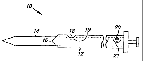
[017] FIG. 1A is a side schematic view of a catheter device having a detection
orifice disposed on a sheath.
[018] FIG. 113 is a side schematic view illustrating two detection orifices
disposed on the sheath.
[019] FIG. 1C is a side schematic view illustrating two, linearly offset
orifices
disposed on the sheath.
[020] FIGS. 2A-2E schematically illustrate the sheath of FIG. 1 puncturing and
entering an artery.
[021] FIG. 3 is a side schematic view of a sheath having two detection
orifices.
[022] FIG 4 is a schematic view of a dial display.
[023] FIG. 5 is a schematic view of an indicator.
[024] FIG. 6 is a side, partially sectional view of a catheter having an
orifice in a
sheath and an orifice in a arteriotomy locator.
[025] FIG. 7 is a flowchart illustrating the steps of sealing a puncture.
[026] FIG. 8 is a side, sectional view of an insertion sheath and arteriotomy
locator in an artery.
[027] FIG. 9 is an illustration of an anchor device being inserted through an
insertion sheath.
[028] FIG. 10 is a side, sectional partially enlarged view of a set anchor.
[029]. FIG. 11 is a flowchart illustrating the steps of inserting and setting
the
anchor.
DETAILED DESCRIPTION
[030] In the present invention, specific reference is made to exemplary
embodiments of a catheter sheath and a vascular closure device and method.
However, vascular closure devices and methods can take many forms and may be
used in various catheterization, laproscopic, and minimally invasive
procedures,
-6-
CA 02523311 2005-10-24
WO 2004/096056 PCT/US2004/012703
and the position detecting features and steps of the present invention are
intended
to be applicable as suitable to any form of vascular closure device and
method.
[031] FIG. 1A is a side view of a vascular closure device 10 that includes a
sheath 12 and a arteriotomy locator 14 that is receivable within the sheath
12.
The arteriotomy locator 14 is positionable over and guidable by a guide wire
16.
It should be appreciated that the various components (e.g., the arteriotomy
locator
14, guide wire 16) may be inserted into and removed from the sheath 12 before
or
after the sheath 12 has been passed through a wall of blood vessel. In other
words, the sheath 12 provides a known access point once so inserted. The
various
components are made from any suitable material such as metal or medical grade
plastic.
[032] In one embodiment, the sheath 12 includes a detection orifice 18 located
near a distal end of the sheath 12. The detection orifice 18 is in fluid
communication with a locator orifice 20 that is located near a proximal end of
the
sheath 10 (or any suitable portion of the device 10), so that the locator
orifice 20
is visible to a surgeon even when the sheath 12 is inserted through the wall
of a
blood vessel. The locator orifice 20 is an indicator that may take any
configuration suitable to provide a visual, audible, and/or tactile indication
of
blood flow.
[033] Fluid communication between the detection orifice 18 and the locator
orifice 20 may be achieved in a number of ways. In one embodiment, the
arteriotomy locator 14 includes a hollow passageway and a arteriotomy locator
orifice 19. When properly positioned within the sheath 12, the arteriotomy
locator orifice 19 aligns with the detection orifice 18. Similarly, the
arteriotomy
locator 14 includes an exit orifice 21 that aligns with locator orifice 20.
Alternatively, the proximal end of arteriotomy locator 14 could be exposed and
exit orifice 21 could function as the indicator (e.g., functionally replacing
locator
orifice 20). In another embodiment, a separate, dedicated lumen is provided
within the sheath 12 between the two orifices 18, 20. In another embodiment,
no
-7-
CA 02523311 2005-10-24
WO 2004/096056 PCT/US2004/012703
such lumen is provided. Rather, the presence of the arteriotomy locator 14
within
the hollow sheath 12 forms a fluid passageway between the detection orifice 18
and the locator orifice 20. In some embodiments, a gap between the interior
cavity of the sheath 12 and the arteriotomy locator 14 forms the fluid
passageway.
It may be formed in other suitable ways, too. For example, the outer surface
of
the arteriotomy locator 14 or the inside surface of the sheath 12 may be
provided
with grooves or channels.
[034] FIGS. 1B and 1C illustrate variations of the device 10, wherein two
detection orifices 18A and 18B are provided and both are in fluid
communication
with the locator orifice 20. As will be explained more fully below, blood flow
is
observed through the locator orifice 20 to indicate proper positioning. Thus,
by
providing two (or more) detection orifices 18A, 18B, a larger volume of blood
flow may be achieved to provide a greater indication. In FIG. 1B, the two
orifices
18A, 18B are provided adjacent to one another. In FIG. 1C, the two orifices
18A,
18B are axially offset from one another. As the sheath 12 is generally
inserted
into the artery (or other blood vessel) at a non-perpendicular angle, this
offset
(indicated generally by the hatched line) will generally be positioned
parallel to a
main, axis of the artery wall. Thus, both detection orifices 18A, 18B will be
exposed to the same flow rates at the same time as the sheath 12 is moved and
positioned through and within the artery.
[035] FIGS. 2A-2E schematically illustrate the deployment of the device 10
into
an artery 30 through an artery wall 32, while using the present invention to
properly position the device. In FIG. 2A, the sheath 12 partially pierces the
artery
wall 32. The arteriotomy locator 14 and the guidewire 16 are passed through
the
sheath and into the artery 30. As the device 10 is advanced in FIG. 2B, the
distal
end of the sheath 12 passes through the artery wall 32 and enters the artery
30. At
this point, the detection orifice 18 is still occluded by the artery wall 32,
thus no
blood flow is detected at the locator orifice 20. The operator knows to
further
advance the device 10, as illustrated in FIG. 2C. As indicated, the detection
-8-
CA 02523311 2005-10-24
WO 2004/096056 PCT/US2004/012703
orifice 18 is within the artery 30 and exposed to blood flow. Blood is
delivered
from the detection orifice 18 to the locator orifice 20 (or otherwise
indicated).
Thus, the operator now knows that the distal end 15 of the sheath and at least
a
portion of the detection orifice 18 is within the artery.
[036] Once blood flow is detected, the operator knows that the sheath is
properly
positioned. That is, the tip or distal end 15 is just inside the artery 32 and
the
device is ready for anchor deployment. This is advantageous in that the device
10
is properly positioned with only one advancing and one retracting stroke,
without
removing the sheath 12 completely from the artery wall 32. That is, because
the
detection orifice 18 is positioned on the sheath 12, the retraction of the
device 10
to find the edge of the artery wall 32 does not require the sheath 12 to be
withdrawn from the wall 32. The space between the detection orifice 18 and the
distal end 15 is such that locating the edge of the artery wall in the above
manner,
positions the distal end 15. Of course, a given operator may advance or
retract the
sheath 12 multiple times based upon a familiarity with previous devices (that
required such actions) or to simply gain a comfort level with the positioning.
The
present invention is advantageous in this regard because such repetitive
strokes
still do not withdraw the sheath 12 from the artery wall 32. Thus, even though
they are ultimately unnecessary, they are generally harmless.
[037] FIGS. 2D-2E , generally illustrate the deployment of an anchor 40. The
anchor 40 is passed through the sheath 12 and into the artery. The sheath 12
is
subsequently extracted and the anchor 40 is used as one half of a sealing
device
(within the artery) to seal the puncture.
[038] FIG. 3 illustrates another embodiment of the device 10 that includes a
first
detection orifice 50 and a second detection orifice 52. The first and second
detection orifices 50, 52 are in fluid communication with a first locator
indicator
54 and a second locator indicator 56 respectively. Once again, blood flow is
detected at the appropriate indicator 54, 56 and blood is allowed to enter the
appropriate orifice 50, 52.
-9-
CA 02523311 2005-10-24
WO 2004/096056 PCT/US2004/012703
[039] In order to differentiate between the first orifice 50 and the second
orifice
52, there are separate paths of fluid communication between the respective
orifice
50, 52 and the indicator 54, 56. This can be accomplished with a separate
lumen
provided within the sheath 12 for each flow path. Alternatively, one such flow
path could be the open interior or the sheath 12 as limited by the presence of
the
arteriotomy locator 14. One flow path could be the hollow interior of the
arteriotomy locator 14, as described above. Thus, as blood flow is achieved
through first detection orifice 50, such flow is indicated at the first
locator
indicator 54. Similarly, as blood flow is achieved through second detection
orifice 52, such flow is indicated at second locator indicator 56. Thus, the
first
and second detection orifices 50, 52 act as position identifiers.
[040] The orifices 50, 52 are selectively positioned on the sheath 12 to
indicate a
selected position within the artery 30. That is, the first detection orifice
50 is
located near the distal end 15 of the sheath 12. Thus, as described above the
first
detection orifice indicates successful entry into the artery 30, beyond the
artery
wall 32. Continued advancement of the device 10 beyond this initial indication
places the sheath 12 further into the artery. Blood flow through second
detection
orifice 52 and indicated by second locator indicator 56 indicates that the
sheath 12
has advanced into the artery sufficiently far to allow second detection
orifice 52 to
be in fluid communication with the blood flow of the artery 30.
[041] In one embodiment, the second orifice 52 is positioned so that if blood
flow is detected, this indicates the sheath has been advanced "too far" and
should
be slightly retracted or at least not advanced farther. Alternatively, the
second
orifice 52 could be positioned so that once blood flow is detected, the
operator
knows the sheath is properly positioned. In either case, the recurrent
advancing
and retracting with previous embodiments may be avoided. That is, the sheath
12
can normally be inserted in a single advancing action, with at most, a slight
retraction if a "too far" condition is reached. Alternatively, such an
embodiment
could be used as previously described with advancing and retracting strokes.
The
-10-
CA 02523311 2005-10-24
WO 2004/096056 PCT/US2004/012703
second indicator would simply provide an additional safety function of
alerting
the surgeon that the device 10 has been inserted too far.
[042] FIGS. 4 and 5 illustrate various indicators 60, 62 that can be provided
as
locator indicators 54, 56. In its simplest form, locator'indicator 54, 56 is
simply
an opening through which blood flow occurs and is viewed or otherwise sensed.
The locator indicator 54, 56 could be open to the environment or shielded by a
viewing port. FIG 4 illustrates dial-type display device 60 that indicates the
absence of blood flow; blood flow at the first orifice 50 (I); or blood flow
at the
second orifice 52 (II). FIG. 5 illustrates a simple fluid communication path
62
interconnecting the two orifices 50, 52. The direction of the blood flow will
move
the indicator ball 64 to an appropriate point to indicate which orifice 50, 52
is
within the artery 30.
[043] FIG. 6 illustrates another embodiment wherein a arteriotomy locator
orifice 70 is provided in the arteriotomy locator 14. The arteriotomy locator
orifice 70 is in fluid communication with a arteriotomy locator indicator 76
disposed at a proximal end of the device 10. Similarly, a sheath orifice 18 is
provided in the sheath 12 and functions in the same manner as previously
described in conjunction with a sheath indicator 20. In this embodiment, blood
flow initially indicated by the arteriotomy locator orifice 70 indicates that
the
distal end 15 of the sheath 12 is proximate the artery wall 32. Blood flow
indicated at the sheath orifice 18 will indicate the location of the sheath
orifice 18
relative to the artery wall 32. That is, depending on where the sheath orifice
is
positioned relative to the distal end 15, the detected blood flow could
indicate
different parameters. For example, when placed near the distal end 15, such
blood flow could indicate the proper positioning of the sheath. If the sheath
orifice 18 is positioned further away from the distal end 15, blood flow could
indicate a "too far" positioning of the sheath 12. In either case, it is
unnecessary
to withdraw the sheath 12 from the artery wall during initial positioning and,
thus,
repetitive entry of the sheath 12 through the artery wall 32 is avoided.
-11-
CA 02523311 2005-10-24
WO 2004/096056 PCT/US2004/012703
(044] Referring to FIG. 7, in one embodiment an insertion procedure utilizing
the device 10 comprises three steps: locate the blood vessel, in this instance
an
artery (180), set the anchor (185), and seal the puncture (190). Before
beginning
the procedure, the surgeon may conduct a fluoroscopic assessment of the
arteriotomy region, to confirm the correct placement of the procedure sheath
that
is already in place, for example, in the common femoral artery.
[045] The procedure will be described in reference to the flowchart of FIG. 11
as
well as the illustrations presented in FIGS. 8-10. To begin the surgeon
inserts and
snaps (200) the arteriotomy locator 14 into the insertion sheath 12. The
locking
interaction between the sheath 12 and the arteriotomy locator 14 at a locking
head
100 assures that the appropriate detection orifices are aligned. Next, the
guide
wire 16 is inserted (210) into the existing procedure sheath. The procedure
sheath
is removed over the guide wire 16. The locator insertion sheath 12 and
arteriotomy locator 14 are inserted (230) over the guide wire 16.
[046] The assembly is advanced through the puncture track and into the artery
30. When the tip 15 of the insertion sheath 12 enters the artery 30, blood
will
begin to flow from the proximal locator orifice 20 (240). The assembly is then
backed out slowly until the blood flow stops (250), indicating that the
detection
orifice 18 has been occluded by the artery wall 32. The device 10 is now
properly
positioned. Earlier devices required subsequent advancement from this
position.
Thus, at least at first, subsequent advancement may be performed as a matter
of
habit for some surgeons. This will simply further advance the sheath 12
further
into the artery. While unnecessary, this action is generally not harmful.
[047] Once positioned, the proximal end of the arteriotomy locator 14 is bent
down slightly, to unlock it from the insertion sheath. The arteriotomy locator
14
and guide wire 16 are removed (240) from the sheath 12. The anchor materials
(110) are then advanced (270) into the sheath 12 as shown in FIG. 9. A secure
cap 120 will only allow the device sleeve and sheath cap to fit together in
the
correct position. While holding the sheath hub steady within the artery, the
secure
-12-
CA 02523311 2005-10-24
WO 2004/096056 PCT/US2004/012703
cap is grasped and slowly pulled back until resistance is felt, indicating the
anchor
is now positioned against (280) the distal end 15 of the insertion sheath 12.
[048] The device-sheath assembly is slowly withdrawn along the angle of the
puncture track to position the anchor 40 against the artery wall 32. As the
sheath
12 clears the skin, a tamper tube and suture will appear. The tamper tube is
advanced to move (290) a collagen sponge 130 down the tissue track. The suture
is retained within the cap 120 and appears as the sheath 12 is withdrawn.
Tension is maintained on suture and the tamper tube is advanced along the
puncture track to help form the collagen anchor seal at the arteriotomy. The
seal
is complete when resistance is felt and the tamping marker is revealed on the
suture, above the tamping tube. This confirms that the self-tightening suture
has
secured the collagen sponge at the arteriotomy as illustrated in FIG. 10.
[049] The procedure is completed by cutting the suture and removing the tamper
tube. The remaining suture is pulled upwards and cut below skin level. The
anchor, collagen sponge, and suture will be naturally absorbed by the body
within
sixty to ninety days.
[050] The present invention is useful for any type of catheter that is placed
within an artery or similar structure. In one context, the present invention
is used
with an artery puncture sealing apparatus, however, such use is merely meant
to
be exemplary and not limiting.
[051] Although the present invention has been described with reference to
preferred embodiments, persons skilled in the art will recognize that changes
may
be made in form and detail without departing from the spirit and scope of the
invention.
-13-