Note: Descriptions are shown in the official language in which they were submitted.
CA 02523557 2005-10-25
WO 2004/096091 PCT/EP2004/004486
Loading and delivery of self-expanding stents
This invention relates in one aspect to a method of loading a
self-expanding stent into a delivery sheath, in which the
stent in a radially confined delivery configuration is
advanced axially into the sheath for delivery to a stenting
site in which the sheath is withdrawn to release the stent
for radial expansion. In another aspect, the invention
relates to a self-expanding stent within a percutaneous
transluminal delivery catheter that includes a sheath that
withdraws proximally to release the stent at a stenting site,
and a pusher within the sheath that retains the stent at the
site during withdrawal of the sheath.
EP-A-788 332 discloses a self-expanding braided metallic
stent tube and a delivery system that includes a soft annulus
within the stent lumen that deforms and mechanically engages
with the mesh of the stent for restraining the stent from
axial movement relative to the inner catheter of the delivery
system, during axial movement of a sleeve surrounding the
stent. The disclosure of EP-A-596 145 is similar.
EP-A-836 447 discloses a system for delivering a self-
expanding stent, in which a stopper ring on an inner catheter
abuts the proximal end of the stent tube during proximal
withdrawal of a sheath which surrounds the stent.
The number of materials that are biologically compatible, and
available for making stents, are comparatively few. One
preferred material is stainless steel. One can make stainless
steel stents that are plastically deformed when they are
expanded radially at the stenting site. One convenient way to
expand such stents is by a balloon at the distal end of a
balloon catheter. Otherwise, one can design a stainless steel
CA 02523557 2005-10-25
WO 2004/096091 PCT/EP2004/004486
2
stent to expand elastically when released at a stenting site.
Typically, this is achieved by proximal withdrawal of a
sheath on the distal end of the delivery catheter, that
withdraws proximally to release the stent progressively,
starting at its distal end.
Another suitable material is the nickel titanium shape memory
alloy known under the trade mark NITINOL. Such stents are
typically loaded into a delivery system at a low temperature
when the crystal structure of the material is martensitic,
and with a memory of a radially expanded shape,
characteristic of a higher temperature austenitic crystalline
structure. Remarkably, the nickel titanium material is
biologically compatible and the martensite/austenite
transformation occurs between room temperature and body
temperature.
This invention is particularly applicable to self-expanding
stents, irrespective of the mechanism of resilient radial
expansion at the stenting site. However, the present
Applicant has particular experience with nickel titanium
shape memory alloy stents and the particular embodiments
described below are based on such materials.
The tubular envelope of a stent usually has apertures through
its wall thickness to permit radial expansion. Thus, an
uncovered or "bare" stent has a tube wall that is normally
liquid-permeable. However, there are many occasions when a
stent with a liquid-impermeable wall that is not apertured
would be desirable. To meet these needs, a family of
"covered" stents have been developed. Applicant has
particular experience with stent tubes provided with a
covering of expanded polytetrafluoroethylene (ePTFE).
Typically, the stent tube is covered by luminal and abluminal
covering layers of ePTFE, which are bonded to each other
through the apertures in the stent tube wall.
CA 02523557 2005-10-25
WO 2004/096091 PCT/EP2004/004486
3
During-manufacture of stents and delivery systems, attention
must be paid to sterility. Specifically, one needs procedures
for loading a covered stent into a catheter delivery system
that will allow sterile conditions to be maintained, or at
least thereafter achieved.
Typically, to introduce a covered self-expanding stent into a
catheter delivery system, a tool needs to be provided that
compresses the covered stent radially inwardly, down to a
diameter which is smaller than the available diameter of the
lumen of the delivery system that is to receive the
compressed covered stent. Clearly, any structure within the
lumen of the stent that resists further inward compression is
better avoided, when the objective is to compress the stent
radially inwardly as much as the system will tolerate, so as
to keep the outside diameter of the delivery system at its
distal tip as small as possible.
However, the stent has to be maintained at the stenting site
during proximal withdrawal of the surrounding sheath, for
progressive release of the stent at the stenting site. If
there is no structure within the lumen of the stent, then the
entire stress imposed on the stent, to prevent it moving
proximally with the proximally withdrawing surrounding
sheath, has to be carried on the proximal end annulus of the
compressed stent. Often this is not really a problem,
especially when the stent is short and not particularly
highly compressed radially inwardly, and especially when
friction between the compressed stent and the surrounding
sheath can be brought to a particularly low value.
Nevertheless, it is important for management of fatigue
resistance to avoid imposing on any point of the stent tube a
level of stress that is higher than the designed maximum. A
stent tube made of metal is susceptible to fatigue failure,
if only because it is subject to cyclic stress at the
frequency of the heartbeat of the body in which it is
CA 02523557 2011-02-10
76186-190
4
installed. For this reason, regulatory authorities require stringent fatigue
performance standards which impose on manufacturers of stents and delivery
systems an onerous burden to avoid any unforeseen stresses on the stent tube.
The state of the art contains numerous suggestions to use an element within
the
lumen of the stent to restrain the stent from proximal withdrawal when the
surrounding sleeve is withdrawn proximally. However, these systems are of
interest only for bare stents, because they rely upon mechanical interaction
between surfaces on the stent pusher within the stent lumen, and boundary
surfaces of apertures within the wall thickness of the stent tube.
It is an object of the present invention to load self-expanding covered stents
into
catheter delivery systems which offers better management of stress within the
stent tube, facilitates quality control and maintenance of sterile conditions,
and is
applicable to a range of stent tube designs.
According to one aspect of the present invention, there is provided a method
of
loading a self-expanding stent into a delivery sheath, in which the stent in a
radially confined delivery configuration is advanced axially into the sheath
for
delivery to a stenting site in which the sheath is withdrawn to release the
stent for
radial expansion at the site characterized by the steps of i) providing said
stent as
a covered stent having a stent matrix with surfaces defining luminal and
abluminal
envelopes spaced apart by a stent wall thickness, a covering material bonded
to
the matrix lying radially inside the luminal envelope ii) providing a stent
pusher
within the lumen of the stent, the stent pusher having radially outwardly
extending
protrusions distributed along the length of the stent lumen iii) compressing
the
stent radially inwardly until the protrusions deform the covering material,
yet
remain radially inside the luminal envelope, and iv) advancing the compressed
stent into the sheath by imposing an endwise force on the stent pusher so that
the
covering material transfers the pushing force from the protrusions of the
stent
pusher to the stent matrix.
According to a second aspect of the present invention there is provided a self-
expanding stent within a percutaneous transluminal delivery catheter that
includes
CA 02523557 2011-02-10
76186-190
4a
a sheath that withdraws proximally to release the stent at a stenting site,
and a
pusher within the sheath that retains the stent at the site during withdrawal
of the
sheath characterised in that i) the pusher extends along the lumen of the
stent and
has radially outwardly extending protrusions distributed along the length of
the
stent lumen ii) the stent is a covered stent having a matrix with surfaces
defining
luminal and abluminal envelopes spaced apart by a stent wall thickness, a
covering material bonded to the matrix lying radially inside the luminal
envelope;
and iii) the protrusions deform the covering material yet remain radially
inside the
luminal envelope.
By distributing over the full length of the stent tube lumen the forces which
necessarily have to be imposed on the stent in order to:
1. load it into a delivery sheath; and/or
2. restrain it from proximal movement during proximal withdrawal of the
delivery sheath during placement of the stent at the stenting site
CA 02523557 2005-10-25
WO 2004/096091 PCT/EP2004/004486
one can manage the distribution of stress within the stent
tube so that it is distributed more or less homogeneously,
rather than concentrated at one end of the stent tube. By
using the covering of the stent as a link in the chain of
stress distribution from the pusher to the sheath, one can
further avoid any point at all within the metal stent tube
which is subject to stress at a level higher than a
prescribed design maximum. By their nature, stent coverings
are more flexible than the stent tube itself, so have the
capability to distribute stress from a point on a metallic
stent pusher to an area, or volume, of the material of the
stent tube.
Furthermore, the flexibility of the stent covering is
sufficient to accommodate the protrusions of the pusher,
irrespective where they lie in relation to the apertures of
the stent lumen. With the present invention, there is no need
to align in any way the protrusions of the stent pusher with
the apertures of the stent lumen. Thus, a further technical
effect of the present invention is valuable simplicity and
speed of operation in loading a range of different covered
stent products into their corresponding delivery systems.
Yet a further advantage of the present invention is that the
stent pusher needs no undercut or rebated surfaces to achieve
its effect, and the pusher has an outside diameter which is
smaller than the inside or luminal diameter of the stent
tube. These factors give greater reassurance that, when the
stent has been placed, and the pusher has to be withdrawn
from the stent lumen, there will be no inadvertent or
unintended snagging of surfaces of the pusher on surfaces of
the covered stent, or indeed of any bodily tissue that might
impinge on the surfaces of the stent pusher after it has been
withdrawn proximally out of the stent lumen.
CA 02523557 2005-10-25
WO 2004/096091 PCT/EP2004/004486
6
Of particular interest in the present invention is a stent
pusher with protrusions arranged helically. Such protrusions
will achieve the desired pushing effect when the pusher is
subject to axial stress. However, arranging the protrusions
helically would allow the pusher to be withdrawn from the
stent lumen, even while the stent is. within the sheath of the
delivery system, simply by "unscrewing" the shaft of the
pusher until the helical protrusions emerge, by continued
rotation of the pusher relative to the stent, out of the
lumen of the stent. In this way, one can employ the stent
pusher of the present invention as part of a system for
loading a covered stent into a sheath, but then remove the
pusher, and pass the sheath stent assembly onwards for
incorporation into a delivery system which will use an
entirely different stent pusher.
For a better understanding of the present invention, and to
show more clearly how the same may be carried into effect,
reference will now be made to the accompanying drawings, in
which:
Fig. 1 is a side view of a tool for loading a covered
self-expanding stent into a sheath;
Fig. 2 is an enlarged view of the distal end (II) of the
tool of Fig. 1; and
Fig. 3 is an axial diametral section through the distal
tip of a stent delivery system which embodies the
present invention.
Fig. 3 shows only the distal tip of the delivery system, but
the remainder of the system is not part of the contribution
which the present invention makes to the art and, in any
event, is familiar to those skilled in this art. The basis
components of a conventional delivery system for a self-
expanding stent are an inner catheter and an outer sheath,
CA 02523557 2005-10-25
WO 2004/096091 PCT/EP2004/004486
7
the purpose of the outer sheath being to confine the self-
expanding stent radially, to the small radius-delivery
configuration, until its release at the site of stenting. The
purpose of the inner catheter is to restrain the stent from
proximal movement with the sheath, while the sheath is being
withdrawn proximally.
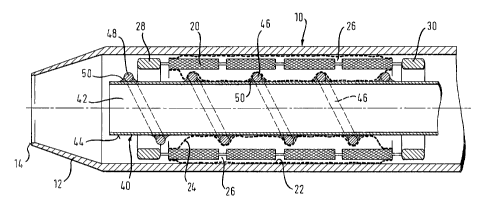
Looking at Fig. 3 of the drawings, the outer sheath 10 of the
delivery system has an integral tapered tip 12 which narrows
down to an end ring 14 of a diameter appropriate to receive a
guidewire (not shown). Confined within the sheath is a
covered stent of which the structural foundation is a stent
body 20 which is an apertured tube of nickel titanium shape
memory alloy. The stent is covered by an outer layer 22 of
ePTFE on the abluminal surface of the stent body, and a
covering layer 2,4 of ePTFE on the luminal inner surface of
the stent body 20, with the. inner and outer layers 24-and 22
being fused together where they can be pressed together
within the apertures 26 of the stent body.
Between the luminal and abluminal surfaces of the stent body
20 is a wall thickness of the metallic stent material
annulus. This annulus lies between the luminal and abluminal
major surfaces of the stent body and, in the specification,
we use the terminology "envelope" to indicate the generalised
surfaces of the luminal and abluminal major wall surfaces of
the stent body. Thus, the outer layer 22 lies outside the
abluminal envelope stent body 20, except where it protrudes
into the apertures 26 for fusing with the inner layer and,
likewise, the inner layer 24 lies radially within the luminal
envelope of the stent body 20 except where it protrudes
radially outwardly into the stent body apertures 26.
The stent body carries a ring of tantalum radiopaque markers
28 at its distal end and a second ring of radiopaque tantalum
markers 30 at its proximal end. It will be appreciated that
the presence of these markers may further militate against
CA 02523557 2011-02-10
76186-190
8
pushing structures that bear against the end surface of the
stent to be deployed.
The-inner catheter 40 defines a guidewire lumen 42.
Conveniently, the inner catheter 40 is based on a stainless
steel hypo tube. This of course endows the entire delivery
system with substantial pushability, but the hypo tube can
also be made remarkably flexible for the desired trackability
of the system through particularly tortuous bodily lumens. In
any event, if stainless steel is not flexible enough for the
distal zone of the delivery system, then it would be feasible
to build the inner catheter 40 from other more flexible
materials such as particular polymers.
The stent delivery system can be arranged as an over the wire
system with a full length guidewire lumen, or a rapid
exchange system with a guidewire lumen only in a distal zone
of the system. The outer sheath 10 can be withdrawn by a full
length outer catheter or' a pull wire within a shaft lumen.
For an example of delivery systems of the present Applicant,
see WO 03/003944 and WO 04/062458.
The inner catheter has an abluminal surface 44 which carries
on it a wire 46 arranged as a helix so as to provide a
plurality of protrusions (at least when seen in section as in
the drawing) on the abluminal surface 44. In the illustrated
embodiment, the wire is of stainless steel, fixed to the
stainless steel tube 40 by deposits 50 of a bonding material
which could be a weld bead or a suitable adhesive.
In any event, as can be seen.on the drawing, when the stent
body is radially inwardly compressed down onto the inner
catheter 40, the inner ePTFE layer 24 deforms to accommodate
the protrusions 48, but the protrusions 48 do not reach
radially outwardly as far as the luminal envelope of the
stent body 20.
CA 02523557 2005-10-25
WO 2004/096091 PCT/EP2004/004486
9
In use, when the illustrated distal tip zone has been brought
to the site of stenting, the outer catheter 12 is carefully
.and progressively withdrawn proximally so that the tip
stretches and slides over the outer ePTFE layer 22 of the
stent, progressively releasing the stent, starting at its
distal end near the markers 28.
As the stent progressively expands, the inner ePTFE layer 24
moves radially outwardly away from the protrusions 48 until,
with complete withdrawal of the tip 12 proximally beyond the
proximal ring of radiopaque markers 30, the stent is fully
released. It will be appreciated that there is then a
substantial annular gap between the lumen of the expanded
stent and the envelope containing the protrusions 48,
enabling the inner catheter 40 also to be withdrawn
proximally from the lumen of the stent without any snagging
of the inner catheter 40 on any part of the stent.
It will be appreciated that, for loading a stent,into a
sheath, an analogous sequence of steps may be performed, with
radially inward compression of the stent body down onto the
protrusions 48 of a loading tool which has a shape in section
analogous to that of the inner catheter 40. Once the stent
has been so compressed, a suitable sheath can be offered up
to one end of the compressed stent tube, and then the stent
can be urged axially into the sheath by imposing an axial
force on the line of protrusions 48 through the tube 40 on
which they amounted, so that this force is transferred from
the protrusions 48 to the inner layer 24 and thence to the
stent body 20 and the outer layer 22, so that the entire
covered stent device is urged by the protrusions 48 into the
receiving sheath.
A particular advantage of the helical structure of
protrusions 48 as shown in the drawing is that the pusher
within the stent lumen can be removed trouble-free from the
lumen of the stent even when it is in a compressed
CA 02523557 2005-10-25
WO 2004/096091 PCT/EP2004/004486
configuration within a sheath-as shown in the drawing, simply
by "unscrewing" the pusher from within the stent lumen.
'Drawing Figures 1 and 2 show a suitable loading tool 60, long
enough to push the covered stent along the full length of the
outer catheter 10, after being compressed and introduced and
advanced into the proximal end of the outer catheter. The
tool 60 features at its distal end a radially-outwardly
protruding wire spiral 62 with a configuration corresponding
to that of the protrusions 48 and the inner catheter 40
(although non-corresponding configurations are also
feasible). The covered stent is compressed around the
protrusions 62 before the tool 60 is used to urge the covered
stent by means of the protrusions 62, from the proximal to
the distal end of the outer catheter.
The illustrated embodiment shows a system in which the
tapered distal tip of the stent delivery system is carried on
the distal end of the outer catheter. Those skilled in the
art are well-aware that many proposed delivery systems
feature a tapered tip on the inner catheter instead. The
present invention is just as useful in such systems as it is
.in systems, as illustrated, with the tapered tip on the outer
catheter.
The stent on which the present device operates can be an
covered.self-expanding stent. The stent which is the basis of
the illustrated embodiment is the one that is the preferred
embodiment of WO 2002/015820 which is cut from a nickel-
titanium tube. However, the invention is equally applicable
to other stent design philosophies, such as stents fabricated
from wire (one example is the Gianturco "Z" stent made from
zig zag wire rings) or other metals, such as stainless steel.
The invention is particular useful for covered stents in
which only the cover connects adjacent ones of a plurality of
stenting rings, because the engagement of the pusher over the
full length of the stent should avoid any tendency for the
CA 02523557 2005-10-25
WO 2004/096091 PCT/EP2004/004486
11
stent covering to "concertina" between the stenting rings
when pushed only from its trailing (usually proximal) end.
Those skilled in the art will be able to recognise from this
disclosure many other ways to realise the present invention
besides that described with reference to the drawings.