Note: Descriptions are shown in the official language in which they were submitted.
CA 02524163 2010-12-17
WO 2001/098694 PCT/US2004f005127
MULTI-FUNCTIONAL MEDICAL CATHETER
CROSS-REFERENCES TO RELATED APPLICATIONS
[0001] This application is a Continuation-in-Part (CIP) of pending U.S. Patent
Application Serial
No. 10/305,256, filed November 25, 2002, published on August 14, 2003 under
publication No.
US 2003-0153907 Al, and entitled "ULTRASOUND-GUIDED ABLATION CATHETER
AND METHODS OF USE;" which is a continuation of U.S. Application Serial No.
09/750,439,
filed on December 28, 2000, and issued on January 21, 2003 as U.S. Patent No.
6,508,765;
which is a continuation of U.S. Application Serial No. 09/227,281, filed
January 6, 1999 and
issued on March 27, 2001 as U.S. Patent No. 6,206,831 B 1.
BACKGROUND OF THE INVENTION
[0002] The invention relates generally to the field of medical catheters, and
in particular, to
multi-functional medical catheters adapted to map, orient and/or provide
treatment for a
variety of medical conditions.
[0003] Physicians make use of catheters today in medical procedures that are
best
performed by gaining access into interior regions of the body. For example, in
electrophysiological therapy, ablation is used to treat cardiac rhythm
disturbances. Such a
therapy may be used, for instance, to treat atrial fibrillation by forming
lesions in heart tissue
at desired locations to interrupt undesirable electrical pathways.
[0004] During these procedures, the physician typically first maps the
electrical activity of
the patient's heart to help determine the location of any abnormalities. The
physician then
steers a catheter through a main vein or artery into the interior region of
the heart that is to be
treated. An ablation element carried on the distal end of the catheter is
positioned near the
tissue that is to be ablated. For such treatments, the delivery of ablating
energy must be
closely governed to avoid incidence of tissue damage and coagulum formation.
Further, the
ablation catheters must be precisely positioned adjacent to and preferably in
contact with the
tissue to be treated, to insure the lesions are properly located.
[0005] Physicians and staff performing diagnostic and therapeutic procedures,
such as
electrophysiological therapy, typically require an imaging system to assist
them in
positioning the ablation catheter. Mini-transesophageal echocardiography (mini-
TEE) probes
CA 02524163 2011-10-06
are available, however, these probes must be swallowed or inserted down the
patient's throat.
Such probes are poorly tolerated by patients unless they are fully
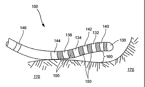
anesthetized. Further, these
probes can be rather large (i.e., 20 French in diameter), use complex
transducer
configurations and may have difficulty in detecting tissue contact by the
ablation elements.
Further, the mapping, imaging and treatment often requires multiple
instruments or catheters,
involving complex procedures as well as the introduction or reintroduction of
multiple
catheters into the patient. Improvements are desired.
BRIEF SUMMARY OF THE INVENTION
[0006] The present invention provides multi-functional medical catheters,
systems and
methods for their use. In some embodiments, the catheters include ultrasound-
guided
ablation catheters. Catheters and systems of the present invention will be
particularly useful
for precise positioning of ablation catheters prior to ablation of cardiac
tissue, such as that
required for the treatment of atrial fibrillation. Further, the functionality
of some of the
embodiments permits a single catheter to be used for tissue mapping, tissue
orientation, tissue
imaging, and/or tissue treatment, including ablation. Some of the systems of
the present
invention use transducers in the distal end of the catheter to assist the
operator in determining
whether or not the ablation elements are in contact with the tissue to be
ablated. Non-
ablation catheters also fall within the scope of the present invention, with
such catheters
providing tissue mapping, tissue orientation and/or tissue imaging functions.
[0007] In one particular embodiment, a medical catheter of the present
invention includes a
flexible elongate body having a proximal end and a distal end. A plurality of
spaced apart
electrodes are operably attached to the flexible body near the distal end. At
least some of the
electrodes are adapted for mapping a tissue. The catheter includes a plurality
of tissue
orientation detectors spaced along an external surface of the flexible body,
wherein at least
some of the tissue orientation detectors are disposed between at least some of
the electrodes.
In this manner, the medical catheter is capable of both tissue mapping and
tissue orientation
functions. In some embodiments, at least one of the electrodes is adapted for
ablating a desired
portion of the tissue, with the catheter capable of tissue ablation or other
treatments.
[0008] In some aspects, at least one of the electrodes is adapted for both
mapping and
ablation. In some aspects, the electrodes adapted for ablating have at least
one tissue
orientation detector adjacent thereto. In such a manner, the detector(s) help
determine the
location of the ablation electrode prior to ablation. For example, the
detectors may operate to
determine tissue contact, to detect a distance to the tissue, to detect a
three-dimensional
2
CA 02524163 2011-10-06
position relative to the tissue, and the like. In some aspects, at least one
of the electrodes
includes a tip electrode coupled to a tip of the distal end.
[00091 The tissue orientation detectors may have a variety of configurations
within the scope
of the present invention. For example, in one embodiment the tissue
orientation detectors
include a plurality of transducers. In a particular embodiment, at least some
of the transducers
include ultrasound transducers. Alternatively, or in addition, at least some
of the transducers
are electric, magnetic, or electromagnetic tracking transducers.
[00101 The present invention further provides exemplary medical catheter
systems according
to the present invention. In one embodiment, the system includes a medical
catheter including
a flexible elongate body having a proximal end and a distal end. A plurality
of spaced apart
electrodes are operably attached to the flexible body near the distal end. At
least some of the
electrodes are adapted for mapping a tissue. The catheter includes a plurality
of tissue
orientation detectors spaced along an external surface of the flexible body,
wherein at least
some of the tissue orientation detectors are disposed between at least some of
the electrodes. A
controller is coupled to the plurality of electrodes and coupled to the
plurality of tissue
orientation detectors. In one aspect, the controller is adapted for
controlling a tissue mapping
function performed by the plurality of electrodes. In a particular aspect, the
tissue mapping
function includes a non-contact tissue mapping function. In one aspect, the
controller is further
adapted for determining a tissue ablation pattern based on a result of the
tissue mapping
function.
[00111 In another aspect, the medical catheter system controller is adapted
for receiving a
plurality of signals from the tissue orientation detectors and determining an
orientation of the
elongate body relative to the tissue.
[00121 In some embodiments, the medical catheter system further includes a
digitizing
system, and/or an RF generator electrically coupled to the plurality of
electrodes. The
digitizing system is adapted for producing a digitized image of the tissue.
These images may
be based in part on the data received by the electrodes and/or the detectors.
The RF generator
may facilitate using one or more electrodes to ablate tissue, or the like.
[00131 The present invention further provides exemplary methods of precisely
positioning a
medical catheter with respect to a tissue. In one such embodiment, the method
includes
providing a medical catheter system, such as one of the systems detailed
herein. The method
further includes inserting the flexible elongate body into a patient, mapping
an electrical
profile of the tissue using at least some of the electrodes, and positioning
the elongate body to
be proximate a tissue using the tissue orientation detectors. The positioning
is based at least in
3
CA 02524163 2011-10-06
part on the electrical profile of the tissue.
[00141 In one aspect, the method further includes activating at least one of
the electrodes to
ablate a desired region of the tissue if the controller determines that at
least one of the tissue
orientation detectors is in contact with the desired region. In particular
aspects, at least one of
the electrodes is activated to ablate a desired region of the tissue if the
controller determines
3a
CA 02524163 2010-12-17
WO 2004/098694 PCT/US2004/005127
that one of the tissue orientation detectors located adjacent the electrode is
in contact with the
desired region, if the tissue orientation detector located directly proximal
of the electrode is in
contact with the tissue, and/or if the tissue orientation detectors closest to
the electrode in
both the proximal and distal directions are in contact with the tissue. In
this manner, tissue
contact may be determined prior to ablation.
[0015] In one aspect, methods of the present invention further include
identifying a desired
region of the tissue to be treated based on the electrical profile of the
tissue. As discussed
herein, the mapping may include a non-contact mapping in some embodiments to
obtain or
help obtain the electrical profile.
[0016] In another method of precisely positioning a catheter within a patient
according to
the present invention, the catheter is inserted into the patient. The method
then includes
mapping a tissue of the patient, using at least some of the plurality of
spaced apart electrodes,
to produce a tissue profile. The tissue profile may include, for example, a
map or other
depiction of a plurality of electrical pathways in the tissue. A tissue region
to be treated is
identified by using, at least in part, the tissue profile. The elongate body
is positioned using
the transducers so that at least one of the electrodes is proximate the tissue
region. In one
aspect, the elongate body positioning includes a three-dimensional
localization positioning.
The electrode(s) may be further operated to ablate the tissue region where
desired to provide
treatment to the patient.
[0017] In another embodiment of the present invention, a method of diagnosing
and
treating cardiac rhythm disturbances includes inserting a catheter into a
patient, and mapping
a tissue of the patient, using at least some of the plurality of spaced apart
electrodes, to
produce a tissue profile. The method includes identifying a tissue to be
treated using the
tissue profile, positioning the elongate body using the tissue orientation
detectors so that at
least one of the electrodes is proximate the tissue to be treated, and
treating the tissue using
the catheter. The treatment may include ablating the tissue using at least one
electrode.
Ablation may occur through the use of RF ablation, through ultrasound
ablation, or the like.
An exemplary description of acoustic ablation using transducer elements is
described in U.S.
Patent No. 5,630,837. It will be appreciated by those skilled in the art that
other ablation elements
may be used within the scope of the present invention.
[0018] Other features and advantages of the invention will appear from the
following
description in which the preferred embodiment has been set forth in detail in
conjunction with
the accompanying drawings.
4
CA 02524163 2005-10-28
WO 2004/098694 PCT/US2004/005127
BRIEF DESCRIPTION OF THE DRAWINGS
[0019] Fig. 1 depicts an overall view of a system for ablating tissue
according to an
embodiment of the present invention;
[0020] Fig. 2 depicts the distal end of a flexible elongate body as part of a
catheter system
according to an embodiment of the present invention;
[0021] Fig. 3 depicts a cross-sectional side view of the flexible elongate
body shown in Fig.
2;
[0022] Fig. 4A depicts a cross-sectional end view of the flexible body shown
in Fig. 3
taken along line 4A-4A;
[0023] Fig. 4B depicts an overall view of a cylindrical transducer element as
part of a
catheter apparatus according to an embodiment of the present invention;
[0024] Figs. 5A and 5B depict alternative embodiments of a medical catheter
apparatus
according to the present invention;
[0025] Fig. 6 depicts a schematic of a multiplexer for use with medical
catheters of the
present invention;
[0026] Figs. 7A-7B depict energizing and reflected signals sent to and
received by a
transducer element of the present invention;
[0027] Fig. 8 depicts an embodiment of a medical catheter apparatus of the
present
invention in contact with tissue;
[0028] Fig. 9 is an overall view of a medical catheter according to an
alternative
embodiment of the present invention;
[0029] Fig. 10 is a simplified overall view of a medical catheter system
according to an
embodiment of the present invention;
[0030] Fig. 11 is a simplified flow chart of a method of the present
invention; and
[0031] Figs. 12A and 12B depict a simplified overall view and a cross-
sectional side view
of an alternative embodiment of a catheter according to the present invention.
DETAILED DESCRIPTION OF THE INVENTION
[0032] Fig. 1 depicts a medical catheter apparatus 2 as part of a catheter
system 4 according
to an embodiment of the present invention. Apparatus 2 comprises a flexible
elongate body
12 having a distal end 10 and a proximal end 14. Proximal end 14 includes a
handle 16
containing a steering mechanism 18. Steering mechanism 18 includes a steering
lever 22
which operates a cam wheel (not shown) to maneuver flexible distal end 10 as
shown by the
CA 02524163 2010-12-17
WO 2004/098694 PCT/US2004/005127
arrows in Fig. 1. System 4 includes a connector 20 which connects with a
controller 23 for
operation of apparatus 2 as further described below. Controller 23 is capable
of providing
electrical input to apparatus 2 as needed to map, image, orient, and/or ablate
a patient tissue.
It will be appreciated by those skilled in the art that steering mechanism 18
can vary from that
shown in Fig. 1 within the scope of the present invention. Exemplary steering
mechanisms are
described in International Application No. PCT/US 1994/011748, published on
January 4, 1996
under publication No. WO 1996/000036.
[0033] Medical catheter apparatus 2 depicted in Fig. 1 will be particularly
useful in the treatment
of atrial fibrillation by positioning distal end 10 within a desired region of
the heart. To enter the
right atrium, the physician can direct elongate body 12 through a conventional
vascular
introducer through the femoral vein. For entry into the left atrium, the
physician can direct
elongate body 12 through a conventional vascular introducer retrograde through
the aortic and
mitral valves. For the treatment of atrial fibrillation, it is believed that
formation of lesions in the
heart muscle tissue is required. Catheters of the present invention may be
used, in some
embodiments, to ablate heart tissue containing abnormal electrical pathways,
such as
arrhythmogenic foci. Further details of apparatus 2 are shown in Figs. 2 and
3.
[00341 Figs. 2 and 3 depict elongate body 12 having a plurality of spaced-
apart ablation
elements 24, each separated by a gap 26 from adjacent ablation elements 24.
Interspaced
amongst ablation elements 24 are a plurality of transducer elements 28. In one
embodiment,
ablation elements 24 and transducer elements 28 are operably attached to body
12 in an
alternating fashion. Apparatus 2 preferably includes between about two (2) and
about
fourteen (14) ablation elements, and between about three (3) and about fifteen
(15) transducer
elements. More preferably, apparatus 2 has at least one more transducer
element 28 than
ablation elements 24. In one embodiment, a temperature sensor 30 is provided
at or near
distal end 10 and a proximal temperature sensor 32 is provided proximal to
ablation elements
24. Temperature sensors 30 and 32 preferably comprise thermocouples.
Temperature
sensors 30 and 32 also may comprise thermistors and the like within the scope
of the present
invention. Temperature sensors or thermocouples 30 and 32 operate to detect
the temperature
in the region of ablation. A plurality of insulators 40 are provided between
transducer
elements 28 and ablation elements 24. Insulators 40 may comprise polyimide,
polyesters,
teflon or the like to insulate transducer elements 28 from ablation elements
24.
[00351 In one embodiment, transducer elements 28 comprise cylindrical
transducer
elements as best shown in Figs. 4A-4B. Transducer elements 28 include an outer
face 46 and
an inner face 48. Inner faces 48 of transducer elements 28 are positioned such
that a
6
CA 02524163 2005-10-28
WO 2004/098694 PCT/US2004/005127
longitudinal axis 38 of body 12 passes through a throughhole 44 of each
transducer element
28. In such a manner, transducer elements 28 are configured to expose outer
faces 46 to
surrounding tissue and fluid within the patient. In this manner, transducer
elements 28 may
operate to image within a three-hundred and sixty degree (360 ) plane that is
generally
perpendicular to longitudinal axis 38 without the need to rotate body 12 or
transducers 28. It
will be appreciated by those skilled in the art that other transducer shapes
may be used within
the scope of the present invention. For example, transducer elements 28 may
comprise
rectangular or elliptical transducer elements operably attached to distal end
10.
[0036] Transducer elements 28 may comprise ultrasound transducers. In this
embodiment,
transducer elements 28 may comprise piezocomposite materials, piezoceramics
(such as
PZT), piezoplastics, and the like. Alternatively, as further detailed below,
transducer
elements 28 may be adapted to transduce between a magnetic field and a
voltage. Other
transducer types also may be used within the scope of the present invention,
including
without limitation, electric, magnetic, electromagnetic, permanent magnets,
wireless, optical,
and the like.
[0037] In the embodiment shown in Fig. 3, transducers 28 comprise ultrasound
transducer
elements 28. Transducers 28 each may include a matching layer 42, or multiple
matching
layers 42, operably attached to the outer face 46 of each transducer element
28. Matching
layers 42 operate to improve transducer element 28 performance. Transducer
elements 28
also can operate without matching layers 42 within the scope of the present
invention.
[0038] Transducer elements 28 have an outer diameter 29. Outer diameter 29 can
be less
than an outer diameter 31 of flexible elongate body 12 or, alternatively,
about equal to
diameter 31. Preferably, diameter 31 of body 12 is less than about eight (8)
French to permit
the introduction of apparatus 2 into a patient's tortuous vasculature.
[0039] Gap 26 separates adjacent ablation elements 24. Gap 26 preferably is
between
about 1.5 mm and about 3.0 mm in width. Gap 26, however, can be larger or
smaller in size
and need not be of uniform size between each two adjacent ablation elements
24. Similarly,
each gap 26 need not contain a transducer element 28, and gap 26 may contain
more than one
transducer element 28 within the scope of the present invention. However,
preferably at least
some gaps 26 contain transducer elements 28, and in some embodiments, each gap
26
between ablation elements 24 contains at least one transducer element 28.
[0040] Elongate body 12 preferably includes a working lumen 39 through which
longitudinal axis 38 passes. As best shown in Fig. 4A, matching layer 42
extends around the
outer surface of transducer element 28. Matching layer 42 is operably attached
to transducer
7
CA 02524163 2005-10-28
WO 2004/098694 PCT/US2004/005127
element 28, preferably using epoxy or the like. Transducer element 28 can be
operably
attached to elongate body 12 in a variety of manners, including by epoxy. The
use of lumen
39 is best shown in Figs. 5A and 5B which depict two alternative embodiments
of apparatus
2 of the present invention.
[0041] Fig. 5A depicts the medical catheter apparatus shown in Fig. 3 without
matching
layers 42. As can be seen in Fig. 5A, a plurality of leads 50 are operably
attached to
thermocouples 30 and 32, to transducer elements 28 and to ablation elements
24. For an
embodiment having electrodes for ablation elements 24, each electrode has a
single lead 50.
Thermocouples 30 and 32 each have a pair of leads 50. Transducer elements 28
have one
lead 50 in electrical communication with outer face 46. Further, a ground 52
extends from
inner face 48 of transducer 28. As shown in Fig. 5A, a common ground can be
used for all
transducer elements 28 within a particular apparatus 2. One benefit of using a
common
ground 52 is that fewer leads or wires 50 are passed from distal end 10,
through lumen 39 to
controller 23.
[0042] The embodiment shown in Fig. 5B depicts the use of a multiplexer 54
operably
attached to distal end 10 of flexible elongate body 12. Multiplexer 54
preferably is disposed
proximal of ablation elements 24 and transducer elements 28. Multiplexer 54
permits the
attachment of leads 50 from transducer elements 28 to multiplexer 54 without
the need to run
those leads 50 to controller 23. Such a configuration can reduce the number of
wires needed
to be extended through lumen 39 to controller 23.
[0043] The operation of multiplexer 54 is best described in conjunction with
Fig. 6. Fig. 6
depicts transducer elements 28 each having ground 52 and lead 50. Leads 50 are
operably
attached to multiplexer 54, preferably on the distal side of multiplexer 54.
Multiplexer 54 has
a ground 62 and a transmission line 60 for providing power to multiplexer
circuit 54.
Transmit and receive lines 56 provide a means to transmit electrical signals
to multiplexer 54.
Multiplexer 54 then directs electrical signals to the appropriate
transducer(s) 28.
Transmit/receive wires 56 carry transducer 28 excitation signals as
differential pulses in
series format from controller 23 to multiplexer 54. At multiplexer 54, each
excitation signal
is routed to an appropriate one of the transducer elements 28 in order to
execute an excitation
sequence used by controller 23. Similarly, return inputs or echoes received by
transducer
element(s) 28 are transferred to multiplexer 54 and return to controller 23
along
transmit/receive lines 56.
[0044] By minimizing the number of wires required to carry the excitation
signals from
controller 23 to each of transducer elements 28, the diameter of elongate body
12, and more
8
CA 02524163 2005-10-28
WO 2004/098694 PCT/US2004/005127
specifically, the size of lumen 39 can be reduced. Alternatively or in
addition, the number of
transducer elements 28 can be increased at distal end 10 without the need to
require wires to
be run through lumen 39 to controller 23.
[0045] Multiplexer 54 further may include a clock line 58 extending from
controller 23 to
multiplexer 54. Clock line 58 assists multiplexer 54 in determining which
transducer element
28 is to receive an excitation signal. Alternatively, as shown in Fig. 6,
clock line 58 operates
by counting the number of excitation signals transmitted through
transmit/receive lines 56
and incrementing a counter in multiplexer 54 to coordinate the transfer of
excitation signals
to the appropriate transducer 28. In one embodiment, multiplexer 54 also
includes a data line
(not shown in Fig. 6) extending from controller 23 to multiplexer 54. This
data line permits
controller 23 to control the operation of multiplexer 54.
[0046] Turning now to Figs. 7 and 8, the operation of medical catheter
apparatus 2 and
system 4 according to an embodiment of the present invention will be
described. Medical
catheter apparatus 2 operates by having transducer elements 28 detect the
proximity of a
tissue 70 with respect to elongate body 12 distal end 10. Controller 23
calculates the time
delay between transducer element 28 excitation and the receipt of a reflected
signal 66 from
surrounding tissue 70 to determine the distance between transducer element 28
and tissue 70,
as further described below.
[0047] As shown by Figs. 7A and 7B, an excitation signal 64 is transmitted
from controller
23 to transducer elements 28, or to multiplexer 54 for transmission to
transducer elements 28.
Excitation signal 64 is converted by transducer 28 into an ultrasound signal
which propagates
out into surrounding fluid and tissues within the patient. Transducer elements
28 detect
reflected signals 66 and transfer electrical representations of those signals
to controller 23 for
processing.
[0048] Controller 23 uses the time delay between the excitation 64 and the
receipt of
reflected signal 66 to calculate the approximate distance to the reflecting
object. Controller
23 is capable of differentiating between low amplitude blood reflections and
larger amplitude
tissue reflections 66 as shown in Fig. 7. Controller 23 further differentiates
from a
randomized back scatter versus more stable tissue scatter. The distance from
each transducer
28 to tissue 70 may be calculated by knowing the speed of sound and measuring
the time
response to the larger amplitude tissue reflections. If the signal completely
consists of larger
amplitude wave forms, intimate contact will be diagnosed. While transducers 28
inherently
have a blind zone/time period in which signals cannot be measured, the
resulting blind zone
distance is rather small. For example, for a 30 Mhz transducer, this distance
is approximately
9
CA 02524163 2005-10-28
WO 2004/098694 PCT/US2004/005127
0.15 mm. Hence, reflected signal 66 measured almost immediately after
excitation 64 occurs
results in the distance from the transducer 28 to tissue 70 being less-than
about 0.15 min blind
distance.
[0049] Medical catheter system 4, therefore, can be operated by inserting
apparatus 2 into
the patient and positioning distal end 10 of apparatus 2 near a desired
location of the patient's
anatomy. Transducer elements 28 are energized with excitation signal 64 and
reflected
signals 66 are received and processed by controller 23. Controller 23
determines whether or
not transducer elements 28 are in contact with tissue 70. If at least one
transducer element 28
is in contact with tissue 70, ablation using an adjacent ablation element 24
may occur.
Preferably, as shown in Fig. 8, it will be desirable to have more than one
transducer element
28 in contact with tissue 70.
[0050] Controller 23 can be operated in a variety of ways to determine the
number and
positioning of transducer elements 28 which maybe in contact with tissue 70.
For example,
as shown in Fig. 8, transducer elements 28A, 28B and 28C would indicate that
they were in
contact with tissue 70. This may permit the physician to ablate tissue 70
using electrode 24A
and electrode 24B. Transducer element 28D would not indicate contact with
tissue 70.
Therefore, it is inconclusive whether ablation element 24C is in contact with
tissue 70.
Hence, the physician may choose not to ablate with ablation element 24C.
[0051] In one embodiment, controller 23 may use a green and red light system
for
indicating when transducer elements 28 are in contact with tissue 70. In one
particular
embodiment, for example, controller 23 has a red light and a green light for
each transducer
element 28A-28D depicted in Fig. 8. The green light would be illuminated by
controller 23
when the corresponding transducer element 28 is in contact with tissue 70. Red
lights would
be illuminated for those transducer elements 28 not in tissue contact.
[0052] Alternatively, a single green and red light may be used for apparatus
2, whereby the
green light is illuminated by controller 23 only when all transducer elements
28 are in tissue
contact. Still another embodiment involves several transducer elements 28
corresponding to
a single green/red light set. For example, elements 28A and 28B may have one
green light
which controller 23 illuminates when both elements 28A and 28B are in tissue
contact. The
red light corresponding to elements 28A and 28B would be illuminated if one or
both
transducer elements 28A and 28B are not in contact with tissue 70. It will be
appreciated by
those skilled in the art that there exist numerous ways within the scope of
the present
invention for controller 23 to indicate when tissue 70 contact has been
achieved by transducer
elements 28, including audible tones and the like.
CA 02524163 2010-12-17
WO 2004/098694 PCT/US2004/005127
[0053] Ablation elements 24 are preferably used for mono-polar ablation,
although bi-polar
ablation also is anticipated within the scope of the present invention.
Ablation elements 24
preferably comprise electrodes. In this manner, RF ablation may occur using
ablation
elements 24.
[0054] Alternatively, ablation elements 24 may comprise ablation ultrasound
transducers.
In this manner, transducer elements 28 are operated in pulse mode to determine
their distance
from tissue 70. Upon tissue contact, ablation transducers 24 would be used to
ablate tissue
70. The use of transducers for acoustic ablation is further described in U.S.
Patent No.
5,630,837.
[0055] Alternatively, transducer elements 28 can be used to both image and
ablate tissue
70. Transducer elements 28 would first be operated in pulse mode, to determine
whether
transducer elements 28 are in contact with tissue 70. Transducer elements 28
then would
receive a continuous wave or gated continuous wave electrical signal having a
frequency of
about 10-15 MHz, and transducer elements 28 would ablate tissue 70 using
ultrasound
ablation.
[0056] Turning now to Figs. 9 and 10, an alternative embodiment of a medical
catheter
100, and a medical catheter system 200 according to the present invention will
be described.
Medical catheter 100 includes an elongate body 105 having a proximal end 110
and a distal
end 120. Proximal end 110 is coupled to a steering device 210 as shown in Fig.
10. Steering
device 210 may, but need not be similar to that described in conjunction with
Fig. 1. The
length of catheter 100 may vary within the scope of the present invention. In
one
embodiment, the length of catheter 100 is sufficient to permit insertion into
the femoral vein
in a patient leg and traverse through the patient vasculature to reach the
heart muscle or other
region to be treated. Distal end 120, as best shown in Fig. 9, includes a
plurality of elements
coupled to or otherwise disposed therewith for tissue mapping, tissue
orientation detection,
tissue imaging, tissue treatment, and the like. In the embodiment shown in
Fig. 9, distal end
120 includes a tip electrode 130 disposed at or near the distal tip of
catheter 100. In one
embodiment, tip electrode 130 provides an exemplary electrode for ablation
treatments as
previously described.
[0057] Catheter 100 includes a plurality of spaced apart electrodes 132, 134,
and 136
coupled to distal end 120. In one embodiment, electrodes 132-136 comprise ring
electrodes.
In a particular embodiment, ring electrodes 134 and 136 operate as an
electrode pair for a
tissue mapping function. Further, electrodes 130 and 132 may operate as an
electrode pair
11
CA 02524163 2005-10-28
WO 2004/098694 PCT/US2004/005127
for a tissue mapping function. Catheter 100 further includes a plurality of
tissue orientation
detectors 140, 142, 144, and 146 spaced along elongate body 105. As shown in
Fig. 9, tissue
orientation detector 140 is disposed near the distal tip of elongate body 105
such that detector
140 is in close proximity to tip electrode 130. Similarly, detector 146 is
disposed proximal to
the remaining elements of distal end 120, and maybe used for orientating or
detecting the
location of distal end 120.
[0058] Distal end 120 further includes a plurality of insulators 150.
Insulators 150 are
adapted to insulate electrodes 130-136 from one another, and/or to insulate
detectors 140-146
from one another, and/or to insulate detectors 140-146 from electrodes 130-
136. In a
particular embodiment, each electrode 130-136 has at least one detector 140-
146 disposed
adjacent thereto, with possibly an intervening insulator 150 therebetween. For
example, tip
electrode 130 has detector 140 located proximal thereto. Electrode 132 has
detector 140
located distal thereto, and detector 142 located proximal thereto. While
electrodes 134 and
136 are separated from one another by only an insulator 150, each electrode
134 and 136 has
an adjacent detector 142 and 144, respectively. In this manner, detectors 140-
146 and
electrodes 130-136 may be used in concert for a variety of procedures as
further described
herein. It will be appreciated by those skilled in the art that the
orientation and order of the
various detectors 140-146, electrodes 130-136 and insulators 150 may vary
within the scope
of the present invention.
[0059] In one embodiment, tissue orientation detectors 140-146 include
transducers.
Transducers 140-146 may be adapted to transduce between a variety of physical
parameters.
For example, in one embodiment, at least some transducers 140-146 are adapted
to transduce
between ultrasound energy and a voltage. This may occur, for example, when one
or more of
detectors 140-146 comprise ultrasound transducers which are adapted to
transmit an
ultrasound energy wave when a voltage is applied across opposing surfaces of
the detector
140-146. The ultrasound wave travels towards a tissue 170, and is reflected by
tissue 170.
The reflected wave is received by detector 140-146, and is converted into a
voltage by
detector 140-146. The voltage is transmitted to a controller 230, such as is
shown in Fig. 10.
In this manner, detectors 140-146 transduce between ultrasound energy and
voltage.
Alternatively, detectors 140-146 may be adapted to transducer between a
voltage and a
magnetic field. For example, a magnetic or electromagnetic field generator can
be placed in
proximity to the patient. In one embodiment, the catheter carries one or more
transducers
that detect the magnetic or electromagnetic field and convert it into a
voltage. The voltage is
then supplied to controller 230 for orientation detection purposes.
Alternatively, other
12
CA 02524163 2010-12-17
WO 2004/098694 PCT/US2003/005127
transducer types may be used, including electrical transducers, permanent
magnets, optical
transducers, and the like.
[0060] Medical catheter 100 is adapted to perform one or more functions, and
may be
adapted to image tissue, map tissue, assist in orienting itself with respect
to tissue, treat
tissue, and the like. For example, catheter 100 may be adapted for mapping a
patient tissue,
such as heart tissue. This may occur a number of ways within the scope of the
present
invention. For example, tissue orientation detectors 140-146 may be used by
inserting
catheter 100 into a patient's vasculature and transferring distal end 120 to a
desired region of
the patient. Catheter 100 then may be used in conjunction with one or more
reference
catheters to perform a three-dimensional localization process to help map the
general shape
of the patient's tissue, such as the heart muscle. Details of a three-
dimensional localization
process are further described in U. S. Patent No. 6,490,474, entitled "System
and Method for
Electrode Localization Using Ultrasound."
[0061] In an alternative embodiment, catheter 100 is used to map the
electrical activity of
tissue 170. For example, in one embodiment, catheter 100 is inserted into a
desired region of
the patient, and positioned such that one or more electrodes 130-136 are in
contact with tissue
170. Tissue mapping procedures may then be performed to map the electrical
activity of the
heart muscle. Such electrode mapping techniques are further described in U. S.
Patent Nos.
5,598,848, entitled "Systems and Methods for Positioning Multiple Electrode
Structures in
Electrical Contact with the Myocardium"; U. S. Patent No. 5,487,391, entitled
"Systems and
Methods for Deriving and Displaying the Propagation Velocities of Electrical
Events in the
Heart"; and U.S. Patent No. 6,516,807, entitled "System and Methods for
Locating and
Guiding Operative Elements within Interior Body Regions," the complete
disclosures of
which are incorporated herein by reference for all purposes.
[0062] While the above-noted references discuss the use of a basket catheter
for placing
electrodes in contact with heart tissue to be mapped, the present invention
may be adapted to
insure tissue contact prior to mapping. For example, the techniques discussed
in conjunction
with Figs. 1-8 may be used, including the time delay of ultrasound signals
transmitted by, and
subsequently received by detectors 140-146, to verify tissue contact.
[0063] In an alternative embodiment, catheter 100 maps the electrical activity
of tissue 170
using a non-contact mapping technique. Non-contact mapping uses electrodes 130-
136 to
sense electrical activity within tissue 170 notwithstanding the fact there may
be a gap 160
between electrode(s) 130-136 and tissue 170. These far field signals received
by electrodes
13
CA 02524163 2010-12-17
WO 2004/098694 PCT/US2004/005127
130-136 are mapped onto the surface of tissue 170 using an algorithm which
takes into
account the relationship between distal end 120 and tissue 170, and the
general orientation of
catheter 100 with respect to tissue 170. In this manner, electrically active
tissue 170 is
mapped. Additional details on mapping tissue, including non-contact mapping,
may be found
in U.S. Patent 6,240,307 entitled "Endocardial Mapping System."
[0064] Data received or generated by detectors 140-146, and/or electrodes 130-
136 may be
optionally transmitted to controller 230 by coupling catheter 100 to
controller 230 using a
cable 220 or other electrically conductive medium. In one embodiment,
controller 230
comprises a microprocessor coupled to a computer readable storage medium
having software
or other programs adapted to perform a variety of procedures. Controller 230
may include an
input device 250 for receipt of a compact disc, a DVD, or the like containing
reference data,
algorithms or related processing software, or the like. In a particular
embodiment, controller
230 further includes a light array 240 that is adapted to visually indicate to
the operator or
physician when one or more detectors 140-146 are in contact with tissue 170.
As previously
described, light array 240 may comprise a green/red light system, and/or may
include some
other visual or audio indicator. In one embodiment, controller 230 includes a
digitizer that is
adapted to digitize the data received from catheter 100 and display an image
of tissue 170 on
a monitor 270. Controller 230 may be coupled to monitor 270 using a cable 260
or the like.
Alternatively, wireless connections may be used to couple controller 230 with
display 270
and/or to couple controller 230 with catheter 100.
[0065] Turning now to Fig. 11, an embodiment of a method 300 of precisely
positioning
catheter 100 according to the present invention will be described. Method 300
includes
inserting catheter 100 into a patient (block 310). As previously described,
this may occur, for
example, by inserting catheter 100 through the femoral vein of the patient.
Catheter 100 is
then used to map tissue (block 320). The mapping of tissue 170 may include
three-
dimensional localization techniques, and/or the mapping of electrical activity
within tissue
170, both as previously described. Method 300 further includes identifying a
tissue region to
be treated (block 330). This may occur, for example, by displaying an image of
tissue 170 on
display 270 for review by a physician or other operator of system 200.
[0066] Method 300 further includes positioning of elongate body 105 (block
340). This
may involve the various procedures as previously described, and may include
the use of
detectors 140-146 to orient catheter 100 within the desired region of the
patient. For
example, detectors 140-146 maybe used to generally determine that distal end
120 is in the
14
CA 02524163 2005-10-28
WO 2004/098694 PCT/US2004/005127
proper region of the patient. Further, the positioning of catheter 100 may
include using one
or more detectors 140-146 to determine that tissue 170 has been contacted. In
another
embodiment, electrodes 130-136 are used to facilitate orientation of catheter
100. This may
occur, for example, by receiving electrical signals from the heart and
comparing the electrical
signals with a previously generated map of electrical signals of tissue 170,
such as that
received as a result of the mapping of tissue in block 320. The comparison may
assist in
determining the orientation of catheter 100 relative to tissue 170.
[0067] Once catheter 100 has been precisely positioned, or if non-contact
techniques are
employed once a cardiac map has been obtained, the physician or operator of
system 200 may
optionally treat tissue 170 (block 350). As previously discussed, one such
treatment involves
the ablation of tissue 170, or a portion of tissue 170, such as may be desired
to treat atrial
fibrillation. The treatment aspects of method 300 may further include the
delivery of
medicines or other therapy to tissue 170 instead of ablation. It will be
appreciated by those
skilled in the art that while method 300 is depicted and described as
including a series of
processes, the procedures identified in Fig. 11 may occur in an order
different than that
shown. For example, the physician may have already identified a tissue region
to be treated.
In this case, block 330 may be removed from method 300. Further, the
positioning of
elongate body in block 340 may occur prior to tissue mapping, and/or after
tissue treatment.
[0068] An alternative embodiment of a medical catheter according to the
present invention
will be described in conjunction with Figs. 12A and 12B. As shown, the
catheter includes an
elongate body 412 having a working lumen 439 and a longitudinal axis 438. A
plurality of
spaced-apart electrodes 424 are disposed on body 412. Interspaced amongst
electrodes 424
are a plurality of tissue orientation detectors 428. In one embodiment, tissue
orientation
detectors 428 include transducer elements 428. For embodiments in which
orientation
detectors 428 comprise transducers, and in particularly ultrasound
transducers, detectors 428
may include one or more matching layers 442 operably attached to the outer
face 446 of at
least some of the detectors 428. Matching layers 442 operate to improve
transducer 428
performance. Detectors 428 also may operate without matching layers 442 within
the scope
of the present invention. Further, while shown coupled to elongate body 412 in
an alternating
fashion, the arrangement of electrodes 424 and detectors 428 may vary within
the scope of
the present invention.
[0069] Detectors 428 have an outer diameter, which may be less than an outer
diameter 431
of flexible elongate body 412 or, alternatively, about equal to diameter 431.
Preferably,
diameter 431 of body 412 is less than about eight (8) French to permit the
introduction of the
CA 02524163 2005-10-28
WO 2004/098694 PCT/US2004/005127
medical catheter into a patient's tortuous vasculature. A plurality of gaps
426 separate
electrodes 424 and detectors 428 from each other and/or from one another. Each
gap 426
need not contain detector 428, and gaps 426 may contain more than one detector
428 within
the scope of the present invention. A plurality of insulators 440 are disposed
between at least
some orientation detectors 428 and/or electrodes 424. Insulators 440 may
comprise
polyimide, polyesters, teflon or the like to insulate adjoining detectors 428
and/or electrodes
424.
[0070] In one embodiment, a temperature sensor 430 is disposed at or near the
distal end of
body 412, and a proximal temperature sensor 432 is disposed proximal to
electrodes 424.
Temperature sensors 430 and 432 may comprise thermocouples, thermistors or the
like
within the scope of the present invention. In an alternative embodiment,
temperature sensor
432 is replaced with a tip electrode. In this manner, the distal tip of
elongate body 412 may
be used for mapping and/or ablation procedures.
[0071] In one embodiment, electrodes 424 are adapted for a tissue mapping
function. In a
particular embodiment, electrodes 424 are adapted for only a tissue mapping
function, and
may be sized accordingly. For example, electrodes 424 may comprise ring
electrodes. In
such an embodiment, electrodes 424 may have a smaller exposed outer surface
436 than
similar ablation electrodes. In a particular embodiment, electrodes 424
further include an
inner surface 434, which facilitates electrical coupling to a controller by
having a wire or
wires (not shown) extending through lumen 439. In this manner, the catheter of
Figs. 12A
and 12B is adapted for tissue mapping and tissue orientation functions, and
optionally, tissue
ablation. Tissue imaging also may be included.
[0072] The invention has now been described in detail. However, it will be
appreciated
that certain changes and modifications may be made. For example, while Figs 2,
3, 5 and 8
depict transducer elements 28 interspaced between all ablation elements 24,
transducers 28
may only exist between some of ablation elements 24 and in some gaps 26.
Therefore, the
scope and content of this invention are not limited by the foregoing
description. Rather, the
scope and content are to be defined by the following claims.
16